Chapter 1 Introduction to virus Prepared by Miss Putri Shareen Binti Rosman.

Machine Learning and Coresets for Automated Real-Time VideoSegmentation of Laparoscopic and Robot-Assisted Surgery
Mikhail Volkov 1 and Daniel A. Hashimoto 2 and Guy Rosman 1 and Ozanan R. Meireles 2 and Daniela Rus1
Abstract— Context-aware segmentation of laparoscopic androbot assisted surgical video has been shown to improveperformance and perioperative workflow efficiency, and canbe used for education and time-critical consultation. Modernpressures on productivity preclude manual video analysis, andhospital policies and legacy infrastructure are often prohibitiveof recording and storing large amounts of data.
In this paper we present a system that automatically gen-erates a video segmentation of laparoscopic and robot-assistedprocedures according to their underlying surgical phases usingminimal computational resources, and low amounts of trainingdata. Our system uses an SVM and HMM in combinationwith an augmented feature space that captures the variabilityof these video streams without requiring analysis of the non-rigid and variable environment. By using the data reductioncapabilities of online k-segment coreset algorithms we can effi-ciently produce results of approximately equal quality, in real-time. We evaluate our system in cross-validation experimentsand propose a blueprint for piloting such a system in a realoperating room environment with minimal risk factors.
I. INTRODUCTION
Video-based coaching and debriefing of laparoscopic androbot-assisted minimally invasive surgery (RMIS) has beendemonstrated to contribute to enhanced surgical performance[3, 27]. These procedures are typically taught in a stepwisefashion by identifying distinct steps or phases or an operation[11]. Context-aware segmentation of surgical video can facil-itate education [25], surgical coaching [9, 27], post-operativereviews [11], time-critical consultation, and can improveperioperative workflow efficiency [9, 17] of operating roomassignment and turnover.
While recording laparoscopic and robotic surgical proce-dures is easy, analyzing them is a time-consuming process,usually done manually. Modern pressures on training andproductivity preclude spending hours viewing and editingsurgical video for the purpose of routine video-based coach-ing or performance review. Moreover, strict hospital policiesand legacy infrastructure are often prohibitive of recordingand storing large amounts of video data as a matter ofroutine. Thus, a key focus of this project was a designthat performs well a minimal amount of annotation. First,interviewing surgeons is expensive and time-consuming, soit is important to develop a protocol that is not disruptiveto the surgeons’ work if the project is to succeed. Second,developing such a system allows us to easily train for newsurgical procedures with little effort. Lastly, these systemrequirements are applicable beyond surgical video to many
1Computer Science and Artificial Intelligence Laboratory, MIT,Cambridge, MA {mikhail,rosman,rus}@csail,mit.edu
2Department of Surgery, Massachusetts General Hospital, Boston, [email protected], [email protected]
Fig. 1: Laparoscopic cholecystectomy performed at MGH (top).Coreset segmentation of recorded laparoscopic procedure and thecorresponding surgical phases detected by our system (bottom).
robotic systems, such as a video summary captured from au-tonomous vehicles, modeling robotic behavior in a fixturelessassembly operation, remotely supervised robots used in spaceexploration and emergency response scenarios, etc.
In this study, we present an online phase recognitionsystem for automatic phase segmentation and identificationof laparoscopic surgical video in real-time. Our system isfast, accurate, efficient and scalable, while requiring minimaltraining data. We focus on laparoscopic video recordedby surgeons instead of robotic video for several reasons.First, human laparoscopic procedures are more common,more highly controlled, and rely on fewer computer-assistedvariables, all of which makes them more challenging inprinciple. Second, the end results of our work are directlyapplicable to robot-assisted surgery (compare Fig. 4(5) vsFig. 4(v)), with the additional benefit of being able to relyon multi-sensor data, such as instrument information, fromthe robotic surgical unit.
We employ coreset algorithms for video segmentation [26]and summarization [31] to reduce large amounts of raw datato a small input to our system. A coreset for the k-segmentmean problem enables us to compute temporal segmentationof a video stream based on a predetermined feature spacerepresentation. The end result is a small subset of videoframes that capture the semantic content of the video. Weshow how coresets allow us to process a large amount

Fig. 2: System overview (left to right) – video stream; feature extraction from video frames; descriptor representation (see Fig. 3); vectorquantization; bag-of-words representation; k-segment coreset reduction; and finally, the coreset frames presented as input to our system.
of unclassified data with minimal computational overhead,while still providing guarantees about the classification rates.
We use priors from the relevant features and temporalchanges, and without relying on methods such as instrumentdetection, 3D modeling, spatial geometry, etc. To this endwe leverage the technical knowledge of expert surgeons todesign a feature space that captures the principal axes ofvariability and other visual discriminant factors for the spe-cific surgical video domain. Using well-established machinelearning methods trained on minimal ground truth data, weshow that we can segment, summarize, and classify a surgicalvideo according to its constituent phases with a high levelof accuracy, on par or better than previous work.
A. Related WorkThe general problem of automated video segmentation has
been researched extensively (see [26], and the referencestherein). There has been a lot of research on surgical phaserecognition [2, 13, 16, 21, 28], but this work has been mostlylimited to offline video of entire procedures.
In [18, 22] labeled instrument signals from the OR wereused as low-level tasks to infer corresponding surgical high-level tasks and showed that instruments are used for maneu-vers other than their primary function. Similarly, [10, 22]focused on investigating manual maneuvers and low-levelsurgical tasks to infer corresponding surgical high-level tasksby detecting specific hand and instrument maneuvers inlaparoscopic surgery
The authors in [29] used instrument signals for phaserecognition, such as those provided by da Vinci; [6, 30]investigated instrument detection; [1] looked at instrumentpose estimation; [12, 19, 23, 24] investigated tool tracking.Our work is agnostic to specific instruments, focusing insteadon lower-level hybrid feature spaces that are based onannotations from experienced surgeons.
In [28] 3D models of instruments were used to train themodel. In [5, 20] tools were used as binary signals indicatingthe progress of laparoscopic procedures, In [32] the authorsused metabolizable fluorescent markers attached to the targetorgan to guide a 2D/3D intra-operative registration algorithm.By contrast, we avoid relying on any ad-hoc detectionsignals, in order to keep our system general and applicableto multiple types of surgical (and robotic) contexts.
For temporal models, all of [2, 4, 8, 25] all used HiddenMarkov Models (HMMs) as the method of choice, although[2] used canonical-correlation analysis (CCA), and [8] also
looked at Hidden Semi-Markov Models (HSMM) with Ad-aBoost. In this work, we used the standard HMM because itis relatively simple and has fewer parameters.
On the clinical side, the studies that motivate the openingparagraph of this Section are comprehensive in their ownright and convey the urgent need for the kind of systemsthat we present in this paper.
B. Key Contributions
The main contributions of this paper are:1) We present a real-time algorithm for surgical phase
segmentation and identification, which requires verylittle training data, and is fast and efficient.
2) We demonstrate the effectiveness and robustness of ouralgorithms on real medical data. Training on a totaldataset of just over 10 hours of video, we are able toachieve a 92.8% prediction accuracy.
3) We show that by using coresets we can reduce the datato a small but informative subset that yields practicallyidentical levels of accuracy.
II. TECHNICAL APPROACH
We now describe in detail the technical approach used toenable our end-to-end system.
A. Features.
When performing temporal segmentation, the choice ofper-frame features is crucial. Since we aim at learning froma limited amount of data and under small computationalresource, it is important that our feature space is conciseand representative, with a balance between invariance andexpressiveness.
Instead of trying to model specific 3D objects in thevideos (see for example [23], and references therein), wefavor a more generic approach for several reasons. Foremost,semantically important objects have an in-class variabilityat the geometric level – consider the set of shapes thatform of a chair or a cup, all of which have the samefunction and semantic meaning. While for some objects,large-scale datasets allow direct learning of the appearancespace, this is prohibitive in the case of few training examplesand/or computational resources, and may result in overfitting.Furthermore, exactly 3D geometry and pose are not alwaysobtainable. Consider specular, metallic, semi-transparent ob-jects, or non-rigid objects – all of which are plentiful insidea patient undergoing surgery. Moreover, videos such as
• Augmenting local descriptor with information from surgeons.
[ 1 … 64 | R G B H S V | 1 ... 64 ]SURF color location
Augmenting Local Descriptors
1. Location encoded by sampling a
2. Gaussian surface centered over the detected feature
3. 8x8 surface is unfolded into a 1x64 descriptor that expresses the location of the feature weighted by distance
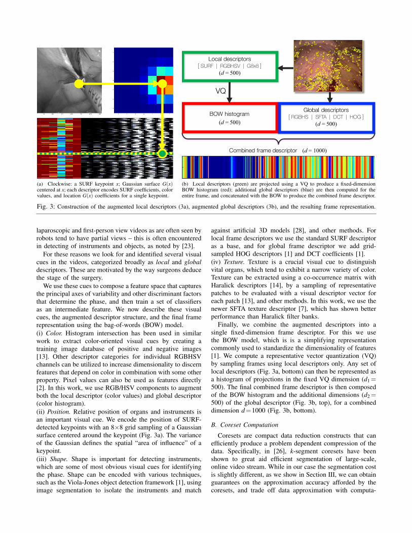
(a) Clockwise: a SURF keypoint x; Gaussian surface G(x)centered at x; each descriptor encodes SURF coefficients, colorvalues, and location G(x) coefficients for a single keypoint.
VQ
BOW histogram(d = 500)
Global descriptors![ RGBHS | SFTA | DCT | HOG ]!
!(d = 500)!.
BOW histogram(d = 500)
.
Local descriptors![ SURF | RGBHSV | G8x8 ]!
!(d = 500)!
Combined frame descriptor (d = 1000)
(b) Local descriptors (green) are projected using a VQ to produce a fixed-dimensionBOW histogram (red); additional global descriptors (blue) are then computed for theentire frame, and concatenated with the BOW to produce the combined frame descriptor.
Fig. 3: Construction of the augmented local descriptors (3a), augmented global descriptors (3b), and the resulting frame representation.
laparoscopic and first-person view videos as are often seen byrobots tend to have partial views – this is often encounteredin detecting of instruments and objects, as noted by [23].
For these reasons we look for and identified several visualcues in the videos, categorized broadly as local and globaldescriptors. These are motivated by the way surgeons deducethe stage of the surgery.
We use these cues to compose a feature space that capturesthe principal axes of variability and other discriminant factorsthat determine the phase, and then train a set of classifiersas an intermediate feature. We now describe these visualcues, the augmented descriptor structure, and the final framerepresentation using the bag-of-words (BOW) model.(i) Color. Histogram intersection has been used in similarwork to extract color-oriented visual cues by creating atraining image database of positive and negative images[13]. Other descriptor categories for individual RGBHSVchannels can be utilized to increase dimensionality to discernfeatures that depend on color in combination with some otherproperty. Pixel values can also be used as features directly[2]. In this work, we use RGB/HSV components to augmentboth the local descriptor (color values) and global descriptor(color histogram).(ii) Position. Relative position of organs and instruments isan important visual cue. We encode the position of SURF-detected keypoints with an 8×8 grid sampling of a Gaussiansurface centered around the keypoint (Fig. 3a). The varianceof the Gaussian defines the spatial “area of influence” of akeypoint.(iii) Shape. Shape is important for detecting instruments,which are some of most obvious visual cues for identifyingthe phase. Shape can be encoded with various techniques,such as the Viola-Jones object detection framework [1], usingimage segmentation to isolate the instruments and match
against artificial 3D models [28], and other methods. Forlocal frame descriptors we use the standard SURF descriptoras a base, and for global frame descriptor we add grid-sampled HOG descriptors [1] and DCT coefficients [1].(iv) Texture. Texture is a crucial visual cue to distinguishvital organs, which tend to exhibit a narrow variety of color.Texture can be extracted using a co-occurrence matrix withHaralick descriptors [14], by a sampling of representativepatches to be evaluated with a visual descriptor vector foreach patch [13], and other methods. In this work, we use thenewer SFTA texture descriptor [7], which has shown betterperformance than Haralick filter banks.
Finally, we combine the augmented descriptors into asingle fixed-dimension frame descriptor. For this we usethe BOW model, which is is a simplifying representationcommonly used to standardize the dimensionality of features[1]. We compute a representative vector quantization (VQ)by sampling frames using local descriptors only. Any set oflocal descriptors (Fig. 3a, bottom) can then be represented asa histogram of projections in the fixed VQ dimension (d1=500). The final combined frame descriptor is then composedof the BOW histogram and the additional dimensions (d2=500) of the global descriptor (Fig. 3b, top), for a combineddimension d=1000 (Fig. 3b, bottom).
B. Coreset Computation
Coresets are compact data reduction constructs that canefficiently produce a problem dependent compression of thedata. Specifically, in [26], k-segment coresets have beenshown to great aid efficient segmentation of large-scale,online video stream. While in our case the segmentation costis slightly different, as we show in Section III, we can obtainguarantees on the approximation accuracy afforded by thecoresets, and trade off data approximation with computa-

(1) (2) (3) (4) (5) (v)
Fig. 4: Phases (1–5 shown) of the laparoscopic sleeve gastrectomy (LSG) procedure: (1) port, (2) biopsy, liver retraction, (3) omentumremoval, dissection, hiatus inspection, (4) stapling, (5) bagging, (6) irrigation, (7) final inspection, withdrawal. Fig. 4(v) shows the baggingphase from a similar surgery performed with a da Vinci Surgical System (compare with Fig. 4(5)).
tional resources. Furthermore, as in our previous work [31],we can augment the coreset by a keyframe compression ofthe video that enables fast retrieval and anytime access forlarge visual stream. We therefore we use an online k-segmentcoreset algorithm to compute a compact representation of thevideo stream over which we can compute the segmentation.This allows our system to run online, in real-time, usingminimal computational resources.
C. Phase prediction.
A binary classifier setup for each phase was used asopposed to a multi-class classifier. Using this approach waspreferred in order to decouple phase transitions, which is themain goal of the classifier layer, from phase identification,which is the goal of the HMM temporal model.
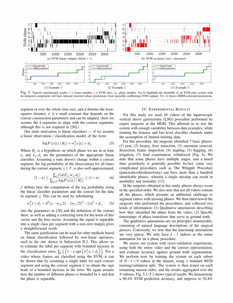
As a first step, we train a series of support vector machinesfor each phase. Each SVM classifies a phase i by outputtinga binary variable pi = 1, P\{pi} = 0. This approach wasshown to be more accurate than a single multi-class SVMin a similar visual domain [14]. This is an iterative stepthat involves interviewing surgeons, re-calibrating the featurespace, re-training the classifiers, etc. Interviewing surgeonsis expensive and time-consuming therefore it is importantto repeat this step first until we achieve the desired levelof accuracy. The first step is to ensure that the augmentedfeature space presented in Section II yields an acceptablelevel of accuracy for this domain with respect to the groundtruth phases. Fig. 5a shows the binary outputs produced bythe SVM. Fig. 5b shows the rate of correct classification(accuracy) of the predictions compared against ground truth.We note that there are two ambiguous cases: (i) multipleSVM outputs yi(t) = 1; and (ii) all SVM outputs yi(t) = 0.
The second step is to make use of the temporal structureof surgical phases (monotonically increasing, mutually exclu-sive, and collectively exhaustive) to correct SVM predictions,resolve the ambiguous cases stated above, and compute asingle time-series of phase predictions. We achieve this usingan observation function φ(V,s,α,β ) that takes a sequence ofSVM outputs V , the current phase hypothesis s, a certaintyparameter α ∈ [0,1], and lookback parameter β ∈Z+, and re-turns the next phase prediction. We start with an initial phasehypothesis s = 0. Then, given the current phase hypothesisand a matrix V where Vi is the column vector of SVM outputsat time i, the current phase estimate p is determined by
φ(V,s,α,β ) =
{argmaxi ∑Vi,1,...,β , if ∑Vi/∑V > α
s, otherwise(1)
The intuition for this function is as follows. We have amatrix consisting of several independent SVM output vectors(with a memory trail of the last β -1 such vectors). Theobservation function then updates the current phase, if andonly if the vector sum for another phase exceeds the currentone by a certainty threshold α , in which case we update ourphase hypothesis to the next phase – otherwise the currentphase persists.
The observation function combines many independentSVM outputs into one single set of phase observations.The values of α and β are essentially high- and low-passfilter parameters, and it is trivial to show correctness byconsidering that both α,β ∈ [0,1].
Phase transitions are modeled using an HMM with the left-right restriction as in [13]. This function is non-restrictive interms of skipping phases and going backwards (thus violatingour assumptions of the phases’ temporal structure), but thisis not necessary to enforce in the classification layer, andit is resolve by the HMM. The final observation sequenceQ = p1, . . . , pN is the emission sequence. Finally, we run theViterbi algorithm [1] on the emission sequence to find themost likely sequence of hidden states (the phases).
III. ANALYSIS
While coresets have been used before for video summa-rization [26] and loop-closure [31], there is the questionof what kind of guarantees are provided by coresets underthe SVM/HMM model that we are using. The followingtheorem shows that under specific conditions, k-segmentcoresets can be used in order to efficiently compute the datalog-likelihood of solutions, including the optimal solution,subject to constraints on the number of location of thetransition boundaries between labels. This includes linearclassifiers, or classifiers whose training hinges on linearclassification, such as SVMs. We give a brief motivation andoutline for the proof, and refer the reader to [26] for moredetails on the specific coreset used.
For a given coreset segment C in the k-segment coreset,it can be shown [26] that for k-segment with segments{x(t) = a jt + b j},x(t),a j,b j ∈ Rd , the coreset provides agood approximation for the fitting cost:
(1− ε)6∑ j d2 (C j,a jti +b j)
∑i d2 (x(ti) ,a jti +b j)6 (1+ ε) , (2)
where C j denotes the sufficient statistic matrix saved for eachcoreset segment j, i denotes the data points indices inside the
500 1000 1500 2000 2500 3000
SV
M c
lass
1234567
500 1000 1500 2000 2500 3000
1234567
(a) SVM binary outputs (black = 1)
500 1000 1500 2000 2500 3000
SV
M c
lass
1234567
500 1000 1500 2000 2500 3000
1234567
(b) SVM accuracy (red = incorrect)
Frames500 1000 1500 2000 2500 3000
Phase
1
2
3
4
5
6
7Ground truthHMM prediction
(1) Example 1 Frames500 1000 1500 2000 2500 3000
1
2
3
4
5
6
7
Ground truthHMM prediction
(2) Example 2 Frames500 1000 1500 2000 2500
1
2
3
4
5
6
7
Ground truthHMM prediction
(3) Example 3
Fig. 5: Typical experimental results: x = frame number, y = SVM class, i.e. phase number. 5(a–b) highlight the shortfalls of an SVM-only system withno temporal component; red lines indicate incorrect phase predictions from (possibly conflicting) SVM outputs. 5(1–3) shows HMM-corrected predictions.
segment or over the whole time axis, and d denotes the least-squares distance. ε is a small constant that depends on thecoreset construction parameters and can be adapted. (here weassume the k-segments to align with the coreset segments,although this is not required in [26].)
Our main motivation is linear classifiers — if we assumea linear observation / classification model, of the form:
logP(x(ti) | H j) = wTj x(ti)+d j, (3)
Where H j is a hypothesis on which phase we are in at timeti, and w j,d j are the parameters of the appropriate linearclassifier. Assuming a state doesn’t change within a coresetsegment, the log-probability of the observations for all timesduring the coreset segment, ti ∈ T , is also well-approximated,
(1− ε)≤∑ j f (d(C j,w j,d j)
∑ti∈T logP(x(ti) | H j)≤ (1+ ε). (4)
f defines here the computation of the log probability usingthe linear classifier parameters and the coreset for the datain segment j. This can be seen by substituting
wTj x+d j = d2(x,−w j/2)−‖w j/2‖2−‖x‖2 +d j. (5)
into the guarantees in [26] and the definition of the coresetthere, as well as adding a correcting term for the norm of thisvector and the bias vector. Assuming the signal is separableinto a single class per segment with a non-zero margin givesa straightforward result.
The same justification can be used for other methods basedon linear classification followed by non-linear operators,such as the one shown in Subsection II.3. This allows usto estimate the label per segment with bounded increase inthe classification error, ∑i
12
(1− yi sgn
(wT
j xi +d j
)). For a
video whose frames are classified using the SVM, it canbe shown that by assuming a single label for each coresetsegment and using the coreset segment C to obtain the signleads to a bounded increase in the error. We again assumehere the number of different phases is bounded by k and thatthe phase is separable.
IV. EXPERIMENTAL RESULTS
For this study we used 10 videos of the laparoscopicvertical sleeve gastrectomy (LSG) procedure performed byexpert surgeons at the MGH. This allowed us to test thesystem with enough variability between data examples, whiletraining the features and low-level classifier channels underthe assumption of limited training data.
For this procedure, the surgeons identified 7 basic phases:(1) port, (2) biopsy, liver retraction, (3) omentum removal,dissection, hiatus inspection, (4) stapling, (5) bagging, (6)irrigation, (7) final examination, withdrawal (Fig. 4). Wenote that some phases have multiple stages, and a muchfiner granularity is generally possible. In-fact, some verycomplicated procedures such as The Whipple Procedure(pancreaticoduodenectomy) can have more than a hundredidentifiable phases, wherein a single misstep can result inmorbidity and mortality [17].
In the surgeries obtained in this study, phases always occurin the specified order. We also note that not all videos containall the phases, which presents an additional challenge tosegment videos with missing phases. We then interviewed thesurgeons who performed the procedures, and collected twokinds of information: (1) Qualitative annotations describinghow they identified the phase from the video; (2) Specifictimestamps of phase transitions that serve as ground truth.
The qualitative annotations are (in principle) innumerable,consisting of natural language descriptions of the surgicalprocess. Conversely, we note that the timestamp annotationsare very sparse. We only have k− 1 indices as the entireannotation for an k-phase procedure.
We assess our system with cross-validation experiments,using both the entire video and the coreset representation,and evaluate accuracy against ground truth segmentation.We perform tests by training the system on each subsetof N−1= 9 videos in the dataset, using a standard 80/20training/validation split. The system is then tested on eachremaining unseen video, and the results aggregated over theN subsets. Fig. 5.1–5.3 shows typical results. We demonstratea 90.4% SVM prediction accuracy, and improve to 92.8%
when combined with HMM. These results are on par withsimilar work in the surgical video domain [15], while achiev-ing a 90+% coreset compression over the original video.
A. Discussion and Conclusions
In this work we showed how a carefully calibrated featurespace can facilitate a very high classification accuracy forphase detection in video streams
The main focus of ongoing work is to improve thephase prediction accuracy. We extend our system to considercontinuous likelihood models, allowing us use temporalregularity to handle ambivalent phase predictions more ef-fectively. We are looking into other temporal models fornon-monotonic phase sequences. With more video data weaim to evaluate our predictive model across different surgicalprocedures. Lastly, our goal is to extend the use of coresets inthis work beyond segmentation, applying the framework wepresented in [31] to generate an interactive visual summaryof laparoscopic and robot-assisted surgeries.
REFERENCES
[1] M. Allan, P.-L. Chang, S. Ourselin, D. J. Hawkes, A. Sridhar, J. Kelly,and D. Stoyanov. Image based surgical instrument pose estimationwith multi-class labelling and optical flow. In International Conferenceon Medical Image Computing and Computer-Assisted Intervention,pages 331–338. Springer, 2015.
[2] T. Blum, H. Feußner, and N. Navab. Modeling and segmentationof surgical workflow from laparoscopic video. In InternationalConference on Medical Image Computing and Computer-AssistedIntervention, pages 400–407. Springer, 2010.
[3] E. M. Bonrath, N. J. Dedy, L. E. Gordon, and T. P. Grantcharov. Com-prehensive surgical coaching enhances surgical skill in the operatingroom: a randomized controlled trial. Annals of surgery, 262(2):205–212, 2015.
[4] L. Bouarfa, P. Jonker, and J. Dankelman. Surgical context discoveryby monitoring low-level activities in the or. In MICCAI workshop onmodeling and monitoring of computer assisted interventions (M2CAI).London, UK, 2009.
[5] L. Bouarfa, P. P. Jonker, and J. Dankelman. Discovery of high-level tasks in the operating room. Journal of biomedical informatics,44(3):455–462, 2011.
[6] D. Bouget, R. Benenson, M. Omran, L. Riffaud, B. Schiele, andP. Jannin. Detecting surgical tools by modelling local appearance andglobal shape. IEEE transactions on medical imaging, 34(12):2603–2617, 2015.
[7] A. F. Costa, G. Humpire-Mamani, and A. J. M. Traina. An efficientalgorithm for fractal analysis of textures. In Graphics, Patterns andImages (SIBGRAPI), 2012 25th SIBGRAPI Conference on, pages 39–46. IEEE, 2012.
[8] O. Dergachyova, D. Bouget, A. Huaulme, X. Morandi, and P. Jannin.Automatic data-driven real-time segmentation and recognition of sur-gical workflow. International journal of computer assisted radiologyand surgery, pages 1–9, 2016.
[9] B. L. Ecker, R. Maduka, A. Ramdon, D. T. Dempsey, K. R. Dumon,and N. N. Williams. Resident education in robotic-assisted verticalsleeve gastrectomy: outcomes and cost-analysis of 411 consecutivecases. Surgery for Obesity and Related Diseases, 2015.
[10] K. Kahol, N. C. Krishnan, V. N. Balasubramanian, S. Panchanathan,M. Smith, and J. Ferrara. Measuring movement expertise in surgicaltasks. In Proceedings of the 14th ACM international conference onMultimedia, pages 719–722. ACM, 2006.
[11] U. Kannan, B. L. Ecker, R. Choudhury, D. T. Dempsey, N. N.Williams, and K. R. Dumon. Laparoscopic hand-assisted versusrobotic-assisted laparoscopic sleeve gastrectomy: experience of 103consecutive cases. Surgery for Obesity and Related Diseases, 2015.
[12] M. Kranzfelder, A. Schneider, A. Fiolka, E. Schwan, S. Gillen,D. Wilhelm, R. Schirren, S. Reiser, B. Jensen, and H. Feussner.Real-time instrument detection in minimally invasive surgery usingradiofrequency identification technology. journal of surgical research,185(2):704–710, 2013.
[13] F. Lalys, L. Riffaud, D. Bouget, and P. Jannin. A framework for therecognition of high-level surgical tasks from video images for cataractsurgeries. IEEE Transactions on Biomedical Engineering, 59(4):966–976, 2012.
[14] F. Lalys, L. Riffaud, X. Morandi, and P. Jannin. Automatic phasesrecognition in pituitary surgeries by microscope images classification.In International Conference on Information Processing in Computer-Assisted Interventions, pages 34–44. Springer, 2010.
[15] F. Lalys, L. Riffaud, X. Morandi, and P. Jannin. Surgical phasesdetection from microscope videos by combining SVM and HMM. InMedical Computer Vision. Recognition Techniques and Applicationsin Medical Imaging, pages 54–62. Springer, 2010.
[16] B. P. Lo, A. Darzi, and G.-Z. Yang. Episode classification for theanalysis of tissue/instrument interaction with multiple visual cues. InMICCAI, pages 230–237. Springer, 2003.
[17] G. Marangoni, G. Morris-Stiff, S. Deshmukh, A. Hakeem, and A. M.Smith. A modern approach to teaching pancreatic surgery. Journal ofgastrointestinal surgery, 16(8):1597–1604, 2012.
[18] N. Mehta, R. Haluck, M. Frecker, and A. Snyder. Sequence andtask analysis of instrument use in common laparoscopic procedures.Surgical Endoscopy And Other Interventional Techniques, 16(2):280–285, 2002.
[19] T. Neumuth and C. Meißner. Online recognition of surgical in-struments by information fusion. International journal of computerassisted radiology and surgery, 7(2):297–304, 2012.
[20] N. Padoy, T. Blum, S.-A. Ahmadi, H. Feussner, M.-O. Berger, andN. Navab. Statistical modeling and recognition of surgical workflow.Medical image analysis, 16(3):632–641, 2012.
[21] N. Padoy, T. Blum, I. Essa, H. Feussner, M.-O. Berger, and N. Navab.A boosted segmentation method for surgical workflow analysis. In In-ternational Conference on Medical Image Computing and Computer-Assisted Intervention, pages 102–109. Springer, 2007.
[22] N. Padoy, T. Blum, H. Feussner, M.-O. Berger, and N. Navab. On-linerecognition of surgical activity for monitoring in the operating room.In AAAI, pages 1718–1724, 2008.
[23] A. Reiter, P. K. Allen, and T. Zhao. Feature classification for trackingarticulated surgical tools. In International Conference on MedicalImage Computing and Computer-Assisted Intervention, pages 592–600. Springer, 2012.
[24] N. Rieke, D. J. Tan, M. Alsheakhali, F. Tombari, C. A. di San Filippo,V. Belagiannis, A. Eslami, and N. Navab. Surgical tool tracking andpose estimation in retinal microsurgery. In International Conference onMedical Image Computing and Computer-Assisted Intervention, pages266–273. Springer, 2015.
[25] J. Rosen, M. Solazzo, B. Hannaford, and M. Sinanan. Task decom-position of laparoscopic surgery for objective evaluation of surgicalresidents’ learning curve using hidden markov model. Computer AidedSurgery, 7(1):49–61, 2002.
[26] G. Rosman, M. Volkov, D. Feldman, J. W. Fisher III, and D. Rus.Coresets for k-segmentation of streaming data. In NIPS, pages 559–567. Curran Associates, Inc., 2014.
[27] P. Singh, R. Aggarwal, M. Tahir, P. H. Pucher, and A. Darzi. A ran-domized controlled study to evaluate the role of video-based coachingin training laparoscopic skills. Annals of surgery, 261(5):862–869,2015.
[28] S. Speidel, J. Benzko, S. Krappe, G. Sudra, P. Azad, B. P. Muller-Stich, C. Gutt, and R. Dillmann. Automatic classification of minimallyinvasive instruments based on endoscopic image sequences. In SPIEMedical Imaging, pages 72610A–72610A. International Society forOptics and Photonics, 2009.
[29] R. Stauder, A. Okur, L. Peter, A. Schneider, M. Kranzfelder, H. Feuss-ner, and N. Navab. Random forests for phase detection in surgicalworkflow analysis. In International Conference on Information Pro-cessing in Computer-Assisted Interventions, pages 148–157. Springer,2014.
[30] R. Sznitman, C. Becker, and P. Fua. Fast part-based classification forinstrument detection in minimally invasive surgery. In InternationalConference on Medical Image Computing and Computer-AssistedIntervention, pages 692–699. Springer, 2014.
[31] M. Volkov, G. Rosman, D. Feldman, J. W. Fisher III, and D. Rus.Coresets for visual summarization with applications to loop closure.In ICRA, Seattle, Washington, USA, May 2015. IEEE.
[32] E. Wild, D. Teber, D. Schmid, T. Simpfendorfer, M. Muller, A.-C. Baranski, H. Kenngott, K. Kopka, and L. Maier-Hein. Robustaugmented reality guidance with fluorescent markers in laparoscopicsurgery. International journal of computer assisted radiology andsurgery, pages 1–9, 2016.