
m6A-eCLIP User Guide
53
eclipsebio.com Protocol v1.01R 1 This product is for research use only, and is not intended for diagnostic or therapeutic uses. m6A-eCLIP User Guide RNA Genomics Solutions Robust next-gen technologies to simplify the complexity of RNA discovery and therapeutic development
Transcript of m6A-eCLIP User Guide

eclipsebio.com Protocol v1.01R 1
This product is for research use only, and is not intended for diagnostic or therapeutic uses.
m6A-eCLIP User Guide
RNA Genomics Solutions
Robust next-gen technologies to simplify the complexity of RNA discovery and therapeutic development

eclipsebio.com Protocol v1.01R 2
Table of Contents
Table of Contents
Chapter 1 Overview ..................................................................................................................... 5
Introduction to m6A eCLIP .............................................................................................................5
Precautions....................................................................................................................................5
Included with Kit ............................................................................................................................7
Equipment Not Included with Kit ....................................................................................................8
Reagents Not Included with Kit ......................................................................................................9
m6A-eCLIP Assay Workflow .......................................................................................................... 10
Chapter 2 RNA Isolation ............................................................................................................. 11
Overview ..................................................................................................................................... 11
Consumables ............................................................................................................................... 11
Preparation ................................................................................................................................. 11
Procedure .................................................................................................................................... 11 Poly(A)-RNA Isolation Procedure ....................................................................................................................... 11 Measure mRNA Concentration .......................................................................................................................... 13 mRNA Fragmentation ........................................................................................................................................ 14
Chapter 3: Crosslink mRNA to m6A Antibody .............................................................................. 15
Overview ..................................................................................................................................... 15
Consumables ............................................................................................................................... 15
Preparation ................................................................................................................................. 15
Procedure .................................................................................................................................... 15 Antibody-Coupling ............................................................................................................................................. 15 Preparation of Protein G Beads for Coupling ..................................................................................................... 15 Crosslink IP Samples and Couple Antibody-mRNA Complexes to Protein G Beads ........................................... 16 Coupling Crosslinked Antibody-mRNA Complexes to Protein G Beads ............................................................. 17 Preparation for Western Blots ........................................................................................................................... 17
Chapter 4: Immunoprecipitation (IP) of Samples ......................................................................... 18
Overview ..................................................................................................................................... 18
Consumables ............................................................................................................................... 18
Preparation ................................................................................................................................. 18
Procedure .................................................................................................................................... 18 First Immunoprecipitation Wash ....................................................................................................................... 18 IP RNA 5’-end repair ........................................................................................................................................... 19 IP RNA 3’-end repair ........................................................................................................................................... 20

eclipsebio.com Protocol v1.01R 3
Second Immunoprecipitation Wash ................................................................................................................... 20 RNA Adapter Ligation to Immunoprecipitation Samples ................................................................................... 21 Third Immunoprecipitation Wash ...................................................................................................................... 21
Chapter 5: Preparation for SDS-PAGE and Membrane Transfer ................................................... 23
Overview ..................................................................................................................................... 23
Consumables ............................................................................................................................... 23
Preparation ................................................................................................................................. 23
Procedure .................................................................................................................................... 23 Prepare Reagents for SDS-PAGE ........................................................................................................................ 23
Chapter 6: SDS-PAGE and Membrane Transfer............................................................................ 25
Overview ..................................................................................................................................... 25
Consumables ............................................................................................................................... 25
Preparation ................................................................................................................................. 25
Procedure .................................................................................................................................... 25 Elution of IP m6A-Ab-RNA complexes for electrophoresis ................................................................................ 25 Load SDS-PAGE preparative and analytical gels ................................................................................................. 26 Prepare gels and transfer to membrane ............................................................................................................ 26
Chapter 7: Chapter Processing of Input RNA samples .................................................................. 28
Overview ..................................................................................................................................... 28
Consumables ............................................................................................................................... 28
Preparation ................................................................................................................................. 28
Procedure .................................................................................................................................... 28 5’-End repair of Input RNA ................................................................................................................................. 28 3’-End repair of Input RNA ................................................................................................................................. 29 Clean Repaired Input Samples ........................................................................................................................... 29 Input Sample Adapter Ligation .......................................................................................................................... 30 Input RNA bead cleanup .................................................................................................................................... 30
Chapter 8: mRNA Recovery, Reverse Transcription, cDNA Adapter Ligation ................................. 32
Overview ..................................................................................................................................... 32
Consumables ............................................................................................................................... 32
Preparation ................................................................................................................................. 32
Procedure .................................................................................................................................... 33 IP Membrane Cutting and mRNA Recovery ....................................................................................................... 33 Digest RBP-RNA Complexes ............................................................................................................................... 33 IP Sample Cleanup .............................................................................................................................................. 34 Reverse Transcription of IP and Input Sample Reagent Preparation ................................................................. 35 Reverse transcription of IP and Input RNA ........................................................................................................ 35 cDNA End Repair of IP and Input Samples ......................................................................................................... 35 cDNA IP and Input Sample Bead Cleanup .......................................................................................................... 36 IP and Input cDNA Ligation on Beads ................................................................................................................. 37
Chapter 9: Library Amplification and Preparation for sequencing ................................................ 38

eclipsebio.com Protocol v1.01R 4
Overview ..................................................................................................................................... 38
Consumables ............................................................................................................................... 38
Preparation ................................................................................................................................. 38
Procedure .................................................................................................................................... 38 Ligated cDNA IP and Input Sample Bead Cleanup .............................................................................................. 38 cDNA IP and Input sample quantification by qPCR ............................................................................................ 39 PCR amplification of IP and Input cDNA and Dual Index Addition ..................................................................... 40
First AMPure library PCR product cleanup .................................................................................... 42
Second AMPure library PCR product cleanup ................................................................................ 42 Library Quantification ........................................................................................................................................ 43 Pool and Sequence Final Library ........................................................................................................................ 43
APPENDIX A: SDS-PAGE Gel Electrophoresis ................................................................................ 44 Required Equipment and Materials ................................................................................................................... 44 Reagents ............................................................................................................................................................. 44
APPENDIX B1: SDS-PAGE Gel Electrophoresis .............................................................................. 46
APPENDIX B2: Membrane Transfer ............................................................................................. 47
Sequencing parameters ................................................................................................................ 50
Sequencing depth ........................................................................................................................ 50
Read structure model ................................................................................................................... 51
Appendix D: Total RNA Isolation ................................................................................................ 52 Total RNA Isolation ............................................................................................................................................. 52 RNA and RIN Measurement Overview ............................................................................................................... 53

eclipsebio.com Protocol v1.01R 5
Chapter 1 Overview
Introduction to m6A eCLIP
The m6A eCLIP (enhanced CrossLinking ImmunoPrecipitation) kit transforms RNA analysis by streamlining mRNA isolation, eCLIP sample prep, and library prep, using a robust and reproducible framework to identify and map methylation sites on target RNA. The methylated RNA is immunoprecipitated using a highly specific Eclipse BioInnovations m6A antibody. RNA is chemically fragmented into 100 nucleotides or smaller fragments to generate high quality libraries.
The m6A eCLIP kit utilizes the eCLIP technology based on the Van Nostrand et al. (Nature Methods, 2016) method which produces high quality libraries that can enable the user to achieve up to single nucleotide resolution.
The m6A eCLIP kit offers:
High throughput and robust workflow
High reproducibility with accurate data
Unbiased and high specificity
Important Note
Before initiating this procedure thoroughly review the required equipment and materials list.
This procedure is to be followed by trained lab personnel.
Term Temperature
Room Temperature 20 – 25 °C
Ice 0 – 4 °C
Freeze -80 °C
Volume units
µL is microliter One millionth (10-6) of a liter
mL is milliliter One thousandth (10-3) of a liter
Precautions This kit contains chemicals which can be hazardous. High-Salt Buffer contains SDS and other
detergents. Enzyme buffers contain reducing agents and nucleotide solutions. Personal protection
equipment (PPE) should be worn during the entirety of this procedure.
➢ Use 1.5 mL DNA LoBind tubes (Eppendorf) during all steps.
➢ During beads washing, ensure DNA LoBind tubes are completely closed.
➢ Do not allow antibody-coupled beads to dry between wash steps.
➢ Beads used in nucleic acid cleanup steps should be completely dried before elution.
➢ Store all reagents on ice between steps unless otherwise indicated.
➢ When not in use, store all reagents at temperature indicated in “Day x Reagents” section.

eclipsebio.com Protocol v1.01R 6
➢ Use only calibrated pipettes. An additional 3% volume is recommended for all master mix
calculations to account for volume inaccuracy.
➢ TipOne® RPT Ultra Low Retention Filter Tips from USA Scientific are strongly recommended for
entire experiment.
➢ Always completely resuspend beads before taking aliquot or adding beads to sample.
➢ All Thermomixer incubations are done with interval mixing (15 seconds ON/15 seconds OFF).
➢ Store all enzymes at –20 °C.
➢ This protocol has been tested with 100 ng to 2 µg of starting mRNA.
IMPORTANT: Materials listed below are for m6A-eCLIP experimental set up ONLY. Additional
equipment, materials and reagents are required for protein gel electrophoresis, membrane transfer,
western blotting and imaging, and quantification of samples. See Appendix for supplemental
information.
eclipsebio.com Protocol v1.01R 7
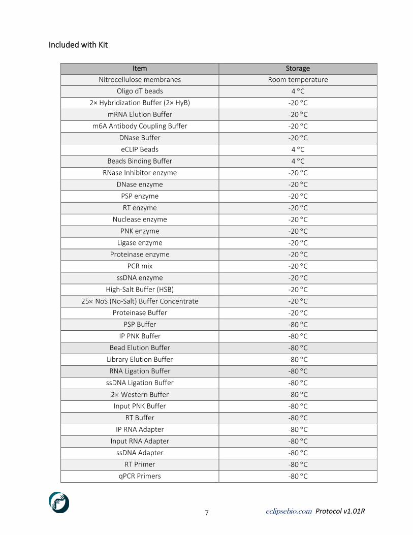
Included with Kit
Item Storage
Nitrocellulose membranes Room temperature
Oligo dT beads 4 C
2× Hybridization Buffer (2× HyB) -20 C
mRNA Elution Buffer -20 C
m6A Antibody Coupling Buffer -20 C
DNase Buffer -20 C
eCLIP Beads 4 C
Beads Binding Buffer 4 C
RNase Inhibitor enzyme -20 C
DNase enzyme -20 C
PSP enzyme -20 C
RT enzyme -20 C
Nuclease enzyme -20 C
PNK enzyme -20 C
Ligase enzyme -20 C
Proteinase enzyme -20 C
PCR mix -20 C
ssDNA enzyme -20 C
High-Salt Buffer (HSB) -20 C
25 NoS (No-Salt) Buffer Concentrate -20 C
Proteinase Buffer -20 C
PSP Buffer -80 C
IP PNK Buffer -80 C
Bead Elution Buffer -80 C
Library Elution Buffer -80 C
RNA Ligation Buffer -80 C
ssDNA Ligation Buffer -80 C
2 Western Buffer -80 C
Input PNK Buffer -80 C
RT Buffer -80 C
IP RNA Adapter -80 C
Input RNA Adapter -80 C
ssDNA Adapter -80 C
RT Primer -80 C
qPCR Primers -80 C
eclipsebio.com Protocol v1.01R 8
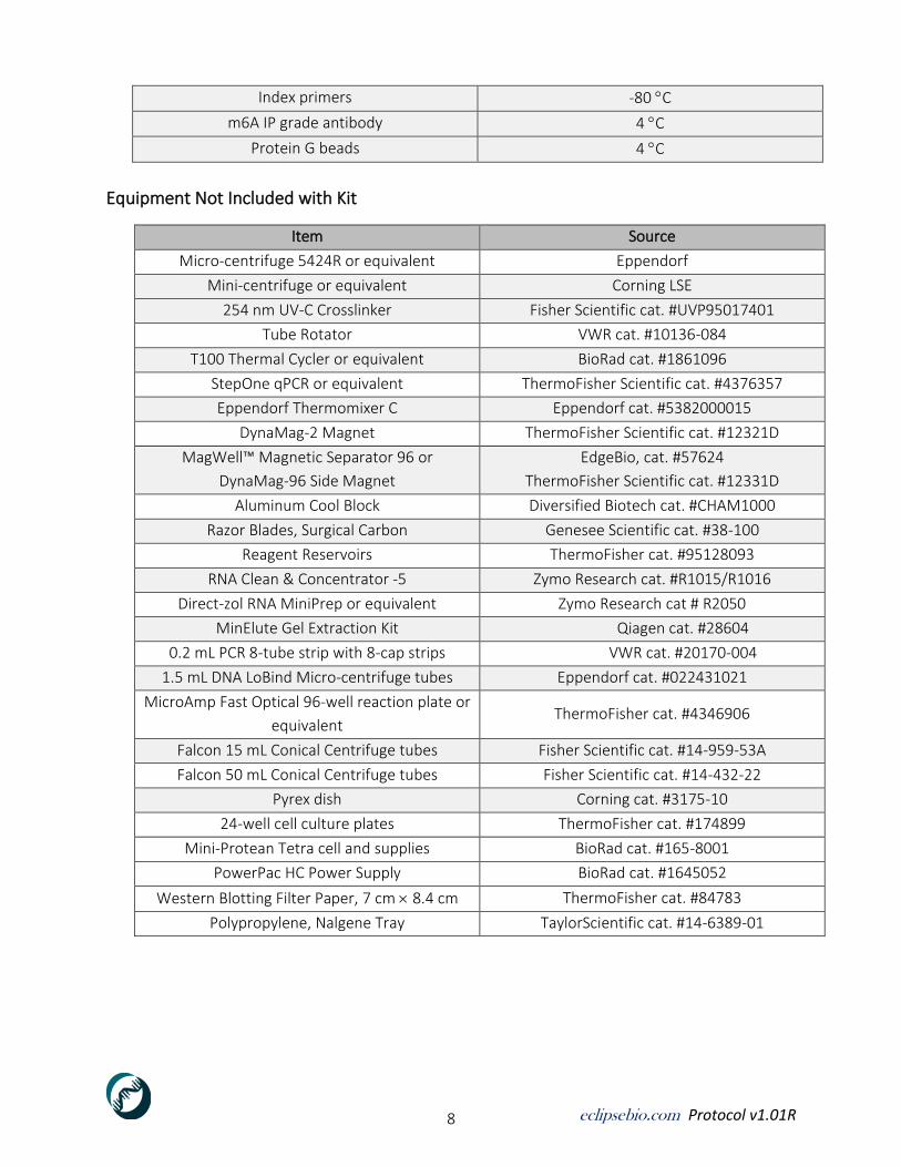
Index primers -80 C
m6A IP grade antibody 4 C
Protein G beads 4 C
Equipment Not Included with Kit
Item Source
Micro-centrifuge 5424R or equivalent Eppendorf
Mini-centrifuge or equivalent Corning LSE
254 nm UV-C Crosslinker Fisher Scientific cat. #UVP95017401
Tube Rotator VWR cat. #10136-084
T100 Thermal Cycler or equivalent BioRad cat. #1861096
StepOne qPCR or equivalent ThermoFisher Scientific cat. #4376357
Eppendorf Thermomixer C Eppendorf cat. #5382000015
DynaMag-2 Magnet ThermoFisher Scientific cat. #12321D
MagWell™ Magnetic Separator 96 or
DynaMag-96 Side Magnet
EdgeBio, cat. #57624
ThermoFisher Scientific cat. #12331D
Aluminum Cool Block Diversified Biotech cat. #CHAM1000
Razor Blades, Surgical Carbon Genesee Scientific cat. #38-100
Reagent Reservoirs ThermoFisher cat. #95128093
RNA Clean & Concentrator -5 Zymo Research cat. #R1015/R1016
Direct-zol RNA MiniPrep or equivalent Zymo Research cat # R2050
MinElute Gel Extraction Kit Qiagen cat. #28604
0.2 mL PCR 8-tube strip with 8-cap strips VWR cat. #20170-004
1.5 mL DNA LoBind Micro-centrifuge tubes Eppendorf cat. #022431021
MicroAmp Fast Optical 96-well reaction plate or
equivalent ThermoFisher cat. #4346906
Falcon 15 mL Conical Centrifuge tubes Fisher Scientific cat. #14-959-53A
Falcon 50 mL Conical Centrifuge tubes Fisher Scientific cat. #14-432-22
Pyrex dish Corning cat. #3175-10
24-well cell culture plates ThermoFisher cat. #174899
Mini-Protean Tetra cell and supplies BioRad cat. #165-8001
PowerPac HC Power Supply BioRad cat. #1645052
Western Blotting Filter Paper, 7 cm 8.4 cm ThermoFisher cat. #84783
Polypropylene, Nalgene Tray TaylorScientific cat. #14-6389-01

eclipsebio.com Protocol v1.01R 9
Reagents Not Included with Kit
Reagent Source
Ethanol, Pure, 200 proof, for Molecular Biology Sigma-Aldrich cat. #E7023-1L
Nuclease-free Molecular Biology Grade Water
or UltraPure™ DEPC-Treated Water
Corning/VWR cat. #95000-094
ThermoFisher Scientific cat. # 750023
DPBS, Corning VWR cat. #21-031-CV
EDTA (0.5 M), pH 8.0, RNase-free ThermoFisher Scientific cat. #AM9261
1 M Sodium Hydroxide solution (NaOH) Sigma-Aldrich cat. #79724-100ML
1 M Hydrogen Chloride (HCl) Any
Agencourt AMPure XP Beckman Coulter cat. #A63881
NEB LUNA Universal qPCR 2 Master Mix New England BioLabs cat. #M3003S
Spectra Multicolor Broad Range Protein Ladder ThermoFisher Scientific cat. #26634
Pierce 20 TBS Tween 20 Buffer ThermoFisher Scientific cat. #28360
20 NuPAGE Transfer Buffer ThermoFisher Scientific cat. #NP00061
Methanol Sigma-Aldrich cat. #494437

eclipsebio.com Protocol v1.01R 10
m6A-eCLIP Assay Workflow
eclipsebio.com Protocol v1.01R 11
Chapter 2 RNA Isolation Overview This section describes the isolation of 50 µg of total RNA from 10 M cells to be subjected to m6A-
eCLIP using Zymo's Direct-zol RNA isolation kit. mRNA is subsequently enriched by double oligo dT
capture to select for polyadenylated transcripts. 500 ng of final mRNA is then heat fragmented to
100-200 nt pieces – 10 ng of the fragmented sample is saved for total RNA-seq (input) and the other
490ng is coupled to m6A antibody (IP sample).
Consumables
➢ Oligo dT beads ➢ TRIzol Reagent ➢ 2× Hybridization Buffer (HyB) (Thaw at room temperature then store on ice) ➢ mRNA Elution Buffer (Thaw at room temperature then store on ice) ➢ DNase Buffer (Thaw at room temperature then store on ice) ➢ RNase Inhibitor enzyme ➢ DNase enzyme ➢ Direct-zol RNA MiniPrep or equivalent cell pellet(s) ➢ Molecular Biology Grade Water ➢ 100% Ethanol
Preparation
1. Centrifugation steps are done at room temperature.
2. Prewarm thermomixer to 60 C.
Procedure
Poly(A)-RNA Isolation Procedure 1. Transfer 50 µg of total RNA to a new 1.5 mL LoBind DNA tube.
2. If volume of RNA is less than 200 µL; bring volume up to 200 µL using Molecular Biology Grade
water. If RNA volume exceeds 200 µL, continue with volume and increase volume of 2× HyB
when resuspending washed Oligo dT beads so final concentration of HyB is 1× during binding.
3. Incubate RNA in thermomixer for 2 minutes at 60 C with interval mixing.
4. After incubation immediately place RNA samples on ice.
5. Transfer 200 µL of Oligo dT beads into new 1.5 mL LoBind DNA tubes for each sample.
6. Add 100 µL of 2× HyB to each tube containing 200 µL Oligo dT beads, invert tube to mix.
7. Place tube on DynaMag-2 magnet and allow 1 minute for beads to separate.
8. Slowly invert closed tubes on magnet as beads start to separate to capture beads from cap.
9. When supernatant is transparent, discard supernatant without disturbing beads.
10. Remove tube from magnet and add 300 µL 2× HyB to each sample.

eclipsebio.com Protocol v1.01R 12
11. Invert tube to mix until homogeneous.
12. Place tube on DynaMag-2 magnet.
13. Allow 1 minute for beads to separate.
14. Slowly invert closed tubes on magnet as beads start to separate to capture beads from cap.
15. When separation is complete, discard supernatant without disturbing beads.
16. Repeat steps 6-15 for a total of two washes.
17. Remove tube from magnet and add 200 µL of 2× HyB.
18. Pipette mix to combine until homogeneous.
19. Add entire volume (200 µL) of beads in 2× HyB to 200 µL of denatured RNA (from step 4).
20. Place tube containing RNA and beads on tube rotator for 20 minutes at room temperature.
21. While the sample is rotating, dilute 2× HyB 5-fold according to Table 1.
22. Place tube containing beads and RNA on DynaMag-2 magnet/
23. Allow 1 minute for beads to separate.
24. Slowly invert closed tubes while on magnet as beads to separate to capture any beads from cap.
25. When separation is complete, discard supernatant without disturbing beads.
26. Remove tube from magnet and add 745 µL of diluted HyB (Table 1).
Table 1. Dilution of 2× Hybridization Buffer (per sample)
Component Volume (µL)
2x Hybridization Buffer (HyB) 300
Molecular Biology Grade water 1200
Total: 1500
27. Invert to mix until homogeneous.
28. Place tube on DynaMag-2 magnet and allow 1 minute for beads to separate.
29. Slowly invert closed tubes while on magnet to capture any beads from cap.
30. When separation is complete, discard supernatant without disturbing beads.
31. Spin tube in mini-centrifuge for 2 seconds.
32. Discard supernatant.
33. Resuspend beads in 200 µL mRNA Elution Buffer.
34. Pipette mix to combine until homogeneous.
35. Incubate sample in thermomixer for 2 minutes at 60 C with interval mixing.
36. After incubation immediately place on ice for 2 minutes.
37. Add 200 µL of 2× HyB into eluted mRNA samples containing original oligo dT beads to have total
volume of 400 µL.
38. Incubate on tube rotator for 20 minutes at room temperature.
39. Once rotation is complete place tube containing beads and RNA on DynaMag-2 magnet.
40. Allow 1 minute for beads to separate.
41. Slowly invert closed tubes to separate and capture any beads from cap.
42. When separation is complete, discard supernatant without disturbing beads.
43. Remove tube from magnet.
eclipsebio.com Protocol v1.01R 13
44. Add 745 µL of diluted HyB (Table 1).
45. Invert to mix until homogeneous.
46. Place tube on DynaMag-2 magnet.
47. Allow 1 minute for beads to separate.
48. Slowly invert closed tubes to separate and capture any beads from cap.
49. When separation is complete and supernatant is transparent, aspirate and discard supernatant
without disturbing beads.
50. Spin tube in mini-centrifuge for 15 seconds.
51. Aspirate all residual liquid.
52. Add 40 µL Molecular Biology Grade water to bead pellet.
53. Pipette mix until homogeneous.
54. Incubate sample in thermomixer for 2 minutes at 60 C with interval mixing.
55. Magnetize immediately and transfer all supernatant to a new 1.5 mL LoBind DNA tube without
disturbing beads and place on ice.
➢ Note: Volume pulled from beads should be ~40 µL
56. Re-elute sample a second time by adding 41 µL Molecular Biology Grade water to the beads.
57. Pipette mix until homogeneous.
58. Place sample in thermomixer set at 60 C with interval mixing.
59. Increase temperature to 70 C, allow sample to transition temperatures
60. Incubate for a total of 3 minutes (starting from when temperature is increased) on thermomixer.
61. Magnetize immediately and pool all supernatant with 1.5 mL LoBind DNA tube containing RNA
(step 36) without disturbing beads.
➢ Note: Total volume of mRNA will be around 80 µL.
62. Take ~80 µL sample into the following section (Measure mRNA Concentration).
Measure mRNA Concentration
mRNA can be measured using a variety of methods. This protocol has been optimized using Agilent
4200 TapeStation with Agilent’s High Sensitivity RNA ScreenTape which measures both total RNA
concentration and RNA integrity number. RIN is based on the ratio of 28S rRNA to 18S rRNA. Oligo dT
beads select out mRNA, so RIN is expected to be low due to depletion of 28/18S rRNA, but
concentration of mRNA is still applicable. Expected mRNA yield is 1-3% of total RNA. For 50 µg
starting RNA, expect 500 ng to 1.5 µg of final mRNA. Take sample into following section (mRNA
Fragmentation)
Optional Stopping Point: RNA samples to be stored at 80C Next stopping point: 1 hour

eclipsebio.com Protocol v1.01R 14
mRNA Fragmentation 1. Aliquot 420 ng of eluted mRNA to new signed 0.2 mL PCR tube strip and prepare
mRNA fragmentation mix for each sample according to Table 2.
Table 2. mRNA fragmentation Mix (per sample)
Component Volume (µL)
RNA + Molecular Biology Grade Water 67
DNase Buffer 8
RNase Inhibitor 2
DNase 3
Total: 80
2. Mix sample well.
3. Incubate samples in PCR machine: 37 C for 10 minutes, 95 C for 16 minutes and 5 C for 10 sec,
with lid at 98 C.
4. Place samples on ice after incubation.

eclipsebio.com Protocol v1.01R 15
Chapter 3: Crosslink mRNA to m6A Antibody
Overview
In this section, input material is saved and immunoprecipitation samples are prepared for crosslinking. Fragmented RNA samples are first coupled with an m6A IP antibody and then crosslinked to the bound RNA fragments using UV light. Lastly, samples are coupled overnight with protein G beads.
Consumables
➢ m6A IP antibody ➢ High-Salt Buffer (Thaw at room temperature then store on ice) ➢ m6A Coupling Buffer (Thaw at room temperature then store on ice) ➢ RNase Inhibitor enzyme ➢ Molecular Biology Grade Water ➢ Protein G beads
Preparation
1. Thaw at room temperature then store on ice 2. 254 nm UV-C mercury (Hg) bulbs MUST be used during crosslinking.
Procedure ➢ Preparation Note: Save input material. Calculate concentration of fragmented mRNA (e.g. If 420
ng starting mRNA was used, the concentration should be (420 ng / 80 µL) = 5.25 ng/µL). Take 20 ng fragmented mRNA and freeze at – 80 °C as input sample.
Antibody-Coupling 1. Dilute 400 ng of fragmented mRNA with m6A Coupling Buffer to 393 µL.
2. Add 4 µL RNase inhibitor enzyme to each sample.
3. Add 3 µL (3 µg) m6A antibody to each sample. Total sample volume should be 400 µL.
4. Rotate for at least 2 hrs up to 12 hrs at 4 °C.
Preparation of Protein G Beads for Coupling 1. Mix Protein G beads until homogeneous.
2. Transfer 10 µL of Protein G beads per sample into a 1.5 mL LoBind tube (e.g. for 8 samples use 80
µL of Protein G beads).
3. Dilute High-Salt Buffer according to Table 3. Invert to mix then store on ice.

eclipsebio.com Protocol v1.01R 16
Table 3. High-Salt Buffer Dilution
Component Volume (mL)
Molecular Biology Grade water 9
High-Salt Buffer 1
Total: 10
4. Add 5 volumes (50 µL per sample) of chilled diluted High-Salt Buffer to the tube containing
Protein G beads.
5. Place tube on DynaMag-2 magnet.
6. After separation is complete, discard supernatant without disturbing beads.
7. Remove tube from magnet.
8. Add 500 µL chilled diluted High Salt Wash Buffer to the tube.
9. Invert mix until homogeneous.
10. Place the tube on DynaMag-2 magnet.
11. After separation is complete, discard supernatant without disturbing beads.
12. Repeat steps 8-11 for a total of two washes.
➢ Note: Do not discard undiluted High-Salt Buffer, store at 4 °C after use.
13. Remove tube from magnet and add 51 µL chilled m6A Coupling Buffer per sample to the tube
(e.g. for 6 samples, add 306 µL chilled m6A Coupling Buffer).
14. Resuspend by pipetting until homogeneous.
15. Store on ice until samples are ready for bead-coupling.
➢ Beads will be used in Coupling Crosslinked Antibody-mRNA Complex to Protein G Beads
section.
Crosslink IP Samples and Couple Antibody-mRNA Complexes to Protein G Beads 1. Label 24-well plate according to samples being crosslinked.
2. Prepare a thin, flat layer of ice in a glass Pyrex dish and place a 24-well plate on top to chill. If not
using flaked ice, add ~0.5 cm of water to ensure entire bottom of plate is in contact with either
ice or ice water.
3. Transfer all antibody-coupled mRNA samples to each corresponding well on 24-well plate (from
step Antibody-Coupling section step 4).
4. Place Pyrex dish containing 24-well plate (without lid) and samples into 254 nm UV-C Ultraviolet
Crosslinker
5. Crosslink twice at Energy = 1500 (150 mJ/cm2), removing dish containing samples from
crosslinker between the rounds of crosslinking, allowing plate to cool on ice for 15-30 seconds.
6. Remove dish containing samples from Crosslinker allowing plate to cool on ice for 15-30 seconds.
7. Carefully transfer crosslinked samples to new 1.5 mL LoBind tubes and place on ice.

eclipsebio.com Protocol v1.01R 17
Coupling Crosslinked Antibody-mRNA Complexes to Protein G Beads 1. Resuspend washed Protein G beads (prepared in Preparation of Protein G Beads for Coupling
section step 13) by pipetting.
2. Add 50 µL of resuspended Protein G bead solution to each crosslinked sample (Crosslink IP
Samples and Couple Antibody-mRNA Complexes to Protein G Beads section step 5).
3. Rotate overnight at 4 C.
Preparation for Western Blots ➢ Preparation Note: Recommended to prepare 1 MOPS SDS Running Buffer, WB Transfer Buffer,
1 NoS Buffer (Table 4, Table 5, Table 6 respectively) in advance for Western blot running and
store at 4 °C overnight.
Table 4. 1× NuPAGE MOPS SDS Running Buffer Preparation
Component Volume (mL)
Molecular Biology Grade Water 1045
20 NuPAGE MOPS SDS Running Buffer 55
Total: 1100*
* 1100 mL is sufficient volume for 2 gel chambers (for a total of 4 gels)
Table 5. 1× NuPAGE Transfer Buffer Preparation
Component Volume (mL)
Molecular Biology Grade Water 935
20 NuPAGE Transfer Buffer 55
100% Methanol 110
Total: 1100*
*This volume is sufficient for 1 standard transfer chamber, which holds 2 transfer stacks.
Table 6. 1× NoS Buffer Preparation
Component Volume (mL)
Molecular Biology Grade Water 48
25 NoS (No-Salt) Buffer Concentrate 2
Total: 50

eclipsebio.com Protocol v1.01R 18
Chapter 4: Immunoprecipitation (IP) of Samples
Overview This section describes immunoprecipitation and end repair of RNA samples, followed by ligation an
adapter to the 3’ end of bound transcripts.
Consumables
➢ PSP Buffer (Thaw at room temperature then store on ice.) ➢ IP PNK Buffer (Thaw at room temperature then store on ice.) ➢ RNA Ligation Buffer ➢ IP RNA Adapter
➢ PSP enzyme ➢ PNK enzyme ➢ RNase Inhibitor enzyme ➢ DNase enzyme ➢ Ligase enzyme
➢ 1 NoS (No-Salt) Buffer Concentrate (Gently invert 5 times to mix then store on ice.) ➢ Undiluted High-Salt Buffer (HSB) (Gently invert 5 times to mix then store on ice) ➢ Molecular Biology Grade Water
Preparation
1. Pre-warm Thermomixer to 37 C.
Procedure
First Immunoprecipitation Wash 1. Obtain immunoprecipitation (IP) tubes from (Coupling Crosslinked Antibody-mRNA Complexes to
Protein G Beads step 3)
2. Put on DynaMag-2 magnet to separate beads,
3. Allow at least 1 minute for bead separation.
4. When separation is complete and liquid is transparent, carefully aspirate and discard supernatant
without disturbing beads.
5. Remove IP tubes from magnet
6. Add 500 µL cold HSB.
7. Invert mix until homogeneous
8. Place on DynaMag-2 magnet.
9. While on magnet, slowly invert closed tubes as beads start to separate to capture any beads
from cap.
10. When separation is complete, and liquid is transparent, gently open tubes and discard
supernatant without disturbing beads.

eclipsebio.com Protocol v1.01R 19
11. Remove IP tubes from magnet and add 500 µL cold HSB.
12. Close tubes well and vortex for 15 seconds.
13. Incubate on tube rotator for 3 min at room temperature, then place on magnet.
14. While on magnet, slowly invert closed tubes as beads start to separate to capture any beads
from cap.
15. When separation is complete, and liquid is transparent, gently open tubes and discard
supernatant without disturbing beads.
16. Repeat steps 11-15 for additional round of wash.
17. Remove IP tubes from magnet.
18. Add 500 µL cold 1 NoS Buffer.
19. Gently invert mix until homogeneous.
20. Separate beads on magnet and remove supernatant without disturbing beads.
21. Remove IP tubes from magnet.
22. Add 500 µL cold 1 NoS Buffer.
23. Gently invert mix until homogeneous.
24. Spin all IP samples in mini-centrifuge for 3 seconds.
25. Place samples back on magnet and allow 1 minute to separate.
26. Pipette and discard any excess liquid without disturbing beads using fresh 1mL tips and 1ml
pipettor.
27. Remove IP tubes from magnet.
28. Add 500 µL cold 1 NoS Buffer.
29. Invert to mix until homogeneous.
30. Place samples on ice and proceed immediately to the next step.
IP RNA 5’-end repair
1. Prepare IP PSP Master Mix according to Table 7 in a fresh 1.5 mL DNA LoBind tube. Pipette mix to
combine and store on ice until use.
➢ Note: Include 3% excess volume to correct for pipetting losses.
Table 7. IP PSP Master Mix (per sample)
Component Volume/ IP (µL)
Molecular Biology Grade Water 23
PSP Buffer 20
RNase Inhibitor 2
DNase 2
PSP enzyme 3
Total: 50
2. Move all IP tubes from ice to DynaMag-2 magnet and allow at least 1 minute for bead separation.
3. Remove and discard supernatant.
4. Spin all samples in mini-centrifuge for 3 seconds.
5. Place samples back on magnet and allow 1 minute to separate.
eclipsebio.com Protocol v1.01R 20
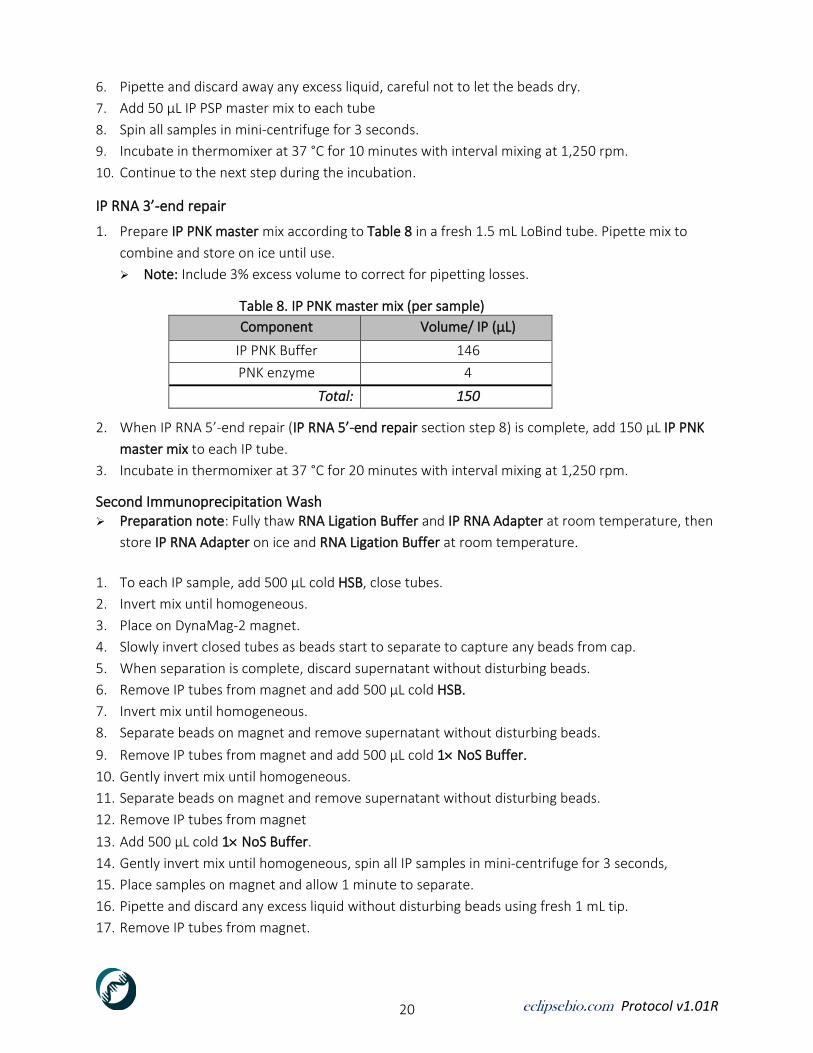
6. Pipette and discard away any excess liquid, careful not to let the beads dry.
7. Add 50 µL IP PSP master mix to each tube
8. Spin all samples in mini-centrifuge for 3 seconds.
9. Incubate in thermomixer at 37 °C for 10 minutes with interval mixing at 1,250 rpm.
10. Continue to the next step during the incubation.
IP RNA 3’-end repair
1. Prepare IP PNK master mix according to Table 8 in a fresh 1.5 mL LoBind tube. Pipette mix to
combine and store on ice until use.
➢ Note: Include 3% excess volume to correct for pipetting losses.
Table 8. IP PNK master mix (per sample)
Component Volume/ IP (µL)
IP PNK Buffer 146
PNK enzyme 4
Total: 150
2. When IP RNA 5’-end repair (IP RNA 5’-end repair section step 8) is complete, add 150 µL IP PNK
master mix to each IP tube.
3. Incubate in thermomixer at 37 °C for 20 minutes with interval mixing at 1,250 rpm.
Second Immunoprecipitation Wash ➢ Preparation note: Fully thaw RNA Ligation Buffer and IP RNA Adapter at room temperature, then
store IP RNA Adapter on ice and RNA Ligation Buffer at room temperature.
1. To each IP sample, add 500 µL cold HSB, close tubes.
2. Invert mix until homogeneous.
3. Place on DynaMag-2 magnet.
4. Slowly invert closed tubes as beads start to separate to capture any beads from cap.
5. When separation is complete, discard supernatant without disturbing beads.
6. Remove IP tubes from magnet and add 500 µL cold HSB.
7. Invert mix until homogeneous.
8. Separate beads on magnet and remove supernatant without disturbing beads.
9. Remove IP tubes from magnet and add 500 µL cold 1 NoS Buffer.
10. Gently invert mix until homogeneous.
11. Separate beads on magnet and remove supernatant without disturbing beads.
12. Remove IP tubes from magnet
13. Add 500 µL cold 1 NoS Buffer.
14. Gently invert mix until homogeneous, spin all IP samples in mini-centrifuge for 3 seconds,
15. Place samples on magnet and allow 1 minute to separate.
16. Pipette and discard any excess liquid without disturbing beads using fresh 1 mL tip.
17. Remove IP tubes from magnet.
eclipsebio.com Protocol v1.01R 21
18. Add 500 µL cold 1 NoS Buffer.
19. Invert to mix until homogeneous.
20. Store on ice and proceed to the next step.
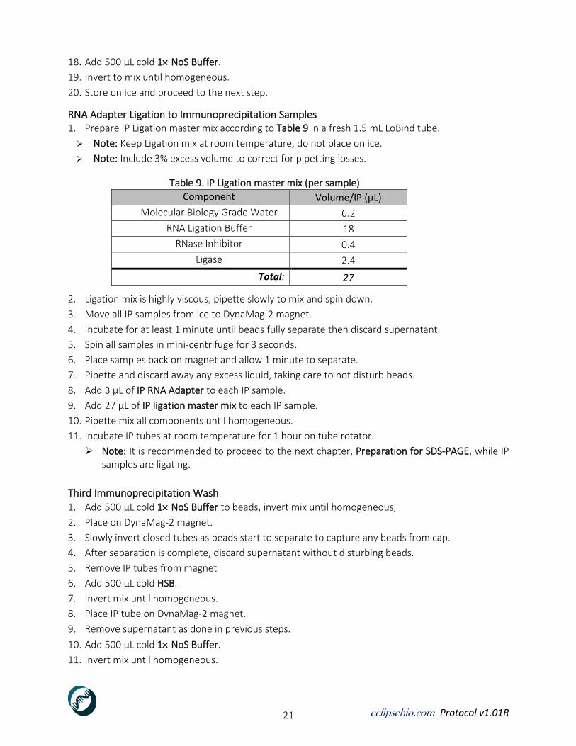
RNA Adapter Ligation to Immunoprecipitation Samples 1. Prepare IP Ligation master mix according to Table 9 in a fresh 1.5 mL LoBind tube.
➢ Note: Keep Ligation mix at room temperature, do not place on ice.
➢ Note: Include 3% excess volume to correct for pipetting losses.
Table 9. IP Ligation master mix (per sample)
Component Volume/IP (µL)
Molecular Biology Grade Water 6.2
RNA Ligation Buffer 18
RNase Inhibitor 0.4
Ligase 2.4
Total: 27
2. Ligation mix is highly viscous, pipette slowly to mix and spin down.
3. Move all IP samples from ice to DynaMag-2 magnet.
4. Incubate for at least 1 minute until beads fully separate then discard supernatant.
5. Spin all samples in mini-centrifuge for 3 seconds.
6. Place samples back on magnet and allow 1 minute to separate.
7. Pipette and discard away any excess liquid, taking care to not disturb beads.
8. Add 3 µL of IP RNA Adapter to each IP sample.
9. Add 27 µL of IP ligation master mix to each IP sample.
10. Pipette mix all components until homogeneous.
11. Incubate IP tubes at room temperature for 1 hour on tube rotator.
➢ Note: It is recommended to proceed to the next chapter, Preparation for SDS-PAGE, while IP samples are ligating.
Third Immunoprecipitation Wash 1. Add 500 µL cold 1 NoS Buffer to beads, invert mix until homogeneous,
2. Place on DynaMag-2 magnet.
3. Slowly invert closed tubes as beads start to separate to capture any beads from cap.
4. After separation is complete, discard supernatant without disturbing beads.
5. Remove IP tubes from magnet
6. Add 500 µL cold HSB.
7. Invert mix until homogeneous.
8. Place IP tube on DynaMag-2 magnet.
9. Remove supernatant as done in previous steps.
10. Add 500 µL cold 1 NoS Buffer.
11. Invert mix until homogeneous.

eclipsebio.com Protocol v1.01R 22
12. Separate beads on magnet and remove supernatant as done in previous steps.
13. Remove IP tubes from magnet.
14. Add 500 µL cold 1 NoS Buffer.
15. Gently invert mix until homogeneous.
16. Move all IP samples to DynaMag-2 magnet.
17. Incubate for at least 1 minute until beads fully separate then discard supernatant.
18. Spin all samples in mini-centrifuge for 3 seconds.
19. Place samples back on magnet.
20. Allow 1 minute to separate.
21. Remove supernatant, taking care to not disturb beads.
22. Remove IP tubes from magnet.
23. Place samples on ice and proceed immediately to the next step.
Proceed immediately to next chapter
Next Stopping point: ~45 minutes

eclipsebio.com Protocol v1.01R 23
Chapter 5: Preparation for SDS-PAGE and Membrane Transfer Overview This section describes the preparative steps required to perform SDS-PAGE on IP samples. It is recommended to perform these steps during IP RNA 3’-adapter ligation.
Consumables
➢ NuPAGE 4-12% Bis-Tris gels
➢ 20 NuPAGE MOPS SDS Running Buffer
➢ 20 NuPAGE Transfer Buffer ➢ Spectra Multicolor Broad Range Protein Ladder (thaw at room temperature then store on
ice)
➢ 2 Western Buffer (thaw at room temperature then store on ice) ➢ 100% Methanol ➢ Molecular Biology Grade Water
Preparation 1. Remove pre-cast gels from plastic and rinse with clean water prior to use
Procedure The following protocol is provided for pre-cast NuPage 4-12% Bis-Tris gels, which have neutral pH and are ideal for RNA stability. NuPAGE Tris-Acetate gels have been tested and are recommended for large molecular weight proteins (>200 kDa), but require different running buffer (see manufacturer recommendations). We have not validated other pre-cast or manually cast polyacrylamide gel formulations.
Prepare Reagents for SDS-PAGE 1. Determine the number of preparative gels (for size selection) required for all IP and Input
samples (see Example: Preparative Gel Loading Scheme below).
2. Label the appropriate number of pre-cast NuPAGE 4-12% Bis-Tris gels with sample/ladder
information, reserve wells between samples for ladders.
Example: Preparative Gel Loading Scheme (2 IP samples + 2 Input samples)
Well 1 2 3 4 5 6 7 8 9 10
Sample/Ladder High Conc. Ladder
IP 1 Low Conc. Ladder
IP 2 Low Conc. Ladder
IP 3 Low Conc. Ladder
IP 4 Low Conc. Ladder

eclipsebio.com Protocol v1.01R 24
3. Dilute 20 NuPAGE MOPS SDS Running Buffer to create 1 Running Buffer according to Table 10.
4. Shake manually to mix, then store on ice.
Table 10. 1 Running Buffer Preparation
Component Volume (mL)
Molecular Biology Grade Water 1000
20 NuPAGE MOPS SDS Running Buffer 53
Total: 1053*
* This volume is sufficient volume for 2 gel chambers (for a total of 4 gels).
5. Dilute 20 NuPAGE Transfer Buffer to create 1 Transfer Buffer according to Table 11.
6. Shake manually to mix, then store on ice.
Table 11. 1 Transfer Buffer Preparation
Component Volume (mL)
Molecular Biology Grade Water 1000
20 NuPAGE Transfer Buffer 59
100% Methanol 118
Total: 1177*
*This volume is sufficient for 1 standard transfer chamber, which holds 2 transfer stacks (2 gels).
7. Dilute protein ladders according to Table 12 and store on ice.
Table 12. Protein Ladder Dilutions
Component (µL)
Ladder Concentration
Molecular Biology Grade Water
Spectra Multicolor Broad Range Protein Ladder
2 Western Buffer
Total volume
High* 60 43 60 163
Low* 150 23 150 323
*High concentration ladder dilution volume is sufficient to run 8 wells; adjust according to sample number.
*Low concentration ladder dilution volume is sufficient to run 16 wells; adjust according to sample number.

eclipsebio.com Protocol v1.01R 25
Chapter 6: SDS-PAGE and Membrane Transfer
Overview This section describes the running of IP samples on SDS-PAGE and subsequent transfer to membranes. IP samples are washed following ligation and then denatured in western buffer. Denatured samples are loaded onto gels, and SDS-PAGE is performed for ~90 minutes. Lastly, samples are horizontally transferred from gels to membranes for size-selection and western blotting.
Consumables
➢ 1 NoS (No-Salt) Buffer Concentrate
➢ 2 Western Buffer
➢ 1 Running Buffer (diluted in previous section) ➢ High-concentration ladder (diluted in previous section) ➢ Low-concentration ladder (diluted in previous section) ➢ Bead Elution Buffer
Preparation
1. Running and transfer buffer should be made previously and pre-chilled to 4 C. Buffers can
be stored at 4 C for up to 1 month.
2. Place 2 Western Buffer and Spectra Multicolor Broad Range Protein Ladder at room temperature until fully thawed.
3. NuPAGE MOPS SDS Running Buffer according to Table 4 (Page 18).
Procedure
Elution of IP m6A-Ab-RNA complexes for electrophoresis 1. Prepare 1 Western Buffer according to Table 13 below and store on ice.
Table 13. 1 Western Buffer Preparation
Reagent Volume (μL)
Molecular Biology Grade Water 30
2 Western Buffer 30
Total: 60
2. Move all IP tubes from ice to DynaMag-2 magnet and allow at least 1 minute for bead separation.
3. Remove and discard supernatant.
4. Spin all samples in mini-centrifuge for 3 seconds.
5. Place samples back on magnet and allow 1 minute for separation.

eclipsebio.com Protocol v1.01R 26
6. Pipette and discard any excess liquid without disturbing beads.
7. Add 21 µL of 1 Western Buffer to IP samples.
8. Pipette mix until homogenous and then store on ice.
9. Incubate IP samples on thermomixer at 65 °C for 10 minutes with interval mixing at 1,200 rpm.
10. Move all samples to ice for 2 minutes.
11. Spin all samples in mini-centrifuge for 3 seconds.
12. Move all tubes from ice to DynaMag-2 magnet and allow at least 1 minute for bead separation.
13. Remove supernatants (containing m6A-Ab-RNA complexes) from beads and place in fresh, labeled
tubes.
14. Discard tubes containing beads.
Optional Stopping Point: If stopping here, eluted cDNA samples should be stored at -80 °C Next stopping point: ~4 hrs
Load SDS-PAGE preparative and analytical gels 1. Assemble electrophoresis tank (see Appendix A: SDS-PAGE for assembly instructions).
2. Remove sticker from all labeled gels.
3. Place labeled preparative and analytical gels inside of electrophoresis tank and clamp shut.
4. Add 1 Running Buffer to central chamber of electrophoresis tank until outer chambers are
halfway filled (~500 mL).
5. Remove combs from gels.
6. Wash wells of gels by gently pipetting 1 mL of 1 Running Buffer from central chamber into
wells.
7. Load 20 µL of High Concentration Ladder and Low Concentration Ladder into appropriate wells of
preparative and analytical gels.
8. Load 20 µL of IP samples into appropriate wells of gel.
➢ Note: Ensure all wells in the middle of the gels are filled, as empty wells can cause uneven
sample electrophoresis.
9. Run gels at constant 160 V until the lower dye front reaches the bottom of the gel (typically 75-
90 minutes).
➢ Optional: During polyacrylamide gel-electrophoresis continue to Chapter 6: Processing
of Input RNA samples.
10. Continue to WB Gel Transfer of IP samples when gel run is completed.
Prepare gels and transfer to membrane 1. After SDS-PAGE is complete, remove gel cassette(s) and place face down on work-space surface.
2. Disassemble the gel cassette by carefully inserting the gel knife into the gap between the two
plates of the cassette. Gently push up and down on the handle to ‘crack’ the cassette but not
fully separate the top and bottom plates. Continue until all 3 sealed edges have been released.

eclipsebio.com Protocol v1.01R 27
3. Carefully separate the top and bottom cassette plates, with the gel attached to one of the two
plates.
➢ Note: If the gel remains attached to both plates, briefly submerging the cassette apparatus in
1 NuPAGE Transfer Buffer will loosen the gel sufficiently to separate the plates without
tearing the gel.
➢ Note: If using the gel knife to separate the gel from the plate, ensure that the knife is wetted
with transfer buffer to avoid tearing the gel.
4. Trim off wells on top of gel and 2-3 mm on bottom of gel using gel knife.
5. Gel is now ready to be transferred.
6. Prepare membranes for transfer (see Appendix B: Membrane Transfer for detailed instructions).
➢ Note: We recommend using the provided nitrocellulose membranes, which were identified
to have decreased background RNA contamination (see Van Nostrand, et al. Methods Mol
Biol. 2017 (PMID 28766298)).
➢ Note: We recommend using a ‘wet’ transfer method as modeled in this protocol.
7. Assemble transfer chamber and transfer stacks (see Appendix B: Membrane Transfer if using
Mini Protean Tetra Apparatus).
8. Run transfer for 2 hours at constant 200 mA or at constant 30 V overnight at 4 °C.
➢ Note: If transferring for 2 hours at constant 200 mA so, surround as much of the outside of
the apparatus with ice as possible to avoid overheating. If using more than one Mini-Protean
Tetra apparatus, connect only one per power supply.

eclipsebio.com Protocol v1.01R 28
Chapter 7: Chapter Processing of Input RNA samples
Overview In the following chapter the user will perform end repair and clean up on RNA then performing
adapter ligation on samples. This chapter use the Zymo RNA Clean and Concentrator-5 kit to clean
input RNA and the reagents for this can be found in the Zymo RNA Clean and Concentrator-5 kit.
Consumables
➢ Input PSP buffer (Thaw at room temperature then store on ice) ➢ Input PNK (Thaw at room temperature then store on ice)
➢ RNA Ligation Buffer (Thaw at room temperature then store on ice) ➢ Input RNA Adapter (Thaw at room temperature then store on ice) ➢ Bead Elution Buffer (Thaw at room temperature then store on ice) ➢ eCLIP beads (Take out of 4 °C and resuspend until homogeneous)
Preparation 1. Prewarm thermomixer to 37 °C 10 minutes with interval mixing. 2. Ensure 100% EtOH is added to RNA Wash Buffer concentrate upon first usage.
3. Centrifugation steps are done at room temperature.
4. Prepare fresh 80% ethanol in Molecular Biology Grade water in a fresh 50 mL tube. Store at room
temperature for up to 1 week.
Procedure 5’-End repair of Input RNA 1. Prepare Input PSP master mix according to Table 14 below in a fresh 1.5 mL LoBind tube. 2. Mix then store on ice until use.
➢ Note: Include 3% excess volume to correct for pipetting losses
Table 14. Input PSP Master Mix (per sample)
Component Volume/Input (µL)
PSP Buffer 8
RNase Inhibitor 1
PSP enzyme 2
Total: 11
3. Dilute each 20 ng of saved fragmented input mRNA (from Antibody-Coupling section step 4) to
10 µL with Molecular Biology Grade water.
4. Add 11 µL of Input PSP Master Mix to each 10 µL input sample, mix by flicking.
5. Spin in mini-centrifuge for 3 seconds.
6. Incubate in Thermomixer for 10 minutes at 37 °C with interval mixing at 1,250rpm.

eclipsebio.com Protocol v1.01R 29
3’-End repair of Input RNA 1. Prepare Input PNK Master Mix according to Table 15 in a fresh 1.5 mL LoBind tube.
2. Mix then store on ice until use.
➢ Note: Include 3% excess volume to correct for pipetting losses
Table 15. Input PNK master mix (per sample)
Component Volume/Input (µL)
Input PNK Buffer 70
DNase 1
PNK enzyme 4
Total: 75
3. Add 75 µL of Input PNK master mix to each tube.
4. Mix by flicking, spin in mini-centrifuge for 3 seconds.
5. Incubate in Thermomixer for 20 minutes at 37 °C with interval mixing at 1,250 rpm.
Clean Repaired Input Samples 1. Add 200 µL of RNA Binding Buffer to the 95 µL of each end-repaired Input RNA. Pipette mix.
2. Add 300 µL of 100% EtOH, pipette 10 times to mix.
3. Transfer the entire sample to a new filter column placed in a collection tube.
4. Centrifuge at 7,000 g for 30 seconds. Discard flow-through.
5. Add 400 µL RNA Prep Buffer to each column.
6. Centrifuge at 7,000 g for 30 seconds. Discard flow-through.
7. Add 480 µL RNA Wash Buffer to each column.
8. Centrifuge at 7,000 g for 30 seconds. Discard flow-through.
9. Repeat step 7-8 for a total of two washes.
10. Centrifuge the column at 10,000 g for 1 minute with emptied collection tube.
11. Carefully transfer filter column to a new 1.5 mL LoBind tube (avoid liquid in collection tube).
12. Discard flow-through and collection tube.
13. Open column caps and allow to air dry for 2 minutes or until column is completely dry.
14. Elute all samples by adding 11 µL of Molecular Biology Grade water directly to filter.
15. Incubate at room temperature for 1 minute.
16. Centrifuge at 12,000 g for 90 seconds.
17. Place RNA samples on ice if continuing to the next step.
Optional Stopping Point: If stopping here, Input RNA samples should be stored at -80 °C Next stopping point: ~2 hrs

eclipsebio.com Protocol v1.01R 30
Input Sample Adapter Ligation ➢ Preparation note: Preheat PCR thermal cycler block to 65 °C (with lid set at 70 °C).
1. Add 5 µL of repaired Input RNA (from Clean Repaired Input Samples section step 17) into pre-
labeled 0.2 mL strip tubes, temporarily place on ice. Store remaining repaired Input RNA at -80 °C
as backup.
2. Add 2 µL of Input RNA Adapter to each Input RNA tube, pipette mix.
3. Spin samples in mini-centrifuge for 5 seconds to draw all liquid to the bottom of the tube.
4. Incubate tubes at 65 °C for 2 minutes in thermal cycler with the lid preheated to 70°C,
5. Immediately place samples on ice for 1 minute.
6. Add 13.5 µL of Input Ligation Master Mix (Table 16) adapter-added input RNA sample.
Table 16. Input Ligation Master Mix (per sample)
Component Volume (µL)
RNA Ligation Buffer 12
RNase Inhibitor 0.3
Ligase 1.2
Total: 13.5
7. Pipette mix until homogeneous.
8. Spin samples in mini-centrifuge for 5 seconds to draw all liquid to the bottom of the tube.
9. Incubate the samples for 1 hour at room temperature on tube rotator.
Input RNA bead cleanup 1. Wash eCLIP Beads prior to addition to samples.
2. Add 10 µL of eCLIP beads to a new 1.5 mL LoBind tube.
➢ Note: For 2 input samples transfer 20 µL of eCLIP beads into new 1.5 mL LoBind tube.
3. Add 5 volume of Bead Binding Buffer.
➢ Note: For 2 input samples add 100 µL buffer to 20 µL of eCLIP beads.
4. Pipette up and down to mix until sample is homogeneous.
5. Place tube on DynaMag-2 magnet.
6. When separation is complete and supernatant is clear, carefully aspirate and discard supernatant
without disturbing beads.
7. Remove tube from magnet.
8. Resuspend eCLIP beads in 63 µL of Bead Binding Buffer per sample and pipette up and down
until beads are fully resuspended.
➢ Note: For 3 input samples resuspend beads in the same tube with 189 µL of Bead Binding
Buffer.
9. Transfer 60 µL of washed eCLIP Beads to each 20 µL tube of ligated input RNA sample.
10. Pipette up and down to mix until sample is homogeneous.
11. Add 60 µL of 100% EtOH to each sample.
12. Pipette mix until homogeneous.

eclipsebio.com Protocol v1.01R 31
13. Incubate at room temperature for 10 minutes, with pipette mixing every 5 minutes.
14. Incubate on magnet for 30 seconds until separation is complete and supernatant is transparent.
15. Carefully aspirate and discard supernatant without disturbing beads.
16. Add 320 µL of 80% EtOH to each sample.
17. Carefully aspirate and discard supernatant without disturbing beads.
18. Add 160 µL of 80% EtOH without disturbing beads.
19. Move samples to different positions on magnet to wash thoroughly.
20. Carefully add an additional 160µL of 80% EtOH.
21. Incubate on magnet for 30 seconds until separation is complete and supernatant is transparent.
22. Carefully aspirate and discard all supernatant while on magnet.
23. Repeat steps 18-22 for a total of two washes.
24. Spin capped samples in mini-centrifuge for 5 seconds to draw all liquid to the bottom of the tube.
25. Place tube back on 96-well magnet.
26. Incubate on magnet for 10 seconds until separation is complete and supernatant is transparent.
27. While on magnet aspirate and discard all residual liquid without disturbing beads.
28. Allow beads to air dry for 5 minutes or until beads no longer appear wet and shiny.
29. Once completely dry, carefully remove tubes from magnet.
30. Add 9.5 µL Bead Elution Buffer to each sample.
31. Pipette up and down to mix until sample is homogeneous.
32. Incubate for 5 minutes at room temperature.
33. After incubation, move tubes to 96-well magnet.
34. Incubate on magnet for 30 seconds until separation is complete and supernatant is transparent.
35. Transfer whole sample to new 0.2 mL strip tubes.
36. Store input RNA samples at -80 C or continue to Reverse Transcription.
➢ Note: If stop here, store IP nitrocellulose membrane flat in sheet protector at -20 C
Optional Stopping Point: If stopping here, Input RNA samples should be stored at -80 °C
Next stopping point: ~4 hrs

eclipsebio.com Protocol v1.01R 32
Chapter 8: mRNA Recovery, Reverse Transcription, cDNA Adapter Ligation
Overview In the following chapter the user will perform mRNA Recovery, Reverse Transcription, and cDNA
Adapter Ligation. IP-ed RNA is recovered from the nitrocellulose membrane by proteinase digestion
from cut membrane fragments. The resulting sample is cleaned using the Zymo RNA clean and
concentrator-5 kit and is then ready for reverse transcription. Reverse transcription is performed for
both IP and input samples, followed by several steps to remove dNTPs and template RNA. ssDNA
samples are then cleaned using beads and overnight ligation is performed to attach an adapter to
the 5’ ends of cDNA
Consumables
➢ 0.5 M EDTA ➢ 1 M NaOH ➢ 1 M HCl ➢ 100% Ethanol ➢ 80% Ethanol ➢ eCLIP beads (Take out of 4 °C and resuspend until homogeneous) ➢ Bead Binding Buffer ➢ Proteinase Buffer (Thaw at room temperature) ➢ Proteinase Enzyme ➢ RT Enzyme ➢ RNase Inhibitor ➢ Nuclease Enzyme ➢ ssDNA Enzyme ➢ Ligase Enzyme ➢ RT Buffer (Thaw at room temperature then store on ice) ➢ RT Primer (Thaw at room temperature then store on ice) ➢ ssDNA Adapter ➢ ssDNA Ligation Buffer
Preparation
1. Ensure 100% EtOH is added to RNA Wash Buffer concentrate upon first usage.
2. Centrifugation steps are done at room temperature.
3. Preheat PCR thermal cycler block to 65 °C (with lid set at 70 °C).

eclipsebio.com Protocol v1.01R 33
Procedure IP Membrane Cutting and mRNA Recovery 1. Place membrane with plastic sheet protector on a flat, hard surface, such as a glass coverslip.
2. Using a clean razor blade for each sample, carefully cut the region from 30 kDa to 150 kDa from
the IP nitrocellulose membrane. (see Figure 4)
Figure 1. IP nitrocellulose membrane region
3. Remove excised membrane piece from sheet protector and place on glass coverslip.
➢ Note: To prevent membrane from drying out, 10 µL of Molecular Biology Grade water can be
added to each strip.
4. Further cut the membrane piece into 1-2 mm square slices.
5. Carefully transfer all slices to a new 1.5 mL LoBind tube.
6. Spin samples in mini-centrifuge for 5 seconds to collect membrane slices at the bottom.
7. Store tubes on ice until all samples are isolated then proceed immediately to the next step.
Digest RBP-RNA Complexes 1. Prepare Digest Mix according to Table 17 in a fresh 1.5 mL LoBind tube.
➢ Note: Include 3% excess volume to correct for pipetting losses.
Table 17. Digest Mix Preparation (per sample)
Component Volume (µL)
Proteinase Buffer 130
Proteinase 20
Total: 150
2. Add 150 µL Digest mix to each sample tube containing membrane slices.
3. Incubate in Thermomixer at 37 °C for 20 minutes
4. Incubate in Thermomixer at 50 °C for 20 minutes with interval mixing at 1,250 rpm.
➢ Note: It is important to ensure all membrane slices are submerged in Digest mix during
incubation; move slices with a clean pipette tip if necessary.
eclipsebio.com Protocol v1.01R 34
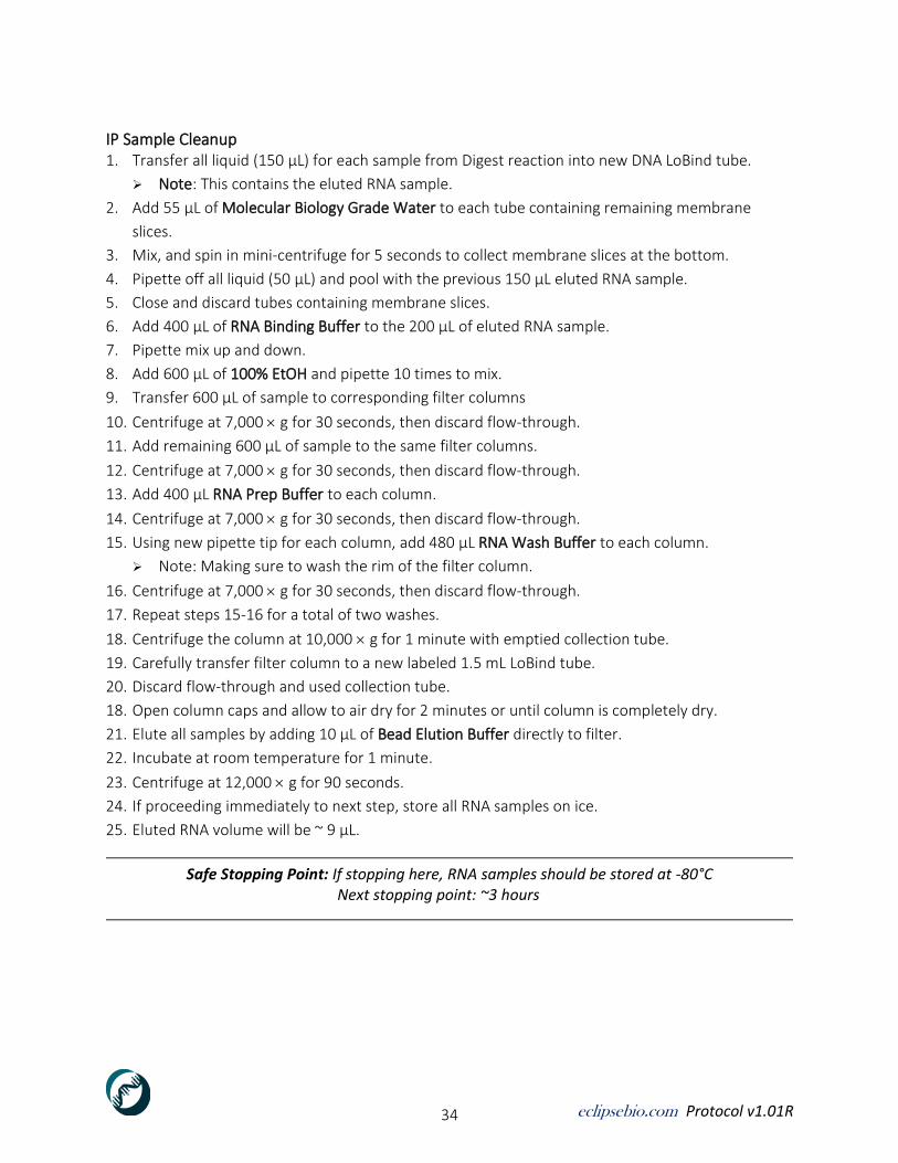
IP Sample Cleanup 1. Transfer all liquid (150 µL) for each sample from Digest reaction into new DNA LoBind tube.
➢ Note: This contains the eluted RNA sample.
2. Add 55 µL of Molecular Biology Grade Water to each tube containing remaining membrane
slices.
3. Mix, and spin in mini-centrifuge for 5 seconds to collect membrane slices at the bottom.
4. Pipette off all liquid (50 µL) and pool with the previous 150 µL eluted RNA sample.
5. Close and discard tubes containing membrane slices.
6. Add 400 µL of RNA Binding Buffer to the 200 µL of eluted RNA sample.
7. Pipette mix up and down.
8. Add 600 µL of 100% EtOH and pipette 10 times to mix.
9. Transfer 600 µL of sample to corresponding filter columns
10. Centrifuge at 7,000 g for 30 seconds, then discard flow-through.
11. Add remaining 600 µL of sample to the same filter columns.
12. Centrifuge at 7,000 g for 30 seconds, then discard flow-through.
13. Add 400 µL RNA Prep Buffer to each column.
14. Centrifuge at 7,000 g for 30 seconds, then discard flow-through.
15. Using new pipette tip for each column, add 480 µL RNA Wash Buffer to each column.
➢ Note: Making sure to wash the rim of the filter column.
16. Centrifuge at 7,000 g for 30 seconds, then discard flow-through.
17. Repeat steps 15-16 for a total of two washes.
18. Centrifuge the column at 10,000 g for 1 minute with emptied collection tube.
19. Carefully transfer filter column to a new labeled 1.5 mL LoBind tube.
20. Discard flow-through and used collection tube.
18. Open column caps and allow to air dry for 2 minutes or until column is completely dry.
21. Elute all samples by adding 10 µL of Bead Elution Buffer directly to filter.
22. Incubate at room temperature for 1 minute.
23. Centrifuge at 12,000 g for 90 seconds.
24. If proceeding immediately to next step, store all RNA samples on ice.
25. Eluted RNA volume will be ~ 9 µL.
Safe Stopping Point: If stopping here, RNA samples should be stored at -80°C Next stopping point: ~3 hours

eclipsebio.com Protocol v1.01R 35
Reverse Transcription of IP and Input Sample Reagent Preparation 1. Add 1.5 µL of RT Primer into the 0.2 mL strip tube containing 9µL of Input samples (“Input RNA
bead Cleanup” section step 36).
2. For each IP RNA sample, transfer 9µL into a new, labeled 0.2mL strip tube.
3. Add 1.5 µL of RT Primer into IP RNA.
4. Mix, and spin all samples in mini-centrifuge for 5 seconds to draw all liquid to the bottom of the
tube.
5. Incubate at 65 °C for 2 minutes in thermal cycler with the lid preheated to 70 °C.
6. After incubation, immediately transfer to ice for 1 minute.
7. Adjust the thermal cycler block temperature to 54 °C – 20 minutes (with lid set to 65 °C)
Reverse transcription of IP and Input RNA 1. Prepare Reverse Transcription Master Mix according to Table 18 in a fresh 1.5mL LoBind tube.
2. Pipette sample up and down 10 times to mix.
3. Store samples on ice until use.
➢ Note: Include 3% excess volume to correct for pipetting losses
Table 18. Reverse Transcription Master-Mix (per sample)
Component Volume (µL)
RT Buffer 9.2
RNase Inhibitor 0.2
RT enzyme 0.6
Total: 10
4. Add 10 µL of the Reverse Transcription Master Mix to each sample leaving samples on ice.
Pipette to mix.
5. Spin samples in mini-centrifuge for 5 seconds to draw all liquid to the bottom of the tube.
6. Immediately incubate samples at 54 °C for 20 minutes in thermal cycler with the lid at 65 °C.
7. After incubation, immediately place samples on ice.
8. Adjust thermal cycler block temperature to 37 °C (with lid set to 45 °C).
cDNA End Repair of IP and Input Samples 1. Add 2.5 µL of Nuclease to each sample. Pipette to mix.
2. Spin samples in mini-centrifuge for 5 seconds to draw all liquid to the bottom of the tube.
3. Incubate in thermal cycler at 37 °C for 15 minutes with the lid at 45 °C.
4. Remove the strip-tube and place samples on ice.
5. Adjust thermal cycler block to 70 °C (with lid set to 75 °C).
6. Add 1 µL of 0.5 M EDTA (pH 8) to each sample.
7. Pipette samples up and down gently 5 times to mix.
8. Add 3 µL of 1 M NaOH to each sample.
9. Pipette samples up and down gently 5 times to mix.
10. Incubate tubes at 70 °C for 10 minutes in thermal cycler with the lid at 75 °C.
11. Place strip-tube on ice for 10 seconds.

eclipsebio.com Protocol v1.01R 36
12. Add 3 µL of 1 M HCl to each sample.
13. Proceed immediately to the next step.
cDNA IP and Input Sample Bead Cleanup ➢ Preparation Note: Thaw ssDNA Adapter and ssDNA Ligation Buffer at room temperature until
completely melted then store ssDNA Adapter on ice and ssDNA Ligation Buffer at room
temperature.
➢ Preparation Note: Prepare fresh 80% Ethanol in Molecular Biology Grade water in a fresh 50mL
tube if was not done previously. Store at room temperature for up to 1 week. Keep tube closed
tightly.
1. Take eCLIP beads (provided) out of 4 °C and resuspend until homogeneous.
2. Wash eCLIP beads prior to addition to samples.
3. For each IP and input cDNA sample, transfer 5µL of eCLIP beads to a new 1.5 mL DNA LoBind tube
(e.g. for 4 samples transfer 20 µL of eCLIP beads).
4. Add 5 volume of Bead Binding Buffer (e.g. for 4 samples add 100 µL buffer to 20 µL of eCLIP
beads). Pipette up and down to mix until sample is homogeneous.
5. Place tube on DynaMag-2 magnet. When separation is complete and supernatant is clear,
carefully aspirate and discard supernatant without disturbing beads.
6. Remove tube from magnet.
7. Resuspend eCLIP beads in 90 µL of Bead Binding Buffer per sample.
8. Pipette up and down until beads are fully resuspended.
9. Add 87 µL of washed eCLIP Beads to each IP and Input cDNA sample.
10. Pipette up and down to mix until sample is homogeneous.
11. Add 105 µL of 100% EtOH to each IP and Input cDNA sample.
12. Pipette mix until homogeneous.
13. Incubate at room temperature for 10 minutes, with pipette mixing every 5 minutes.
14. Move samples to fresh strip tube: place a new, labeled 0.2 mL strip tube on 96-well magnet and
transfer sample from old to new strip tube.
15. Allow to incubate for 1 minute or until separation is complete and liquid is transparent.
16. Carefully discard supernatant without disturbing beads.
17. Add 150 µL of 80% EtOH,
18. Move samples to different positions on magnet to wash thoroughly.
19. Carefully add an additional 150 µL of 80% EtOH.
20. Incubate on magnet for 30 seconds until separation is complete and supernatant is transparent.
21. Carefully aspirate and discard all supernatant while on magnet.
22. Repeat steps 17-21 once for a total of two washes.
23. Spin capped samples in mini-centrifuge for 5 seconds to draw all liquid to the bottom of the tube.
24. Place tube back on 96-well magnet.
25. Incubate on magnet for 10 seconds until separation is complete and supernatant is transparent.
26. Using fine tips, aspirate and discard all residual liquid without disturbing beads while on magnet.

eclipsebio.com Protocol v1.01R 37
27. Allow beads to air dry for 5 minutes or until beads no longer appear wet and shiny.
➢ Note: Do not over dry samples.
28. Once completely dry, carefully remove tubes from magnet.
29. Resuspend beads in 2.5 µL of ssDNA Adapter.
30. Pipette to mix until homogeneous.
31. Incubate in thermal cycler at 70 °C for 2 minutes with the lid at 75 °C.
32. Following incubation, immediately place on ice for 1 minute.
IP and Input cDNA Ligation on Beads 1. Prepare cDNA Ligation master mix according to Table 19 in a fresh 1.5mL LoBind tube. Pipette
mix to combine (do not vortex). Use immediately.
➢ Note: Include 3% excess volume to correct for pipetting losses
Table 19. cDNA Ligation Master Mix (per sample)
Component Volume (µL)
ssDNA Ligation Buffer 6.5
Ligase 1
ssDNA enzyme 0.3
Total: 7.8
2. Slowly add 7.8 µL of cDNA Ligation master mix to each sample from previous section cDNA IP
and Input Sample Bead Clean Up) and pipette mix until homogeneous.
3. Incubate at room temperature overnight on a tube rotator.
Safe Stopping Point

eclipsebio.com Protocol v1.01R 38
Chapter 9: Library Amplification and Preparation for sequencing
Overview
In the following chapter you will be bead purifying cDNA and prepping samples for sequencing.
Consumables
➢ Bead Elution Buffer (Thaw and keep at room temperature) ➢ Library Elution Buffer ➢ 50(5,6,7,8) Index Primer ➢ 70(5,6,7,8) Index Primer ➢ qPCR Primers
➢ NEB LUNA Universal qPCR 2 Master Mix (Thaw at room temperature then keep on ice) ➢ PCR mix ➢ eCLIP Beads (Take out of 4 °C and resuspend until homogeneous) ➢ eCLIP Bead Binding Buffer ➢ Agencourt AMPure XP beads ➢ Molecular Biology Grade Water ➢ 80% and 100% Ethanol
Preparation 1. Thaw NEB LUNA Universal qPCR 2 Master Mix at room temperature until completely melted
then store on ice until use.
Procedure
Ligated cDNA IP and Input Sample Bead Cleanup 1. Add 5 µL of Bead Elution Buffer to 10 µL adapter-ligated IP and Input cDNA sample from IP and
Input cDNA Ligation on Beads (p.37, step 3).
2. Mix until sample is homogeneous.
3. Add 45 µL of Bead Binding Buffer to each IP and Input sample.
4. Pipette mix until homogeneous.
5. Add 45 µL of 100% EtOH to each sample.
6. Pipette mix until homogeneous.
7. Incubate at room temperature for 10 minutes, with pipette mixing every 5 minutes.
8. Move samples to 96-well magnet.
9. Incubate on magnet for 30 seconds or until separation is complete and supernatant is
transparent.
10. Carefully discard supernatant without disturbing beads.
11. Wash beads with 320 µL of 80% EtOH without disturbing beads or overfilling wells.
12. Carefully discard supernatant without disturbing beads.

eclipsebio.com Protocol v1.01R 39
13. Add 160 µL of 80% EtOH without disturbing beads, move samples to different positions on
magnet to wash thoroughly.
14. Add an additional 160 µL of 80% EtOH.
15. Incubate on magnet for 30 seconds until separation is complete and supernatant is transparent.
16. Carefully discard all supernatant while on magnet.
17. Repeat steps 11-16 once for a total of two washes.
18. Spin capped samples in mini-centrifuge for 5 seconds to draw all liquid to the bottom of the tube.
19. Place tube back on 96-well magnet.
20. Incubate on magnet for 30 seconds or until separation is complete and supernatant is
transparent.
21. Carefully discard all supernatant while on magnet.
22. Allow beads to air dry for 5 minutes or until beads no longer appear wet and shiny.
➢ Note: Do not over dry samples.
23. Carefully remove tubes from magnet.
24. For each sample, resuspend beads in 25 µL Bead Elution Buffer.
25. Pipette up and down to mix until sample is homogeneous.
26. Incubate beads in Bead Elution Buffer on ice if continuing to next step.
27. Move tubes to 96-well magnet.
28. Incubate on magnet for 30 seconds or until separation is complete and supernatant is
transparent.
29. Transfer 25 µL to new 0.2 mL strip tubes.
30. Store samples on ice if proceeding to the next step.
Optional Stopping Point: If stopping here, eluted cDNA samples should be stored at -80 °C Next stopping point: ~2 hrs
cDNA IP and Input sample quantification by qPCR 1. Prepare qPCR master mix for the appropriate number of reactions according to Table 20 in a
fresh 1.5 mL LoBind tube.
➢ Note: Include 3% excess volume to correct for pipetting losses
Table 20. qPCR quantification master mix (per sample)
Component Volume (µL)
NEB LUNA Universal qPCR 2 Master Mix 5
qPCR Primers 4
Total: 9
2. Obtain and label a 96- or 384-well qPCR reaction plate (See Table 21 for suggested 96-well
layout).

eclipsebio.com Protocol v1.01R 40
Table 21. 96-well qPCR plate layout for 21 samples
1 2 3 4 5 6 7 8 9 10 11 12
A
B water water water water water water water water water water water
C water Sample 1 Sample 2 Sample 3 Sample 4 Sample 5 Sample 6 Sample 7 Sample 8 Neg Control water
D water Sample 1 Sample 2 Sample 3 Sample 4 Sample 5 Sample 6 Sample 7 Sample 8 Neg Control water
E water Sample 9 Sample 10 Sample 11 Sample 12 Sample 13 Sample 14 Sample 15 Sample 16 Neg Control water
F water Sample 9 Sample 10 Sample 11 Sample 12 Sample 13 Sample 14 Sample 15 Sample 16 Neg Control water
G water water water water water water water water water water water
H
Note: We recommend running each sample in biological duplicate. Negative controls use water in place of cDNA.
3. Add 1 µL of eluted cDNA samples to 9 µL of Molecular Biology Grade Water for a 1:10 dilution. 4. Add 9 µL of qPCR master mix into all assay wells on ice.
5. Add 1 µL of each diluted cDNA (or water for negative controls) into the designated well.
➢ Note: Store remaining diluted cDNA samples at -20°C.
6. Cover the plate with a MicroAmp adhesive film and seal with MicroAmp adhesive film applicator.
7. Spin plate at 3,000 g for 1 minute.
8. Run qPCR assay according to the user manual for the specific instrument in your laboratory.
9. Run parameters appropriate for SYBR.
➢ Note: For example, for the StepOnePlus qPCR system the appropriate program is:
95 °C – 30 sec
(95 °C – 10 sec, 65 °C – 30 sec) 32 cycles; No melting curve
10. Record qPCR Ct values for all samples.
11. Set threshold to 0.5 – this recommendation is for StepOnePlus System.
➢ Typical acceptable Ct values range from 10 to 23 (with input samples typically <10). For
robust estimation, Ct values for samples should be 10. If values are below 9, dilute the 1:10
diluted cDNA an additional 10-fold, and re-perform qPCR using the 1:100 diluted cDNA.
PCR amplification of IP and Input cDNA and Dual Index Addition ➢ Preparation Note: For library pooling strategies, see Illumina documentation. We recommend
multiplexing at least 8 libraries with diverse indexes.
➢ Preparation Note: See APPENDIX : SEQUENCING SPECIFICATIONS for read structure.
1. Thaw Index primers at room temperature until fully melted. Shake to mix and spin in mini-
centrifuge for 5 seconds. Store on ice until use.
2. Prepare PCR amplification reaction mix according to Table 22 in fresh 0.2 mL PCR strip-tubes.
Keep tubes on ice.
➢ Note: If samples are going to be multiplexed during high-throughput sequencing, ensure that
all samples to be pooled together have a unique combination of indexing primers.

eclipsebio.com Protocol v1.01R 41
Table 22. PCR amplification reaction mix contents (prepare individually for each sample)
Component Volume (µL)
Ligated cDNA 16
50(5,6,7,8) Index Primer 2
70(5,6,7,8) Index Primer 2
PCR mix 20
Total: 40
3. Pipette mix to combine.
4. Spin samples in mini-centrifuge for 3 seconds to draw all liquid to the bottom of the tube.
5. Keep samples on ice.
6. Refer to Ct values recorded to calculate the appropriate number of cycles for each sample. Use
formula to calculate N = Ct – 9, where N is the number of cycles performed using the second
(two-step) cycling conditions:
Total cycles = Ct – 3
N = Total cycles – 6 = Ct – 9
TOTAL number of PCR cycles for final library amplification = 6+N.
➢ Note: e.g. If Ct = 13.1, then N = 4 and Total number of PCR cycles equal 10 (6+4).
7. Run PCR for the specific number of cycles calculated for each sample according to the PCR
program shown in Table 23.
Table 23. PCR amplification program
Temperature Time Cycles
98°C 30 seconds
98°C 15 seconds
6 70°C 30 seconds
72°C 40 seconds
Extra N cycles (N = Ct value – 9)
98°C 15 seconds N*
72°C 45 seconds
72°C 1 minute
4°C
Total number of PCR cycles 6+N
*N should be 1 and < 14.
8. Immediately put samples on ice to cool following PCR amplification.
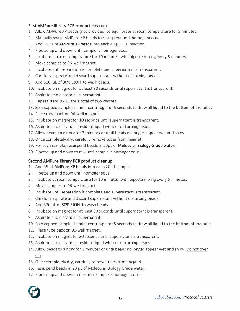
eclipsebio.com Protocol v1.01R 42
First AMPure library PCR product cleanup 1. Allow AMPure XP beads (not provided) to equilibrate at room temperature for 5 minutes.
2. Manually shake AMPure XP beads to resuspend until homogeneous.
3. Add 70 µL of AMPure XP beads into each 40 µL PCR reaction.
4. Pipette up and down until sample is homogeneous.
5. Incubate at room temperature for 10 minutes, with pipette mixing every 5 minutes.
6. Move samples to 96-well magnet.
7. Incubate until separation is complete and supernatant is transparent.
8. Carefully aspirate and discard supernatant without disturbing beads.
9. Add 320 µL of 80% EtOH to wash beads.
10. Incubate on magnet for at least 30 seconds until supernatant is transparent.
11. Aspirate and discard all supernatant.
12. Repeat steps 9 - 11 for a total of two washes.
13. Spin capped samples in mini-centrifuge for 5 seconds to draw all liquid to the bottom of the tube.
14. Place tube back on 96-well magnet.
15. Incubate on magnet for 10 seconds until supernatant is transparent.
16. Aspirate and discard all residual liquid without disturbing beads.
17. Allow beads to air dry for 3 minutes or until beads no longer appear wet and shiny.
18. Once completely dry, carefully remove tubes from magnet.
19. For each sample, resuspend beads in 20µL of Molecular Biology Grade water.
20. Pipette up and down to mix until sample is homogeneous.
Second AMPure library PCR product cleanup 1. Add 35 µL AMPure XP beads into each 20 µL sample
2. Pipette up and down until homogeneous.
3. Incubate at room temperature for 10 minutes, with pipette mixing every 5 minutes.
4. Move samples to 96-well magnet.
5. Incubate until separation is complete and supernatant is transparent.
6. Carefully aspirate and discard supernatant without disturbing beads.
7. Add 320 µL of 80% EtOH to wash beads.
8. Incubate on magnet for at least 30 seconds until supernatant is transparent.
9. Aspirate and discard all supernatant.
10. Spin capped samples in mini-centrifuge for 5 seconds to draw all liquid to the bottom of the tube.
11. Place tube back on 96-well magnet.
12. Incubate on magnet for 30 seconds until supernatant is transparent.
13. Aspirate and discard all residual liquid without disturbing beads.
14. Allow beads to air dry for 3 minutes or until beads no longer appear wet and shiny. Do not over
dry.
15. Once completely dry, carefully remove tubes from magnet.
16. Resuspend beads in 20 µL of Molecular Biology Grade water.
17. Pipette up and down to mix until sample is homogeneous.

eclipsebio.com Protocol v1.01R 43
➢ Note: Do not use other buffers for final library.
18. Incubate 5 minutes at room temperature.
19. After incubation, move tubes to 96-well magnet.
20. Incubate on magnet for 1 minute until supernatant is transparent.
21. Transfer 20 µL to a new 0.2 mL strip-tube.
22. Store samples on ice if proceeding to the next step.
Optional Stopping Point: If stopping here, libraries should be quickly frozen at -80 °C Next stopping point: ~20 minutes or ~2 hrs if pooling immediately
Library Quantification Libraries can be quantified using a variety of methods. Protocol has been optimized using
Agilent4200 TapeStation, which quantifies both library concentration, molarity and size distribution.
See Agilent4200 TapeStation manual for operation instructions.
Pool and Sequence Final Library Final library pooling concentration and conditions are typically set by high-throughput sequencing
provider. See provider information or Illumina website for additional details, especially for sequencing
color-balancing.
Protocol End

eclipsebio.com Protocol v1.01R 44
APPENDIX A: SDS-PAGE Gel Electrophoresis
Required Equipment and Materials
Item Source
NuPAGE 4-12% Bis-Tris Gels, 1.0mm, 12-well ThermoFisher Scientific cat. #NP0322BOX
NuPAGE 4-12% Bis-Tris Gels, 1.0mm, 10-well ThermoFisher Scientific cat. #NP0321BOX
XCell Surelock Mini-Cell ThermoFisher Scientific cat. #EI0001
PowerEase 300W Power Supply (230VAC) ThermoFisher Scientific cat. #PS0301
Polypropylene, Nalgene Tray TaylorScientific cat. #14-6389-01
Reagents
Not Included in kit
Item Source
20 NuPAGE MOPS SDS Running Buffer ThermoFisher Scientific cat. #NP001
Molecular Biology Grade Water, Corning VWR cat. #95000-094
Spectra Multicolor Broad Range Protein Ladder ThermoFisher Scientific cat. #26634
Included in kit
Item Source Storage
2× Western Buffer Eclipse BioInnovations -80C
eclipsebio.com Protocol v1.01R 45
Procedure
Prepare NuPAGE 4-12% Bis-Tris gels.
1 All samples will be run on two gels (PREPARATIVE), select 10-well or 12-well gel(s) according to
sample number and loading scheme.
2 Cut open gel cassette pouch with scissors, remove cassettes from pouch and remove white
sealing tape from bottom of gel cassette – rinse with clean water (diH2O). Dry with kimwipes.
Prepare XCell Surelock Mini-Cell gel apparatus for loading gel cassettes
1. Rinse XCell Surelock Mini-Cell gel apparatus thoroughly with diH2O then dry with kimwipes.
2. Place XCell Surelock Mini-Cell gel apparatus into Polypropylene Nalgene ice tray.
3. Insert first labeled NuPAGE 4-12% Bis-Tris gel cassette in either the FRONT ‘Pre-Cast Gel slot’ or
in the BACK ‘Pre-Cast Gel slot’.
4. If only running one gel insert ‘Buffer Dam’ in BACK portion of apparatus.
5. Insert ‘Gel Tension Wedge’.
6. Add 200 mL of cold Running Buffer into ‘Inner Buffer Chamber’.
7. Pull forward on the ‘Gel Tension Wedge level’ until lever stops, and the gels or gel/Buffer Dam
appears snug.
8. Add remaining cold Running Buffer into ‘Inner Buffer Chamber’. Continue to fill until Outer Buffer
Chambers are approximately half full (roughly 500-550mL per whole chamber).
9. Carefully remove gel comb(s).
10. Rinse gel wells with cold Running Buffer from the Outer Buffer Chamber using a p1000 pipettor.
Be careful not to disrupt well walls of gel, this will create difficulties when loading samples. Check
carefully for broken wells.
11. Carefully load all ladders (see m6A-eCLIP Protocol for volume) in predesignated wells by lowering
the tip to the bottom of the well and slowly pipetting ladder without carry over.
12. Next carefully load all samples (see m6A-eCLIP Protocol for volume) in predesignated wells by
lowering the tip to the bottom of the well and slowly pipetting sample without carry over.
13. Load any undesignated wells with diluted Western Buffer.
14. Once all wells have been filled, place cell safety lid with cables on XCell Surelock gel apparatus.
The lid can only be firmly closed if the (-) electrode is aligned over the banana plug. If the lid is
not properly seated, error message can appear on power supply.
15. With the Power Supply off, connect the electrode cords. Check labels: Red to (+) and black to (-).
16. Turn on the power.
17. Adjust settings to 150V and run for 75 minutes or until dye front is at the bottom of gel.
18. Cover hole in cell safety lid with a paper towel.
19. Pour a sufficient amount of ice into Nalgene ice tray so XCell Surelock Mini-Cell gel apparatus is ¾
covered then remove the paper towel from lid.
eclipsebio.com Protocol v1.01R 46
Disassembling XCell Surelock Mini-Cell gel apparatus
1. At the end of run turn off the power and disconnect the cables from power supply. Remove the
lid and unlock the lever.
2. Remove gel cassette(s) and place on work space surface.
3. Discard Running Buffer accordingly.
4. Store all reagents under appropriate storage conditions.
5. Rinse XCell Surelock Mini-Cell gel apparatus and all associated parts with clean water.
6. Dry thoroughly with kimwipes.

eclipsebio.com Protocol v1.01R 47
APPENDIX B: Membrane Transfer
Required Equipment and Materials
Item Source
Mini-Protean Tetra cell and supplies BioRad cat. #165-8001
PowerPac HC Power Supply BioRad cat. #1645052
Western Blotting Filter Paper, 7cm 8.4cm ThermoFisher cat. #84783
Polypropylene, Nalgene Tray TaylorScientific cat. #14-6389-01
Reagents
Not Included in kit
Item Source
Pierce 20 TBS Tween 20 Buffer ThermoFisher Scientific cat. #28360
20 NuPAGE Transfer Buffer ThermoFisher Scientific cat. #NP00061
Methanol Sigma-Aldrich cat. #494437
DPBS VWR cat. #21-031-CV
Molecular Biology Grade Water VWR catalog #95000-094
Procedure
Prepare Mini-Protean Tetra System
1. Rinse Mini-Protean Tetra cell thoroughly with diH2O and dry with kimwipes.
2. Place cell into transfer tank.
Prepare transfer stack for PREPARATIVE gel transfer
1. Place stack cassette into Pyrex drying dish with the black side facing down (Refer to Figure 2).
Included in kit
Item Source
Nitrocellulose membrane Eclipse BioInnovations
PVDF membrane Any
Figure 2. Transfer stack layer composition

eclipsebio.com Protocol v1.01R 48
2. Pour chilled 1 NuPAGE Transfer Buffer into Pyrex drying dish so that the black side of stack
cassette (1) is submerged.
3. Wet ‘Fiber pad’ in 1 NuPAGE Transfer Buffer and place on top of the black side of stack
cassette. Roll with a Blotting Roller to remove air bubbles.
4. Fully saturate a piece of Western blotting ‘Filter paper’ in 1 NuPAGE Transfer Buffer. Place
wetted filter paper on top of Fiber pad. Use Blotting Roller to remove any bubbles between
layers.
5. Carefully move gel from cassette to top of wetted filter paper using gel knife (see Figure 3).
Ensure gel knife is wetted with transfer buffer so not to tear the gel.
6. Analytical gel: PVDF membrane transfer
a. Let PVDF membrane sit in 100% Methanol for 1 minute.
b. Using tweezers, remove PVDF membrane from methanol and transfer to 1 NuPAGE
Transfer Buffer. Let sit for 1 minute.
7. Preparative gel: Nitrocellulose membrane transfer
a. Let nitrocellulose membrane equilibrate in transfer buffer for 1 minute
8. Analytical and Preparative gels:
a. Using tweezers, carefully place the membrane on top of the polyacrylamide gel. Use
Blotting Roller to remove any bubbles between layers.
b. Fully saturate a second piece of filter paper in 1 NuPAGE Transfer Buffer and place on
top of ‘Membrane’. Use Blotting Roller to remove any bubbles.
c. Wet second Fiber pad in 1 NuPAGE Transfer Buffer and roll out any bubbles. Place gray
Fiber pad on top of the Filter paper. Ensure that no parts are sticking outside of the stack
cassette that would prevent insertion into apparatus. Close the stack cassette firmly and
lock with the white latch.
Figure 3. Gel transfer schematic

eclipsebio.com Protocol v1.01R 49
Assembling and running the Mini-Protean Tetra System
1. Assemble the Mini-Protean Tetra System apparatus (Refer to Figure 5 for component
identification).
2. When placing the stack cassette into the ‘Electrode module’ it is essential that the black
(cathode -) side of the cassette is facing the black side of the module.
3. After all stack cassettes have been loaded, place a blue cooling ice pack (stored at -20C) in
Mini-Protean Tetra apparatus. Pour 1 NuPAGE Transfer Buffer from Pyrex tray into the
apparatus.
4. Add remaining 900mL of 1 NuPAGE Transfer Buffer inside the electrode module between
the two gel stack cassettes. Continue adding any remaining Transfer Buffer to the chamber
until chamber is full (see indicator line on transfer tank). Securely close the lid by placing the
electrodes over the ‘Buffer tank banana plugs’.
5. Connect cables to corresponding colors on power supply and turn on power supply.
6. Run gel transfer overnight (> 10 hours) in a cold (4C) room at constant 30 Volts.
➢ Note: Transfer can alternatively be performed for 2 hours at constant 200 mA. If doing so,
surround as much of the outside of the apparatus with ice as possible to avoid overheating.
If using more than one Mini-Protean Tetra apparatus, connect only one per power supply
or increase current to 210 mA. For 1mm gels, 90 min at 210 mA can be enough to
complete transfer (connect only one chamber with 1-2 gels per power supply)
Figure 5. Mini-Protean Tetra components

eclipsebio.com Protocol v1.01R 50
Appendix C: Sequencing Specifications
IMPORTANT: If user has purchased an eCLIP kit with data analysis package (Eclipse BioInnovations
cat. #ECEKD-0001) single-end sequencing must be performed in order to be compatible with Eclipse
Bio’s software pipeline.
Sequencing parameters: Eclipse Bio's eCLIP kit is based on the single-end eCLIP variant described in:
https://www.ncbi.nlm.nih.gov/pubmed/28766298 Van Nostrand EL, Nguyen TB, et al. Robust, Cost-Effective Profiling of RNA Binding Protein Targets with Single-end Enhanced Crosslinking and Immunoprecipitation (seCLIP). Methods Mol Biol. 2017;1648:177-200. PMID: 28766298. Libraries generated using the m6A-eCLIP method are typically sequenced using standard SE100 conditions on the Illumina HiSeq, NovaSeq, or NextSeq platforms. m6A-eCLIP libraries are compatible with paired-end sequencing if desired by the user, however due to the small size of typical eCLIP RNA fragments (~200bp), most fragments are fully sequenced in standard single-end formats.
Sequencing depth: Eclipse Bio’s target is 40-50 million reads per m6A-eCLIP dataset. How deeply to sequence an m6A-eCLIP dataset is a challenging balance between cost and sufficient read depth to detect true binding events. In an effort to experimentally address this question, an analysis of eCLIP-seq datasets for 150 RNA binding proteins suggested that for 90% of datasets, saturation of peak information occurred at or below 8.5 million reads (See Supplementary Fig. 11 of Van Nostrand EL, et al. A Large-Scale Binding and Functional Map of Human RNA Binding Proteins. Nature (Accepted, in press) (preprint available at https://www.biorxiv.org/content/early/2018/10/05/179648). However, we have found that targeting 25 million reads (for eCLIP) provides better coverage for abundant, broadly binding RNA binding proteins (such as HNRNPs) while still allowing pooling of ~14 eCLIP libraries per standard Illumina HiSeq 4000 lane.
➢ Adapter sequences: Illumina TruSeq HT adapters (provided) RNA adapter: 5Phos/rArGrArUrCrGrGrArArGrArGrCrArCrArCrGrUrCrUrG/3SpC3/ ssDNA adapter: 5Phos/NNNNNNNNNNAGATCGGAAGAGCGTCGTGT/3SpC3/
Index primer sequences: Illumina dual index primers (provided)
i7 index name i7 bases on Sample Sheet i5 index name i5 bases bases on Sample Sheet
705 ATTCAGAA 505 AGGCGAAG

eclipsebio.com Protocol v1.01R 51
706 GAATTCGT 506 TAATCTTA
707 CTGAAGCT 507 CAGGACGT
708 TAATGCGC 508 GTACTGAC
Read structure model

eclipsebio.com Protocol v1.01R 52
Appendix D: Total RNA Isolation
Total RNA Isolation 1. Obtain 10 million cells for RNA isolation either fresh or from frozen pellets. 2. Resuspend cells in 500 µL of TRIzol. 3. Mix until sample is homogeneous.
➢ Note: If sample cannot easily be pipetted due to viscosity, add additional TRIzol in 100 µL increments.
4. Centrifuge sample at 12,000 × g at room temperature for 3 minutes to pellet cellular debris.
5. Transfer supernatant to a fresh 1.5 mL LoBind DNA tube. Supernatant may be viscous.
6. Add an equal volume of 100% Ethanol to supernatant and pipette mix thoroughly.
7. Use one Zymo-Spin IIC column per 5 million cells used.
8. Split volume in increments of 600µL into each Zymo-Spin IIC column in a collection tube.
9. Centrifuge at 5,000 × g for 1 minute or until all liquid has passed through filter.
10. Rebind flow-through a second time by transferring it from the collection tube back to the filter.
11. Centrifuge at 5,000 × g for 1 minute or until all liquid has passed through filter.
12. Discard the flow-through.
13. Repeat with remaining lysate-ethanol mixture until entire volume has been transferred.
14. Add 400 µL Direct-zol RNA PreWash to each column.
15. Centrifuge at 5,000 × g for 1 minute or until all liquid has passed through filter.
16. Discard the flow-through.
17. Repeat steps 14-16 for a total of two washes.
18. Add 700 µL RNA Wash Buffer to column
19. Centrifuge at 5,000 × g for 1 minute or until all liquid has passed through filter.
20. Discard flow-through.
21. Add 350 µL RNA Wash Buffer to column
22. Centrifuge at 5,000 × g for 1 minute or until all liquid has passed through filter.
23. Discard flow-through.
24. Repeat step 21-23 for a total of two washes.
25. Transfer column filter to a new collection tube.
26. Spin at 12,000 × g for 2 min to dry column filter.
27. Transfer filter to a new 1.5 mL LoBind DNA tube.
28. Open cap and allow filter to air dry for 2 minutes.
29. Add 50 µL of Molecular Biology Grade Water to filter in each column to elute RNA.
30. Allow to incubate at room temperature for 1 minute.
31. Centrifuge at 12,000 × g for 90 seconds.
➢ Note: For high concentrations of RNA, elute in <50 µL and >25 µL
32. Re-elute RNA by transferring flow-through back to filter and centrifuge at 15,000 × g for 3 min.
➢ Note: If post-elution volume is < 25 µL add an additional 30 µL of Molecular Biology
Grade water to filter and spin at 15,000 × g for 3 minutes
33. Put samples on ice.

eclipsebio.com Protocol v1.01R 53
RNA and RIN Measurement Overview Total RNA can be measured using a variety of methods. This protocol has been optimized using
Agilent 4200 TapeStation with Agilent’s High Sensitivity RNA ScreenTape, which measures both total
RNA concentration and RNA Integrity Number (RIN). RNA must have a RIN score of 7 or higher to
continue.
![Natural Variation in RNA m6A Methylation and Its ......Natural Variation in RNA m6A Methylation and Its Relationship with Translational Status1[OPEN] Jin-Hong Luo,a,b,2 Ye Wang,c,2](https://static.fdocuments.in/doc/165x107/60e9600d2e83be1a38617ed7/natural-variation-in-rna-m6a-methylation-and-its-natural-variation-in-rna.jpg)


![The m6A Reader ECT2 Controls Trichome Morphology by ... · The m6A Reader ECT2 Controls Trichome Morphology by Affecting mRNA Stability in Arabidopsis[OPEN] Lian-Huan Wei,a Peizhe](https://static.fdocuments.in/doc/165x107/5f076dc27e708231d41ceff2/the-m6a-reader-ect2-controls-trichome-morphology-by-the-m6a-reader-ect2-controls.jpg)


