M100-S25: Performance Standards for Antimicrobial ...
240
January 2015 M100-S25 Performance Standards for Antimicrobial Susceptibility Testing; Twenty-Fifth Informational Supplement This document provides updated tables for the Clinical and Laboratory Standards Institute antimicrobial susceptibility testing standards M02-A12, M07-A10, and M11-A8. An informational supplement for global application developed through the Clinical and Laboratory Standards Institute consensus process. Licensed to: Diagnostic Medicine Services This document is protected by copyright. CLSI order # 0, Downloaded on 01/06/2014.
Transcript of M100-S25: Performance Standards for Antimicrobial ...

January 2015
M100-S25Performance Standards for Antimicrobial Susceptibility Testing; Twenty-Fifth Informational Supplement
This document provides updated tables for the Clinical and Laboratory Standards Institute antimicrobial susceptibility testing standards M02-A12, M07-A10, and M11-A8.
An informational supplement for global application developed through the Clinical and Laboratory Standards Institute consensus process.
Licensed�to:��Diagnostic�Medicine�ServicesThis�document�is�protected�by�copyright.�CLSI�order�#�0,�Downloaded�on�01/06/2014.
Clinical and Laboratory Standards Institute Setting the standard for quality in clinical laboratory testing around the world.
The Clinical and Laboratory Standards Institute (CLSI) is a not-for-profit membership organization that brings together the varied perspectives and expertise of the worldwide laboratory community for the advancement of a common cause: to foster excellence in laboratory medicine by developing and implementing clinical laboratory standards and guidelines that help laboratories fulfill their responsibilities with efficiency, effectiveness, and global applicability. Consensus Process
Consensus—the substantial agreement by materially affected, competent, and interested parties—is core to the development of all CLSI documents. It does not always connote unanimous agreement, but does mean that the participants in the development of a consensus document have considered and resolved all relevant objections and accept the resulting agreement. Commenting on Documents
CLSI documents undergo periodic evaluation and modification to keep pace with advancements in technologies, procedures, methods, and protocols affecting the laboratory or health care.
CLSI’s consensus process depends on experts who volunteer to serve as contributing authors and/or as participants in the reviewing and commenting process. At the end of each comment period, the committee that developed the document is obligated to review all comments, respond in writing to all substantive comments, and revise the draft document as appropriate.
Comments on published CLSI documents are equally essential, and may be submitted by anyone, at any time, on any document. All comments are addressed according to the consensus process by a committee of experts. Appeals Process
If it is believed that an objection has not been adequately addressed, the process for appeals is documented in the CLSI Standards Development Policies and Process document.
All comments and responses submitted on draft and published documents are retained on file at CLSI and are available upon request.
Get Involved—Volunteer!Do you use CLSI documents in your workplace? Do you see room for improvement? Would you like to get involved in the revision process? Or maybe you see a need to develop a new document for an emerging technology? CLSI wants to hear from you. We are always looking for volunteers. By donating your time and talents to improve the standards that affect your own work, you will play an active role in improving public health across the globe.
For further information on committee participation or to submit comments, contact CLSI.
Clinical and Laboratory Standards Institute950 West Valley Road, Suite 2500 Wayne, PA 19087 USA P: 610.688.0100F: [email protected]
Licensed�to:��Diagnostic�Medicine�ServicesThis�document�is�protected�by�copyright.�CLSI�order�#�0,�Downloaded�on�01/06/2014.
Vol. 35 No. 3 M100-S25
1
Performance Standards for Antimicrobial Susceptibility Testing; Twenty-Fifth Informational Supplement Abstract The supplemental information presented in this document is intended for use with the antimicrobial susceptibility testing procedures published in the following Clinical and Laboratory Standards Institute (CLSI)–approved standards: M02-A12—Performance Standards for Antimicrobial Disk Susceptibility Tests; Approved Standard—Twelfth Edition; M07-A10—Methods for Dilution Antimicrobial Susceptibility Tests for Bacteria That Grow Aerobically; Approved Standard—Tenth Edition; and M11-A8—Methods for Antimicrobial Susceptibility Testing of Anaerobic Bacteria; Approved Standard—Eighth Edition. The standards contain information about both disk (M02) and dilution (M07 and M11) test procedures for aerobic and anaerobic bacteria. Clinicians depend heavily on information from the clinical microbiology laboratory for treatment of their seriously ill patients. The clinical importance of antimicrobial susceptibility test results requires that these tests be performed under optimal conditions and that laboratories have the capability to provide results for the newest antimicrobial agents. The tabular information presented here represents the most current information for drug selection, interpretation, and QC using the procedures standardized in the most current editions of M02, M07, and M11. Users should replace the tables published earlier with these new tables. (Changes in the tables since the previous edition appear in boldface type.) Clinical and Laboratory Standards Institute. Performance Standards for Antimicrobial Susceptibility Testing; Twenty-Fifth Informational Supplement. CLSI document M100-S25 (ISBN 1-56238-989-0 [Print]; ISBN 1-56238-990-4 [Electronic]). Clinical and Laboratory Standards Institute, 950 West Valley Road, Suite 2500, Wayne, Pennsylvania 19087 USA, 2015.
The data in the interpretive tables in this supplement are valid only if the methodologies in M02-A12—Performance Standards for Antimicrobial Disk Susceptibility Tests; Approved Standard—Twelfth Edition; M07-A10—Methods for Dilution Antimicrobial Susceptibility Tests for Bacteria That Grow Aerobically; Approved Standard—Tenth Edition; and M11-A8—Methods for Antimicrobial Susceptibility Testing of Anaerobic Bacteria; Approved Standard—Eighth Edition are followed.
Licensed�to:��Diagnostic�Medicine�ServicesThis�document�is�protected�by�copyright.�CLSI�order�#�0,�Downloaded�on�01/06/2014.

January 2015 M100-S25
2 Licensed�to:��Diagnostic�Medicine�ServicesThis�document�is�protected�by�copyright.�CLSI�order�#�0,�Downloaded�on�01/06/2014.

ISBN 1-56238-989-0 (Print) M100-S25 ISBN 1-56238-990-4 (Electronic) Vol. 35 No. 3 ISSN 1558-6502 (Print) Replaces M100-S24 ISSN 2162-2914 (Electronic) Vol. 34 No. 1
Performance Standards for Antimicrobial Susceptibility Testing; Twenty-Fifth Informational Supplement Volume 35 Number 3 Jean B. Patel, PhD, D(ABMM) Franklin R. Cockerill III, MD Patricia A. Bradford, PhD George M. Eliopoulos, MD Janet A. Hindler, MCLS, MT(ASCP) Stephen G. Jenkins, PhD, D(ABMM), F(AAM) James S. Lewis II, PharmD Brandi Limbago, PhD Linda A. Miller, PhD David P. Nicolau, PharmD, FCCP, FIDSA Mair Powell, MD, FRCP, FRCPath Jana M. Swenson, MMSc Maria M. Traczewski, BS, MT(ASCP) John D. Turnidge, MD Melvin P. Weinstein, MD Barbara L. Zimmer, PhD
Licensed�to:��Diagnostic�Medicine�ServicesThis�document�is�protected�by�copyright.�CLSI�order�#�0,�Downloaded�on�01/06/2014.
January 2015 M100-S25
4
Copyright ©2015 Clinical and Laboratory Standards Institute. Except as stated below, any reproduction of content from a CLSI copyrighted standard, guideline, companion product, or other material requires express written consent from CLSI. All rights reserved. Interested parties may send permission requests to [email protected]. CLSI hereby grants permission to each individual member or purchaser to make a single reproduction of this publication for use in its laboratory procedure manual at a single site. To request permission to use this publication in any other manner, e-mail [email protected]. Suggested Citation CLSI. Performance Standards for Antimicrobial Susceptibility Testing; Twenty-Fifth Informational Supplement. CLSI document M100-S25. Wayne, PA: Clinical and Laboratory Standards Institute; 2015. Twenty-Fifth Informational Supplement January 2015
Seventeenth Informational Supplement January 2007
Twenty-Fourth Informational Supplement January 2014
Sixteenth Informational Supplement January 2006
Twenty-Third Informational Supplement January 2013
Fifteenth Informational Supplement January 2005
Twenty-Second Informational Supplement January 2012
Fourteenth Informational Supplement January 2004
Twenty-First Informational Supplement January 2011
Thirteenth Informational Supplement January 2003
Twentieth Informational Supplement (Update) June 2010
Twelfth Informational Supplement January 2002
Twentieth Informational Supplement January 2010
Eleventh Informational Supplement January 2001
Nineteenth Informational Supplement January 2009
Tenth Informational Supplement January 2000
Eighteenth Informational Supplement January 2008
Ninth Informational Supplement January 1999
ISBN 1-56238-989-0 (Print) ISBN 1-56238-990-4 (Electronic) ISSN 1558-6502 (Print) ISSN 2162-2914 (Electronic)
Licensed�to:��Diagnostic�Medicine�ServicesThis�document�is�protected�by�copyright.�CLSI�order�#�0,�Downloaded�on�01/06/2014.

Vol. 35 No. 3 M100-S25
5
Committee Membership Consensus Committee on Microbiology
Subcommittee on Antimicrobial Susceptibility Testing Jean B. Patel, PhD, D(ABMM) Chairholder Centers for Disease Control and Prevention USA Franklin R. Cockerill III, MD Vice-Chairholder Mayo Clinic USA Patricia A. Bradford, PhD AstraZeneca Pharmaceuticals USA George M. Eliopoulos, MD Beth Israel Deaconess Medical Center USA
Janet A. Hindler, MCLS, MT(ASCP) UCLA Medical Center USA Stephen G. Jenkins, PhD, D(ABMM), F(AAM) New York Presbyterian Hospital USA James S. Lewis II, PharmD Oregon Health and Science University USA Brandi Limbago, PhD Centers for Disease Control and Prevention USA Linda A. Miller, PhD GlaxoSmithKline USA
David P. Nicolau, PharmD, FCCP, FIDSA Hartford Hospital USA Mair Powell, MD, FRCP, FRCPath MHRA United Kingdom John D. Turnidge, MD SA Pathology at Women’s and Children’s Hospital Australia Melvin P. Weinstein, MD Robert Wood Johnson University Hospital USA Barbara L. Zimmer, PhD Siemens Healthcare Diagnostics Inc. USA
Acknowledgment CLSI, the Consensus Committee on Microbiology, and the Subcommittee on Antimicrobial Susceptibility Testing gratefully acknowledge the following volunteers for their important contributions to the development of this document: Jana M. Swenson, MMSc USA
Maria M. Traczewski, BS, MT(ASCP) The Clinical Microbiology Institute USA
Richard B. Thomson, Jr., PhD, D(ABMM), FAAM Chairholder Evanston Hospital, NorthShore University HealthSystem USA John H. Rex, MD, FACP Vice-Chairholder AstraZeneca Pharmaceuticals USA Thomas R. Fritsche, MD, PhD Marshfield Clinic USA
Patrick R. Murray, PhD BD Diagnostic Systems USA Jean B. Patel, PhD, D(ABMM) Centers for Disease Control and Prevention USA Kerry Snow, MS, MT(ASCP) FDA Center for Drug Evaluation and Research USA
John D. Turnidge, MD SA Pathology at Women’s and Children’s Hospital Australia Jeffrey L. Watts, PhD, RM(NRCM) Zoetis USA Nancy L. Wengenack, PhD, D(ABMM) Mayo Clinic USA Barbara L. Zimmer, PhD Siemens Healthcare Diagnostics Inc. USA
Licensed�to:��Diagnostic�Medicine�ServicesThis�document�is�protected�by�copyright.�CLSI�order�#�0,�Downloaded�on�01/06/2014.
January 2015 M100-S25
6
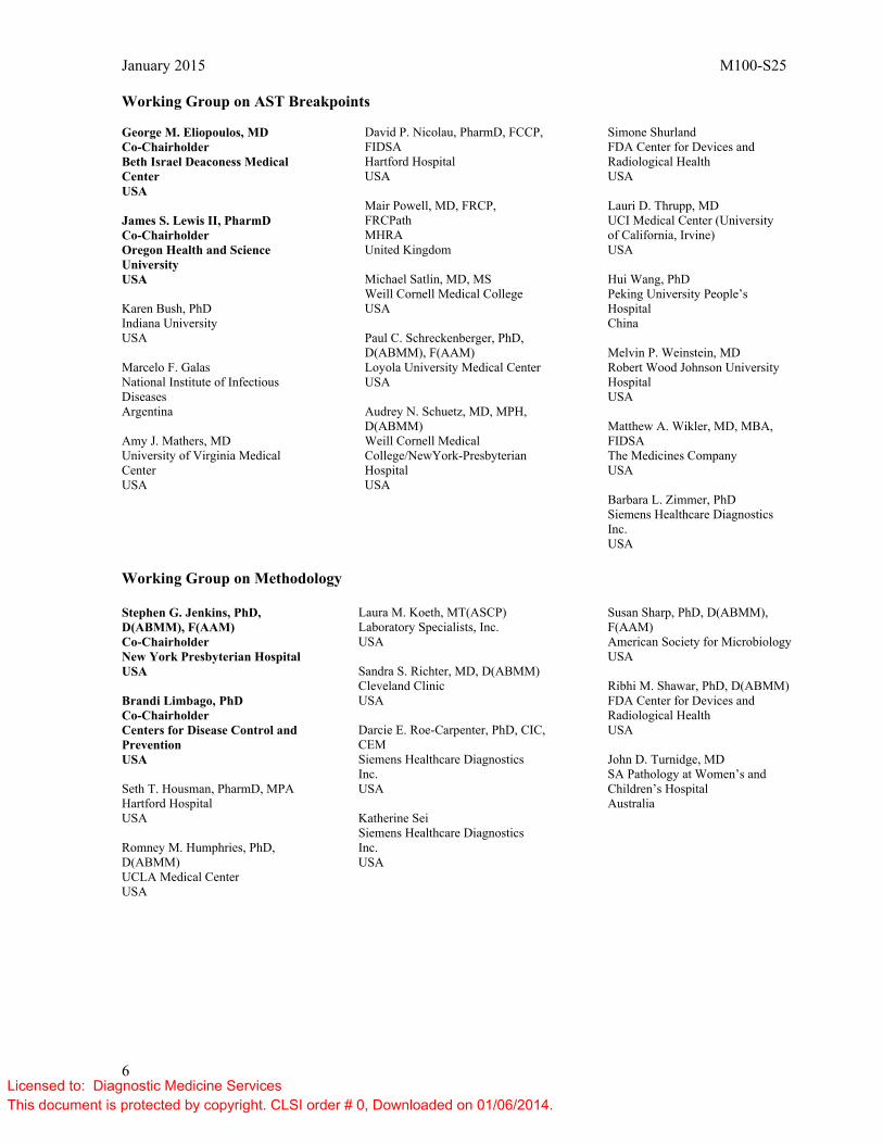
Working Group on AST Breakpoints George M. Eliopoulos, MD Co-Chairholder Beth Israel Deaconess Medical Center USA James S. Lewis II, PharmD Co-Chairholder Oregon Health and Science University USA Karen Bush, PhD Indiana University USA Marcelo F. Galas National Institute of Infectious Diseases Argentina Amy J. Mathers, MD University of Virginia Medical Center USA
David P. Nicolau, PharmD, FCCP, FIDSA Hartford Hospital USA Mair Powell, MD, FRCP, FRCPath MHRA United Kingdom Michael Satlin, MD, MS Weill Cornell Medical College USA Paul C. Schreckenberger, PhD, D(ABMM), F(AAM) Loyola University Medical Center USA Audrey N. Schuetz, MD, MPH, D(ABMM) Weill Cornell Medical College/NewYork-Presbyterian Hospital USA
Simone Shurland FDA Center for Devices and Radiological Health USA Lauri D. Thrupp, MD UCI Medical Center (University of California, Irvine) USA Hui Wang, PhD Peking University People’s Hospital China Melvin P. Weinstein, MD Robert Wood Johnson University Hospital USA Matthew A. Wikler, MD, MBA, FIDSA The Medicines Company USA Barbara L. Zimmer, PhD Siemens Healthcare Diagnostics Inc. USA
Working Group on Methodology Stephen G. Jenkins, PhD, D(ABMM), F(AAM) Co-Chairholder New York Presbyterian Hospital USA Brandi Limbago, PhD Co-Chairholder Centers for Disease Control and Prevention USA Seth T. Housman, PharmD, MPA Hartford Hospital USA Romney M. Humphries, PhD, D(ABMM) UCLA Medical Center USA
Laura M. Koeth, MT(ASCP) Laboratory Specialists, Inc. USA Sandra S. Richter, MD, D(ABMM) Cleveland Clinic USA Darcie E. Roe-Carpenter, PhD, CIC, CEM Siemens Healthcare Diagnostics Inc. USA Katherine Sei Siemens Healthcare Diagnostics Inc. USA
Susan Sharp, PhD, D(ABMM), F(AAM) American Society for Microbiology USA Ribhi M. Shawar, PhD, D(ABMM) FDA Center for Devices and Radiological Health USA John D. Turnidge, MD SA Pathology at Women’s and Children’s Hospital Australia
Licensed�to:��Diagnostic�Medicine�ServicesThis�document�is�protected�by�copyright.�CLSI�order�#�0,�Downloaded�on�01/06/2014.

Vol. 35 No. 3 M100-S25
7
Working Group on Quality Control Steven D. Brown, PhD, ABMM Co-Chairholder USA Sharon K. Cullen, BS, RAC Co-Chairholder Siemens Healthcare Diagnostics Inc. USA William B. Brasso BD Diagnostic Systems USA Patricia S. Conville, MS, MT(ASCP) FDA Center for Devices and Radiological Health USA Robert K. Flamm, PhD JMI Laboratories USA
Stephen Hawser, PhD IHMA Europe Sàrl Switzerland Janet A. Hindler, MCLS, MT(ASCP) UCLA Medical Center USA Denise Holliday, MT(ASCP) BD Diagnostic Systems USA Michael D. Huband AstraZeneca Pharmaceuticals USA Erika Matuschek, PhD ESCMID Sweden
Ross Mulder, MT(ASCP) bioMérieux, Inc. USA Susan D. Munro, MT(ASCP), CLS USA Robert P. Rennie, PhD Provincial Laboratory for Public Health Canada Frank O. Wegerhoff, PhD, MSc(Epid), MBA USA Mary K. York, PhD, ABMM MKY Microbiology Consulting USA
Working Group on Text and Tables Jana M. Swenson, MMSc Co-Chairholder USA Maria M. Traczewski, BS, MT(ASCP) Co-Chairholder The Clinical Microbiology Institute USA Janet A. Hindler, MCLS, MT(ASCP) UCLA Medical Center USA Peggy Kohner, BS, MT(ASCP) Mayo Clinic USA Dyan Luper, BS, MT(ASCP)SM, MB BD Diagnostic Systems USA
Linda M. Mann, PhD, D(ABMM) USA Melissa B. Miller, PhD, D(ABMM) UNC Hospitals USA Susan D. Munro, MT(ASCP), CLS USA Flavia Rossi, MD University of São Paulo Brazil Jeff Schapiro, MD Kaiser Permanente USA
Dale A. Schwab, PhD, D(ABMM) Quest Diagnostics Nichols Institute USA Richard B. Thomson, Jr., PhD, D(ABMM), FAAM Evanston Hospital, NorthShore University HealthSystem USA Nancy E. Watz, MS, MT(ASCP), CLS Stanford Hospital and Clinics USA Mary K. York, PhD, ABMM MKY Microbiology Consulting USA
Staff Clinical and Laboratory Standards Institute USA Luann Ochs, MS Senior Vice President – Operations Tracy A. Dooley, MLT(ASCP) Project Manager
Megan L. Tertel, MA Editorial Manager Joanne P. Christopher, MA Editor Patrice E. Polgar Editor
Licensed�to:��Diagnostic�Medicine�ServicesThis�document�is�protected�by�copyright.�CLSI�order�#�0,�Downloaded�on�01/06/2014.

January 2015 M100-S25
8 Licensed�to:��Diagnostic�Medicine�ServicesThis�document�is�protected�by�copyright.�CLSI�order�#�0,�Downloaded�on�01/06/2014.

Vol. 35 No. 3 M100-S25
9
Contents Abstract ......................................................................................................................................................... 1 Committee Membership ................................................................................................................................ 5 Summary of Changes .................................................................................................................................. 13 Summary of CLSI Processes for Establishing Interpretive Criteria and Quality Control Ranges .............. 16 CLSI Reference Methods vs Commercial Methods and CLSI vs US Food and Drug Administration Interpretive Criteria (Breakpoints) .............................................................................................................. 17 CLSI Breakpoint Additions/Revisions Since 2010 ..................................................................................... 18 Subcommittee on Antimicrobial Susceptibility Testing Mission Statement .............................................. 20 Instructions for Use of Tables ..................................................................................................................... 21 Table 1A. Suggested Groupings of Antimicrobial Agents With US Food and Drug Administration Clinical Indications That Should Be Considered for Routine Testing and Reporting on Nonfastidious Organisms by Clinical Microbiology Laboratories in the United States .................................................... 32 Table 1B. Suggested Groupings of Antimicrobial Agents With US Food and Drug Administration Clinical Indications That Should Be Considered for Routine Testing and Reporting on Fastidious Organisms by Clinical Microbiology Laboratories in the United States .................................................... 38 Table 1C. Suggested Groupings of Antimicrobial Agents With US Food and Drug Administration Clinical Indications That Should Be Considered for Routine Testing and Reporting on Anaerobic Organisms by Clinical Microbiology Laboratories in the United States .................................................... 42 Tables 2A–2J. Zone Diameter and Minimal Inhibitory Concentration Interpretive Standards for: 2A. Enterobacteriaceae .............................................................................................................................. 44 2B-1. Pseudomonas aeruginosa ................................................................................................................. 52 2B-2. Acinetobacter spp. ............................................................................................................................. 56 2B-3. Burkholderia cepacia complex ......................................................................................................... 58 2B-4. Stenotrophomonas maltophilia ......................................................................................................... 60 2B-5. Other Non-Enterobacteriaceae ......................................................................................................... 62 2C. Staphylococcus spp. ............................................................................................................................. 64 2D. Enterococcus spp. ................................................................................................................................ 72 2E. Haemophilus influenzae and Haemophilus parainfluenzae ................................................................. 76 2F. Neisseria gonorrhoeae .......................................................................................................................... 80
Tabl
e of
Con
tent
sTa
ble
of C
onte
nts
Licensed�to:��Diagnostic�Medicine�ServicesThis�document�is�protected�by�copyright.�CLSI�order�#�0,�Downloaded�on�01/06/2014.

January 2015 M100-S25
10
Contents (Continued) 2G. Streptococcus pneumoniae ................................................................................................................... 84 2H-1. Streptococcus spp. β-Hemolytic Group ............................................................................................ 90 2H-2. Streptococcus spp. Viridans Group .................................................................................................. 94 2I. Neisseria meningitidis ........................................................................................................................... 98 2J-1. Anaerobes ......................................................................................................................................... 102 2J-2. Epidemiological Cutoff Values for Propionibacterium acnes ......................................................... 106 Table 3A. Screening and Confirmatory Tests for Extended-Spectrum β-Lactamases in Klebsiella pneumoniae, Klebsiella oxytoca, Escherichia coli, and Proteus mirabilis ............................................... 108 Introduction to Tables 3B and 3C. Tests for Carbapenemases in Enterobacteriaceae, Pseudomonas aeruginosa, and Acinetobacter spp. .......................................................................................................... 112 Table 3B. The Modified Hodge Confirmatory Test for Suspected Carbapenemase Production in Enterobacteriaceae ............................................................................................................................. 114 Table 3B-1. Modifications of Table 3B When Using Interpretive Criteria for Carbapenems Described in M100-S20 (January 2010) .............................................................................................. 116 Table 3C. Carba NP Confirmatory Test for Suspected Carbapenemase Production in Enterobacteriaceae, Pseudomonas aeruginosa, and Acinetobacter spp. ............................................. 120 Table 3C-1. Modifications of Table 3C When Using Minimal Inhibitory Concentration Interpretive Criteria for Carbapenems Described in M100-S20 (January 2010) .................................. 123 Table 3D. Screening Test for Detection of β-Lactamase Production in Staphylococcus species ............. 128 Table 3E. Screening Test for Detection of Methicillin Resistance (Oxacillin Resistance) in Staphylococcus species ............................................................................................................................. 132 Table 3F. Screening Test for Detection of Vancomycin Minimal Inhibitory Concentration ≥ 8 µg/mL in Staphylococcus aureus and Enterococcus species ................................................................................ 136 Table 3G. Screening Test for Detection of Inducible Clindamycin Resistance in Staphylococcus species, Streptococcus pneumoniae, and Streptococcus spp. β-Hemolytic Group ................................... 138 Table 3H. Screening Test for Detection of High-Level Mupirocin Resistance in Staphylococcus aureus ........................................................................................................................................................ 142 Table 3I. Screening Test for Detection of High-Level Aminoglycoside Resistance in Enterococcus species ....................................................................................................................................................... 144
Tabl
e of
Con
tent
s
Licensed�to:��Diagnostic�Medicine�ServicesThis�document�is�protected�by�copyright.�CLSI�order�#�0,�Downloaded�on�01/06/2014.
Vol. 35 No. 3 M100-S25
11
Contents (Continued) Table 4A. Disk Diffusion: Quality Control Ranges for Nonfastidious Organisms (Unsupplemented Mueller-Hinton Medium) .......................................................................................................................... 146 Table 4B. Disk Diffusion: Quality Control Ranges for Fastidious Organisms ......................................... 150 Table 4C. Disk Diffusion: Reference Guide to Quality Control Frequency ............................................. 152 Table 4D. Disk Diffusion: Troubleshooting Guide................................................................................... 156 Table 5A. MIC: Quality Control Ranges for Nonfastidious Organisms (Unsupplemented Mueller-Hinton Medium [Cation-Adjusted if Broth]) ............................................................................................ 158 Table 5B. MIC: Quality Control Ranges for Fastidious Organisms (Broth Dilution Methods) ............... 162 Table 5C. MIC: Quality Control Ranges for Neisseria gonorrhoeae (Agar Dilution Method) ................ 166 Table 5D. MIC: Quality Control Ranges for Anaerobes (Agar Dilution Method) ................................... 168 Table 5E. MIC: Quality Control Ranges for Anaerobes (Broth Microdilution Method).......................... 170 Table 5F. MIC: Reference Guide to Quality Control Frequency .............................................................. 172 Table 5G. MIC: Troubleshooting Guide ................................................................................................... 176 Table 6A. Solvents and Diluents for Preparation of Stock Solutions of Antimicrobial Agents ............... 180 Table 6B. Preparation of Stock Solutions for Antimicrobial Agents Provided With Activity Expressed as Units. ................................................................................................................................... 184 Table 6C. Preparation of Solutions and Media Containing Combinations of Antimicrobial Agent ......... 186 Table 7A. Scheme for Preparing Dilutions of Antimicrobial Agents to Be Used in Agar Dilution Susceptibility Tests ................................................................................................................................... 188 Table 8A. Scheme for Preparing Dilutions of Antimicrobial Agents to Be Used in Broth Dilution Susceptibility Tests ................................................................................................................................... 190 Table 8B. Scheme for Preparing Dilutions of Water-Insoluble Antimicrobial Agents to Be Used in Broth Dilution Susceptibility Tests ........................................................................................................... 192 Appendix A. Suggestions for Confirmation of Resistant (R), Intermediate (I), or Nonsusceptible (NS) Antimicrobial Susceptibility Test Results and Organism Identification .......................................... 194 Appendix B. Intrinsic Resistance .............................................................................................................. 198 Appendix C. Quality Control Strains for Antimicrobial Susceptibility Tests ........................................... 204 Appendix D. Cumulative Antimicrobial Susceptibility Report for Anaerobic Organisms ....................... 208 Appendix E. Dosing Regimens Used to Establish Susceptible or Susceptible-Dose Dependent Interpretive Criteria…………………………………………........... ........................................................ 214
Tabl
e of
Con
tent
s
Licensed�to:��Diagnostic�Medicine�ServicesThis�document�is�protected�by�copyright.�CLSI�order�#�0,�Downloaded�on�01/06/2014.

January 2015 M100-S25
12
Contents (Continued) Appendix F. Cefepime Breakpoint Change for Enterobacteriaceae and Introduction of the Susceptible-Dose Dependent Interpretive Category ................................................................................. 216 Appendix G. Epidemiological Cutoff Values ........................................................................................... 220 Glossary I (Part 1). β-Lactams: Class and Subclass Designation and Generic Name .............................. 222 Glossary I (Part 2). Non–β-Lactams: Class and Subclass Designation and Generic Name ...................... 224 Glossary II. Abbreviations/Routes of Administration/Drug Class for Antimicrobial Agents Listed in M100-S25 ................................................................................................................................................. 226 Glossary III. List of Identical Abbreviations Used for More Than One Antimicrobial Agent in US Diagnostic Products .................................................................................................................................. 229 The Quality Management System Approach ............................................................................................ 230 Related CLSI Reference Materials ........................................................................................................... 231
The Clinical and Laboratory Standards Institute consensus process, which is the mechanism for moving a document through two or more levels of review by the health care community, is an ongoing process. Users should expect revised editions of any given document. Because rapid changes in technology may affect the procedures, methods, and protocols in a standard or guideline, users should replace outdated editions with the current editions of CLSI documents. Current editions are listed in the CLSI catalog and posted on our website at www.clsi.org. If you or your organization is not a member and would like to become one, and to request a copy of the catalog, contact us at: Telephone: +610.688.0100; Fax: +610.688.0700; E-mail: [email protected]; Website: www.clsi.org.
Tabl
e of
Con
tent
s
Licensed�to:��Diagnostic�Medicine�ServicesThis�document�is�protected�by�copyright.�CLSI�order�#�0,�Downloaded�on�01/06/2014.

Vol. 35 No. 3 M100-S25
13
Summary of Changes This list includes the “major” changes in this document. Other minor or editorial changes were made to the general formatting and to some of the table footnotes and comments. Changes to the tables since the previous edition appear in boldface type. Additions, Changes, and Deletions The following are additions or changes unless otherwise noted as a “deletion.” Instructions for Use of Tables Noted that cefazolin is a surrogate agent in Test and Report Group U for Enterobacteriaceae and is not reported exclusively on urine isolates (p. 22). Described the concept of epidemiological cutoff value (ECV), which is being introduced for Propionibacterium acnes and vancomycin (p. 25). Clarified recommendations for the β-lactamase screen in coagulase-negative staphylococci (p. 28). Tables 1A, 1B, 1C – Drugs Recommended for Testing and Reporting Deleted from Tables 1A, 1B, and 1C – gatifloxacin, grepafloxacin, lomefloxacin, ticarcillin, trovafloxacin. Enterobacteriaceae: Added fosfomycin to Test Report Group U for testing and reporting of E. coli urinary tract isolates only (p. 32). Enterococcus spp.: Added fosfomycin to Test Report Group U with indications for use against E. faecalis urinary tract isolates only (p. 32). Expanded recommendations for performing susceptibility testing on anaerobic isolates associated with polymicrobial infections (p. 43). Tables 2A Through 2J-2 – Interpretive Criteria (Breakpoints) Added instructions for following the manufacturer’s recommendations for QC when using a commercial test system. Enterobacteriaceae (Table 2A): Added azithromycin disk diffusion and MIC interpretive criteria for Salmonella Typhi (p. 49). Added pefloxacin disk diffusion interpretive criteria for Salmonella spp. for use as a surrogate test for detecting nonsusceptibility to ciprofloxacin (p. 49). Haemophilus influenzae and Haemophilus parainfluenzae (Table 2E): Clarified recommendations for selecting QC strains based on the antimicrobial agents tested (p. 76).
Sum
mar
y of
Cha
nges
Licensed�to:��Diagnostic�Medicine�ServicesThis�document�is�protected�by�copyright.�CLSI�order�#�0,�Downloaded�on�01/06/2014.

January 2015 M100-S25
14
Summary of Changes (Continued) Streptococcus pneumoniae (Table 2G): Added suggestions for assessing deterioration of oxacillin disk content (p. 84). Anaerobes (Table 2J-1): Clarified recommendations for selecting QC strains tested for routine QC (p. 102). Expanded the definition of the intermediate interpretive category when used with anaerobic bacteria and addressed several clinical factors associated with this definition (p. 102). Epidemiological Cutoff Values for Propionibacterium acnes (Table 2J-2): New table with epidemiological cutoff values (ECVs) for vancomycin related to therapy of P. acnes infections (p. 106). Tables 3A Through 3I – Screening and Confirmatory Tests Tests for Carbapenemases in Enterobacteriaceae, Pseudomonas aeruginosa, and Acinetobacter spp. (Introduction to Tables 3B and 3C): Added table that introduces Tables 3B and 3B-1 by summarizing methods for detecting carbapenemase-producing Enterobacteriaceae, P. aeruginosa, and Acinetobacter spp. (p. 112). The Modified Hodge Confirmatory Test for Suspected Carbapenemase Production in Enterobacteriaceae (Table 3B): Expanded recommendations for when the modified Hodge test might be used (pp. 114 to 115). Modifications of Table 3B When Using Interpretive Criteria for Carbapenems Described in M100-S20 (January 2010) (Table 3B-1): Eliminated details of MHT performance (now only in Table 3B) and included only steps related to testing and reporting decisions for the MHT (p. 116). Carba NP Confirmatory Test for Suspected Carbapenemase Production in Enterobacteriaceae, Pseudomonas aeruginosa, and Acinetobacter spp. (Table 3C): Added new table with detailed instructions for performance of this phenotypic test for carbapenemase production in Enterobacteriaceae, P. aeruginosa, and Acinetobacter spp. (pp. 120 to 126). Modifications of Table 3C When Using Minimal Inhibitory Concentration Interpretive Criteria for Carbapenems Described in M100-S20 (January 2010) (Table 3C-1): Added new table that includes only steps related to testing and reporting decisions for the Carba NP Test (pp. 123 to 126). Tables 4 and 5 – Quality Control Table 4A (p. 146): Added QC range for: Escherichia coli ATCC® 25922 Pefloxacin Klebsiella pneumoniae ATCC® 700603 Ceftaroline-avibactam Ceftazidime-avibactam Ceftolozane-tazobactam
Sum
mar
y of
Cha
nges
Licensed�to:��Diagnostic�Medicine�ServicesThis�document�is�protected�by�copyright.�CLSI�order�#�0,�Downloaded�on�01/06/2014.

Vol. 35 No. 3 M100-S25
15
Summary of Changes (Continued) Added recommendations for handling E. coli ATCC® 35218 to ensure it maintains its β-lactamase production integrity. Table 5A (p. 158): Added QC ranges for: Klebsiella pneumoniae ATCC® 700603 Amoxicillin Amoxicillin-clavulanate Ampicillin Ampicillin-sulbactam Ceftaroline Ceftazidime Piperacillin-tazobactam Ticarcillin Ticarcillin-clavulanate Added recommendations for handling E. coli ATCC® 35218 to ensure it maintains its β-lactamase production integrity. Added footnote to piperacillin for K. pneumoniae ATCC® 700603 that explains no range is recommended due to exquisite susceptibility of this organism to piperacillin (very low and off-scale MICs). Table 6A – Solvents and Diluents (p. 180): Revised diluent for tedizolid along with instructions for preparation of stock solutions. Appendixes and Glossaries Appendix A. Suggestions for Confirmation of Resistant (R), Intermediate (I), or Nonsusceptible (NS) Antimicrobial Susceptibility Test Results and Organism Identification: Corrected susceptibility category result that should be investigated for S. pneumoniae with ceftaroline (previously “R”; now “NS”) (p. 196). Appendix D. Cumulative Antimicrobial Susceptibility Report for Anaerobic Organisms (p. 208): Updated table with current data available. New Appendix F. Cefepime Breakpoint Change for Enterobacteriaceae and Introduction of the Susceptible-Dose Dependent Interpretive Category (p. 216): Relocated information previously positioned in the front of M100 to new Appendix F (no changes to content). New Appendix G. Epidemiological Cutoff Values (p. 220): Added new appendix containing a detailed description of ECVs that is aimed at answering questions about this concept, which is appearing in M100 for the first time. Content defines ECVs and describes their intended use. Glossary II – added pefloxacin (p. 228).
Sum
mar
y of
Cha
nges
Licensed�to:��Diagnostic�Medicine�ServicesThis�document�is�protected�by�copyright.�CLSI�order�#�0,�Downloaded�on�01/06/2014.

January 2015 M100-S25
16
Summary of CLSI Processes for Establishing Interpretive Criteria and Quality Control Ranges The Clinical and Laboratory Standards Institute (CLSI) is an international, voluntary, not-for-profit, interdisciplinary, standards-developing, and educational organization accredited by the American National Standards Institute (ANSI) that develops and promotes use of consensus-developed standards and guidelines within the health care community. These consensus standards and guidelines are developed to address critical areas of diagnostic testing and patient health care, and are developed in an open and consensus-seeking forum. CLSI is open to anyone or any organization that has an interest in diagnostic testing and patient care. Information about CLSI can be found at www.clsi.org. The CLSI Subcommittee on Antimicrobial Susceptibility Testing reviews data from a variety of sources and studies (eg, in vitro, pharmacokinetics-pharmacodynamics, and clinical studies) to establish antimicrobial susceptibility test methods, interpretive criteria, and QC parameters. The details of the data required to establish interpretive criteria, QC parameters, and how the data are presented for evaluation are described in CLSI document M23—Development of In Vitro Susceptibility Testing Criteria and Quality Control Parameters. Over time, a microorganism’s susceptibility to an antimicrobial agent may decrease, resulting in a lack of clinical efficacy and/or safety. In addition, microbiological methods and QC parameters may be refined to ensure more accurate and better performance of susceptibility test methods. Because of this, CLSI continually monitors and updates information in its documents. Although CLSI standards and guidelines are developed using the most current information and thinking available at the time, the field of science and medicine is ever changing; therefore, standards and guidelines should be used in conjunction with clinical judgment, current knowledge, and clinically relevant laboratory test results to guide patient treatment. Additional information, updates, and changes in this document are found in the meeting summary minutes of the Subcommittee on Antimicrobial Susceptibility Testing at www.clsi.org.
Licensed�to:��Diagnostic�Medicine�ServicesThis�document�is�protected�by�copyright.�CLSI�order�#�0,�Downloaded�on�01/06/2014.

Vol. 35 No. 3 M100-S25
17
CLSI Reference Methods vs Commercial Methods and CLSI vs US Food and Drug Administration Interpretive Criteria (Breakpoints)
It is important for users of M02-A12, M07-A10, and the M100 Informational Supplement to recognize that the standard methods described in CLSI documents are reference methods. These methods may be used for routine antimicrobial susceptibility testing of clinical isolates, for evaluation of commercial devices that will be used in clinical laboratories, or by drug or device manufacturers for testing of new agents or systems. Results generated by reference methods, such as those contained in CLSI documents, may be used by regulatory authorities to evaluate the performance of commercial susceptibility testing devices as part of the approval process. Clearance by a regulatory authority indicates that the commercial susceptibility testing device provides susceptibility results that are substantially equivalent to results generated using reference methods for the organisms and antimicrobial agents described in the device manufacturer’s approved package insert. CLSI breakpoints may differ from those approved by various regulatory authorities for many reasons, including the following: different databases, differences in interpretation of data, differences in doses used in different parts of the world, and public health policies. Differences also exist because CLSI proactively evaluates the need for changing breakpoints. The reasons why breakpoints may change and the manner in which CLSI evaluates data and determines breakpoints are outlined in CLSI document M23—Development of In Vitro Susceptibility Testing Criteria and Quality Control Parameters. Following a decision by CLSI to change an existing breakpoint, regulatory authorities may also review data in order to determine how changing breakpoints may affect the safety and effectiveness of the antimicrobial agent for the approved indications. If the regulatory authority changes breakpoints, commercial device manufacturers may have to conduct a clinical laboratory trial, submit the data to the regulatory authority, and await review and approval. For these reasons, a delay of one or more years may be required if an interpretive breakpoint change is to be implemented by a device manufacturer. In the United States, it is acceptable for laboratories that use US Food and Drug Administration (FDA)–cleared susceptibility testing devices to use existing FDA interpretive breakpoints. Either FDA or CLSI susceptibility interpretive breakpoints are acceptable to clinical laboratory accrediting bodies. Policies in other countries may vary. Each laboratory should check with the manufacturer of its antimicrobial susceptibility test system for additional information on the interpretive criteria used in its system’s software. Following discussions with appropriate stakeholders, such as infectious diseases practitioners and the pharmacy department, as well as the pharmacy and therapeutics and infection control committees of the medical staff, newly approved or revised breakpoints may be implemented by clinical laboratories. Following verification, CLSI disk diffusion test breakpoints may be implemented as soon as they are published in M100. If a device includes antimicrobial test concentrations sufficient to allow interpretation of susceptibility and resistance to an agent using the CLSI breakpoints, a laboratory could choose to, after appropriate verification, interpret and report results using CLSI breakpoints.
Licensed�to:��Diagnostic�Medicine�ServicesThis�document�is�protected�by�copyright.�CLSI�order�#�0,�Downloaded�on�01/06/2014.

January 2015 M100-S25
18
CLSI Breakpoint Additions/Revisions Since 2010
Antimicrobial Agent Date of Revision* (M100 version) Comments
Enterobacteriaceae Aztreonam January 2010 (M100-S20) Cefazolin January 2010 (M100-S20)
January 2011 (M100-S21) Breakpoints were revised twice since 2010.
Cefazolin January 2014 (M100-S24) Breakpoints predict results for oral cephalosporins when used for therapy of uncomplicated UTIs.
Cefepime January 2014 (M100-S24) Cefotaxime January 2010 (M100-S20) Ceftazidime January 2010 (M100-S20) Ceftizoxime January 2010 (M100-S20) Ceftriaxone January 2010 (M100-S20) Doripenem June 2010 (M100-S20-U) No previous CLSI breakpoints existed for
doripenem. Ertapenem June 2010 (M100-S20-U)
January 2012 (M100-S22) Breakpoints were revised twice since 2010.
Imipenem June 2010 (M100-S20-U) Meropenem June 2010 (M100-S20-U) Ciprofloxacin – Salmonella spp. (including S. Typhi)
January 2012 (M100-S22) Removed body site–specific breakpoint recommendations in 2013.
Ceftaroline January 2013 (M100-S23) No previous CLSI breakpoints existed for ceftaroline.
Levofloxacin – Salmonella spp. (including S. Typhi)
January 2013 (M100-S23)
Ofloxacin – Salmonella spp. (including S. Typhi)
June 2013 (M100-S23)
Pefloxacin – Salmonella spp. (including S. Typhi)
January 2015 (M100-S25) Surrogate test for ciprofloxacin.
Azithromycin – S. Typhi only January 2015 (M100-S25) Pseudomonas aeruginosa Piperacillin-tazobactam January 2012 (M100-S22) Ticarcillin-clavulanate January 2012 (M100-S22) Doripenem January 2012 (M100-S22) Imipenem January 2012 (M100-S22) Meropenem January 2012 (M100-S22) Ticarcillin January 2012 (M100-S22) Piperacillin January 2012 (M100-S22) Acinetobacter spp. Doripenem January 2014 (M100-S24) Imipenem January 2014 (M100-S24) Meropenem January 2014 (M100-S24) Staphylococcus spp. Ceftaroline January 2013 (M100-S23) No previous CLSI breakpoints existed for
ceftaroline. Haemophilus influenzae and Haemophilus parainfluenzae Ceftaroline January 2013 (M100-S23) No previous CLSI breakpoints existed for
ceftaroline.
Licensed�to:��Diagnostic�Medicine�ServicesThis�document�is�protected�by�copyright.�CLSI�order�#�0,�Downloaded�on�01/06/2014.
Vol. 35 No. 3 M100-S25
19
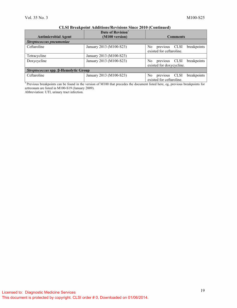
CLSI Breakpoint Additions/Revisions Since 2010 (Continued)
Antimicrobial Agent Date of Revision* (M100 version) Comments
Streptococcus pneumoniae Ceftaroline January 2013 (M100-S23) No previous CLSI breakpoints
existed for ceftaroline. Tetracycline January 2013 (M100-S23) Doxycycline January 2013 (M100-S23) No previous CLSI breakpoints
existed for doxycycline. Streptococcus spp. β-Hemolytic Group Ceftaroline January 2013 (M100-S23) No previous CLSI breakpoints
existed for ceftaroline. * Previous breakpoints can be found in the version of M100 that precedes the document listed here, eg, previous breakpoints for aztreonam are listed in M100-S19 (January 2009). Abbreviation: UTI, urinary tract infection.
Licensed�to:��Diagnostic�Medicine�ServicesThis�document�is�protected�by�copyright.�CLSI�order�#�0,�Downloaded�on�01/06/2014.

January 2015 M100-S25
20
Subcommittee on Antimicrobial Susceptibility Testing Mission Statement The Subcommittee on Antimicrobial Susceptibility Testing is composed of representatives from the professions, government, and industry, including microbiology laboratories, government agencies, health care providers and educators, and pharmaceutical and diagnostic microbiology industries. Using the CLSI voluntary consensus process, the subcommittee develops standards that promote accurate antimicrobial susceptibility testing and appropriate reporting. The mission of the Subcommittee on Antimicrobial Susceptibility Testing is to: Develop standard reference methods for antimicrobial susceptibility tests. Provide quality control parameters for standard test methods. Establish interpretive criteria for the results of standard antimicrobial susceptibility tests. Provide suggestions for testing and reporting strategies that are clinically relevant and cost-effective. Continually refine standards and optimize detection of emerging resistance mechanisms through
development of new or revised methods, interpretive criteria, and quality control parameters. Educate users through multimedia communication of standards and guidelines. Foster a dialogue with users of these methods and those who apply them. The ultimate purpose of the subcommittee’s mission is to provide useful information to enable laboratories to assist the clinician in the selection of appropriate antimicrobial therapy for patient care. The standards and guidelines are meant to be comprehensive and to include all antimicrobial agents for which the data meet established CLSI guidelines. The values that guide this mission are quality, accuracy, fairness, timeliness, teamwork, consensus, and trust.
Licensed�to:��Diagnostic�Medicine�ServicesThis�document�is�protected�by�copyright.�CLSI�order�#�0,�Downloaded�on�01/06/2014.
For Use With M02-A12 and M07-A10 M100-S25
Clinical and Laboratory Standards Institute. All rights reserved. 21
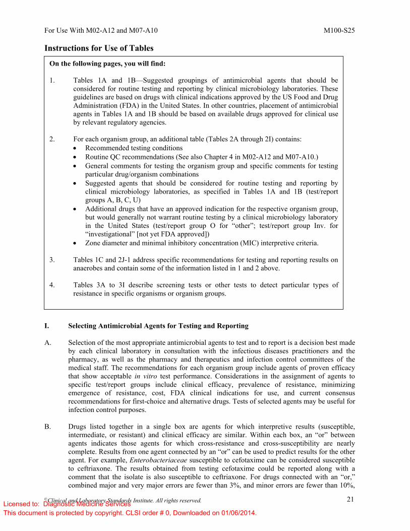
Instructions for Use of Tables I. Selecting Antimicrobial Agents for Testing and Reporting A. Selection of the most appropriate antimicrobial agents to test and to report is a decision best made
by each clinical laboratory in consultation with the infectious diseases practitioners and the pharmacy, as well as the pharmacy and therapeutics and infection control committees of the medical staff. The recommendations for each organism group include agents of proven efficacy that show acceptable in vitro test performance. Considerations in the assignment of agents to specific test/report groups include clinical efficacy, prevalence of resistance, minimizing emergence of resistance, cost, FDA clinical indications for use, and current consensus recommendations for first-choice and alternative drugs. Tests of selected agents may be useful for infection control purposes.
B. Drugs listed together in a single box are agents for which interpretive results (susceptible,
intermediate, or resistant) and clinical efficacy are similar. Within each box, an “or” between agents indicates those agents for which cross-resistance and cross-susceptibility are nearly complete. Results from one agent connected by an “or” can be used to predict results for the other agent. For example, Enterobacteriaceae susceptible to cefotaxime can be considered susceptible to ceftriaxone. The results obtained from testing cefotaxime could be reported along with a comment that the isolate is also susceptible to ceftriaxone. For drugs connected with an “or,” combined major and very major errors are fewer than 3%, and minor errors are fewer than 10%,
On the following pages, you will find: 1. Tables 1A and 1B—Suggested groupings of antimicrobial agents that should be
considered for routine testing and reporting by clinical microbiology laboratories. These guidelines are based on drugs with clinical indications approved by the US Food and Drug Administration (FDA) in the United States. In other countries, placement of antimicrobial agents in Tables 1A and 1B should be based on available drugs approved for clinical use by relevant regulatory agencies.
2. For each organism group, an additional table (Tables 2A through 2I) contains:
Recommended testing conditions Routine QC recommendations (See also Chapter 4 in M02-A12 and M07-A10.) General comments for testing the organism group and specific comments for testing
particular drug/organism combinations Suggested agents that should be considered for routine testing and reporting by
clinical microbiology laboratories, as specified in Tables 1A and 1B (test/report groups A, B, C, U)
Additional drugs that have an approved indication for the respective organism group, but would generally not warrant routine testing by a clinical microbiology laboratory in the United States (test/report group O for “other”; test/report group Inv. for “investigational” [not yet FDA approved])
Zone diameter and minimal inhibitory concentration (MIC) interpretive criteria.
3. Tables 1C and 2J-1 address specific recommendations for testing and reporting results on anaerobes and contain some of the information listed in 1 and 2 above.
4. Tables 3A to 3I describe screening tests or other tests to detect particular types of
resistance in specific organisms or organism groups.
Licensed�to:��Diagnostic�Medicine�ServicesThis�document�is�protected�by�copyright.�CLSI�order�#�0,�Downloaded�on�01/06/2014.

January 2015 Vol. 35 No. 3
Clinical and Laboratory Standards Institute. All rights reserved. 22
based on a large population of bacteria tested (see CLSI document M23 for description of error types). In addition, to qualify for an “or,” at least 100 strains with resistance to the agents in question must be tested, and a result of “resistant” must be obtained with all agents for at least 95% of the strains. “Or” is also used for comparable agents when tested against organisms for which “susceptible-only” interpretive criteria are provided (eg, cefotaxime or ceftriaxone with Haemophilus influenzae). When no “or” connects agents within a box, testing of one agent cannot be used to predict results for another, owing either to discrepancies or insufficient data.
C. Test/Report Groups 1. As listed in Tables 1A, 1B, and 1C, agents in Group A are considered appropriate for inclusion
in a routine, primary testing panel, as well as for routine reporting of results for the specific organism groups.
2. Group B includes antimicrobial agents that may warrant primary testing, but they may be
reported only selectively, such as when the organism is resistant to agents of the same antimicrobial class, as in Group A. Other indications for reporting the result might include a selected specimen source (eg, a third-generation cephalosporin for enteric bacilli from CSF or trimethoprim-sulfamethoxazole for urinary tract isolates); a polymicrobial infection; infections involving multiple sites; cases of patient allergy, intolerance, or failure to respond to an antimicrobial agent in Group A; or for purposes of infection control.
3. Group C includes alternative or supplemental antimicrobial agents that may require testing in
those institutions that harbor endemic or epidemic strains resistant to several of the primary drugs (especially in the same class, eg, -lactams); for treatment of patients allergic to primary drugs; for treatment of unusual organisms (eg, chloramphenicol for extraintestinal isolates of Salmonella spp.); or for reporting to infection control as an epidemiological aid.
4. Group U (“urine”) includes certain antimicrobial agents (eg, nitrofurantoin and certain
quinolones) that are used only or primarily for treating urinary tract infections. These agents should not be routinely reported against pathogens recovered from other sites of infection. An exception to this rule is for Enterobacteriaceae in Table 1A, where cefazolin is listed as a surrogate agent for oral cephalosporins. Other antimicrobial agents with broader indications may be included in Group U for specific urinary pathogens (eg, P. aeruginosa and ofloxacin).
5. Group O (“other”) includes antimicrobial agents that have a clinical indication for the organism
group but are generally not candidates for routine testing and reporting in the United States.
6. Group Inv. (“investigational”) includes antimicrobial agents that are investigational for the organism group and have not yet been approved by the FDA for use in the United States.
D. Selective Reporting
Each laboratory should decide which agents in the tables to report routinely (Group A) and which might be reported only selectively (from Group B), in consultation with the infectious diseases practitioners, the pharmacy, and the pharmacy and therapeutics and infection control committees of the health care institution. Selective reporting should improve the clinical relevance of test reports and help minimize the selection of multiresistant, health care–associated strains by overuse of broad-spectrum agents. Results for Group B antimicrobial agents tested but not reported routinely should be available on request, or they may be reported for selected specimen types. Unexpected resistance, when confirmed, should be reported (eg, resistance to a secondary agent but susceptibility to a primary agent, such as a P. aeruginosa isolate resistant to amikacin but susceptible to tobramycin; as such, both drugs should be reported). In addition, each
Licensed�to:��Diagnostic�Medicine�ServicesThis�document�is�protected�by�copyright.�CLSI�order�#�0,�Downloaded�on�01/06/2014.

For Use With M02-A12 and M07-A10 M100-S25
Clinical and Laboratory Standards Institute. All rights reserved. 23
laboratory should develop a protocol to address isolates that are confirmed as resistant to all agents on its routine test panels. This protocol should include options for testing additional agents in-house or sending the isolate to a reference laboratory.
II. Reporting Results The minimal inhibitory concentration (MIC) values determined as described in M07-A10 may be
reported directly to clinicians for patient care purposes. However, it is essential that an interpretive category result (S, I, or R) also be provided routinely to facilitate understanding of the MIC report by clinicians. Zone diameter measurements without an interpretive category should not be reported. Recommended interpretive categories for various MIC and zone diameter values are included in tables for each organism group and are based on evaluation of data as described in CLSI document M23.
Recommended MIC and disk diffusion interpretive criteria are based on usual dosage regimens and routes of administration in the United States.
A. Susceptible, susceptible-dose dependent, intermediate, resistant or nonsusceptible interpretations
are reported and defined as follows: 1. Susceptible (S)
The “susceptible” category implies that isolates are inhibited by the usually achievable concentrations of antimicrobial agent when the dosage recommended to treat the site of infection is used.
2. Susceptible-Dose Dependent (SDD)
The “susceptible-dose dependent” category implies that susceptibility of an isolate is dependent on the dosing regimen that is used in the patient. In order to achieve levels that are likely to be clinically effective against isolates for which the susceptibility testing results (either MICs or disk diffusion) are in the SDD category, it is necessary to use a dosing regimen (ie, higher doses, more frequent doses, or both) that results in higher drug exposure than the dose that was used to establish the susceptible breakpoint. Consideration should be given to the maximum approved dosage regimen, because higher exposure gives the highest probability of adequate coverage of an SDD isolate. The dosing regimens used to set the SDD interpretive criterion are provided in Appendix E. The drug label should be consulted for recommended doses and adjustment for organ function. NOTE: The SDD interpretation is a new category for antibacterial susceptibility testing, although it has been previously applied for interpretation of antifungal susceptibility test results (see CLSI document M27-S4, the supplement to CLSI document M27). The concept of SDD has been included within the intermediate category definition for antimicrobial agents. However, this is often overlooked or not understood by clinicians and microbiologists when an intermediate result is reported. The SDD category may be assigned when doses well above those used to calculate the susceptible breakpoint are approved and used clinically, and where sufficient data to justify the designation exist and have been reviewed. When the intermediate category is used, its definition remains unchanged. See Appendix F for further information.
Licensed�to:��Diagnostic�Medicine�ServicesThis�document�is�protected�by�copyright.�CLSI�order�#�0,�Downloaded�on�01/06/2014.

January 2015 Vol. 35 No. 3
Clinical and Laboratory Standards Institute. All rights reserved. 24
3. Intermediate (I)
The “intermediate” category includes isolates with antimicrobial agent MICs that approach usually attainable blood and tissue levels, and for which response rates may be lower than for susceptible isolates. The intermediate category implies clinical efficacy in body sites where the drugs are physiologically concentrated (eg, quinolones and -lactams in urine) or when a higher than normal dosage of a drug can be used (eg, -lactams). This category also includes a buffer zone, which should prevent small, uncontrolled, technical factors from causing major discrepancies in interpretations, especially for drugs with narrow pharmacotoxicity margins.
4. Resistant (R) The “resistant” category implies that isolates are not inhibited by the usually achievable
concentrations of the agent with normal dosage schedules and/or that demonstrate MICs or zone diameters that fall in the range where specific microbial resistance mechanisms (eg, -lactamases) are likely, and clinical efficacy of the agent against the isolate has not been reliably shown in treatment studies.
5. Nonsusceptible (NS)
The “nonsusceptible” category is used for isolates for which only a susceptible interpretive criterion has been designated because of the absence or rare occurrence of resistant strains. Isolates for which the antimicrobial agent MICs are above or zone diameters below the value indicated for the susceptible breakpoint should be reported as nonsusceptible. NOTE 1: An isolate that is interpreted as nonsusceptible does not necessarily mean that the isolate has a resistance mechanism. It is possible that isolates with MICs above the susceptible breakpoint that lack resistance mechanisms may be encountered within the wild-type distribution subsequent to the time the susceptible-only breakpoint is set. NOTE 2: For strains yielding results in the “nonsusceptible” category, organism identification and antimicrobial susceptibility test results should be confirmed (see Appendix A).
6. Interpretive Criteria
Interpretive criteria are the MIC or zone diameter values used to indicate susceptible, intermediate, and resistant breakpoints.
Antimicrobial Agent
Disk Content
Zone DiameterInterpretive Criteria (nearest whole mm)
MIC Interpretive Criteria (µg/mL)
S I R S I R X 30 μg 20 15–19 14 4 8–16 32Y — — — — 1 2 4Z 10 μg 16 — — 1 — —
For example, for antimicrobial agent X with interpretive criteria in the table above, the susceptible breakpoint is 4 g/mL or 20 mm and the resistant breakpoint is 32 g/mL or 14 mm. For some antimicrobial agents (eg, antimicrobial agent Y), only MIC interpretive criteria may be available. For these agents, the disk diffusion zone diameters do not correlate with MIC values. Technical issues may also preclude the use of the disk diffusion method for some agents.
Licensed�to:��Diagnostic�Medicine�ServicesThis�document�is�protected�by�copyright.�CLSI�order�#�0,�Downloaded�on�01/06/2014.

For Use With M02-A12 and M07-A10 M100-S25
Clinical and Laboratory Standards Institute. All rights reserved. 25
For some antimicrobial agents (eg, antimicrobial agent Z) only susceptible criteria exist. For these agents, the absence or rare occurrence of resistant strains precludes defining any results categories other than “susceptible.” For strains yielding results suggestive of a “nonsusceptible” category, organism identification and antimicrobial susceptibility test results should be confirmed (see Appendix A). In both cases, a dash mark (—) indicates that interpretive criteria are not applicable. Laboratories should only report results for agents listed in the Table 2 specific to the organism being tested; it is not appropriate to apply disk diffusion or MIC interpretive criteria taken from an alternative Table 2. There may be rare cases where an agent may be appropriate for an isolate but for which there are no CLSI interpretive criteria (eg, tigecycline). In these cases the FDA prescribing information document for the agent should be consulted.
B. In place of interpretive criteria (“breakpoints” or “clinical breakpoints”) an epidemiological cutoff value (ECV) may be listed for specific organism/antimicrobial agent combinations (see Table 2J-2 and Appendix G). ECVs and breakpoints are very different. Breakpoints are established using MIC distributions, pharmacokinetic-pharmacodynamic (PK-PD) data, and clinical outcome data (as described in CLSI document M23). Because breakpoints are based on pharmacologically and clinically rich datasets, they are considered to be robust predictors of likely clinical outcome. By contrast, ECVs are MIC values that separate bacterial populations into those with (non-wild-type [NWT]) and without (wild-type [WT]) acquired and/or mutational resistance mechanisms based on their phenotypes (MICs). They are, therefore, based on in vitro data only.
ECVs are principally used to signal the emergence or evolution of NWT strains. ECVs are not clinical breakpoints, and, thus, proven clinical relevance of ECVs has not yet been identified or approved by CLSI or any regulatory agency.
C. For some organism groups excluded from Tables 2A through 2J-1, CLSI document M45—
Methods for Antimicrobial Dilution and Disk Susceptibility Testing of Infrequently Isolated or Fastidious Bacteria provides suggestions for standardized methods for susceptibility testing, including information about drug selection, interpretation, and QC. The organism groups covered in that document are Abiotrophia and Granulicatella spp. (formerly known as nutritionally deficient or nutritionally variant streptococci); Aeromonas spp.; Bacillus spp. (not B. anthracis); Campylobacter jejuni/coli; Corynebacterium spp. (including C. diphtheriae); Erysipelothrix rhusiopathiae; the HACEK group: Aggregatibacter spp. (formerly Haemophilus aphrophilus, H. paraphrophilus, H. segnis, and Actinobacillus actinomycetemcomitans), Cardiobacterium spp., Eikenella corrodens, and Kingella spp.; Helicobacter pylori; Lactobacillus spp.; Leuconostoc spp.; Listeria monocytogenes; Moraxella catarrhalis; Pasteurella spp.; Pediococcus spp.; potential agents of bioterrorism; and Vibrio spp., including V. cholerae.
For organisms other than those in the groups mentioned above, studies are not yet adequate to develop reproducible, definitive standards to interpret results. These organisms may require different media or different atmospheres of incubation, or they may show marked strain-to-strain variation in growth rate. For these microorganisms, consultation with an infectious diseases specialist is recommended for guidance in determining the need for susceptibility testing and in the interpretation of results. Published reports in the medical literature and current consensus recommendations for therapy of uncommon microorganisms may obviate the need for testing. If necessary, a dilution method usually is the most appropriate testing method, and this may require submitting the organism to a reference laboratory. Physicians should be informed of the limitations of results and advised to interpret results with caution.
Licensed�to:��Diagnostic�Medicine�ServicesThis�document�is�protected�by�copyright.�CLSI�order�#�0,�Downloaded�on�01/06/2014.

January 2015 Vol. 35 No. 3
Clinical and Laboratory Standards Institute. All rights reserved. 26
D. Policies regarding the generation of cumulative antibiograms should be developed in concert with the infectious diseases service, infection control personnel, and the pharmacy and therapeutics committee. In most circumstances, the percentage of susceptible and intermediate results should not be combined into the same statistics. See CLSI document M39—Analysis and Presentation of Cumulative Antimicrobial Susceptibility Test Data.
III. Therapy-Related Comments
Some of the comments in the tables relate to therapy concerns. These are denoted with an Rx symbol. It may be appropriate to include some of these comments (or modifications thereof) on the patient report. An example would be inclusion of a comment on Enterococcus susceptibility reports from blood cultures that “combination therapy with ampicillin, penicillin, or vancomycin (for susceptible strains) plus an aminoglycoside is usually indicated for serious enterococcal infections, such as endocarditis, unless high-level resistance to both gentamicin and streptomycin is documented; such combinations are predicted to result in synergistic killing of the Enterococcus.”
Antimicrobial dosage regimens often vary widely among practitioners and institutions. In some
cases, the MIC interpretive criteria rely on PK-PD data, using specific human dosage regimens. In cases where specific dosage regimens are important for proper application of breakpoints, the dosage regimen is listed. These dosage regimen comments are not generally intended for use on individual patient reports.
IV. Confirmation of Patient Results Multiple test parameters are monitored by following the QC recommendations described in
M100. However, acceptable results derived from testing QC strains do not guarantee accurate results when testing patient isolates. It is important to review all of the results obtained from all drugs tested on a patient’s isolate before reporting the results. This should include, but not be limited to, ensuring that 1) the antimicrobial susceptibility results are consistent with the identification of the isolate; 2) the results from individual agents within a specific drug class follow the established hierarchy of activity rules (eg, in general, third-generation cephems are more active than first- or second-generation cephems against Enterobacteriaceae); and 3) the isolate is susceptible to those agents for which resistance has not been documented (eg, vancomycin and Streptococcus spp.) and for which only “susceptible” interpretive criteria exist in M100.
Unusual or inconsistent results should be confirmed by rechecking various parameters of testing
detailed in Appendix A. Each laboratory must develop its own policies for confirmation of unusual or inconsistent antimicrobial susceptibility test results. The list provided in Appendix A emphasizes those results that are most likely to affect patient care.
V. Development of Resistance and Testing of Repeat Isolates
Isolates that are initially susceptible may become intermediate or resistant after initiation of therapy. Therefore, subsequent isolates of the same species from a similar body site should be tested in order to detect resistance that may have developed. This can occur within as little as three to four days and has been noted most frequently in Enterobacter, Citrobacter, and Serratia spp. with third-generation cephalosporins; in P. aeruginosa with all antimicrobial agents; and in staphylococci with quinolones. For S. aureus, vancomycin-susceptible isolates may become vancomycin intermediate during the course of prolonged therapy.
Licensed�to:��Diagnostic�Medicine�ServicesThis�document�is�protected�by�copyright.�CLSI�order�#�0,�Downloaded�on�01/06/2014.

For Use With M02-A12 and M07-A10 M100-S25
Clinical and Laboratory Standards Institute. All rights reserved. 27
In certain circumstances, testing of subsequent isolates to detect resistance that may have developed might be warranted earlier than within three to four days. The decision to do so requires knowledge of the specific situation and the severity of the patient’s condition (eg, an isolate of Enterobacter cloacae from a blood culture on a premature infant). Laboratory guidelines on when to perform susceptibility testing on repeat isolates should be determined after consultation with the medical staff.
VI. Warning Some of the comments in the tables relate to dangerously misleading results that can occur when
certain antimicrobial agents are tested and reported as susceptible against specific organisms. These are denoted with the word “Warning.”
“Warning”: The following antimicrobial agent/organism combinations may appear active in vitro, but are not effective clinically and must not be reported as susceptible.
Location Organism Antimicrobial Agents That Must Not Be
Reported as Susceptible Table
2A Salmonella spp., Shigella spp. 1st- and 2nd-generation cephalosporins,
cephamycins, and aminoglycosides Table
2C Oxacillin-resistant Staphylococcus spp.
Penicillins, -lactam/-lactamase inhibitor combinations, antistaphylococcal cephems (except cephalosporins with anti-MRSA activity), and carbapenems
Table 2D
Enterococcus spp. Aminoglycosides (except high concentrations), cephalosporins, clindamycin, and trimethoprim-sulfamethoxazole
Abbreviation: MRSA, methicillin-resistant Staphylococcus aureus. VII. Screening Tests
Screening tests, as described in this document, characterize an isolate based on a specific resistance mechanism or phenotype. Some screening tests have sufficient sensitivity and specificity such that results of the screen can be reported without additional testing. Others provide presumptive results and require further testing for confirmation. A summary of the screening tests is provided here; the details for each screening test, including test specifications, limitations, and additional tests needed for confirmation, are provided in the tables listed below.
Licensed�to:��Diagnostic�Medicine�ServicesThis�document�is�protected�by�copyright.�CLSI�order�#�0,�Downloaded�on�01/06/2014.

January 2015 Vol. 35 No. 3
Clinical and Laboratory Standards Institute. All rights reserved. 28
Organism Group
Table Location
Resistance Phenotype or Mechanism Screening Tests
Further Testing or Confirmation Required?
Enterobacteriaceae 3A ESBL production Broth microdilution and disk diffusion with various cephalosporins and aztreonam
Yes, if screen test positivea
3B, 3B-1, 3C,and 3C-1
Carbapenemase production
Broth microdilution and disk diffusion with various carbapenems
Yes, if screen test positive
Staphylococcus aureus
3D β-lactamase production
Penicillin disk diffusion zone-edge test
No
Chromogenic cephalosporin
No, if screen test is positive Yes, if screen test is negative perform the penicillin zone-edge test
3E Oxacillin resistance Agar dilution; MHA with 4% NaCl and 6 µg/mL oxacillin
No
mecA-mediated oxacillin resistance
Broth microdilution and disk diffusion with cefoxitin
No
3F Vancomycin MIC ≥ 8 µg/mL
Agar dilution; BHI with 6 µg/mL vancomycin
Yes, if screen test positive
3G Inducible clindamycin resistance
Broth microdilution and disk diffusion with clindamycin and erythromycin
No
3H High-level mupirocin resistance
Broth microdilution and disk diffusion with mupirocin
No
Coagulase-negative staphylococci
3D β-lactamase production
Chromogenic cephalosporin
No, if the screen test is positive Yes, if screen test is negative and testing was performed using uninduced growth, repeat using induced growth
3E mecA-mediated oxacillin resistance
Disk diffusion with cefoxitin
No
3G Inducible clindamycin resistance
Broth microdilution and disk diffusion with clindamycin and erythromycin
No
Licensed�to:��Diagnostic�Medicine�ServicesThis�document�is�protected�by�copyright.�CLSI�order�#�0,�Downloaded�on�01/06/2014.

For Use With M02-A12 and M07-A10 M100-S25
Clinical and Laboratory Standards Institute. All rights reserved. 29
Organism Group Table
Location
Resistance Phenotype or Mechanism Screening Tests
Further Testing or Confirmation Required?
Enterococci 3F Vancomycin MIC ≥ 8 µg/mL
Agar dilution; BHI with 6 µg/mL vancomycin
Yes, if screen test positive
3I HLAR Broth microdilution, agar dilution, and disk diffusion with gentamicin and streptomycin
No for MIC; yes for disk, if inconclusive
Streptococcus pneumoniae
2G Penicillin resistance Disk diffusion with oxacillin
Yes, if nonsusceptible (oxacillin zone ≤ 19 mm)
Streptococcus pneumoniae
3G Inducible clindamycin resistance
Broth microdilution and disk diffusion with clindamycin and erythromycin
No
Streptococcus spp. β-hemolytic Group
3G Inducible clindamycin resistance
Broth microdilution and disk diffusion with clindamycin and erythromycin
No
a If the current cephalosporin, aztreonam, and carbapenem breakpoints are used, ESBL and/or modified Hodge testing is not required, but may be used to determine the presence of a resistance mechanism that may be of epidemiological significance. However, if the ESBL and/or carbapenemase screen is performed and positive, the confirmatory test must be performed to establish the presence of an ESBL or a carbapenemase.
Abbreviations: BHI, Brain Heart Infusion; ESBL, extended-spectrum -lactamase; HLAR, high-level aminoglycoside resistance; MHA, Mueller-Hinton agar; MIC, minimal inhibitory concentration. VIII. Quality Control and Verification
Recommendations for QC are addressed in various tables and appendixes. Acceptable ranges for QC strains are provided in Tables 4A and 4B for disk diffusion and Tables 5A through 5E for MIC testing. Guidance for frequency of QC and modifications of antimicrobial susceptibility testing (AST) systems is found in Table 4C for disk diffusion and Table 5F for MIC testing. Guidance for troubleshooting out-of-range results is addressed in Table 4D for disks and Table 5G for MIC testing. Additional information is available in Appendix C, Quality Control Strains for Antimicrobial Susceptibility Tests (eg, QC organism characteristics, QC testing recommendations). Implementation of any new diagnostic test requires verification.1 Each laboratory that introduces a new AST system or adds a new antimicrobial agent to an existing AST system must verify or establish that, before reporting patient test results, the system meets performance specifications for that system. Verification generally involves testing clinical isolates with the new AST system and comparing results to those obtained with an established reference method or a system that has been previously verified. Testing clinical isolates may be done concurrently with the two systems. Alternatively, organisms with known MICs or zone sizes may be used for the verification. Guidance on verification studies is not addressed in this document. Other publications describe verification of AST systems (eg, ASM Cumitech 31A2
and Patel J, et al.3). References 1 Centers for Medicare & Medicaid Services, US Department of Health and Human Services. Part 493—Laboratory Requirements; Standard: Establishment and verification of
Licensed�to:��Diagnostic�Medicine�ServicesThis�document�is�protected�by�copyright.�CLSI�order�#�0,�Downloaded�on�01/06/2014.

January 2015 Vol. 35 No. 3
Clinical and Laboratory Standards Institute. All rights reserved. 30
performance specifications (Codified at 42 CFR §493.1253). US Government Printing Office; published annually. 2 Clark RB, Lewinski MA, Loeffelholz MJ, Tibbetts RJ. Cumitech 31A: verification and validation of procedures in the clinical microbiology laboratory. Washington, DC: ASM Press; 2009. 3 Patel J, Sharp S, Novak-Weekley S. Verification of antimicrobial susceptibility testing
methods: a practical approach. Clin Microbiol Newslett. 2013;35(13):103-109. IX. Abbreviations and Acronyms
AST antimicrobial susceptibility testing ATCC®a American Type Culture Collection BHI Brain Heart Infusion BLNAR -lactamase negative, ampicillin-resistant BSC biological safety cabinet BSL-2 Biosafety Level 2 BSL-3 Biosafety Level 3 CAMHB cation-adjusted Mueller-Hinton broth CFU colony-forming unit(s) CMRNG chromosomally mediated penicillin-resistant Neisseria gonorrhoeae CoNS coagulase-negative staphylococci CSF cerebrospinal fluid DMF dimethylformamide DMSO dimethyl sulfoxide ECV epidemiological cutoff value ESBL extended-spectrum -lactamase FDA US Food and Drug Administration HLAR high-level aminoglycoside resistance HTM Haemophilus Test Medium I intermediate ID identification KPC Klebsiella pneumoniae carbapenemase LHB lysed horse blood MHA Mueller-Hinton agar MHB Mueller-Hinton broth MHT modified Hodge test MIC minimal inhibitory concentration MRS methicillin-resistant staphylococci MRSA methicillin-resistant Staphylococcus aureus NAD nicotinamide adenine dinucleotide NDM New Delhi metallo--lactamase NWT non-wild-type PBP 2a penicillin-binding protein 2a PCR polymerase chain reaction PK-PD pharmacokinetic-pharmacodynamic QC quality control R resistant S susceptible
a ATCC® is a registered trademark of the American Type Culture Collection.
Licensed�to:��Diagnostic�Medicine�ServicesThis�document�is�protected�by�copyright.�CLSI�order�#�0,�Downloaded�on�01/06/2014.

For Use With M02-A12 and M07-A10 M100-S25
Clinical and Laboratory Standards Institute. All rights reserved. 31
SDD susceptible-dose dependent TSA tryptic soy agar UTI urinary tract infection WT wild-type
Licensed�to:��Diagnostic�Medicine�ServicesThis�document�is�protected�by�copyright.�CLSI�order�#�0,�Downloaded�on�01/06/2014.

January 2015 Vol. 35 No. 3
Clinical and Laboratory Standards Institute. All rights reserved. 32
Table 1A. Suggested Groupings of Antimicrobial Agents With US Food and Drug Administration Clinical Indications That Should Be Considered for Routine Testing and Reporting on Nonfastidious Organisms by Clinical Microbiology Laboratories in the United States
GR
OU
P A
PR
IMA
RY
TEST
A
ND
REP
OR
T Enterobacteriaceae Pseudomonas aeruginosa Staphylococcus spp. Enterococcus spp.n
Ampicillind Ceftazidime Azithromycinb or clarithromycinb or erythromycinb
Ampicillino Penicillinp Clindamycinb
*,†Oxacillinj,l
†Cefoxitinj,l
(surrogate test for oxacillin)
Cefazoline
Gentamicin Tobramycin
Penicillinj
Gentamicin Tobramycin
Piperacillin Trimethoprim- sulfamethoxazole
GR
OU
P B
PRIM
AR
Y TE
ST
REP
OR
T SE
LEC
TIVE
LY
Amikacin
Amikacin Ceftarolinei *Daptomycink *Daptomycink
Aztreonam Linezolid Linezolid Amoxicillin-clavulanate Ampicillin-sulbactam Piperacillin-tazobactam Ticarcillin-clavulanate
Cefepime Doxycycline Minocyclineb
Tetracyclinea
Cefuroxime Vancomycin
Ciprofloxacin Levofloxacin
*Vancomycin
Cefepime Doripenem Imipenem Meropenem
Rifampinh
Cefotetan Cefoxitin
Piperacillin-tazobactam
Cefotaximed,e or ceftriaxoned,e
Ciprofloxacind Levofloxacind
Doripenem Ertapenem Imipenem Meropenem
Piperacillin Trimethoprim-sulfamethoxazoled
* MIC testing only; disk diffusion test unreliable. † See oxacillin and cefoxitin comments in Table 2C for using cefoxitin as a surrogate for oxacillin.
GR
OU
P C
SU
PPLE
MEN
TAL
REP
OR
T SE
LEC
TIVE
LY
Aztreonam Ceftazidime
Chloramphenicolb Gentamicin (high-level resistance screen only)
Ceftaroline Ciprofloxacin or levofloxacin or ofloxacin Moxifloxacin
Streptomycin (high-level resistance screen only)
Chloramphenicolb,d Gentamicinm Tetracyclinea
GR
OU
P U
SU
PPLE
MEN
TAL
FOR
UR
INE
ON
LY
Cefazolinc
(surrogate test for uncomplicated UTI)
Norfloxacin Ofloxacin
Norfloxacin Ciprofloxacin Levofloxacin Norfloxacin
Fosfomycinf Norfloxacin Ofloxacin
Nitrofurantoin Fosfomycinq Sulfisoxazole Nitrofurantoin Nitrofurantoin Trimethoprim Sulfisoxazole
Trimethoprim Tetracyclinea
Tabl
e 1A
Sugg
este
d N
onfa
stid
ious
Gro
upin
gsM
02 a
nd M
07
Licensed�to:��Diagnostic�Medicine�ServicesThis�document�is�protected�by�copyright.�CLSI�order�#�0,�Downloaded�on�01/06/2014.
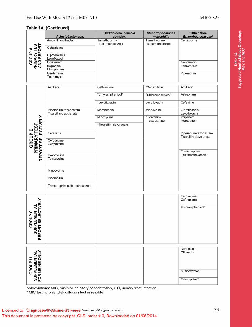
For Use With M02-A12 and M07-A10 M100-S25
Clinical and Laboratory Standards Institute. All rights reserved. 33
Table 1A. (Continued) G
RO
UP
A
PR
IMA
RY
TEST
A
ND
REP
OR
T Acinetobacter spp.
Burkholderia cepacia complex
Stenotrophomonas maltophilia
*Other Non-Enterobacteriaceaeg
Ampicillin-sulbactam
Trimethoprim- sulfamethoxazole
Trimethoprim- sulfamethoxazole
Ceftazidime
Ceftazidime Ciprofloxacin Levofloxacin Doripenem Imipenem Meropenem
Gentamicin Tobramycin
Gentamicin Tobramycin
Piperacillin
GR
OU
P B
PR
IMA
RY
TEST
R
EPO
RT
SELE
CTI
VELY
Amikacin
Ceftazidime
*Ceftazidime
Amikacin
*Chloramphenicolb
*Chloramphenicolb
Aztreonam
*Levofloxacin
Levofloxacin Cefepime
Piperacillin-tazobactam Ticarcillin-clavulanate
Meropenem Minocycline Ciprofloxacin Levofloxacin
Minocycline *Ticarcillin- clavulanate
Imipenem Meropenem
*Ticarcillin-clavulanate
Cefepime
Piperacillin-tazobactam Ticarcillin-clavulanate
Cefotaxime Ceftriaxone Trimethoprim-
sulfamethoxazole Doxycycline Tetracycline Minocycline Piperacillin Trimethoprim-sulfamethoxazole
GR
OU
P C
SU
PPLE
MEN
TAL
REP
OR
T SE
LEC
TIVE
LY
Cefotaxime Ceftriaxone Chloramphenicolb
GR
OU
P U
SU
PPLE
MEN
TAL
FOR
UR
INE
ON
LY Norfloxacin
Ofloxacin
Sulfisoxazole
Tetracyclinea
Abbreviations: MIC, minimal inhibitory concentration, UTI, urinary tract infection. * MIC testing only; disk diffusion test unreliable.
Tabl
e 1A
Sugg
este
d N
onfa
stid
ious
Gro
upin
gsM
02 a
nd M
07
Licensed�to:��Diagnostic�Medicine�ServicesThis�document�is�protected�by�copyright.�CLSI�order�#�0,�Downloaded�on�01/06/2014.

January 2015 Vol. 35 No. 3
Clinical and Laboratory Standards Institute. All rights reserved. 34
Table 1A. (Continued)
“Warning”: The following antimicrobial agents should not be routinely reported for bacteria isolated from CSF that are included in this document. These antimicrobial agents are not the drugs of choice and may not be effective for treating CSF infections caused by these organisms (ie, the bacteria included in Tables 2A through 2J):
agents administered by oral route only 1st- and 2nd-generation cephalosporins (except cefuroxime parenteral)
and cephamycins clindamycin macrolides
tetracyclines fluoroquinolones
NOTE 1: For information about the selection of appropriate antimicrobial agents; explanation of Test and
Report Groups A, B, C, and U; and explanation of the listing of agents within boxes, including the meaning of “or” between agents, refer to the Instructions for Use of Tables that precede Table 1A.
NOTE 2: Information in boldface type is new or modified since the previous edition.
Footnotes General Comments a. Organisms that are susceptible to tetracycline are also considered susceptible to doxycycline and
minocycline. However, some organisms that are intermediate or resistant to tetracycline may be susceptible to doxycycline, minocycline, or both.
b. Not routinely reported on organisms isolated from the urinary tract. Enterobacteriaceae c. Rx: Cefazolin results predict results for the oral agents cefaclor, cefdinir, cefpodoxime, cefprozil,
cefuroxime, cephalexin, and loracarbef when used for therapy of uncomplicated UTIs due to E. coli, K. pneumoniae, and P. mirabilis. Cefpodoxime, cefdinir, and cefuroxime may be tested individually because some isolates may be susceptible to these agents while testing resistant to cefazolin.
d. WARNING: For Salmonella spp. and Shigella spp., first- and second-generation cephalosporins and
cephamycins may appear active in vitro, but are not effective clinically and should not be reported as susceptible.
When fecal isolates of Salmonella and Shigella spp. are tested, only ampicillin, a fluoroquinolone, and trimethoprim-sulfamethoxazole should be reported routinely. In addition, for extraintestinal isolates of Salmonella spp., a third-generation cephalosporin should be tested and reported, and chloramphenicol may be tested and reported, if requested. Susceptibility testing is indicated for typhoidal Salmonella (S. Typhi and Salmonella Paratyphi A–C) isolated from extraintestinal and intestinal sources. Routine susceptibility testing is not indicated for nontyphoidal Salmonella spp. isolated from intestinal sources.
e. Cefotaxime or ceftriaxone should be tested and reported on isolates from CSF in place of cefazolin. f. For testing and reporting of E. coli urinary tract isolates only.
Tabl
e 1A
Sugg
este
d N
onfa
stid
ious
Gro
upin
gsM
02 a
nd M
07
Licensed�to:��Diagnostic�Medicine�ServicesThis�document�is�protected�by�copyright.�CLSI�order�#�0,�Downloaded�on�01/06/2014.

For Use With M02-A12 and M07-A10 M100-S25
Clinical and Laboratory Standards Institute. All rights reserved. 35
Table 1A. (Continued) Other Non-Enterobacteriaceae g. Other non-Enterobacteriaceae include Pseudomonas spp. and other nonfastidious, glucose-
nonfermenting, gram-negative bacilli, but exclude Pseudomonas aeruginosa, Acinetobacter spp., Burkholderia cepacia, and Stenotrophomonas maltophilia, because there are separate lists of suggested drugs to test and report for them.
Recommendations for testing and reporting of Aeromonas hydrophila complex, B. mallei, B.
pseudomallei, and Vibrio species (including V. cholerae) are found in CLSI document M45. Staphylococcus spp. h. Rx: Rifampin should not be used alone for antimicrobial therapy. i. For S. aureus only including methicillin-resistant Staphylococcus aureus (MRSA). j. Penicillin-susceptible staphylococci are also susceptible to other -lactam agents with established
clinical efficacy for staphylococcal infections. Penicillin-resistant staphylococci are resistant to penicillinase-labile penicillins. Oxacillin-resistant staphylococci are resistant to all currently available -lactam antimicrobial agents, with the exception of the newer cephalosporins with anti-MRSA activity. Thus, susceptibility or resistance to a wide array of -lactam antimicrobial agents may be deduced from testing only penicillin and either cefoxitin or oxacillin. Routine testing of other -lactam agents, except those with anti-MRSA activity, is not advised.
k. Daptomycin should not be reported for isolates from the respiratory tract. l. The results of either cefoxitin disk diffusion or cefoxitin MIC tests can be used to predict the presence
of mecA-mediated oxacillin resistance in S. aureus and S. lugdunensis. For coagulase-negative staphylococci (except S. lugdunensis), the cefoxitin disk diffusion test is the preferred method for detection of mecA-mediated oxacillin resistance. Cefoxitin is used as a surrogate for detection of oxacillin resistance; report oxacillin as susceptible or resistant based on cefoxitin results. If a penicillinase-stable penicillin is tested, oxacillin is the preferred agent, and results can be applied to the other penicillinase-stable penicillins. Please refer to Glossary I.
m. For staphylococci that test susceptible, aminoglycosides are used only in combination with other
active agents that test susceptible. Enterococcus spp. n. Warning: For Enterococcus spp., cephalosporins, aminoglycosides (except for high-level resistance
screening), clindamycin, and trimethoprim-sulfamethoxazole may appear active in vitro, but are not effective clinically and should not be reported as susceptible.
o. The results of ampicillin susceptibility tests should be used to predict the activity of amoxicillin.
Ampicillin results may be used to predict susceptibility to amoxicillin-clavulanate, ampicillin-sulbactam, piperacillin, and piperacillin-tazobactam among non–-lactamase-producing enterococci. Ampicillin susceptibility can be used to predict imipenem susceptibility, providing the species is confirmed to be E. faecalis.
Tabl
e 1A
Sugg
este
d N
onfa
stid
ious
Gro
upin
gsM
02 a
nd M
07
Licensed�to:��Diagnostic�Medicine�ServicesThis�document�is�protected�by�copyright.�CLSI�order�#�0,�Downloaded�on�01/06/2014.
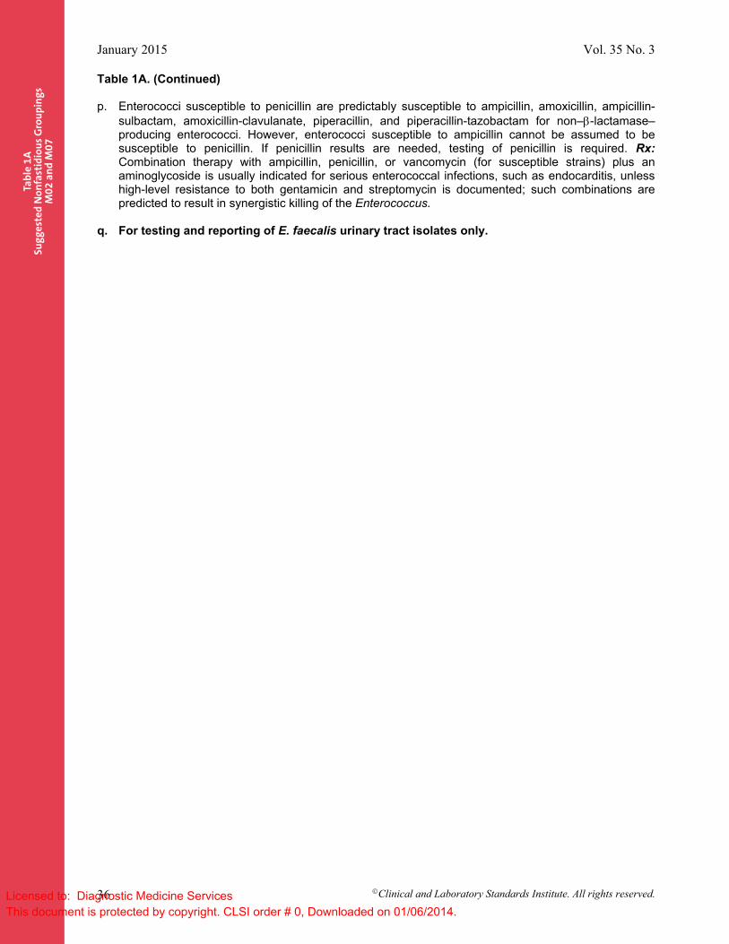
January 2015 Vol. 35 No. 3
Clinical and Laboratory Standards Institute. All rights reserved. 36
Table 1A. (Continued) p. Enterococci susceptible to penicillin are predictably susceptible to ampicillin, amoxicillin, ampicillin-
sulbactam, amoxicillin-clavulanate, piperacillin, and piperacillin-tazobactam for non–-lactamase–producing enterococci. However, enterococci susceptible to ampicillin cannot be assumed to be susceptible to penicillin. If penicillin results are needed, testing of penicillin is required. Rx: Combination therapy with ampicillin, penicillin, or vancomycin (for susceptible strains) plus an aminoglycoside is usually indicated for serious enterococcal infections, such as endocarditis, unless high-level resistance to both gentamicin and streptomycin is documented; such combinations are predicted to result in synergistic killing of the Enterococcus.
q. For testing and reporting of E. faecalis urinary tract isolates only.
Tabl
e 1A
Sugg
este
d N
onfa
stid
ious
Gro
upin
gsM
02 a
nd M
07
Licensed�to:��Diagnostic�Medicine�ServicesThis�document�is�protected�by�copyright.�CLSI�order�#�0,�Downloaded�on�01/06/2014.

For Use With M02-A12 and M07-A10 M100-S25
Clinical and Laboratory Standards Institute. All rights reserved. 37
This page is intentionally left blank.
Licensed�to:��Diagnostic�Medicine�ServicesThis�document�is�protected�by�copyright.�CLSI�order�#�0,�Downloaded�on�01/06/2014.
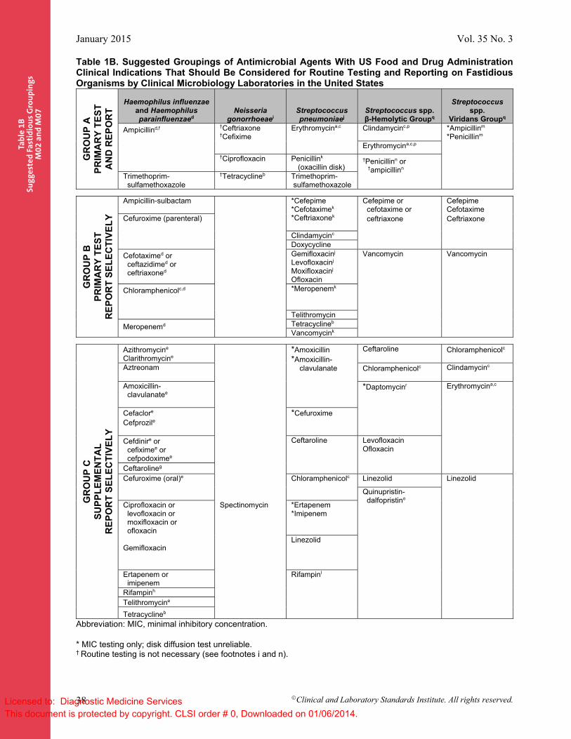
January 2015 Vol. 35 No. 3
Clinical and Laboratory Standards Institute. All rights reserved. 38
Table 1B. Suggested Groupings of Antimicrobial Agents With US Food and Drug Administration Clinical Indications That Should Be Considered for Routine Testing and Reporting on Fastidious Organisms by Clinical Microbiology Laboratories in the United States
GR
OU
P A
PR
IMA
RY
TEST
A
ND
REP
OR
T
Haemophilus influenzae and Haemophilus parainfluenzaed
Neisseria gonorrhoeaei
Streptococcus pneumoniaej
Streptococcus spp. β-Hemolytic Groupq
Streptococcus spp.
Viridans Groupq
Ampicillind,f †Ceftriaxone †Cefixime
Erythromycina,c Clindamycinc,p
*Ampicillinm
*Penicillinm Erythromycina,c,p
†Ciprofloxacin Penicillink (oxacillin disk)
†Penicillinn or †ampicillinn
Trimethoprim- sulfamethoxazole
†Tetracyclineb Trimethoprim- sulfamethoxazole
GR
OU
P B
PR
IMA
RY
TEST
R
EPO
RT
SELE
CTI
VELY
Ampicillin-sulbactam
*Cefepime *Cefotaximek *Ceftriaxonek
Cefepime or cefotaxime or ceftriaxone
Cefepime Cefotaxime Ceftriaxone Cefuroxime (parenteral)
Clindamycinc Doxycycline
Cefotaximed or ceftazidimed or ceftriaxoned
Gemifloxacinj Levofloxacinj Moxifloxacinj Ofloxacin
Vancomycin Vancomycin
Chloramphenicolc,d
*Meropenemk
Telithromycin
Meropenemd Tetracyclineb Vancomycink
GR
OU
P C
SU
PPLE
MEN
TAL
R
EPO
RT
SELE
CTI
VELY
Azithromycine Clarithromycine
*Amoxicillin *Amoxicillin- clavulanate
Ceftaroline
Chloramphenicolc
Aztreonam Chloramphenicolc
Clindamycinc
Amoxicillin- clavulanatee
*Daptomycinr
Erythromycina,c
Cefaclore
Cefprozile
*Cefuroxime
Cefdinire or cefiximee or cefpodoximee
Ceftaroline Levofloxacin Ofloxacin
Ceftarolineg Cefuroxime (oral)e Chloramphenicolc Linezolid Linezolid
Quinupristin- dalfopristino Ciprofloxacin or
levofloxacin or moxifloxacin or ofloxacin Gemifloxacin
Spectinomycin *Ertapenem *Imipenem
Linezolid
Ertapenem or imipenem
Rifampinl
Rifampinh Telithromycine Tetracyclineb
Abbreviation: MIC, minimal inhibitory concentration. * MIC testing only; disk diffusion test unreliable. † Routine testing is not necessary (see footnotes i and n).
Tabl
e 1B
Sugg
este
d Fa
stid
ious
Gro
upin
gsM
02 a
nd M
07
Licensed�to:��Diagnostic�Medicine�ServicesThis�document�is�protected�by�copyright.�CLSI�order�#�0,�Downloaded�on�01/06/2014.

For Use With M02-A12 and M07-A10 M100-S25
Clinical and Laboratory Standards Institute. All rights reserved. 39
Table 1B. (Continued)
“Warning”: The following antimicrobial agents should not be routinely reported for bacteria isolated from CSF that are included in this document. These antimicrobial agents are not the drugs of choice and may not be effective for treating CSF infections caused by these organisms (ie, the bacteria included in Tables 2A through 2J):
agents administered by oral route only 1st- and 2nd-generation cephalosporins (except cefuroxime parenteral)
and cephamycins clindamycin macrolides
tetracyclines fluoroquinolones
NOTE 1: For information about the selection of appropriate antimicrobial agents; explanation of Test and
Report Groups A, B, C, and U; and explanation of the listing of agents within boxes, including the meaning of “or” between agents, refer to the Instructions for Use of Tables that precede Table 1A.
NOTE 2: Information in boldface type is new or modified since the previous edition.
Footnotes
General Comments a. Susceptibility and resistance to azithromycin, clarithromycin, and dirithromycin can be predicted by
testing erythromycin. b. Organisms that are susceptible to tetracycline are also considered susceptible to doxycycline and
minocycline. c. Not routinely reported for organisms isolated from the urinary tract. Haemophilus spp. d. For isolates of H. influenzae from CSF, only results of testing with ampicillin, one of the third-
generation cephalosporins, chloramphenicol, and meropenem are appropriate to report routinely. e. Amoxicillin-clavulanate, azithromycin, cefaclor, cefdinir, cefixime, cefpodoxime, cefprozil, cefuroxime,
clarithromycin, loracarbef, and telithromycin are oral agents that may be used as empiric therapy for respiratory tract infections due to Haemophilus spp. The results of susceptibility tests with these antimicrobial agents are often not useful for management of individual patients. However, susceptibility testing of Haemophilus spp. with these compounds may be appropriate for surveillance or epidemiological studies.
f. The results of ampicillin susceptibility tests should be used to predict the activity of amoxicillin. The
majority of isolates of H. influenzae that are resistant to ampicillin and amoxicillin produce a TEM-type -lactamase. In most cases, a direct -lactamase test can provide a rapid means of detecting ampicillin and amoxicillin resistance.
g. For H. influenzae only. h. May be appropriate only for prophylaxis of case contacts. Refer to Table 2E.
Tabl
e 1B
Sugg
este
d Fa
stid
ious
Gro
upin
gsM
02 a
nd M
07
Licensed�to:��Diagnostic�Medicine�ServicesThis�document�is�protected�by�copyright.�CLSI�order�#�0,�Downloaded�on�01/06/2014.

January 2015 Vol. 35 No. 3
Clinical and Laboratory Standards Institute. All rights reserved. 40
Table 1B. (Continued) Neisseria gonorrhoeae i. Culture and susceptibility testing of N. gonorrhoeae should be considered in cases of treatment
failure. Antimicrobial agents recommended for testing include, at a minimum, those agents listed in Group A. The most recent guidelines from the Centers for Disease Control and Prevention for treatment and testing are available at http://www.cdc.gov/std/Gonorrhea/.
Streptococcus pneumoniae j. S. pneumoniae isolates susceptible to levofloxacin are predictably susceptible to gemifloxacin and
moxifloxacin. However, S. pneumoniae susceptible to gemifloxacin or moxifloxacin cannot be assumed to be susceptible to levofloxacin.
k. Penicillin and cefotaxime, ceftriaxone, or meropenem should be tested by a reliable MIC method
(such as that described in M07-A10), and reported routinely with CSF isolates of S. pneumoniae. Such isolates can also be tested against vancomycin using the MIC or disk method. With isolates from other sites, the oxacillin disk screening test may be used. If the oxacillin zone size is ≤ 19 mm, penicillin, cefotaxime, ceftriaxone, or meropenem MICs should be determined.
l. Rx: Rifampin should not be used alone for antimicrobial therapy. Streptococcus spp. m. Rx: Penicillin- or ampicillin-intermediate isolates may require combined therapy with an
aminoglycoside for bactericidal action. n. Penicillin and ampicillin are drugs of choice for treatment of β-hemolytic streptococcal infections.
Susceptibility testing of penicillins and other -lactams approved by the US Food and Drug Administration for treatment of β-hemolytic streptococcal infections need not be performed routinely, because nonsusceptible isolates (ie, penicillin MICs > 0.12 and ampicillin MICs > 0.25 g/mL) are extremely rare in any β-hemolytic streptococcus and have not been reported for Streptococcus pyogenes. If testing is performed, any β-hemolytic streptococcal isolate found to be nonsusceptible should be re-identified, retested, and, if confirmed, submitted to a public health laboratory. (See Appendix A for further instructions.)
o. Report against S. pyogenes. p. Rx: Recommendations for intrapartum prophylaxis for Group B streptococci are penicillin or
ampicillin. Although cefazolin is recommended for penicillin-allergic women at low risk for anaphylaxis, those at high risk for anaphylaxis may receive clindamycin. Group B streptococci are susceptible to ampicillin, penicillin, and cefazolin, but may be resistant to erythromycin and clindamycin. When Group B Streptococcus is isolated from a pregnant woman with severe penicillin allergy (high risk for anaphylaxis), erythromycin and clindamycin (including inducible clindamycin resistance) should be tested, and only clindamycin should be reported. See Table 3G.
q. For this table, the β-hemolytic group includes the large colony–forming pyogenic strains of
streptococci with Group A (S. pyogenes), C, or G antigens and strains with Group B (S. agalactiae) antigen. Small colony–forming β-hemolytic strains with Group A, C, F, or G antigens (S. anginosus group, previously termed “S. milleri”) are considered part of the viridans group, and interpretive criteria for the viridans group should be used.
r. Daptomycin should not be reported for isolates from the respiratory tract.
Tabl
e 1B
Sugg
este
d Fa
stid
ious
Gro
upin
gsM
02 a
nd M
07
Licensed�to:��Diagnostic�Medicine�ServicesThis�document�is�protected�by�copyright.�CLSI�order�#�0,�Downloaded�on�01/06/2014.

For Use With M02-A12 and M07-A10 M100-S25
Clinical and Laboratory Standards Institute. All rights reserved. 41
This page is intentionally left blank.
Licensed�to:��Diagnostic�Medicine�ServicesThis�document�is�protected�by�copyright.�CLSI�order�#�0,�Downloaded�on�01/06/2014.

January 2015 Vol. 35 No. 3
Clinical and Laboratory Standards Institute. All rights reserved. 42
Table 1C. Suggested Groupings of Antimicrobial Agents With US Food and Drug Administration Clinical Indications That Should Be Considered for Routine Testing and Reporting on Anaerobic Organisms by Clinical Microbiology Laboratories in the United States
Bacteroides fragilis Group and Other Gram-Negative Anaerobes
Gram-Positive Anaerobesb
Gro
up A
Pr
imar
y Te
st a
nd R
epor
t
Amoxicillin-clavulanate Ampicillin-sulbactam Piperacillin-tazobactam Ticarcillin-clavulanate
Ampicillina
Penicillina Amoxicillin-clavulanate Ampicillin-sulbactam Piperacillin-tazobactam Ticarcillin-clavulanate
Clindamycin Clindamycin
Doripenem Ertapenem Imipenem Meropenem
Doripenem Ertapenem Imipenem Meropenem
Metronidazole Metronidazole
Gro
up C
Su
pple
men
tal
Rep
ort S
elec
tivel
y
Penicillina
Ampicillina
Cefotetan Cefoxitin
Cefotetan Cefoxitin
Ceftizoxime Ceftriaxone
Ceftizoxime Ceftriaxone
Chloramphenicol Moxifloxacin Moxifloxacin
Piperacillin
Piperacillin Tetracycline
Tabl
e 1C
Sugg
este
d An
aero
be G
roup
ings
M11
Licensed�to:��Diagnostic�Medicine�ServicesThis�document�is�protected�by�copyright.�CLSI�order�#�0,�Downloaded�on�01/06/2014.

For Use With M11-A8 M100-S25
Clinical and Laboratory Standards Institute. All rights reserved. 43
Table 1C. (Continued) NOTE 1: For information about the selection of appropriate antimicrobial agents; explanation of Test and
Report Groups A and C; and explanation of the listing of agents within boxes, refer to the Instructions for Use of Tables that precede Table 1A.
NOTE 2: Most anaerobic infections are polymicrobial, including both β-lactamase–positive and β-
lactamase–negative strains. Testing may not be necessary for polymicrobial anaerobic infections. However, if requested, only the organism most likely to be resistant (eg, B. fragilis group) should be tested and results reported.
NOTE 3: Specific Clostridium species (eg, C. perfringens, C. septicum, C. sordellii) may be the singular
cause of an infection, are typically susceptible to penicillin and ampicillin, and should be tested and reported.
NOTE 4: Information in boldface type is new or modified since the previous edition.
Footnotes
General Comment a. If -lactamase positive, report as resistant to penicillin and ampicillin. Be aware that β-lactamase–
negative isolates may be resistant to penicillin and ampicillin by other mechanisms. Gram-positive Anaerobes b. Many non–spore-forming, gram-positive anaerobic rods are resistant to metronidazole.
Tabl
e 1C
Sugg
este
d An
aero
be G
roup
ings
M11
Licensed�to:��Diagnostic�Medicine�ServicesThis�document�is�protected�by�copyright.�CLSI�order�#�0,�Downloaded�on�01/06/2014.

January 2015 Vol. 35 N
o. 3
Table 2A. Zone Diameter and Minimal Inhibitory Concentration Interpretive Standards for Enterobacteriaceae
* ATCC® is a registered trademark of the American Type Culture Collection. Refer to Tables 3A, 3B, and 3C for additional testing recommendations, reporting suggestions, and QC.
General Comments
(1) For disk diffusion, test a maximum of 12 disks on a 150-mm plate and no more than 6 disks on a 100-mm plate; disks should be placed no less than 24
mm apart, center to center (see M02, Subchapter 3.6). Each zone diameter should be clearly measurable; overlapping zones prevent accurate measurement. Measure the diameter of the zones of complete inhibition (as judged by the unaided eye), including the diameter of the disk. Hold the Petri plate a few inches above a black background illuminated with reflected light. The zone margin should be considered the area showing no obvious, visible growth that can be detected with the unaided eye. Ignore faint growth of tiny colonies that can be detected only with a magnifying lens at the edge of the zone of inhibited growth. Strains of Proteus spp. may swarm into areas of inhibited growth around certain antimicrobial agents. With Proteus spp., ignore the thin veil of swarming growth in an otherwise obvious zone of growth inhibition. With trimethoprim and the sulfonamides, antagonists in the medium may allow some slight growth; therefore, disregard slight growth (20% or less of the lawn of growth) and measure the more obvious margin to determine the zone diameter.
(2) When fecal isolates of Salmonella and Shigella spp. are tested, only ampicillin, a fluoroquinolone, and trimethoprim-sulfamethoxazole should be reported
routinely. In addition, for extraintestinal isolates of Salmonella spp., a third-generation cephalosporin should be tested and reported, and chloramphenicol may be tested and reported if requested. Susceptibility testing is indicated for typhoidal Salmonella (S. Typhi and Salmonella Paratyphi A–C) isolated from extraintestinal and intestinal sources. Routine susceptibility testing is not indicated for nontyphoidal Salmonella spp. isolated from intestinal sources.
(3) The dosage regimens shown in the comment column below are those required to achieve plasma drug exposures (in adults with normal renal and hepatic
functions) on which breakpoints were based. When implementing new breakpoints, it is strongly recommended that laboratories share this information with infectious diseases practitioners, pharmacists, pharmacy and therapeutics committees, and infection control committees.
NOTE: Information in boldface type is new or modified since the previous edition.
44 ©Clinical and Laboratory Standards Institute. All rights reserved.
Testing Conditions Medium: Disk diffusion: Mueller-Hinton agar (MHA) Broth dilution: cation-adjusted Mueller-Hinton broth Agar dilution: MHA Inoculum: Growth method or direct colony suspension, equivalent to a
0.5 McFarland standard Incubation: 35°C 2°C; ambient air Disk diffusion: 16 to 18 hours Dilution methods: 16 to 20 hours
Routine QC Recommendations (See Tables 4A and 5A for acceptable QC ranges.) Escherichia coli ATCC®* 25922 Pseudomonas aeruginosa ATCC® 27853 (for carbapenems) Escherichia coli ATCC® 35218 (for -lactam/-lactamase inhibitor combinations) When a commercial test system is used for susceptibility testing, refer to the manufacturer’s instructions for QC test recommendations and QC ranges.
Table 2AEnterobacteriaceae
M02 and M07
Licensed�to:��Diagnostic�M
edicine�Services
This�document�is�protected�by�copyright.�C
LSI�order�#�0,�D
ownloaded�on�01/06/2014.

Table 2A. (Continued)
Test/Report Group
Antimicrobial Agent
Disk Content
Zone Diameter Interpretive Criteria (nearest whole mm)
MIC Interpretive Criteria (µg/mL)
Comments S SDD I R S SDD I R PENICILLINS
A Ampicillin 10 g ≥ 17 14–16 ≤ 13 ≤ 8 16 ≥ 32 (4) Results of ampicillin testing can be used to predict results for amoxicillin. See comment (2).
B Piperacillin 100 g ≥ 21 18–20 ≤ 17 16 32–64 128 O Mecillinam 10 g ≥ 15 12–14 ≤ 11 8 16 32 (5) For testing and reporting of E. coli
urinary tract isolates only. O Ticarcillin 75 g ≥ 20 15–19 ≤ 14 ≤ 16 32–64 128
β-LACTAM/β-LACTAMASE INHIBITOR COMBINATIONS B Amoxicillin-clavulanate 20/10 µg ≥ 18 14–17 ≤ 13 ≤ 8/4 16/8 ≥ 32/16 B Ampicillin-sulbactam 10/10 µg ≥ 15 12–14 ≤ 11 ≤ 8/4 16/8 ≥ 32/16 B Piperacillin-tazobactam 100/10 µg ≥ 21 18–20 ≤ 17 ≤ 16/4 32/4–64/4 ≥ 128/4 B Ticarcillin-clavulanate 75/10 µg ≥ 20 15–19 ≤ 14 ≤ 16/2 32/2–64/2 ≥ 128/2
CEPHEMS (PARENTERAL) (Including cephalosporins I, II, III, and IV. Please refer to Glossary I.) (6) WARNING: For Salmonella spp. and Shigella spp., first- and second-generation cephalosporins and cephamycins may appear active in vitro, but are not effective clinically and should not be reported as susceptible.
(7) Following evaluation of PK-PD properties, limited clinical data, and MIC distributions, revised interpretive criteria for cephalosporins (cefazolin, cefotaxime, ceftazidime, ceftizoxime, and ceftriaxone) and aztreonam were first published in January 2010 (M100-S20) and are listed in this table. Cefuroxime (parenteral) was also evaluated; however, no change in interpretive criteria was required for the dosage indicated below. When using the current interpretive criteria, routine ESBL testing is no longer necessary before reporting results (ie, it is no longer necessary to edit results for cephalosporins, aztreonam, or penicillins from susceptible to resistant). However, ESBL testing may still be useful for epidemiological or infection control purposes. For laboratories that have not implemented the current interpretive criteria, ESBL testing should be performed as described in Table 3A.
Note that interpretive criteria for drugs with limited availability in many countries (eg, moxalactam, cefonicid, cefamandole, and cefoperazone) were not evaluated. If considering use of these drugs for E. coli, Klebsiella, or Proteus spp., ESBL testing should be performed (see Table 3A). If isolates test ESBL positive, the results for moxalactam, cefonicid, cefamandole, and cefoperazone should be reported as resistant.
(8) Enterobacter, Citrobacter, and Serratia may develop resistance during prolonged therapy with third-generation cephalosporins as a result of derepression of AmpC -lactamase. Therefore, isolates that are initially susceptible may become resistant within three to four days after initiation of therapy. Testing of repeat isolates may be warranted.
A Cefazolin 30 µg 23 20–22 ≤ 19 ≤ 2
4 ≥ 8 (9) Interpretive criteria are based on a dosage regimen of 2 g every 8 h. See comment (7). For UTI interpretive criteria, see below under CEPHEMS (ORAL).
C Ceftaroline 30 µg ≥ 23 20–22 ≤ 19 ≤ 0.5 1 ≥ 2 (10) Interpretive criteria are based on a dosage regimen of 600 mg every 12 h.
©Clinical and Laboratory Standards Institute. All rights reserved. 45
For Use W
ith M02-A
12 and M07-A
10 M100-S25
Table 2AEnterobacteriaceae
M02 and M07
Licensed�to:��Diagnostic�M
edicine�Services
This�document�is�protected�by�copyright.�C
LSI�order�#�0,�D
ownloaded�on�01/06/2014.

January 2015 Vol. 35 N
o. 3
Table 2A. (Continued)
Test/Report Group
Antimicrobial Agent
Disk Content
Zone Diameter Interpretive Criteria (nearest whole mm)
MIC Interpretive Criteria (µg/mL)
Comments S SDD I R S SDD I R CEPHEMS (PARENTERAL) (Including cephalosporins I, II, III, and IV. Please refer to Glossary I.) (Continued)
U Cephalothin (surrogate test for uncomplicated UTI)
30 µg ≥ 18 15–17 ≤ 14 ≤ 8 16 ≥ 32 (11) Cephalothin interpretive criteria can be used only to predict susceptibility to the oral agents, cefadroxil, cefpodoxime, cephalexin, and loracarbef. Older data that suggest that cephalothin results could predict susceptibility to some other cephalosporins may still be correct, but there are no recent data to confirm this. (12) To predict results for oral cephalosporins when used for therapy of uncomplicated UTIs, testing cefazolin is preferred to testing cephalothin.
B Cefepime 30 µg ≥ 25 19–24
– ≤ 18 ≤ 2 4–8 – ≥ 16 (13) The interpretive criterion for susceptible is based on a dosage regimen of 1 g every 12 h. The interpretive criterion for SDD is based on dosing regimens that result in higher cefepime exposure, either higher doses or more frequent doses or both, up to approved maximum dosing regimens. See Appendix E for more information about interpretive criteria and dosing regimens. Also see the definition of SDD in the Instructions for Use of Tables section.
B B
Cefotaxime or ceftriaxone
30 µg 30 µg
≥ 26 ≥ 23
23–25 20–22
≤ 22 ≤ 19
≤ 1 ≤ 1
2 2
≥ 4 ≥ 4
(14) Interpretive criteria are based on a dosage regimen of 1 g every 24 h for ceftriaxone and 1 g every 8 h for cefotaxime. See comment (7).
B Cefotetan 30 g ≥ 16 13–15 12 ≤ 16 32 ≥ 64 B Cefoxitin 30 g ≥ 18 15–17 14 ≤ 8 16 ≥ 32 (15) The interpretive criteria are based on a
dosage regimen of at least 8 g per day (eg, 2 g every 6 h).
B Cefuroxime (parenteral)
30 g ≥ 18 15–17 14 ≤ 8 16 ≥ 32 (16) Interpretive criteria are based on a dosage regimen of 1.5 g every 8 h. See comment (7).
46 ©Clinical and Laboratory Standards Institute. All rights reserved.
Table 2AEnterobacteriaceae
M02 and M07
Licensed�to:��Diagnostic�M
edicine�Services
This�document�is�protected�by�copyright.�C
LSI�order�#�0,�D
ownloaded�on�01/06/2014.

For Use W
ith M02-A
12 and M07-A
10 M100-S25
Table 2A. (Continued)
Test/Report Group
Antimicrobial Agent
Disk Content
Zone Diameter Interpretive Criteria (nearest whole mm)
MIC Interpretive Criteria (µg/mL)
Comments S SDD I R S SDD I R
CEPHEMS (PARENTERAL) (Including cephalosporins I, II, III, and IV. Please refer to Glossary I.) (Continued)
C Ceftazidime 30 g ≥ 21 18–20 17 ≤ 4 8 ≥ 16 (17) Interpretive criteria are based on a dosage regimen of 1 g every 8 h. See comment (7).
O Cefamandole 30 g ≥ 18 15–17 14 ≤ 8 16 ≥ 32 See comment (7). O Cefmetazole 30 g ≥ 16 13–15 12 ≤ 16 32 ≥ 64 (18) Insufficient new data exist to reevaluate
interpretive criteria listed here. O Cefonicid 30 g ≥ 18 15–17 14 ≤ 8 16 ≥ 32 See comment (7). O Cefoperazone 75 g ≥ 21 16–20 15 ≤ 16 32 ≥ 64 See comment (7). O Ceftizoxime 30 g ≥ 25 22–24 21 ≤ 1 2 ≥ 4 (19) Interpretive criteria are based on a dosage
regimen of 1 g every 12 h. See comment (7).
O Moxalactam 30 g ≥ 23 15–22 14 ≤ 8 16–32 ≥ 64 See comment (7). CEPHEMS (ORAL)
B Cefuroxime (oral) 30 g 23 15–22 14 ≤ 4 8–16 ≥ 32 See comments (12 and 20). U Cefazolin
(surrogate test for uncomplicated UTI)
30 g ≥ 15 – ≤ 14 ≤ 16 – – ≥ 32 (20) Rx: Cefazolin results predict results for the oral agents cefaclor, cefdinir, cefpodoxime, cefprozil, cefuroxime, cephalexin, and loracarbef when used for therapy of uncomplicated UTIs due to E. coli, K. pneumoniae, and P. mirabilis. Cefpodoxime, cefdinir, and cefuroxime may be tested individually because some isolates may be susceptible to these agents while testing resistant to cefazolin. See comment (12).
O Loracarbef 30 g 18 15–17 14 ≤ 8 16 ≥ 32 (21) Do not test Citrobacter, Providencia, or Enterobacter spp. with cefdinir or loracarbef by disk diffusion because false-susceptible results have been reported. See comments (12 and 20).
O Cefaclor 30 g 18 15–17 14 8 16 32 See comments (12 and 20). O Cefdinir 5 g 20 17–19 16 1 2 4 See comments (12, 20, and 21). O Cefixime 5 g 19 16–18 15 1 2 4 (22) Do not test Morganella spp. with cefixime,
cefpodoxime, or cefetamet by disk diffusion. O Cefpodoxime 10 g 21 18–20 17 2 4 8 See comments (12, 20, and 22). O Cefprozil 30 g 18 15–17 14 8 16 32 (23) Do not test Providencia spp. with cefprozil
by disk diffusion because false-susceptible results have been reported. See comments (12 and 20).
Inv. Cefetamet 10 g 18 15–17 14 4 8 16 See comment (22). Inv. Ceftibuten 30 g 21 18–20 17 8 16 32 (24) For testing and reporting of urine isolates only.
©Clinical and Laboratory Standards Institute. All rights reserved. 47
Table 2AEnterobacteriaceae
M02 and M07
Licensed�to:��Diagnostic�M
edicine�Services
This�document�is�protected�by�copyright.�C
LSI�order�#�0,�D
ownloaded�on�01/06/2014.
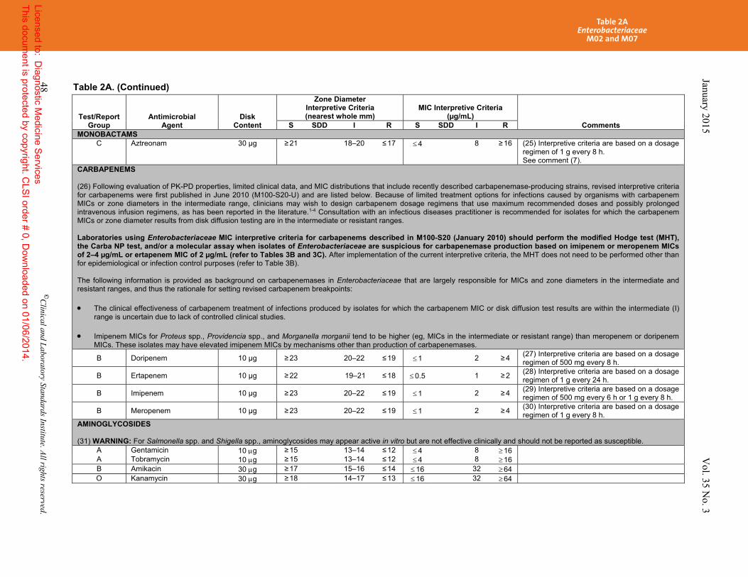
Table 2A. (Continued)
Test/Report Group
Antimicrobial Agent
Disk Content
Zone Diameter Interpretive Criteria (nearest whole mm)
MIC Interpretive Criteria (µg/mL)
Comments S SDD I R S SDD I R MONOBACTAMS
C Aztreonam 30 µg ≥ 21 18–20 ≤ 17 4 8 ≥ 16 (25) Interpretive criteria are based on a dosage regimen of 1 g every 8 h. See comment (7).
CARBAPENEMS (26) Following evaluation of PK-PD properties, limited clinical data, and MIC distributions that include recently described carbapenemase-producing strains, revised interpretive criteria for carbapenems were first published in June 2010 (M100-S20-U) and are listed below. Because of limited treatment options for infections caused by organisms with carbapenem MICs or zone diameters in the intermediate range, clinicians may wish to design carbapenem dosage regimens that use maximum recommended doses and possibly prolonged intravenous infusion regimens, as has been reported in the literature.1-4 Consultation with an infectious diseases practitioner is recommended for isolates for which the carbapenem MICs or zone diameter results from disk diffusion testing are in the intermediate or resistant ranges. Laboratories using Enterobacteriaceae MIC interpretive criteria for carbapenems described in M100-S20 (January 2010) should perform the modified Hodge test (MHT), the Carba NP test, and/or a molecular assay when isolates of Enterobacteriaceae are suspicious for carbapenemase production based on imipenem or meropenem MICs of 2–4 µg/mL or ertapenem MIC of 2 µg/mL (refer to Tables 3B and 3C). After implementation of the current interpretive criteria, the MHT does not need to be performed other than for epidemiological or infection control purposes (refer to Table 3B). The following information is provided as background on carbapenemases in Enterobacteriaceae that are largely responsible for MICs and zone diameters in the intermediate and resistant ranges, and thus the rationale for setting revised carbapenem breakpoints: The clinical effectiveness of carbapenem treatment of infections produced by isolates for which the carbapenem MIC or disk diffusion test results are within the intermediate (I)
range is uncertain due to lack of controlled clinical studies.
Imipenem MICs for Proteus spp., Providencia spp., and Morganella morganii tend to be higher (eg, MICs in the intermediate or resistant range) than meropenem or doripenem MICs. These isolates may have elevated imipenem MICs by mechanisms other than production of carbapenemases.
B Doripenem 10 µg ≥ 23 20–22 ≤ 19 1 2 ≥ 4 (27) Interpretive criteria are based on a dosage regimen of 500 mg every 8 h.
B Ertapenem 10 µg ≥ 22 19–21 ≤ 18 0.5 1 ≥ 2 (28) Interpretive criteria are based on a dosage regimen of 1 g every 24 h.
B Imipenem 10 µg ≥ 23 20–22 ≤ 19 1 2 ≥ 4 (29) Interpretive criteria are based on a dosage regimen of 500 mg every 6 h or 1 g every 8 h.
B Meropenem 10 µg ≥ 23 20–22 ≤ 19 1 2 ≥ 4 (30) Interpretive criteria are based on a dosage regimen of 1 g every 8 h.
AMINOGLYCOSIDES (31) WARNING: For Salmonella spp. and Shigella spp., aminoglycosides may appear active in vitro but are not effective clinically and should not be reported as susceptible.
A Gentamicin 10 g ≥ 15 13–14 ≤ 12 4 8 16 A Tobramycin 10 g ≥ 15 13–14 ≤ 12 4 8 16 B Amikacin 30 g ≥ 17 15–16 ≤ 14 16 32 64 O Kanamycin 30 g ≥ 18 14–17 ≤ 13 16 32 64
January 2015
Vol. 35 N
o. 3
48 ©Clinical and Laboratory Standards Institute. All rights reserved.
Table 2AEnterobacteriaceae
M02 and M07
Licensed�to:��Diagnostic�M
edicine�Services
This�document�is�protected�by�copyright.�C
LSI�order�#�0,�D
ownloaded�on�01/06/2014.

©Clinical and Laboratory Standards Institute. All rights reserved. 49
Table 2A. (Continued)
Test/Report Group
Antimicrobial Agent
Disk Content
Zone Diameter Interpretive Criteria (nearest whole mm)
MIC Interpretive Criteria (µg/mL)
Comments S SDD I R S SDD I R AMINOGLYCOSIDES (Continued)
O Netilmicin 30 g ≥ 15 13–14 ≤ 12 8 16 32 O Streptomycin 10 g ≥ 15 12–14 ≤ 11 – – – (32) There are no MIC interpretive
standards. MACROLIDES
Inv. Azithromycin 15 g ≥ 13 – ≤ 12 16 – 32 (33) Salmonella Typhi only: Interpretive criteria are based on MIC distribution data.
TETRACYCLINES
(34) Organisms that are susceptible to tetracycline are also considered susceptible to doxycycline and minocycline. However, some organisms that are intermediate or resistant to tetracycline may be susceptible to doxycycline, minocycline, or both.
C Tetracycline 30 g ≥ 15 12–14 ≤ 11 4 8 16 O Doxycycline 30 g ≥ 14 11–13 ≤ 10 4 8 16 O Minocycline 30 g ≥ 16 13–15 ≤ 12 4 8 16
FLUOROQUINOLONES NOTE: Reevaluation of fluoroquinolones is ongoing. See comment (2).
B Ciprofloxacin 5 µg ≥ 21 16–20 ≤ 15 ≤ 1 2 ≥ 4 (35) For testing and reporting of Enterobacteriaceae except for Salmonella spp. B Levofloxacin 5 µg ≥ 17 14–16 ≤ 13 ≤ 2 4 ≥ 8
B
Inv.
B
B
Ciprofloxacin Pefloxacin Levofloxacin Ofloxacin
5 µg
5 g (surrogate
test for cipro-floxacin)
–
–
≥ 31
24
–
–
21–30
–
–
–
≤ 20
23
–
–
≤ 0.06
–
≤ 0.12
≤ 0.12
0.12–0.5
–
0.25–1
0.25–1
≥ 1
–
≥ 2
≥ 2
(36) For testing and reporting of Salmonella spp. (including S. Typhi and S. Paratyphi A–C). See comment (2). (37) Strains of Salmonella that test nonsusceptible to ciprofloxacin, levofloxacin, ofloxacin, pefloxacin, or nalidixic acid may be associated with clinical failure or delayed response in fluoroquinolone-treated patients with salmonellosis. (38) If a ciprofloxacin, levofloxacin, or ofloxacin MIC test cannot be done, pefloxacin disk diffusion may be used as a surrogate test. Because pefloxacin is not available in the United States, a ciprofloxacin disk alone or both ciprofloxacin and nalidixic acid disks could also be tested. (39) No single screening test will detect resistance resulting from all possible fluoroquinolone resistance mechanisms that have been identified in Salmonella spp.
For Use W
ith M02-A
12 and M07-A
10 M100-S25
Table 2AEnterobacteriaceae
M02 and M07
Licensed�to:��Diagnostic�M
edicine�Services
This�document�is�protected�by�copyright.�C
LSI�order�#�0,�D
ownloaded�on�01/06/2014.

Table 2A. (Continued)
Abbreviations: ATCC®, American Type Culture Collection; ESBL, extended-spectrum -lactamase; FDA, US Food and Drug Administration; I, intermediate; MHT, modified Hodge test; MIC, minimal inhibitory concentration; PK-PD, pharmacokinetic-pharmacodynamic; R, resistant; S, susceptible; SDD, susceptible-dose dependent; UTI, urinary tract infection.
Test/Report Group
Antimicrobial Agent
Disk Content
Zone Diameter Interpretive Criteria (nearest whole mm)
MIC Interpretive Criteria (µg/mL)
Comments S SDD I R S SDD I R FLUOROQUINOLONES (Continued)
U Lomefloxacin or 10 µg ≥ 22 19–21 ≤ 18 ≤ 2 4 ≥ 8 U ofloxacin 5 µg ≥ 16 13–15 ≤ 12 ≤ 2 4 ≥ 8 U Norfloxacin 10 µg ≥ 17 13–16 ≤ 12 ≤ 4 8 16 O Enoxacin 10 µg ≥ 18 15–17 ≤ 14 2 4 8 O Gatifloxacin 5 µg ≥ 18 15–17 ≤ 14 2 4 8 O Gemifloxacin 5 µg ≥ 20 16–19 ≤ 15 0.25 0.5 1 (40) FDA-approved for Klebsiella
pneumoniae.O Grepafloxacin 5 µg ≥ 18 15–17 ≤ 14 1 2 4
Inv. Fleroxacin 5 µg ≥ 19 16–18 ≤ 15 2 4 8 QUINOLONES
O Cinoxacin 100 µg ≥ 19 15–18 ≤ 14 16 32 64 See comment (24). O Nalidixic acid 30 µg ≥ 19 14–18 ≤ 13 16 – 32 (41) These interpretive criteria are for urinary
tract isolates of Enterobacteriaceae and for all isolates of Salmonella. See comments (37) and (38).
FOLATE PATHWAY INHIBITORS B Trimethoprim-
sulfamethoxazole 1.25/
23.75 µg ≥ 16 11–15 ≤ 10 2/38
– 4/76
See comment (2).
U Sulfonamides 250 or 300 µg
≥ 17 13–16 ≤ 12 256 – 512 (42) Sulfisoxazole can be used to represent any of the currently available sulfonamide preparations.
U Trimethoprim 5 µg ≥ 16 11–15 ≤ 10 8 – 16 PHENICOLS
C Chloramphenicol 30 µg ≥ 18 13–17 ≤ 12 8 16 32 (43) Not routinely reported on isolates from the urinary tract.
FOSFOMYCINS U Fosfomycin 200 µg ≥ 16 13–15 ≤ 12 64 128 256 (44) For testing and reporting of E. coli
urinary tract isolates only. (45) The 200-g fosfomycin disk contains 50 g of glucose-6-phosphate. (46) The only approved MIC method for testing is agar dilution using agar media supplemented with 25 g/mL of glucose-6-phosphate. Broth dilution MIC testing should not be performed.
NITROFURANS U Nitrofurantoin 300 µg ≥ 17 15–16 ≤ 14 32 64 128
January 2015
Vol. 35 N
o. 3
50 ©Clinical and Laboratory Standards Institute. All rights reserved.
Table 2AEnterobacteriaceae
M02 and M07
Licensed�to:��Diagnostic�M
edicine�Services
This�document�is�protected�by�copyright.�C
LSI�order�#�0,�D
ownloaded�on�01/06/2014.

For Use With M02-A12 and M07-A10 M100-S25
Clinical and Laboratory Standards Institute. All rights reserved. 51
This page is intentionally left blank.
Licensed�to:��Diagnostic�Medicine�ServicesThis�document�is�protected�by�copyright.�CLSI�order�#�0,�Downloaded�on�01/06/2014.

Table 2B-1. Zone Diameter and Minimal Inhibitory Concentration Interpretive Standards for Pseudomonas aeruginosa
General Comments
(1) For disk diffusion, test a maximum of 12 disks on a 150-mm plate and no more than 6 disks on a 100-mm plate; disks should be placed no less than 24 mm
apart, center to center (see M02, Subchapter 3.6). Each zone diameter should be clearly measurable; overlapping zones prevent accurate measurement. Measure the diameter of the zones of complete inhibition (as judged by the unaided eye), including the diameter of the disk. Hold the Petri plate a few inches above a black background illuminated with reflected light. The zone margin should be considered the area showing no obvious, visible growth that can be detected with the unaided eye. Ignore faint growth of tiny colonies that can be detected only with a magnifying lens at the edge of the zone of inhibited growth.
(2) The susceptibility of P. aeruginosa isolated from patients with cystic fibrosis can be reliably determined by disk diffusion or dilution methods, but may require
extended incubation for up to 24 hours before reporting as susceptible. (3) P. aeruginosa may develop resistance during prolonged therapy with all antimicrobial agents. Therefore, isolates that are initially susceptible may become
resistant within three to four days after initiation of therapy. Testing of repeat isolates may be warranted. (4) The dosage regimens shown in the comment column below are those required to achieve plasma drug exposures (in adults with normal renal and hepatic
functions) on which breakpoints were derived. When implementing new breakpoints, it is strongly recommended that laboratories share this information with infectious diseases practitioners, pharmacists, pharmacy and therapeutics committees, and infection control committees.
NOTE: Information in boldface type is new or modified since the previous edition.
Test/Report Group
Antimicrobial Agent
Disk Content
Zone Diameter Interpretive Criteria (nearest whole mm)
MIC Interpretive Criteria (µg/mL)
Comments S I R S I R PENICILLINS
A Piperacillin 100 g 21 15–20 14 16 32–64 128 (5) Interpretive criteria for piperacillin (alone or with tazobactam) are based on a piperacillin dosage regimen of at least 3 g every 6 h.
Testing Conditions Medium: Disk diffusion: Mueller-Hinton agar (MHA)
Broth dilution: cation-adjusted Mueller-Hinton broth Agar dilution: MHA Inoculum: Growth method or direct colony suspension, equivalent to a
0.5 McFarland standard Incubation: 35°C 2°C; ambient air Disk diffusion: 16 to 18 hours Dilution methods: 16 to 20 hours
Routine QC Recommendations (See Tables 4A and 5A for acceptable QC ranges.) Pseudomonas aeruginosa ATCC® 27853 Escherichia coli ATCC® 35218 (for -lactam/-lactamase inhibitor combinations) When a commercial test system is used for susceptibility testing, refer to the manufacturer’s instructions for QC test recommendations and QC ranges.
52 ©Clinical and Laboratory Standards Institute. All rights reserved.
January 2015
Vol. 35 N
o. 3
Table 2B-1Pseudomonas aeruginosa
M02 and M07
Licensed�to:��Diagnostic�M
edicine�Services
This�document�is�protected�by�copyright.�C
LSI�order�#�0,�D
ownloaded�on�01/06/2014.

Table 2B-1. (Continued)
Test/Report Group
Antimicrobial Agent
Disk Content
Zone Diameter Interpretive Criteria (nearest whole mm)
MIC Interpretive Criteria (µg/mL)
Comments S I R S I R PENICILLINS (Continued)
B Ticarcillin 75 g 24 16–23 15 16 32–64 128 (6) Interpretive criteria for ticarcillin (alone or with clavulanate) are based on a ticarcillin dosage regimen of at least 3 g every 6 h.
-LACTAM/-LACTAMASE INHIBITOR COMBINATIONS B Piperacillin-tazobactam 100/10 g 21 15–20 14 16/4 32/4–64/4 128/4 (7) Interpretive criteria for piperacillin (alone or
with tazobactam) are based on a piperacillin dosage regimen of at least 3 g every 6 h.
O Ticarcillin-clavulanate 75/10 g 24 16–23 15 16/2 32/2–64/2 128/2 (8) Interpretive criteria for ticarcillin (alone or with clavulanate) are based on a ticarcillin dosage regimen of at least 3 g every 6 h.
CEPHEMS (PARENTERAL) (Including cephalosporins I, II, III, and IV. Please refer to Glossary I.) A Ceftazidime 30 g 18 15–17 14 8 16 32 (9) Interpretive criteria are based on a dosage
regimen of 1 g every 6 h or 2 g every 8 h. B Cefepime 30 g 18 15–17 14 8 16 32 (10) Interpretive criteria are based on a
dosage regimen of 1 g every 8 h or 2 g every 12 h.
MONOBACTAMS B Aztreonam 30 g 22 16–21 15 8 16 32 (11) Interpretive criteria are based on a
dosage regimen of 1 g every 6 h or 2 g every 8 h.
CARBAPENEMS B Doripenem 10 g 19 16–18 15 2 4 8 (12) Interpretive criteria for doripenem are
based on a dosage regimen of 500 mg every 8 h.
B Imipenem 10 g 19 16–18 15 2 4 8 (13) Interpretive criteria for imipenem are based on a dosage regimen of 1 g every 8 h or 500 mg every 6 h. (14) Interpretive criteria for meropenem are based on a dosage regimen of 1 g every 8 h.
B
Meropenem
10 g
19
16–18
15
2
4
8
LIPOPEPTIDES O Colistin 10 g 11 – 10 2 4 8 O Polymyxin B 300 units 12 – 11 2 4 8
AMINOGLYCOSIDES A Gentamicin 10 g 15 13–14 12 4 8 16 A Tobramycin 10 g 15 13–14 12 4 8 16 B Amikacin 30 g 17 15–16 14 16 32 64 O Netilmicin 30 g 15 13–14 12 8 16 32
©Clinical and Laboratory Standards Institute. All rights reserved. 53
For Use W
ith M02-A
12 and M07-A
10 M100-S25
Table 2B-1Pseudomonas aeruginosa
M02 and M07
Licensed�to:��Diagnostic�M
edicine�Services
This�document�is�protected�by�copyright.�C
LSI�order�#�0,�D
ownloaded�on�01/06/2014.

Table 2B-1. (Continued)
Abbreviations: ATCC®, American Type Culture Collection; I, intermediate; MIC, minimal inhibitory concentration; R, resistant; S, susceptible.
Test/Report Group
Antimicrobial Agent
Disk Content
Zone Diameter Interpretive Criteria (nearest whole mm)
MIC Interpretive Criteria (µg/mL)
Comments S I R S I R FLUOROQUINOLONES
B B
Ciprofloxacin Levofloxacin
5 g 5 g
21 17
16–20 14–16
15 13
1 2
2 4
4 8
U U U
Lomefloxacin or ofloxacin Norfloxacin
10 g 5 g 10 g
22 16 17
19–21 13–15 13–16
18 12 12
2 2 4
4 4 8
8 8 16
O Gatifloxacin 5 g 18 15–17 14 2 4 8 (15) For testing and reporting of urinary tract isolates only.
54 ©Clinical and Laboratory Standards Institute. All rights reserved.
January 2015
Vol. 35 N
o. 3
Table 2B-1Pseudomonas aeruginosa
M02 and M07
Licensed�to:��Diagnostic�M
edicine�Services
This�document�is�protected�by�copyright.�C
LSI�order�#�0,�D
ownloaded�on�01/06/2014.

For Use With M02-A12 and M07-A10 M100-S25
©Clinical and Laboratory Standards Institute. All rights reserved. 55
This page is intentionally left blank.
Licensed�to:��Diagnostic�Medicine�ServicesThis�document�is�protected�by�copyright.�CLSI�order�#�0,�Downloaded�on�01/06/2014.

Table 2B-2. Zone Diameter and Minimal Inhibitory Concentration Interpretive Standards for Acinetobacter spp.
General Comments
(1) For disk diffusion, test a maximum of 12 disks on a 150-mm plate and no more than 6 disks on a 100-mm plate; disks should be placed no less than 24 mm
apart, center to center (see M02, Subchapter 3.6). Each zone diameter should be clearly measurable; overlapping zones prevent accurate measurement. Measure the diameter of the zones of complete inhibition (as judged by the unaided eye), including the diameter of the disk. Hold the Petri plate a few inches above a black background illuminated with reflected light. The zone margin should be considered the area showing no obvious, visible growth that can be detected with the unaided eye. Ignore faint growth of tiny colonies that can be detected only with a magnifying lens at the edge of the zone of inhibited growth. With trimethoprim and the sulfonamides, antagonists in the medium may allow some slight growth; therefore, disregard slight growth (20% or less of the lawn of growth) and measure the more obvious margin to determine the zone diameter.
NOTE: Information in boldface type is new or modified since the previous edition.
Test/Report Group
Antimicrobial Agent
Disk Content
Zone Diameter Interpretive Criteria (nearest whole mm)
MIC Interpretive Criteria (µg/mL)
Comments S I R S I R PENICILLINS
B Piperacillin 100 g 21 18–20 17 16 32–64 128 O Mezlocillin 75 g 21 18–20 17 16 32–64 128 O Ticarcillin 75 g 20 15–19 14 16 32–64 128
-LACTAM/-LACTAMASE INHIBITOR COMBINATIONS A Ampicillin-sulbactam 10/10 g 15 12–14 11 8/4 16/8 32/16 B Piperacillin-tazobactam 100/10 g 21 18–20 17 16/4 32/4–64/4 128/4 B Ticarcillin-clavulanate 75/10 g 20 15–19 14 16/2 32/2–64/2 128/2
Testing Conditions Medium: Disk diffusion: Mueller-Hinton agar (MHA) Broth dilution: cation-adjusted Mueller-Hinton broth Agar dilution: MHA Inoculum: Growth method or direct colony suspension, equivalent to a 0.5
McFarland standard Incubation: 35°C 2°C; ambient air; 20 to 24 hours, all methods
Routine QC Recommendations (See Tables 4A and 5A for acceptable QC ranges.) Pseudomonas aeruginosa ATCC® 27853 Escherichia coli ATCC® 25922 (for tetracyclines and trimethoprim-sulfamethoxazole) Escherichia coli ATCC® 35218 (for -lactam/-lactamase inhibitor combinations) When a commercial test system is used for susceptibility testing, refer to the manufacturer’s instructions for QC test recommendations and QC ranges.
January 2015
Vol. 35 N
o. 3
56 ©Clinical and Laboratory Standards Institute. All rights reserved.
Table 2B-2Acinetobacter spp.
M02 and M07
Licensed�to:��Diagnostic�M
edicine�Services
This�document�is�protected�by�copyright.�C
LSI�order�#�0,�D
ownloaded�on�01/06/2014.

©Clinical and Laboratory Standards Institute. All rights reserved. 57
For Use W
ith M02-A
12 and M07-A
10 M100-S25
Table 2B-2. (Continued)
Abbreviations: ATCC®, American Type Culture Collection; I, intermediate; MIC, minimal inhibitory concentration; R, resistant; S, susceptible.
Test/Report Group
Antimicrobial Agent
Disk Content
Zone Diameter Interpretive Criteria (nearest whole mm)
MIC Interpretive Criteria (µg/mL)
Comments S I R S I R CEPHEMS (PARENTERAL) (Including cephalosporins I, II, III, and IV. Please refer to Glossary I.)
A Ceftazidime 30 g 18 15–17 14 8 16 32 B Cefepime 30 g 18 15–17 14 8 16 32 B B
Cefotaxime Ceftriaxone
30 g 30 g
23 21
15–22 14–20
14 13
8 8
16–32 16–32
64 64
CARBAPENEMS A
A
Doripenem Imipenem
10 g
10 g
≥ 18
22
15–17
19–21
≤ 14
18
2
2
4
4
8 8
(2) Interpretive criteria for doripenem are based on a dosage regimen of 500 mg every 8 h. (3) Interpretive criteria for imipenem are based on a dosage regimen of 500 mg every 6 h.
A Meropenem 10 g 18 15–17 14 2 4 8 (4) Interpretive criteria for meropenem are based on a dosage regimen of 1 g every 8 h or 500 mg every 6 h.
LIPOPEPTIDES O O
Polymyxin B Colistin
– – –
– –
– –
2 2
– –
4 4
AMINOGLYCOSIDES A Gentamicin 10 g 15 13–14 12 4 8 16 A Tobramycin 10 g 15 13–14 12 4 8 16 B Amikacin 30 g 17 15–16 14 16 32 64 O Netilmicin – – – – 8 16 32
TETRACYCLINES (5) Organisms that are susceptible to tetracycline are also considered susceptible to doxycycline and minocycline. However, some organisms that are intermediate or resistant to tetracycline may be susceptible to doxycycline, minocycline, or both.
B Tetracycline 30 g 15 12–14 11 4 8 16 B Doxycycline 30 g 13 10–12 9 4 8 16 B Minocycline 30 g 16 13–15 12 4 8 16
FLUOROQUINOLONES A A
Ciprofloxacin Levofloxacin
5 g 5 g
21 17
16–20 14–16
15 13
1 2
2 4
4 8
O Gatifloxacin 5 g 18 15–17 14 2 4 8 AMINOGLYCOSIDES
B Trimethoprim- sulfamethoxazole
1.25/23.75 g
16 11–15 10 2/38 – 4/76
Table 2B-2Acinetobacter spp.
M02 and M07
Licensed�to:��Diagnostic�M
edicine�Services
This�document�is�protected�by�copyright.�C
LSI�order�#�0,�D
ownloaded�on�01/06/2014.
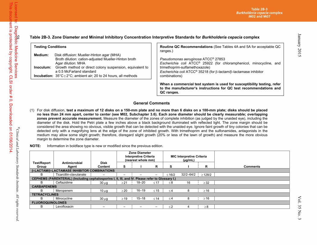
Table 2B-3. Zone Diameter and Minimal Inhibitory Concentration Interpretive Standards for Burkholderia cepacia complex
General Comments
(1) For disk diffusion, test a maximum of 12 disks on a 150-mm plate and no more than 6 disks on a 100-mm plate; disks should be placed
no less than 24 mm apart, center to center (see M02, Subchapter 3.6). Each zone diameter should be clearly measurable; overlapping zones prevent accurate measurement. Measure the diameter of the zones of complete inhibition (as judged by the unaided eye), including the diameter of the disk. Hold the Petri plate a few inches above a black background illuminated with reflected light. The zone margin should be considered the area showing no obvious, visible growth that can be detected with the unaided eye. Ignore faint growth of tiny colonies that can be detected only with a magnifying lens at the edge of the zone of inhibited growth. With trimethoprim and the sulfonamides, antagonists in the medium may allow some slight growth; therefore, disregard slight growth (20% or less of the lawn of growth) and measure the more obvious margin to determine the zone diameter.
NOTE: Information in boldface type is new or modified since the previous edition.
Test/Report Group
Antimicrobial Agent
Disk Content
Zone Diameter Interpretive Criteria (nearest whole mm)
MIC Interpretive Criteria (µg/mL)
Comments S I R S I R -LACTAM/-LACTAMASE INHIBITOR COMBINATIONS
B Ticarcillin-clavulanate – – – – 16/2 32/2–64/2 128/2 CEPHEMS (PARENTERAL) (Including cephalosporins I, II, III, and IV. Please refer to Glossary I.)
B Ceftazidime 30 g 21 18–20 17 8 16 32 CARBAPENEMS
B Meropenem 10 g 20 16–19 15 4 8 16 TETRACYCLINES
B Minocycline 30 g 19 15–18 14 4 8 16 FLUOROQUINOLONES
B Levofloxacin – – – – 2 4 8
Testing Conditions Medium: Disk diffusion: Mueller-Hinton agar (MHA) Broth dilution: cation-adjusted Mueller-Hinton broth Agar dilution: MHA Inoculum: Growth method or direct colony suspension, equivalent to
a 0.5 McFarland standard Incubation: 35°C 2°C; ambient air; 20 to 24 hours, all methods
Routine QC Recommendations (See Tables 4A and 5A for acceptable QC ranges.) Pseudomonas aeruginosa ATCC® 27853 Escherichia coli ATCC® 25922 (for chloramphenicol, minocycline, and trimethoprim-sulfamethoxazole) Escherichia coli ATCC® 35218 (for -lactam/-lactamase inhibitor combinations) When a commercial test system is used for susceptibility testing, refer to the manufacturer’s instructions for QC test recommendations and QC ranges.
58 ©Clinical and Laboratory Standards Institute. All rights reserved.
January 2015
Vol. 35 N
o. 3
Table 2B-3Burkholderia cepacia complex
M02 and M07
Licensed�to:��Diagnostic�M
edicine�Services
This�document�is�protected�by�copyright.�C
LSI�order�#�0,�D
ownloaded�on�01/06/2014.

©Clinical and Laboratory Standards Institute. All rights reserved. 59
Table 2B-3. (Continued)
Abbreviations: ATCC®, American Type Culture Collection; I, intermediate; MIC, minimal inhibitory concentration; R, resistant; S, susceptible.
Test/Report Group
Antimicrobial Agent
Disk Content
Zone Diameter Interpretive Criteria (nearest whole mm)
MIC Interpretive Criteria (µg/mL)
Comments S I R S I R FOLATE PATHWAY INHIBITORS
A Trimethoprim- sulfamethoxazole
1.25/23.75 g 16 11–15 10 2/38
– 4/76
PHENICOLS
B Chloramphenicol – – – – 8 16 32 (2) Not routinely reported on isolates from the
urinary tract.
For Use W
ith M02-A
12 and M07-A
10 M100-S25
Table 2B-3Burkholderia cepacia complex
M02 and M07
Licensed�to:��Diagnostic�M
edicine�Services
This�document�is�protected�by�copyright.�C
LSI�order�#�0,�D
ownloaded�on�01/06/2014.

Table 2B-4. Zone Diameter and Minimal Inhibitory Concentration Interpretive Standards for Stenotrophomonas maltophilia
General Comments
(1) For disk diffusion, test a maximum of 12 disks on a 150-mm plate and no more than 6 disks on a 100-mm plate; disks should be placed no less than
24 mm apart, center to center (see M02, Subchapter 3.6). Each zone diameter should be clearly measurable; overlapping zones prevent accurate measurement. Measure the diameter of the zones of complete inhibition (as judged by the unaided eye), including the diameter of the disk. Hold the Petri plate a few inches above a black background illuminated with reflected light. The zone margin should be considered the area showing no obvious, visible growth that can be detected with the unaided eye. Ignore faint growth of tiny colonies that can be detected only with a magnifying lens at the edge of the zone of inhibited growth. With trimethoprim and the sulfonamides, antagonists in the medium may allow some slight growth; therefore, disregard slight growth (20% or less of the lawn of growth) and measure the more obvious margin to determine the zone diameter.
NOTE: Information in boldface type is new or modified since the previous edition.
Test/Report Group
Antimicrobial Agent
Disk Content
Zone Diameter Interpretive Criteria (nearest whole mm)
MIC Interpretive Criteria (µg/mL)
Comments S I R S I R -LACTAM/-LACTAMASE INHIBITOR COMBINATIONS
B Ticarcillin-clavulanate – – – – 16/2 32/2–64/2 128/2 CEPHEMS (PARENTERAL) (Including cephalosporins I, II, III, and IV. Please refer to Glossary I.)
B Ceftazidime – – – – 8 16 32 TETRACYCLINES
B Minocycline 30 g 19 15–18 14 4 8 16 FLUOROQUINOLONES
B Levofloxacin 5 g 17 14–16 13 2 4 8
Testing Conditions Medium: Disk diffusion: Mueller-Hinton agar (MHA) Broth dilution: cation-adjusted Mueller-Hinton broth Agar dilution: MHA Inoculum: Growth method or direct colony suspension, equivalent to a
0.5 McFarland standard Incubation: 35°C 2°C; ambient air; 20 to 24 hours, all methods
Routine QC Recommendations (See Tables 4A and 5A for acceptable QC ranges.) Pseudomonas aeruginosa ATCC® 27853 Escherichia coli ATCC® 25922 (for chloramphenicol, minocycline, and trimethoprim-sulfamethoxazole) Escherichia coli ATCC® 35218 (for -lactam/-lactamase inhibitor combinations) When a commercial test system is used for susceptibility testing, refer to the manufacturer’s instructions for QC test recommendations and QC ranges.
January 2015
Vol. 35 N
o. 3
60 ©Clinical and Laboratory Standards Institute. All rights reserved.
Table 2B-4Stenotrophomonas maltophilia
M02 and M07
Licensed�to:��Diagnostic�M
edicine�Services
This�document�is�protected�by�copyright.�C
LSI�order�#�0,�D
ownloaded�on�01/06/2014.

©Clinical and Laboratory Standards Institute. All rights reserved. 61
Table 2B-4. (Continued)
Abbreviations: ATCC®, American Type Culture Collection; I, intermediate; MIC, minimal inhibitory concentration; R, resistant; S, susceptible.
Test/Report Group
Antimicrobial Agent
Disk Content
Zone Diameter Interpretive Criteria (nearest whole mm)
MIC Interpretive Criteria (µg/mL)
Comments S I R S I R FOLATE PATHWAY INHIBITORS
A Trimethoprim- sulfamethoxazole
1.25/23.75 g
16 11–15 10 2/38 – 4/76
PHENICOLS
B Chloramphenicol – – – – 8 16 32 (2) Not routinely reported on isolates from the urinary tract.
For Use W
ith M02-A
12 and M07-A
10 M100-S25
Table 2B-4Stenotrophomonas maltophilia
M02 and M07
Licensed�to:��Diagnostic�M
edicine�Services
This�document�is�protected�by�copyright.�C
LSI�order�#�0,�D
ownloaded�on�01/06/2014.

Table 2B-5. Minimal Inhibitory Concentration Interpretive Standards (g/mL) for Other Non-Enterobacteriaceae (Refer to Comment 1)
General Comments (1) Other non-Enterobacteriaceae include Pseudomonas spp. (not P. aeruginosa) and other nonfastidious, glucose-nonfermenting, gram-negative bacilli,
but exclude P. aeruginosa, Acinetobacter spp., Burkholderia cepacia, B. mallei, B. pseudomallei, and Stenotrophomonas maltophilia. Refer to Tables 2B-2, 2B-3, and 2B-4 for testing of Acinetobacter spp., B. cepacia complex, and S. maltophilia, respectively, and CLSI document M45 for testing of Burkholderia mallei, B. pseudomallei, Aeromonas spp., and Vibrio spp.
(2) For other non-Enterobacteriaceae, the disk diffusion method has not been systematically studied by the subcommittee nor have clinical data been
collected for review. Therefore, for this organism group, disk diffusion testing is not currently recommended. NOTE: Information in boldface type is new or modified since the previous edition.
Test/Report Group
Antimicrobial Agent
Disk Content
Zone Diameter Interpretive Criteria (nearest whole mm)
MIC Interpretive Criteria (µg/mL)
Comments S I R S I R PENICILLINS
A Piperacillin – – – – 16 32–64 128 O Carbenicillin – – – – 16 32 64 O Mezlocillin – – – – 16 32–64 128 O Ticarcillin – – – – 16 32–64 128
-LACTAM/-LACTAMASE INHIBITOR COMBINATIONS B B
Piperacillin-tazobactam Ticarcillin-clavulanate
– –
– –
– –
– –
16/4 16/2
32/4–64/4 32/2–64/2
128/4 128/2
CEPHEMS (PARENTERAL) (Including cephalosporins I, II, III, and IV. Please refer to Glossary I.) A Ceftazidime – – – – 8 16 32 B Cefepime – – – – 8 16 32 C C
Cefotaxime Ceftriaxone
– –
– –
– –
– –
8 8
16–32 16–32
64 64
Testing Conditions Medium: Broth dilution: cation-adjusted Mueller-Hinton broth Agar dilution: Mueller-Hinton agar Inoculum: Growth method or direct colony suspension, equivalent to a 0.5
McFarland standard Incubation: 35°C ± 2°C; ambient air; 16 to 20 hours
Routine QC Recommendations (See Tables 4A and 5A for acceptable QC ranges.) Pseudomonas aeruginosa ATCC® 27853 Escherichia coli ATCC® 25922 (for chloramphenicol, tetracyclines, sulfonamides, and trimethoprim-sulfamethoxazole) Escherichia coli ATCC® 35218 (for -lactam/-lactamase inhibitor combinations) When a commercial test system is used for susceptibility testing, refer to the manufacturer’s instructions for QC test recommendations and QC ranges.
62 ©Clinical and Laboratory Standards Institute. All rights reserved.
January 2015
Vol. 35 N
o. 3
Table 2B-5Other Non-Enterobacteriaceae
M07
Licensed�to:��Diagnostic�M
edicine�Services
This�document�is�protected�by�copyright.�C
LSI�order�#�0,�D
ownloaded�on�01/06/2014.

©Clinical and Laboratory Standards Institute. All rights reserved. 63
Table 2B-5. (Continued)
Abbreviations: ATCC®, American Type Culture Collection; I, intermediate; MIC, minimal inhibitory concentration; R, resistant; S, susceptible.
Test/Report Group
Antimicrobial Agent
Disk Content
Zone Diameter Interpretive Criteria (nearest whole mm)
MIC Interpretive Criteria (µg/mL)
Comments S I R S I R CEPHEMS (PARENTERAL) (Including cephalosporins I, II, III, and IV. Please refer to Glossary I.) (Continued)
O Cefoperazone – – – – 16 32 64 O Ceftizoxime – – – – 8 16–32 64 O Moxalactam – – – – 8 16–32 64
MONOBACTAMS B Aztreonam – – – – 8 16 32
CARBAPENEMS B Imipenem – – – – 4 8 16 B Meropenem – – – – 4 8 16
LIPOPEPTIDES O Colistin – – – – 2 4 8 O Polymyxin B – – – – 2 4 8
AMINOGLYCOSIDES A Gentamicin – – – – 4 8 16 A Tobramycin – – – – 4 8 16 B Amikacin – – – – 16 32 64 O Netilmicin – – – – 8 16 32
TETRACYCLINES (3) Organisms that are susceptible to tetracycline are also considered susceptible to doxycycline and minocycline. However, some organisms that are intermediate or resistant to tetracycline may be susceptible to doxycycline, minocycline, or both.
U Tetracycline – – – – 4 8 16 O Doxycycline – – – – 4 8 16 O Minocycline – – – – 4 8 16
FLUOROQUINOLONES B B
Ciprofloxacin Levofloxacin
– –
– –
– – 1 2
2 4
4 8
U U U
Lomefloxacin or ofloxacin Norfloxacin
– – –
– – –
– – –
– – –
2 2 4
4 4 8
8 8 16
O Gatifloxacin – – – – 2 4 8 (4) For testing and reporting of urinary tract isolates only.
FOLATE PATHWAY INHIBITORS B Trimethoprim-
sulfamethoxazole – – – – 2/38 – 4/76
U Sulfonamides – – – – 256 – 512
(5) Sulfisoxazole can be used to represent any of the currently available sulfonamide preparations.
PHENICOLS C Chloramphenicol – – – – 8 16 32 (6) Not routinely reported on isolates from the
urinary tract.
For Use W
ith M02-A
12 and M07-A
10 M100-S25
Table 2B-5Other Non-Enterobacteriaceae
M07
Licensed�to:��Diagnostic�M
edicine�Services
This�document�is�protected�by�copyright.�C
LSI�order�#�0,�D
ownloaded�on�01/06/2014.

Table 2C. Zone Diameter and Minimum Inhibitory Concentration Interpretive Standards for Staphylococcus spp.
General Comments (1) For disk diffusion, test a maximum of 12 disks on a 150-mm plate and no more than 6 disks on a 100-mm plate; disks should be placed no less than 24
mm apart, center to center (see M02, Subchapter 3.6). Each zone diameter should be clearly measurable; overlapping zones prevent accurate measurement. Measure the diameter of the zones of complete inhibition (as judged by the unaided eye), including the diameter of the disk. Hold the Petri plate a few inches above a black background illuminated with reflected light, except for linezolid, which should be read with transmitted light (plate held up to light source). The zone margin should be considered the area showing no obvious, visible growth that can be detected with the unaided eye. Ignore faint growth of tiny colonies that can be detected only with a magnifying lens at the edge of the zone of inhibited growth. With trimethoprim and the sulfonamides, antagonists in the medium may allow some slight growth; therefore, disregard slight growth (20% or less of the lawn of growth) and measure the more obvious margin to determine the zone diameter. For linezolid, any discernible growth within the zone of inhibition is indicative of resistance to the respective agent.
(2) Historically, resistance to the penicillinase-stable penicillins (see Glossary I) has been referred to as “methicillin resistance” or “oxacillin resistance.”
MRSAs are those strains of S. aureus that express mecA or another mechanism of methicillin resistance, such as changes in affinity of penicillin-binding proteins for oxacillin (modified S. aureus strains).
Testing Conditions Medium: Disk diffusion: Mueller-Hinton agar (MHA) Broth dilution: cation-adjusted Mueller-Hinton broth (CAMHB); CAMHB +
2% NaCl for oxacillin; CAMHB supplemented to 50 µg/mL calcium for daptomycin
Agar dilution: MHA; MHA + 2% NaCl for oxacillin. Agar dilution has not been validated for daptomycin.
Inoculum: Direct colony suspension, equivalent to a 0.5 McFarland standard Incubation: 35°C 2°C; ambient air Disk diffusion: 16 to 18 hours; 24 hours (coagulase-negative staphylococci [CoNS] and cefoxitin) Dilution methods: 16 to 20 hours; 24 hours for oxacillin and vancomycin; Testing at temperatures above 35°C may not detect methicillin-resistant staphylococci (MRS).
Routine QC Recommendations (See Tables 4A and 5A for acceptable QC ranges.) Staphylococcus aureus ATCC® 25923 (disk diffusion) Staphylococcus aureus ATCC® 29213 (MIC) When a commercial test system is used for susceptibility testing, refer to the manufacturer’s instructions for QC test recommendations and QC ranges.
64 ©Clinical and Laboratory Standards Institute. All rights reserved.
January 2015
Vol. 35 N
o. 3
Table 2CStaphylococcus spp.
M02 and M07
Licensed�to:��Diagnostic�M
edicine�Services
This�document�is�protected�by�copyright.�C
LSI�order�#�0,�D
ownloaded�on�01/06/2014.

©Clinical and Laboratory Standards Institute. All rights reserved. 65
Table 2C. (Continued) (3) In most staphylococcal isolates, oxacillin resistance is mediated by mecA, encoding the penicillin-binding protein 2a (PBP 2a, also called PBP2'). Isolates
that test positive for mecA or PBP 2a should be reported as oxacillin resistant.
Isolates that test resistant by oxacillin minimal inhibitory concentration (MIC), cefoxitin MIC, or cefoxitin disk test should be reported as oxacillin resistant.
Mechanisms of oxacillin resistance other than mecA are rare and include a novel mecA homologue, mecC.1 MICs for strains with mecC are typically in the resistant range for cefoxitin and/or oxacillin; mecC resistance cannot be detected by tests directed at mecA or PBP 2a.
(4) Oxacillin-resistant S. aureus and CoNS (MRS), are considered resistant to other -lactam agents, ie, penicillins, -lactam/-lactamase inhibitor combinations, cephems (with the exception of the cephalosporins with anti-MRSA activity), and carbapenems. This is because most cases of documented MRS infections have responded poorly to -lactam therapy, or because convincing clinical data that document clinical efficacy for those agents have not been presented.
(5) Routine testing of urine isolates of S. saprophyticus is not advised, because infections respond to concentrations achieved in urine of antimicrobial agents
commonly used to treat acute, uncomplicated urinary tract infections (eg, nitrofurantoin, trimethoprim ± sulfamethoxazole, or a fluoroquinolone). (6) For screening tests for β-lactamase production, oxacillin resistance, mecA-mediated oxacillin resistance using cefoxitin, reduced susceptibility to
vancomycin, inducible clindamycin resistance, and high-level mupirocin resistance (S. aureus only), refer to Tables 3D, 3E, 3F, 3G, and 3H, respectively. NOTE: Information in boldface type is new or modified since the previous edition.
_____________________________________________
1 García-Álvarez L, Holden MT, Lindsay H, et al. Methicillin-resistant Staphylococcus aureus with a novel mecA homologue in human and bovine populations in the UK and Denmark: a descriptive study. Lancet Infect Dis. 2011;11(8):595-603.
For Use W
ith M02-A
12 and M07-A
10 M100-S25
Table 2CStaphylococcus spp.
M02 and M07
Licensed�to:��Diagnostic�M
edicine�Services
This�document�is�protected�by�copyright.�C
LSI�order�#�0,�D
ownloaded�on�01/06/2014.

Table 2C. (Continued)
Test/Report
Group Antimicrobial
Agent Disk
Content
Zone Diameter Interpretive Criteria (nearest whole mm)
MIC Interpretive Criteria (µg/mL)
Comments S I R S I R PENICILLINASE-LABILE PENICILLINS (7) Penicillin-susceptible staphylococci are also susceptible to other β-lactam agents with established clinical efficacy for staphylococcal infections. Penicillin-resistant staphylococci are resistant to penicillinase-labile penicillins, including ampicillin, amoxicillin, azlocillin, carbenicillin, mezlocillin, piperacillin, and ticarcillin.
A Penicillin 10 units 29 – 28 0.12 – 0.25 (8) Penicillin should be used to test the susceptibility of all staphylococci to all penicillinase-labile penicillins. Penicillin-resistant strains of staphylococci produce -lactamase. Perform test(s) to detect -lactamase production on staphylococci for which the penicillin MICs are ≤ 0.12 µg/mL or zone diameters ≥
29 mm before reporting the isolate as penicillin susceptible. Rare isolates of staphylococci that contain genes for -lactamase production may appear negative by -lactamase tests. Consequently, for serious infections requiring penicillin therapy, laboratories should perform MIC tests and -lactamase testing on all subsequent isolates from the same patient. PCR testing of the isolate for the blaZ -lactamase gene may be considered. See Tables 3D and 3E. (9) For oxacillin-resistant staphylococci report penicillin as resistant or do not report.
PENICILLINASE-STABLE PENICILLINS (10) Oxacillin (or cefoxitin) results can be applied to the other penicillinase-stable penicillins (cloxacillin, dicloxacillin, flucloxacillin, methicillin, and nafcillin). For agents with established clinical efficacy and considering site of infection and appropriate dosing, oxacillin (cefoxitin)-susceptible staphylococci can be considered susceptible to:
-lactam/-lactamase inhibitor combinations (amoxicillin-clavulanate, ampicillin-sulbactam, piperacillin-tazobactam, ticarcillin-clavulanate) Oral cephems (cefaclor, cefdinir, cefpodoxime, cefprozil, cefuroxime, loracarbef) Parenteral cephems including cephalosporins I, II, III, and IV (cefamandole, cefazolin, cefepime, cefmetazole, cefonicid, cefoperazone, cefotaxime, cefotetan, ceftizoxime,
ceftriaxone, cefuroxime, cephalothin, ceftaroline, moxalactam) Carbapenems (doripenem, ertapenem, imipenem, meropenem)
Oxacillin-resistant staphylococci are resistant to all currently available -lactam antimicrobial agents, with the exception of the newer cephalosporins with anti-MRSA activity. Thus, susceptibility or resistance to a wide array of -lactam antimicrobial agents may be deduced from testing only penicillin and either cefoxitin or oxacillin. Testing of other -lactam agents, except those with anti-MRSA activity, is not advised. See comments (3) and (4). In addition, further explanation on the use of cefoxitin for prediction of mecA-mediated oxacillin resistance can be found in Subchapter 3.13 of M07-A10 and Subchapter 3.9 of M02-A12.
66 ©Clinical and Laboratory Standards Institute. All rights reserved.
January 2015
Vol. 35 N
o. 3
Table 2CStaphylococcus spp.
M02 and M07
Licensed�to:��Diagnostic�M
edicine�Services
This�document�is�protected�by�copyright.�C
LSI�order�#�0,�D
ownloaded�on�01/06/2014.

©Clinical and Laboratory Standards Institute. All rights reserved. 67
Table 2C. (Continued)
Test/Report Group
Antimicrobial Agent
Disk Content
Zone Diameter Interpretive Criteria (nearest whole mm)
MIC Interpretive Criteria (µg/mL)
Comments S I R S I R PENICILLINASE-STABLE PENICILLINS (Continued)
A Oxacillin For S. aureus and S. lugdunensis.
30 µg cefoxitin (surrogate test for oxacillin)
–
22
–
–
–
21
≤ 2 (oxacillin)
≤ 4 (cefoxitin)
–
–
≥ 4 (oxacillin)
≥ 8 (cefoxitin)
For use with S. aureus and S. lugdunensis. (11) Oxacillin disk testing is not reliable. See cefoxitin and comment (4) for reporting oxacillin when testing cefoxitin as a surrogate agent. (12) Cefoxitin is tested as a surrogate for oxacillin; report oxacillin susceptible or resistant based on the cefoxitin result. See comments (4), (7), and (10).
A Oxacillin For CoNS except S. lugdunensis.
– – – – ≤ 0.25 (oxacillin)
–
≥ 0.5 (oxacillin)
For use with CoNS except S. lugdunensis. (13) Oxacillin MIC interpretive criteria may overcall resistance for some CoNS, because some non–S. epidermidis strains for which the oxacillin MICs are 0.5–2 µg/mL lack mecA. For serious infections with CoNS other than S. epidermidis, testing for mecA or for PBP 2a or with cefoxitin disk diffusion may be appropriate for strains for which the oxacillin MICs are 0.5–2 µg/mL.
30 µg cefoxitin(surrogate test for oxacillin)
25
– 24
– –
–
See comments (4), (7), (10), and (12).
CEPHEMS (PARENTERAL) B Ceftaroline 30 µg ≥ 24 21–23 ≤ 20 ≤ 1 2 ≥ 4 (14) For use with S. aureus only, including
MRSA. (15) Interpretive criteria are based on a dosage regimen of 600 mg every 12 h.
For Use W
ith M02-A
12 and M07-A
10 M100-S25
Table 2CStaphylococcus spp.
M02 and M07
Licensed�to:��Diagnostic�M
edicine�Services
This�document�is�protected�by�copyright.�C
LSI�order�#�0,�D
ownloaded�on�01/06/2014.

Table 2C. (Continued)
Test/Report Group
Antimicrobial Agent
Disk Content
Zone Diameter Interpretive Criteria (nearest whole mm)
MIC Interpretive Criteria (µg/mL)
Comments S I R S I R GLYCOPEPTIDES (16) For S. aureus, vancomycin-susceptible isolates may become vancomycin intermediate during the course of prolonged therapy.
B
Vancomycin
– – – – 2 4–8 16 For use with S. aureus. (17) MIC tests should be performed to determine the susceptibility of all isolates of staphylococci to vancomycin. The disk test does not differentiate vancomycin-susceptible isolates of S. aureus from vancomycin-intermediate isolates, nor does the test differentiate among vancomycin-susceptible, -intermediate, and -resistant isolates of CoNS, all of which will give similar size zones of inhibition. (18) Send any S. aureus for which the vancomycin is 8 g/mL to a reference laboratory. See Appendix A. Also refer to Table 3F for S. aureus, Subchapter 3.13.1.7 in M07-A10, and Subchapter 3.9.1.7 in M02-A12.
B
Vancomycin
– – – – 4
8–16 32 For use with CoNS. See comment (17). (19) Send any CoNS for which the vancomycin MIC is 32 g/mL to a reference laboratory. See Appendix A. See also Subchapter 3.13.1.7 in M07-A10, and Subchapter 3.9.1.7 in M02-A12.
Inv. Teicoplanin 30 g 14 11–13 10 8 16 32 (20) Teicoplanin disk diffusion interpretive criteria were not reevaluated concurrent with the reevaluation of vancomycin disk diffusion interpretive criteria. Therefore, the ability of these teicoplanin interpretive criteria to differentiate teicoplanin-intermediate and teicoplanin-resistant staphylococci from teicoplanin-susceptible strains is not known.
LIPOPEPTIDES B Daptomycin – – – – 1 – – (21) Daptomycin should not be reported for
isolates from the respiratory tract.
68 ©Clinical and Laboratory Standards Institute. All rights reserved.
January 2015
Vol. 35 N
o. 3
Table 2CStaphylococcus spp.
M02 and M07
Licensed�to:��Diagnostic�M
edicine�Services
This�document�is�protected�by�copyright.�C
LSI�order�#�0,�D
ownloaded�on�01/06/2014.

©Clinical and Laboratory Standards Institute. All rights reserved. 69
Table 2C. (Continued)
Test/Report Group
Antimicrobial Agent
Disk Content
Zone Diameter Interpretive Criteria (nearest whole mm)
MIC Interpretive Criteria (µg/mL)
Comments S I R S I R AMINOGLYCOSIDES (22) For staphylococci that test susceptible, aminoglycosides are used only in combination with other active agents that test susceptible.
C Gentamicin 10 g 15 13–14 12 4 8 16 O Amikacin 30 g 17 15–16 14 16 32 64 O Kanamycin 30 g 18 14–17 13 16 32 64 O Netilmicin 30 g 15 13–14 12 8 16 32 O Tobramycin 10 g 15 13–14 12 4 8 16
MACROLIDES (23) Not routinely reported on organisms isolated from the urinary tract.
A A A
Azithromycin or clarithromycin or erythromycin
15 g 15 g 15 g
18 18 23
14–17 14–17 14–22
13 13 13
2 2 0.5
4 4
1–4
8 8 8
O Telithromycin 15 g 22 19–21 18 1 2 4 O Dirithromycin 15 g 19 16–18 15 2 4 8
TETRACYCLINES (24) Organisms that are susceptible to tetracycline are also considered susceptible to doxycycline and minocycline. However, some organisms that are intermediate or resistant to tetracycline may be susceptible to doxycycline, minocycline, or both.
B Tetracycline 30 g 19 15–18 14 4 8 16 B Doxycycline 30 g 16 13–15 12 4 8 16 B Minocycline 30 g 19 15–18 14 4 8 16 See comment (23).
FLUOROQUINOLONES (25) Staphylococcus spp. may develop resistance during prolonged therapy with quinolones. Therefore, isolates that are initially susceptible may become resistant within three to four days after initiation of therapy. Testing of repeat isolates may be warranted.
C Ciprofloxacin or 5 g 21 16–20 15 1 2 4 C levofloxacin or 5 g 19 16–18 15 1 2 4 C ofloxacin 5 g 18 15–17 14 1 2 4 C Moxifloxacin 5 g 24 21–23 20 0.5 1 2 U Lomefloxacin 10 g 22 19–21 18 2 4 8 U Norfloxacin 10 g 17 13–16 12 4 8 16 O Enoxacin 10 g 18 15–17 14 2 4 8 (26) FDA approved for S. saprophyticus and S.
epidermidis (but not for S. aureus). O Gatifloxacin 5 g 23 20–22 19 0.5 1 2 O Grepafloxacin 5 g 18 15–17 14 1 2 4
For Use W
ith M02-A
12 and M07-A
10 M100-S25
Table 2CStaphylococcus spp.
M02 and M07
Licensed�to:��Diagnostic�M
edicine�Services
This�document�is�protected�by�copyright.�C
LSI�order�#�0,�D
ownloaded�on�01/06/2014.
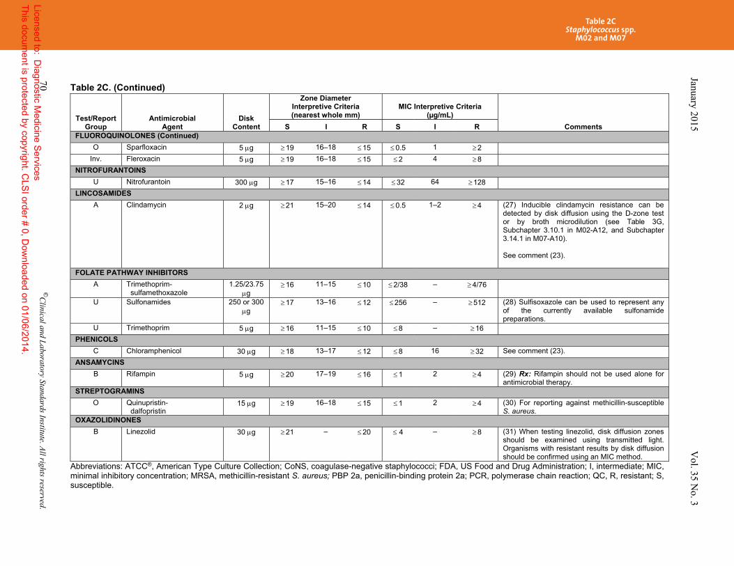
Table 2C. (Continued)
Abbreviations: ATCC®, American Type Culture Collection; CoNS, coagulase-negative staphylococci; FDA, US Food and Drug Administration; I, intermediate; MIC, minimal inhibitory concentration; MRSA, methicillin-resistant S. aureus; PBP 2a, penicillin-binding protein 2a; PCR, polymerase chain reaction; QC, R, resistant; S, susceptible.
Test/Report Group
Antimicrobial Agent
Disk Content
Zone Diameter Interpretive Criteria (nearest whole mm)
MIC Interpretive Criteria (µg/mL)
Comments S I R S I R FLUOROQUINOLONES (Continued)
O Sparfloxacin 5 g 19 16–18 15 0.5 1 2 Inv. Fleroxacin 5 g 19 16–18 15 2 4 8
NITROFURANTOINS U Nitrofurantoin 300 g 17 15–16 14 32 64 128
LINCOSAMIDES A Clindamycin 2 g 21 15–20 14 0.5 1–2 4 (27) Inducible clindamycin resistance can be
detected by disk diffusion using the D-zone test or by broth microdilution (see Table 3G, Subchapter 3.10.1 in M02-A12, and Subchapter 3.14.1 in M07-A10). See comment (23).
FOLATE PATHWAY INHIBITORS A Trimethoprim-
sulfamethoxazole 1.25/23.75
g 16 11–15 10 2/38
–
4/76
U Sulfonamides 250 or 300 g
17 13–16 12 256
–
512
(28) Sulfisoxazole can be used to represent any of the currently available sulfonamide preparations.
U Trimethoprim 5 g 16 11–15 10 8 – 16 PHENICOLS
C Chloramphenicol 30 g 18 13–17 12 8 16 32 See comment (23). ANSAMYCINS
B Rifampin 5 g 20 17–19 16 1 2 4 (29) Rx: Rifampin should not be used alone for antimicrobial therapy.
STREPTOGRAMINS O Quinupristin-
dalfopristin 15 g 19 16–18 15 1 2 4 (30) For reporting against methicillin-susceptible
S. aureus.OXAZOLIDINONES
B Linezolid 30 g 21 – 20 4 – 8 (31) When testing linezolid, disk diffusion zones should be examined using transmitted light. Organisms with resistant results by disk diffusion should be confirmed using an MIC method.
70 ©Clinical and Laboratory Standards Institute. All rights reserved.
January 2015
Vol. 35 N
o. 3
Table 2CStaphylococcus spp.
M02 and M07
Licensed�to:��Diagnostic�M
edicine�Services
This�document�is�protected�by�copyright.�C
LSI�order�#�0,�D
ownloaded�on�01/06/2014.
For Use With M02-A12 and M07-A10 M100-S25
©Clinical and Laboratory Standards Institute. All rights reserved. 71
This page is intentionally left blank.
Licensed�to:��Diagnostic�Medicine�ServicesThis�document�is�protected�by�copyright.�CLSI�order�#�0,�Downloaded�on�01/06/2014.

Table 2D. Zone Diameter and Minimal Inhibitory Concentration Interpretive Standards for Enterococcus spp.
Refer to Tables 3F and 3I for additional testing recommendations, reporting suggestions, and QC.
General Comments (1) For disk diffusion, test a maximum of 12 disks on a 150-mm plate and no more than 6 disks on a 100-mm plate; disks should be placed no less
than 24 mm apart, center to center (see M02, Subchapter 3.6). Each zone diameter should be clearly measurable; overlapping zones prevent accurate measurement. Measure the diameter of the zones of complete inhibition (as judged by the unaided eye), including the diameter of the disk. Hold the Petri plate a few inches above a black background illuminated with reflected light, except for vancomycin, which should be read with transmitted light (plate held up to light source). The zone margin should be considered the area showing no obvious, visible growth that can be detected with the unaided eye. Ignore faint growth of tiny colonies that can be detected only with a magnifying lens at the edge of the zone of inhibited growth. Any discernible growth within the zone of inhibition indicates vancomycin resistance.
(2) WARNING: For Enterococcus spp., cephalosporins, aminoglycosides (except for high-level resistance screening), clindamycin, and trimethoprim-
sulfamethoxazole may appear active in vitro, but they are not effective clinically, and isolates should not be reported as susceptible. (3) Synergy between ampicillin, penicillin, or vancomycin and an aminoglycoside can be predicted for enterococci by using a high-level aminoglycoside
(gentamicin and streptomycin) screening test (see Table 3I). NOTE: Information in boldface type is new or modified since the previous edition.
Testing Conditions Medium: Disk diffusion: Mueller-Hinton agar (MHA) Broth dilution: cation-adjusted Mueller-Hinton broth (CAMHB);
CAMHB supplemented to 50 µg/mL calcium for daptomycin Agar dilution: MHA; agar dilution has not been validated for
daptomycin Inoculum: Growth method or direct colony suspension, equivalent to a 0.5
McFarland standard Incubation: 35°C ± 2°C; ambient air
Disk diffusion: 16 to 18 hours Dilution methods: 16 to 20 hours All methods: 24 hours for vancomycin
Routine QC Recommendations (See Tables 4A and 5A for acceptable QC ranges.) Disk diffusion: Staphylococcus aureus ATCC® 25923 Dilution methods: Enterococcus faecalis ATCC® 29212 When a commercial test system is used for susceptibility testing, refer to the manufacturer’s instructions for QC test recommendations and QC ranges.
72 ©Clinical and Laboratory Standards Institute. All rights reserved.
January 2015
Vol. 35 N
o. 3
Table 2DEnterococcus spp.
M02 and M07
Licensed�to:��Diagnostic�M
edicine�Services
This�document�is�protected�by�copyright.�C
LSI�order�#�0,�D
ownloaded�on�01/06/2014.

©Clinical and Laboratory Standards Institute. All rights reserved. 73
Table 2D. (Continued)
Test/Report Group
Antimicrobial Agent
Disk Content
Zone Diameter Interpretive Criteria (nearest whole mm)
MIC Interpretive Criteria (µg/mL)
Comments S I R S I R PENICILLINS
A A
Penicillin Ampicillin
10 units 10 g
15 17
– –
14 16
8 8
– –
16 16
(4) The results of ampicillin susceptibility tests should be used to predict the activity of amoxicillin. Ampicillin results may be used to predict susceptibility to amoxicillin-clavulanate, ampicillin-sulbactam, piperacillin, and piperacillin-tazobactam among non–-lactamase-producing enterococci. Ampicillin susceptibility can be used to predict imipenem susceptibility, providing the species is confirmed to be E. faecalis. (5) Enterococci susceptible to penicillin are predictably susceptible to ampicillin, amoxicillin, ampicillin-sulbactam, amoxicillin-clavulanate, piperacillin, and piperacillin-tazobactam for non–-lactamase-producing enterococci. However, enterococci susceptible to ampicillin cannot be assumed to be susceptible to penicillin. If penicillin results are needed, testing of penicillin is required. (6) Rx: Combination therapy with ampicillin, penicillin, or vancomycin (for susceptible strains), plus an aminoglycoside, is usually indicated for serious enterococcal infections, such as endocarditis, unless high-level resistance to both gentamicin and streptomycin is documented; such combinations are predicted to result in synergistic killing of the Enterococcus. (7) Penicillin or ampicillin resistance among enterococci due to -lactamase production has been reported very rarely. Penicillin or ampicillin resistance due to -lactamase production is not reliably detected with routine disk or dilution methods, but is detected using a direct, nitrocefin-based -lactamase test. Because of the rarity of -lactamase-positive enterococci, this test need not be performed routinely, but can be used in selected cases. A positive -lactamase test predicts resistance to penicillin, as well as amino- and ureidopenicillins (see Glossary I).
For Use W
ith M02-A
12 and M07-A
10 M100-S25
Table 2DEnterococcus spp.
M02 and M07
Licensed�to:��Diagnostic�M
edicine�Services
This�document�is�protected�by�copyright.�C
LSI�order�#�0,�D
ownloaded�on�01/06/2014.

Table 2D. (Continued)
Test/Report Group
Antimicrobial Agent
Disk Content
Zone Diameter Interpretive Criteria (nearest whole mm)
MIC Interpretive Criteria (µg/mL)
Comments S I R S I R GLYCOPEPTIDES
B Vancomycin 30 g 17 15–16 14 4 8–16 32 (8) When testing vancomycin against enterococci, plates should be held a full 24 hours for accurate detection of resistance. Zones should be examined using transmitted light; the presence of a haze or any growth within the zone of inhibition indicates resistance. Organisms with intermediate zones should be tested by an MIC method as described in M07-A10. For isolates for which the vancomycin MICs are 8 to 16 g/mL, perform biochemical tests for identification as listed under the “Vancomycin MIC ≥ 8 µg/mL” test found in Table 3F. See comments (3) and (6).
Inv. Teicoplanin 30 g 14 11–13 10 8 16 32 LIPOPEPTIDES
B
Daptomycin
– – – – 4 – – (9) Daptomycin should not be reported for isolates from the respiratory tract.
MACROLIDES O Erythromycin 15 g 23 14–22 13 0.5 1–4 8 (10) Not routinely reported on isolates from the urinary tract.
TETRACYCLINES (11) Organisms that are susceptible to tetracycline are also considered susceptible to doxycycline and minocycline. However, some organisms that are intermediate or resistant to tetracycline may be susceptible to doxycycline, minocycline, or both.
U Tetracycline 30 g 19 15–18 14 4 8 16 O Doxycycline 30 g 16 13–15 12 4 8 16 O Minocycline 30 g 19 15–18 14 4 8 16
FLUOROQUINOLONES U U U
Ciprofloxacin Levofloxacin Norfloxacin
5 g 5 g 10 g
21 17 17
16–20 14–16 13–16
15 13 12
1 2 4
2 4 8
4 8 16
O Gatifloxacin 5 g 18 15–17 14 2 4 8 (12) These interpretive criteria apply to urinary tract isolates only.
NITROFURANTOINS U Nitrofurantoin 300 g 17 15–16 14 32 64 128
74 ©Clinical and Laboratory Standards Institute. All rights reserved.
January 2015
Vol. 35 N
o. 3
Table 2DEnterococcus spp.
M02 and M07
Licensed�to:��Diagnostic�M
edicine�Services
This�document�is�protected�by�copyright.�C
LSI�order�#�0,�D
ownloaded�on�01/06/2014.

©Clinical and Laboratory Standards Institute. All rights reserved. 75
Table 2D. (Continued)
Abbreviations: ATCC®, American Type Culture Collection; I, intermediate; MIC, minimal inhibitory concentration; R, resistant; S, susceptible.
Test/Report Group
Antimicrobial Agent
Disk Content
Zone Diameter Interpretive Criteria (nearest whole mm)
MIC Interpretive Criteria (µg/mL)
Comments S I R S I R ANSAMYCINS
O Rifampin 5 g 20 17–19 16 1 2 4 (13) Rx: Rifampin should not be used alone for antimicrobial therapy.
FOSFOYCINS U Fosfomycin 200 g 16 13–15 12 64 128 256 (14) For testing and reporting of E. faecalis urinary tract
isolates only. (15) The approved MIC testing method is agar dilution. Agar media should be supplemented with 25 µg/mL of glucose-6-phosphate. Broth dilution testing should not be performed. (16) The 200-g fosfomycin disk contains 50 g of glucose-6-phosphate.
PHENICOLS O Chloramphenicol 30 g 18 13–17 12 8 16 32 See comment (10).
STREPTOGRAMINS O Quinupristin-
dalfopristin 15 g 19 16–18 15 1 2 4 (17) For reporting against vancomycin-resistant E.
faecium. OXAZOLIDINONES
B Linezolid 30 g 23 21–22 20 2 4 8
For Use W
ith M02-A
12 and M07-A
10 M100-S25
Table 2DEnterococcus spp.
M02 and M07
Licensed�to:��Diagnostic�M
edicine�Services
This�document�is�protected�by�copyright.�C
LSI�order�#�0,�D
ownloaded�on�01/06/2014.

Table 2E. Zone Diameter and Minimal Inhibitory Concentration Interpretive Standards for Haemophilus influenzae and Haemophilus parainfluenzae
General Comments
(1) Haemophilus spp., as used in this table, includes only H. influenzae and H. parainfluenzae. See CLSI document M45 for testing and reporting recommendations for other species of Haemophilus.
(2) The 0.5 McFarland suspension will contain approximately 1 to 4 × 10⁸ CFU/mL. Exercise care in preparing this suspension, because higher inoculum
concentrations may lead to false-resistant results with some -lactam antimicrobial agents, particularly when -lactamase–producing strains of H. influenzae are tested.
(3) For disk diffusion, test a maximum of 9 disks on a 150-mm plate and 4 disks on a 100-mm plate. Measure the diameter of the zones of complete inhibition (as
judged by the unaided eye), including the diameter of the disk. Hold the Petri plate a few inches above a black background illuminated with reflected light. The zone margin should be considered the area showing no obvious, visible growth that can be detected with the unaided eye. Ignore faint growth of tiny colonies that can be detected only with a magnifying lens at the edge of the zone of inhibited growth. With trimethoprim and the sulfonamides, antagonists in the medium may allow some slight growth; therefore, disregard slight growth (20% or less of the lawn of growth) and measure the more obvious margin to determine the zone diameter.
(4) For isolates of H. influenzae from CSF, only results of testing with ampicillin, one of the third-generation cephalosporins, chloramphenicol, and meropenem
are appropriate to report routinely. (5) Amoxicillin-clavulanate, azithromycin, clarithromycin, cefaclor, cefprozil, loracarbef, cefdinir, cefixime, cefpodoxime, cefuroxime, and telithromycin are oral
agents that may be used as empiric therapy for respiratory tract infections due to Haemophilus spp. The results of susceptibility tests with these antimicrobial agents are often not useful for management of individual patients. However, susceptibility testing of Haemophilus spp. with these compounds may be appropriate for surveillance or epidemiological studies.
Testing Conditions Medium: Disk diffusion: Haemophilus Test Medium (HTM) Broth dilution: HTM broth Inoculum: Direct colony suspension, equivalent to a 0.5
McFarland standard prepared using colonies from an overnight (preferably 20- to 24-hour) chocolate agar plate [see comment (2)]
Incubation: 35°C ± 2°C; Disk diffusion: 5% CO2; 16 to 18 hours Broth dilution: ambient air; 20 to 24 hours
Routine QC Recommendations (See Tables 4A, 4B, 5A, and 5B for acceptable QC ranges.) Haemophilus influenzae ATCC® 49247 Haemophilus influenzae ATCC® 49766 Use either Haemophilus influenzae ATCC® 49247 or Haemophilus influenzae ATCC® 49766 or both of these strains, based on the antimicrobial agents to be tested. Neither strain has QC ranges for all agents that might be tested against H. influenzae or H. parainfluenzae. Escherichia coli ATCC® 35218 (when testing amoxicillin-clavulanate) When a commercial test system is used for susceptibility testing, refer to the manufacturer’s instructions for QC test recommendations and QC ranges.
76 ©Clinical and Laboratory Standards Institute. All rights reserved.
January 2015
Vol. 35 N
o. 3
Table 2EHaemophilus influenzae and Haemophilus
parainfluenzae M02 and M07
Licensed�to:��Diagnostic�M
edicine�Services
This�document�is�protected�by�copyright.�C
LSI�order�#�0,�D
ownloaded�on�01/06/2014.

©Clinical and Laboratory Standards Institute. All rights reserved. 77
For Use W
ith M02-A
12 and M07-A
10 M100-S25
Table 2E. (Continued) (6) To make HTM: Prepare a fresh hematin stock solution by dissolving 50 mg of hematin powder in 100 mL of 0.01 mol/L NaOH with heat and stirring until the
powder is thoroughly dissolved. Add 30 mL of the hematin stock solution and 5 g of yeast extract to 1 L of Mueller-Hinton agar and autoclave. After autoclaving and cooling, add 3 mL of a nicotinamide adenine dinucleotide (NAD) stock solution (50 mg of NAD dissolved in 10 mL of distilled water, filter sterilized) aseptically.
NOTE: Information in boldface type is new or modified since the previous edition.
Test/Report Group
Antimicrobial Agent
Disk Content
Zone Diameter Interpretive Criteria (nearest whole mm)
MIC Interpretive Criteria (µg/mL)
Comments S I R S I R PENICILLINS
A Ampicillin 10 g 22 19–21 18 1 2 4 See comment (4). (7) The results of ampicillin susceptibility tests should be used to predict the activity of amoxicillin. The majority of isolates of H. influenzae that are resistant to ampicillin and amoxicillin produce a TEM-type -lactamase. In most cases, a direct -lactamase test can provide a rapid means of detecting resistance to ampicillin and amoxicillin. (8) Rare BLNAR strains of H. influenzae should be considered resistant to amoxicillin-clavulanate, ampicillin-sulbactam, cefaclor, cefamandole, cefetamet, cefonicid, cefprozil, cefuroxime, loracarbef, and piperacillin-tazobactam, despite apparent in vitro susceptibility of some BLNAR strains to these agents.
-LACTAM/-LACTAMASE INHIBITOR COMBINATIONS B Ampicillin-sulbactam 10/10 g 20 – 19 2/1 – 4/2 See comment (8). C Amoxicillin-clavulanate 20/10 g 20 – 19 4/2 – 8/4 See comments (5) and (8). O Piperacillin-tazobactam 100/10 g 21 – – 1/4 – 2/4 See comment (8).
CEPHEMS (PARENTERAL) (Including cephalosporins I, II, III, and IV. Please refer to Glossary I.) B B B
Cefotaxime or ceftazidime or ceftriaxone
30 g 30 g 30 g
26 26 26
– – –
– – –
2 2 2
– – –
– – –
See comment (4).
B Cefuroxime 30 g 20 17–19 16 4 8 16 See comments (5) and (8). C Ceftaroline 30 g 30 – – 0.5 – – (9) For H. influenzae only.
(10) Interpretive criteria are based on a dosage regimen of 600 mg every 12 h.
O Cefonicid 30 g 20 17–19 16 4 8 16 See comment (8). O Cefamandole – – – – 4 8 16 See comment (8).
Table 2EHaemophilus influenzae and Haemophilus
parainfluenzae M02 and M07
Licensed�to:��Diagnostic�M
edicine�Services
This�document�is�protected�by�copyright.�C
LSI�order�#�0,�D
ownloaded�on�01/06/2014.

Table 2E. (Continued)
Test/Report Group
Antimicrobial Agent
Disk Content
Zone Diameter Interpretive Criteria (nearest whole mm)
MIC Interpretive Criteria (µg/mL)
Comments S I R S I R CEPHEMS (PARENTERAL) (Including cephalosporins I, II, III, and IV. Please refer to Glossary I.) (Continued)
O Cefepime 30 g 26 – – 2 – – O Ceftizoxime 30 g 26 – – 2 – – See comment (4).
CEPHEMS (ORAL) C C
Cefaclor Cefprozil
30 g 30 g
20 18
17–19 15–17
16 14
8 8
16 16
32 32
See comments (5) and (8).
C C C
Cefdinir or cefixime or cefpodoxime
5 g 5 g
10 g
20 21 21
– – –
– – –
1 1 2
– – –
– – –
See comment (5).
C Cefuroxime 30 g 20 17–19 16 4 8 16 See comments (5) and (8). O Loracarbef 30 g 19 16–18 15 8 16 32 See comments (5) and (8). O Ceftibuten 30 g 28 – – 2 – –
Inv. Cefetamet 10 g 18 15–17 14 4 8 16 See comment (8). MONOBACTAMS
C Aztreonam 30 g 26 – – 2 – – CARBAPENEMS
B Meropenem 10 g 20 – – 0.5 – – See comment (4). C C
Ertapenem or imipenem
10 g 10 g
19 16
– –
– –
0.5 4
– –
– –
O Doripenem 10 g 16 – – 1 – – MACROLIDES
C C
Azithromycin Clarithromycin
15 g 15 g
12 13
– 11–12
– 10
4 8
– 16
– 32
See comment (5). See comment (5).
KETOLIDES C Telithromycin 15 g 15 12–14 11 4 8 16 See comment (5).
TETRACYCLINES (11) Organisms that are susceptible to tetracycline are also considered susceptible to doxycycline and minocycline.
C Tetracycline 30 g 29 26–28 25 2 4 8 FLUOROQUINOLONES
C C C C C
Ciprofloxacin or levofloxacin or lomefloxacin or moxifloxacin or ofloxacin
5 g 5 g
10 g 5 g 5 g
21 17
22 18 16
– – – – –
– – – – –
1 2 2 1 2
– – – – –
– – – – –
C Gemifloxacin 5 g 18 – – 0.12 – – O Gatifloxacin 5 g 18 – – 1 – – O Grepafloxacin 5 g 24 – – 0.5 – – O Sparfloxacin – – – – 0.25 – –
78 ©Clinical and Laboratory Standards Institute. All rights reserved.
January 2015
Vol. 35 N
o. 3
Table 2EHaemophilus influenzae and Haemophilus
parainfluenzae M02 and M07
Licensed�to:��Diagnostic�M
edicine�Services
This�document�is�protected�by�copyright.�C
LSI�order�#�0,�D
ownloaded�on�01/06/2014.

©Clinical and Laboratory Standards Institute. All rights reserved. 79
Table 2E. (Continued)
Abbreviations: ATCC®, American Type Culture Collection; BLNAR, -lactamase negative, ampicillin-resistant; CFU, colony-forming unit(s); I, intermediate; MIC, minimal inhibitory concentration; R, resistant; S, susceptible.
Test/Report Group
Antimicrobial Agent
Disk Content
Zone Diameter Interpretive Criteria (nearest
whole mm) MIC Interpretive Criteria
(µg/mL) Comments S I R S I R
FLUOROQUINOLONES (Continued) O Trovafloxacin 10 g 22 – – 1 – –
Inv. Fleroxacin 5 g 19 – – 2 – – FOLATE PATHWAY INHIBITORS
A Trimethoprim- sulfamethoxazole
1.25/23.75 g
16 11–15 10
0.5/9.5 1/19–2/38
4/76
PHENICOLS B Chloramphenicol 30 g 29 26–28 25 2 4 8 See comment (4).
(12) Not routinely reported on isolates from the urinary tract.
ANSAMYCINS C Rifampin 5 g 20 17–19 16 1 2 4 (13) May be appropriate only for prophylaxis of
case contacts. These interpretive criteria do not apply to therapy of patients with invasive H. influenzae disease.
For Use W
ith M02-A
12 and M07-A
10 M100-S25
Table 2EHaemophilus influenzae and Haemophilus
parainfluenzae M02 and M07
Licensed�to:��Diagnostic�M
edicine�Services
This�document�is�protected�by�copyright.�C
LSI�order�#�0,�D
ownloaded�on�01/06/2014.

Table 2F. Zone Diameter and Minimal Inhibitory Concentration Interpretive Standards for Neisseria gonorrhoeae
General Comments (1) For disk diffusion, test a maximum of 9 disks on a 150-mm plate and 4 disks on a 100-mm plate. For some agents, eg, fluoroquinolones or cephalosporins,
only 2 to 3 disks may be tested per plate. Measure the diameter of the zones of complete inhibition (as judged by the unaided eye), including the diameter of the disk. Hold the Petri plate a few inches above a black background illuminated with reflected light. The zone margin should be considered the area showing no obvious, visible growth that can be detected with the unaided eye. Ignore faint growth of tiny colonies that can be detected only with a magnifying lens at the edge of the zone of inhibited growth.
(2) The clinical effectiveness of cefmetazole, cefotetan, cefoxitin, and spectinomycin for treating infections due to organisms that produce intermediate results
with these agents is unknown. (3) For disk diffusion testing of N. gonorrhoeae, an intermediate result for an antimicrobial agent indicates either a technical problem that should be resolved by
repeat testing or a lack of clinical experience in treating infections due to organisms with these zones. Strains with intermediate zones to agents other than cefmetazole, cefotetan, cefoxitin, and spectinomycin have a documented lower clinical cure rate (85% to 95%) compared with > 95% for susceptible strains.
(4) The recommended medium for testing N. gonorrhoeae consists of GC agar to which a 1% defined growth supplement (1.1 g L-cystine, 0.03 g guanine HCl,
0.003 g thiamine HCl, 0.013 g para-aminobenzoic acid, 0.01 g B12, 0.1 g cocarboxylase, 0.25 g nicotinamide adenine dinucleotide, 1 g adenine, 10 g L-glutamine, 100 g glucose, 0.02 g ferric nitrate, 25.9 g L-cysteine HCl [in 1 L H2O]) is added after autoclaving.
NOTE: Information in boldface type is new or modified since the previous edition.
Testing Conditions Medium: Disk diffusion: GC agar base and 1% defined growth supplement. (The use
of a cysteine-free growth supplement is not required for disk diffusion testing.)
Agar dilution: GC agar base and 1% defined growth supplement. (The use of a cysteine-free growth supplement is required for agar dilution tests with carbapenems and clavulanate. Cysteine-containing defined growth supplement does not significantly alter dilution test results with other drugs.)
Inoculum: Direct colony suspension, equivalent to a 0.5 McFarland standard prepared in Mueller-Hinton broth or 0.9% phosphate-buffered saline, pH 7.0, using colonies from an overnight (20- to 24-hour) chocolate agar plate incubated in 5% CO2
Incubation: 36°C ± 1°C (do not exceed 37°C); 5% CO2; all methods, 20 to 24 hours
Routine QC Recommendations (See Tables 4B and 5C for acceptable QC ranges.) Neisseria gonorrhoeae ATCC® 49226 When a commercial test system is used for susceptibility testing, refer to the manufacturer’s instructions for QC test recommendations and QC ranges.
80 ©Clinical and Laboratory Standards Institute. All rights reserved.
January 2015
Vol. 35 N
o. 3
Table 2FNeisseria gonorrhoeae
M02 and M07
Licensed�to:��Diagnostic�M
edicine�Services
This�document�is�protected�by�copyright.�C
LSI�order�#�0,�D
ownloaded�on�01/06/2014.

©Clinical and Laboratory Standards Institute. All rights reserved. 81
For Use W
ith M02-A
12 and M07-A
10 M100-S25
Table 2F. (Continued)
Test/Report Group
Antimicrobial Agent
Disk Content
Zone Diameter Interpretive Criteria (nearest whole mm)
MIC Interpretive Criteria (µg/mL)
Comments S I R S I R PENICILLINS
O Penicillin 10 units 47 27–46 26 0.06 0.12–1 2 See comment (3). (5) A positive -lactamase test predicts resistance to penicillin, ampicillin, and amoxicillin. (6) A -lactamase test detects one form of penicillin resistance in N. gonorrhoeae and also may be used to provide epidemiological information. Strains with chromosomally mediated resistance can be detected only by the disk diffusion method or the agar dilution MIC method. (7) Gonococci that produce zones of inhibition of 19 mm around a 10-unit penicillin disk are likely to be -lactamase–producing strains. However, the -lactamase test remains preferable to other susceptibility methods for rapid, accurate recognition of this plasmid-mediated penicillin resistance.
CEPHEMS (PARENTERAL) (Including cephalosporins I, II, III, and IV. Please refer to Glossary I.) A Ceftriaxone 30 g 35 – – 0.25 – – O O
Cefoxitin Cefuroxime
30 g 30 g
28 31
24–27 26–30
23 25
2 1
4 2
8 4
See comment (2). See comment (3).
O Cefepime 30 g 31 – – 0.5 – – O Cefmetazole 30 g 33 28–32 27 2 4 8 See comment (2).
O Cefotaxime 30 g 31 – – 0.5 – – O Cefotetan 30 g 26 20–25 19 2 4 8 See comment (2).
O Ceftazidime 30 g 31 – – 0.5 – – O Ceftizoxime 30 g 38 – – 0.5 – –
CEPHEMS (ORAL) A Cefixime 5 g 31 – – 0.25 – – O Cefpodoxime 10 g 29 – – 0.5 – –
Inv. Cefetamet 10 g 29 – – 0.5 – –
Table 2FNeisseria gonorrhoeae
M02 and M07
Licensed�to:��Diagnostic�M
edicine�Services
This�document�is�protected�by�copyright.�C
LSI�order�#�0,�D
ownloaded�on�01/06/2014.

Table 2F. (Continued)
Abbreviations: ATCC®, American Type Culture Collection; I, intermediate; MIC, minimal inhibitory concentration; R, resistant; S, susceptible.
Test/Report Group
Antimicrobial Agent
Disk Content
Zone Diameter Interpretive Criteria (nearest whole mm)
MIC Interpretive Criteria (µg/mL)
Comments S I R S I R TETRACYCLINES (8) Organisms that are susceptible to tetracycline are also considered susceptible to doxycycline and minocycline.
A Tetracycline 30 g 38 31–37 30 0.25
0.5–1 2 (9) Gonococci with 30-g tetracycline disk zone diameters of 19 mm usually indicate a plasmid-mediated tetracycline-resistant Neisseria gonorrhoeae isolate. Resistance in these strains should be confirmed by a dilution test (MIC 16 g/mL).
FLUOROQUINOLONES See comment (3). A Ciprofloxacin 5 g 41 28–40 27 0.06 0.12–0.5 1 O Enoxacin 10 g 36 32–35 31 0.5 1 2 O Lomefloxacin 10 g 38 27–37 26 0.12 0.25–1 2 O Ofloxacin 5 g 31 25–30 24 0.25 0.5–1 2
Inv. Fleroxacin 5 g 35 29–34 28 0.25 0.5 1 AMINOCYCLITOLS
C Spectinomycin 100 g 18 15–17 14 32 64 128 See comment (2).
82 ©Clinical and Laboratory Standards Institute. All rights reserved.
January 2015
Vol. 35 N
o. 3
Table 2FNeisseria gonorrhoeae
M02 and M07
Licensed�to:��Diagnostic�M
edicine�Services
This�document�is�protected�by�copyright.�C
LSI�order�#�0,�D
ownloaded�on�01/06/2014.

For Use With M02-A12 and M07-A10 M100-S25
Clinical and Laboratory Standards Institute. All rights reserved. 83
This page is intentionally left blank.
Licensed�to:��Diagnostic�Medicine�ServicesThis�document�is�protected�by�copyright.�CLSI�order�#�0,�Downloaded�on�01/06/2014.
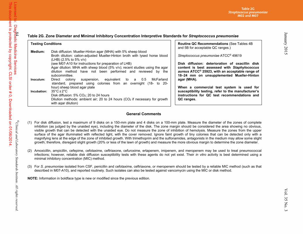
Table 2G. Zone Diameter and Minimal Inhibitory Concentration Interpretive Standards for Streptococcus pneumoniae
General Comments (1) For disk diffusion, test a maximum of 9 disks on a 150-mm plate and 4 disks on a 100-mm plate. Measure the diameter of the zones of complete
inhibition (as judged by the unaided eye), including the diameter of the disk. The zone margin should be considered the area showing no obvious, visible growth that can be detected with the unaided eye. Do not measure the zone of inhibition of hemolysis. Measure the zones from the upper surface of the agar illuminated with reflected light, with the cover removed. Ignore faint growth of tiny colonies that can be detected only with a magnifying lens at the edge of the zone of inhibited growth. With trimethoprim and the sulfonamides, antagonists in the medium may allow some slight growth; therefore, disregard slight growth (20% or less of the lawn of growth) and measure the more obvious margin to determine the zone diameter.
(2) Amoxicillin, ampicillin, cefepime, cefotaxime, ceftriaxone, cefuroxime, ertapenem, imipenem, and meropenem may be used to treat pneumococcal
infections; however, reliable disk diffusion susceptibility tests with these agents do not yet exist. Their in vitro activity is best determined using a minimal inhibitory concentration (MIC) method.
(3) For S. pneumoniae isolated from CSF, penicillin and cefotaxime, ceftriaxone, or meropenem should be tested by a reliable MIC method (such as that
described in M07-A10), and reported routinely. Such isolates can also be tested against vancomycin using the MIC or disk method. NOTE: Information in boldface type is new or modified since the previous edition.
Testing Conditions
Medium: Disk diffusion: Mueller-Hinton agar (MHA) with 5% sheep blood Broth dilution: cation-adjusted Mueller-Hinton broth with lysed horse blood (LHB) (2.5% to 5% v/v) (see M07-A10 for instructions for preparation of LHB) Agar dilution: MHA with sheep blood (5% v/v); recent studies using the agar dilution method have not been performed and reviewed by the subcommittee.
Inoculum: Direct colony suspension, equivalent to a 0.5 McFarland standard, prepared using colonies from an overnight (18- to 20- hour) sheep blood agar plate
Incubation: 35°C ± 2°C Disk diffusion: 5% CO2; 20 to 24 hours Dilution methods: ambient air; 20 to 24 hours (CO2 if necessary for growth
with agar dilution)
Routine QC Recommendations (See Tables 4B and 5B for acceptable QC ranges.) Streptococcus pneumoniae ATCC® 49619 Disk diffusion: deterioration of oxacillin disk content is best assessed with Staphylococcus aureus ATCC® 25923, with an acceptable range of 18–24 mm on unsupplemented Mueller-Hinton agar (MHA). When a commercial test system is used for susceptibility testing, refer to the manufacturer’s instructions for QC test recommendations and QC ranges.
84 ©Clinical and Laboratory Standards Institute. All rights reserved.
January 2015
Vol. 35 N
o. 3
Table 2GStreptococcus pneumoniae
M02 and M07
Licensed�to:��Diagnostic�M
edicine�Services
This�document�is�protected�by�copyright.�C
LSI�order�#�0,�D
ownloaded�on�01/06/2014.
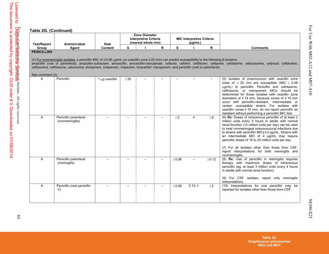
©Clinical and Laboratory Standards Institute. All rights reserved. 85
For Use W
ith M02-A
12 and M07-A
10 M100-S25
Table 2G. (Continued)
Test/Report Group
Antimicrobial Agent
Disk Content
Zone Diameter Interpretive Criteria (nearest whole mm)
MIC Interpretive Criteria (µg/mL)
Comments S I R S I R PENICILLINS (4) For nonmeningitis isolates, a penicillin MIC of ≤ 0.06 g/mL (or oxacillin zone ≥ 20 mm) can predict susceptibility to the following β-lactams: ampicillin (oral or parenteral), ampicillin-sulbactam, amoxicillin, amoxicillin-clavulanate, cefaclor, cefdinir, cefditoren, cefepime, cefotaxime, cefpodoxime, cefprozil, ceftaroline, ceftizoxime, ceftriaxone, cefuroxime, doripenem, ertapenem, imipenem, loracarbef, meropenem, and penicillin (oral or parenteral). See comment (3).
A Penicillin 1 g oxacillin 20 – – – – – (5) Isolates of pneumococci with oxacillin zone sizes of 20 mm are susceptible (MIC 0.06 g/mL) to penicillin. Penicillin and cefotaxime, ceftriaxone, or meropenem MICs should be determined for those isolates with oxacillin zone diameters of ≤ 19 mm, because zones of ≤ 19 mm occur with penicillin-resistant, -intermediate, or certain -susceptible strains. For isolates with oxacillin zones ≤ 19 mm, do not report penicillin as resistant without performing a penicillin MIC test.
A Penicillin parenteral (nonmeningitis)
– – – – 2 4 8 (6) Rx: Doses of intravenous penicillin of at least 2 million units every 4 hours in adults with normal renal function (12 million units per day) can be used to treat nonmeningeal pneumococcal infections due to strains with penicillin MICs ≤ 2 µg/mL. Strains with an intermediate MIC of 4 µg/mL may require penicillin doses of 18 to 24 million units per day. (7) For all isolates other than those from CSF, report interpretations for both meningitis and nonmeningitis.
A Penicillin parenteral (meningitis)
– – – – 0.06 – 0.12 (8) Rx: Use of penicillin in meningitis requires therapy with maximum doses of intravenous penicillin (eg, at least 3 million units every 4 hours in adults with normal renal function). (9) For CSF isolates, report only meningitis interpretations.
A Penicillin (oral penicillin V)
– – – – 0.06 0.12–1 2 (10) Interpretations for oral penicillin may be reported for isolates other than those from CSF.
Table 2GStreptococcus pneumoniae
M02 and M07
Licensed�to:��Diagnostic�M
edicine�Services
This�document�is�protected�by�copyright.�C
LSI�order�#�0,�D
ownloaded�on�01/06/2014.

Table 2G. (Continued)
Test/Report
Group Antimicrobial
Agent Disk
Content
Zone Diameter Interpretive Criteria (nearest whole mm)
MIC Interpretive Criteria (µg/mL)
Comments S I R S I R PENICILLINS (Continued)
C
C
Amoxicillin (nonmeningitis) Amoxicillin-clavulanate (nonmeningitis)
– – – – 2
2/1
4
4/2
8
8/4
CEPHEMS (PARENTERAL) (Including cephalosporins I, II, III, and IV. Please refer to Glossary I.) See comment (4).
O Cefepime (meningitis) – – – – 0.5 1 2 (11) In the United States, for CSF isolates, report only nonmeningitis interpretations. There is not an FDA-approved indication for the use of cefepime for meningitis in the United States.
B Cefepime (nonmeningitis) – – – – 1 2 4 (12) In the United States, only report interpretations for nonmeningitis and include the nonmeningitis notation on the report.
B B
Cefotaxime (meningitis) Ceftriaxone (meningitis)
– –
– –
– –
– –
0.5 0.5
1 1
2 2
(13) For CSF isolates, report only meningitis interpretations. (14) Rx: Use of cefotaxime or ceftriaxone in meningitis requires therapy with maximum doses. See comment (3).
B B
Cefotaxime (nonmeningitis) Ceftriaxone (nonmeningitis)
– –
– –
– –
– –
1 1
2 2
4 4
(15) For all isolates other than those from CSF, report interpretations for both meningitis and nonmeningitis.
C Ceftaroline (nonmeningitis) 30 g ≥ 26 – – ≤ 0.5 – – (16) Interpretive criteria are based on a dosage regimen of 600 mg every 12 h.
C Cefuroxime (parenteral) – – – – 0.5 1 2 CEPHEMS (ORAL) See comment (4).
C Cefuroxime (oral) – – – – 1 2 4 O Cefaclor – – – – 1 2 4 O Cefdinir – – – – 0.5 1 2 O Cefpodoxime – – – – 0.5 1 2 O Cefprozil – – – – 2 4 8 O Loracarbef – – – – 2 4 8
86 ©Clinical and Laboratory Standards Institute. All rights reserved.
January 2015
Vol. 35 N
o. 3
Table 2GStreptococcus pneumoniae
M02 and M07
Licensed�to:��Diagnostic�M
edicine�Services
This�document�is�protected�by�copyright.�C
LSI�order�#�0,�D
ownloaded�on�01/06/2014.

©Clinical and Laboratory Standards Institute. All rights reserved. 87
Table 2G. (Continued)
Test/Report
Group Antimicrobial
Agent Disk
Content
Zone Diameter Interpretive Criteria (nearest whole mm)
MIC Interpretive Criteria (µg/mL)
Comments S I R S I R CARBAPENEMS See comment (4).
B Meropenem – – – – 0.25 0.5 1 See comments (3) and (5). C Ertapenem – – – – 1 2 4 C Imipenem – – – – 0.12 0.25–0.5 1 O Doripenem – – – – 1 – –
GLYCOPEPTIDES B Vancomycin 30 g 17 – – 1 – – See comment (3).
MACROLIDES (17) Susceptibility and resistance to azithromycin, clarithromycin, and dirithromycin can be predicted by testing erythromycin. (18) Not routinely reported for organisms isolated from the urinary tract.
A Erythromycin 15 g 21 16–20 15 0.25 0.5 1 B Telithromycin 15 g 19 16–18 15 1 2 4 O Azithromycin 15 g 18 14–17 13 0.5 1 2 O Clarithromycin 15 g 21 17–20 16 0.25 0.5 1 O Dirithromycin 15 g 18 14–17 13 0.5 1 2
TETRACYCLINES (19) Organisms that are susceptible to tetracycline are also considered susceptible to doxycycline and minocycline.
B B
Tetracycline Doxycycline
30 g 30 g
28 28
25–27 25–27
24 24
1 0.25
2 0.5
4 1
FLUOROQUINOLONES B B B B
Gemifloxacin Levofloxacin Moxifloxacin Ofloxacin
5 g 5 g 5 g 5 g
23 17 18 16
20–22 14–16 15–17 13–15
19 13 14 12
0.12 2 1 2
0.25 4 2 4
0.5 8 4 8
(20) S. pneumoniae isolates susceptible to levofloxacin are predictably susceptible to gemifloxacin and moxifloxacin. However, S. pneumoniae susceptible to gemifloxacin or moxifloxacin cannot be assumed to be susceptible to levofloxacin.
O Gatifloxacin 5 g 21 18–20 17 1 2 4 O Sparfloxacin 5 g 19 16–18 15 0.5 1 2
FOLATE PATHWAY INHIBITORS A Trimethoprim-
sulfamethoxazole 1.25/
23.75 g 19 16–18 15
0.5/9.5 1/19–2/38
4/76
PHENICOLS C Chloramphenicol 30 g 21 – 20 4 – 8 See comment (18).
ANSAMYCINS C Rifampin 5 g 19 17–18 16 1 2 4 (21) Rx: Rifampin should not be used alone for
antimicrobial therapy.
For Use W
ith M02-A
12 and M07-A
10 M100-S25
Table 2GStreptococcus pneumoniae
M02 and M07
Licensed�to:��Diagnostic�M
edicine�Services
This�document�is�protected�by�copyright.�C
LSI�order�#�0,�D
ownloaded�on�01/06/2014.

Table 2G. (Continued)
Abbreviations: ATCC®, American Type Culture Collection; CSF, cerebrospinal fluid; FDA, US Food and Drug Administration; I, intermediate; MIC, minimal inhibitory concentration; R, resistant; S, susceptible.
Test/Report Group
Antimicrobial Agent
Disk Content
Zone Diameter Interpretive Criteria (nearest whole mm)
MIC Interpretive Criteria (µg/mL)
Comments S I R S I R LINCOSAMIDES
B Clindamycin 2 g 19 16–18 15 0.25 0.5 1 (22) Inducible clindamycin resistance can be detected by disk diffusion using the D-zone test or by broth microdilution using the single-well test (containing both erythromycin and clindamycin) (see Table 3G, Subchapter 3.10.1 in M02-A12, and Subchapter 3.14.1 in M07-A10). See comment (18).
STREPTOGRAMINS O Quinupristin-dalfopristin 15 g 19 16–18 15 1 2 4
OXAZOLIDINONES C Linezolid 30 g 21 – – 2 – –
88 ©Clinical and Laboratory Standards Institute. All rights reserved.
January 2015
Vol. 35 N
o. 3
Table 2GStreptococcus pneumoniae
M02 and M07
Licensed�to:��Diagnostic�M
edicine�Services
This�document�is�protected�by�copyright.�C
LSI�order�#�0,�D
ownloaded�on�01/06/2014.

For Use With M02-A12 and M07-A10 M100-S25
Clinical and Laboratory Standards Institute. All rights reserved. 89
This page is intentionally left blank.
Licensed�to:��Diagnostic�Medicine�ServicesThis�document�is�protected�by�copyright.�CLSI�order�#�0,�Downloaded�on�01/06/2014.

Table 2H-1. Zone Diameter and Minimal Inhibitory Concentration Interpretive Standards for Streptococcus spp. β-Hemolytic Group
Refer to Table 3G for additional testing recommendations, reporting suggestions, and QC.
General Comments
(1) For disk diffusion, test a maximum of 9 disks on a 150-mm plate and 4 disks on a 100-mm plate. Measure the diameter of the zones of complete inhibition
(as judged by the unaided eye), including the diameter of the disk. The zone margin should be considered the area showing no obvious, visible growth that can be detected with the unaided eye. Do not measure the zone of inhibition of hemolysis. Measure the zones from the upper surface of the agar illuminated with reflected light, with the cover removed. Ignore faint growth of tiny colonies that can be detected only with a magnifying lens at the edge of the zone of inhibited growth.
(2) For this table, the β-hemolytic group includes the large colony–forming pyogenic strains of streptococci with Group A (Streptococcus pyogenes), C, or G
antigens and strains with Group B (S. agalactiae) antigen. Small colony–forming β-hemolytic strains with Group A, C, F, or G antigens (S. anginosus group, previously termed “S. milleri”) are considered part of the viridans group, and interpretive criteria for the viridans group should be used (see Table 2H-2).
(3) Penicillin and ampicillin are drugs of choice for treatment of β-hemolytic streptococcal infections. Susceptibility testing of penicillins and other -lactams
approved by the US Food and Drug Administration for treatment of β-hemolytic streptococcal infections need not be performed routinely, because nonsusceptible isolates (ie, penicillin minimal inhibitory concentration [MICs] > 0.12 and ampicillin MICs > 0.25 µg/mL) are extremely rare in any β-hemolytic streptococcus and have not been reported for S. pyogenes. If testing is performed, any β-hemolytic streptococcal isolate found to be nonsusceptible should be re-identified, retested, and, if confirmed, submitted to a public health laboratory. (See Appendix A for further instructions.)
(4) Interpretive criteria for Streptococcus spp. β-hemolytic group are proposed based on population distributions of various species, pharmacokinetics of the
antimicrobial agents, previously published literature, and the clinical experience of members of the subcommittee. Systematically collected clinical data were not available for review with many of the antimicrobial agents in this table.
Testing Conditions Medium: Disk diffusion: Mueller-Hinton agar (MHA) with 5% sheep blood Broth dilution: cation-adjusted Mueller-Hinton broth (CAMHB) with lysed horse blood
(LHB) (2.5% to 5% v/v); the CAMHB should be supplemented to 50 µg/mL calcium for daptomycin (see M07-A10 for instructions for preparation of LHB)
Agar dilution: MHA with sheep blood (5% v/v); recent studies using the agar dilution method have not been performed and reviewed by the subcommittee.
Inoculum: Direct colony suspension, equivalent to a 0.5 McFarland standard, using colonies from an overnight (18- to 20-hour) sheep blood agar plate
Incubation: 35°C 2°C Disk diffusion: 5% CO2; 20 to 24 hours Dilution methods: ambient air; 20 to 24 hours (CO2 if necessary for growth with agar
dilution)
Routine QC Recommendations (See Tables 4B and 5B for acceptable QC ranges.) Streptococcus pneumoniae ATCC® 49619 When a commercial test system is used for susceptibility testing, refer to the manufacturer’s instructions for QC test recommendations and QC ranges.
90 ©Clinical and Laboratory Standards Institute. All rights reserved.
January 2015
Vol. 35 N
o. 3
Table 2H-1Streptococcus spp. β-Hemolytic Group
M02 and M07
Licensed�to:��Diagnostic�M
edicine�Services
This�document�is�protected�by�copyright.�C
LSI�order�#�0,�D
ownloaded�on�01/06/2014.

©Clinical and Laboratory Standards Institute. All rights reserved. 91
Table 2H-1. (Continued) NOTE: Information in boldface type is new or modified since the previous edition.
Test/Report Group
Antimicrobial Agent
Disk Content
Zone Diameter Interpretive Criteria (nearest whole mm)
MIC Interpretive Criteria (µg/mL)
Comments S I R S I R PENICILLINS (5) For the following organism groups, an organism that is susceptible to penicillin can be considered susceptible to the listed antimicrobial agents when used for approved indications. For β-hemolytic streptococci (Groups A, B, C, G): ampicillin, amoxicillin, amoxicillin-clavulanate, ampicillin-sulbactam, cefazolin, cefepime, ceftaroline, cephradine, cephalothin, cefotaxime, ceftriaxone, ceftizoxime, imipenem, ertapenem, and meropenem. In addition, for β-hemolytic streptococci Group A: cefaclor, cefdinir, cefprozil, ceftibuten, cefuroxime, cefpodoxime, and cephapirin.
A A
Penicillin or ampicillin
10 units 10 g
24 24
– –
– –
0.12 0.25
– –
– –
See comment (3).
CEPHEMS (PARENTERAL) (Including cephalosporins I, II, III, and IV. Please refer to Glossary I.) See comment (5).
B B B
Cefepime or cefotaxime or ceftriaxone
30 g 30 g 30 g
24 24 24
– – –
– – –
0.5 0.5 0.5
– – –
– – –
C Ceftaroline 30 g ≥ 26 – – 0.5 – – (6) Interpretive criteria are based on a dosage regimen of 600 mg every 12 h.
CARBAPENEMS See comment (5).
O Doripenem – – – – 0.12 – – O Ertapenem – – – – 1 – – O Meropenem – – – – 0.5 – –
GLYCOPEPTIDES B Vancomycin 30 g 17 – – 1 – –
LIPOPEPTIDES C Daptomycin – – – – 1 – – (7) Daptomycin should not be reported for isolates
from the respiratory tract.
For U
se With M
02-A12 and M
07-A10 M
100-S25
Table 2H-1Streptococcus spp. β-Hemolytic Group
M02 and M07
Licensed�to:��Diagnostic�M
edicine�Services
This�document�is�protected�by�copyright.�C
LSI�order�#�0,�D
ownloaded�on�01/06/2014.

Table 2H-1. (Continued)
Test/Report Group
Antimicrobial Agent
Disk Content
Zone Diameter Interpretive Criteria (nearest whole mm)
MIC Interpretive Criteria (µg/mL)
Comments S I R S I R MACROLIDES (8) Susceptibility and resistance to azithromycin, clarithromycin, and dirithromycin can be predicted by testing erythromycin. (9) Not routinely reported on isolates from the urinary tract.
A Erythromycin 15 g 21 16–20 15 0.25 0.5 1 (10) Rx: Recommendations for intrapartum prophylaxis for Group B streptococci are penicillin or ampicillin. Although cefazolin is recommended for penicillin-allergic women at low risk for anaphylaxis, those at high risk for anaphylaxis may receive clindamycin. Group B streptococci are susceptible to ampicillin, penicillin, and cefazolin, but may be resistant to erythromycin and clindamycin. When a Group B Streptococcus is isolated from a pregnant woman with severe penicillin allergy (high risk for anaphylaxis), erythromycin and clindamycin (including inducible clindamycin resistance) should be tested, and only clindamycin should be reported. See Table 3G.
O Azithromycin 15 g 18 14–17 13 0.5 1 2 O Clarithromycin 15 g 21 17–20 16 0.25 0.5 1 O Dirithromycin 15 g 18 14–17 13 0.5 1 2
TETRACYCLINES (11) Organisms that are susceptible to tetracycline are also considered susceptible to doxycycline and minocycline.
O Tetracycline 30 g 23 19–22 18 2 4 8 FLUOROQUINOLONES
C Levofloxacin 5 g 17 14–16 13 2 4 8 C Ofloxacin 5 g 16 13–15 12 2 4 8 O Gatifloxacin 5 g 21 18–20 17 1 2 4 O Grepafloxacin 5 g 19 16–18 15 0.5 1 2 O Trovafloxacin 10 g 19 16–18 15 1 2 4
PHENICOLS C Chloramphenicol 30 g 21 18–20 17 4 8 16 See comment (9).
LINCOSAMIDES A Clindamycin 2 g 19 16–18 15 0.25 0.5 1 See comments (9) and (10).
(12) Inducible clindamycin resistance can be detected by disk diffusion using the D-zone test and broth microdilution. See Table 3G, Subchapter 3.10.1 in M02-A12, and Subchapter 3.14.1 in M07-A10.
92 ©Clinical and Laboratory Standards Institute. All rights reserved.
January 2015
Vol. 35 N
o. 3
Table 2H-1Streptococcus spp. β-Hemolytic Group
M02 and M07
Licensed�to:��Diagnostic�M
edicine�Services
This�document�is�protected�by�copyright.�C
LSI�order�#�0,�D
ownloaded�on�01/06/2014.
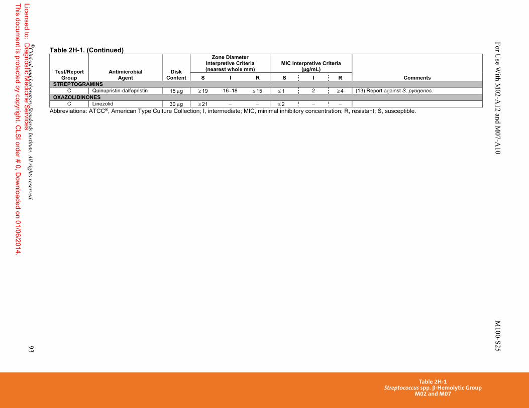
©Clinical and Laboratory Standards Institute. All rights reserved. 93
Table 2H-1. (Continued)
Abbreviations: ATCC®, American Type Culture Collection; I, intermediate; MIC, minimal inhibitory concentration; R, resistant; S, susceptible.
Test/Report Group
Antimicrobial Agent
Disk Content
Zone Diameter Interpretive Criteria (nearest whole mm)
MIC Interpretive Criteria (µg/mL)
Comments S I R S I R STREPTOGRAMINS
C Quinupristin-dalfopristin 15 g 19 16–18 15 1 2 4 (13) Report against S. pyogenes. OXAZOLIDINONES
C Linezolid 30 g 21 – – 2 – –
For Use W
ith M02-A
12 and M07-A
10 M100-S25
Table 2H-1Streptococcus spp. β-Hemolytic Group
M02 and M07
Licensed�to:��Diagnostic�M
edicine�Services
This�document�is�protected�by�copyright.�C
LSI�order�#�0,�D
ownloaded�on�01/06/2014.

Table 2H-2. Zone Diameter and Minimal Inhibitory Concentration Interpretive Standards for Streptococcus spp. Viridans Group
General Comments (1) For disk diffusion, measure the diameter of the zones of complete inhibition (as judged by the unaided eye), including the diameter of the disk. The zone
margin should be considered the area showing no obvious, visible growth that can be detected with the unaided eye. Do not measure the zone of inhibition of hemolysis. Measure the zones from the upper surface of the agar illuminated with reflected light, with the cover removed. Ignore faint growth of tiny colonies that can be detected only with a magnifying lens at the edge of the zone of inhibited growth.
(2) The viridans group of streptococci includes the following five groups, with several species within each group: mutans group, salivarius group, bovis group,
anginosus group (previously “S. milleri” group), and mitis group. The anginosus group includes small colony–forming β-hemolytic strains with Groups A, C, F, and G antigens. For detailed information on the species within the groups, please refer to recent clinical microbiology literature.
(3) Interpretive criteria for Streptococcus spp. viridans group are proposed based on population distributions of various species, pharmacokinetics of the
antimicrobial agents, previously published literature, and the clinical experience of members of the subcommittee. Systematically collected clinical data were not available for review with many of the antimicrobial agents in this table.
NOTE: Information in boldface type is new or modified since the previous edition.
Testing Conditions Medium: Disk diffusion: Mueller-Hinton agar (MHA) with 5% sheep blood Broth dilution: cation-adjusted Mueller-Hinton broth (CAMHB) with lysed horse blood (LHB)
(2.5% to 5% v/v); the CAMHB should be supplemented to 50 µg/mL calcium for daptomycin (see M07-A10 for instructions for preparation of LHB)
Agar dilution: MHA with sheep blood (5% v/v); recent studies using the agar dilution method have not been performed and reviewed by the subcommittee.
Inoculum: Direct colony suspension, equivalent to a 0.5 McFarland standard using colonies from an overnight (18- to 20-hour) sheep blood agar plate
Incubation: 35°C 2°C Disk diffusion: 5% CO2; 20 to 24 hours Dilution methods: ambient air; 20 to 24 hours (CO2 if necessary for growth with agar dilution)
Routine QC Recommendations (See Tables 4B and 5B for acceptable QC ranges.) Streptococcus pneumoniae ATCC® 49619 When a commercial test system is used for susceptibility testing, refer to the manufacturer’s instructions for QC test recommendations and QC ranges.
94 ©Clinical and Laboratory Standards Institute. All rights reserved.
January 2015
Vol. 35 N
o. 3
Table 2H-2Streptococcus spp. Viridans Group
M02 and M07
Licensed�to:��Diagnostic�M
edicine�Services
This�document�is�protected�by�copyright.�C
LSI�order�#�0,�D
ownloaded�on�01/06/2014.

©Clinical and Laboratory Standards Institute. All rights reserved. 95
For Use W
ith M02-A
12 and M07-A
10 M100-S25
Table 2H-2. (Continued)
Test/Report Group
Antimicrobial Agent
Disk Content
Zone Diameter Interpretive Criteria (nearest whole mm)
MIC Interpretive Criteria (µg/mL)
Comments S I R S I R PENICILLINS
A A
Penicillin Ampicillin
– – – – 0.12 0.25
0.25–2 0.5–4
4 8
(4) Viridans streptococci isolated from normally sterile body sites (eg, CSF, blood, bone) should be tested for penicillin susceptibility using an MIC method. (5) Rx: Penicillin- or ampicillin-intermediate isolates may require combined therapy with an aminoglycoside for bactericidal action.
CEPHEMS (PARENTERAL) (Including cephalosporins I, II, III, and IV. Please refer to Glossary I.) B B B
Cefepime Cefotaxime Ceftriaxone
30 g 30 g 30 g
24 28 27
22–23 26–27 25–26
21 25 24
1 1 1
2 2 2
4 4 4
CARBAPENEMS O Doripenem – – – – 1 – – O Ertapenem – – – – 1 – – O Meropenem – – – – 0.5 – –
GLYCOPEPTIDES B Vancomycin 30 g 17 – – 1 – –
LIPOPEPTIDES O Daptomycin – – – – 1 – – (6) Daptomycin should not be reported for
isolates from the respiratory tract.
MACROLIDES (7) Susceptibility and resistance to azithromycin, clarithromycin, and dirithromycin can be predicted by testing erythromycin. (8) Not routinely reported on isolates from the urinary tract.
C Erythromycin 15 g 21 16–20 15 0.25 0.5 1 O Azithromycin 15 g 18 14–17 13 0.5 1 2 O Clarithromycin 15 g 21 17–20 16 0.25 0.5 1 O Dirithromycin 15 g 18 14–17 13 0.5 1 2
TETRACYCLINES (9) Organisms that are susceptible to tetracycline are also considered susceptible to doxycycline and minocycline.
O Tetracycline 30 g 23 19–22 18 2 4 8
Table 2H-2Streptococcus spp. Viridans Group
M02 and M07
Licensed�to:��Diagnostic�M
edicine�Services
This�document�is�protected�by�copyright.�C
LSI�order�#�0,�D
ownloaded�on�01/06/2014.

Table 2H-2. (Continued)
Abbreviations: ATCC®, American Type Culture Collection; CSF, cerebrospinal fluid; I, intermediate; MIC, minimal inhibitory concentration; R, resistant; S, susceptible.
Test/Report Group
Antimicrobial Agent
Disk Content
Zone Diameter Interpretive Criteria (nearest whole mm)
MIC Interpretive Criteria (µg/mL)
Comments S I R S I R FLUOROQUINOLONES
O Levofloxacin 5 g 17 14–16 13 2 4 8 O Ofloxacin 5 g 16 13–15 12 2 4 8 O Gatifloxacin 5 g 21 18–20 17 1 2 4 O Grepafloxacin 5 g 19 16–18 15 0.5 1 2 O Trovafloxacin 10 g 19 16–18 15 1 2 4
PHENICOLS C Chloramphenicol 30 g 21 18–20 17 4 8 16 See comment (8).
LINCOSAMIDES C Clindamycin 2 g 19 16–18 15 0.25 0.5 1 See comment (8).
STREPTOGRAMINS O Quinupristin-dalfopristin 15 g 19 16–18 15 1 2 4
OXAZOLIDINONES C Linezolid 30 g 21 – – 2 – –
96 ©Clinical and Laboratory Standards Institute. All rights reserved.
January 2015
Vol. 35 N
o. 3
Table 2H-2Streptococcus spp. Viridans Group
M02 and M07
Licensed�to:��Diagnostic�M
edicine�Services
This�document�is�protected�by�copyright.�C
LSI�order�#�0,�D
ownloaded�on�01/06/2014.

For Use With M02-A12 and M07-A10 M100-S25
Clinical and Laboratory Standards Institute. All rights reserved. 97
This page is intentionally left blank.
Licensed�to:��Diagnostic�Medicine�ServicesThis�document�is�protected�by�copyright.�CLSI�order�#�0,�Downloaded�on�01/06/2014.

Table 2I. Zone Diameter and Minimal Inhibitory Concentration Interpretive Standards for Neisseria meningitidis
General Comments
Important: For complete information on safety precautions, see Biosafety in Microbiological and Biomedical Laboratories. 5th ed. Washington, DC: US Department of Health and Human Services; 2009. http://www.cdc.gov/biosafety/publications/bmbl5/. (1) Recommended precautions: Perform all antimicrobial susceptibility testing (AST) of N. meningitidis in a biological safety cabinet (BSC). Manipulating N.
meningitidis outside a BSC is associated with increased risk for contracting meningococcal disease. Laboratory-acquired meningococcal disease is associated with a case fatality rate of 50%. Exposure to droplets or aerosols of N. meningitidis is the most likely risk for laboratory-acquired infection. Rigorous protection from droplets or aerosols is mandated when microbiological procedures (including AST) are performed on all N. meningitidis isolates.
(2) If a BSC is unavailable, manipulation of these isolates should be minimized, limited to Gram staining or serogroup identification using phenolized saline
solution, while wearing a laboratory coat and gloves and working behind a full face splash shield. Use Biosafety Level 3 (BSL-3) practices, procedures, and containment equipment for activities with a high potential for droplet or aerosol production and for activities involving production quantities or high concentrations of infectious materials. If Biosafety Level 2 (BSL-2) or BSL-3 facilities are not available, forward isolates to a reference or public health laboratory with a minimum of BSL-2 facilities.
(3) Laboratorians who are exposed routinely to potential aerosols of N. meningitidis should consider vaccination according to the current recommendations of the
Centers for Disease Control and Prevention Advisory Committee on Immunization Practices (http://www.cdc.gov/vaccines/acip/index.html). Vaccination will decrease but not eliminate the risk of infection, because it is less than 100% effective and does not provide protection against serogroup B, a frequent cause of laboratory-acquired cases.
Testing Conditions Medium: Disk diffusion: Mueller-Hinton agar (MHA) with 5% sheep blood Broth microdilution: cation-adjusted Mueller-Hinton broth
supplemented with lysed horse blood (LHB) (2.5% to 5% v/v) (see M07-A10 for preparation of LHB)
Agar dilution: MHA supplemented with sheep blood (5% v/v) Inoculum: Direct colony suspension from 20 to 24 hours growth from chocolate
agar incubated at 35°C; 5% CO2; equivalent to a 0.5 McFarland standard. Colonies grown on sheep blood agar may be used for inoculum preparation. However, the 0.5 McFarland suspension obtained from sheep blood agar will contain approximately 50% fewer CFU/mL. This must be taken into account when preparing the final dilution before panel inoculation, as guided by colony counts.
Incubation: 35°C 2°C; 5% CO2; 20 to 24 hours
Routine QC Recommendations (See Tables 4A, 4B, 5A, and 5B for acceptable QC ranges.) Streptococcus pneumoniae ATCC® 49619:
Disk diffusion: incubate in 5% CO2. Broth microdilution: incubate in ambient air or CO2 (except azithromycin QC tests that must be incubated in ambient air).
E. coli ATCC® 25922
Disk diffusion, broth microdilution or agar dilution for ciprofloxacin, nalidixic acid, minocycline, and sulfisoxazole: incubate in ambient air or CO2.
When a commercial test system is used for susceptibility testing, refer to the manufacturer’s instructions for QC test recommendations and QC ranges.
98 ©Clinical and Laboratory Standards Institute. All rights reserved.
January 2015
Vol. 35 N
o. 3
Table 2INeisseria meningitidis
M02 and M07
Licensed�to:��Diagnostic�M
edicine�Services
This�document�is�protected�by�copyright.�C
LSI�order�#�0,�D
ownloaded�on�01/06/2014.

©Clinical and Laboratory Standards Institute. All rights reserved. 99
Table 2I. (Continued)
(4) For disk diffusion, test a maximum of 5 disks on a 150-mm plate and 2 disks on a 100-mm plate. Measure the diameter of the zones of complete inhibition (as judged by the unaided eye), including the diameter of the disk. Measure the zones from the upper surface of the agar illuminated with reflected light, with the cover removed. Ignore faint growth of tiny colonies that can be detected only with a magnifying lens at the edge of the zone of inhibited growth. With trimethoprim and the sulfonamides, antagonists in the medium may allow some slight growth; therefore, disregard slight growth (20% or less of the lawn of growth) and measure the more obvious margin to determine the zone diameter.
(5) Interpretive criteria are based on population distributions of minimal inhibitory concentrations (MICs) of various agents, pharmacokinetics of the agents,
previously published literature, and the clinical experience of members of the subcommittee. Systematically collected clinical data were not available to review with many of the antimicrobial agents in this table.
(6) With azithromycin, interpretive criteria were developed initially using MICs determined by incubation in ambient air for the pharmacodynamic calculations. NOTE: Information in boldface type is new or modified since the previous edition.
Test/Report Group
Antimicrobial Agent
Disk Content
Zone Diameter Interpretive Criteria (nearest whole mm)
MIC Interpretive Criteria (µg/mL)
Comments S I R S I R PENICILLINS
C C
Penicillin Ampicillin
– –
– –
– –
0.06 0.12
0.12–0.25 0.25–1
0.5 2
CEPHEMS
C Cefotaxime or 30 g 34 – – 0.12 – – C ceftriaxone 30 g 34 – – 0.12 – –
CARBAPENEMS C Meropenem 10 g 30 – – 0.25 – –
MACROLIDES C Azithromycin 15 g 20 – – 2 – – See comment (6).
(7) May be appropriate only for prophylaxis of meningococcal case contacts. These interpretive criteria do not apply to therapy of patients with invasive meningococcal disease.
For Use W
ith M02-A
12 and M07-A
10 M100-S25
Table 2INeisseria meningitidis
M02 and M07
Licensed�to:��Diagnostic�M
edicine�Services
This�document�is�protected�by�copyright.�C
LSI�order�#�0,�D
ownloaded�on�01/06/2014.

Table 2I. (Continued)
Abbreviations: ATCC®, American Type Culture Collection; CFU, colony-forming unit(s); MIC, minimal inhibitory concentration; R, resistant; S, susceptible.
Test/Report Group
Antimicrobial Agent
Disk Content
Zone Diameter Interpretive Criteria (nearest whole mm)
MIC Interpretive Criteria (µg/mL)
Comments S I R S I R TETRACYCLINES
C Minocycline 30 g 26 – – 2 – – See comment (7). FLUOROQUINOLONES (8) For surveillance purposes, a nalidixic acid MIC 8 g/mL or a zone ≤ 25 mm may correlate with diminished fluoroquinolone susceptibility.
C Ciprofloxacin 5 g 35 33–34 32 0.03 0.06 0.12 See comment (7).
C Levofloxacin – – – – 0.03 0.06 0.12 FOLATE PATHWAY INHIBITORS
C Sulfisoxazole – – – – 2 4 8 See comment (7). C Trimethoprim-
sulfamethoxazole 1.25/
23.75 g 30 26–29 25 0.12/
2.4 0.25/4.75 0.5/
9.5 (9) Trimethoprim-sulfamethoxazole is the preferred disk for detection of sulfonamide resistance. Trimethoprim-sulfamethoxazole testing predicts susceptibility and resistance to trimethoprim-sulfamethoxazole and sulfonamides. Sulfonamides may be appropriate only for prophylaxis of meningococcal case contacts.
PHENICOLS C Chloramphenicol 30 g 26 20–25 19 2 4 8 (10) Not routinely reported on isolates from the
urinary tract. ANSAMYCINS
C Rifampin 5 g 25 20–24 19 0.5 1 2 See comment (7).
100 ©Clinical and Laboratory Standards Institute. All rights reserved.
January 2015
Vol. 35 N
o. 3
Table 2INeisseria meningitidis
M02 and M07
Licensed�to:��Diagnostic�M
edicine�Services
This�document�is�protected�by�copyright.�C
LSI�order�#�0,�D
ownloaded�on�01/06/2014.

For Use With M02-A12 and M07-A10 M100-S25
Clinical and Laboratory Standards Institute. All rights reserved. 101
This page is intentionally left blank.
Licensed�to:��Diagnostic�Medicine�ServicesThis�document�is�protected�by�copyright.�CLSI�order�#�0,�Downloaded�on�01/06/2014.

Table 2J-1. Minimal Inhibitory Concentration Interpretive Standards for Anaerobes
General Comments
(1) For isolates for which the antimicrobial agent minimal inhibitory concentrations (MICs) fall within the intermediate category, maximum dosages,
along with proper ancillary therapy, should be used to achieve the best possible levels of drug in abscesses and/or poorly perfused tissues. If this approach is taken, organisms for which the antimicrobial agent MICs fall within the susceptible range are generally amenable to therapy. Those organisms for which the antimicrobial agent MICs are in the intermediate range may respond, but in such cases efficacy as measured by patient clinical response should be carefully monitored. Ancillary therapy, such as drainage procedures and debridement, are of great importance for proper management of anaerobic infections.
(2) Refer to Figures 2 and 3 in CLSI document M11 for examples of reading end points. (3) MIC values using either Brucella blood agar or Wilkins Chalgren agar (former reference medium) are considered equivalent. (4) Broth microdilution is only recommended for testing the B. fragilis group. MIC values for agar or broth microdilution are considered equivalent for
that group. (5) Until further studies are performed to validate broth microdilution for testing other organisms, it should be used only for testing members of the B. fragilis
group. NOTE: Information in boldface type is new or modified since the previous edition.
Testing Conditions Medium: Agar dilution: (for all anaerobes): Brucella agar
supplemented with hemin (5 g/mL), Vitamin K1 (1 g/mL), and laked sheep blood (5% v/v)
Broth microdilution (for Bacteroides fragilis group only): Brucella broth supplemented with hemin (5 g/mL), Vitamin K1 (1 g/mL), and lysed horse blood (5% v/v)
Inoculum: Growth method or direct colony suspension, equivalent to 0.5 McFarland suspension; Agar: 105 CFU per spot
Broth: 106 CFU/mL Incubation: 36°C ± 1°C, anaerobically Broth microdilution: 46 to 48 hours
Agar dilution: 42 to 48 hours
Routine QC Recommendations (See Tables 5D and 5E for acceptable QC ranges.) Test one or more of the following organisms. The choice and number of QC strains tested should be based on obtaining on-scale end points for the antimicrobial agent tested. Bacteroides fragilis ATCC® 25285 Bacteroides thetaiotaomicron ATCC® 29741 Clostridium difficile ATCC® 700057 Eggerthella Ienta ATCC® 43055 (formerly Eubacterium lentum) When a commercial test system is used for susceptibility testing, refer to the manufacturer’s instructions for QC test recommendations and QC ranges.
102 ©Clinical and Laboratory Standards Institute. All rights reserved.
January 2015
Vol. 35 N
o. 3
Table 2J-1Anaerobes
M11
Licensed�to:��Diagnostic�M
edicine�Services
This�document�is�protected�by�copyright.�C
LSI�order�#�0,�D
ownloaded�on�01/06/2014.

For Use W
ith M11-A
8 M100-S25
©Clinical and Laboratory Standards Institute. All rights reserved. 103
Table 2J-1. (Continued)
Test/Report Group
Antimicrobial Agent
MIC Interpretive Criteria (µg/mL)
Comments S I R PENICILLINS
A/C A/C
Ampicillina
Penicillina
0.5 0.5
1 1
2 2
(6) Ampicillin and penicillin are recommended for primary testing for gram-positive organisms (Group A) because most of them are β-lactamase negative, but not for gram-negative organisms (Group C) because many are β-lactamase positive. (7) Members of the B. fragilis group are presumed to be resistant. Other gram-negative and gram-positive anaerobes may be screened for -lactamase activity with a chromogenic cephalosporin; if -lactamase positive, report as resistant to penicillin, ampicillin, and amoxicillin. Be aware that -lactamase-negative isolates may be resistant to -lactams by other mechanisms. Because higher blood levels are achievable with these antimicrobials, infection with non–-lactamase-producing organisms with higher MICs (2–4 µg/mL) with adequate dosage regimen might be treatable. (8) Results of ampicillin testing can be used to predict results for amoxicillin.
C Piperacillin 32 64 128 C Ticarcillin 32 64 128 C Mezlocillin 32 64 128
-LACTAM/-LACTAMASE INHIBITOR COMBINATIONS A Amoxicillin-clavulanate 4/2 8/4 16/8 A Ampicillin-sulbactam 8/4 16/8 32/16 A Piperacillin-tazobactam 32/4 64/4 128/4 A Ticarcillin-clavulanate 32/2 64/2 128/2
CEPHEMS (PARENTERAL) (Including cephalosporins I, II, III, and IV. Please refer to Glossary I.) C Cefotetan 16 32 64 C Cefoxitin 16 32 64 C Ceftizoxime 32 64 128 C Ceftriaxone 16 32 64 O Cefmetazole 16 32 64 O Cefoperazone 16 32 64 O Cefotaxime 16 32 64
CARBAPENEMS A Doripenem 2 4 8 A Ertapenem 4 8 16 A Imipenem 4 8 16 A Meropenem 4 8 16
TETRACYCLINES C Tetracycline 4 8 16
FLUOROQUINOLONES C Moxifloxacin 2 4 8
Table 2J-1Anaerobes
M11
Licensed�to:��Diagnostic�M
edicine�Services
This�document�is�protected�by�copyright.�C
LSI�order�#�0,�D
ownloaded�on�01/06/2014.

Table 2J-1. (Continued)
Abbreviations: ATCC®, American Type Culture Collection; CFU, colony-forming unit(s); I, intermediate; MIC, minimal inhibitory concentration; R, resistant; S, susceptible.
Footnote
a. A/C: Group A for gram-positive organisms and Group C for B. fragilis and other gram-negative organisms. Refer to Table 1C.
Test/Report Group
Antimicrobial Agent
MIC Interpretive Criteria (µg/mL)
Comments S I R LINCOSAMIDES
A Clindamycin 2 4 8 PHENICOLS
C Chloramphenicol 8 16 32 NITROIMIDAZOLES
A Metronidazole 8 16 32 (9) Many non–spore-forming, gram-positive anaerobic rods are resistant to metronidazole.
104 ©Clinical and Laboratory Standards Institute. All rights reserved.
January 2015
Vol. 35 N
o. 3
Table 2J-1Anaerobes
M11
Licensed�to:��Diagnostic�M
edicine�Services
This�document�is�protected�by�copyright.�C
LSI�order�#�0,�D
ownloaded�on�01/06/2014.

For Use With M11-A8 M100-S25
Clinical and Laboratory Standards Institute. All rights reserved. 105
This page is intentionally left blank.
Licensed�to:��Diagnostic�Medicine�ServicesThis�document�is�protected�by�copyright.�CLSI�order�#�0,�Downloaded�on�01/06/2014.
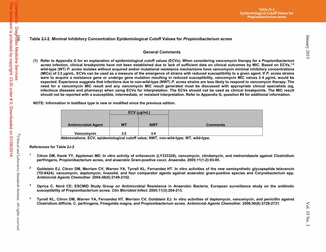
Table 2J-2. Minimal Inhibitory Concentration Epidemiological Cutoff Values for Propionibacterium acnes
General Comments
(1) Refer to Appendix G for an explanation of epidemiological cutoff values (ECVs). When considering vancomycin therapy for a Propionibacterium acnes infection, clinical breakpoints have not been established due to lack of sufficient data on clinical outcomes by MIC. Based on ECVs,1-4 wild-type (WT) P. acnes isolates without acquired and/or mutational resistance mechanisms have vancomycin minimal inhibitory concentrations (MICs) of ≤ 2 µg/mL. ECVs can be used as a measure of the emergence of strains with reduced susceptibility to a given agent. If P. acnes strains were to acquire a resistance gene or undergo gene mutation resulting in reduced susceptibility, vancomycin MIC values ≥ 4 µg/mL would be expected. Experience suggests that infections due to non-wild-type (NWT) P. acnes strains are less likely to respond to vancomycin therapy. The need for a vancomycin MIC result and any vancomycin MIC result generated must be discussed with appropriate clinical specialists (eg, infectious diseases and pharmacy) when using ECVs for interpretation. The ECVs should not be used as clinical breakpoints. The MIC result should not be reported with a susceptible, intermediate, or resistant interpretation. Refer to Appendix G, question #4 for additional information.
NOTE: Information in boldface type is new or modified since the previous edition.
Antimicrobial Agent
ECV (g/mL)
Comments WT NWT
Vancomycin ≤ 2 ≥ 4
Abbreviations: ECV, epidemiological cutoff value; NWT, non-wild-type; WT, wild-type. References for Table 2J-2 1 Citron DM, Kwok YY, Appleman MD. In vitro activity of oritavancin (LY333328), vancomycin, clindamycin, and metronidazole against Clostridium
perfringens, Propionibacterium acnes, and anaerobic Gram-positive cocci. Anaerobe. 2005;11(1-2):93-95.
2 Goldstein EJ, Citron DM, Merriam CV, Warren YA, Tyrrell KL, Fernandez HT. In vitro activities of the new semisynthetic glycopeptide telavancin (TD-6424), vancomycin, daptomycin, linezolid, and four comparator agents against anaerobic gram-positive species and Corynebacterium spp. Antimicrob Agents Chemother. 2004;48(6):2149-2152.
3 Oprica C, Nord CE; ESCMID Study Group on Antimicrobial Resistance in Anaerobic Bacteria. European surveillance study on the antibiotic susceptibility of Propionibacterium acnes. Clin Microbiol Infect. 2005;11(3):204-213.
4 Tyrrell KL, Citron DM, Warren YA, Fernandez HT, Merriam CV, Goldstein EJ. In vitro activities of daptomycin, vancomycin, and penicillin against Clostridium difficile, C. perfringens, Finegoldia magna, and Propionibacterium acnes. Antimicrob Agents Chemother. 2006;50(8):2728-2731.
106 ©Clinical and Laboratory Standards Institute. All rights reserved.
January 2015
Vol. 35 N
o. 3
Table 2J-2Epidemiological Cutoff Values for
Propionibacterium acnes
Licensed�to:��Diagnostic�M
edicine�Services
This�document�is�protected�by�copyright.�C
LSI�order�#�0,�D
ownloaded�on�01/06/2014.

For Use With M11-A8 M100-S25
Clinical and Laboratory Standards Institute. All rights reserved. 107
This page is intentionally left blank.
Licensed�to:��Diagnostic�Medicine�ServicesThis�document�is�protected�by�copyright.�CLSI�order�#�0,�Downloaded�on�01/06/2014.
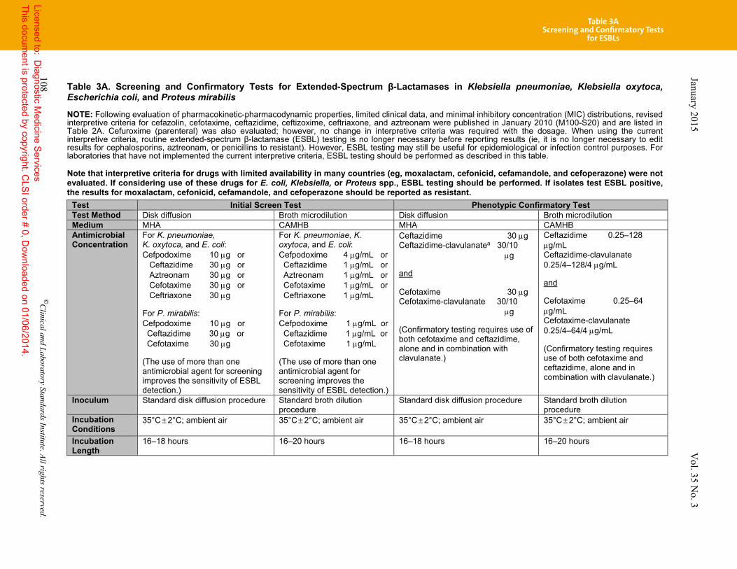
Table 3A. Screening and Confirmatory Tests for Extended-Spectrum β-Lactamases in Klebsiella pneumoniae, Klebsiella oxytoca, Escherichia coli, and Proteus mirabilis
NOTE: Following evaluation of pharmacokinetic-pharmacodynamic properties, limited clinical data, and minimal inhibitory concentration (MIC) distributions, revised interpretive criteria for cefazolin, cefotaxime, ceftazidime, ceftizoxime, ceftriaxone, and aztreonam were published in January 2010 (M100-S20) and are listed in Table 2A. Cefuroxime (parenteral) was also evaluated; however, no change in interpretive criteria was required with the dosage. When using the current interpretive criteria, routine extended-spectrum β-lactamase (ESBL) testing is no longer necessary before reporting results (ie, it is no longer necessary to edit results for cephalosporins, aztreonam, or penicillins to resistant). However, ESBL testing may still be useful for epidemiological or infection control purposes. For laboratories that have not implemented the current interpretive criteria, ESBL testing should be performed as described in this table. Note that interpretive criteria for drugs with limited availability in many countries (eg, moxalactam, cefonicid, cefamandole, and cefoperazone) were not evaluated. If considering use of these drugs for E. coli, Klebsiella, or Proteus spp., ESBL testing should be performed. If isolates test ESBL positive, the results for moxalactam, cefonicid, cefamandole, and cefoperazone should be reported as resistant.
Test Initial Screen Test Phenotypic Confirmatory TestTest Method Disk diffusion Broth microdilution Disk diffusion Broth microdilution Medium MHA CAMHB MHA CAMHBAntimicrobial Concentration
For K. pneumoniae, K. oxytoca, and E. coli: Cefpodoxime 10 g or Ceftazidime 30 g or Aztreonam 30 g or Cefotaxime 30 g or Ceftriaxone 30 g For P. mirabilis: Cefpodoxime 10 g or Ceftazidime 30 g or Cefotaxime 30 g (The use of more than one antimicrobial agent for screening improves the sensitivity of ESBL detection.)
For K. pneumoniae, K. oxytoca, and E. coli: Cefpodoxime 4 g/mL or Ceftazidime 1 g/mL or Aztreonam 1 g/mL or Cefotaxime 1 g/mL or Ceftriaxone 1 g/mL For P. mirabilis: Cefpodoxime 1 g/mL or Ceftazidime 1 g/mL or Cefotaxime 1 g/mL (The use of more than one antimicrobial agent for screening improves the sensitivity of ESBL detection.)
Ceftazidime 30 g Ceftazidime-clavulanatea 30/10 g and Cefotaxime 30 g Cefotaxime-clavulanate 30/10 g (Confirmatory testing requires use of both cefotaxime and ceftazidime, alone and in combination with clavulanate.)
Ceftazidime 0.25–128 g/mL Ceftazidime-clavulanate 0.25/4–128/4 g/mL and Cefotaxime 0.25–64 g/mL Cefotaxime-clavulanate 0.25/4–64/4 g/mL (Confirmatory testing requires use of both cefotaxime and ceftazidime, alone and in combination with clavulanate.)
Inoculum Standard disk diffusion procedure Standard broth dilution procedure
Standard disk diffusion procedure Standard broth dilution procedure
Incubation Conditions
35°C 2°C; ambient air 35°C 2°C; ambient air 35°C 2°C; ambient air 35°C 2°C; ambient air
Incubation Length
16–18 hours 16–20 hours 16–18 hours 16–20 hours
108 ©Clinical and Laboratory Standards Institute. All rights reserved.
January 2015
Vol. 35 N
o. 3
Table 3AScreening and Confirmatory Tests
for ESBLs
Licensed�to:��Diagnostic�M
edicine�Services
This�document�is�protected�by�copyright.�C
LSI�order�#�0,�D
ownloaded�on�01/06/2014.
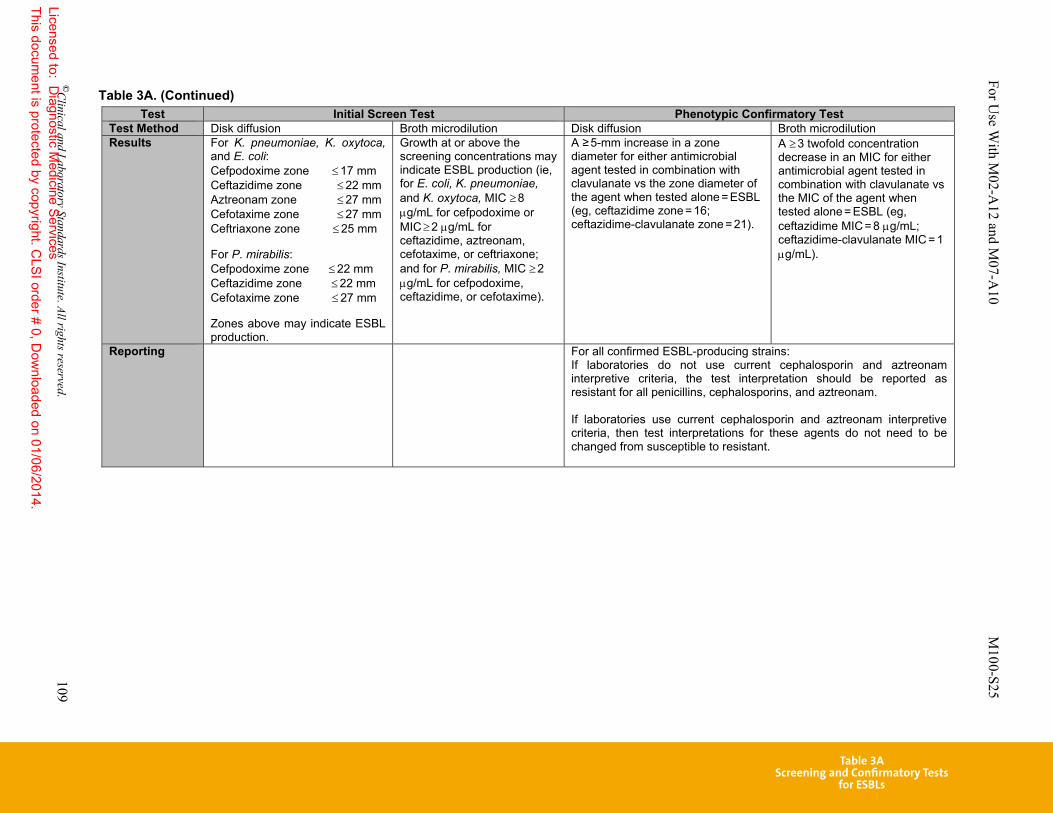
©Clinical and Laboratory Standards Institute. All rights reserved. 109
Table 3A. (Continued)
Test Initial Screen Test Phenotypic Confirmatory TestTest Method Disk diffusion Broth microdilution Disk diffusion Broth microdilution Results For K. pneumoniae, K. oxytoca,
and E. coli: Cefpodoxime zone 17 mm Ceftazidime zone 22 mm Aztreonam zone 27 mm Cefotaxime zone 27 mm Ceftriaxone zone 25 mm For P. mirabilis: Cefpodoxime zone 22 mm Ceftazidime zone 22 mm Cefotaxime zone 27 mm Zones above may indicate ESBL production.
Growth at or above the screening concentrations may indicate ESBL production (ie, for E. coli, K. pneumoniae, and K. oxytoca, MIC 8 g/mL for cefpodoxime or MIC 2 g/mL for ceftazidime, aztreonam, cefotaxime, or ceftriaxone; and for P. mirabilis, MIC 2 g/mL for cefpodoxime, ceftazidime, or cefotaxime).
A ≥ 5-mm increase in a zone diameter for either antimicrobial agent tested in combination with clavulanate vs the zone diameter of the agent when tested alone = ESBL (eg, ceftazidime zone = 16; ceftazidime-clavulanate zone = 21).
A 3 twofold concentration decrease in an MIC for either antimicrobial agent tested in combination with clavulanate vs the MIC of the agent when tested alone = ESBL (eg, ceftazidime MIC = 8 g/mL; ceftazidime-clavulanate MIC = 1 g/mL).
Reporting For all confirmed ESBL-producing strains: If laboratories do not use current cephalosporin and aztreonam interpretive criteria, the test interpretation should be reported as resistant for all penicillins, cephalosporins, and aztreonam. If laboratories use current cephalosporin and aztreonam interpretive criteria, then test interpretations for these agents do not need to be changed from susceptible to resistant.
For Use W
ith M02-A
12 and M07-A
10 M100-S25
Table 3AScreening and Confirmatory Tests
for ESBLs
Licensed�to:��Diagnostic�M
edicine�Services
This�document�is�protected�by�copyright.�C
LSI�order�#�0,�D
ownloaded�on�01/06/2014.

Table 3A. (Continued)
Abbreviations: ATCC®, American Type Culture Collection; CAMHB, cation-adjusted Mueller-Hinton broth; ESBL, extended-spectrum -lactamase; MHA, Mueller-Hinton agar; MIC, minimal inhibitory concentration; QC, quality control.
Test Initial Screen Test Phenotypic Confirmatory TestTest Method Disk diffusion Broth microdilution Disk diffusion Broth microdilution QC Recommendations
When testing ESBL-screening antimicrobial agents, K. pneumoniae ATCC®b 700603 is provided as a supplemental QC strain (eg, for training, competency, or test evaluation). Either strain, K. pneumoniae ATCC® 700603 or E. coli ATCC® 25922, may then be used for routine QC (eg, weekly or daily). E. coli ATCC® 25922 (see acceptable QC ranges in Table 4A) K. pneumoniae ATCC® 700603: Cefpodoxime zone 9–16 mm Ceftazidime zone 10–18 mm Aztreonam zone 9–17 mm Cefotaxime zone 17–25 mm Ceftriaxone zone 16–24 mm
When testing ESBL-screening antimicrobial agents, K. pneumoniae ATCC® 700603 is provided as a supplemental QC strain (eg, for training, competency, or test evaluation). Either strain, K. pneumoniae ATCC® 700603 or E. coli ATCC® 25922, may then be used for routine QC (eg, weekly or daily). E. coli ATCC® 25922 = No growth (see acceptable QC ranges listed in Table 5A) K. pneumoniae ATCC® 700603 = Growth: Cefpodoxime MIC 8 g/mL Ceftazidime MIC 2 g/mL Aztreonam MIC 2 g/mL Cefotaxime MIC 2 g/mL Ceftriaxone MIC 2 g/mL
When performing the ESBL confirmatory tests, K. pneumoniae ATCC® 700603 and E. coli ATCC® 25922 should be used for routine QC (eg, weekly or daily). Acceptable QC: E. coli ATCC® 25922: ≤ 2-mm increase in zone diameter for antimicrobial agent tested in combination with clavulanate vs the zone diameter when tested alone. K. pneumoniae ATCC® 700603:
≥ 5-mm increase in zone diameter of ceftazidime-clavulanate vs ceftazidime alone; ≥ 3-mm increase in zone diameter of cefotaxime-clavulanate vs cefotaxime alone.
When performing the ESBL confirmatory tests, K. pneumoniae ATCC® 700603 and E. coli ATCC® 25922 should be tested routinely (eg, weekly or daily). Acceptable QC: E. coli ATCC® 25922: < 3 twofold concentration decrease in MIC for antimicrobial agent tested in combination with clavulanate vs the MIC of the agent when tested alone. K. pneumoniae ATCC® 700603: 3 twofold concentration decrease in MIC for an antimicrobial agent tested in combination with clavulanate vs the MIC of the agent when tested alone.
110 ©Clinical and Laboratory Standards Institute. All rights reserved.
January 2015
Vol. 35 N
o. 3
Table 3AScreening and Confirmatory Tests
for ESBLs
Licensed�to:��Diagnostic�M
edicine�Services
This�document�is�protected�by�copyright.�C
LSI�order�#�0,�D
ownloaded�on�01/06/2014.

©Clinical and Laboratory Standards Institute. All rights reserved. 111
Table 3A. (Continued)
Footnotes
a. Preparation of ceftazidime-clavulanate (30 g/10 µg) and cefotaxime-clavulanate (30 g/10 g) disks: Using a stock solution of clavulanate at 1000 g/mL (either freshly prepared or taken from small aliquots that have been frozen at −70°C), add 10 L of clavulanate to ceftazidime (30 g) and cefotaxime (30 g) disks. Use a micropipette to apply the 10 L of stock solution to the ceftazidime and cefotaxime disks within one hour before they are applied to the plates, allowing about 30 minutes for the clavulanate to absorb and the disks to be dry enough for application. Use disks immediately after preparation or discard; do not store.
b. ATCC® is a registered trademark of the American Type Culture Collection.
For Use W
ith M02-A
12 and M07-A
10 M100-S25
Table 3AScreening and Confirmatory Tests
for ESBLs
Licensed�to:��Diagnostic�M
edicine�Services
This�document�is�protected�by�copyright.�C
LSI�order�#�0,�D
ownloaded�on�01/06/2014.

Introduction to Tables 3B and 3C. Tests for Carbapenemases in Enterobacteriaceae, Pseudomonas aeruginosa, and Acinetobacter spp. Institutional infection control procedures or epidemiological investigations may require identification of carbapenemase-producing Enterobacteriaceae, P. aeruginosa, and Acinetobacter spp. Such testing is not currently recommended for routine use. Carbapenemase-producing isolates of Enterobacteriaceae usually test intermediate or resistant to one or more carbapenems using the current interpretive criteria as listed in Table 2A (NOTE: Ertapenem nonsusceptibility is the most sensitive indicator of carbapenemase production), and usually test resistant to one or more agents in cephalosporin subclass III (eg, cefoperazone, cefotaxime, ceftazidime, ceftizoxime, and ceftriaxone). However, some isolates that produce carbapenemases such as SME or IMI often test susceptible to these cephalosporins. Laboratories using Enterobacteriaceae minimal inhibitory concentration (MIC) interpretive criteria for carbapenems described in M100-S20 (January 2010) should perform the modified Hodge test (MHT), the Carba NP test, and/or a molecular assay as described below when isolates of Enterobacteriaceae are suspicious for carbapenemase production based on imipenem or meropenem MICs of 2–4 µg/mL or ertapenem MIC of 2 µg/mL. Refer to Tables 3B-1 or 3C-1 for specific steps to use with interpretive criteria for carbapenems listed in M100-S20 (January 2010).
Abbreviations: ESBL, extended-spectrum β-lactamase; MHT, modified Hodge test; NDM, New Delhi metallo--lactamase.
Tests Used for Epidemiological or Infection Control–Related Testing
MHT Carba NP Other (eg, molecular assays) Organisms Enterobacteriaceae that are nonsusceptible
to one or more carbapenems Enterobacteriaceae, P. aeruginosa, and Acinetobacter spp. that are nonsusceptible to one or more carbapenems
Enterobacteriaceae, P. aeruginosa, and Acinetobacter spp. that are nonsusceptible to one or more carbapenems to determine the presence of a carbapenemase, or to determine carbapenemase type in isolates positive by MHT or Carba NP
Strengths Simple to perform No special reagents or media required
Rapid
Determines type of carbapenemase in addition to absence or presence of the enzyme
Limitations False-positive results can occur in isolates that produce ESBL or AmpC enzymes coupled with porin loss. False-negative results are occasionally noted (eg, some isolates producing NDM carbapenemase). Only applies to Enterobacteriaceae.
Special reagents are required, some of which require in-house preparation (and have a short shelf life). Invalid results occur with some isolates. Certain carbapenemase types (eg, OXA-type, chromosomally encoded) are not consistently detected.
Special reagents and equipment required Specific to targeted genes; false-negative result if specific carbapenemase gene present is not targeted
112 ©Clinical and Laboratory Standards Institute. All rights reserved.
January 2015
Vol. 35 N
o. 3 Introduction to Tables 3B and 3C
Tests for Carbapenemases
Licensed�to:��Diagnostic�M
edicine�Services
This�document�is�protected�by�copyright.�C
LSI�order�#�0,�D
ownloaded�on�01/06/2014.

For Use With M02-A12 and M07-A10 M100-S25
Clinical and Laboratory Standards Institute. All rights reserved. 113
This page is intentionally left blank.
Licensed�to:��Diagnostic�Medicine�ServicesThis�document�is�protected�by�copyright.�CLSI�order�#�0,�Downloaded�on�01/06/2014.

Table 3B. The Modified Hodge Confirmatory Test for Suspected Carbapenemase Production in Enterobacteriaceae
Test Confirmatory Test
When to Do This Test: For epidemiological or infection control purposes. NOTE: No change in the interpretation of carbapenem susceptibility test results is required for carbapenemase-positive isolates.
Test Method MHT Medium MHA
Antimicrobial Concentration
Ertapenem disk 10 g or Meropenem disk 10 g
Inoculum (1) Prepare a 0.5 McFarland standard suspension (using either direct colony suspension or growth method) of E. coli ATCC® 25922 (the indicator organism) in broth or saline, and dilute 1:10 in saline or broth. Inoculate an MHA plate as for the routine disk diffusion procedure. Allow the plate to dry 3 to 10 minutes. Place the appropriate number of ertapenem or meropenem disks on the plate as noted below and shown in Figures 1 and 2. (2) Using a 10-µL loop or swab, pick 3 to 5 colonies of test or QC organism grown overnight on a blood agar plate and inoculate in a straight line out from the edge of the disk. The streak should be at least 20–25 mm in length. Test the number of isolates per plate as noted below and shown in Figures 1 and 2. Capacity of small and large MHA plates (100-mm or 150-mm diameter, respectively): Small Large Disks 1 1–4 Test isolates 1 1–6 QC isolates 2 2
Incubation Conditions 35°C ± 2°C; ambient air
Incubation Length 16–20 hours
NOTE: If using FORMER minimal inhibitory concentration (MIC) interpretive criteria for carbapenems described in M100-S20 (January 2010), please refer to modifications in Table 3B-1 below.
114 ©Clinical and Laboratory Standards Institute. All rights reserved.
January 2015
Vol. 35 N
o. 3
Table 3BThe Modified Hodge Confirmatory Test for
Suspected Carbapenemase Production in Enterobacteriaceae
Licensed�to:��Diagnostic�M
edicine�Services
This�document�is�protected�by�copyright.�C
LSI�order�#�0,�D
ownloaded�on�01/06/2014.

©Clinical and Laboratory Standards Institute. All rights reserved. 115
Table 3B. (Continued)
Abbreviations: ATCC®, American Type Culture Collection; MHA, Mueller-Hinton agar; MHT, modified Hodge test; QC, quality control. NOTE 1: Test recommendations were largely derived following testing of US isolates of Enterobacteriaceae, and provide for a high level of sensitivity (> 90%)
and specificity (> 90%) in detecting Klebsiella pneumoniae carbapenemase–type carbapenemases in these isolates.1 The sensitivity and specificity of the test for detecting other carbapenemase production can vary.
NOTE 2: No data exist on the usefulness of the MHT for the detection of carbapenemase production in nonfermenting gram-negative bacilli. Reference for Table 3B 1 Anderson KF, Lonsway DR, Rasheed JK, et al. Evaluation of methods to identify the Klebsiella pneumoniae carbapenemase in Enterobacteriaceae.
J Clin Microbiol. 2007;45(8):2723-2725.
Test Confirmatory Test Results Following incubation, examine the MHA plate for enhanced growth around the test or QC organism streak at the intersection of the
streak and the zone of inhibition (see Figures 1 and 2). Enhanced growth = positive for carbapenemase production. No enhanced growth = negative for carbapenemase production. Some test isolates may produce substances that will inhibit growth of E. coli ATCC® 25922. When this occurs, a clear area will be seen around the streak (see Figure 3), and the MHT is uninterpretable for these isolates. NOTE: Not all carbapenemase-producing isolates of Enterobacteriaceae are MHT positive, and MHT-positive results may be encountered in isolates with carbapenem resistance mechanisms other than carbapenemase production.
Further Testing and Reporting
Report results of the MHT to infection control or those requesting epidemiological information. No change in the interpretation of carbapenem susceptibility test results is required for MHT-positive isolates.
QC Recommendations Test positive and negative QC organisms each day of testing. K. pneumoniae ATCC® BAA-1705—MHT positive K. pneumoniae ATCC® BAA-1706—MHT negative
For Use W
ith M02-A
12 and M07-A
10 M100-S25
Table 3BThe Modified Hodge Confirmatory Test for
Suspected Carbapenemase Production in Enterobacteriaceae
Licensed�to:��Diagnostic�M
edicine�Services
This�document�is�protected�by�copyright.�C
LSI�order�#�0,�D
ownloaded�on�01/06/2014.
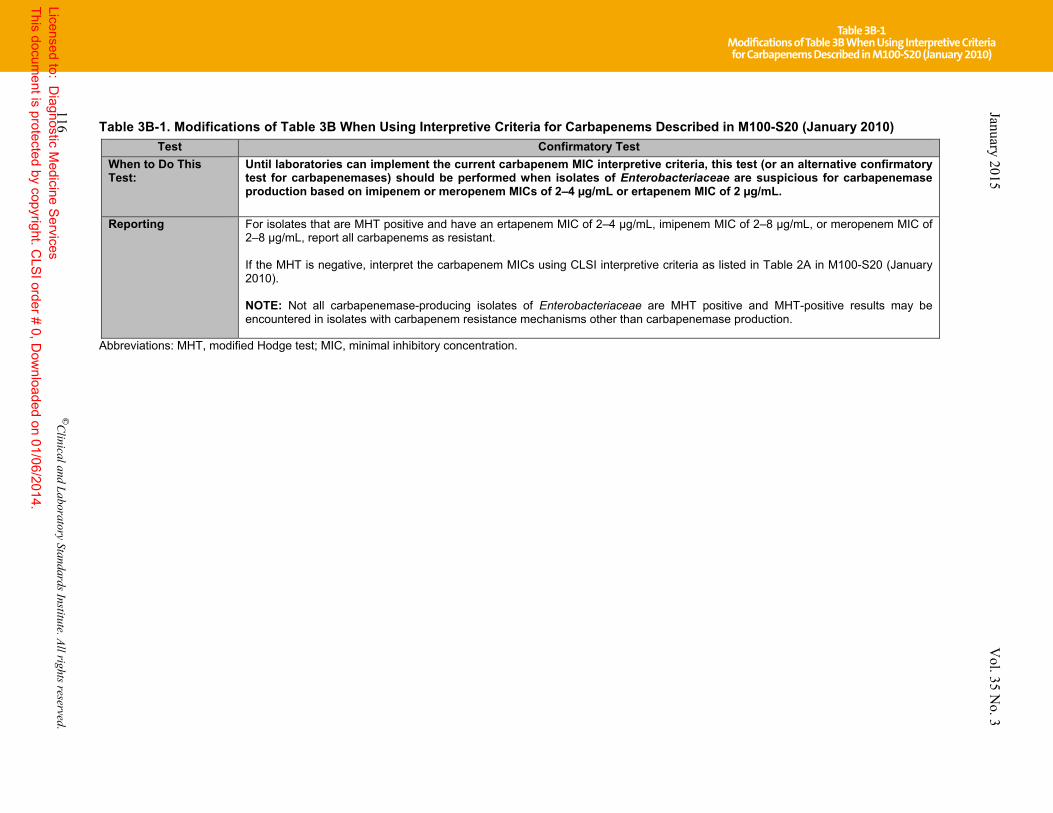
Table 3B-1. Modifications of Table 3B When Using Interpretive Criteria for Carbapenems Described in M100-S20 (January 2010)
Abbreviations: MHT, modified Hodge test; MIC, minimal inhibitory concentration.
Test Confirmatory Test When to Do This Test:
Until laboratories can implement the current carbapenem MIC interpretive criteria, this test (or an alternative confirmatory test for carbapenemases) should be performed when isolates of Enterobacteriaceae are suspicious for carbapenemase production based on imipenem or meropenem MICs of 2–4 µg/mL or ertapenem MIC of 2 µg/mL.
Reporting For isolates that are MHT positive and have an ertapenem MIC of 2–4 µg/mL, imipenem MIC of 2–8 µg/mL, or meropenem MIC of 2–8 µg/mL, report all carbapenems as resistant. If the MHT is negative, interpret the carbapenem MICs using CLSI interpretive criteria as listed in Table 2A in M100-S20 (January 2010). NOTE: Not all carbapenemase-producing isolates of Enterobacteriaceae are MHT positive and MHT-positive results may be encountered in isolates with carbapenem resistance mechanisms other than carbapenemase production.
116 ©Clinical and Laboratory Standards Institute. All rights reserved.
January 2015
Vol. 35 N
o. 3
Table 3B-1Modifications of Table 3B When Using Interpretive Criteria for Carbapenems Described in M100-S20 (January 2010)
Licensed�to:��Diagnostic�M
edicine�Services
This�document�is�protected�by�copyright.�C
LSI�order�#�0,�D
ownloaded�on�01/06/2014.

©Clinical and Laboratory Standards Institute. All rights reserved. 117
Tables 3B and 3B-1. (Continued)
1 2
3
Figure 1. The MHT Performed on a Small MHA Plate. (1) K. pneumoniae ATCC® BAA-1705, positive result; (2) K. pneumoniae ATCC® BAA-1706, negative result; and (3) a clinical isolate, positive result.
E. coli ATCC® 25922
Inhibition of E. coli ATCC® 25922 by ertapenem
Enhanced growth of E. coli ATCC®
25922. Carbapenemase produced by K. pneumoniae ATCC ® BAA-1705 inactivated ertapenem that diffused into the media. Thus, there is no longer sufficient ertapenem here to inhibit E. coli ATCC® 25922 and an indentation of the zone is noted.
For Use W
ith M02-A
12 and M07-A
10 M100-S25
Tables 3B and 3B-1The Modified Hodge Confirmatory Test for Suspected Carbapenemase Production andModifications When Using Interpretive Criteria Described in M100-S20 (January 2010)
Licensed�to:��Diagnostic�M
edicine�Services
This�document�is�protected�by�copyright.�C
LSI�order�#�0,�D
ownloaded�on�01/06/2014.

Tables 3B and 3B-1. (Continued)
Figure 2. The MHT Performed on a Large MHA Plate With Ertapenem. (1) K. pneumoniae ATCC® BAA-1705, positive result; (2) K. pneumoniae ATCC® BAA-1706, negative result; (3–8) clinical isolates; (6) negative result; (3, 4, 5, 7, 8) positive result.
Figure 3. An Example of an Indeterminate Result. (1) A clinical isolate with an indeterminate result; and (2) a clinical isolate with a negative result.
118 ©Clinical and Laboratory Standards Institute. All rights reserved.
January 2015
Vol. 35 N
o. 3
Tables 3B and 3B-1The Modified Hodge Confirmatory Test for Suspected Carbapenemase Production andModifications When Using Interpretive Criteria Described in M100-S20 (January 2010)
Licensed�to:��Diagnostic�M
edicine�Services
This�document�is�protected�by�copyright.�C
LSI�order�#�0,�D
ownloaded�on�01/06/2014.
For Use With M02-A12 and M07-A10 M100-S25
Clinical and Laboratory Standards Institute. All rights reserved. 119
This page is intentionally left blank.
Licensed�to:��Diagnostic�Medicine�ServicesThis�document�is�protected�by�copyright.�CLSI�order�#�0,�Downloaded�on�01/06/2014.

Table 3C. Carba NP Confirmatory Test for Suspected Carbapenemase Production in Enterobacteriaceae, Pseudomonas aeruginosa, and Acinetobacter spp.1-7
NOTE: If using FORMER minimal inhibitory concentration (MIC) interpretive criteria for carbapenems described in M100-S20 (January 2010), please refer to modifications in Table 3C-1 below.
Test Confirmatory Test When to Do This Test:
For epidemiological or infection control purposes. NOTE: No change in the interpretation of carbapenem susceptibility test results is required for Carba NP–positive isolates. Such testing is not currently recommended for routine use.
Test Method Colorimetric microtube assayTest Reagents and Materials
Clinical laboratory reagent water Imipenem reference standard powder Commercially available bacterial protein extraction reagent in Tris HCl buffer, pH 7.4 Zinc sulfate heptahydrate Phenol red powder 1N NaOH solution 10% HCl solution Microcentrifuge tubes 1.5 mL, clear 1 μL inoculation loops Containers to store prepared solutions
Use reagents above to prepare the following solutions (instructions for preparation are provided below this table): 10mM zinc sulfate heptahydrate solution 0.5% phenol red solution 0.1 N sodium hydroxide solution Carba NP Solution A Carba NP Solution B (solution A + imipenem)
Test Procedure 1. Label two microcentrifuge tubes (one “a” and one “b”) for each patient isolate, QC organism, and uninoculatedreagent control.
2. Add 100 μL of bacterial protein extraction reagent to each tube. 3. For each isolate to be tested, emulsify a 1-µL loopful of bacteria from an overnight blood agar plate in both tubes
“a” and “b.” Vortex each tube for 5 seconds. (Uninoculated reagent control tubes should contain only bacterial protein extraction reagent, no organism.) NOTE: Do not use growth from selective media or plates containing antibiotics or other agents that select for certain bacteria.
4. Add 100 μL of Solution A to tube “a.” 5. Add 100 μL of Solution B to tube “b.” 6. Vortex tubes well. 7. Incubate at 35°C ± 2°C for up to 2 hours. Isolates that demonstrate positive results before 2 hours can be reported as
carbapenemase producers.
120 ©Clinical and Laboratory Standards Institute. All rights reserved.
January 2015
Vol. 35 N
o. 3
Table 3CCarba NP Confirmatory Test for Suspected Carbapenemase Production in
Enterobacteriaceae, Pseudomonas aeruginosa, and Acinetobacter spp.
Licensed�to:��Diagnostic�M
edicine�Services
This�document�is�protected�by�copyright.�C
LSI�order�#�0,�D
ownloaded�on�01/06/2014.

©Clinical and Laboratory Standards Institute. All rights reserved. 121
Table 3C. (Continued) Test Confirmatory Test
Test Interpretation Strategy for reading (see Figure 1, below): 1. Read uninoculated reagent control tubes “a” and ‘b’ (ie, “blanks”). Both tubes must be red or red-orange. If either tube is any other color, the test is invalid.
2. Read inoculated tube “a.” Tube “a” must be red or red-orange. If tube “a” is any other color, the test is invalid.
3. Read inoculated tube “b.” Red or red-orange = negative Light orange, dark yellow, or yellow = positive Orange = invalid
4. Interpret results as follows:
Results for Patient and QC TubesTube “a”:Solution A
(serves as internal control) Tube “b”: Solution B Interpretation
Red or red-orange Red or red-orange Negative, no carbapenemase detected
Red or red-orange Light-orange, dark yellow, or yellow
Positive, carbapenemase producer
Red or red-orange Orange Invalid Orange, light-orange, dark yellow, or yellow
Any color Invalid
For Use W
ith M02-A
12 and M07-A
10 M100-S25
Table 3CCarba NP Confirmatory Test for Suspected Carbapenemase Production in
Enterobacteriaceae, Pseudomonas aeruginosa, and Acinetobacter spp.
Licensed�to:��Diagnostic�M
edicine�Services
This�document�is�protected�by�copyright.�C
LSI�order�#�0,�D
ownloaded�on�01/06/2014.

Table 3C. (Continued) Test Confirmatory Test
NOTES: 1. A slight color change may be observed with the addition of imipenem to Solution A. Compare patient tubes to the uninoculated reagent control tubes when interpreting questionable results. 2. For invalid results: Check reagents for QC strains and uninoculated reagent controls.
Reagent deterioration can cause invalid results. An invalid result for an uninoculated reagent control test indicates a problem with Solution A and/or Solution B. Check the pH of Solution A. If pH is < 7.8, prepare fresh Solution A and Solution B.
Repeat the test, including the uninoculated reagent controls. If the repeat test is invalid, perform molecular assay.
Reporting
Report positive as “Carbapenemase producer.” Report negative as “No carbapenemase detected.”
QC Recommendations Test positive and negative QC strains and uninoculated reagent control tubes each day of testing. K. pneumoniae ATCC® BAA-1705—Carbapenemase Positive K. pneumoniae ATCC® BAA-1706—Carbapenemase Negative Results for uninoculated reagent control tubes “a” and “b” must be negative (ie, red or red-orange). Any other result invalidates all tests performed on that day with the same lot of reagents. The addition of imipenem to tube “b” might cause tube “b” to appear red-orange when tube “a” is red.
Abbreviations: ATCC®, American Type Culture Collection; KPC, Klebsiella pneumoniae carbapenemase; QC, quality control. NOTE 1: Test recommendations were largely derived following testing of US isolates of Enterobacteriaceae, Pseudomonas aeruginosa, and
Acinetobacter spp., and provide for a high level of sensitivity (> 90%) and specificity (> 90%) in detecting Klebsiella pneumoniae carbapenemase (KPC), New Delhi metallo--lactamase, VIM, IMP, SPM, and SME-type carbapenemases in these isolates. The sensitivity and specificity of the test for detecting other carbapenemase production can vary. For example, the sensitivity of the Carba NP test for detecting OXA-48-type carbapenemases is low (ie, 11%).
NOTE 2: In CLSI studies, two KPC-positive strains with low carbapenem MICs (one E. cloacae susceptible by MIC to all three carbapenems and one E. coli that was susceptible to meropenem and intermediate to imipenem and ertapenem) were not detected by this test.
122 ©Clinical and Laboratory Standards Institute. All rights reserved.
January 2015
Vol. 35 N
o. 3
Table 3CCarba NP Confirmatory Test for Suspected Carbapenemase Production in
Enterobacteriaceae, Pseudomonas aeruginosa, and Acinetobacter spp.
Licensed�to:��Diagnostic�M
edicine�Services
This�document�is�protected�by�copyright.�C
LSI�order�#�0,�D
ownloaded�on�01/06/2014.
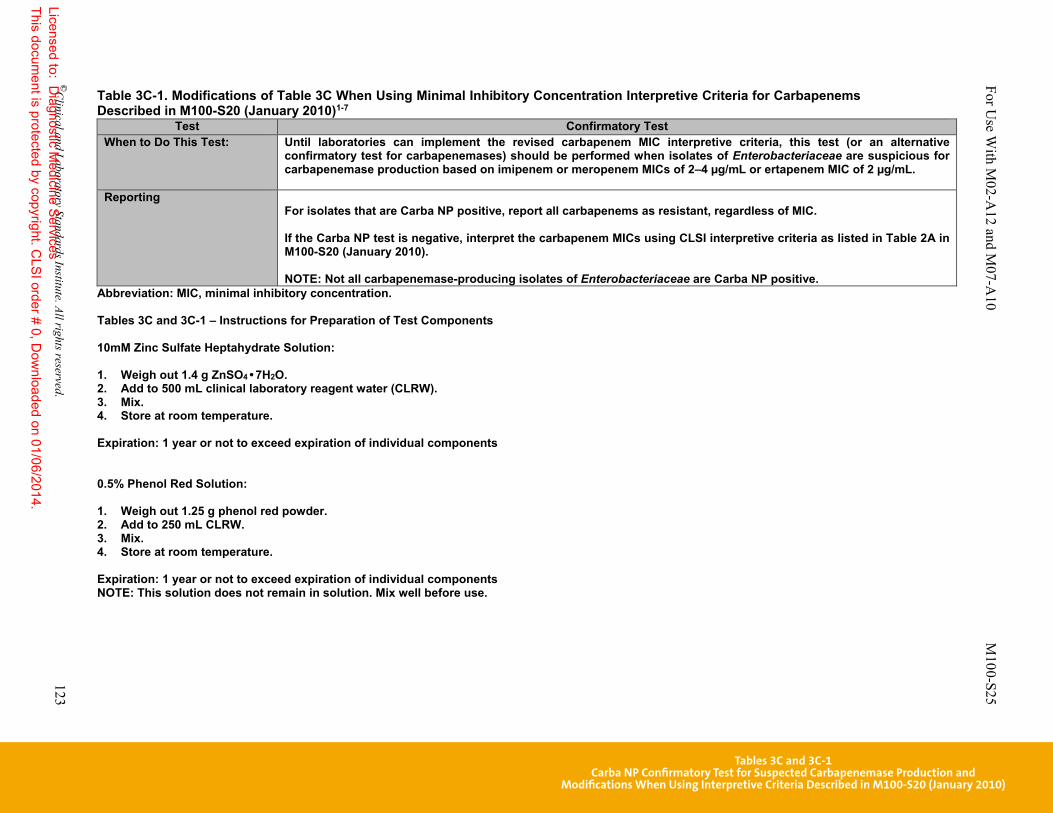
©Clinical and Laboratory Standards Institute. All rights reserved. 123
Table 3C-1. Modifications of Table 3C When Using Minimal Inhibitory Concentration Interpretive Criteria for Carbapenems Described in M100-S20 (January 2010)1-7
Abbreviation: MIC, minimal inhibitory concentration.
Tables 3C and 3C-1 – Instructions for Preparation of Test Components 10mM Zinc Sulfate Heptahydrate Solution: 1. Weigh out 1.4 g ZnSO4 • 7H2O. 2. Add to 500 mL clinical laboratory reagent water (CLRW). 3. Mix. 4. Store at room temperature. Expiration: 1 year or not to exceed expiration of individual components 0.5% Phenol Red Solution: 1. Weigh out 1.25 g phenol red powder. 2. Add to 250 mL CLRW. 3. Mix. 4. Store at room temperature.
Expiration: 1 year or not to exceed expiration of individual components NOTE: This solution does not remain in solution. Mix well before use.
Test Confirmatory Test When to Do This Test:
Until laboratories can implement the revised carbapenem MIC interpretive criteria, this test (or an alternativeconfirmatory test for carbapenemases) should be performed when isolates of Enterobacteriaceae are suspicious for carbapenemase production based on imipenem or meropenem MICs of 2–4 µg/mL or ertapenem MIC of 2 µg/mL.
Reporting For isolates that are Carba NP positive, report all carbapenems as resistant, regardless of MIC. If the Carba NP test is negative, interpret the carbapenem MICs using CLSI interpretive criteria as listed in Table 2A in M100-S20 (January 2010). NOTE: Not all carbapenemase-producing isolates of Enterobacteriaceae are Carba NP positive.
For Use W
ith M02-A
12 and M07-A
10 M100-S25
Tables 3C and 3C-1Carba NP Confirmatory Test for Suspected Carbapenemase Production and
Modifications When Using Interpretive Criteria Described in M100-S20 (January 2010)
Licensed�to:��Diagnostic�M
edicine�Services
This�document�is�protected�by�copyright.�C
LSI�order�#�0,�D
ownloaded�on�01/06/2014.

Tables 3C and 3C-1. (Continued) 0.1 N Sodium Hydroxide Solution: 1. Add 20 mL 1N NaOH to 180 mL CLRW. 2. Store at room temperature.
Expiration: 1 year or not to exceed expiration of individual components Carba NP Solution A: 1. In a 25- to 50-mL beaker, add 2 mL 0.5% phenol red solution to 16.6 mL CLRW. 2. Add 180 μL 10 mM zinc sulfate solution. 3. Adjust pH to 7.8 ± 0.1 with 0.1N NaOH solution (or 10% HCl solution if pH is too high). 4. Store at 4 to 8°C in a small vial or bottle, and protect from prolonged light exposure. Expiration: 2 weeks or not to exceed expiration of individual components (solution should remain red or red-orange; do not use if solution turns any other color) Carba NP Solution B (Solution A + 6 mg/mL Imipenem): 1. Determine the amount of Solution B required, allowing 100 μL per tube for each patient, QC strain, and uninoculated reagent control.
Example: To test 2 patient isolates, positive and negative controls and an uninoculated reagent control, 500 μL of Solution B is needed.
2. Weigh out approximately 10–20 mg of imipenem powder. NOTE: It is advisable to weigh out at least 10 mg of powder. Divide the actual weight by 6
to determine the amount (in mL) of Solution A to add to the powder.
Example: 18 mg of imipenem / 6 = 3 mL of Solution A, which is sufficient for 30 tubes.
3. Store at 4 to 8°C for up to 3 days.
124 ©Clinical and Laboratory Standards Institute. All rights reserved.
January 2015
Vol. 35 N
o. 3
Tables 3C and 3C-1Carba NP Confirmatory Test for Suspected Carbapenemase Production and
Modifications When Using Interpretive Criteria Described in M100-S20 (January 2010)
Licensed�to:��Diagnostic�M
edicine�Services
This�document�is�protected�by�copyright.�C
LSI�order�#�0,�D
ownloaded�on�01/06/2014.

Orange
Light Orange
Dark Yellow
Yellow
Yellow
Red
Red
Red
Red-orange
Red-orange
©Clinical and Laboratory Standards Institute. All rights reserved. 125
Tables 3C and 3C-1. (Continued)
Solution A Solution B
Figure 1. Interpretation of Color Reactions Comparing Tube “a” (Solution A without imipenem) to Tube “b” (Solution B with imipenem)
For Use W
ith M02-A
12 and M07-A
10 M100-S25
Tables 3C and 3C-1Carba NP Confirmatory Test for Suspected Carbapenemase Production and
Modifications When Using Interpretive Criteria Described in M100-S20 (January 2010)
Licensed�to:��Diagnostic�M
edicine�Services
This�document�is�protected�by�copyright.�C
LSI�order�#�0,�D
ownloaded�on�01/06/2014.

Tables 3C and 3C-1. (Continued) References for Tables 3C and 3C-1 1 Carvalhaes CG, Picão RC, Nicoletti AG, Xavier DE, Gales AC. Cloverleaf test (modified Hodge test) for detecting carbapenemase production in
Klebsiella pneumoniae: be aware of false positive results. J Antimicrob Chemother. 2010;65(2):249-251.
2 Girlich D, Poirel L, Nordmann P. Value of the modified Hodge test for detection of emerging carbapenemases in Enterobacteriaceae. J Clin Microbiol. 2012;50(2):477-479.
3 Nordmann P, Poirel L, Dortet L. Rapid detection of carbapenemase-producing Enterobacteriaceae. Emerg Infect Dis. 2012;18(9):1503-1507.
4 Dortet L, Poirel L, Nordmann P. Rapid detection of carbapenemase-producing Pseudomonas spp. J Clin Microbiol. 2012;50(11):3773-3776.
5 Dortet L, Poirel L, Nordmann P. Rapid identification of carbapenemase types in Enterobacteriaceae and Pseudomonas spp. by using a biochemical test. Antimicrob Agents Chemother. 2012;56(12):6437-6440.
6 Cunningham SA, Noorie T, Meunier D, Woodford N, Patel R. Rapid and simultaneous detection of genes encoding Klebsiella pneumoniae carbapenemase (blaKPC) and New Delhi metallo-β-lactamase (blaNDM) in Gram-negative bacilli. J Clin Microbiol. 2013;51(4):1269-1271.
7 Vasoo S, Cunningham SA, Kohner PC, et al. Comparison of a novel, rapid chromogenic biochemical assay, the Carba NP test, with the modified Hodge test for detection of carbapenemase-producing Gram-negative bacilli. J Clin Microbiol. 2013;51(9):3097-3101.
126 ©Clinical and Laboratory Standards Institute. All rights reserved.
January 2015
Vol. 35 N
o. 3
Tables 3C and 3C-1Carba NP Confirmatory Test for Suspected Carbapenemase Production and
Modifications When Using Interpretive Criteria Described in M100-S20 (January 2010)
Licensed�to:��Diagnostic�M
edicine�Services
This�document�is�protected�by�copyright.�C
LSI�order�#�0,�D
ownloaded�on�01/06/2014.

For Use With M02-A12 and M07-A10 M100-S25
Clinical and Laboratory Standards Institute. All rights reserved. 127
This page is intentionally left blank.
Licensed�to:��Diagnostic�Medicine�ServicesThis�document�is�protected�by�copyright.�CLSI�order�#�0,�Downloaded�on�01/06/2014.

Table 3D. Screening Test for Detection of β-Lactamase Production in Staphylococcus species Screen Test β-Lactamase Production
Organism Group S. aureus with penicillin MICs ≤ 0.12 µg/mL or zones ≥ 29 mma
S. aureusa and CoNS (including S. lugdunensisb) with penicillin MICs ≤ 0.12 µg/mL or zones ≥ 29 mm
Test Method Disk diffusion (Penicillin zone-edge test)
Nitrocefin-based test
Medium MHA N/A Antimicrobial Concentration
10 units penicillin disk N/A
Inoculum Standard disk diffusion procedure
Induced growth (ie, growth taken from the zone margin surrounding a penicillin or cefoxitin disk test on either MHA or a blood agar plate after 16–18 hours of incubation)
Incubation Conditions
35°C ± 2°C; ambient air
Room temperature
Incubation Length 16–18 hours Up to 1 hour for nitrocefin-based test or follow manufacturer’s directions
Results Sharp zone edge (“cliff”) = β-lactamase positive. Fuzzy zone edge (“beach”) = β-lactamase negative.
Nitrocefin-based test: conversion from yellow to red/pink =
β-lactamase positive.
Organism Group
S. aureus with penicillin MICs ≤ 0.12 µg/mL or zones ≥ 29 mma
S. aureusa and CoNS (including S. lugdunensisb) with penicillin MICs ≤ 0.12 µg/mL or zones ≥ 29 mm
Test Method Disk diffusion (Penicillin zone-edge test)
Nitrocefin-based test
Further Testing and Reporting
β-lactamase-positive staphylococci are resistant to penicillin, amino-, carboxy-, and ureidopenicillins.
Nitrocefin-based tests can be used for S. aureus, but negative results should be confirmed with the penicillin zone-edge test before reporting penicillin as susceptible. β-lactamase-positive staphylococci are resistant to penicillin, amino-, carboxy-, and ureidopenicillins.
QC Recommendations – Routinec
S. aureus ATCC® 25923 for routine QC of penicillin disk to include examination of zone edge test (fuzzy edge = "beach")
128 ©Clinical and Laboratory Standards Institute. All rights reserved.
January 2015
Vol. 35 N
o. 3
Table 3DScreening Test for β-Lactamase Production
in Staphylococcus spp.
Licensed�to:��Diagnostic�M
edicine�Services
This�document�is�protected�by�copyright.�C
LSI�order�#�0,�D
ownloaded�on�01/06/2014.

©Clinical and Laboratory Standards Institute. All rights reserved. 129
Table 3D. (Continued) Screen Test β-Lactamase ProductionQC Recommendations – Lot/shipmentd
S. aureus ATCC® 29213 – positive S. aureus ATCC® 25923 – negative (or see local regulations and manufacturers’ recommendations)
QC Recommendations –Supplementale
S. aureus ATCC® 29213 – positive penicillin zone-edge test (sharp edge = “cliff”)
Abbreviations: ATCC®, American Type Culture Collection; CoNS, coagulase-negative staphylococci; MHA, Mueller-Hinton agar; MIC, minimal inhibitory concentration; QC, quality control.
Footnotes
a. The penicillin disk diffusion zone-edge test was shown to be more sensitive than nitrocefin-based tests for detection of β-lactamase production in S. aureus. The penicillin zone-
edge test is recommended if only one test is used for β-lactamase detection. However, some laboratories may choose to perform a nitrocefin-based test first and, if this test is positive, report the results as positive for β-lactamase (or penicillin resistant). If the nitrocefin test is negative, the penicillin zone-edge test should be performed before reporting the isolate as penicillin susceptible in cases where penicillin may be used for therapy (eg, endocarditis).
References: Kaase M, Lenga S, Friedrich S, et al. Comparison of phenotypic methods for penicillinase detection in Staphylococcus aureus. Clin Microbiol Infect. 2008;14(6):614-616. Gill VJ, Manning CB, and Ingalls CM. Correlation of penicillin minimum inhibitory concentrations and penicillin zone edge appearance with staphylococcal beta-lactamase
production. J Clin Microbiol. 1981;14(4):437-440. b. For S. lugdunensis, tests for β-lactamase detection are not necessary because isolates producing a β-lactamase will test penicillin resistant (MIC > 0.12 μg/mL and zone
diameters < 29 mm). If a laboratory is using a method other than the CLSI disk diffusion or MIC reference method and is unsure if the method can reliably detect penicillin resistance with contemporary isolates of S. lugdunensis, the laboratory should perform an induced nitrocefin assay or other CLSI reference method on isolates that test penicillin susceptible before reporting the isolate as penicillin susceptible.
c. QC recommendations – Routine
Test negative (susceptible) QC strain:
With each new lot/shipment of testing materials Weekly if the test is performed at least once a week and criteria for converting from daily to weekly QC testing have been met (see Subchapters 4.7.2.1 and 4.7.2.2 in M02 and M07) Daily if the test is performed less than once per week and/or if criteria for converting from daily to weekly QC testing have not been met
d. QC recommendations – Lot/shipment Test positive (resistant) QC strain at minimum with each new lot/shipment of testing materials.
For Use W
ith M02-A
12 and M07-A
10 M100-S25
Table 3DScreening Test for β-Lactamase Production
in Staphylococcus spp.
Licensed�to:��Diagnostic�M
edicine�Services
This�document�is�protected�by�copyright.�C
LSI�order�#�0,�D
ownloaded�on�01/06/2014.

Table 3D. (Continued) e. QC recommendations – Supplemental
Supplemental QC strains can be used to assess a new test, for training personnel, and for competency assessment. It is not necessary to include supplemental QC strains in routine daily or weekly antimicrobial susceptibility testing QC programs. See Appendix C, footnote h, which describes use of supplemental QC strains.
Figure 1. A Positive Penicillin Disk Zone-Edge Test for β-Lactamase Detection. The zone edge is sharp or like a “cliff” indicating β-lactamase production.
Figure 2. A Negative Penicillin Disk Zone-Edge Test for β-Lactamase Detection. The zone edge is fuzzy or like a “beach” indicating no β-lactamase production.
130 ©Clinical and Laboratory Standards Institute. All rights reserved.
January 2015
Vol. 35 N
o. 3
Table 3DScreening Test for β-Lactamase Production
in Staphylococcus spp.
Licensed�to:��Diagnostic�M
edicine�Services
This�document�is�protected�by�copyright.�C
LSI�order�#�0,�D
ownloaded�on�01/06/2014.
For Use With M02-A12 and M07-A10 M100-S25
Clinical and Laboratory Standards Institute. All rights reserved. 131
This page is intentionally left blank.
Licensed�to:��Diagnostic�Medicine�ServicesThis�document�is�protected�by�copyright.�CLSI�order�#�0,�Downloaded�on�01/06/2014.

Table 3E. Screening Test for Detection of Methicillin Resistance (Oxacillin Resistance) in Staphylococcus species
Screen Test Oxacillin Resistance mecA-Mediated Oxacillin Resistance
Using Cefoxitin Organism Group
S. aureus S. aureus and S. lugdunensis
CoNSa S. aureus and S. lugdunensis
Test Method
Agar dilution Disk diffusion Broth microdilution
Medium MHA with 4% NaCl MHA CAMHB
Antimicrobial Concentration
6 g/mL oxacillin 30 µg cefoxitin disk 4 µg/mL cefoxitin
Inoculum Direct colony suspension to obtain 0.5 McFarland turbidity. Using a 1-L loop that was dipped in the suspension, spot an area 10–15 mm in diameter. Alternatively, using a swab dipped in the suspension and expressed, spot a similar area or streak an entire quadrant.
Standard disk diffusion procedure
Standard broth microdilution procedure
Incubation Conditions
33 to 35°C; ambient air. (Testing at temperatures above 35°C may not detect MRSA.)
33 to 35°C; ambient air. (Testing at temperatures above 35°C may not detect MRSA.)
33 to 35°C; ambient air. (Testing at temperatures above 35°C may not detect MRSA.)
Incubation Length
24 hours; read with transmitted light
16–18 hours 24 hours (may be reported after 18 hours, if resistant)
16–20 hours
132 ©Clinical and Laboratory Standards Institute. All rights reserved.
January 2015
Vol. 35 N
o. 3
Table 3EScreening Test for Methicillin Resistance (Oxacillin Resistance)
in Staphylococcus spp.
Licensed�to:��Diagnostic�M
edicine�Services
This�document�is�protected�by�copyright.�C
LSI�order�#�0,�D
ownloaded�on�01/06/2014.

©Clinical and Laboratory Standards Institute. All rights reserved. 133
Table 3E. (Continued)
Screen Test Oxacillin Resistance mecA-Mediated Oxacillin Resistance
Using Cefoxitin Organism Group
S. aureus S. aureus and S. lugdunensis CoNSa S. aureus and S. lugdunensis
Test Method
Agar dilution Disk diffusion Broth microdilution
Results Examine carefully with transmitted light for > 1 colony or light film of growth. > 1 colony = oxacillin resistant
≤ 21 mm = mecA positive ≥ 22 mm = mecA negative
≤ 24 mm = mecA positive ≥ 25 mm = mecA negative.
> 4 µg/mL = mecA positive ≤ 4 µg/mL = mecA negative
Further Testing and Reporting
Oxacillin-resistant staphylococci are resistant to all β-lactam agents; other β-lactam agents should be reported as resistant or should not be reported.
Cefoxitin is used as a surrogate for mecA-mediated oxacillin resistance. Isolates that test as mecA positive should be reported as oxacillin (not cefoxitin) resistant; other β-lactam agents, except those with anti-MRSA activity, should be reported as resistant or should not be reported.
Cefoxitin is used as a surrogate for mecA-mediated oxacillin resistance. Isolates that test as mecA positive should be reported as oxacillin (not cefoxitin) resistant; routine testing of other β-lactam agents, except those with anti-MRSA activity, is not advised. Because of the rare occurrence of oxacillin resistance mechanisms other than mecA, isolates that test as mecA negative, but for which the oxacillin MICs are resistant (MIC ≥ 4 µg/mL), should be reported as oxacillin resistant.
QC Recommend-ations –Routineb
S. aureus ATCC® 29213 – Susceptible (with each test day)
S. aureus ATCC® 25923 – mecA negative (cefoxitin zone 23–29 mm)
S. aureus ATCC® 29213 – mecA negative (cefoxitin MIC 1–4 µg/mL)
QC Recommend-ations – Lot/shipmentc
S. aureus ATCC® 43300 – Resistant
S. aureus ATCC® 43300 – mecA positive (zone ≤ 21 mm) S. aureus ATCC® 43300 – mecA positive (MIC > 4 µg/mL)
Abbreviations: ATCC®, American Type Culture Collection; CAMHB, cation-adjusted Mueller-Hinton broth; CoNS, coagulase-negative staphylococci; MHA, Mueller-Hinton agar; MIC, minimal inhibitory concentration; MRSA, methicillin-resistant S. aureus; QC, quality control.
For Use W
ith M02-A
12 and M07-A
10 M100-S25
Table 3EScreening Test for Methicillin Resistance (Oxacillin Resistance)
in Staphylococcus spp.
Licensed�to:��Diagnostic�M
edicine�Services
This�document�is�protected�by�copyright.�C
LSI�order�#�0,�D
ownloaded�on�01/06/2014.

Table 3E. (Continued)
Footnotes a. Except S. lugdunensis, which is included in the S. aureus group.
b. QC recommendations – Routine Test negative (susceptible) QC strain:
With each new lot/shipment of testing materials Weekly if the test is performed at least once a week and criteria for converting from daily to weekly QC testing have been met (see Subchapters 4.7.2.1 and
4.7.2.2 in M02 and M07) Daily if the test is performed less than once per week and/or if criteria for converting from daily to weekly QC testing have not been met
c. QC recommendations – Lot/shipment
Test positive (resistant) QC strain at minimum with each new lot/shipment of testing materials.
134 ©Clinical and Laboratory Standards Institute. All rights reserved.
January 2015
Vol. 35 N
o. 3
Table 3EScreening Test for Methicillin Resistance (Oxacillin Resistance)
in Staphylococcus spp.
Licensed�to:��Diagnostic�M
edicine�Services
This�document�is�protected�by�copyright.�C
LSI�order�#�0,�D
ownloaded�on�01/06/2014.

For Use With M02-A12 and M07-A10 M100-S25
Clinical and Laboratory Standards Institute. All rights reserved. 135
This page is intentionally left blank.
Licensed�to:��Diagnostic�Medicine�ServicesThis�document�is�protected�by�copyright.�CLSI�order�#�0,�Downloaded�on�01/06/2014.

Table 3F. Screening Test for Detection of Vancomycin Minimal Inhibitory Concentration ≥ 8 µg/mL in Staphylococcus aureus and Enterococcus species
Screen Test Vancomycin MIC 8 g/mLOrganism Group S. aureus Enterococcus spp.
Test Method Agar dilution Agar dilution
Medium BHI agar BHIa agar
Antimicrobial Concentration 6 g/mL vancomycin 6 g/mL vancomycin
Inoculum Direct colony suspension to obtain 0.5 McFarland turbidity. Preferably, using a micropipette, spot a 10-L drop onto agar surface. Alternatively, using a swab dipped in the suspension and the excess liquid expressed, spot an area 10–15 mm in diameter or streak a portion of the plate.
1–10 µL of a 0.5 McFarland suspension spotted onto agar surface. Alternatively, using a swab dipped in the suspension and the excess liquid expressed, spot an area 10–15 mm in diameter or streak a portion of the plate.
Incubation Conditions 35°C 2°C; ambient air 35°C ± 2°C; ambient air
Incubation Length 24 hours 24 hours
Results Examine carefully with transmitted light for 1 colony or light film of growth. 1 colony = Presumptive reduced susceptibility to vancomycin
> 1 colony = Presumptive vancomycin resistance
Further Testing and Reporting Perform a vancomycin MIC using a validated MIC method to determine vancomycin MICs on S. aureus that grow on BHI–vancomycin screening agar. Testing on BHI–vancomycin screening agar does not reliably detect all vancomycin-intermediate S. aureus strains. Some strains for which the vancomycin MICs are 4 g/mL will fail to grow.
Perform vancomycin MIC on Enterococcus spp. that grow on BHI–vancomycin screening agar and test for motility and pigment production to distinguish species with acquired resistance (eg, vanA and vanB) from those with intrinsic, intermediate-level resistance to vancomycin (eg, vanC), such as Enterococcus gallinarum and Enterococcus casseliflavus, which often grow on the vancomycin screen plate. In contrast to other enterococci, E. casseliflavus and E. gallinarum with vancomycin MICs of 8–16 g/mL (intermediate) differ from vancomycin-resistant enterococcus for infection control purposes.
QC Recommendations – Routineb Enterococcus faecalis ATCC 29212 – Susceptible E. faecalis ATCC® 29212 – Susceptible
QC Recommendations – Lot/shipmentc E. faecalis ATCC 51299 – Resistant E. faecalis ATCC® 51299 – Resistant
Abbreviations: ATCC®, American Type Culture Collection; BHI, Brain Heart Infusion; MIC, minimal inhibitory concentration; QC, quality control.
136 ©Clinical and Laboratory Standards Institute. All rights reserved.
January 2015
Vol. 35 N
o. 3
Table 3FScreening Test for Vancomycin MIC ≥ 8 µg/mL
in Staphylococcus aureus and Enterococcus spp.
Licensed�to:��Diagnostic�M
edicine�Services
This�document�is�protected�by�copyright.�C
LSI�order�#�0,�D
ownloaded�on�01/06/2014.

©Clinical and Laboratory Standards Institute. All rights reserved. 137
Table 3F. (Continued)
Footnotes
a. BHI: even though not as widely available, dextrose phosphate agar and broth have been shown in limited testing to perform comparably. b. QC recommendations – Routine
Test negative (susceptible) QC strain:
With each new lot/shipment of testing materials
Weekly if the screening test is performed at least once a week and criteria for converting from daily to weekly QC testing have been met (see
Subchapters 4.7.2.1 and 4.7.2.2 in M02 and M07)
Daily if the screening test is performed less than once per week and/or if criteria for converting from daily to weekly QC testing have not been met
c. QC recommendations – Lot/shipment Test positive (resistant) QC strain at minimum with each new lot/shipment of testing materials.
For Use W
ith M02-A
12 and M07-A
10 M100-S25
Table 3FScreening Test for Vancomycin MIC ≥ 8 µg/mL
in Staphylococcus aureus and Enterococcus spp.
Licensed�to:��Diagnostic�M
edicine�Services
This�document�is�protected�by�copyright.�C
LSI�order�#�0,�D
ownloaded�on�01/06/2014.
Table 3G. Screening Test for Detection of Inducible Clindamycin Resistance in Staphylococcus species, Streptococcus pneumoniae, and Streptococcus spp. β-Hemolytic Groupa
Screen Test
Inducible Clindamycin Resistance
Test Method Disk Diffusion (D-zone test) Broth Microdilution
Organism Group (applies only to organisms resistant to erythromycin and susceptible or intermediate to clindamycin)
S. aureus, S. lugdunensis, and CoNS
S. pneumoniae and β-hemolytic Streptococcus spp.
S. aureus, S. lugdunensis, and CoNSb
S. pneumoniae and β-hemolytic Streptococcus spp.
Medium MHA or blood agar purity plate used with MIC tests
MHA supplemented with sheep blood (5% v/v) or TSA supplemented with sheep blood (5% v/v)
CAMHB CAMHB with LHB (2.5% to 5% v/v)
Antimicrobial Concentration 15-µg erythromycin and 2-µg clindamycin disks spaced 15–26 mm apart
15-µg erythromycin disk and 2-µg clindamycin disk spaced 12 mm apart
4 µg/mL erythromycin and 0.5 µg/mL clindamycin in same well
1 µg/mL erythromycin and 0.5 µg/mL clindamycin in same well
Inoculum Standard disk diffusion procedure or heavily inoculated area of purity plate
Standard disk diffusion procedure
Standard broth microdilution procedure
Incubation Conditions 35°C ± 2°C; ambient air 35°C 2°C; 5% CO2 35°C ± 2°C; ambient air
Incubation Length 16–18 hours 20–24 hours 18–24 hours 20–24 hours
Results Flattening of the zone of inhibition adjacent to the erythromycin disk (referred to as a D-zone) = inducible clindamycin resistance. Hazy growth within the zone of inhibition around clindamycin =
clindamycin resistance, even if no D-zone is apparent.
Any growth = inducible clindamycin resistance. No growth = no inducible clindamycin resistance.
138 ©Clinical and Laboratory Standards Institute. All rights reserved.
January 2015
Vol. 35 N
o. 3
Table 3GScreening Test for Inducible Clindamycin in Staphylococcus spp.,
Streptococcus pneumoniae, and Streptococcus spp. β-Hemolytic Group
Licensed�to:��Diagnostic�M
edicine�Services
This�document�is�protected�by�copyright.�C
LSI�order�#�0,�D
ownloaded�on�01/06/2014.

©Clinical and Laboratory Standards Institute. All rights reserved. 139
Table 3G. (Continued) Screen Test Inducible Clindamycin Resistance
Test Method Disk diffusion(D-zone test) Broth microdilution
Organism Group (applies only to organisms resistant to erythromycin and susceptible or intermediate to clindamycin)
S. aureus, S. lugdunensis, and CoNS
S. pneumoniae and β-hemolytic Streptococcus spp.
S. aureus, S. lugdunensis, and CoNSb
S. pneumoniae and β-hemolytic Streptococcus spp.
Further Testing and Reporting
Report isolates with inducible clindamycin resistance as “clindamycin resistant.”
The following comment may be included with the report: “This isolate is presumed to be resistant based on detection of inducible clindamycin resistance.”
QC Recommendations – Routineb
S. aureus ATCC® 25923 for routine QC of erythromycin and clindamycin disks
S. pneumoniae ATCC 49619 for routine QC of erythromycin and clindamycin disks See Appendix C for use of supplemental QC strains.
S. aureus ATCC® BAA-976 or S. aureus ATCC® 29213 – no growth
S. pneumoniae ATCC 49619 or S. aureus ATCC BAA-976 – no growth
QC Recommendations – Lot/shipmentc
S. aureus ATCC BAA-977 – growth
QC Recommendations – Supplementald
S. aureus ATCC® BAA-976 (D-zone test negative) S. aureus ATCC® BAA-977 (D-zone test positive) Use of unsupplemented MHA is acceptable for these strains.
S. aureus ATCC® BAA-976 (no growth) S. aureus ATCC® BAA-977 (growth)
Abbreviations: ATCC®, American Type Culture Collection; CAMHB, cation-adjusted Mueller-Hinton broth; CoNS, coagulase-negative staphylococci; LHB, lysed horse blood; MHA, Mueller-Hinton agar; MIC, minimal inhibitory concentration ; QC, quality control; TSA, tryptic soy agar.
Footnotes
a. NOTE: Antimicrobial susceptibility testing (AST) of β-hemolytic streptococci need not be performed routinely (see comment [3] in Table 2H-1). When susceptibility testing is clinically indicated, it should include testing for inducible clindamycin resistance. In accordance with 2010 guidance from the Centers for Disease Control and Prevention, colonizing isolates of group B streptococci from penicillin-allergic pregnant women should be tested for inducible clindamycin resistance (see comment [10] in Table 2H-1).
For Use W
ith M02-A
12 and M07-A
10 M100-S25
Table 3GScreening Test for Inducible Clindamycin in Staphylococcus spp.,
Streptococcus pneumoniae, and Streptococcus spp. β-Hemolytic Group
Licensed�to:��Diagnostic�M
edicine�Services
This�document�is�protected�by�copyright.�C
LSI�order�#�0,�D
ownloaded�on�01/06/2014.

Table 3G. (Continued)
Reference Verani JR, McGee L, Schrag SJ; Division of Bacterial Diseases, National Center for Immunization and Respiratory Diseases, Centers for Disease Control and Prevention. Prevention of perinatal group B streptococcal disease – revised guidelines from CDC, 2010. MMWR Recomm Rep. 2010;59(RR-10):1-36.
b. QC recommendations – Routine
Test negative (susceptible) QC strain: With each new lot/shipment of testing materials Weekly if the screening test is performed at least once a week and criteria for converting from daily to weekly QC testing have been met
(see Subchapters 4.7.2.1 and 4.7.2.2 in M02 and M07)
Daily if the screening test is performed less than once per week and/or if criteria for converting from daily to weekly QC testing have not been met
c. QC recommendations – Lot/shipment
Test positive (resistant) QC strain at minimum with each new lot/shipment of testing materials. d. QC recommendations – Supplemental
Supplemental QC strains can be used to assess a new test, for training personnel, and for competency assessment. It is not necessary to
include supplemental QC strains in routine daily or weekly AST QC programs. See Appendix C, footnote g, which describes use of supplemental QC strains.
140 ©Clinical and Laboratory Standards Institute. All rights reserved.
January 2015
Vol. 35 N
o. 3
Table 3GScreening Test for Inducible Clindamycin in Staphylococcus spp.,
Streptococcus pneumoniae, and Streptococcus spp. β-Hemolytic Group
Licensed�to:��Diagnostic�M
edicine�Services
This�document�is�protected�by�copyright.�C
LSI�order�#�0,�D
ownloaded�on�01/06/2014.

For Use With M02-A12 and M07-A10 M100-S25
©Clinical and Laboratory Standards Institute. All rights reserved. 141
This page is intentionally left blank.
Licensed�to:��Diagnostic�Medicine�ServicesThis�document�is�protected�by�copyright.�CLSI�order�#�0,�Downloaded�on�01/06/2014.

January 2015 Vol. 35 No. 3
142 ©Clinical and Laboratory Standards Institute. All rights reserved.
Table 3H. Screening Test for Detection of High-Level Mupirocin Resistance in Staphylococcus aureus
Footnotes a. Although not formally validated by CLSI document M23–based analyses, some studies have linked a lack of response to
mupirocin-based decolonization regimens with isolates for which the mupirocin MICs are 512 µg/mL. Although this document does not provide guidance on interpretive criteria for mupirocin, disk-based testing and the MIC screening test described here identify isolates for which the mupirocin MICs are 512 µg/mL.
b. References:
Simor AE. Randomized controlled trial of chlorhexidine gluconate for washing intranasal mupirocin, and rifampin and doxycycline versus no treatment for the eradication of methicillin-resistant Staphylococcus aureus colonization. Clin Infect Dis. 2007;44:178-185.
Harbarth S, Dharan S, Liassine N, Herrault P, Auckenthaler R, Pittet D. Randomized, placebo-controlled, double-blind trial to evaluate the efficacy of mupirocin for eradicating carriage of methicillin-resistant Staphylococcus aureus. Antimicrob Agents Chemother. 1999;43:1412-1416.
Walker ES, Vasquez JE, Dula R, Bullock H, Sarubbi FA. Mupirocin-resistant, methicillin-resistant Staphylococcus aureus; does mupirocin remain effective? Infect Control Hosp Epidemiol. 2003;24:342-346.
Screen Test High-Level
Mupirocin Resistancea,b
Organism Group S. aureus Test Method Disk diffusion Broth microdilution Medium MHA CAMHB
Antimicrobial Concentration
200-µg mupirocin disk Single mupirocin 256-g/mL well
Inoculum Standard disk diffusion procedure Standard broth microdilution procedure
Incubation Conditions
35°C 2°C; ambient air 35°C 2°C; ambient air
Incubation Length 24 hours; read with transmitted light 24 hours
Results Examine carefully with transmitted light for light growth within the zone of inhibition. No zone = high-level mupirocin resistance. Any zone = the absence of high-level mupirocin resistance.
For single 256-µg/mL well:
Growth = high-level mupirocin resistance.
No growth = the absence of high-level mupirocin resistance.
Further Testing and Reporting
Report isolates with no zone as high-level mupirocin resistant. Report any zone of inhibition as the absence of high-level resistance.
Report growth in the 256-µg/mL well as high-level mupirocin resistant. Report no growth in the 256-µg/mL well as the absence of high-level resistance.
QC Recommend-ations – Routinec
S. aureus ATCC® 25923 (200-µg disk) – mupA negative (zone 29–38 mm)
S. aureus ATCC® 29213 – mupA negative (MIC 0.06–0.5 µg/mL) or E. faecalis ATCC® 29212 – mupA negative (MIC 16–128 µg/mL)
QC Recommend-ations – Lot/shipmentd
S. aureus ATCC® BAA-1708 – mupA positive (no zone)
S. aureus ATCC® BAA-1708 – mupA positive (growth in 256-µg/mL well)
Abbreviations: ATCC®, American Type Culture Collection; CAMHB, cation-adjusted Mueller Hinton broth; MHA, Mueller-Hinton agar; MIC, minimal inhibitory concentration; QC, quality control.
Tabl
e 3H
Scre
enin
g Te
st fo
r Hig
h-Le
vel M
upiro
cin
Resi
stan
ce in
St
aphy
loco
ccus
aur
eus
Licensed�to:��Diagnostic�Medicine�ServicesThis�document�is�protected�by�copyright.�CLSI�order�#�0,�Downloaded�on�01/06/2014.

For Use With M02-A12 and M07-A10 M100-S25
©Clinical and Laboratory Standards Institute. All rights reserved. 143
Table 3H. (Continued) c. QC recommendations – Routine
Test negative (susceptible) QC strain:
With each new lot/shipment of testing materials
Weekly if the screening test is performed at least once a week and criteria for converting from daily to weekly QC testing have been met (see Subchapters 4.7.2.1 and 4.7.2.2 in M02 and M07)
Daily if the screening test is performed less than once per week and/or if criteria for converting from daily to weekly QC testing have not been met
d. QC recommendations – Lot/shipment
Test positive (resistant) QC strain at minimum with each new lot/shipment of testing materials.
Tabl
e 3H
Scre
enin
g Te
st fo
r Hig
h-Le
vel M
upiro
cin
Resi
stan
ce in
St
aphy
loco
ccus
aur
eus
Licensed�to:��Diagnostic�Medicine�ServicesThis�document�is�protected�by�copyright.�CLSI�order�#�0,�Downloaded�on�01/06/2014.

Table 3I. Screening Test for Detection of High-Level Aminoglycoside Resistance in Enterococcus speciesa Screen Test Gentamicin HLAR Streptomycin HLARTest Method Disk diffusion Broth microdilution Agar dilution Disk diffusion Broth microdilution Agar dilution Medium MHA BHIb broth BHIb agar MHA BHIb broth BHIb agar Antimicrobial Concentration
120-µg gentamicin disk Gentamicin, 500 g/mL
Gentamicin, 500 µg/mL
300-µg streptomycin disk Streptomycin, 1000 µg/mL
Streptomycin, 2000 g/mL
Inoculum Standard disk diffusion procedure
Standard broth dilution procedure
10 L of a 0.5 McFarland suspension spotted onto agar surface
Standard disk diffusion procedure
Standard broth dilution procedure
10 L of a 0.5 McFarland suspension spotted onto agar surface
Incubation Conditions
35°C ± 2°C; ambient air 35°C ± 2°C; ambient air 35°C ± 2°C; ambient air 35°C ± 2°C; ambient air 35°C ± 2°C; ambient air 35°C ± 2°C; ambient air
Incubation Length 16–18 hours 24 hours 24 hours 16–18 hours 24–48 hours (if susceptible at 24 hours, reincubate)
24–48 hours (if susceptible at 24 hours, reincubate)
Results 6 mm = Resistant 7–9 mm = Inconclusive ≥ 10 mm = Susceptible MIC correlates: R = > 500 µg/mL S = ≤ 500 µg/mL
Any growth = Resistant
> 1 colony = Resistant
6 mm = Resistant 7–9 mm = Inconclusive ≥ 10 mm = Susceptible MIC correlates: R = > 1000 µg/mL (broth) and > 2000 µg/mL (agar)
S = ≤ 500 µg/mL (broth) and ≤ 1000 µg/mL (agar)
Any growth = Resistant
> 1 colony = Resistant
Further Testing and Reporting
Resistant: is not synergistic with cell wall–active agent (eg, ampicillin, penicillin, and vancomycin). Susceptible: is synergistic with cell wall–active agent (eg, ampicillin, penicillin, and vancomycin) that is also susceptible.
If disk diffusion result is inconclusive: perform an agar dilution or broth dilution MIC test to confirm.
QC Recommendations – Routinec
E. faecalis ATCC® 29212:
16–23 mm
E. faecalis ATCC® 29212 – Susceptible
E. faecalis ATCC® 29212 – Susceptible
E. faecalis ATCC® 29212: 14–20 mm
E. faecalis ATCC® 29212 – Susceptible
E. faecalis ATCC® 29212 – Susceptible
QC Recommendations – Lot/shipmentd
E. faecalis ATCC® 51299 – Resistant
E. faecalis ATCC® 51299 – Resistant
E. faecalis ATCC® 51299 – Resistant
E. faecalis ATCC® 51299 – Resistant
Abbreviations: ATCC®, American Type Culture Collection; BHI, Brain Heart Infusion; HLAR, high-level aminoglycoside resistance; MHA, Mueller-Hinton agar; MIC, minimal inhibitory concentration; QC, quality control.
144 ©Clinical and Laboratory Standards Institute. All rights reserved.
January 2015
Vol. 35 N
o. 3
Table 3IScreening Test for High-Level Aminoglycoside
Resistance in Enterococcus spp.
Licensed�to:��Diagnostic�M
edicine�Services
This�document�is�protected�by�copyright.�C
LSI�order�#�0,�D
ownloaded�on�01/06/2014.

©Clinical and Laboratory Standards Institute. All rights reserved. 145
Table 3I. (Continued)
Footnotes
a. Other aminoglycosides need not be tested, because their activities against enterococci are not superior to gentamicin and streptomycin. b. BHI: even though not as widely available, dextrose phosphate agar and broth have been shown in limited testing to perform comparably.
c. QC recommendations – Routine Test negative (susceptible) QC strain:
With each new lot/shipment of testing materials
Weekly if the screening test is performed at least once a week and criteria for converting from daily to weekly QC testing have been met (see Subchapters
4.7.2.1 and 4.7.2.2 in M02 and M07) Daily if the screening test is performed less than once per week and/or if criteria for converting from daily to weekly QC testing have not been met
d. QC recommendations – Lot/shipment
Test positive (resistant) QC strain at minimum with each new lot/shipment of testing materials.
For Use W
ith M02-A
12 and M07-A
10 M100-S25
Table 3IScreening Test for High-Level Aminoglycoside
Resistance in Enterococcus spp.
Licensed�to:��Diagnostic�M
edicine�Services
This�document�is�protected�by�copyright.�C
LSI�order�#�0,�D
ownloaded�on�01/06/2014.

January 2015 Vol. 35 No. 3
146 ©Clinical and Laboratory Standards Institute. All rights reserved.
Table 4A. Disk Diffusion: Quality Control Ranges for Nonfastidious Organisms (Unsupplemented Mueller-Hinton Medium)
Antimicrobial
Agent
Disk Content
Escherichia coli
ATCC®a 25922
Staphylococcus aureus
ATCC® 25923
Pseudomonas aeruginosa
ATCC® 27853
Escherichia coli
ATCC® 35218b,c
Klebsiella pneumoniae
ATCC®
700603 Amikacin 30 g 19–26 20–26 18–26 – –Amoxicillin-clavulanate 20/10 g 18–24 28–36 – 17–22 –Ampicillin 10 g 15–22 27–35 – 6 –Ampicillin-sulbactam 10/10 g 19–24 29–37 – 13–19 –Azithromycin 15 g – 21–26 – – –Azlocillin 75 g – – 24–30 – –Aztreonam 30 g 28–36 – 23–29 – –Carbenicillin 100 g 23–29 – 18–24 – –Cefaclor 30 g 23–27 27–31 – – –Cefamandole 30 g 26–32 26–34 – – –Cefazolin 30 g 21–27 29–35 – – –Cefdinir 5 g 24–28 25–32 – – –Cefditoren 5 g 22–28 20–28 – – –Cefepime 30 g 31–37 23–29 24–30 – –Cefetamet 10 g 24–29 – – – –Cefixime 5 g 23–27 – – – –Cefmetazole 30 g 26–32 25–34 – – –Cefonicid 30 g 25–29 22–28 – – –Cefoperazone 75 g 28–34 24–33 23–29 – –Cefotaxime 30 g 29–35 25–31 18–22 – –Cefotetan 30 g 28–34 17–23 – – –Cefoxitin 30 g 23–29 23–29 – – –Cefpodoxime 10 g 23–28 19–25 – – –Cefprozil 30 g 21–27 27–33 – – –Ceftaroline 30 g 26–34 26–35 – – –Ceftaroline-avibactam 30/15 g 27–34 25–34 17–26 27–35 21–27d Ceftazidime 30 g 25–32 16–20 22–29 – – Ceftazidime-avibactam 30/20 g 27–35 16–22 25–31 28–35 21–27d Ceftibuten 30 g 27–35 – – – – Ceftizoxime 30 g 30–36 27–35 12–17 – – Ceftobiprole 30 g 30–36 26–34 24–30 – – Ceftolozane-tazobactam 30/10 g 24–32 10–18 25–31 25–31 17–25 Ceftriaxone 30 g 29–35 22–28 17–23 – –Cefuroxime 30 g 20–26 27–35 – – –Cephalothin 30 g 15–21 29–37 – – –Chloramphenicol 30 g 21–27 19–26 – – –Cinoxacin 100 g 26–32 – – – –Ciprofloxacin 5 g 30–40 22–30 25–33 – –Clarithromycin 15 g – 26–32 – – –Clinafloxacin 5 g 31–40 28–37 27–35 – –Clindamycine 2 g – 24–30 – – –Colistin 10 g 11–17 – 11–17 – –Dirithromycin 15 g – 18–26 – – –Doripenem 10 g 27–35 33–42 28–35 – –Doxycycline 30 g 18–24 23–29 – – –Enoxacin Eravacycline
10 g 20 g
28–36 16–23
22–28 19–26
22–28 –
– –
––
Ertapenem 10 g 29–36 24–31 13–21 – –Erythromycine 15 g – 22–30 – – –Faropenem 5 g 20–26 27–34 – – –Fleroxacin 5 g 28–34 21–27 12–20 – –Fosfomycinf 200 g 22–30 25–33 – – –Fusidic acid 10 g – 24–32 – – –Garenoxacin 5 g 28–35 30–36 19–25 – –Gatifloxacin 5 g 30–37 27–33 20–28 – –Gemifloxacin 5 g 29–36 27–33 19–25 – –Gentamicing 10 g 19–26 19–27 17–23 – –Grepafloxacin 5 g 28–36 26–31 20–27 – –Iclaprim 5 g 14–22 25–33 – – –Imipenem 10 g 26–32 – 20–28 – –Kanamycin 30 g 17–25 19–26 – – –Levofloxacin 5 g 29–37 25–30 19–26 – –Linezolid Linopristin-flopristin
30 g 10 g
– –
25–32 25–31
– –
– –
––
Lomefloxacin 10 g 27–33 23–29 22–28 – –
Tabl
e 4A
Non
fast
idio
us Q
ualit
y Co
ntro
lM
02
Licensed�to:��Diagnostic�Medicine�ServicesThis�document�is�protected�by�copyright.�CLSI�order�#�0,�Downloaded�on�01/06/2014.

For Use With M02-A12—Disk Diffusion M100-S25
©Clinical and Laboratory Standards Institute. All rights reserved. 147
Table 4A. (Continued)
Antimicrobial Agent
Disk Content
Escherichia
coli ATCC® 25922
Staphylococcus
aureus ATCC® 25923
Pseudomonas
aeruginosa ATCC® 27853
Escherichia
coli ATCC® 35218b,c
Klebsiella pneumoniae
ATCC®
700603 Loracarbef 30 g 23–29 23–31 – – – Mecillinam 10 g 24–30 – – – – Meropenem 10 g 28–34 29–37 27–33 – – Methicillin 5 g – 17–22 – – – Mezlocillin 75 g 23–29 – 19–25 – – Minocycline 30 g 19–25 25–30 – – – Moxalactam 30 g 28–35 18–24 17–25 – – Moxifloxacin 5 g 28–35 28–35 17–25 – – Nafcillin 1 g – 16–22 – – – Nalidixic acid 30 g 22–28 – – – – Netilmicin 30 g 22–30 22–31 17–23 – – Nitrofurantoin 300 g 20–25 18–22 – – – Norfloxacin 10 g 28–35 17–28 22–29 – – Ofloxacin 5 g 29–33 24–28 17–21 – – Omadacycline 30 g 22–28 22–30 – – – Oxacillin 1 g – 18–24 – – – Pefloxacin 5 g 25–33 – – – –Penicillin 10 units – 26–37 – – – Piperacillin 100 g 24–30 – 25–33 12–18 – Piperacillin-tazobactam 100/10 g 24–30 27–36 25–33 24–30 – Plazomicin 30 g 21–27 19–25 15–21 – – Polymyxin B 300 units 13–19 – 14–18 – – Quinupristin-dalfopristin 15 g – 21–28 – – – Razupenem 10 g 21–26 – j – – – Rifampin 5 g 8–10 26–34 – – – Solithromycin 15 g – 22–30 – – – Sparfloxacin 5 g 30–38 27–33 21–29 – – Streptomycing 10 g 12–20 14–22 – – –
Sulfisoxazolei 250 g or 300 g 15–23 24–34 – – – Tedizolid Teicoplanin
20 g 30 g
– –
22–29 15–21
– –
– –
–
Telavancin 30 g – 16–20 – – – Telithromycin 15 g – 24–30 – – – Tetracycline 30 g 18–25 24–30 – – – Ticarcillin 75 g 24–30 – 21–27 6 – Ticarcillin-clavulanate 75/10 g 24–30 29–37 20–28 21–25 – Tigecycline 15 g 20–27 20–25 9–13 – – Tobramycin 10 g 18–26 19–29 20–26 – – Trimethoprimi 5 g 21–28 19–26 – – – Trimethoprim- sulfamethoxazolei
1.25/23.75 g 23–29 24–32 – – –
Trospectomycin 30 g 10–16 15–20 – – – Trovafloxacin 10 g 29–36 29–35 21–27 – – Ulifloxacin (prulifloxacin)h
5 g 32–38 20–26 27–33 – –
Vancomycin 30 g – 17–21 – – – Abbreviation: ATCC®, American Type Culture Collection. NOTE: Information in boldface type is new or modified since the previous edition.
Footnotes a. ATCC® is a registered trademark of the American Type Culture Collection. b. QC strain recommended when testing β-lactam/β-lactamase inhibitors.
Tabl
e 4A
Non
fast
idio
us Q
ualit
y Co
ntro
lM
02
Licensed�to:��Diagnostic�Medicine�ServicesThis�document�is�protected�by�copyright.�CLSI�order�#�0,�Downloaded�on�01/06/2014.
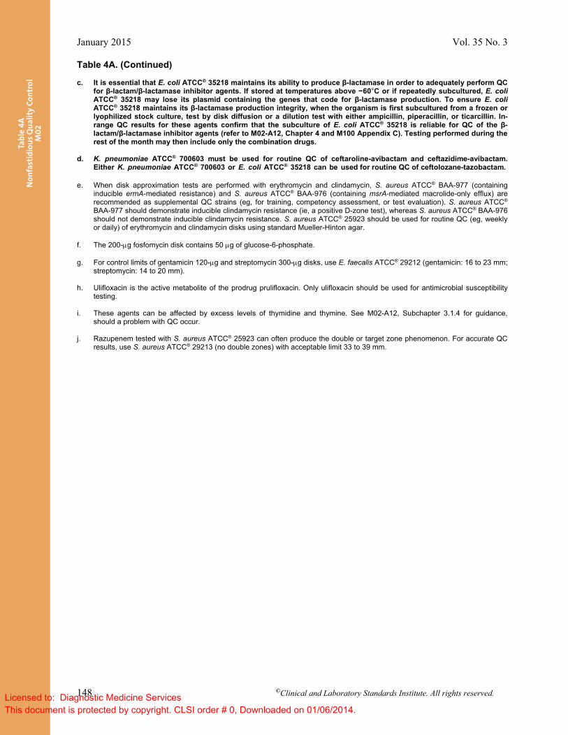
January 2015 Vol. 35 No. 3
148 ©Clinical and Laboratory Standards Institute. All rights reserved.
Table 4A. (Continued) c. It is essential that E. coli ATCC® 35218 maintains its ability to produce β-lactamase in order to adequately perform QC
for β-lactam/β-lactamase inhibitor agents. If stored at temperatures above −60°C or if repeatedly subcultured, E. coli ATCC® 35218 may lose its plasmid containing the genes that code for β-lactamase production. To ensure E. coli ATCC® 35218 maintains its β-lactamase production integrity, when the organism is first subcultured from a frozen or lyophilized stock culture, test by disk diffusion or a dilution test with either ampicillin, piperacillin, or ticarcillin. In-range QC results for these agents confirm that the subculture of E. coli ATCC® 35218 is reliable for QC of the β-lactam/β-lactamase inhibitor agents (refer to M02-A12, Chapter 4 and M100 Appendix C). Testing performed during the rest of the month may then include only the combination drugs.
d. K. pneumoniae ATCC® 700603 must be used for routine QC of ceftaroline-avibactam and ceftazidime-avibactam.
Either K. pneumoniae ATCC® 700603 or E. coli ATCC® 35218 can be used for routine QC of ceftolozane-tazobactam. e. When disk approximation tests are performed with erythromycin and clindamycin, S. aureus ATCC® BAA-977 (containing
inducible ermA-mediated resistance) and S. aureus ATCC® BAA-976 (containing msrA-mediated macrolide-only efflux) are recommended as supplemental QC strains (eg, for training, competency assessment, or test evaluation). S. aureus ATCC® BAA-977 should demonstrate inducible clindamycin resistance (ie, a positive D-zone test), whereas S. aureus ATCC® BAA-976 should not demonstrate inducible clindamycin resistance. S. aureus ATCC® 25923 should be used for routine QC (eg, weekly or daily) of erythromycin and clindamycin disks using standard Mueller-Hinton agar.
f. The 200-g fosfomycin disk contains 50 g of glucose-6-phosphate. g. For control limits of gentamicin 120-g and streptomycin 300-g disks, use E. faecalis ATCC® 29212 (gentamicin: 16 to 23 mm;
streptomycin: 14 to 20 mm). h. Ulifloxacin is the active metabolite of the prodrug prulifloxacin. Only ulifloxacin should be used for antimicrobial susceptibility
testing. i. These agents can be affected by excess levels of thymidine and thymine. See M02-A12, Subchapter 3.1.4 for guidance,
should a problem with QC occur. j. Razupenem tested with S. aureus ATCC® 25923 can often produce the double or target zone phenomenon. For accurate QC
results, use S. aureus ATCC® 29213 (no double zones) with acceptable limit 33 to 39 mm.
Tabl
e 4A
Non
fast
idio
us Q
ualit
y Co
ntro
lM
02
Licensed�to:��Diagnostic�Medicine�ServicesThis�document�is�protected�by�copyright.�CLSI�order�#�0,�Downloaded�on�01/06/2014.

For Use With M02-A12—Disk Diffusion M100-S25
©Clinical and Laboratory Standards Institute. All rights reserved. 149
This page is intentionally left blank.
Licensed�to:��Diagnostic�Medicine�ServicesThis�document�is�protected�by�copyright.�CLSI�order�#�0,�Downloaded�on�01/06/2014.

January 2015 Vol. 35 No. 3
150 ©Clinical and Laboratory Standards Institute. All rights reserved.
Table 4B. Disk Diffusion: Quality Control Ranges for Fastidious Organisms
Antimicrobial Agent
Disk Content
Haemophilus
influenzae ATCC® 49247
Haemophilus
influenzae ATCC® 49766
Neisseria
gonorrhoeae ATCC® 49226
Streptococcus pneumoniae
ATCC® 49619a Amoxicillin-clavulanateb 20/10 g 15–23 – – – Ampicillin 10 g 13–21 – – 30–36 Ampicillin-sulbactam 10/10 g 14–22 – – – Azithromycin 15 g 13–21 – – 19–25 Aztreonam 30 g 30–38 – – – Cefaclor 30 g – 25–31 – 24–32 Cefdinir 5 g – 24–31 40–49 26–31 Cefditoren 5 g 25–34 – – 27–35 Cefepime 30 g 25–31 – 37–46 28–35 Cefetamet 10 g 23–28 – 35–43 – Cefixime 5 g 25–33 – 37–45 16–23 Cefmetazole 30 g 16–21 – 31–36 – Cefonicid 30 g – 30–38 – – Cefotaxime 30 g 31–39 – 38–48 31–39 Cefotetan 30 g – – 30–36 – Cefoxitin 30 g – – 33–41 – Cefpodoxime 10 g 25–31 – 35–43 28–34 Cefprozil 30 g – 20–27 – 25–32 Ceftaroline 30 g 29–39 – – 31–41 Ceftaroline-avibactamc 30/15 g 30–38 – – – Ceftazidime 30 g 27–35 – 35–43 – Ceftazidime-avibactamc 30/20 g 28–34 – – 23–31 Ceftibuten 30 g 29–36 – – – Ceftizoxime 30 g 29–39 – 42–51 28–34 Ceftobiproled
Ceftolozane-tazobactamc 30 g
30/10 g 28–36 23–29
30–38 –
– –
33–39 21–29
Ceftriaxone 30 g 31–39 – 39–51 30–35 Cefuroxime 30 g – 28–36 33–41 – Cephalothin 30 g – – – 26–32 Chloramphenicol 30 g 31–40 – – 23–27 Ciprofloxacin 5 g 34–42 – 48–58 – Clarithromycin 15 g 11–17 – – 25–31 Clinafloxacin 5 g 34–43 – – 27–34 Clindamycin 2 g – – – 19–25 Dirithromycin 15 g – – – 18–25 Doripenem 10 g 21–31 – – 30–38 Doxycycline 30 g – – – 25–34 Enoxacin Eravacycline
10 g 20 g
– –
– –
43–51 –
– 23–30
Ertapenemd 10 g 20–28 27–33 – 28–35 Erythromycin 15 g – – – 25–30 Faropenem 5 g 15–22 – – 27–35 Fleroxacin 5 g 30–38 – 43–51 – Fusidic acid 10 g – – – 9–16 Garenoxacin 5 g 33–41 – – 26–33 Gatifloxacin 5 g 33–41 – 45–56 24–31 Gemifloxacin 5 g 30–37 – – 28–34 Grepafloxacin 5 g 32–39 – 44–52 21–28 Iclaprim 5 g 24–33 – – 21–29 Imipenem 10 g 21–29 – – – Levofloxacin 5 g 32–40 – – 20–25 Linezolid Linopristin-flopristin
30 g 10 g
– 25–31
– –
– –
25–34 22–28
Lomefloxacin 10 g 33–41 – 45–54 – Loracarbef 30 g – 26–32 – 22–28 Meropenem 10 g 20–28 – – 28–35 Moxifloxacin 5 g 31–39 – – 25–31 Nitrofurantoin 300 g – – – 23–29 Norfloxacin 10 g – – – 15–21 Ofloxacin 5 g 31–40 – 43–51 16–21 Omadacycline 30 g 21–29 – – 24–32 Oxacillin 1 g – – – 12e Penicillin 10 units – – 26–34 24–30 Piperacillin-tazobactam 100/10 g 33–38 – – – Quinupristin-dalfopristin 15 g 15–21 – – 19–24 Razupenem 10 g 24–30 – – 29–36 Rifampin 5 g 22–30 – – 25–30
Tabl
e 4B
Fast
idio
us Q
ualit
y Co
ntro
lM
02
Licensed�to:��Diagnostic�Medicine�ServicesThis�document�is�protected�by�copyright.�CLSI�order�#�0,�Downloaded�on�01/06/2014.

For Use With M02-A12—Disk Diffusion M100-S25
©Clinical and Laboratory Standards Institute. All rights reserved. 151
Table 4B. (Continued)
Antimicrobial Agent
Disk Content
Haemophilus
influenzae ATCC® 49247
Haemophilus
influenzae ATCC® 49766
Neisseria
gonorrhoeae ATCC® 49226
Streptococcus pneumoniae
ATCC® 49619a Solithromycin 15 g 16–23 – – 25–33 Sparfloxacin 5 g 32–40 – 43–51 21–27 Spectinomycin 100 g – – 23–29 – Tedizolid Telavancin
20 g 30 g
– –
– –
– –
24–30 17–24
Telithromycin 15 g 17–23 – – 27–33 Tetracycline 30 g 14–22 – 30–42 27–31 Tigecycline 15 g 23–31 – 30–40 23–29 Trimethoprim- sulfamethoxazole
1.25/23.75 g 24–32 – – 20–28
Trospectomycin 30 g 22–29 – 28–35 – Trovafloxacin 10 g 32–39 – 42–55 25–32 Vancomycin 30 g – – – 20–27
Disk Diffusion Testing Conditions for Clinical Isolates and Performance of QC
Organism Haemophilus influenzae Neisseria gonorrhoeae Streptococci and Neisseria meningitidis
Medium HTM GC agar base and 1% defined growth supplement. The use of a cysteine-free growth supplement is not required for disk diffusion testing.
MHA supplemented with 5% defibrinated sheep blood
Inoculum
Direct colony suspension Direct colony suspension Direct colony suspension
Incubation Characteristics
5% CO2; 16–18 hours; 35°C 5% CO2; 20–24 hours; 35°C 5% CO2; 20–24 hours; 35°C
Abbreviations: ATCC®, American Type Culture Collection; HTM, Haemophilus Test Medium; MHA, Mueller-Hinton agar; QC, quality control. NOTE: Information in boldface type is new or modified since the previous edition.
Footnotes a. Despite the lack of reliable disk diffusion interpretive criteria for S. pneumoniae with certain -lactams, Streptococcus
pneumoniae ATCC® 49619 is the strain designated for QC of all disk diffusion tests with all Streptococcus spp. b. When testing Haemophilus on HTM incubated in ambient air, the acceptable QC limits for E. coli ATCC® 35218 are 17 to 22
mm for amoxicillin-clavulanate. c. QC limits for E. coli ATCC® 35218 in HTM: ceftaroline-avibactam 26 to 34 mm; ceftazidime-avibactam 27 to 34 mm;
ceftolozane-tazobactam 25 to 31 mm. d. Either H. influenzae ATCC® 49247 or 49766 may be used for routine QC testing. e. Deterioration in oxacillin disk content is best assessed with QC organism S. aureus ATCC® 25923, with an acceptable zone
diameter of 18 to 24 mm. Ta
ble
4BFa
stid
ious
Qua
lity
Cont
rol
M02
Licensed�to:��Diagnostic�Medicine�ServicesThis�document�is�protected�by�copyright.�CLSI�order�#�0,�Downloaded�on�01/06/2014.

January 2015 Vol. 35 No. 3
152 ©Clinical and Laboratory Standards Institute. All rights reserved.
Table 4C. Disk Diffusion: Reference Guide to Quality Control Frequency
Conversion From Daily to Weekly QC Routine QC is performed each day the test is performed unless an alternative plan has been established (see CLSI document EP23TM).1 M02-A12, Subchapter 4.7.2.1 describes a 20- or 30-day plan that, if successfully completed, allows a user to convert from daily to weekly QC. An alternative option using a two-phase, 15-replicate (3 × 5 day) plan is described as follows: Test 3 replicates using individual inoculum preparations of the appropriate QC strains for 5
consecutive test days. Evaluate each QC strain/antimicrobial agent combination separately using acceptance criteria and
following recommended actions as described in the flow diagram below. Upon successful completion of either the 20- or 30-day plan or the 15-replicate (3 × 5 day) plan,
the laboratory can convert from daily to weekly QC testing. If unsuccessful, investigate, take corrective action as appropriate, and continue daily QC testing until either the 20- or 30-day plan or 15-replicate (3 × 5 day) plan is successfully completed. At that time weekly QC testing can be initiated.
15-Replicate (3 × 5 Day) Plan: Acceptance Criteria and Recommended Action*
Number Out of Range With Initial Testing
(Based on 15 Replicates)
Conclusion From Initial Testing (Based on 15
Replicates)
Number Out of Range After Repeat Testing (Based on
All 30 Replicates) Conclusion After Repeat Testing
0–1
Plan is successful. Convert to weekly QC testing.
N/A
N/A
2–3
Test another 3 replicates for 5 days.
2–3
Plan is successful. Convert to weekly QC testing.
≥ 4
Plan fails. Investigate and take corrective action as
appropriate. Continue QC each test day.
≥ 4
Plan fails. Investigate and take corrective action as
appropriate. Continue QC each test day.
* Assess each QC strain/antimicrobial agent combination separately. Abbreviations: N/A, not applicable; QC, quality control.
Tabl
e 4C
QC
Test
ing
Freq
uenc
yM
02
Licensed�to:��Diagnostic�Medicine�ServicesThis�document�is�protected�by�copyright.�CLSI�order�#�0,�Downloaded�on�01/06/2014.
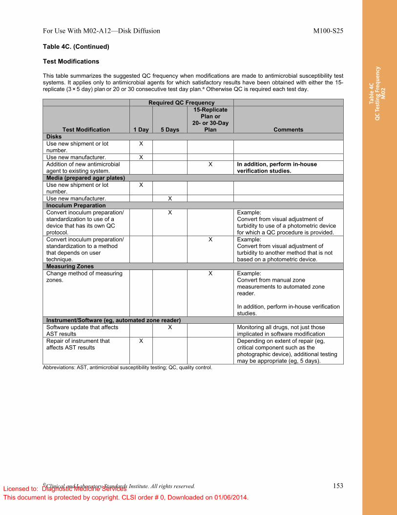
For Use With M02-A12—Disk Diffusion M100-S25
©Clinical and Laboratory Standards Institute. All rights reserved. 153
Table 4C. (Continued) Test Modifications This table summarizes the suggested QC frequency when modifications are made to antimicrobial susceptibility test systems. It applies only to antimicrobial agents for which satisfactory results have been obtained with either the 15-replicate (3 × 5 day) plan or 20 or 30 consecutive test day plan.a Otherwise QC is required each test day.
Required QC Frequency
Test Modification 1 Day 5 Days
15-Replicate Plan or
20- or 30-Day Plan Comments
Disks Use new shipment or lot number.
X
Use new manufacturer. X Addition of new antimicrobial agent to existing system.
X In addition, perform in-house verification studies.
Media (prepared agar plates) Use new shipment or lot number.
X
Use new manufacturer. X Inoculum Preparation Convert inoculum preparation/ standardization to use of a device that has its own QC protocol.
X Example: Convert from visual adjustment of turbidity to use of a photometric device for which a QC procedure is provided.
Convert inoculum preparation/ standardization to a method that depends on user technique.
X Example: Convert from visual adjustment of turbidity to another method that is not based on a photometric device.
Measuring Zones Change method of measuring zones.
X Example: Convert from manual zone measurements to automated zone reader. In addition, perform in-house verification studies.
Instrument/Software (eg, automated zone reader)Software update that affects AST results
X Monitoring all drugs, not just those implicated in software modification
Repair of instrument that affects AST results
X Depending on extent of repair (eg, critical component such as the photographic device), additional testing may be appropriate (eg, 5 days).
Abbreviations: AST, antimicrobial susceptibility testing; QC, quality control.
Tabl
e 4C
QC
Test
ing
Freq
uenc
yM
02
Licensed�to:��Diagnostic�Medicine�ServicesThis�document�is�protected�by�copyright.�CLSI�order�#�0,�Downloaded�on�01/06/2014.

January 2015 Vol. 35 No. 3
154 ©Clinical and Laboratory Standards Institute. All rights reserved.
Table 4C. (Continued) NOTE 1: QC can be performed before or concurrent with testing patient isolates. Patient results can be reported for that day if QC
results are within the acceptable limits. NOTE 2: Manufacturers of commercial or in-house-prepared tests should follow their own internal procedures and applicable
regulations. NOTE 3: For troubleshooting out-of-range results, refer to M02-A12, Subchapter 4.8.1 and M100 Table 4D. Additional information
is available in Appendix C, Quality Control Strains for Antimicrobial Susceptibility Tests (eg, QC organism characteristics, QC testing recommendations).
NOTE 4: Broth, saline, and/or water used to prepare an inoculum does not require routine QC. Reference 1 CLSI. Laboratory Quality Control Based on Risk Management; Approved Guideline. CLSI document EP23-ATM. Wayne, PA:
Clinical and Laboratory Standards Institute; 2011.
Tabl
e 4C
QC
Test
ing
Freq
uenc
yM
02
Licensed�to:��Diagnostic�Medicine�ServicesThis�document�is�protected�by�copyright.�CLSI�order�#�0,�Downloaded�on�01/06/2014.

For Use With M02-A12—Disk Diffusion M100-S25
©Clinical and Laboratory Standards Institute. All rights reserved. 155
This page is intentionally left blank.
Licensed�to:��Diagnostic�Medicine�ServicesThis�document�is�protected�by�copyright.�CLSI�order�#�0,�Downloaded�on�01/06/2014.
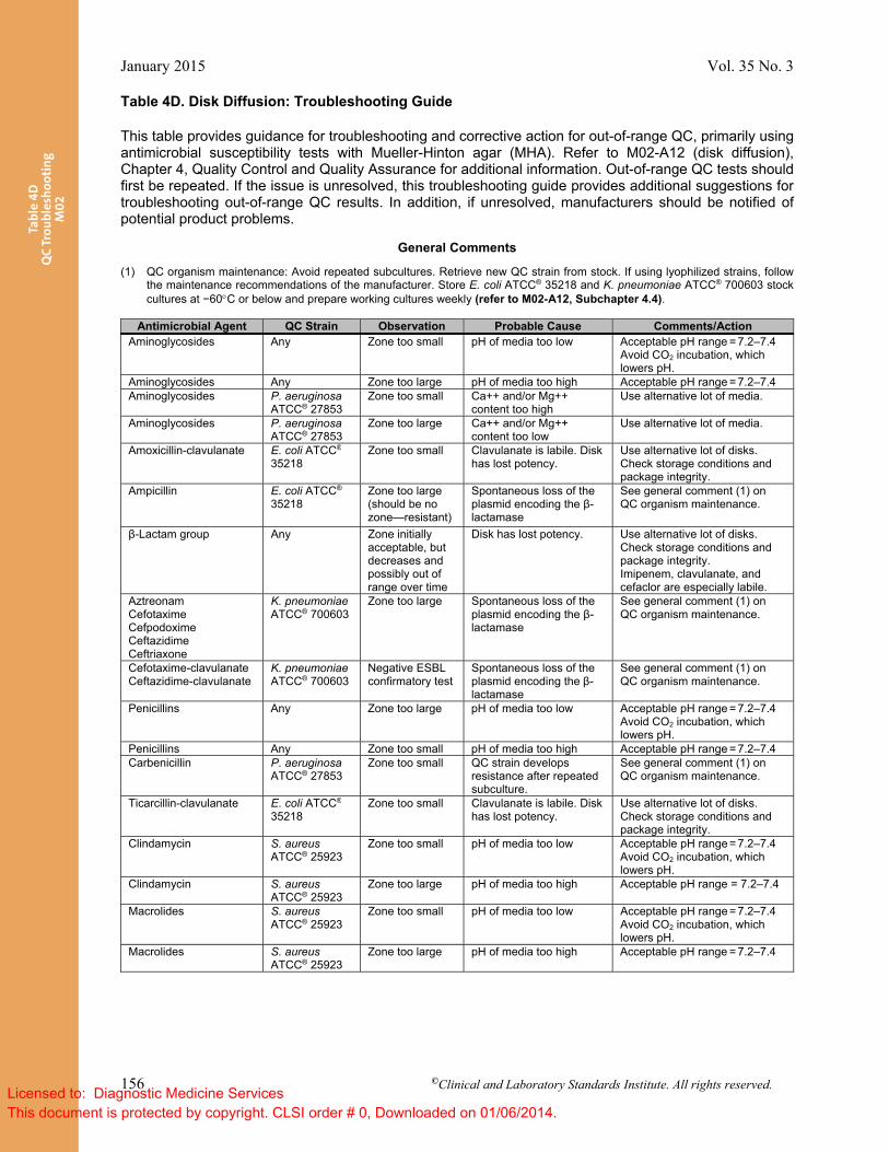
January 2015 Vol. 35 No. 3
156 ©Clinical and Laboratory Standards Institute. All rights reserved.
Table 4D. Disk Diffusion: Troubleshooting Guide
This table provides guidance for troubleshooting and corrective action for out-of-range QC, primarily using antimicrobial susceptibility tests with Mueller-Hinton agar (MHA). Refer to M02-A12 (disk diffusion), Chapter 4, Quality Control and Quality Assurance for additional information. Out-of-range QC tests should first be repeated. If the issue is unresolved, this troubleshooting guide provides additional suggestions for troubleshooting out-of-range QC results. In addition, if unresolved, manufacturers should be notified of potential product problems. General Comments
(1) QC organism maintenance: Avoid repeated subcultures. Retrieve new QC strain from stock. If using lyophilized strains, follow
the maintenance recommendations of the manufacturer. Store E. coli ATCC® 35218 and K. pneumoniae ATCC® 700603 stock cultures at −60C or below and prepare working cultures weekly (refer to M02-A12, Subchapter 4.4).
Antimicrobial Agent QC Strain Observation Probable Cause Comments/Action
Aminoglycosides Any Zone too small pH of media too low Acceptable pH range = 7.2–7.4 Avoid CO2 incubation, which lowers pH.
Aminoglycosides Any Zone too large pH of media too high Acceptable pH range = 7.2–7.4 Aminoglycosides P. aeruginosa
ATCC® 27853 Zone too small Ca++ and/or Mg++
content too high Use alternative lot of media.
Aminoglycosides P. aeruginosa ATCC® 27853
Zone too large Ca++ and/or Mg++ content too low
Use alternative lot of media.
Amoxicillin-clavulanate E. coli ATCC® 35218
Zone too small Clavulanate is labile. Disk has lost potency.
Use alternative lot of disks. Check storage conditions and package integrity.
Ampicillin E. coli ATCC®
35218 Zone too large (should be no zone—resistant)
Spontaneous loss of the plasmid encoding the β-lactamase
See general comment (1) on QC organism maintenance.
β-Lactam group Any Zone initially acceptable, but decreases and possibly out of range over time
Disk has lost potency. Use alternative lot of disks. Check storage conditions and package integrity. Imipenem, clavulanate, and cefaclor are especially labile.
Aztreonam Cefotaxime Cefpodoxime Ceftazidime Ceftriaxone
K. pneumoniae ATCC® 700603
Zone too large Spontaneous loss of the plasmid encoding the β-lactamase
See general comment (1) on QC organism maintenance.
Cefotaxime-clavulanate Ceftazidime-clavulanate
K. pneumoniae ATCC® 700603
Negative ESBL confirmatory test
Spontaneous loss of the plasmid encoding the β-lactamase
See general comment (1) on QC organism maintenance.
Penicillins Any Zone too large pH of media too low Acceptable pH range = 7.2–7.4 Avoid CO2 incubation, which lowers pH.
Penicillins Any Zone too small pH of media too high Acceptable pH range = 7.2–7.4 Carbenicillin P. aeruginosa
ATCC® 27853 Zone too small QC strain develops
resistance after repeated subculture.
See general comment (1) on QC organism maintenance.
Ticarcillin-clavulanate E. coli ATCC® 35218
Zone too small Clavulanate is labile. Disk has lost potency.
Use alternative lot of disks. Check storage conditions and package integrity.
Clindamycin S. aureus ATCC® 25923
Zone too small pH of media too low Acceptable pH range = 7.2–7.4 Avoid CO2 incubation, which lowers pH.
Clindamycin S. aureus ATCC® 25923
Zone too large pH of media too high Acceptable pH range = 7.2–7.4
Macrolides S. aureus ATCC® 25923
Zone too small pH of media too low Acceptable pH range = 7.2–7.4 Avoid CO2 incubation, which lowers pH.
Macrolides S. aureus ATCC® 25923
Zone too large pH of media too high Acceptable pH range = 7.2–7.4
Tabl
e 4D
QC
Trou
bles
hoot
ing
M02
Licensed�to:��Diagnostic�Medicine�ServicesThis�document�is�protected�by�copyright.�CLSI�order�#�0,�Downloaded�on�01/06/2014.

For Use With M02-A12—Disk Diffusion M100-S25
©Clinical and Laboratory Standards Institute. All rights reserved. 157
Table 4D. (Continued)
Abbreviations: ATCC®, American Type Culture Collection; ESBL, extended-spectrum -lactamase; MHA, Mueller-Hinton agar; MIC, minimal inhibitory concentration; QC, quality control.
Antimicrobial Agent QC Strain Observation Probable Cause Comments/Action
Quinolones Any Zone too small pH of media too low Acceptable pH range = 7.2–7.4 Avoid CO2 incubation, which lowers pH.
Quinolones Any Zone too large pH of media too high Acceptable pH range = 7.2–7.4 Tetracyclines Any Zone too large pH of media too low Acceptable pH range = 7.2–7.4
Avoid CO2 incubation, which lowers pH.
Tetracyclines Any Zone too small pH of media too high Acceptable pH range = 7.2–7.4 Tetracyclines Any Zone too small Ca++ and/or Mg++ content
too high Use alternative lot of media.
Tetracyclines Any Zone too large Ca++ and/or Mg++ content too low
Use alternative lot of media.
Sulfonamides Trimethoprim Trimethoprim- sulfamethoxazole
E. faecalis ATCC® 29212
Zone ≤ 20 mm Media too high in thymidine content
Use alternative lot of media.
Various Any Many zones too large
Inoculum too light Error in inoculum preparation Media depth too thin MHA nutritionally unacceptable
Repeat using McFarland 0.5 turbidity standard or standardizing device. Check expiration date and proper storage if using barium sulfate or latex standards. Use agar with depth approximately 4 mm. Recheck alternate lots of MHA.
Various Any Many zones too small
Inoculum too heavy Error in inoculum preparation Media depth too thick MHA nutritionally unacceptable
Repeat using McFarland 0.5 turbidity standard or standardizing device. Check expiration date and proper storage if using barium sulfate or latex standards. Use agar with depth approximately 4 mm. Recheck alternate lots of MHA.
Various Any One or more zones too small or too large
Measurement error
Transcription error
Random defective disk
Disk not pressed firmly against agar
Recheck readings for measurement or transcription errors. Retest. If retest results are out of range and no errors are detected, initiate corrective action.
Various S. pneumoniae ATCC® 49619
Zones too large. Lawn of growth scanty.
Inoculum source plate too old and contains too many nonviable cells. Plate used to prepare inoculum should be 18–20 hours.
Subculture QC strain and repeat QC test or retrieve new QC strain from stock.
Various Any One QC strain is out of range, but other QC organism(s) are in range with the same antimicrobial agent.
One QC organism may be a better indicator of a QC problem.
Retest this strain to confirm reproducibility of acceptable results. Evaluate with alternative strains with known MICs. Initiate corrective action with problem QC strain/antimicrobial agents.
Various Any Two QC strains are out of range with the same antimicrobial agent.
Indicates a problem with the disk
Use alternative lot of disks. Check storage conditions and package integrity.
Various Any Zones overlap. Too many disks per plate Place no more than 12 disks on a 150-mm plate and 5 disks on a 100-mm plate; for some fastidious bacteria that produce large zones, use fewer.
Tabl
e 4D
QC
Trou
bles
hoot
ing
M02
Licensed�to:��Diagnostic�Medicine�ServicesThis�document�is�protected�by�copyright.�CLSI�order�#�0,�Downloaded�on�01/06/2014.

January 2015 Vol. 35 No. 3
158 ©Clinical and Laboratory Standards Institute. All rights reserved.
Table 5A. MIC: Quality Control Ranges for Nonfastidious Organisms (Unsupplemented Mueller-Hinton Medium [Cation-Adjusted if Broth])
Antimicrobial Agent
Staphylococcus aureus
ATCC®a 29213
Enterococcus faecalis
ATCC® 29212
Escherichia coli
ATCC® 25922
Pseudomonas aeruginosa
ATCC® 27853
Escherichia coli
ATCC® 35218b,c
Klebsiella pneumoniae
ATCC®
700603 Amikacin 1–4 64–256 0.5–4 1–4 – –Amoxicillin – – – – > 128Amoxicillin-clavulanate 0.12/0.06–0.5/0.25 0.25/0.12–
1.0/0.5 2/1–8/4 – 4/2–16/8 4/2–16/8
Ampicillin 0.5–2 0.5–2 2–8 – > 32 >128Ampicillin-sulbactam – – 2/1–8/4 – 8/4–32/16 8/4–32/16Azithromycin 0.5–2 – – – – –Azlocillin 2–8 1–4 8–32 2–8 – –Aztreonam – – 0.06–0.25 2–8 0.03–0.12 8–64 Aztreonam-avibactam – – 0.03/4–0.12/4 2/4–8/4 0.015/4–0.06/4 0.06/4–0.5/4e Besifloxacin 0.015–0.06 0.06–0.25 0.06–0.25 1–4 – –Biapenem 0.03–0.12 – 0.03–0.12 0.5–2 0.03–0.12 0.03–0.12 Carbenicillin 2–8 16–64 4–16 16–64 – –Cefaclor 1–4 – 1–4 – – –Cefamandole 0.25–1 – 0.25–1 – – –Cefazolin 0.25–1 – 1–4 – – –Cefdinir 0.12–0.5 – 0.12–0.5 – – –Cefditoren 0.25–2 – 0.12–1 – – –Cefepime 1–4 – 0.015–0.12 0.5–4 – –Cefetamet – – 0.25–1 – – –Cefixime 8–32 – 0.25–1 – – –Cefmetazole 0.5–2 – 0.25–1 > 32 – –Cefonicid 1–4 – 0.25–1 – – –Cefoperazone 1–4 – 0.12–0.5 2–8 – –Cefotaxime 1–4 – 0.03–0.12 8–32 – –Cefotetan 4–16 – 0.06–0.25 – – –Cefoxitin 1–4 – 2–8 – – –Cefpodoxime 1–8 – 0.25–1 – – –Cefprozil 0.25–1 – 1–4 – – –Ceftaroline 0.12–0.5 0.25–2d 0.03–0.12 – – 2–8Ceftaroline-avibactam 0.12/4–0.5/4 – 0.03/4–0.12/4 – 0.015/4–0.06/4d 0.25/4–1/4e Ceftazidime 4–16 – 0.06–0.5 1–4 – 16–64Ceftazidime-avibactam 4/4–16/4 – 0.06/4–0.5/4 0.5/4–4/4 0.03/4–0.12/4 0.25/4–2/4e Ceftibuten – – 0.12–0.5 – – –Ceftizoxime 2–8 – 0.03–0.12 16–64 – –Ceftobiprole 0.12–1 0.06–0.5 0.03–0.12 1–4 – – Ceftolozane-tazobactam 16/4–64/4 – 0.12/4–0.5/4 0.25/4–1/4 0.06/4–0.25/4 0.5/4–2/4e Ceftriaxone 1–8 – 0.03–0.12 8–64 – –Cefuroxime 0.5–2 – 2–8 – – –Cephalothin 0.12–0.5 – 4–16 – – –Chloramphenicol 2–16 4–16 2–8 – – –Cinoxacin – – 2–8 – – –Ciprofloxacing 0.12–0.5 0.25–2 0.004–0.015 0.25–1 – – Clarithromycin 0.12–0.5 – – – – –Clinafloxacin 0.008–0.06 0.03–0.25 0.002–0.015 0.06–0.5 – –Clindamycinh 0.06–0.25 4–16 – – – – Colistin – – 0.25–2 0.5–4 – –Dalbavancinj 0.03–0.12 0.03–0.12 – – – –
Daptomycink 0.12–1 1–4 – – – –
Dirithromycin 1–4 – – – – –Doripenem 0.015–0.06 1–4 0.015–0.06 0.12–0.5 – –Doxycycline 0.12–0.5 2–8 0.5–2 – – –Enoxacin Eravacycline
0.5–2 0.015–0.12
2–16 0.015–0.06
0.06–0.25 0.03–0.12
2–8 2–16
– –
––
Ertapenem 0.06–0.25 4–16 0.004–0.015 2–8 – –Erythromycinh 0.25–1 1–4 – – – –
Tabl
e 5A
Non
fast
idio
us Q
ualit
y Co
ntro
lM
07
Licensed�to:��Diagnostic�Medicine�ServicesThis�document�is�protected�by�copyright.�CLSI�order�#�0,�Downloaded�on�01/06/2014.

For Use With M07-A10—MIC Testing M100-S25
©Clinical and Laboratory Standards Institute. All rights reserved. 159
Table 5A. (Continued)
Antimicrobial Agent
Staphylococcus aureus
ATCC®a 29213
Enterococcus faecalis
ATCC® 29212
Escherichia coli
ATCC® 25922
Pseudomonas aeruginosa
ATCC® 27853
Escherichia coli
ATCC® 35218b,c
Klebsiella pneumoniae
ATCC®
700603 Faropenem 0.03–0.12 – 0.25–1 – – –Fidaxomicin 2–16 1–4 – – – –Finafloxacin 0.03–0.25 0.25–1 0.004–0.03 1–8 – –Fleroxacin 0.25–1 2–8 0.03–0.12 1–4 – –Fosfomycinl 0.5–4 32–128 0.5–2 2–8 – –Fusidic acid Garenoxacin
0.06–0.25 0.004–0.03
– 0.03–0.25
– 0.004–0.03
– 0.5–2
– –
––
Gatifloxacin 0.03–0.12 0.12–1.0 0.008–0.03 0.5–2 – –Gemifloxacin 0.008–0.03 0.015–0.12 0.004–0.015 0.25–1 – –Gentamicinm 0.12–1 4–16 0.25–1 0.5–2 – –Grepafloxacin 0.03–0.12 0.12–0.5 0.004–0.03 0.25–2.0 – –Iclaprim 0.06–0.25 0.004–0.03 1–4 – – –Imipenem 0.015–0.06 0.5–2 0.06–0.25 1–4 – –Kanamycin 1–4 16–64 1–4 – – –Levofloxacin 0.06–0.5 0.25–2 0.008–0.06 0.5–4 – –Linezolid Linopristin-flopristin
1–4 0.06–0.25
1–4 0.5–2
– –
– –
– –
––
Lomefloxacin 0.25–2 2–8 0.03–0.12 1–4 – –Loracarbef 0.5–2 – 0.5–2 > 8 – –Mecillinam – – 0.03–0.25n – – –Meropenem 0.03–0.12 2–8 0.008–0.06 0.25–1 – –Methicillin 0.5–2 > 16 – – – –Mezlocillin 1–4 1–4 2–8 8–32 – –Minocyclineg 0.06–0.5 1–4 0.25–1 – – –Moxalactam 4–16 – 0.12–0.5 8–32 – –Moxifloxacin 0.015–0.12 0.06–0.5 0.008–0.06 1–8 – –Nafcillin 0.12–0.5 2–8 – – – –Nalidixic acidg – – 1–4 – – –Netilmicin 0.25 4–16 0.5–1 0.5–8 – –Nitrofurantoin 8–32 4–16 4–16 – – –Norfloxacin 0.5–2 2–8 0.03–0.12 1–4 – –Ofloxacin Omadacyclinei
0.12–1 0.12–1
1–4 0.06–0.5
0.015–0.12 0.25–2
1–8 –
– –
––
Oritavancinj 0.015–0.12 0.008–0.03 – – – –Oxacillin 0.12–0.5 8–32 – – – –Penicillin 0.25–2 1–4 – – – –Piperacillin 1–4 1–4 1–4 1–8 > 64 –f Piperacillin-tazobactam 0.25/4–2/4 1/4–4/4 1/4–4/4 1/4–8/4 0.5/4–2/4 8/4–32/4 Plazomicin 0.25–2 32–128 0.25–2 1–4 – –Polymyxin B – – 0.25–2 0.5–2 – –Quinupristin-dalfopristin 0.25–1 2–8 – – – –Razupenem 0.008–0.03 0.25–1 0.06–0.5 – – –Rifampin 0.004–0.015 0.5–4 4–16 16–64 – –Solithromycin 0.03–0.12 0.015–0.06 – – – –Sparfloxacin 0.03–0.12 0.12–0.5 0.004–0.015 0.5–2 – –Sulfisoxazoleg,p 32–128 32–128 8–32 – – –Sulopenem 0.015–0.12 2–8 0.015–0.06 – – –Tedizolid Teicoplanin
0.25–1 0.25–1
0.25–1 0.25–1
– –
– –
– –
–
Telavancinj 0.03–0.12 0.03–0.12 – – – –Telithromycin 0.06–0.25 0.015–0.12 – – – –Tetracycline 0.12–1 8–32 0.5–2 8–32 – –Ticarcillin 2–8 16–64 4–16 8–32 > 128 > 256Ticarcillin-clavulanate 0.5/2–2/2 16/2–64/2 4/2–16/2 8/2–32/2 8/2–32/2 32/2–128/2Tigecyclinei 0.03–0.25 0.03–0.12 0.03–0.25 – – –Tobramycin 0.12–1 8–32 0.25–1 0.25–1 – –Trimethoprimp 1–4 0.12–0.5 0.5–2 > 64 – –Trimethoprim- sulfamethoxazole
0.5/9.5 0.5/9.5 0.5/9.5 8/152–32/608 – –
Trospectomycin 2–16 2–8 8–32 – – –Trovafloxacin 0.008–0.03 0.06–0.25 0.004–0.015 0.25–2 – –Ulifloxacin (prulifloxacin)o
– – 0.004–0.015 0.12–0.5 – –
Vancomycinq 0.5–2 1–4 – – – –Abbreviations: ATCC®, American Type Culture Collection; MIC, minimal inhibitory concentration.
Tabl
e 5A
Non
fast
idio
us Q
ualit
y Co
ntro
lM
07
Licensed�to:��Diagnostic�Medicine�ServicesThis�document�is�protected�by�copyright.�CLSI�order�#�0,�Downloaded�on�01/06/2014.

January 2015 Vol. 35 No. 3
160 ©Clinical and Laboratory Standards Institute. All rights reserved.
Table 5A. (Continued) NOTE 1: These MICs were obtained in several reference laboratories by dilution methods. If four or fewer concentrations are
tested, QC may be more difficult. NOTE 2: Information in boldface type is new or modified since the previous edition.
Footnotes
a. ATCC® is a registered trademark of the American Type Culture Collection. b. QC strain recommended when testing β-lactam/β-lactamase inhibitors. c. It is essential that E. coli ATCC® 35218 maintains its ability to produce β-lactamase in order to adequately perform QC
for β-lactam/β-lactamase inhibitor agents. If stored at temperatures above −60°C or if repeatedly subcultured, E. coli ATCC® 35218 may lose its plasmid containing the genes that code for β-lactamase production. To ensure E. coli ATCC® 35218 maintains its β-lactamase production integrity, when the organism is first subcultured from a frozen or lyophilized stock culture, test by disk diffusion or a dilution test with either ampicillin, piperacillin, or ticarcillin. In-range QC results for these agents confirm that the subculture of E. coli ATCC® 35218 is reliable for QC of the β-lactam/β-lactamase inhibitor agents (refer to M07-A10, Subchapter 4.4 and M100 Appendix C). Testing performed during the rest of the month may then include only the combination drugs.
d. Testing this strain with this antimicrobial agent is considered supplemental QC only and is not required as routine user QC
testing. e. K. pneumoniae ATCC® 700603 must be used for routine QC of ceftazidime-avibactam, ceftaroline-avibactam,
aztreonam-avibactam, and ceftolozane-tazobactam. Either K. pneumoniae ATCC® 700603 or E. coli ATCC® 35218 can be used for routine QC of other β-lactam/β-lactamase inhibitor combination agents.
K. pneumoniae ATCC® 700603 should be tested against ceftazidime-avibactam and ceftazidime alone, ceftaroline-
avibactam and ceftaroline alone, or aztreonam-avibactam and aztreonam alone to confirm the activity of avibactam in the combination and to ensure that the plasmid encoding the β-lactamase has not been lost in this QC strain. Currently, there are no MIC QC ranges for ceftolozane without avibactam. Either aztreonam, ceftaroline, or ceftazidime can be tested to ensure K. pneumoniae ATCC® 700603 has not lost its β-lactamase resistance plasmid.
f. No range recommended due to off-scale results on the low end. g. QC limits for E. coli ATCC® 25922 with ciprofloxacin, nalidixic acid, minocycline, and sulfisoxazole when tested in cation-
adjusted Mueller-Hinton broth (CAMHB) with 2.5% to 5% lysed horse blood incubated either in ambient air or 5% CO2 (when testing N. meningitidis) are the same as those listed in Table 5A.
h. When the erythromycin/clindamycin combination well for detection of inducible clindamycin resistance is used, S. aureus
ATCC® BAA-977 (containing inducible ermA-mediated resistance) and S. aureus ATCC® 29213 or S. aureus ATCC® BAA-976 (containing msrA-mediated macrolide-only efflux) are recommended for QC purposes. S. aureus ATCC® BAA-977 should demonstrate inducible clindamycin resistance (ie, growth in the well), whereas S. aureus ATCC® 29213 and S. aureus ATCC® BAA-976 should not demonstrate inducible clindamycin resistance (ie, no growth in the well).
i. For broth microdilution testing of omadacycline and tigecycline, when MIC panels are prepared, the medium must be prepared
fresh on the day of use. The medium must be no more than 12 hours old at the time the panels are made; however, the panels may then be frozen for later use.
j. QC ranges reflect MICs obtained when CAMHB is supplemented with 0.002% polysorbate-80. k. QC ranges reflect MICs obtained when Mueller-Hinton broth is supplemented with calcium to a final concentration of 50 g/mL.
Agar dilution has not been validated for daptomycin. l. The approved MIC susceptibility testing method is agar dilution. Agar media should be supplemented with 25 g/mL of
glucose-6-phosphate. Broth dilution should not be performed. m. For control organisms for gentamicin and streptomycin high-level aminoglycoside screen tests for enterococci, see Table 3I. n. This test should be performed by agar dilution only. o. Ulifloxacin is the active metabolite of the prodrug prulifloxacin. Only ulifloxacin should be used for antimicrobial susceptibility
testing. p. Very medium-dependent, especially with enterococci. q. For QC organisms for vancomycin screen test for enterococci, see Table 3F.
Tabl
e 5A
Non
fast
idio
us Q
ualit
y Co
ntro
lM
07
Licensed�to:��Diagnostic�Medicine�ServicesThis�document�is�protected�by�copyright.�CLSI�order�#�0,�Downloaded�on�01/06/2014.

For Use With M07-A10—MIC Testing M100-S25
©Clinical and Laboratory Standards Institute. All rights reserved. 161
This page is intentionally left blank.
Licensed�to:��Diagnostic�Medicine�ServicesThis�document�is�protected�by�copyright.�CLSI�order�#�0,�Downloaded�on�01/06/2014.

January 2015 Vol. 35 No. 3
162 ©Clinical and Laboratory Standards Institute. All rights reserved.
Table 5B. MIC: Quality Control Ranges for Fastidious Organisms (Broth Dilution Methods)
Antimicrobial Agent
Haemophilus influenzae
ATCC® 49247
Haemophilus influenzae
ATCC® 49766
Streptococcus pneumoniae ATCC® 49619
Amoxicillina – – 0.03–0.12 Amoxicillin-clavulanatea 2/1–16/8 – 0.03/0.015–0.12/0.06 Ampicillin 2–8 – 0.06–0.25 Ampicillin-sulbactam 2/1–8/4 – – Azithromycin 1–4 – 0.06–0.25 Aztreonam 0.12–0.5 – – Besifloxacin 0.015–0.06 – 0.03–0.12 Cefaclor – 1–4 1–4 Cefamandole – 0.25–1 – Cefdinir – 0.12–0.5 0.03–0.25 Cefditoren 0.06–0.25 – 0.015–0.12 Cefepime 0.5–2 – 0.03–0.25 Cefetamet 0.5–2 – 0.5–2 Cefixime 0.12–1 – – Cefmetazole 2–16 – – Cefonicid – 0.06–0.25 – Cefotaxime 0.12–0.5 – 0.03–0.12 Cefotetan – – – Cefoxitin – – – Cefpirome 0.25–1 – – Cefpodoxime 0.25–1 – 0.03–0.12 Cefprozil – 1–4 0.25–1 Ceftaroline 0.03–0.12 – 0.008–0.03 Ceftaroline-avibactam 0.015/4–0.12/4 – – Ceftazidime 0.12–1 – – Ceftazidime-avibactamb 0.06/4–0.5/4 0.015/4–0.06/4 0.25/4–2/4 Ceftibuten 0.25–1 – – Ceftizoxime 0.06–0.5 – 0.12–0.5 Ceftobiprolec
Ceftolozane-tazobactam 0.12–1
0.5/4–2/4 0.016–0.06
– 0.004–0.03 0.25/4–1/4
Ceftriaxone 0.06–0.25 – 0.03–0.12 Cefuroxime – 0.25–1 0.25–1 Cephalothin – – 0.5–2 Chloramphenicol 0.25–1 – 2–8 Ciprofloxacind 0.004–0.03 – – Clarithromycin 4–16 – 0.03–0.12 Clinafloxacin 0.001–0.008 – 0.03–0.12 Clindamycin – – 0.03–0.12 Dalbavancinf – – 0.008–0.03
Daptomycing – – 0.06–0.5 Dirithromycin 8–32 – 0.06–0.25 Doripenem – 0.06–0.25 0.03–0.12 Doxycycline – – 0.015–0.12 Enoxacin Eravacycline
– 0.06–0.5
– –
– 0.004–0.03
Ertapenem – 0.015–0.06 0.03–0.25 Erythromycin – – 0.03–0.12 Faropenem – 0.12–0.5 0.03–0.25 Finafloxacin – 0.002–0.008 0.25–1 Fleroxacin 0.03–0.12 – – Fusidic acid – – 4–32 Garenoxacin 0.002–0.008 – 0.015–0.06 Gatifloxacin 0.004–0.03 – 0.12–0.5 Gemifloxacin 0.002–0.008 – 0.008–0.03 Gentamicin – – – Grepafloxacin 0.002–0.015 – 0.06–0.5 Iclaprim 0.12–1 – 0.03–0.12 Imipenem – 0.25–1 0.03–0.12 Levofloxacin 0.008–0.03 – 0.5–2 Linezolid Linopristin- flopristin
– 0.25–2
– –
0.25–2 0.12–0.5
Lomefloxacin 0.03–0.12 – – Loracarbef – 0.5–2 2–8
Tabl
e 5B
Fast
idio
us Q
ualit
y Co
ntro
l Bro
th D
ilutio
nM
07
Licensed�to:��Diagnostic�Medicine�ServicesThis�document�is�protected�by�copyright.�CLSI�order�#�0,�Downloaded�on�01/06/2014.

For Use With M07-A10—MIC Testing M100-S25
©Clinical and Laboratory Standards Institute. All rights reserved. 163
Table 5B. (Continued)
Antimicrobial Agent
Haemophilus influenzae
ATCC® 49247
Haemophilus influenzae
ATCC® 49766
Streptococcus pneumoniae ATCC® 49619
Meropenem – 0.03–0.12 0.06–0.25 Metronidazole – – – Minocyclined – – – Moxifloxacin 0.008–0.03 – 0.06–0.25 Nalidixic acidd – – – Nitrofurantoin – – 4–16 Norfloxacin – – 2–8 Ofloxacin 0.015–0.06 – 1–4 Omadacyclinee 0.5–2 – 0.015–0.12 Oritavancinf – – 0.001–0.004 Penicillin – – 0.25–1 Piperacillin- tazobactam
0.06/4–0.5/4 – –
Quinupristin- dalfopristin
2–8 – 0.25–1
Razupenem – 0.008–0.03 0.008–0.06 Rifampin 0.25–1 – 0.015–0.06 Solithromycin 1–4 – 0.004–0.015 Sparfloxacin 0.004–0.015 – 0.12–0.5 Spectinomycin – – – Sulfisoxazoled – – – Sulopenem – 0.06–0.25 0.03–0.12 Tedizolid – – 0.12–0.5 Telavancinf – – 0.004–0.015 Telithromycin 1–4 – 0.004–0.03 Tetracycline 4–32 – 0.06–0.5 Tigecyclinee 0.06–0.5 – 0.015–0.12 Trimethoprim- sulfamethoxazole
0.03/0.59– 0.25/4.75
– 0.12/2.4– 1/19
Trospectomycin 0.5–2 – 1–4 Trovafloxacin 0.004–0.015 – 0.06–0.25 Vancomycin – – 0.12–0.5
Testing Conditions for Clinical Isolates and Performance of QC
Organism Haemophilus influenzae Streptococcus pneumoniae and streptococci Neisseria meningitidis
Medium Broth dilution: HTM broth
Broth dilution: CAMHB with LHB (2.5% to 5% v/v)
Broth dilution: CAMHB with LHB (2.5% to 5% v/v)
Inoculum Direct colony suspension Direct colony suspension Direct colony suspension
Incubation Characteristics
Ambient air; 20–24 hours; 35°C Ambient air; 20–24 hours; 35°C 5% CO2; 20–24 hours; 35°C (for QC with S. pneumoniae ATCC® 49619, 5% CO2 or ambient air, except for azithromycin, ambient air only)
Abbreviations: ATCC®, American Type Culture Collection; CAMHB, cation-adjusted Mueller-Hinton broth; HTM, Haemophilus Test Medium; LHB, lysed horse blood; MIC, minimal inhibitory concentration; QC, quality control.
Tabl
e 5B
Fast
idio
us Q
ualit
y Co
ntro
l Bro
th D
ilutio
nM
07
Licensed�to:��Diagnostic�Medicine�ServicesThis�document�is�protected�by�copyright.�CLSI�order�#�0,�Downloaded�on�01/06/2014.
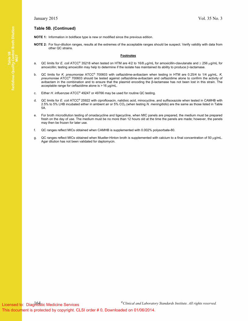
January 2015 Vol. 35 No. 3
164 ©Clinical and Laboratory Standards Institute. All rights reserved.
Table 5B. (Continued) NOTE 1: Information in boldface type is new or modified since the previous edition. NOTE 2: For four-dilution ranges, results at the extremes of the acceptable ranges should be suspect. Verify validity with data from
other QC strains.
Footnotes a. QC limits for E. coli ATCC® 35218 when tested on HTM are 4/2 to 16/8 g/mL for amoxicillin-clavulanate and 256 g/mL for
amoxicillin; testing amoxicillin may help to determine if the isolate has maintained its ability to produce -lactamase. b. QC limits for K. pneumoniae ATCC® 700603 with ceftazidime-avibactam when testing in HTM are 0.25/4 to 1/4 µg/mL. K.
pneumoniae ATCC® 700603 should be tested against ceftazidime-avibactam and ceftazidime alone to confirm the activity of avibactam in the combination and to ensure that the plasmid encoding the β-lactamase has not been lost in this strain. The acceptable range for ceftazidime alone is > 16 µg/mL.
c. Either H. influenzae ATCC® 49247 or 49766 may be used for routine QC testing. d. QC limits for E. coli ATCC® 25922 with ciprofloxacin, nalidixic acid, minocycline, and sulfisoxazole when tested in CAMHB with
2.5% to 5% LHB incubated either in ambient air or 5% CO2 (when testing N. meningitidis) are the same as those listed in Table 5A.
e. For broth microdilution testing of omadacycline and tigecycline, when MIC panels are prepared, the medium must be prepared
fresh on the day of use. The medium must be no more than 12 hours old at the time the panels are made; however, the panels may then be frozen for later use.
f. QC ranges reflect MICs obtained when CAMHB is supplemented with 0.002% polysorbate-80. g. QC ranges reflect MICs obtained when Mueller-Hinton broth is supplemented with calcium to a final concentration of 50 g/mL.
Agar dilution has not been validated for daptomycin.
Tabl
e 5B
Fast
idio
us Q
ualit
y Co
ntro
l Bro
th D
ilutio
nM
07
Licensed�to:��Diagnostic�Medicine�ServicesThis�document�is�protected�by�copyright.�CLSI�order�#�0,�Downloaded�on�01/06/2014.

For Use With M07-A10—MIC Testing M100-S25
©Clinical and Laboratory Standards Institute. All rights reserved. 165
This page is intentionally left blank.
Licensed�to:��Diagnostic�Medicine�ServicesThis�document�is�protected�by�copyright.�CLSI�order�#�0,�Downloaded�on�01/06/2014.

January 2015 Vol. 35 No. 3
166 ©Clinical and Laboratory Standards Institute. All rights reserved.
Table 5C. MIC: Quality Control Ranges for Neisseria gonorrhoeae (Agar Dilution Method)
Testing Conditions for Clinical Isolates and Performance of QC
Organism Neisseria gonorrhoeae Medium Agar dilution: GC agar base and 1% defined
growth supplement. The use of a cysteine-free supplement is required for agar dilution tests with carbapenems and clavulanate. Cysteine-containing defined growth supplements do not significantly alter dilution test results with other drugs.
Inoculum Direct colony suspension, equivalent to a 0.5 McFarland standard
Incubation Characteristics
36°C 1°C (do not exceed 37°C); 5% CO2; 20–24 hours
Abbreviations: ATCC®, American Type Culture Collection; MIC, minimal inhibitory concentration; QC; quality control. NOTE 1: Information in boldface type is new or modified since the previous edition. NOTE 2: For four-dilution ranges, results at the extremes of the acceptable ranges should be suspect. Verify validity with data from
other QC strains.
Antimicrobial Agent
Neisseria gonorrhoeae ATCC® 49226
Cefdinir 0.008–0.03 Cefepime 0.015–0.06 Cefetamet 0.015–0.25 Cefixime 0.004–0.03 Cefmetazole 0.5–2 Cefotaxime 0.015–0.06 Cefotetan 0.5–2 Cefoxitin 0.5–2 Cefpodoxime 0.03–0.12 Ceftazidime 0.03–0.12 Ceftizoxime 0.008–0.03 Ceftriaxone 0.004–0.015 Cefuroxime 0.25–1 Ciprofloxacin 0.001–0.008 Enoxacin 0.015–0.06 Fleroxacin 0.008–0.03 Gatifloxacin 0.002–0.015 Grepafloxacin 0.004–0.03 Lomefloxacin 0.008–0.03 Moxifloxacin 0.008–0.03 Ofloxacin 0.004–0.015 Penicillin 0.25–1 Sparfloxacin 0.004–0.015 Spectinomycin 8–32 Tetracycline 0.25–1 Trospectomycin 1–4 Trovafloxacin 0.004–0.015
Tabl
e 5C
Qua
lity
Cont
rol f
or N
eiss
eria
gon
orrh
oeae
Agar
Dilu
tion
M07
Licensed�to:��Diagnostic�Medicine�ServicesThis�document�is�protected�by�copyright.�CLSI�order�#�0,�Downloaded�on�01/06/2014.

For Use With M07-A10—MIC Testing M100-S25
©Clinical and Laboratory Standards Institute. All rights reserved. 167
This page is intentionally left blank.
Licensed�to:��Diagnostic�Medicine�ServicesThis�document�is�protected�by�copyright.�CLSI�order�#�0,�Downloaded�on�01/06/2014.
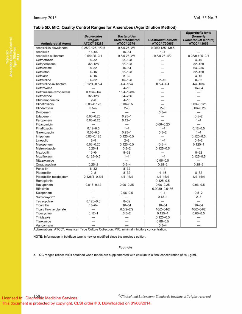
January 2015 Vol. 35 No. 3
168 ©Clinical and Laboratory Standards Institute. All rights reserved.
Table 5D. MIC: Quality Control Ranges for Anaerobes (Agar Dilution Method)
Abbreviations: ATCC®, American Type Culture Collection; MIC, minimal inhibitory concentration. NOTE: Information in boldface type is new or modified since the previous edition.
Footnote
a. QC ranges reflect MICs obtained when media are supplemented with calcium to a final concentration of 50 g/mL.
Antimicrobial Agent
Bacteroides fragilis
ATCC® 25285
Bacteroides thetaiotaomicron
ATCC® 29741 Clostridium difficile
ATCC® 700057
Eggerthella lenta (formerly
Eubacterium lentum) ATCC® 43055
Amoxicillin-clavulanate 0.25/0.125–1/0.5 0.5/0.25–2/1 0.25/0.125–1/0.5 — Ampicillin 16–64 16–64 1–4 — Ampicillin-sulbactam 0.5/0.25–2/1 0.5/0.25–2/1 0.5/0.25–4/2 0.25/0.125–2/1 Cefmetazole 8–32 32–128 — 4–16 Cefoperazone 32–128 32–128 — 32–128 Cefotaxime 8–32 16–64 — 64–256 Cefotetan 4–16 32–128 — 32–128 Cefoxitin 4–16 8–32 — 4–16 Ceftaroline 4–32 16–128 2–16 8–32 Ceftaroline-avibactam 0.12/4–0.5/4 4/4–16/4 0.5/4–4/4 4/4–16/4 Ceftizoxime Ceftolozane-tazobactam
— 0.12/4–1/4
4–16 16/4–128/4
— —
16–64 —
Ceftriaxone 32–128 64–256 — — Chloramphenicol 2–8 4–16 — —
Clinafloxacin 0.03–0.125 0.06–0.5 — 0.03–0.125 Clindamycin 0.5–2 2–8 2–8 0.06–0.25 Doripenem — — 0.5–4 — Ertapenem 0.06–0.25 0.25–1 — 0.5–2 Faropenem Fidaxomicin
0.03–0.25 —
0.12–1 —
— 0.06–0.25
1–4 —
Finafloxacin 0.12–0.5 1–4 1–4 0.12–0.5 Garenoxacin 0.06–0.5 0.25–1 0.5–2 1–4 Imipenem 0.03–0.125 0.125–0.5 — 0.125–0.5 Linezolid 2–8 2–8 1–4 0.5–2 Meropenem 0.03–0.25 0.125–0.5 0.5–4 0.125–1 Metronidazole 0.25–1 0.5–2 0.125–0.5 — Mezlocillin 16–64 8–32 — 8–32 Moxifloxacin 0.125–0.5 1–4 1–4 0.125–0.5 Nitazoxanide — — 0.06–0.5 — Omadacycline 0.25–2 0.5–4 0.25–2 0.25–2 Penicillin 8–32 8–32 1–4 — Piperacillin 2–8 8–32 4–16 8–32 Piperacillin-tazobactam 0.125/4–0.5/4 4/4–16/4 4/4–16/4 4/4–16/4 Ramoplanin — — 0.125–0.5 — Razupenem 0.015–0.12 0.06–0.25 0.06–0.25 0.06–0.5 Rifaximin — — 0.0039–0.0156 — Sulopenem Surotomycina
— —
0.06–0.5 —
1–4 0.12–1
0.5–2 2–8
Tetracycline 0.125–0.5 8–32 — — Ticarcillin 16–64 16–64 16–64 16–64 Ticarcillin-clavulanate — 0.5/2–2/2 16/2–64/2 16/2–64/2 Tigecycline 0.12–1 0.5–2 0.125–1 0.06–0.5 Tinidazole — — 0.125–0.5 — Tizoxanide — — 0.06–0.5 — Vancomycin — — 0.5–4 —
Tabl
e 5D
Anae
robe
Qua
lity
Cont
rol
Agar
Dilu
tion
M11
Licensed�to:��Diagnostic�Medicine�ServicesThis�document�is�protected�by�copyright.�CLSI�order�#�0,�Downloaded�on�01/06/2014.

For Use With M11-A8 M100-S25
©Clinical and Laboratory Standards Institute. All rights reserved. 169
This page is intentionally left blank.
Licensed�to:��Diagnostic�Medicine�ServicesThis�document�is�protected�by�copyright.�CLSI�order�#�0,�Downloaded�on�01/06/2014.

January 2015 Vol. 35 No. 3
Clinical and Laboratory Standards Institute. All rights reserved. 170
Table 5E. MIC: Quality Control Ranges for Anaerobes (Broth Microdilution Method)
Antimicrobial Agent
Bacteroides fragilis
ATCC® 25285
Bacteroides thetaiotaomicron
ATCC® 29741
Clostridium difficile
ATCC® 700057
Eggerthella lenta (formerly
Eubacterium lentum)
ATCC® 43055 Amoxicillin-clavulanate 0.25/0.125–1/0.5 0.25/0.125–1/0.5 — —Ampicillin-sulbactam 0.5/0.25–2/1 0.5/0.25–2/1 — 0.5/0.25–2/1 Cefotetan 1–8 16–128 — 16–64 Cefoxitin 2–8 8–64 — 2–16 Ceftaroline 2–16 8–64 0.5–4 – Ceftaroline-avibactam 0.06/4–0.5/4 2/4–8/4 0.25/4–1/4 4/4–16/4 Ceftizoxime Ceftolozane-tazobactam
— 0.12/4–1/4
— 16/4–64/4
— —
8–32 —
Chloramphenicol 4–16 8–32 — 4–16 Clindamycin 0.5–2 2–8 — 0.06–0.25 Doripenem 0.12–0.5 0.12–1 — —Doxycycline — 2–8 — 2–16 Ertapenem 0.06–0.5 0.5–2 — 0.5–4 Faropenem 0.015–0.06 0.12–1 — 0.5–2 Garenoxacin 0.06–0.25 0.25–2 — 0.5–2 Imipenem 0.03–0.25 0.25–1 — 0.25–2 Linezolid 2–8 2–8 — 0.5–2 Meropenem 0.03–0.25 0.06–0.5 — 0.125–1 Metronidazole 0.25–2 0.5–4 — 0.125–0.5 Moxifloxacin 0.12–0.5 1.0–8 — 0.12–0.5 Omadacyclinea 0.12–1 0.25–1 0.06–0.25 0.06–5 Penicillin 8–32 8–32 — — Piperacillin 4–16 8–64 — 8–32 Piperacillin-tazobactam Razupenem Sulopenem Surotomycinb
0.03/4–0.25/4 0.03–0.25
— —
2/4–16/4 0.12–0.5
0.03–0.25 —
— 0.06–0.5
0.5–2 0.12–1
8/4–32/4 0.12–0.5 0.25–1
1–4 Ticarcillin-clavulanate Tigecyclinea
0.06/2–0.5/2 0.06–0.5
0.5/2–2/2 0.25–1
— 0.03–0.12
8/2–32/2 —
Abbreviations: ATCC®, American Type Culture Collection; MIC, minimal inhibitory concentration. NOTE 1: Information in boldface type is new or modified since the previous edition. NOTE 2: For four-dilution ranges, results at the extremes of the acceptable range(s) should be suspect. Verify validity with data
from other QC strains.
Footnotes
a. For broth microdilution testing of tigecycline and omadacycline, when MIC panels are prepared, the medium must be prepared fresh on the day of use. The medium must be no greater than 12 hours old at the time the panels are made; however, the panels may then be frozen for later use.
b. QC ranges reflect MICs obtained when broth is supplemented with calcium to a final concentration of 50 g/mL.
Tabl
e 5E
Anae
robe
Qua
lity
Cont
rol
Brot
h M
icro
dilu
tion
M11
Licensed�to:��Diagnostic�Medicine�ServicesThis�document�is�protected�by�copyright.�CLSI�order�#�0,�Downloaded�on�01/06/2014.

For Use With M11-A8 M100-S25
©Clinical and Laboratory Standards Institute. All rights reserved. 171
This page is intentionally left blank.
Licensed�to:��Diagnostic�Medicine�ServicesThis�document�is�protected�by�copyright.�CLSI�order�#�0,�Downloaded�on�01/06/2014.

January 2015 Vol. 35 No. 3
Clinical and Laboratory Standards Institute. All rights reserved. 172
Table 5F. MIC: Reference Guide to Quality Control Frequency Conversion From Daily to Weekly QC Routine QC is performed each day the test is performed unless an alternative plan has been established (see CLSI document EP231). M07-A10, Subchapter 4.7.2.1 describes a 20- or 30-day plan that, if successfully completed, allows a user to convert from daily to weekly QC. An alternative option using a two-phase, 15-replicate (3 × 5 day) plan is described as follows: Test 3 replicates using individual inoculum preparations of the appropriate QC strains for 5
consecutive test days. Evaluate each QC strain/antimicrobial agent combination separately using acceptance criteria and
following recommended actions as described in the flow diagram below. Upon successful completion of either the 20- or 30-day plan or the 15-replicate (3 × 5 day) plan,
the laboratory can convert from daily to weekly QC testing. If unsuccessful, investigate, take corrective action as appropriate, and continue daily QC testing until either the 20- or 30-day plan or 15-replicate (3 × 5 day) plan is successfully completed. At that time weekly QC testing can be initiated.
15-Replicate (3 × 5 Day) Plan: Acceptance Criteria and Recommended Action*
Number Out of Range With Initial Testing
(Based on 15 Replicates)
Conclusion From Initial Testing (Based on 15
Replicates)
Number Out of Range After Repeat Testing (Based on All
30 Replicates) Conclusion After Repeat
Testing
0–1 Plan is successful. Convert
to weekly QC testing.
N/A
N/A
2–3 Test another 3 replicates for
5 days.
2–3 Plan is successful. Convert
to weekly QC testing.
≥ 4 Plan fails. Investigate and take corrective action as
appropriate. Continue QC each test day.
≥ 4
Plan fails. Investigate and take corrective action as
appropriate. Continue QC each test day.
* Assess each QC strain/antimicrobial agent combination separately. Abbreviations: N/A, not applicable; QC, quality control.
Tabl
e 5F
QC
Test
ing
Freq
uenc
yM
07
Licensed�to:��Diagnostic�Medicine�ServicesThis�document�is�protected�by�copyright.�CLSI�order�#�0,�Downloaded�on�01/06/2014.

For Use With M07-A10—MIC Testing M100-S25
©Clinical and Laboratory Standards Institute. All rights reserved. 173
Table 5F. (Continued) Test Modifications This table summarizes the suggested QC frequency when modifications are made to antimicrobial susceptibility test systems. It applies only to antimicrobial agents for which satisfactory results have been obtained with either the 15-replicate (3 × 5 day) plan or 20 or 30 consecutive test day plan.a Otherwise QC is required each test day.
Required QC Frequency
Test Modification 1
Day 5
Days
15-Replicate Plan or
20- or 30-Day Plan Comments
MIC Tests(s) Use new shipment or lot number. X Expand dilution range. X Example:
Convert from breakpoint to expanded range MIC panels.
Reduce dilution range. X Example: Convert from expanded dilution range to breakpoint panels.
Use new method (same company).
X Examples: Convert from visual to instrument reading of panel. Convert from overnight to rapid MIC test. In addition, perform in-house verification studies.
Use new manufacturer of MIC test.
X In addition, perform in-house verification studies.
Use new manufacturer of broth or agar.
X
Addition of new antimicrobial agent to existing system
X In addition, perform in-house verification studies.
Inoculum Preparation Convert inoculum preparation/ standardization to use of a device that has its own QC protocol.
X Example: Convert from visual adjustment of turbidity to use of a photometric device for which a QC procedure is provided.
Convert inoculum preparation/ standardization to a method that is dependent on user technique.
X Example: Convert from visual adjustment of turbidity to another method that is not based on a photometric device.
Instrument/Software Software update that affects AST results
X Monitor all drugs, not just those implicated in software modification.
Repair of instrument that affects AST results
X Depending on extent of repair (eg, critical component such as the optics), additional testing may be appropriate (eg, 5 days).
Abbreviations: AST, antimicrobial susceptibility testing; MIC, minimal inhibitory concentration; QC, quality control.
Tabl
e 5F
QC
Test
ing
Freq
uenc
yM
07
Licensed�to:��Diagnostic�Medicine�ServicesThis�document�is�protected�by�copyright.�CLSI�order�#�0,�Downloaded�on�01/06/2014.

January 2015 Vol. 35 No. 3
Clinical and Laboratory Standards Institute. All rights reserved. 174
Table 5F. (Continued) NOTE 1: QC can be performed before or concurrent with testing patient isolates. Patient results can be reported for that day if QC
results are within the acceptable limits. NOTE 2: Manufacturers of commercial or in-house-prepared tests should follow their own internal procedures and applicable
regulations. NOTE 3: Acceptable MIC QC limits for US Food and Drug Administration–cleared antimicrobial susceptibility tests may differ
slightly from acceptable CLSI QC limits. Users of each device should use the manufacturer’s procedures and QC limits as indicated in the instructions for use.
NOTE 4: For troubleshooting out-of-range results, refer to M07-A10, Subchapter 4.8.1 and M100 Table 5G. Additional information
is available in Appendix C, Quality Control Strains for Antimicrobial Susceptibility Tests (eg, organism characteristics, QC testing recommendations).
NOTE 5: Broth, saline, and/or water used to prepare an inoculum does not require routine QC.
Reference 1 CLSI. Laboratory Quality Control Based on Risk Management; Approved Guideline. CLSI document EP23-ATM. Wayne, PA:
Clinical and Laboratory Standards Institute; 2011.
Tabl
e 5F
QC
Test
ing
Freq
uenc
yM
07
Licensed�to:��Diagnostic�Medicine�ServicesThis�document�is�protected�by�copyright.�CLSI�order�#�0,�Downloaded�on�01/06/2014.

For Use With M07-A10—MIC Testing M100-S25
©Clinical and Laboratory Standards Institute. All rights reserved. 175
This page is intentionally left blank.
Licensed�to:��Diagnostic�Medicine�ServicesThis�document�is�protected�by�copyright.�CLSI�order�#�0,�Downloaded�on�01/06/2014.

January 2015 Vol. 35 No. 3
Clinical and Laboratory Standards Institute. All rights reserved. 176
Table 5G. MIC: Troubleshooting Guide This table provides guidance for troubleshooting and corrective action for out-of-range QC primarily using antimicrobial susceptibility tests with cation-adjusted Mueller-Hinton broth (CAMHB) for broth microdilution. Refer to M07-A10 (MIC), Chapter 4, Quality Control and Quality Assurance. Out-of-range QC tests should first be repeated. If the issue is unresolved, this troubleshooting guide provides additional suggestions for troubleshooting out-of-range QC results and unusual clinical isolate results. In addition, if unresolved, manufacturers should be notified of potential product problems.
General Comments
(1) QC organism maintenance: Avoid repeated subcultures. Retrieve new QC strain from stock. If using lyophilized strains, follow the maintenance recommendations of the manufacturer. Store E. coli ATCC® 35218 and K. pneumoniae ATCC® 700603 stock cultures at −60C or below and prepare working cultures weekly (refer to M07-A10, Subchapter 4.4).
Antimicrobial
Agent QC Strain Observation Probable Cause Comments/Suggested Actions Aminoglycosides Any MIC too high pH of media too low Acceptable pH range = 7.2–7.4
Avoid CO2 incubation, which lowers pH. Aminoglycosides Any MIC too low pH of media too high Acceptable pH range = 7.2–7.4 Aminoglycosides P. aeruginosa
ATCC® 27853 MIC too high Ca++ and/or Mg++
content too high Acceptable range = Ca++ 20–25 mg/L Mg++ 10–12.5 mg/L
Aminoglycosides P. aeruginosa ATCC® 27853
MIC too low Ca++ and/or Mg++ content too low
Acceptable range = Ca++ 20–25 mg/L Mg++ 10–12.5 mg/L
Amoxicillin- clavulanate
E. coli ATCC® 35218
MIC too high Clavulanate is labile. Antimicrobial agent is degrading.
Use alternative lot. Check storage and package integrity.
-Lactam group Any MIC initially acceptable, but increases possibly out of range over time
Antimicrobial agent is degrading.
Use alternative lot. Check storage and package integrity. Imipenem, cefaclor, and clavulanate are especially labile.
Aztreonam Cefotaxime Cefpodoxime Ceftazidime Ceftriaxone
K. pneumoniae ATCC® 700603
MIC too low Spontaneous loss of the plasmid encoding the -lactamase.
See general comment (1) on QC organism maintenance.
Cefotaxime- clavulanate Ceftazidime- clavulanate
K. pneumoniae ATCC® 700603
Negative ESBL confirmatory test
Spontaneous loss of the plasmid encoding the -lactamase.
See general comment (1) on QC organism maintenance.
Carbapenems P. aeruginosa ATCC® 27853
MIC too high Zn++ concentration in media is too high.
Use alternative lot.
Carbapenems P. aeruginosa ATCC® 27853
MIC too high Antimicrobial agent is degrading.
Use alternative lot. Check storage and package integrity. Repeated imipenem results of 4 g/mL with P. aeruginosa ATCC® 27853 may indicate deterioration of the drug.
Penicillin S. aureus ATCC® 29213
MIC too high QC strain is a -lactamase producer; overinoculation may yield increased MICs.
Repeat with a carefully adjusted inoculum.
Penicillins Any MIC too low pH of media too low Acceptable pH range = 7.2–7.4 Avoid CO2 incubation, which lowers pH.
Penicillins Any MIC too high pH of media too high Acceptable pH range = 7.2–7.4 Carbenicillin P. aeruginosa
ATCC® 27853 MIC too high QC strain develops
resistance after repeated subculture.
See general comment (1) on QC organism maintenance.
Ticarcillin- clavulanate
E. coli ATCC® 35218
MIC too high Clavulanate is labile. Antimicrobial agent is degrading.
Use alternative lot. Check storage and package integrity.
Clindamycin S. aureus ATCC® 29213 E. faecalis ATCC® 29212
MIC too high pH of media too low Acceptable pH range = 7.2–7.4 Avoid CO2 incubation, which lowers pH.
Tabl
e 5G
QC
Trou
bles
hoot
ing
M07
Licensed�to:��Diagnostic�Medicine�ServicesThis�document�is�protected�by�copyright.�CLSI�order�#�0,�Downloaded�on�01/06/2014.

For Use With M07-A10—MIC Testing M100-S25
©Clinical and Laboratory Standards Institute. All rights reserved. 177
Table 5G. (Continued) Antimicrobial
Agent QC Strain Observation Probable Cause Comments/Suggested Actions Clindamycin S. aureus
ATCC® 29213 E. faecalis ATCC® 29212
MIC too low pH of media too high Acceptable pH range = 7.2–7.4
Daptomycin S. aureus ATCC® 29213 E. faecalis ATCC® 29212
MICs too high MICs too low
Ca++ content too low Ca++ content too high
Acceptable Ca++ content 50 µg/mL in CAMHB Adjust Ca++ concentration in or try alternative lots.
Macrolides and Ketolides
S. aureus ATCC® 29213 E. faecalis ATCC® 29212
MIC too high pH of media too low Acceptable pH range = 7.2–7.4 Avoid CO2 incubation, which lowers pH.
Macrolides and Ketolides
S. aureus ATCC® 29213 E. faecalis ATCC® 29212
MIC too low pH of media too high Acceptable pH range = 7.2–7.4
Quinolones Any MIC too high pH of media too low Acceptable pH range = 7.2–7.4 Avoid CO2 incubation, which lowers pH.
Quinolones Any MIC too low pH of media too high Acceptable pH range = 7.2–7.4 Tetracyclines Any MIC too low pH of media too low Acceptable pH range = 7.2–7.4 Tetracyclines Any MIC too high pH of media too high Acceptable pH range = 7.2–7.4 Tetracyclines Any MIC too high Ca++ and/or Mg++ content
too high Acceptable range = Ca++ 20–25 mg/L Mg++ 10–12.5 mg/L
Tetracyclines Any MIC too low Ca++ and/or Mg++ content too low
Acceptable range = Ca++ 20–25 mg/L Mg++ 10–12.5 mg/L
Omadacycline Tigecycline
Any MIC too high CAMHB has not been freshly prepared.
Reference panels must be used or frozen within 12 hours of CAMHB preparation.
Various Any Many MICs too low
Inoculum too light; error in inoculum preparation
Repeat using McFarland 0.5 turbidity standard or standardizing device. Check expiration date and proper storage if using barium sulfate or latex standards. Check steps in inoculum preparation and inoculation procedure. Perform colony count check of growth control well immediately after inoculation and before incubation (E. coli ATCC® 25922 closely approximates 5 105 CFU/mL).
Various Any Many MICs too high or too low
CAMHB not optimal Use alternative lot.
Various Any Many MICs too high
Inoculum too heavy Repeat using McFarland 0.5 turbidity standard or standardizing device. Check expiration date and proper storage if using barium sulfate or latex standards. Check steps in inoculum preparation and inoculation procedure. Perform colony count check of growth control well immediately after inoculation and before incubation (E. coli ATCC® 25922 closely approximates 5 105 CFU/mL).
Various Any Skipped wells Contamination. Improper inoculation of panel or inadequate mixing of inoculum. Actual concentration of drug in wells inaccurate. Volume of broth in wells inaccurate.
Repeat QC test. Use alternative lot.
Various Any Several MICs too high or too low
Possible reading/transcription error
Recheck readings. Use alternative lot.
Tabl
e 5G
QC
Trou
bles
hoot
ing
M07
Licensed�to:��Diagnostic�Medicine�ServicesThis�document�is�protected�by�copyright.�CLSI�order�#�0,�Downloaded�on�01/06/2014.

January 2015 Vol. 35 No. 3
Clinical and Laboratory Standards Institute. All rights reserved. 178
Table 5G. (Continued) Antimicrobial
Agent QC Strain Observation Probable Cause Comments/Suggested Actions Various S. pneumoniae
ATCC® 49619 MICs too low Inoculum source plate too
old and contains too many nonviable cells. Plate used to prepare inoculum should be 18–20 hours. MHB with LHB not optimal.
Subculture QC strain and repeat QC test; or subculture new QC strain from stock culture. Use alternative lot.
Various Any One QC strain is out of range, but other QC strains are in range with the same antimicrobial agent.
One QC organism may be a better indicator of a QC problem (eg, P. aeruginosa ATCC® 27853 is a better indicator of imipenem deterioration than E. coli ATCC® 25922).
Determine if the in-range QC strain has an on-scale end point for the agent in question. Retest this strain to confirm reproducibility of acceptable results. Evaluate with alternative strains with known MICs. Initiate corrective action with problem QC strain/antimicrobial agent(s).
Various Any Two QC strains are out of range with the same antimicrobial agent.
Indicates a problem with the antimicrobial agent. May be a systemic problem.
Initiate corrective action.
Various Any One QC result is out of range, but the antimicrobial agent is not an agent reported for patient results (eg, not on hospital formulary).
If antimicrobial agent is not normally reported, no repeat is necessary if adequate controls are in place to prevent reporting of the out-of-range antimicrobial agent. Carefully check antimicrobial agents of the same class for similar trend toward out-of-control results. If the antimicrobial agent in question is consistently out of control, contact the manufacturer.
Abbreviations: ATCC®, American Type Culture Collection; CAMHB, cation-adjusted Mueller-Hinton broth; CFU, colony-forming unit(s); ESBL, extended-spectrum -lactamase; LHB, lysed horse blood; MHB, Mueller-Hinton broth; MIC, minimal inhibitory concentration; QC, quality control.
Tabl
e 5G
QC
Trou
bles
hoot
ing
M07
Licensed�to:��Diagnostic�Medicine�ServicesThis�document�is�protected�by�copyright.�CLSI�order�#�0,�Downloaded�on�01/06/2014.

For Use With M07-A10—MIC Testing M100-S25
©Clinical and Laboratory Standards Institute. All rights reserved. 179
This page is intentionally left blank.
Licensed�to:��Diagnostic�Medicine�ServicesThis�document�is�protected�by�copyright.�CLSI�order�#�0,�Downloaded�on�01/06/2014.

January 2015 Vol. 35 No. 3
Clinical and Laboratory Standards Institute. All rights reserved. 180
Table 6A. Solvents and Diluents for Preparation of Stock Solutions of Antimicrobial Agentse Antimicrobial Agent Solvent Diluent
Unless otherwise stated, use a minimum amount of the listed solvent to solubilize the antimicrobial powder.
Finish diluting the final stock solution as stated below.
Amikacin Water Water Amoxicillin Phosphate buffer, pH 6.0, 0.1 mol/L Phosphate buffer, pH 6.0, 0.1 mol/L Ampicillin Phosphate buffer, pH 8.0, 0.1 mol/L Phosphate buffer, pH 6.0, 0.1 mol/L Avibactam Water Water Azithromycin 95% ethanol or glacial acetic acide,f Broth media Azlocillin Water Water Aztreonam Saturated solution sodium bicarbonate Water Besifloxacin Methanol Water Biapenem Salinem Salinem Carbenicillin Water Water Cefaclor Water Water Cefadroxil Phosphate buffer, pH 6.0, 0.1 mol/L Water Cefamandole Water Water Cefazolin Phosphate buffer, pH 6.0, 0.1 mol/L Phosphate buffer, pH 6.0, 0.1 mol/L Cefdinir Phosphate buffer, pH 6.0, 0.1 mol/L Water Cefditoren Phosphate buffer, pH 6.0, 0.1 mol/L Water Cefepime Phosphate buffer, pH 6.0, 0.1 mol/L Phosphate buffer, pH 6.0, 0.1 mol/L Cefetamet Phosphate buffer, pH 6.0, 0.1 mol/L Water Cefixime Phosphate buffer, pH 7.0, 0.1 mol/L Phosphate buffer, pH 7.0, 0.1 mol/L Cefmetazole Water Water Cefonicid Water Water Cefoperazone Water Water Cefotaxime Water Water Cefotetan DMSOe Water Cefoxitin Water Water Cefpodoxime 0.10% (11.9 mmol/L) aqueous sodium bicarbonate Water Cefprozil Water Water Ceftaroline DMSOe to 30% of total volume Salinem Ceftazidime Sodium carbonated Water Ceftibuten 1/10 vol DMSOe Water Ceftizoxime Water Water Ceftobiprole DMSO plus glacial acetic acide,h Water, vortex vigorously Ceftolozane Water or salinem Water or salinem Ceftriaxone Water Water Cefuroxime Phosphate buffer, pH 6.0, 0.1 mol/L Phosphate buffer, pH 6.0, 0.1 mol/L Cephalexin Phosphate buffer, pH 6.0, 0.1 mol/L Water Cephalothin Phosphate buffer, pH 6.0, 0.1 mol/L Water Cephapirin Phosphate buffer, pH 6.0, 0.1 mol/L Water Cephradine Phosphate buffer, pH 6.0, 0.1 mol/L Water Chloramphenicol 95% ethanol Water Cinoxacin 1/2 volume of water, then add 1 mol/L NaOH dropwise
to dissolve Water
Ciprofloxacin Water Water Clarithromycin Methanole or glacial acetic acide,f Phosphate buffer, pH 6.5, 0.1 mol/L Clavulanate Phosphate buffer, pH 6.0, 0.1 mol/L Phosphate buffer, pH 6.0, 0.1 mol/L Clinafloxacin Water Water Clindamycin Water Water Colistina Water Water Dalbavancin DMSOe DMSOe,g
Daptomycin Water Water
Tabl
e 6A
Solv
ents
and
Dilu
ents
M07
Licensed�to:��Diagnostic�Medicine�ServicesThis�document�is�protected�by�copyright.�CLSI�order�#�0,�Downloaded�on�01/06/2014.

For Use With M07-A10—MIC Testing M100-S25
©Clinical and Laboratory Standards Institute. All rights reserved. 181
Table 6A. (Continued) Antimicrobial Agent Solvent Diluent
Unless otherwise stated, use a minimum amount of the listed solvent to solubilize the antimicrobial powder.
Finish diluting the final stock solution as stated below.
Dirithromycin Glacial acetic acidf Water Doripenem Salinem Salinem Doxycycline Water Water Enoxacin 1/2 volume of water, then 0.1 mol/L NaOH dropwise
to dissolve Water
Eravacycline Water Water Ertapenem Phosphate buffer, pH 7.2, 0.01 mol/L Phosphate buffer, pH 7.2, 0.01 mol/L Erythromycin 95% ethanol or glacial acetic acide,f Water Faropenem Water Water Fidaxomicin DMSOe Water Finafloxacin Water Water Fleroxacin 1/2 volume of water, then 0.1 mol/L NaOH dropwise
to dissolve Water
Fosfomycin Water Water Fusidic acid Water Water Garenoxacin Water (with stirring) Water Gatifloxacin Water (with stirring) Water Gemifloxacin Water Water Gentamicin Water Water Iclaprim DMSOe Water Imipenem Phosphate buffer, pH 7.2, 0.01 mol/L Phosphate buffer, pH 7.2, 0.01 mol/L Kanamycin Water Water Levofloxacin 1/2 volume of water, then 0.1 mol/L NaOH dropwise
to dissolve Water
Linezolid Water Water Linopristin-flopristin DMFk Water Lomefloxacin Water Water Loracarbef Water Water Mecillinam Water Water Meropenem Water Water Methicillin Water Water Metronidazole DMSOe Water Mezlocillin Water Water Minocycline Water Water Moxalactam (diammonium salt)b
0.04 mol/L HCI (let sit for 1.5 to 2 hours) Phosphate buffer, pH 6.0, 0.1 mol/L
Moxifloxacin Water Water Mupirocin Water Water Nafcillin Water Water Nalidixic acid 1/2 volume of water, then add 1 mol/L NaOH dropwise to
dissolve
Netilmicin Water Water Nitazoxanide DMSOe,l DMSOe,l Nitrofurantoinc Phosphate buffer, pH 8.0, 0.1 mol/L Phosphate buffer, pH 8.0, 0.1 mol/L Norfloxacin 1/2 volume of water, then 0.1 mol/L NaOH dropwise
to dissolve Water
Tabl
e 6A
Solv
ents
and
Dilu
ents
M07
Licensed�to:��Diagnostic�Medicine�ServicesThis�document�is�protected�by�copyright.�CLSI�order�#�0,�Downloaded�on�01/06/2014.
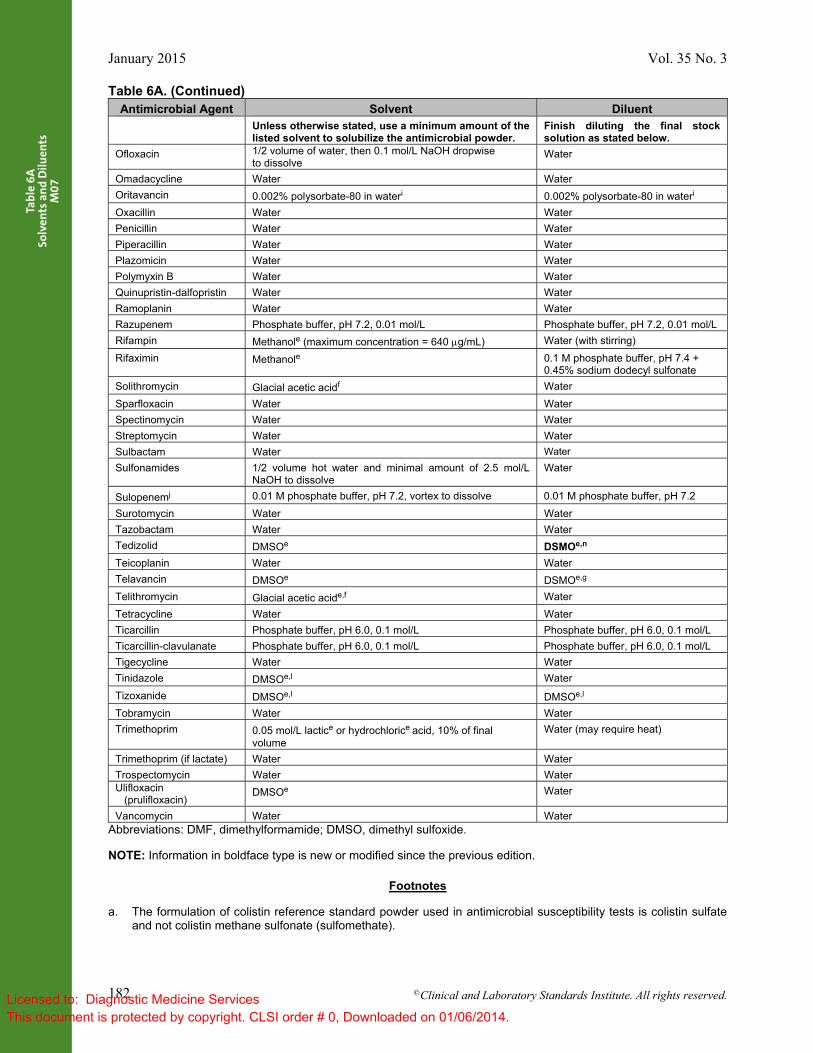
January 2015 Vol. 35 No. 3
Clinical and Laboratory Standards Institute. All rights reserved. 182
Table 6A. (Continued) Antimicrobial Agent Solvent Diluent
Unless otherwise stated, use a minimum amount of the listed solvent to solubilize the antimicrobial powder.
Finish diluting the final stock solution as stated below.
Ofloxacin 1/2 volume of water, then 0.1 mol/L NaOH dropwise to dissolve
Water
Omadacycline Water Water Oritavancin 0.002% polysorbate-80 in wateri 0.002% polysorbate-80 in wateri
Oxacillin Water Water Penicillin Water Water Piperacillin Water Water Plazomicin Water Water Polymyxin B Water Water Quinupristin-dalfopristin Water Water Ramoplanin Water Water Razupenem Phosphate buffer, pH 7.2, 0.01 mol/L Phosphate buffer, pH 7.2, 0.01 mol/L Rifampin Methanole (maximum concentration = 640 g/mL) Water (with stirring) Rifaximin Methanole 0.1 M phosphate buffer, pH 7.4 +
0.45% sodium dodecyl sulfonate Solithromycin Glacial acetic acidf Water Sparfloxacin Water Water Spectinomycin Water Water Streptomycin Water Water Sulbactam Water Water Sulfonamides 1/2 volume hot water and minimal amount of 2.5 mol/L
NaOH to dissolve Water
Sulopenemj 0.01 M phosphate buffer, pH 7.2, vortex to dissolve 0.01 M phosphate buffer, pH 7.2 Surotomycin Water Water Tazobactam Water Water Tedizolid DMSOe DSMOe,n Teicoplanin Water Water Telavancin DMSOe DSMOe,g Telithromycin Glacial acetic acide,f Water Tetracycline Water Water Ticarcillin Phosphate buffer, pH 6.0, 0.1 mol/L Phosphate buffer, pH 6.0, 0.1 mol/L Ticarcillin-clavulanate Phosphate buffer, pH 6.0, 0.1 mol/L Phosphate buffer, pH 6.0, 0.1 mol/L Tigecycline Water Water Tinidazole DMSOe,l Water Tizoxanide DMSOe,l DMSOe,l Tobramycin Water Water Trimethoprim 0.05 mol/L lactice or hydrochlorice acid, 10% of final
volume Water (may require heat)
Trimethoprim (if lactate) Water Water Trospectomycin Water Water Ulifloxacin (prulifloxacin)
DMSOe Water
Vancomycin Water Water Abbreviations: DMF, dimethylformamide; DMSO, dimethyl sulfoxide. NOTE: Information in boldface type is new or modified since the previous edition.
Footnotes a. The formulation of colistin reference standard powder used in antimicrobial susceptibility tests is colistin sulfate
and not colistin methane sulfonate (sulfomethate).
Tabl
e 6A
Solv
ents
and
Dilu
ents
M07
Licensed�to:��Diagnostic�Medicine�ServicesThis�document�is�protected�by�copyright.�CLSI�order�#�0,�Downloaded�on�01/06/2014.

For Use With M07-A10—MIC Testing M100-S25
©Clinical and Laboratory Standards Institute. All rights reserved. 183
Table 6A. (Continued) b. The diammonium salt of moxalactam is very stable, but it is almost pure R isomer. Moxalactam for clinical use is
a 1:1 mixture of R and S isomers. Therefore, the salt is dissolved in 0.04 mol/L HCl and allowed to react for 1.5 to 2 hours to convert it to equal parts of both isomers.
c. Alternatively, nitrofurantoin is dissolved in DMSO. d. Anhydrous sodium carbonate is used at a weight of exactly 10% of the ceftazidime to be used. The sodium
carbonate is dissolved in solution in most of the required water. The antimicrobial agent is dissolved in this sodium carbonate solution, and water is added to the desired volume. The solution is to be used as soon as possible, but it can be stored up to six hours at no more than 25°C.
e. Consult the safety data sheets before working with any antimicrobial reference standard powder, solvent, or
diluent. Some of the compounds (eg, solvents such as DMSO, methanol) are more toxic than others and may necessitate handling in a chemical fume hood.
f. For glacial acetic acid, use 1/2 volume of water, then add glacial acetic acid dropwise until dissolved, not to
exceed 2.5 µL/mL. g. Starting stock solutions of dalbavancin and telavancin should be prepared at concentrations no higher than 1600
µg/mL. Intermediate 100× concentrations should then be diluted in DMSO. Final 1:100 dilutions should then be made directly into cation-adjusted Mueller-Hinton broth (CAMHB) supplemented with 0.002% (v/v) polysorbate-80, so the final concentration of DMSO in the wells is no greater than 1%. See also Table 8B.
h. For each 1.5 mg of ceftobiprole, add 110 L of a 10:1 mixture of DMSO and glacial acetic acid. Vortex vigorously
for one minute, then intermittently for 15 minutes. Dilute to 1.0 mL with distilled water. i. Starting stock solutions of oritavancin should be prepared at concentrations no higher than 1600 g/mL in
0.002% polysorbate-80 in water. Intermediate 100× oritavancin concentrations should then be prepared in 0.002% polysorbate-80 in water. Final 1:100 dilutions should be made directly into CAMHB supplemented with 0.002% polysorbate-80, so the final concentration of polysorbate-80 in the wells is 0.002%.
j. Must be made fresh on the day of use. k. DMF to 25% of final volume/water. l. Final concentration of DMSO should not exceed 1%. This may be accomplished as follows: 1) prepare the stock
solution at 10 times higher concentration than planned stock solution (ie, prepare at 12 800 g/mL, rather than 1280 µg/mL); 2) add 1.8 mL sterile water to each agar deep; 3) add 0.2 mL of each antibiotic dilution to each agar deep.
m. Saline – a solution of 0.85% to 0.9% NaCl (w/v). n. Starting stock solutions of tedizolid should be prepared at concentrations no higher than 1600 g/mL.
Intermediate 100× concentrations should be diluted in DMSO. Final 1:100 dilutions should be made directly into CAMHB, so that the final concentration of DMSO in the wells is no greater than 1%. See also Table 8B.
Ta
ble
6ASo
lven
ts a
nd D
iluen
tsM
07
Licensed�to:��Diagnostic�Medicine�ServicesThis�document�is�protected�by�copyright.�CLSI�order�#�0,�Downloaded�on�01/06/2014.
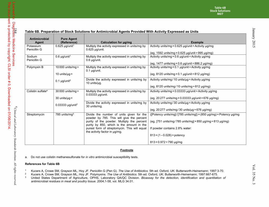
Table 6B. Preparation of Stock Solutions for Antimicrobial Agents Provided With Activity Expressed as Units
Footnote
a. Do not use colistin methanesulfonate for in vitro antimicrobial susceptibility tests. References for Table 6B 1 Kucers A, Crowe SM, Grayson ML, Hoy JF. Penicillin G (Pen G). The Use of Antibiotics. 5th ed. Oxford, UK: Butterworth-Heinemann; 1997:3-70. 2 Kucers A, Crowe SM, Grayson ML, Hoy JF. Polymyxins. The Use of Antibiotics. 5th ed. Oxford, UK: Butterworth-Heinemann; 1997:667-675. 3 United States Department of Agriculture, OPHS, Laboratory QA/QC Division. Bioassay for the detection, identification and quantitation of
antimicrobial residues in meat and poultry tissue. 2004;1-58, vol. MLG 34.01.
Antimicrobial Agent
Pure Agent (Reference) Calculation for µg/mg Example
Potassium Penicillin G
0.625 µg/unit1 Multiply the activity expressed in units/mg by 0.625 µg/unit.
Activity units/mg × 0.625 µg/unit = Activity µg/mg (eg, 1592 units/mg × 0.625 µg/unit = 995 µg/mg)
Sodium Penicillin G
0.6 µg/unit1 Multiply the activity expressed in units/mg by 0.6 µg/unit.
Activity units/mg × 0.6 µg/unit = Activity µg/mg (eg, 1477 units/mg × 0.6 µg/unit = 886.2 µg/mg)
Polymyxin B 10 000 units/mg =
10 units/µg =
0.1 µg/unit2
Multiply the activity expressed in units/mg by 0.1 µg/unit.
Activity units/mg × 0.1 µg/unit = Activity µg/mg (eg, 8120 units/mg × 0.1 µg/unit = 812 µg/mg)
Divide the activity expressed in units/mg by 10 units/µg.
Activity units/mg / 10 units/µg = Activity µg/mg (eg, 8120 units/mg / 10 units/mg = 812 µg/mg)
Colistin sulfatea 30 000 units/mg =
30 units/µg =
0.03333 µg/unit2
Multiply the activity expressed in units/mg by 0.03333 µg/unit.
Activity units/mg × 0.03333 µg/unit = Activity µg/mg (eg, 20 277 units/mg × 0.03333 µg/unit = 676 µg/mg)
Divide the activity expressed in units/mg by 30 units/mg.
Activity units/mg / 30 units/µg = Activity µg/mg (eg, 20 277 units/mg / 30 units/µg = 676 µg/mg)
Streptomycin 785 units/mg3 Divide the number of units given for the powder by 785. This will give the percent purity of the powder. Multiply the percent purity by 850, which is the amount in the purest form of streptomycin. This will equal the activity factor in µg/mg.
([Potency units/mg] / [785 units/mg]) × (850 µg/mg) = Potency µg/mg (eg, [751 units/mg / 785 units/mg] × 850 µg/mg = 813 µg/mg) If powder contains 2.8% water: 813 × (1 – 0.028) = potency 813 × 0.972 = 790 µg/mg
184 ©Clinical and Laboratory Standards Institute. All rights reserved.
January 2015
Vol. 35 N
o. 3
Table 6BStock Solutions
M07
Licensed�to:��Diagnostic�M
edicine�Services
This�document�is�protected�by�copyright.�C
LSI�order�#�0,�D
ownloaded�on�01/06/2014.

For Use With M07-A10—MIC Testing M100-S25
Clinical and Laboratory Standards Institute. All rights reserved. 185
This page is intentionally left blank.
Licensed�to:��Diagnostic�Medicine�ServicesThis�document�is�protected�by�copyright.�CLSI�order�#�0,�Downloaded�on�01/06/2014.

Table 6C. Preparation of Solutions and Media Containing Combinations of Antimicrobial Agents Antimicrobial
Agent
Combination Tested Preparation Example Amoxicillin-clavulanate
2:1 ratio (amoxicillin:clavulanate)
Prepare 10× starting concentration as 2:1 ratio and dilute as needed.
For a starting concentration of 128/64 in the panel, prepare a 10× stock concentration of 2560 µg/mL for amoxicillin and 1280 µg/mL for clavulanate. Then combine equal amounts of each to the first dilution tube, which will then contain 1280/640 µg/mL of the combination. Dilute 1:10 with broth to achieve the final concentration in microdilution wells.
Ampicillin- sulbactam
2:1 ratio (ampicillin:sulbactam) Same as amoxicillin-clavulanate.
Aztreonam-avibactam
Fixed concentration of avibactam at 4 µg/mL
Prepare 10× starting concentration of aztreonam at twice the concentration needed and dilute as usual using serial twofold dilutions. Add an equal volume of avibactam 80 µg/mL to each of the diluted tubes.
For a starting concentration of 128/4 in the panel, prepare a 10× stock concentration of aztreonam at 2560 µg/mL and dilute by serial twofold increments down to the final concentration needed in the panel. Prepare a stock concentration of avibactam at 80 µg/mL. Then add an equal volume of the avibactam 80 µg/mL solution to each diluted tube of aztreonam. For example, 5 mL of 2560 µg/mL aztreonam + 5 mL of 80 µg/mL avibactam = 10 mL of 1280/40 µg/mL aztreonam-avibactam. Dilute 1:10 with broth to achieve the final concentration in microdilution wells.
Ceftaroline-avibactam
Fixed concentration of avibactam at 4 µg/mL
Same as aztreonam-avibactam.
Ceftazidime-avibactam
Fixed concentration of avibactam at 4 µg/mL
Same as aztreonam-avibactam.
Ceftolozane- tazobactam
Fixed concentration of tazobactam at 4 μg/mL
Same as aztreonam-avibactam.
Piperacillin-tazobactam
Fixed concentration of tazobactam at 4 µg/mL
Same as aztreonam-avibactam.
Ticarcillin-clavulanate
Fixed concentration of clavulanate at 2 µg/mL
Prepare 10× starting concentration of ticarcillin at twice the concentration needed and dilute as usual using serial twofold dilutions. Add an equal volume of clavulanate 40 µg/mL to each of the diluted tubes.
For a starting concentration of 128/2 in the panel, prepare a 10× stock concentration of ticarcillin at 2560 µg/mL and dilute by serial twofold increments down to the final concentration needed. Prepare a stock concentration of clavulanate at 40 µg/mL. Then add an equal volume of the clavulanate 40 µg/mL solution to each diluted tube of ticarcillin. For example, 5 mL of 2560 µg/mL ticarcillin + 5 mL of 40 µg/mL clavulanate = 10 mL of 1280/20 µg/mL ticarcillin-clavulanate. Dilute 1:10 with broth to achieve the final concentration in microdilution wells.
186 ©Clinical and Laboratory Standards Institute. All rights reserved.
January 2015
Vol. 35 N
o. 3
Table 6CSolutions and Media
M07
Licensed�to:��Diagnostic�M
edicine�Services
This�document�is�protected�by�copyright.�C
LSI�order�#�0,�D
ownloaded�on�01/06/2014.

©Clinical and Laboratory Standards Institute. All rights reserved. 187
Table 6C. (Continued) Antimicrobial
Agent Combination Tested Preparation Example Trimethoprim-sulfamethoxazole
1:19 ratio (trimethoprim:sulfamethoxazole)
Prepare a 10× starting concentration of trimethoprim at 1600 µg/mL (or at 1280 µg/mL that will require dilution to 160 µg/mL). Prepare a 10× starting concentration of sulfamethoxazole at a log2 multiple of 1520 µg/mL (eg, 1520, 3040, or 6080 µg/mL) depending on the starting concentration needed.
For a starting concentration of 8/152 in the panel, prepare a 10× concentration of trimethoprim at 160 µg/mL. Prepare a 10× starting concentration of sulfamethoxazole at 3040 µg/mL. Add an equal volume of the 160 µg/mL trimethoprim and the 3040 µg/mL sulfamethoxazole to the first dilution tube, and then dilute by serial twofold dilutions as usual. For example, 5 mL of 160 µg/mL trimethoprim and 5 mL of 3040 µg/mL sulfamethoxazole = 10 mL of 80/1520 trimethoprim-sulfamethoxazole. Dilute 1:10 with broth to achieve the final concentration in microdilution wells.
Quinupristin- dalfopristin Linopristin- flopristin
Preparation usually not required, because drug powder is received as combination.
NOTE: To prepare intermediate dilutions of antimicrobial agents, a convenient formula to use is C1 V1 = C2 V2, where C1 is the concentration of stock solution of the antimicrobial agent (usually 1280 µg/mL or greater); V1 is the unknown volume that will be needed to make the intermediate concentration; C2 is the intermediate concentration needed; and V2 is the volume of the intermediate stock solution needed.
For example: To prepare 20 mL of a 40 µg/mL solution from a 1280 µg/mL stock solution:
C1 V1 = C2 V2
1280 µg/mL V1 = 40 µg/mL 20 mL
V1 = 40 µg/mL 20 mL
1280 µg/mL V1 = 0.625 mL
Therefore, add 0.625 mL of the 1280 µg/mL stock solution to 19.375 mL of diluent (usually water) for a final volume of 20 mL of a 40 µg/mL solution.
For Use W
ith M07-A
10—M
IC Testing M
100-S25
Table 6CSolutions and Media
M07
Licensed�to:��Diagnostic�M
edicine�Services
This�document�is�protected�by�copyright.�C
LSI�order�#�0,�D
ownloaded�on�01/06/2014.

January 2015 Vol. 35 No. 3
188 Clinical and Laboratory Standards Institute. All rights reserved.
Table 7A. Scheme for Preparing Dilutions of Antimicrobial Agents to Be Used in Agar Dilution Susceptibility Tests
Antimicrobial Solution
Step Concentration
(g/mL) Source Volume
(mL) Diluent
(mL)
Intermediate Concentration
(g/mL)
Final Concentration
at 1:10 Dilution in Agar (g/mL) Log2
5120 Stock – – 5120 512 9 1 5120 Stock 2 2 2560 256 8 2 5120 Stock 1 3 1280 128 7 3 5120 Stock 1 7 640 64 6 4 640 Step 3 2 2 320 32 5 5 640 Step 3 1 3 160 16 4 6 640 Step 3 1 7 80 8 3 7 80 Step 6 2 2 40 4 2 8 80 Step 6 1 3 20 2 1 9 80 Step 6 1 7 10 1 0 10 10 Step 9 2 2 5 0.5 −1 11 10 Step 9 1 3 2.5 0.25 −2 12 10 Step 9 1 7 1.25 0.125 −3
NOTE: This table is modified from Ericsson HM, Sherris JC. Antibiotic sensitivity testing. Report of an international collaborative study. Acta Pathol Microbiol Scand. 1971;217(suppl B):1-98.
Tabl
e 7A
Dilu
tion
Sche
me
for A
gar D
ilutio
n Te
sts
M07
Licensed�to:��Diagnostic�Medicine�ServicesThis�document�is�protected�by�copyright.�CLSI�order�#�0,�Downloaded�on�01/06/2014.

For Use With M07-A10—MIC Testing M100-S25
Clinical and Laboratory Standards Institute. All rights reserved. 189
This page is intentionally left blank.
Licensed�to:��Diagnostic�Medicine�ServicesThis�document�is�protected�by�copyright.�CLSI�order�#�0,�Downloaded�on�01/06/2014.

January 2015 Vol. 35 No. 3
190 Clinical and Laboratory Standards Institute. All rights reserved.
Table 8A. Scheme for Preparing Dilutions of Antimicrobial Agents to Be Used in Broth Dilution Susceptibility Tests
Antimicrobial Solution
Step
Concentration
(g/mL)
Source
Volumea
(mL)
+
CAMHBb
Volumea
(mL)
=
Final Concentration
(g/mL)
Log2
1 5120 Stock 1 9 512 9
2 512 Step 1 1 1 256 8 3 512 Step 1 1 3 128 7 4 512 Step 1 1 7 64 6 5 64 Step 4 1 1 32 5 6 64 Step 4 1 3 16 4 7 64 Step 4 1 7 8 3 8 8 Step 7 1 1 4 2 9 8 Step 7 1 3 2 1 10 8 Step 7 1 7 1 0 11 1 Step 10 1 1 0.5 −1 12 1 Step 10 1 3 0.25 −2 13 1 Step 10 1 7 0.125 −3
Abbreviation: CAMHB, cation-adjusted Mueller-Hinton broth. NOTE: This table is modified from Ericsson HM, Sherris JC. Antibiotic sensitivity testing. Report of an international collaborative study. Acta Pathol Microbiol Scand. 1971;217(suppl B):1-90.
Footnotes a. The volumes selected can be any multiple of these figures, depending on the number of tests to be performed. b. Adjustment with cations, if necessary, occurs before this step.
Tabl
e 8A
Dilu
tion
Sche
me
for A
ntim
icro
bial
Age
nts
for B
roth
Dilu
tion
Test
sM
07
Licensed�to:��Diagnostic�Medicine�ServicesThis�document�is�protected�by�copyright.�CLSI�order�#�0,�Downloaded�on�01/06/2014.

For Use With M07-A10—MIC Testing M100-S25
Clinical and Laboratory Standards Institute. All rights reserved. 191
This page is intentionally left blank.
Licensed�to:��Diagnostic�Medicine�ServicesThis�document�is�protected�by�copyright.�CLSI�order�#�0,�Downloaded�on�01/06/2014.

January 2015 Vol. 35 No. 3
192 Clinical and Laboratory Standards Institute. All rights reserved.
Table 8B. Scheme for Preparing Dilutions of Water-Insoluble Antimicrobial Agents to Be Used in Broth Dilution Susceptibility Tests
Antimicrobial Solution
Step
Concentration
(µg/mL)
Source
Volume
(mL) +
Solvent
(mL) (eg,
DMSO)
=
Intermediate
Concentration (g/mL)
=
Final
Concentration at 1:100 (g/mL) Log2
1 1600 Stock 1600 16 4 2 1600 Stock 0.5 0.5 800 8.0 3 3 1600 Stock 0.5 1.5 400 4.0 2 4 1600 Stock 0.5 3.5 200 2.0 1 5 200 Step 4 0.5 0.5 100 1.0 0 6 200 Step 4 0.5 1.5 50 0.5 −1 7 200 Step 4 0.5 3.5 25 0.25 −2 8 25 Step 7 0.5 0.5 12.5 0.125 −3 9 25 Step 7 0.5 1.5 6.25 0.0625 −4 10 25 Step 7 0.5 3.5 3.1 0.03 −5 11 3.1 Step 10 0.5 0.5 1.6 0.016 −6 12 3.1 Step 10 0.5 1.5 0.8 0.008 −7 13 3.1 Step 10 0.5 3.5 0.4 0.004 −8 14 0.4 Step 13 0.5 0.5 0.2 0.002 −9
Abbreviation: DMSO, dimethyl sulfoxide.
Tabl
e 8B
Dilu
tion
Sche
me
for W
ater
-Inso
lubl
e An
timic
robi
al A
gent
s fo
r Bro
th D
ilutio
n Te
sts
M07
Licensed�to:��Diagnostic�Medicine�ServicesThis�document�is�protected�by�copyright.�CLSI�order�#�0,�Downloaded�on�01/06/2014.

For Use With M07-A10—MIC Testing M100-S25
Clinical and Laboratory Standards Institute. All rights reserved. 193
This page is intentionally left blank.
Licensed�to:��Diagnostic�Medicine�ServicesThis�document�is�protected�by�copyright.�CLSI�order�#�0,�Downloaded�on�01/06/2014.
Appendix A. Suggestions for Confirmation of Resistant (R), Intermediate (I), or Nonsusceptible (NS) Antimicrobial Susceptibility Test Results and Organism Identification
Organism or Organism Group Resistance Phenotype Detecteda
Occurrence and Significance of Resistance and Actions to Take Following Confirmation of Resultsa
Category I Category II Category III
Not reported or only
rarely reported to date Uncommon in most
institutions
May be common, but is generally considered of
epidemiological concern
Action Steps:
Confirm ID and susceptibility.a
Report to infection control.
Send to public health laboratory.
Save isolate. NOTE: May be appropriate to notify infection control of preliminary findings before confirmation of results.
Confirm ID and susceptibility if uncommon in your institution.a
Check with infection control in your facility to determine if special reporting procedures or further action are needed.
Check with your local public health department to determine which isolates should be reported to them and when isolates should be sent to the public health laboratory.
Confirm ID and susceptibility if uncommon in your institution.a
Check with infection control in your facility to determine if special reporting procedures or further action are needed.
Any Enterobacteriaceae
Carbapenem – I or Rb x Amikacin, gentamicin, and tobramycin – R x
Escherichia coli Klebsiella spp. Proteus mirabilis
Extended-spectrum cephalosporinc – I or R x
Salmonella and Shigella spp.d
Cephalosporin III – I or R x Fluoroquinolone – I or R x
Acinetobacter baumannii
Colistin/polymyxin – R x Carbapenem – I or R x
Pseudomonas aeruginosa
Colistin/polymyxin – I or R x Amikacin, gentamicin, and tobramycin – R Carbapenem – I or R
x
194 ©Clinical and Laboratory Standards Institute. All rights reserved.
January 2015
Vol. 35 N
o. 3
Appendix ASuggested Test Result Confirmation
and Organism Identification
Licensed�to:��Diagnostic�M
edicine�Services
This�document�is�protected�by�copyright.�C
LSI�order�#�0,�D
ownloaded�on�01/06/2014.

©Clinical and Laboratory Standards Institute. All rights reserved. 195
Appendix A. (Continued)
Organism or Organism Group Resistance Phenotype Detecteda
Occurrence and Significance of Resistance and Actions to Take Following Confirmation of Resultsa
Category I Category II Category III
Not reported or only rarely reported to date
Uncommon in most institutions
May be common, but is generally considered of
epidemiological concern
Stenotrophomonas maltophilia
Trimethoprim-sulfamethoxazole – I or R x
Haemophilus influenzae
Carbapenem – NS Ceftaroline – NS Extended-spectrum cephalosporinc – NS Fluoroquinolone – NS
x
Amoxicillin-clavulanate – R Ampicillin – R and β-lactamase negative
x
Neisseria gonorrhoeae
Extended-spectrum cephalosporinc – NS x Fluoroquinolone – I or R x
Neisseria meningitidis Ampicillin or penicillin – R Extended-spectrum cephalosporinc – NS Meropenem – NS
x
Ampicillin or penicillin – I Azithromycin – NS Chloramphenicol – I or R Fluoroquinolone – I or R Minocycline – NS Rifampin – I or R
x
Enterococcus spp. Daptomycin – NS Linezolid – R
x
Vancomycin – R High-level aminoglycoside – R
x
Staphylococcus aureus Vancomycin MIC ≥ 8 µg/mLe xe Ceftaroline – R Daptomycin – NS Linezolid – R Quinupristin-dalfopristin – I or R Vancomycin MIC = 4 µg/mL
x
Oxacillin – R x Staphylococcus, coagulase-negative
Daptomycin – NS Linezolid – R Quinupristin-dalfopristin – I or R Vancomycin – I or Rf
x
For Use W
ith M02-A
12 and M07-A
10 M100-S25
Appendix ASuggested Test Result Confirmation
and Organism Identification
Licensed�to:��Diagnostic�M
edicine�Services
This�document�is�protected�by�copyright.�C
LSI�order�#�0,�D
ownloaded�on�01/06/2014.

Appendix A. (Continued)
Organism or Organism Group Resistance Phenotype Detecteda
Occurrence and Significance of Resistance and Actions to Take Following Confirmation of Resultsa
Category I Category II Category III
Not reported or only
rarely reported to date Uncommon in most
institutions
May be common, but is generally considered of
epidemiological concern
Streptococcus pneumoniae
Ceftaroline – NSLinezolid – NS Vancomycin – NS
x
Fluoroquinolone – I or R Imipenem or meropenem – I or R Quinupristin-dalfopristin – I or R Rifampin – I or R
x
Using nonmeningitis breakpoints: Amoxicillin or penicillin – R Extended-spectrum cephalosporinc – R
x
Streptococcus, β-hemolytic groupg
Ampicillin or penicillin – NSCeftaroline – NS Daptomycin – NS Ertapenem or meropenem – NS Extended-spectrum cephalosporinc – NS Linezolid – NS Vancomycin – NS
x
Quinupristin-dalfopristin – I or R x Streptococcus, viridans
group Daptomycin – NS Ertapenem or meropenem – NS Linezolid – NS Quinupristin-dalfopristin – I or R Vancomycin – NS
x
Abbreviations: I, intermediate; ID, identification; MIC, minimal inhibitory concentration; NS, nonsusceptible; R, resistant. Nonsusceptible (NS): A category used for isolates for which only a susceptible interpretive criterion has been designated because of the absence or rare occurrence of resistant strains. Isolates that have MICs above or zone diameters below the value indicated for the susceptible breakpoint should be reported as nonsusceptible. NOTE 1: An isolate that is interpreted as nonsusceptible does not necessarily mean that the isolate has a resistance mechanism. It is possible that isolates with MICs above
the susceptible breakpoint that lack resistance mechanisms may be encountered within the wild-type distribution subsequent to the time the susceptible-only breakpoint is set.
NOTE 2: For strains yielding results in the “nonsusceptible” category, organism identification and antimicrobial susceptibility test results should be confirmed (see footnote
“a”).
196 ©Clinical and Laboratory Standards Institute. All rights reserved.
January 2015
Vol. 35 N
o. 3
Appendix ASuggested Test Result Confirmation
and Organism Identification
Licensed�to:��Diagnostic�M
edicine�Services
This�document�is�protected�by�copyright.�C
LSI�order�#�0,�D
ownloaded�on�01/06/2014.

©Clinical and Laboratory Standards Institute. All rights reserved. 197
Appendix A. (Continued)
Footnotes
a. Ensure antimicrobial susceptibility test results and organism identification are accurate and reproducible. Consider the following steps: 1. Check for transcription errors, contamination, or defective panel, plate, or card. 2. Check previous reports on the patient to determine if the isolate was encountered and confirmed earlier. 3. Repeat organism identification and antimicrobial susceptibility tests with initial method to ensure they reproduce. (For category I and II, may elect to skip step 3 and
go to steps 4 and 5. For category III, repeat and/or confirmatory testing may not be needed if resistance is common in your institution.) 4. Confirm organism identification with second method performed in-house or at a referral laboratory. 5. Confirm antimicrobial susceptibility results with second method (eg, in-house or referral laboratory). The second method might be a CLSI reference method (eg,
broth microdilution, agar dilution, or disk diffusion) or a US Food and Drug Administration–cleared commercial test. b. Imipenem MICs for Proteus spp., Providencia spp., and Morganella morganii tend to be higher (eg, MICs in the intermediate or resistant category first published in June
2010 [M100-S20-U]) than those with meropenem or doripenem MICs. These isolates may have elevated MICs by mechanisms other than production of carbapenemases.
c. Extended-spectrum cephalosporin = cephalosporin III or IV (see Glossary I). d. When submitting the report to a public health department, include antimicrobial susceptibility results for Salmonella spp. that are intermediate or resistant to third-
generation cephalosporins (cephalosporin III) and/or intermediate or resistant to fluoroquinolone or resistant to nalidixic acid. e. Rarely encountered. Because of significant infection control and public health implications, follow Category I recommendations for notifying infection control and public
health authorities. f. There are some species of coagulase-negative staphylococci (CoNS) for which vancomycin MICs may test within the intermediate range. In contrast, vancomycin-
resistant CoNS are rare. g. Confirm that Groups C and G are large colony and not small colony variants. Groups C and G small colony variants are included with the viridans group.
For Use W
ith M02-A
12 and M07-A
10 M100-S25
Appendix ASuggested Test Result Confirmation
and Organism Identification
Licensed�to:��Diagnostic�M
edicine�Services
This�document�is�protected�by�copyright.�C
LSI�order�#�0,�D
ownloaded�on�01/06/2014.

Appendix B. Intrinsic Resistance Intrinsic resistance is defined as inherent or innate (not acquired) antimicrobial resistance, which is reflected in wild-type antimicrobial patterns of all or almost all representatives of a species. Intrinsic resistance is so common that susceptibility testing is unnecessary. For example, Citrobacter species are intrinsically resistant to ampicillin. These tables can be helpful in at least three ways: 1) they provide a way to evaluate the accuracy of testing methods; 2) they aid in the recognition of common phenotypes; and 3) they can assist with verification of cumulative antimicrobial susceptibility test data. In the tables, an “R” occurring with an organism-antimicrobial combination means that strains should test resistant. A small percentage (1% to 3%) may appear susceptible due to method variation, mutation, or low levels of resistance expression. A “susceptible” result should be viewed with caution. Ensure antimicrobial susceptibility test results and identification are accurate and reproducible. See Appendix A, footnote “a.” B1. Enterobacteriaceae
Antimicrobial Agent Organism
Am
pici
llin
Am
oxic
illin
-cl
avul
anat
e
Am
pici
llin-
su
lbac
tam
Pipe
raci
llin
Tica
rcill
in
Cep
halo
spor
in I:
C
efaz
olin
, Cep
halo
thin
Cep
ham
ycin
s:
Cef
oxiti
n, C
efot
etan
Cep
halo
spor
in II
: C
efur
oxim
e
Imip
enem
Tetr
acyc
lines
/ Ti
gecy
clin
e
Nitr
ofur
anto
in
Poly
myx
in B
C
olis
tin
Am
inog
lyco
side
s
Citrobacter freundii R R R R R R Citrobacter koseri R R R Enterobacter aerogenes R R R R R R Enterobacter cloacae complex R R R R R R Escherichia coli There is no intrinsic resistance to β-lactams in this organism. Escherichia hermannii R R Hafnia alvei R R R R R Klebsiella pneumoniae R R Morganella morganii R R R R * R R R
Proteus mirabilis There is no intrinsic resistance to penicillins and cephalosporins in this organism.
* R R R
Proteus penneri R R R * R R R Proteus vulgaris R R R * R R R Providencia rettgeri R R R * R R R Providencia stuartii R R R R R R †
Salmonella and Shigella spp. There is no intrinsic resistance to β-lactams in these organisms; see Table 2A, comment (6) for reporting.
Serratia marcescens R R R R R R R R Yersinia enterocolitica R R R R
198 ©Clinical and Laboratory Standards Institute. All rights reserved.
January 2015
Vol. 35 N
o. 3 Appendix B
Intrinsic Resistance
Licensed�to:��Diagnostic�M
edicine�Services
This�document�is�protected�by�copyright.�C
LSI�order�#�0,�D
ownloaded�on�01/06/2014.

©Clinical and Laboratory Standards Institute. All rights reserved. 199
Appendix B. (Continued) B1. (Continued) WARNING: For Salmonella spp. and Shigella spp., aminoglycosides, first- and second-generation cephalosporins, and cephamycins may appear active in vitro, but are not effective clinically and should not be reported as susceptible. * Proteus species, Providencia species, and Morganella species may have elevated minimal inhibitory concentrations to imipenem by mechanisms other than by
production of carbapenemases. Isolates that test as susceptible should be reported as susceptible. † Providencia stuartii should be considered resistant to gentamicin, netilmicin, and tobramycin but not intrinsically resistant to amikacin. NOTE 1: Cephalosporins III, cefepime, aztreonam, ticarcillin-clavulanate, piperacillin-tazobactam, and the carbapenems are not listed, because there is no intrinsic
resistance in Enterobacteriaceae. NOTE 2: Enterobacteriaceae are also intrinsically resistant to clindamycin, daptomycin, fusidic acid, glycopeptides (vancomycin, teicoplanin), linezolid, quinupristin-
dalfopristin, rifampin, and macrolides (erythromycin, clarithromycin, and azithromycin). However, there are some exceptions with macrolides (ie, Salmonella and Shigella spp. with azithromycin).
For Use W
ith M02-A
12 and M07-A
10 M100-S25
Appendix BIntrinsic Resistance
Licensed�to:��Diagnostic�M
edicine�Services
This�document�is�protected�by�copyright.�C
LSI�order�#�0,�D
ownloaded�on�01/06/2014.
Appendix B. (Continued) B2. Non-Enterobacteriaceae
Antimicrobial Agent Organism
Am
pici
llin,
Am
oxic
illin
Pipe
raci
llin
Tica
rcill
in
Am
pici
llin-
sulb
acta
m
Am
oxic
illin
- cla
vula
nate
Pipe
raci
llin-
tazo
bact
am
Cef
otax
ime
Cef
tria
xone
Cef
tazi
dim
e
Cef
epim
e
Azt
reon
am
Imip
enem
Mer
open
em
Erta
pene
m
Poly
myx
in B
C
olis
tin
Am
inog
lyco
side
s
Tetr
acyc
lines
/ Ti
gecy
clin
e
Trim
etho
prim
Trim
etho
prim
-su
lfam
etho
xazo
le
Chl
oram
phen
icol
Fosf
omyc
in
Acinetobacter baumannii/ Acinetobacter calcoaceticus complex
R
*
R
R R R R R Burkholderia cepacia complex
R R R R R R R R R R R R R R R R
Pseudomonas aeruginosa
R R R R R R R R R R R
Stenotrophomonas maltophilia
R R R R R R R R R R R R R † R R
* Acinetobacter baumannii/calcoaceticus may appear to be susceptible to ampicillin-sulbactam due to the activity of sulbactam with this species. † Stenotrophomonas maltophilia is intrinsically resistant to tetracycline but not to doxycycline, minocycline, or tigecycline. NOTE: These nonfermentative gram-negative bacteria are also intrinsically resistant to penicillin (ie, benzylpenicillin), cephalosporin I (cephalothin,
cefazolin), cephalosporin II (cefuroxime), cephamycins (cefoxitin, cefotetan), clindamycin, daptomycin, fusidic acid, glycopeptides (vancomycin, teicoplanin), linezolid, macrolides (erythromycin, azithromycin, clarithromycin), quinupristin-dalfopristin, and rifampin.
200 ©Clinical and Laboratory Standards Institute. All rights reserved.
January 2015
Vol. 35 N
o. 3 Appendix B
Intrinsic Resistance
Licensed�to:��Diagnostic�M
edicine�Services
This�document�is�protected�by�copyright.�C
LSI�order�#�0,�D
ownloaded�on�01/06/2014.

©Clinical and Laboratory Standards Institute. All rights reserved. 201
Appendix B. (Continued) B3. Staphylococci
Antimicrobial Agent Organism N
ovob
ioci
n
Fosf
omyc
in
Fusi
dic
Aci
d
S. aureus/S. lugdunensis There is no intrinsic resistance in these species.
S. epidermidis
S. haemolyticus
S. saprophyticus R R R
S. capitis R
S. cohnii R
S. xylosus R
NOTE 1: These gram-positive bacteria are also intrinsically resistant to aztreonam, polymyxin B/colistin, and nalidixic acid. NOTE 2: Oxacillin-resistant S. aureus and coagulase-negative staphylococci (methicillin-resistant staphylococci [MRS]) are considered resistant to other -lactam
agents, ie, penicillins, -lactam/-lactamase inhibitor combinations, cephems (with the exception of the cephalosporins with anti-MRSA [methicillin-resistant S. aureus] activity), and carbapenems. This is because most cases of documented MRS infections have responded poorly to -lactam therapy, or because convincing clinical data that document clinical efficacy for those agents have not been presented.
For Use W
ith M02-A
12 and M07-A
10 M100-S25
Appendix BIntrinsic Resistance
Licensed�to:��Diagnostic�M
edicine�Services
This�document�is�protected�by�copyright.�C
LSI�order�#�0,�D
ownloaded�on�01/06/2014.

Appendix B. (Continued) B4. Enterococcus spp.
Antimicrobial Agent
Organism Cep
halo
spor
ins
Vanc
omyc
in
Teic
opla
nin
Am
inog
lyco
side
s
Clin
dam
ycin
Qui
nupr
istin
-dal
fopr
istin
Trim
etho
prim
Trim
etho
prim
- su
lfam
etho
xazo
le
Fusi
dic
Aci
d
Enterococcus faecalis R* R* R* R R R* R
Enterococcus faecium R* R* R* R R* R
Enterococcus gallinarum / E. casseliflavus R* R R* R* R R R* R
* Warning: For Enterococcus spp., cephalosporins, aminoglycosides (except for high-level resistance screening), clindamycin, and trimethoprim-sulfamethoxazole
may appear active in vitro, but are not effective clinically and should not be reported as susceptible. NOTE: These gram-positive bacteria are also intrinsically resistant to aztreonam, polymyxin B/colistin, and nalidixic acid.
202 ©Clinical and Laboratory Standards Institute. All rights reserved.
January 2015
Vol. 35 N
o. 3
Appendix BIntrinsic Resistance
Licensed�to:��Diagnostic�M
edicine�Services
This�document�is�protected�by�copyright.�C
LSI�order�#�0,�D
ownloaded�on�01/06/2014.

For Use With M02-A12 and M07-A10 M100-S25
©Clinical and Laboratory Standards Institute. All rights reserved. 203
This page is intentionally left blank.
Licensed�to:��Diagnostic�Medicine�ServicesThis�document�is�protected�by�copyright.�CLSI�order�#�0,�Downloaded�on�01/06/2014.

Appendix C. Quality Control Strains for Antimicrobial Susceptibility Tests Routine QC Strains Organism Characteristics Disk Diffusion Tests MIC Tests Screening Tests Other
B. fragilis ATCC®a 25285
-lactamase positive All anaerobes
B. thetaiotaomicron ATCC® 29741
-lactamase positive All anaerobes
C. difficile ATCC® 700057
-lactamase negative Gram-positive anaerobes
E. faecalis ATCC® 29212
Nonfastidious gram- positive bacteria
Vancomycin agar
HLAR High-level
mupirocin resistance MIC test
Assess suitability of medium for sulfonamide or trimethoprim MIC tests.e
Assess suitability of cation content in each batch/lot of MHB for daptomycin broth microdilution.
E. faecalis ATCC® 51299
Resistant to vancomycin (vanB) and high-level aminoglycosides
Vancomycin agar HLAR
E. coli ATCC® 25922
-lactamase negative Nonfastidious gram-negative bacteria
N. meningitidis
Nonfastidious gram-negative bacteria
N. meningitidis
E. coli ATCC® 35218
Contains plasmid-encoded TEM-1 -lactamase (non- ESBL)b,c,f,g
-lactam/-lactamase inhibitor combinations
-lactam/-lactamase inhibitor combinations
E. lenta (formerly E. lentum) ATCC® 43055
All anaerobes Growth on Brucella media not optimum
H. influenzae ATCC® 49247
BLNAR Haemophilus spp. Haemophilus spp.
H. influenzae ATCC® 49766
Ampicillin susceptible Haemophilus spp. (more reproducible with selected β-lactams)
Haemophilus spp. (more reproducible with selected β-lactams)
K. pneumoniae ATCC® 700603
Contains SHV-18 ESBLc,f,g ESBL screen and confirmatory tests
-lactam/-lactamase inhibitor combinations
ESBL screen and confirmatory tests
-lactam/-lactamase inhibitor combinations
N. gonorrhoeae ATCC® 49226
CMRNG N. gonorrhoeae N. gonorrhoeae
204 ©Clinical and Laboratory Standards Institute. All rights reserved.
January 2015
Vol. 35 N
o. 3 Appendix C
Quality Control Strains
Licensed�to:��Diagnostic�M
edicine�Services
This�document�is�protected�by�copyright.�C
LSI�order�#�0,�D
ownloaded�on�01/06/2014.
©Clinical and Laboratory Standards Institute. All rights reserved. 205
Appendix C. (Continued) Routine QC Strains Organism Characteristics Disk Diffusion Tests MIC Tests Screening Tests Other
P. aeruginosa ATCC® 27853d
Contains inducible AmpC β-lactamase
Nonfastidious gram-negative bacteria
Nonfastidious gram-negative bacteria
Assess suitability of cation content in each batch/lot of Mueller-Hinton for gentamicin MIC and disk diffusion.
S. aureus ATCC® 25923
-lactamase negative mecA negative Little value in MIC testing
due to its extreme susceptibility to most drugs
Nonfastidious gram-positive bacteria
High-level mupirocin resistance disk diffusion test
Inducible clindamycin resistance disk diffusion test (D-zone test)
S. aureus ATCC® 29213
Weak -lactamase–producing strain
mecA negative
Nonfastidious gram-positive bacteria
Oxacillin agar High-level
mupirocin resistance MIC test
Inducible clindamycin resistance MIC test
Assess suitability of cation content in each batch/lot of MHB for daptomycin broth microdilution.
S. aureus ATCC® 43300
Oxacillin-resistant, mecA positive
Cefoxitin disk diffusion testing
Cefoxitin MIC testing Oxacillin agar
S. aureus ATCC® BAA-1708
High-level mupirocin resistance mediated by the mupA gene
High-level mupirocin resistance test
S. pneumoniae ATCC® 49619
Penicillin intermediate by altered penicillin-binding protein
S. pneumoniae Streptococcus spp. N. meningitidis
S. pneumoniae Streptococcus spp. N. meningitidis
Inducible clindamycin resistance MIC test
For Use W
ith M02-A
12 and M07-A
10 M100-S25
Appendix CQuality Control Strains
Licensed�to:��Diagnostic�M
edicine�Services
This�document�is�protected�by�copyright.�C
LSI�order�#�0,�D
ownloaded�on�01/06/2014.

Appendix C. (Continued)
Supplemental QC Strainsh Organism Characteristics
Disk Diffusion Tests
MIC Tests
Screening Tests
Other
E. faecalis ATCC® 29212
Ceftaroline MIC testing
E. faecalis ATCC® 33186
Alternative to E. faecalis ATCC® 29212 to assess suitability of medium for sulfonamide or trimethoprim MIC and disk diffusion tests.e End points are the same as for E. faecalis ATCC® 29212.
H. influenzae ATCC® 10211
Assess each batch/lot for growth capabilities of HTM.
K. pneumoniae ATCC® BAA-1705
KPC-producing strainc MHT positive
Phenotypic confirmatory test for carbapenemase production (MHT)
K. pneumoniae ATCC® BAA-1706
Resistant to carbapenems by mechanisms other than carbapenemase
MHT negative
Phenotypic confirmatory test for carbapenemase production (MHT)
S. aureus ATCC® 29213
Weak -lactamase–producing strain
mecA negative
Penicillin zone-edge test
S. aureus ATCC® BAA-976
Contains msr(A)-mediated macrolide-only resistance
Assess disk approximation tests with erythromycin and clindamycin (D-zone test negative).
S. aureus ATCC® BAA-977
Contains inducible erm(A)-mediated resistance
Assess disk approximation tests with erythromycin and clindamycin (D-zone test positive).
Abbreviations: ATCC®, American Type Culture Collection; BLNAR, -lactamase negative, ampicillin-resistant; CMRNG, chromosomally mediated penicillin-resistant Neisseria gonorrhoeae; ESBL, extended-spectrum -lactamase; HLAR, high-level aminoglycoside resistance; HTM, Haemophilus Test Medium; KPC, Klebsiella pneumoniae carbapenemase; MHB, Mueller-Hinton broth; MHT, modified Hodge test; MIC, minimal inhibitory concentration; QC, quality control.
206 ©Clinical and Laboratory Standards Institute. All rights reserved.
January 2015
Vol. 35 N
o. 3
Appendix CQuality Control Strains
Licensed�to:��Diagnostic�M
edicine�Services
This�document�is�protected�by�copyright.�C
LSI�order�#�0,�D
ownloaded�on�01/06/2014.

©Clinical and Laboratory Standards Institute. All rights reserved. 207
Appendix C. (Continued)
Footnotes a. ATCC® is a registered trademark of the American Type Culture Collection.
b. E. coli ATCC® 35218 is recommended only as a control organism for β-lactamase inhibitor combinations, such as those containing clavulanate, sulbactam, or
tazobactam. This strain contains a plasmid-encoded β-lactamase (non-ESBL); subsequently, the organism is resistant to many penicillinase-labile drugs but susceptible to β-lactam/β-lactamase inhibitor combinations. The plasmid must be present in the QC strain for the QC test to be valid; however, the plasmid may be lost during storage at refrigerator or freezer temperatures. To ensure the plasmid is present, test the strain with a β-lactam agent alone (ampicillin, amoxicillin, piperacillin, or ticarcillin) in addition to a β-lactam/β-lactamase inhibitor agent (eg, amoxicillin-clavulanate). If the strain loses the plasmid, it will be susceptible to the β-lactam agent when tested alone, indicating that the QC test is invalid and a new culture of E. coli ATCC 35218 must be used.
c. Careful attention to organism maintenance (eg, minimal subcultures) and storage (eg, −60C or below) is especially important for QC strains E. coli ATCC®
35218, K. pneumoniae ATCC® 700603, and K. pneumoniae ATCC® BAA-1705 because spontaneous loss of the plasmid encoding the β-lactamase or carbapenemase has been documented. Plasmid loss leads to QC results outside the acceptable limit, such as decreased MICs for E. coli ATCC® 35218 with enzyme-labile penicillins (eg, ampicillin, piperacillin, and ticarcillin), decreased MICs for K. pneumoniae ATCC® 700603 with cephalosporins and aztreonam, and false-negative MHT with K. pneumoniae ATCC® BAA-1705.
d. Develops resistance to -lactam antimicrobial agents after repeated transfers onto laboratory media. Minimize by removing new culture from storage at least monthly or whenever the strain begins to demonstrate results outside the acceptable range.
e. End points should be easy to read (as 80% or greater reduction in growth as compared with the control) if media have acceptable levels of thymidine.
f. Rasheed JK, Anderson GJ, Yigit H, et al. Characterization of the extended-spectrum beta-lactamase reference strain, Klebsiella pneumoniae K6 (ATCC® 700603), which produces the novel enzyme SHV-18. Antimicrob Agents Chemother. 2000;44(9):2382-2388.
g. Queenan AM, Foleno B, Gownley C, Wira E, Bush K. Effects of inoculum and beta-lactamase activity in AmpC- and extended-spectrum beta-lactamase (ESBL)-producing Escherichia coli and Klebsiella pneumoniae clinical isolates tested by using NCCLS ESBL methodology. J Clin Microbiol. 2004;42(1):269-275.
h. QC strains are tested regularly (eg, daily or weekly) to ensure the test system is working and produces results that fall within specified limits listed in M100. The QC strains recommended in this document should be included if a laboratory performs CLSI reference disk diffusion or MIC testing as described herein. For commercial test systems, manufacturer’s recommendations should be followed for all QC procedures. Supplemental QC strains are used to assess particular characteristics of a test or test system in select situations, or may represent alternative QC strains. For example, Haemophilus influenzae ATCC® 10211 is more fastidious than H. influenzae ATCC® 49247 or H. influenzae ATCC® 49766, and is used to ensure HTM can adequately support the growth of clinical isolates of H. influenzae and H. parainfluenzae. Supplemental QC strains may possess susceptibility or resistance characteristics specific for one or more special tests listed in M02-A12 and M07-A10. They can be used to assess a new test, for training new personnel, and for competency assessment. It is not necessary to include supplemental QC strains in routine daily or weekly antimicrobial susceptibility testing QC programs.
For Use W
ith M02-A
12 and M07-A
10 M100-S25
Appendix CQuality Control Strains
Licensed�to:��Diagnostic�M
edicine�Services
This�document�is�protected�by�copyright.�C
LSI�order�#�0,�D
ownloaded�on�01/06/2014.

Appendix D. Cumulative Antimicrobial Susceptibility Report for Anaerobic Organisms
Isolates collected from selected US hospitals 1 January 2010 – 31 December 2012a
Bacteroides fragilis Group
Anaerobic Organisms
Num
ber o
f Str
ains
Am
pici
llin-
sulb
acta
m
Num
ber o
f Str
ains
Pipe
raci
llin-
tazo
bact
am
Num
ber o
f Str
ains
Cef
oxiti
n
Num
ber o
f Str
ains
Erta
pene
m
Num
ber o
f Str
ains
Imip
enem
Num
ber o
f Str
ains
Mer
open
em
Percent Susceptible (%S)
and Percent Resistant (%R)c
%S
%R %S %R %S %R %S %R %S %R %S %R
Breakpoints in µg/mL ≤ 8/4 ≥ 32/16 ≤ 32/4 ≥ 128/4 ≤ 16 ≥ 64 ≤ 4 ≥ 16 ≤ 4 ≥ 16 ≤ 4 ≥ 16
B. fragilis 768 90 3 1497 98 1 1403 87 3 770 97 2 234 98 1 1503 96 1
B.
thetaiotaomicron
349 80 4 467 79 8 469 48 8 348 98 1 134 99 1 470 98 1
B. ovatus 77 88 1 127 95 4 130 58 9 77 95 1 52 100 0 130 98 0
B. vulgatus 106 70 5 174 97 2 153 82 7 106 99 1 56 100 0 153 98 1
B. uniformis 94 88 4 128 95 2 129 60 9 94 100 0 24 100 0 128 99 0
B. eggerthii 60 93 0 70 89 11 73 34 21 60 100 0 ─ ─ ─ 72 100 0 Parabacteroides distasonis 220 66 20 265 56 30 265 42 15 220 97 2 33 97 0 265 97 2
B. fragilis group without B. fragilis 906 78 8 1231 81 11 1219 53 10 905 98 1 299 99 0 1218 98 1
B. fragilis group (all 7 species listed)
2580 82 6 3959 87 7 3841 65 7 2580 98 1 832 99 1 3939 98 1
208 ©Clinical and Laboratory Standards Institute. All rights reserved.
January 2015
Vol. 35 N
o. 3
Appendix DCumulative Susceptibility
Report for Anaerobic Organisms
Licensed�to:��Diagnostic�M
edicine�Services
This�document�is�protected�by�copyright.�C
LSI�order�#�0,�D
ownloaded�on�01/06/2014.

©Clinical and Laboratory Standards Institute. All rights reserved. 209
Appendix D. (Continued)
Bacteroides fragilis Group (Continued)
Footnotes
a. Data were generated from unique isolates from patient specimens submitted to four referral laboratories: Tufts New England Medical Center, Boston, MA; Loyola University Medical Center, Maywood, IL; International Health Management Associates, Inc., Schaumburg, IL; and R.M. Alden Research Laboratory, Culver City, CA. Testing was performed by the agar dilution method.
b. Resistance to metronidazole occurs infrequently. c. Intermediate category is not shown, but can be derived by subtraction of %S and %R for each antimicrobial agent from %100.
Anaerobic Organisms
Num
ber o
f Str
ains
C
linda
myc
in
Num
ber o
f Str
ains
M
oxifl
oxac
in
Num
ber o
f Str
ains
M
etro
nida
zole
b
Percent Susceptible (%S) and Percent Resistant (%R)c
%S %R %S %R %S %R
Breakpoints in µg/mL ≤ 2 ≥ 8 ≤ 2 ≥ 8 ≤ 8 ≥ 32
B. fragilis 1423 72 23 769 65 26 1503 96 2
B. thetaiotaomicron 469 32 55 348 47 34 470 100 0
B. ovatus 129 43 46 77 32 40 130 99 0
B. vulgatus 152 47 52 92 20 76 174 100 0
B. uniformis 121 44 40 94 27 53 128 99 0
B. eggerthii 72 29 63 61 25 38 72 100 0 Parabacteroides distasonis 265 25 57 220 69 27 265 100 0
B. fragilis group without B. fragilis 1208 35 53 892 45 40 1239 100 0
B. fragilis group (all 7 species listed) 3839 48 42 2553 51 36 3981 98 1
For Use W
ith M02-A
12 and M07-A
10 M100-S25
Appendix DCumulative Susceptibility
Report for Anaerobic Organisms
Licensed�to:��Diagnostic�M
edicine�Services
This�document�is�protected�by�copyright.�C
LSI�order�#�0,�D
ownloaded�on�01/06/2014.

Appendix D. (Continued)
Isolates collected from selected US hospitals
1 January 2010 – 31 December 2012a
Anaerobic Organisms Other Than Bacteroides fragilis Group
Anaerobic Organisms
Num
ber o
f St
rain
s
Am
pici
llin-
sulb
acta
m
Num
ber o
f St
rain
s
Pipe
raci
llin-
tazo
bact
am
Num
ber o
f St
rain
s
Cef
oxiti
n
Num
ber o
f St
rain
s
Erta
pene
m
Num
ber o
f St
rain
s
Mer
open
em
Percent Susceptible
(%S) and Percent
Resistant (%R)d
%S
%R %S %R %S %R %S %R %S %R
Breakpoints in µg/mL ≤ 8/4 ≥ 32/16 ≤ 32/4 ≥ 128/4 ≤ 16 ≥ 64 ≤ 4 ≥ 16 ≤ 4 ≥ 16
Prevotella spp. 229 99 0 800 100 0 806 97 1 196 100 0 234 100 0 Fusobacterium nucleatum-necrophorumb
27 100 0 27 100 0 27 100 0 15 100 0 27 100 0
Anaerobic gram-positive coccie
150 88 9 614 99 0 148 94 3 150 83 9 614 98 1
Veillonella spp. 31 90 6 32 84 16 32 97 0 26 85 8 32 97 0
P. acnes 58 100 0 58 100 0 58 100 0 58 100 0 58 100 0 Clostridium perfringens 108 100 0 348 100 0 108 99 0 69 100 0 348 100 0
C. difficilec 34 100 0 494 99 1 34 3 97 24 100 0 494 93 0
Other Clostridium spp.
71 100 0 266 98 2 77 70 17 39 100 0 266 99 0
210 ©Clinical and Laboratory Standards Institute. All rights reserved.
January 2015
Vol. 35 N
o. 3
Appendix DCumulative Susceptibility
Report for Anaerobic Organisms
Licensed�to:��Diagnostic�M
edicine�Services
This�document�is�protected�by�copyright.�C
LSI�order�#�0,�D
ownloaded�on�01/06/2014.
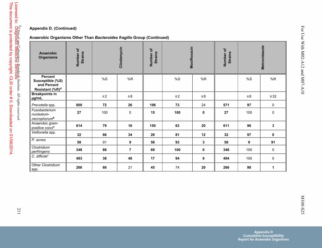
©Clinical and Laboratory Standards Institute. All rights reserved. 211
Appendix D. (Continued)
Anaerobic Organisms Other Than Bacteroides fragilis Group (Continued)
Anaerobic Organisms
Num
ber o
f St
rain
s
C
linda
myc
in
Num
ber o
f St
rain
s
M
oxifl
oxac
in
Num
ber o
f St
rain
s
M
etro
nida
zole
Percent Susceptible (%S)
and Percent Resistant (%R)d
%S %R %S %R %S %R
Breakpoints in µg/mL ≤ 2 ≥ 8 ≤ 2 ≥ 8 ≤ 8 ≥ 32
Prevotella spp. 800 72 26 196 73 24 571 97 0 Fusobacterium nucleatum-necrophorumb
27 100 0 15 100 0 27 100 0
Anaerobic gram-positive coccie 614 79 16 150 63 20 611 96 3
Veillonella spp. 32 66 34 26 81 12 32 97 0 P. acnes 58 91 9 58 93 3 58 9 91 Clostridium perfringens 348 86 7 69 100 0 348 100 0
C. difficilec 493 38 48 17 94 6 494 100 0
Other Clostridium spp. 266 66 21 45 74 20 266 98 1
For Use W
ith M02-A
12 and M07-A
10 M100-S25
Appendix DCumulative Susceptibility
Report for Anaerobic Organisms
Licensed�to:��Diagnostic�M
edicine�Services
This�document�is�protected�by�copyright.�C
LSI�order�#�0,�D
ownloaded�on�01/06/2014.

Appendix D. (Continued)
Footnotes
a. Data were generated from unique isolates from patient specimens submitted to four referral laboratories: Tufts New England Medical Center, Boston, MA; Loyola University Medical Center, Maywood, IL; International Health Management Associates, Inc., Schaumburg IL; and R.M. Alden Research Laboratory, Culver City, CA. Testing was performed by the agar dilution method. b. Calculated from fewer than the CLSI document M391 recommendation of 30 isolates. c. C. difficile isolates are from intestinal source; these results do not imply efficacy for intraluminal infections. Vancomycin minimal inhibitory concentrations for isolates were < 4 µg/mL. d. Intermediate category is not shown, but can be derived by subtraction of %S and %R for each antimicrobial agent from %100. e. Anaerobic gram-positive cocci include Peptococcus, Peptostreptococcus, Finegoldia, Peptoniphilus, and Anaerococcus species. Reference 1 CLSI. Analysis and Presentation of Cumulative Antimicrobial Susceptibility Test Data; Approved Guideline—Fourth Edition. CLSI document M39-A4. Wayne, PA: Clinical and Laboratory Standards Institute; 2014.
212 ©Clinical and Laboratory Standards Institute. All rights reserved.
January 2015
Vol. 35 N
o. 3
Appendix DCumulative Susceptibility
Report for Anaerobic Organisms
Licensed�to:��Diagnostic�M
edicine�Services
This�document�is�protected�by�copyright.�C
LSI�order�#�0,�D
ownloaded�on�01/06/2014.

For Use With M02-A12 and M07-A10 M100-S25
©Clinical and Laboratory Standards Institute. All rights reserved. 213
This page is intentionally left blank.
Licensed�to:��Diagnostic�Medicine�ServicesThis�document�is�protected�by�copyright.�CLSI�order�#�0,�Downloaded�on�01/06/2014.

January 2015 Vol. 35 No. 3
214 Clinical and Laboratory Standards Institute. All rights reserved.
Appendix E. Dosing Regimens Used to Establish Susceptible or Susceptible-Dose Dependent Interpretive Criteria The evolving science of pharmacokinetics-pharmacodynamics has become increasingly important in recent years in determining minimal inhibitory concentration (MIC) interpretive criteria. Recently approved susceptible or susceptible-dose dependent (SDD) interpretive criteria for a number of agents have been based on a specific dosing regimen(s); these dosing regimens are listed in the table below. Proper application of the interpretive criteria requires drug exposure at the site of infection that corresponds to or exceeds the expected systemic drug exposure at the dose listed in adult patients with normal renal function. This information should be shared with pharmacists, infectious diseases staff, and others making dosing recommendations for the institution.
Antimicrobial Agent
Interpretive Criteria Susceptible SDD
MIC (µg/mL) Dose MIC (µg/mL) Dose Table 2A. Enterobacteriaceae Aztreonam ≤ 4 1 g every 8 h N/A Cefazolin ≤ 2 2 g every 8 h N/A Ceftaroline ≤ 0.5 600 mg every 12 h N/A Cefepime ≤ 2 1 g every 12 h 4 1 g every 8 h or 2 g
every 12 h
8 or zone diameter: 19–24 mm
2 g every 8 h
(because it is not possible to correlate specific zone diameters with specific MICs, an isolate with a zone diameter in the SDD range should be treated as if it might be an MIC of 8 µg/mL)
Cefotaxime ≤ 1 1 g every 8 h N/A Ceftriaxone ≤ 1 1 g every 24 h N/A Cefoxitin ≤ 8 8 g per day (eg, 2 g every 6 h) N/A Cefuroxime ≤ 8 1.5 g every 8 h N/A Ceftazidime ≤ 4 1 g every 8 h N/A Ceftizoxime ≤ 1 1 g every 12 h N/A Doripenem ≤ 1 500 mg every 8 h N/A Ertapenem ≤ 0.5 1 g every 24 h N/A Imipenem ≤ 1 500 mg every 6 h or 1 g every 8 h N/A Table 2B-1. Pseudomonas aeruginosa Aztreonam ≤ 8 1 g every 6 h or 2 g every 8 h N/A Cefepime ≤ 8 1 g every 8 h or 2g every 12 h N/A Ceftazidime ≤ 8 1 g every 6 h or 2 g every 8 h N/A Doripenem ≤ 2 500 mg every 8 h N/A Imipenem ≤ 2 1 g every 8 h or 500 mg every 6 h N/A Meropenem ≤ 2 1 g every 8 h N/A Piperacillin ≤ 16 3 g every 6 h N/A Piperacillin-
tazobactam ≤ 16/4 3 g every 6 h N/A
Ticarcillin ≤ 16 3 g every 6 h N/A Ticarcillin-
clavulanate ≤ 16/2 3 g every 6 h N/A
Table 2B-2. Acinetobacter spp. Doripenem ≤ 2 500 mg every 8 h N/A Imipenem ≤ 2 500 mg every 6 h N/A Meropenem ≤ 2 1 g every 8 h or 500 mg every 6 h N/A Table 2C. Staphylococcus spp. Ceftaroline ≤ 1 600 mg every 12 h N/A Table 2E. Haemophilus influenzae and Haemophilus parainfluenzae Ceftaroline ≤ 0.5 600 mg every 12 h N/A
Appe
ndix
ED
osin
g Re
gim
ens U
sed
to E
stab
lish
Susc
eptib
le o
r Su
scep
tible
-Dos
e D
epen
dent
Inte
rpre
tive
Crite
ria
Licensed�to:��Diagnostic�Medicine�ServicesThis�document�is�protected�by�copyright.�CLSI�order�#�0,�Downloaded�on�01/06/2014.

For Use With M02-A12 and M07-A10 M100-S25
©Clinical and Laboratory Standards Institute. All rights reserved. 215
Appendix E. (Continued)
Abbreviations: MIC, minimal inhibitory concentration; N/A, not applicable; SDD, susceptible-dose dependent.
Antimicrobial Agent
Interpretive Criteria Susceptible SDD
MIC (µg/mL) Dose MIC (µg/mL) Dose Table 2G. Streptococcus pneumoniae Ceftaroline (nonmeningitis)
≤ 0.5 600 mg every 12 h N/A
Penicillin (nonmeningitis)
≤ 2 2 million units every 4 h (12 million units per day)
N/A
Penicillin parenteral (meningitis)
≤ 0.06 3 million units every 4 h N/A
Table 2H-1. Streptococcus spp. β-Hemolytic Group Ceftaroline ≤ 0.5 600 mg every 12 h N/A
Appe
ndix
ED
osin
g Re
gim
ens U
sed
to E
stab
lish
Susc
eptib
le o
r Su
scep
tible
-Dos
e D
epen
dent
Inte
rpre
tive
Crite
ria
Licensed�to:��Diagnostic�Medicine�ServicesThis�document�is�protected�by�copyright.�CLSI�order�#�0,�Downloaded�on�01/06/2014.

January 2015 Vol. 35 No. 3
216 Clinical and Laboratory Standards Institute. All rights reserved.
Appendix F. Cefepime Breakpoint Change for Enterobacteriaceae and Introduction of the Susceptible-Dose Dependent Interpretive Category What Changed? The CLSI Subcommittee on Antimicrobial Susceptibility Testing revised the cefepime interpretive criteria (breakpoints) and introduced the susceptible-dose dependent (SDD) category with this breakpoint revision. Below is a summary of the changes.
Previous – 2013
Method Susceptible Intermediate Resistant
MIC ≤ 8 µg/mL 16 µg/mL ≥ 32 µg/mL
Zone Diameter (Disk Diffusion)
≥ 18 mm 15–17 mm ≤ 14 mm
Revised – 2014
Method Susceptible Susceptible-Dose Dependent Resistant
MIC ≤ 2 µg/mL 4–8 µg/mL ≥ 16 µg/mL
Zone Diameter (Disk Diffusion)
≥ 25 mm 19–24 mm ≤ 18 mm
Abbreviation: MIC, minimal inhibitory concentration.
Why were the cefepime breakpoints reconsidered? The issue of new breakpoints for cefepime became apparent for several reasons: Previous breakpoints were based on a higher dose of cefepime than is often used.
Clinical failures were noted for isolates with cefepime MICs of 4 and 8 µg/mL, especially when lower doses of
cefepime were used. There are limited new drugs in the pipeline that show activity against multidrug-resistant gram-negative bacteria;
thus, there is a need to optimize use of drugs currently available. Designing susceptibility reports to correlate better with dosages of the drug used is one way to help accomplish this goal.
What does “susceptible-dose dependent” (SDD) mean? SDD interpretation is a new interpretive category for antibacterial susceptibility testing, although it has been applied for interpretation of antifungal susceptibility test results for several years.
Definition: The “susceptible-dose dependent” category implies that susceptibility of an isolate is dependent on the dosing regimen that is used in the patient. In order to achieve levels that are likely to be clinically effective against isolates for which the susceptibility testing results (either MICs or disk diffusion) are in the SDD category, it is necessary to use a dosing regimen (ie, higher doses, more frequent doses, or both) that results in higher drug exposure than the dose that was used to establish the susceptible breakpoint. Consideration should be given to the maximum approved dosage regimen, because higher exposure gives the highest probability of adequate coverage of an SDD isolate. The dosing regimens used to set the SDD interpretive criterion are provided in Appendix E. The drug label should be consulted for recommended doses and adjustment for organ function.
Appe
ndix
FCe
fepi
me
Brea
kpoi
nt C
hang
e fo
r Ent
erob
acte
riace
aean
d In
trod
uctio
n of
the
Susc
eptib
le-D
ose
Dep
ende
ntIn
terp
retiv
e Ca
tego
ry
Licensed�to:��Diagnostic�Medicine�ServicesThis�document�is�protected�by�copyright.�CLSI�order�#�0,�Downloaded�on�01/06/2014.

For Use With M02-A12 and M07-A10 M100-S25
©Clinical and Laboratory Standards Institute. All rights reserved. 217
Appendix F. (Continued)
NOTE: The SDD interpretation is a new category for antibacterial susceptibility testing, although it has been previously applied for interpretation of antifungal susceptibility test results (see CLSI document M27-S4). The concept of SDD has been included within the intermediate category definition for antimicrobial agents. However, this is often overlooked or not understood by clinicians and microbiologists when an intermediate result is reported. The SDD category may be assigned when doses well above those used to calculate the susceptible breakpoint are approved and used clinically, and where sufficient data to justify the designation exist and have been reviewed. When the intermediate category is used, its definition remains unchanged.
SDD is recommended instead of “intermediate” when reporting cefepime results for Enterobacteriaceae isolates because there are multiple approved dosing options for cefepime, and SDD highlights the option of using higher doses to treat infections caused by isolates when the cefepime MIC is 4 or 8 µg/mL or the zone is 19 to 24 mm. Why is SDD being used now? It has become apparent that there is a growing need to refine susceptibility reporting to maximize clinicians’ use
of available drugs.
Intermediate too often means “resistant” to clinicians because they do not appreciate the full definition of “intermediate.”
SDD is more specific and it conveys what we know—a higher dose can be considered for isolates with MICs (or zones) that fall in this interpretive category.
SDD is already well established for use in antifungal susceptibility testing.
It is anticipated that reporting a cefepime SDD result will encourage clinicians to consider the possibility that cefepime may be an option for treatment.
Antibiotic stewardship programs, which emphasize dosing regimen and duration of therapy options, are increasing awareness of appropriate use of antibiotics. Personnel from these programs should be able to describe the significance to clinicians of an SDD result for cefepime.
How should this change be implemented? Meet with the appropriate practitioners at your institution (members of the antimicrobial stewardship team,
infectious diseases staff, pathology group, pharmacy, etc.) to inform them of these changes and agree on a plan to inform your clinicians of this change.
Talk to the manufacturer of your antimicrobial susceptibility testing (AST) device to determine how to implement
the revised breakpoints on your device. – NOTE: Because the US Food and Drug Administration (FDA) has not revised the cefepime breakpoints and
commercial manufacturers must use FDA breakpoints, the manufacturer cannot adopt the new CLSI cefepime breakpoints. However, for most systems, you can manually change the breakpoints and implement following a verification study.
Work with your laboratory information system staff to report “SDD” or “D” for Enterobacteriaceae when the
cefepime MIC is 4 or 8 µg/mL. Make certain that SDD will be transmitted to the hospital information system and appropriately displayed on reports viewed by clinicians.
Distribute user-specific educational materials to laboratory staff and clinicians receiving AST results from your
laboratory. Examples of these materials can be found on the CLSI Subcommittee on Antimicrobial Susceptibility Testing webpage at www.clsi.org.
Additional Questions and Answers: 1. Q: Does CLSI recommend a comment to be reported with the new cefepime breakpoints?
A: If a laboratory chooses to report a comment explaining the SDD range, CLSI recommends the
following: “The interpretive criterion for susceptible is based on a dosage regimen of 1 g every 12 h. The interpretive criterion for susceptible-dose dependent is based on dosing regimens that result in higher cefepime exposure, either higher doses or more frequent doses or both, up to approved maximum dosing regimens.”
Appe
ndix
FCe
fepi
me
Brea
kpoi
nt C
hang
e fo
r Ent
erob
acte
riace
aean
d In
trod
uctio
n of
the
Susc
eptib
le-D
ose
Dep
ende
ntIn
terp
retiv
e Ca
tego
ry
Licensed�to:��Diagnostic�Medicine�ServicesThis�document�is�protected�by�copyright.�CLSI�order�#�0,�Downloaded�on�01/06/2014.

January 2015 Vol. 35 No. 3
218 Clinical and Laboratory Standards Institute. All rights reserved.
Appendix F. (Continued) 2. Q: Will all intermediate ranges become SDD?
A: No, the SDD category will be implemented for drug/organism combinations only when there is
sufficient evidence to suggest alternative approved dosing regimens may be appropriate for organisms that have MICs or zone diameters between the susceptible and resistant categories.
3. Q: Will SDD be applied to other antimicrobial agents?
A: CLSI will examine the SDD category possibility for additional drug/organism combinations where multiple dosing options exist (eg, other extended-spectrum cephalosporins).
4. Q: How do we perform a verification study before implementing the new cefepime breakpoints on our AST
device? A: Guidelines for performance of such a verification study are provided in the following publication: Clark RB, Lewinski MA, Loeffelholz MJ, Tibbetts RJ. Cumitech 31A: verification and validation of procedures in the clinical microbiology laboratory. Washington, DC: ASM Press; 2009.
5. Q: Does SDD apply to all patients and specimen types (eg, pediatric, geriatric, immunosuppressed)?
A: Yes, in terms of laboratory reporting. Clinicians must decide how to use an SDD result for a specific patient in consideration of all other clinical and physiological parameters for that patient.
6. Q: Do the new cefepime breakpoints apply to Pseudomonas aeruginosa and other gram-negative bacteria also?
A: No, currently they are only applicable to members of the Enterobacteriaceae.
7. Q: Is any special QC required once the SDD breakpoints are implemented?
A: No, currently recommended routine QC is sufficient.
8. Q: Will we be required to report SDD on proficiency testing survey samples?
A: Sponsors of proficiency testing surveys are aware of the difficulties encountered by clinical laboratories in implementing newer CLSI breakpoints. It is highly unlikely that there will be a mandate to report SDD in the near future, but it would be best to check with your proficiency testing survey provider.
9. Q: If we can implement the revised cefepime breakpoints but cannot facilitate reporting of SDD, can we report
“intermediate” instead of SDD? A: A decision related to this question should be made following consultation with your laboratory director, antibiotic stewardship team (if available), infectious diseases practitioners, pharmacists, and infection control practitioners.
10. Q: If we can implement the revised cefepime breakpoints but cannot facilitate reporting of SDD, can we report an
MIC or zone diameter without an MIC? A: A zone diameter should never be reported without an interpretation because there is a high risk of misinterpretation of this value and this poses patient safety issues. There is a lesser danger of reporting an MIC without an interpretation, but this should not be done without an accompanying qualifying comment. See answer to question 9, above.
11. Q: If we are still doing extended-spectrum β-lactamase (ESBL) testing and implement the new cefepime
breakpoints, do we change a susceptible or SDD result to resistant for ESBL-positive isolates? A: No. When CLSI changed the other cephem breakpoints in 2010, the recommendation to perform routine ESBL testing was eliminated. When using the new cefepime breakpoints, there is no need to perform routine ESBL testing for patient reporting purposes. However, ESBL testing might be done for infection control or epidemiological purposes.
Appe
ndix
FCe
fepi
me
Brea
kpoi
nt C
hang
e fo
r Ent
erob
acte
riace
aean
d In
trod
uctio
n of
the
Susc
eptib
le-D
ose
Dep
ende
ntIn
terp
retiv
e Ca
tego
ry
Licensed�to:��Diagnostic�Medicine�ServicesThis�document�is�protected�by�copyright.�CLSI�order�#�0,�Downloaded�on�01/06/2014.

For Use With M02-A12 and M07-A10 M100-S25
©Clinical and Laboratory Standards Institute. All rights reserved. 219
Appendix F. (Continued) 12. Q: What does the dosing information that is given with breakpoints mean?
A: The evolving science of pharmacokinetics-pharmacodynamics has become increasingly important in recent years in determining MIC interpretive criteria. Recently approved susceptible or SDD interpretive criteria for a number of agents have been based on a specific dosing regimen(s); these dosing regimens are listed in Appendix E. Proper application of the interpretive criteria requires drug exposure at the site of infection that corresponds to or exceeds the expected systemic drug exposure, at the dose listed, in adult patients with normal renal function. This information should be shared with pharmacists, infectious diseases staff, and others making dosing recommendations for the institution.
Appe
ndix
FCe
fepi
me
Brea
kpoi
nt C
hang
e fo
r Ent
erob
acte
riace
aean
d In
trod
uctio
n of
the
Susc
eptib
le-D
ose
Dep
ende
ntIn
terp
retiv
e Ca
tego
ry
Licensed�to:��Diagnostic�Medicine�ServicesThis�document�is�protected�by�copyright.�CLSI�order�#�0,�Downloaded�on�01/06/2014.

January 2015 Vol. 35 No. 3
220 Clinical and Laboratory Standards Institute. All rights reserved.
Appendix G. Epidemiological Cutoff Values 1. Q: What are epidemiological cutoff values (ECVs)?
A: ECVs are minimal inhibitory concentration (MIC) values that separate bacterial populations into those with and without acquired and/or mutational resistance mechanisms based on their phenotypes (MICs). ECVs are based solely on in vitro data. The term “wild-type” (WT) is used to describe strains with MIC values at or below the ECV that are presumed not to possess acquired and/or mutational resistance mechanisms, while the term “non-wild-type” (NWT) is used to describe strains with MIC values above the ECV that are presumed to possess acquired and/or mutational resistance mechanisms. ECVs are principally used to signal the emergence and evolution of NWT strains. They are not the same as clinical breakpoints. The ECV is defined as the MIC value that best defines the estimated upper end of the WT population.
2. Q: How are ECVs determined?
A: ECVs are determined by collecting and merging MIC distribution data obtained by testing bacteria from a variety of sources, and then applying techniques for estimating the MIC at the upper end of the WT distribution. In order to be reliable, ECVs are estimated by accounting for both biological (strain-to-strain) variation and MIC assay variation within and between laboratories. They are based on the assumption that the WT distribution of a particular antimicrobial agent/organism combination does not vary geographically or over time.
Several conditions must be fulfilled in order to generate reliable ECVs. The most important are: An ECV can only be determined within a single species because of the genetic diversity between
species within a genus.
All MIC values included in the merged dataset must have been determined using a recognized reference method such as the CLSI MIC broth dilution method (M071), which is also the methodology outlined in an international reference standard (ISO-20776-12).
Data must be sourced from at least three separate laboratories, and there should be at least 100 unique strains included in the merged dataset.
As much as possible, the MIC values included in an individual laboratory’s dataset must be “on scale.” This condition applies particularly to MICs of the presumptive WT strains. Before merging data for ECV estimation, the MIC distribution from each individual laboratory is inspected, and if the lowest concentration tested is also a mode, then these data cannot be included in the merged dataset.
Once acceptable data are merged, there are several methods that can be used to estimate the ECV. The simplest method is visual inspection. Visual inspection generally works for MIC distributions when there is clear separation of WT and NWT. When there is obvious overlap between WT and NWT strains, visual inspection becomes too subjective. In general, statistical methods are preferred because they remove any potential observer bias from the estimation. The two most widely referenced methods are those of Turnidge et al.3 and Kronvall.4
Estimation of ECVs from MIC distributions may be supplemented with molecular tests for known resistance mechanisms, as a form a validation. The detection of a resistance gene per se in strains with MICs at or below the ECV does not necessarily contradict the choice of ECV, unless it can be accompanied by evidence that the gene is being expressed.
3. Q: How are ECVs used to set clinical breakpoints?
A: Clinical breakpoints are set using many criteria as detailed in CLSI document M23,5 including MIC distributions for the antimicrobial and relevant populations of bacteria, in vitro and in vivo pharmacodynamics, human pharmacokinetics, and clinical outcome. MIC distributions and ECVs are thus just one component of a range of data used to set clinical breakpoints.
Appe
ndix
GEp
idem
iolo
gica
l Cut
off V
alue
s
Licensed�to:��Diagnostic�Medicine�ServicesThis�document�is�protected�by�copyright.�CLSI�order�#�0,�Downloaded�on�01/06/2014.

For Use With M02-A12 and M07-A10 M100-S25
©Clinical and Laboratory Standards Institute. All rights reserved. 221
Appendix G. (Continued)
4. Q: How can ECVs be used by the clinical microbiology laboratory?
A: In rare clinical circumstances, experience may suggest an antimicrobial agent for use where no clinical breakpoints exist. For example, vancomycin may be considered for treatment of a P. acnes infection, but there are insufficient data available to establish clinical breakpoints with interpretive criteria. This is due principally to the absence of strains with acquired resistance and a lack of clinical outcome data.
MIC testing using a reference or approved method and ECVs for the drug/organism combination might then be used to determine if the patient’s isolate of P. acnes is a WT or NWT strain. If the vancomycin MIC is at or below the ECV (≤ 2 µg/mL) it can be assumed that the isolate is a WT strain. If the vancomycin MIC is > 4 µg/mL, the strain should be retested to confirm the NWT result. The confirmed MIC result and the ECV data should be discussed with relevant clinicians/pharmacists. A comment could be added to the report indicating that MIC results were discussed with relevant clinical services (infectious diseases/pharmacists). The MIC result should not be reported with an interpretation.
References for Appendix G 1 CLSI. Methods for Dilution Antimicrobial Susceptibility Tests for Bacteria That Grow Aerobically;
Approved Standard—Tenth Edition. CLSI document M100-S25. Wayne, PA: Clinical and Laboratory Standards Institute; 2015.
2 ISO. Clinical laboratory testing and in vitro diagnostic test systems – Susceptibility testing of infectious
agents and evaluation of performance of antimicrobial susceptibility test devices – Part 1: Reference method for testing the in vitro activity of antimicrobial agents against rapidly growing aerobic bacteria involved in infectious diseases. ISO 20776-1. Geneva, Switzerland: International Organization for Standardization; 2006.
3 Turnidge J, Kahlmeter G, Kronvall G. Statistical characterisation of bacterial wild-type MIC value distributions and the determination of epidemiological cut-off values. Clin Microbiol Infect. 2006; 12(5):418-425.
4 Kronvall G. Normalized resistance interpretation as a tool for establishing epidemiological MIC susceptibility breakpoints. J Clin Microbiol. 2010;48(12):4445-4452.
5 CLSI. Development of In Vitro Susceptibility Testing Criteria and Quality Control Parameters; Approved Guideline—Third Edition. CLSI document M23-A3. Wayne, PA: Clinical and Laboratory Standards Institute; 2008.
Appe
ndix
GEp
idem
iolo
gica
l Cut
off V
alue
s
Licensed�to:��Diagnostic�Medicine�ServicesThis�document�is�protected�by�copyright.�CLSI�order�#�0,�Downloaded�on�01/06/2014.

January 2015 Vol. 35 No. 3
222 Clinical and Laboratory Standards Institute. All rights reserved.
Glossary I (Part 1). -Lactams: Class and Subclass Designation and Generic Name Antimicrobial Class Antimicrobial Subclass Agents Included; Generic Names
Penicillins Penicillinase-labile penicillinsa
Penicillin Penicillin Aminopenicillin Amoxicillin
Ampicillin Ureidopenicillin Azlocillin
Mezlocillin Piperacillin
Carboxypenicillin Carbenicillin Ticarcillin
Penicillinase-stable penicillinsb
Cloxacillin Dicloxacillin Methicillin Nafcillin Oxacillin
Amidinopenicillin Mecillinam -Lactam/-lactamase inhibitor combinations
Amoxicillin-clavulanate Ampicillin-sulbactam Aztreonam-avibactam Ceftaroline-avibactam Ceftazidime-avibactam Ceftolozane-tazobactam Piperacillin-tazobactam Ticarcillin-clavulanate
Cephems (parenteral) Cephalosporin Ic Cefazolin Cephalothin Cephapirin Cephradine
Cephalosporin IIc Cefamandole Cefonicid Cefuroxime (parenteral)
Cephalosporin IIIc Cefoperazone Cefotaxime Ceftazidime Ceftizoxime Ceftriaxone
Cephalosporin IVc Cefepime Cephalosporins with anti-MRSA activity Ceftaroline
Ceftobiprole Cephamycin Cefmetazole
Cefotetan Cefoxitin
Oxacephem Moxalactam Cephems (oral) Cephalosporin Cefaclor
Cefadroxil Cefdinir Cefditoren Cefetamet Cefixime Cefpodoxime Cefprozil Ceftibuten Cefuroxime (oral) Cephalexin Cephradine
Carbacephem Loracarbef
Glo
ssar
y I
Licensed�to:��Diagnostic�Medicine�ServicesThis�document�is�protected�by�copyright.�CLSI�order�#�0,�Downloaded�on�01/06/2014.

For Use With M02-A12 and M07-A10 M100-S25
©Clinical and Laboratory Standards Institute. All rights reserved. 223
Glossary I (Part 1). (Continued) Antimicrobial Class Antimicrobial Subclass Agents Included; Generic Names
Monobactams Aztreonam Penems Carbapenem Biapenem
Doripenem Ertapenem Imipenem Meropenem Razupenem
Penem Faropenem Sulopenem
Abbreviation: MRSA, methicillin-resistant S. aureus. a. Hydrolyzed by staphylococcal penicillinase. b. Not hydrolyzed by staphylococcal penicillinase. c. Cephalosporin I, II, III, and IV are sometimes referred to as first-, second-, third-, and fourth-generation cephalosporins,
respectively. Cephalosporin III and IV are also referred to as “extended-spectrum cephalosporins.” This does not imply activity against extended-spectrum -lactamase–producing gram-negative bacteria.
Glo
ssar
y I
Licensed�to:��Diagnostic�Medicine�ServicesThis�document�is�protected�by�copyright.�CLSI�order�#�0,�Downloaded�on�01/06/2014.

January 2015 Vol. 35 No. 3
224 Clinical and Laboratory Standards Institute. All rights reserved.
Glossary I (Part 2). Non–-Lactams: Class and Subclass Designation and Generic Name Antimicrobial Class Antimicrobial Subclass Agents Included; Generic Names
Aminocyclitols Spectinomycin Aminoglycosides Amikacin
Gentamicin Kanamycin Netilmicin Plazomicin Streptomycin Tobramycin
Ansamycins Rifampin Folate pathway inhibitors Iclaprim
Sulfonamides Trimethoprim Trimethoprim-sulfamethoxazole
Fosfomycins Fosfomycin Glycopeptides Glycopeptide Vancomycin
Lipoglycopeptide Dalbavancin Oritavancin Teicoplanin Telavancin Ramoplanin
Lincosamides Clindamycin Lipopeptides Daptomycin
Surotomycin Polymyxins Colistin
Polymyxin B Macrocyclic Fidaxomicin Macrolides Azithromycin
Clarithromycin Dirithromycin Erythromycin
Ketolide Telithromycin Fluoroketolide Solithromycin
Nitrofurans Nitrofurantoin Nitroimidazoles Metronidazole
Tinidazole Oxazolidinones Linezolid
Tedizolid Phenicols Chloramphenicol Pseudomonic acid Mupirocin Quinolones Quinolone Cinoxacin
Garenoxacin Nalidixic acid
Fluoroquinolone Besifloxacin Ciprofloxacin Clinafloxacin Enoxacin Finafloxacin Fleroxacin Gatifloxacin Gemifloxacin Grepafloxacin Levofloxacin Lomefloxacin Moxifloxacin Norfloxacin Ofloxacin Pefloxacin Sparfloxacin Trovafloxacin Ulifloxacin (prulifloxacin)
Glo
ssar
y I
Licensed�to:��Diagnostic�Medicine�ServicesThis�document�is�protected�by�copyright.�CLSI�order�#�0,�Downloaded�on�01/06/2014.

For Use With M02-A12 and M07-A10 M100-S25
©Clinical and Laboratory Standards Institute. All rights reserved. 225
Glossary I (Part 2). (Continued) Antimicrobial Class Antimicrobial Subclass Agents Included; Generic Names
Steroidal Fusidanes Fusidic acid Streptogramins
Linopristin-flopristin Quinupristin-dalfopristin
Tetracyclines Doxycycline Minocycline Tetracycline
Fluorocycline Eravacycline
Glycylcyclines Tigecycline Aminomethylcycline Omadacycline
Thiazolide Nitazoxanide Tizoxanide
Glo
ssar
y I
Licensed�to:��Diagnostic�Medicine�ServicesThis�document�is�protected�by�copyright.�CLSI�order�#�0,�Downloaded�on�01/06/2014.
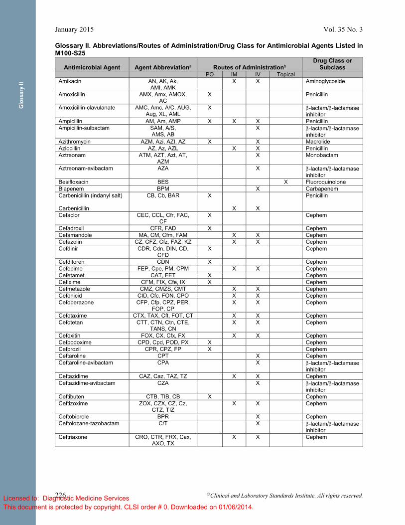
January 2015 Vol. 35 No. 3
226 Clinical and Laboratory Standards Institute. All rights reserved.
Glossary II. Abbreviations/Routes of Administration/Drug Class for Antimicrobial Agents Listed in M100-S25
Antimicrobial Agent Agent Abbreviationa Routes of Administrationb Drug Class or
Subclass PO IM IV Topical Amikacin AN, AK, Ak,
AMI, AMK X X Aminoglycoside
Amoxicillin AMX, Amx, AMOX, AC
X Penicillin
Amoxicillin-clavulanate AMC, Amc, A/C, AUG, Aug, XL, AML
X -lactam/-lactamase inhibitor
Ampicillin AM, Am, AMP X X X Penicillin Ampicillin-sulbactam SAM, A/S,
AMS, AB X -lactam/-lactamase
inhibitor Azithromycin AZM, Azi, AZI, AZ X X Macrolide Azlocillin AZ, Az, AZL X X Penicillin Aztreonam ATM, AZT, Azt, AT,
AZM X Monobactam
Aztreonam-avibactam AZA X -lactam/-lactamase inhibitor
Besifloxacin BES X Fluoroquinolone Biapenem BPM X Carbapenem Carbenicillin (indanyl salt) Carbenicillin
CB, Cb, BAR X
X
X
Penicillin
Cefaclor CEC, CCL, Cfr, FAC, CF
X Cephem
Cefadroxil CFR, FAD X Cephem Cefamandole MA, CM, Cfm, FAM X X Cephem Cefazolin CZ, CFZ, Cfz, FAZ, KZ X X Cephem Cefdinir CDR, Cdn, DIN, CD,
CFD X Cephem
Cefditoren CDN X Cephem Cefepime FEP, Cpe, PM, CPM X X Cephem Cefetamet CAT, FET X Cephem Cefixime CFM, FIX, Cfe, IX X Cephem Cefmetazole CMZ, CMZS, CMT X X Cephem Cefonicid CID, Cfc, FON, CPO X X Cephem Cefoperazone CFP, Cfp, CPZ, PER,
FOP, CP X X Cephem
Cefotaxime CTX, TAX, Cft, FOT, CT X X Cephem Cefotetan CTT, CTN, Ctn, CTE,
TANS, CN X X Cephem
Cefoxitin FOX, CX, Cfx, FX X X Cephem Cefpodoxime CPD, Cpd, POD, PX X Cephem Cefprozil CPR, CPZ, FP X Cephem Ceftaroline CPT X Cephem Ceftaroline-avibactam CPA X -lactam/-lactamase
inhibitor Ceftazidime CAZ, Caz, TAZ, TZ X X Cephem Ceftazidime-avibactam CZA X -lactam/-lactamase
inhibitor Ceftibuten CTB, TIB, CB X Cephem Ceftizoxime ZOX, CZX, CZ, Cz,
CTZ, TIZ X X Cephem
Ceftobiprole BPR X Cephem Ceftolozane-tazobactam C/T X -lactam/-lactamase
inhibitor Ceftriaxone CRO, CTR, FRX, Cax,
AXO, TX X X Cephem
Glo
ssar
y II
Licensed�to:��Diagnostic�Medicine�ServicesThis�document�is�protected�by�copyright.�CLSI�order�#�0,�Downloaded�on�01/06/2014.

For Use With M02-A12 and M07-A10 M100-S25
©Clinical and Laboratory Standards Institute. All rights reserved. 227
Glossary II. (Continued)
Antimicrobial Agent Agent Abbreviationa Routes of Administrationb Drug Class or
Subclass PO IM IV Topical Cefuroxime (oral) Cefuroxime (parenteral)
CXM, CFX, ROX, Crm, FUR, XM
X
X
X
Cephem
Cephalexin CN, LEX, CFL X Cephem Cephalothin CF, Cf, CR, CL, CEP,
CE, KF X Cephem
Cephapirin CP, HAP X X Cephem Cephradine RAD, CH X Cephem Chloramphenicol C, CHL, CL X X Phenicol Cinoxacin CIN, Cn X Quinolone Ciprofloxacin CIP, Cp, CI X X Fluoroquinolone Clarithromycin CLR, CLM,
CLA, Cla, CH X Macrolide
Clinafloxacin CFN, CLX, LF X X Fluoroquinolone Clindamycin CC, CM, CD, Cd, CLI,
DA X X X Lincosamide
Colistin CL, CS, CT X Lipopeptide Dalbavancin DAL X Glycopeptide Daptomycin DAP X Lipopeptide Dicloxacillin DX, DIC X Penicillin Dirithromycin DTM, DT X Macrolide Doripenem DOR X Carbapenem Doxycycline DOX, DC, DOXY X X Tetracycline Eravacycline ERV X X Tetracycline Ertapenem ETP X X Carbapenem Erythromycin E, ERY, EM X X Macrolide Faropenem FAR, FARO X Penem Fidaxomicin FDX X Macrocyclic Finafloxacin FIN X X X Fluoroquinolone Fleroxacin FLE, Fle, FLX, FO X X Fluoroquinolone Fosfomycin FOS, FF, FO, FM X Fosfomycin Fusidic acid FA, FC X X X Steroidal Garenoxacin GRN X X Quinolone Gatifloxacin GAT X X Fluoroquinolone Gemifloxacin GEM X Fluoroquinolone Gentamicin Gentamicin synergy
GM, Gm, CN, GEN GM500, HLG, Gms
X X Aminoglycoside
Grepafloxacin GRX, Grx, GRE, GP X Fluoroquinolone Iclaprim ICL X Folate pathway
inhibitor Imipenem IPM, IMI, Imp, IP X Carbapenem Kanamycin K, KAN, HLK, KM X X Aminoglycoside Levofloxacin LVX, Lvx,
LEV, LEVO, LE X X Fluoroquinolone
Linezolid LNZ, LZ, LZD X X Oxazolidinone Linopristin- flopristin
LFE X Streptogramin
Lomefloxacin LOM, Lmf X Fluoroquinolone Loracarbef LOR, Lor, LO X Cephem Mecillinam MEC X Penicillin Meropenem MEM, Mer, MERO,
MRP, MP X Carbapenem
Methicillin DP, MET, ME, SC X X Penicillin Metronidazole MTZ X X Nitroimidazole Mezlocillin MZ, Mz, MEZ X X Penicillin Minocycline MI, MIN, Min, MN,
MNO, MC, MH X X Tetracycline
Moxalactam MOX X X Cephem Moxifloxacin MXF X X Fluoroquinolone Mupirocin MUP, MOP, MU X Pseudomonic acid Nafcillin NF, NAF, Naf X X Penicillin
Glo
ssar
y II
Licensed�to:��Diagnostic�Medicine�ServicesThis�document�is�protected�by�copyright.�CLSI�order�#�0,�Downloaded�on�01/06/2014.

January 2015 Vol. 35 No. 3
228 Clinical and Laboratory Standards Institute. All rights reserved.
Glossary II. (Continued)
Antimicrobial Agent Agent Abbreviationa Routes of Administrationb Drug Class or
Subclass PO IM IV Topical Nalidixic acid NA, NAL X Quinolone Netilmicin NET, Nt, NC X X Aminoglycoside Nitazoxanide NIT X Thiazolide Nitrofurantoin F/M, FD, Fd, FT,
NIT, NI, F X Nitrofurantoin
Norfloxacin NOR, Nxn, NX X Fluoroquinolone Ofloxacin OFX, OFL, Ofl, OF X X X Fluoroquinolone Omadacycline OMC X X Tetracycline Oritavancin ORI X Lipoglycopeptide Oxacillin OX, Ox, OXS, OXA X X X Penicillin Pefloxacin PEF, PF FluoroquinolonePenicillin P, PEN, PV X X X Penicillin Piperacillin PIP, PI, PP, Pi X X Penicillin Piperacillin-tazobactam TZP, PTZ, P/T, PTc X -lactam/-
lactamase inhibitor combination
Plazomicin PLZ X Aminoglycoside Polymyxin B PB X Lipopeptide Quinupristin-dalfopristin SYN, Syn, QDA, RP X Streptogramin Razupenem RZM X Carbapenem Ramoplanin RAM X Lipoglycopeptide Rifampin RA, RIF, Rif, RI, RD X X Ansamycin Solithromycin SOL X X X Fluoroketolide Sparfloxacin SPX, Sfx, SPA, SO X Fluoroquinolone Spectinomycin SPT, SPE, SC X X Aminocyclitol Streptomycin Streptomycin synergy
S, STR, StS, SM,
ST2000, HLS
X X Aminoglycoside
Sulfonamides SSS, S3 X X Folate pathway inhibitor (some PO only)
Sulopenem SLP, SULO X X Penem Surotomycin SUR X Lipopeptide Tedizolid TZD X X Oxazolidinone Teicoplanin TEC, TPN, Tei,
TEI, TP, TPL X X Glycopeptide
Telavancin TLV X LipoglycopeptideTelithromycin TEL X Ketolide Tetracycline TE, Te, TET, TC X X Tetracycline Ticarcillin TIC, TC, TI, Ti X X Penicillin Ticarcillin-clavulanate TIM, Tim, T/C, TCC,
TLc X -lactam/-
lactamase inhibitor Tigecycline TGC X Glycylcycline Tinoxanide TIN X Thiazolide Tinidazole TNZ X NitroimidazolesTobramycin NN, TM, TO, To, TOB X X Aminoglycoside Trimethoprim TMP, T, TR, W X Folate pathway
inhibitor Trimethoprim- sulfamethoxazole
SXT, SxT, T/S, TS, COT
X X Folate pathway inhibitor
Trovafloxacin TVA, Tva, TRV, TV X X Fluoroquinolone Ulifloxacin (prulifloxacin) PRU X Fluoroquinolone Vancomycin VA, Va, VAN X X Glycopeptide
Abbreviations: PO, per OS (oral); IM, intramuscular; IV, intravenous. a. Abbreviations assigned to one or more diagnostic products in the United States. If no diagnostic product is available,
abbreviation is that of the manufacturer. b. As available in the United States.
Glo
ssar
y II
Licensed�to:��Diagnostic�Medicine�ServicesThis�document�is�protected�by�copyright.�CLSI�order�#�0,�Downloaded�on�01/06/2014.

For Use With M02-A12 and M07-A10 M100-S25
©Clinical and Laboratory Standards Institute. All rights reserved. 229
Glossary III. List of Identical Abbreviations Used for More Than One Antimicrobial Agent in US Diagnostic Products
Agent Abbreviation Antimicrobial Agents for Which Respective
Abbreviation Is Used AZM Azithromycin, Aztreonam AZ Azithromycin, Azlocillin CB, Cb Ceftibuten, Carbenicillin CFR, Cfr Cefaclor, Cefadroxil CF, Cf Cefaclor, Cephalothin CM Clindamycin, Cefamandole CFM, Cfm Cefixime, Cefamandole CZ, Cz Ceftizoxime, Cefazolin CD, Cd Clindamycin, Cefdinir CPZ Cefprozil, Cefoperazone CP, Cp Cephapirin, Cefoperazone, Ciprofloxacin CN, Cn Cephalexin, Cefotetan, Cinoxacin, Gentamicin CFX, Cfx Cefoxitin, Cefuroxime CL Cephalothin, Chloramphenicol CH Clarithromycin, Cephradine DX Doxycycline, Dicloxacillin FO Fleroxacin, Fosfomycin NIT Nitrofurantoin SC Spectinomycin, Methicillin SO Sparfloxacin, Oxacillin TC Tetracycline, Ticarcillin
Glo
ssar
y III
Licensed�to:��Diagnostic�Medicine�ServicesThis�document�is�protected�by�copyright.�CLSI�order�#�0,�Downloaded�on�01/06/2014.

January 2015 Vol. 35 No. 3
230 Clinical and Laboratory Standards Institute. All rights reserved.
The Quality Management System Approach Clinical and Laboratory Standards Institute (CLSI) subscribes to a quality management system (QMS) approach in the development of standards and guidelines, which facilitates project management; defines a document structure via a template; and provides a process to identify needed documents. The QMS approach applies a core set of “quality system essentials” (QSEs), basic to any organization, to all operations in any health care service’s path of workflow (ie, operational aspects that define how a particular product or service is provided). The QSEs provide the framework for delivery of any type of product or service, serving as a manager’s guide. The QSEs are as follows: Organization Personnel Process Management Nonconforming Event Management Customer Focus Purchasing and Inventory Documents and Records Assessments Facilities and Safety Equipment Information Management Continual Improvement M100-S25 does not address any of the QSEs. For a description of the documents listed in the grid, please refer to the Related CLSI Reference Materials section on the following page.
Org
aniz
atio
n
Cus
tom
er F
ocus
Faci
litie
s and
Sa
fety
Pers
onne
l
Purc
hasi
ng a
nd
Inve
ntor
y
Equi
pmen
t
Proc
ess
Man
agem
ent
Doc
umen
ts a
nd
Rec
ords
Info
rmat
ion
Man
agem
ent
Non
conf
orm
ing
Even
t Man
agem
ent
Ass
essm
ents
Con
tinua
l Im
prov
emen
t
EP23 M02 M07 M11 M23 M27
M27-S4 M39 M45
M07
Path of Workflow A path of workflow is the description of the necessary processes to deliver the particular product or service that the organization or entity provides. A laboratory path of workflow consists of the sequential processes: preexamination, examination, and postexamination and their respective sequential subprocesses. All laboratories follow these processes to deliver the laboratory’s services, namely quality laboratory information. M100-S25 addresses the clinical laboratory path of workflow steps indicated by an “X.” For a description of the other documents listed in the grid, please refer to the Related CLSI Reference Materials section on the following page.
Preexamination Examination Postexamination
Exam
inat
ion
orde
ring
Sam
ple
colle
ctio
n
Sam
ple
trans
port
Sam
ple
rece
ipt/p
roce
ssin
g
Exam
inat
ion
Res
ults
revi
ew a
nd
follo
w-u
p
Inte
rpre
tatio
n
Res
ults
repo
rting
an
d ar
chiv
ing
Sam
ple
man
agem
ent
EP23 M02 M07 M11 M27
M27-S4
X EP23 M02 M07 M11 M27
M27-S4
X EP23 M02 M07 M11 M27
M27-S4
X
M02 M07 M11 M27
M27-S4 M39
M27 M27-S4
Licensed�to:��Diagnostic�Medicine�ServicesThis�document�is�protected�by�copyright.�CLSI�order�#�0,�Downloaded�on�01/06/2014.

For Use With M02-A12 and M07-A10 M100-S25
©Clinical and Laboratory Standards Institute. All rights reserved. 231
Related CLSI Reference Materials EP23-ATM M02-A12
Laboratory Quality Control Based on Risk Management; Approved Guideline (2011). This document provides guidance based on risk management for laboratories to develop quality control plans tailored to the particular combination of measuring system, laboratory setting, and clinical application of the test. Performance Standards for Antimicrobial Disk Susceptibility Tests; Approved Standard—Twelfth Edition (2015). This standard contains the current Clinical and Laboratory Standards Institute–recommended methods for disk susceptibility testing, criteria for quality control testing, and updated tables for interpretive zone diameters.
M07-A10 Methods for Dilution Antimicrobial Susceptibility Tests for Bacteria That Grow Aerobically; Approved
Standard—Tenth Edition (2015). This standard addresses reference methods for the determination of minimal inhibitory concentrations of aerobic bacteria by broth macrodilution, broth microdilution, and agar dilution.
M11-A8 Methods for Antimicrobial Susceptibility Testing of Anaerobic Bacteria; Approved Standard—Eighth
Edition (2012). This standard provides reference methods for the determination of minimal inhibitory concentrations of anaerobic bacteria by agar dilution and broth microdilution.
M23-A3
Development of In Vitro Susceptibility Testing Criteria and Quality Control Parameters; Approved Guideline—Third Edition (2008). This document addresses the required and recommended data needed for the selection of appropriate interpretive criteria and quality control ranges for antimicrobial agents.
M27-A3 Reference Method for Broth Dilution Antifungal Susceptibility Testing of Yeasts; Approved Standard—
Third Edition (2008). This document addresses the selection and preparation of antifungal agents; implementation and interpretation of test procedures; and quality control requirements for susceptibility testing of yeasts that cause invasive fungal infections.
M27-S4 Reference Method for Broth Dilution Antifungal Susceptibility Testing of Yeasts; Fourth Informational
Supplement (2012). This document provides updated tables for the CLSI antimicrobial susceptibility testing standard M27-A3.
M39-A4 Analysis and Presentation of Cumulative Antimicrobial Susceptibility Test Data; Approved Guideline—
Fourth Edition (2014). This document describes methods for recording and analysis of antimicrobial susceptibility test data, consisting of cumulative and ongoing summaries of susceptibility patterns of clinically significant microorganisms.
M45-A2 Methods for Antimicrobial Dilution and Disk Susceptibility Testing of Infrequently Isolated or
Fastidious Bacteria; Approved Guideline—Second Edition (2010). This document provides guidance to clinical microbiology laboratories for standardized susceptibility testing of infrequently isolated or fastidious bacteria that are not presently included in CLSI documents M02 or M07. The tabular information in this document presents the most current information for drug selection, interpretation, and quality control for the infrequently isolated or fastidious bacterial pathogens included in this guideline.
CLSI documents are continually reviewed and revised through the CLSI consensus process; therefore, readers should refer to the most current editions.
Licensed�to:��Diagnostic�Medicine�ServicesThis�document�is�protected�by�copyright.�CLSI�order�#�0,�Downloaded�on�01/06/2014.

Active Membership (As of 1 December 2014)
Industry and Large Commercial
Laboratories Abbott (IL) Abbott Point of Care Inc. (NJ) AdvaMed (DC) Aria Diagnostics (CA) ARUP Laboratories (UT) Astellas Pharma (IL) AstraZeneca Pharmaceuticals (MA) Astute Medical, Inc. (CA) Axis-Shield PoC AS (United Kingdom
[GB]) Bayer Healthcare, LLC Diagnostic
Division (IN) BD (NJ) Beckman Coulter, Inc. (PA) Bioanalyse, Ltd. (Turkey) Biohit Oyj. (Finland) BioMerieux, Inc. (MO) Bio-Rad Laboratories, Inc. (CA) Canon U.S. Life Sciences, Inc. (MD) Cempra Pharmaceuticals, Inc. (NC) Cepheid (CA) Abbott (IL) Accelerate Diagnostics Inc. (AZ) Accriva Diagnostics (NJ) AdvaMed (DC) Anaerobe Systems (CA) ARH Regional Medical Center (KY) ARUP Laboratories (UT) Astellas Pharma (IL) AstraZeneca Pharmaceuticals (MA) Astute Medical, Inc. (CA) Axis-Shield PoC AS (United Kingdom
[GB]) Bayer Healthcare, LLC Diagnostic
Division (KS) BD (NJ) Beckman Coulter (PA) Bioanalyse, Ltd. (Turkey) Biohit Oyj. (Finland) Biomedia Co., Ltd. (Thailand) bioMerieux, Inc. (MO) Bio-Rad Laboratories, Inc. (CA) Canon U.S. Life Sciences, Inc. (MD) Cempra Pharmaceuticals, Inc. (NC) Cepheid (CA) Cerexa, Inc. (CA) Clinical Reference Laboratory (KS) Cubist Pharmaceuticals, Inc. (MA) Diagnostica Stago (NJ) Eiken Chemical Company, Ltd. (Japan) Elanco Animal Health (IN) EMH Regional Medical Center (OH) Enzo Clinical Labs (NY) Exosome Diagnostics, Inc. (MN) Greiner Bio-One GmbH (Austria) Greiner Bio-One Inc. (NC) Guangzhou Daan Clinical Laboratory
Center Co. Ltd (China) Himedia Labs Ltd (India) Hinsdale Pathology Associates (IL) Hologic, Inc. (MA) Icon Laboratories, Inc. (NY) Instrumentation Laboratory (MA) Johnson & Johnson Pharmaceutical
Research & Development, L.L.C. (NJ) Kaiser Permanente (CA) Laboratory Corporation of America (NC) Life Laboratories (MA) LifeLabs (Canada) Luminex Corporation (WI) Masimo Corp. (CA) Mbio Diagnostics, Inc. (CO) Melinta Therapeutics, Inc. (CT) Merck & Company, Inc. (NJ) Merial Limited & Newport Laboratories
(MO) Metabolon (NC) Microbiologics (MN) Micromyx, LLC (MI) Myraqa, Inc. (CA) Nihon Kohden Corporation (Japan) Nissui Pharmaceutical Co., Ltd. (Japan) Nova Biomedical Corporation (MA) NovaBiotics (United Kingdom [GB]) Novartis Institutes for Biomedical
Research (CA) Ortho-Clinical Diagnostics, Inc. (NY) Oxyrase, Inc. (OH) PathCare Pathology Laboratory (South
Africa) PerkinElmer (Finland) PerkinElmer Genetics, Inc. (PA) Pfizer Inc (PA) Phadia AB (Sweden) Philips Healthcare Incubator
(Netherlands) QML Pathology (Australia) Quest Diagnostics Nichols Institute (CA) Radiometer Medical A/S (Denmark) Roche Diagnostics Corporation (IN)
Sanofi Pasteur (PA) Sarstedt, Inc. (NC) Sekisui Diagnostics (MA) Siemens Healthcare Diagnostics, Inc.
(GA) Sonic Healthcare USA (TX) Streck Laboratories, Inc. (NE) Sysmex America, Inc. (Singapore) The Binding Site (CA) The Binding Site Group Ltd (United
Kingdom [GB]) The Medicines Company (Canada) Theranos (CA) Theravance Inc. (CA) Thermo Fisher Scientific (CA) Thermo Scientific Microbiology Sdn Bhd
(Malaysia) Ventana Medical Systems Inc. (AZ) Verinata Health, Inc. (CA) Viracor-IBT Reference Laboratory (MO) XDx, Inc. (CA) Zoetis (MI) Health Care Professions/Government 436 Medical Group - Dover Air Force
Base (DE) Academisch Ziekenhuis-VUB (Belgium) ACL Laboratories (IL) ACL Laboratories (WI) ACM Medical Laboratory (NY) Adams Memorial Hospital (IN) Adventist Health System (FL) Affiliated Laboratory, Inc. (ME) AHS Morristown (NJ) Akron Children’s Hospital (OH) Al Noor Hospital (United Arab Emirates) Al Rahba Hospital (United Arab
Emirates) Alaska Native Medical Center (AK) Alaska Regional Hospital (AK) Albany Medical Center Hospital (NY) Alberta Health Services (Canada) Alexandra Health Pte Ltd (Singapore) Alfred I. du Pont Hospital for Children
(DE) All Children’s Hospital (FL) Allegiance Health (MI) Alliance Community Hospital (OH) Allina Labs (MN) Alpena Regional Medical Center (MI) Alta Bates Summit Medical Center (CA) Alverno Clinical Laboratories, Inc. (IN) American Association for Clinical
Chemistry (DC) American Association for Laboratory
Accreditation (MD) American Bio-Clinical Laboratories (CA) American Medical Technologists (VA) American Society for Microbiology (DC) American Society of Phlebotomy
Technicians (SC) American Type Culture Collection (VA) American University of Beirut Medical
Ce (Lebanon) Ampath (South Africa) Anderson Cancer Center (TX) Ann & Robert H. Lurie Children’s
Hospital of Chicago (IL) Anna Jaques Hospital (MA) Anne Arundel Medical Center (MD) Anson General Hospital (Canada) Appalachian Regional Healthcare System
(NC) Applied Proteomics Inc (CA) Arhus Universitets Hospital (Denmark) Arizona State Health Laboratory (AZ) Arkansas Children’s Hospital (AR) Armed Forces Health Surveillance Center
(AFHSC) (MD) Arrowhead Regional Medical Center
(CA) Asan Medical Center (Korea, Republic
of) Asante Health System (OR) Ashe Memorial Hospital (NC) Asia Pacific Regional - FHI360
(Thailand) Asiri Group of Hospitals Ltd. (Sri Lanka) ASPETAR (Qatar Orthopedic and Sports
Medicine Hospital) (Qatar) Aspirus Wausau Hospital (WI) Associacao Das Pioneiras Sociais
(Brazil) Association of Public Health
Laboratories (MD) Atlantic Diagnostics Laboratories (PA) Atlanticare Regional Medical Center (NJ) Atrium Medical Center (OH) Augusta Health (VA) Aultman Hospital (OH) Aultman North Canton Medical
Foundation (OH)
Austin Diagnostic Clinic (TX) Avera McKennan Laboratory (SD) AZ Sint-Lucas Hospital (Belgium) Azienda Ospedale Di Lecco (Italy) Bahrain Defense Force Hospital
(Bahrain) Banyan Biomarkers (CA) Baptist Health Medical Center (FL) Baptist Health Paducah (KY) Baptist Hospital Laboratory (FL) Baptist Hospital of Miami (FL) Baptist Memorial Health Care
Corporation - Hospital Laboratories Works (TN)
Barnes-Jewish Hospital (VT) Bassett Healthcare (NY) Baton Rouge General (LA) Baxter Regional Medical Center (AR) Bay Area Hospital (OR) Bay Medical Center (FL) BayCare Health System (FL) Bayfront Medical Center (FL) Bayhealth Medical Center-Kent General
Hospital (DE) Baylor Health Care System (TX) Baylor St. Luke’s Medical Center (TX) Baystate Medical Center (MA) BC Centre for Disease Control (Canada) Beaumont Health System (MI) Beaver Dam Reference Lab (WI) Beebe Medical Center (DE) Berlin Memorial Hospital (WI) Berwick Hospital Center (PA) Beth Goldstein Consultant (PA) Beth Israel Deaconess Medical Center
(MA) Beth Israel Medical Center (NY) Billings Clinic (MT) Biodesign Institute at ASU (AZ) Bio-Reference Laboratories (NJ) Biothera, The Immune Health Company
(MN) Blanchard Valley Hospital (OH) BloodCenter of Wisconsin (WI) Blue Mountain Health System (PA) Bon Secours Health Partners (VA) Bon Secours Hospital (Ireland) Boyce & Bynum Pathology Labs (MO) Bozeman Deaconess Laboratory (MT) Brant Community Healthcare
System/Brant General Hospital (Canada)
Brazosport Regional Health System (TX) Breathitt Veterinary Center, Murray State
University (KY) Bridgeport Hospital (CT) Bristol Hospital (CT) British Columbia Institute of Technology
(Canada) Brockville General Hospital (Canada) Bronson Methodist Hospital (MI) Broward General Medical Center (FL) Brownwood Regional Medical Center
(TX) Bryan Medical Center (NE) BSA Health System (TX) Cadham Provincial Laboratory-MB
Health (Canada) California Pacific Medical Center (CA) Cambridge Health Alliance (MA) Camden Clark Memorial Hospital (WV) Campbellford Memorial Hospital
(Canada) Canadian Science Center for Human and
Animal Health (Canada) Canadian Society for Medical Laboratory
Science (Canada) Canberra Hospital (Australia) Cape Fear Valley Medical Center
Laboratory (NC) Capital Health Regional Medical Center
(NJ) Capital Region Medical Center (MO) Care Medics (Canada) Carle Foundation Hospital (IL) Carlinville Area Hospital (IL) Carlisle Regional Medical Center (PA) Carolinas Healthcare System (NC) Carolinas Hospital System (SC) Carpermor S.A. de C.V. (Mexico) Carteret General Hospital (NC) Cary Medical Center (ME) Cass County Memorial Hospital (IA) Catholic Health Initiatives (KY) Catholic Health Systems-Sisters of
Charity Hospital (NY) Catholic Medical Center (NH) Cayuga Medical Center at Ithaca (NY) CD Diagnostics, Inc. (DE) Cedars-Sinai Medical Center (CA) Cedimat Medical Center (FL) Center for Disease Detection (TX) Center for Phlebotomy Education (IN)
Centers for Disease Control and
Prevention (GA) Centers for Medicare & Medicaid
Services (MD) Centers for Medicare & Medicaid
Services/CLIA Program (TX) Central Baptist Hospital (KY) Central Mississippi Medical Center (MS) Central Newfoundland Regional Health
Center (Canada) Central Ohio Primary Care Physicians
(OH) Central Pennsylvania Alliance
Laboratory (PA) Central Washington Hospital (WA) Centre Hospitalier Anna-Laberge
(Canada) Centre Hospitalier Lyon SUD (France) Ceylon Hospitals Limited (Sri Lanka) Chaleur Regional Hospital (Canada) Chambersburg Hospital (PA) Champlain Valley Physicians Hospital
(NY) Chang Gung Memorial Hospital
(Taiwan) Chatham - Kent Health Alliance
(Canada) Chesapeake General Hospital (VA) Chester County Hospital (PA) Chi Solutions, Inc. (MI) Children’s Healthcare of Atlanta (GA) Children’s Hospital (AL) Childrens Hospital - Kings Daughters
(VA) Children’s Hospital & Medical Center
(NE) Children’s Hospital & Research Center at
Oakland (CA) Children’s Hospital Boston (MA) Childrens Hospital Los Angeles (CA) Children’s Hospital of Central California
(CA) Children’s Hospital of Philadelphia (PA) Children’s Hospital of Pittsburgh of
UPMC (PA) Childrens Hospital of Wisconsin (WI) Children’s Hospitals and Clinics (MN) Children’s Medical Center (TX) Chino Valley Medical Center (CA) Christiana Care Health Services (DE) CHUM Hospital Saint-Luc (Canada) CHU-St. Justine (Canada) CHW-St. Mary’s Medical Center (CA) Cibola General Hospital (NM) Cincinnati Children’s Hospital Medical
Center (OH) Citizens Memorial Hospital (MO) City of Hope National Medical Center
(CA) City of Milwaukee Health Department
(WI) Cleveland Clinic (OH) Clifton Fine Hospital (NY) Clinica Hospital San Fernando (Panama) Clinical Hospital Merkur
(Croatia/Hrvatska) CLMA (IL) COLA (MD) College of American Pathologists (IL) College of Physicians and Surgeons of
Alberta (Canada) College of Physicians and Surgeons of
Saskatchewan (Canada) College of the North Atlantic (Canada) College of Veterinary Medicine, Auburn
University (AL) Collingwood General & Marine Hospital
(Canada) Colorado State University (CO) Columbia Memorial Hospital (OR) Columbia St. Mary’s Milwaukee (WI) Commonwealth of Kentucky (KY) Commonwealth of Virginia (DCLS)
(VA) Community College of Rhode Island-
Flanagan Campus (RI) Community Foundation of Northwest
Indiana: Community Hospital (IN) Community Hospital of the Monterey
Peninsula (CA) Community Medical Center (MT) Complexe Hospitalier de la Sagamie
(Canada) CompuNet Clinical Laboratories (OH) Concord Hospital (NH) Coney Island Hospital (NY) Consultants Laboratory of WI LLC (WI) Contra Costa Regional Medical Center
(CA) Cook Children’s Medical Center (TX) Cooper University Hospital (NJ) Countess of Chester Hospital (United
Kingdom [GB])
Licensed�to:��Diagnostic�Medicine�ServicesThis�document�is�protected�by�copyright.�CLSI�order�#�0,�Downloaded�on�01/06/2014.

Counties Manukau District Health Board,
Middlemore Hospital (New Zealand) Covenant Medical Center (TX) Crozer-Chester Medical Center (PA) Curry General Hospital (OR) Danat Al Emarat, Women and Children’s
Hospital (United Arab Emirates) Danbury Hospital (CT) Darwin Health Library, NT Dept. of
Health (Australia) Daviess Community Hospital (IN) DaVita Labs (WA) Dayton Children’s Medical Center (OH) Deaconess Hospital Laboratory (IN) Dean Medical Center (WI) Delano Regional Medical
Center/Laboratory (CA) Delaware Public Health Laboratory (DE) Delnor Community Hospital (IL) Department of Defense (VA) Department of Veterans Affairs (DC) DHHS NC State Lab of Public Health
(NC) Diagnostic Accreditation Program
(Canada) Diagnostic Center for Population &
Animal Health (MI) Diagnostic Laboratory Services, Inc. (HI) Diagnostic Medicine Services (Iceland) Diagnostic Services of Manitoba
(Canada) Dialysis Clinic, Inc. Laboratory (TN) Dimensions Healthcare System Prince
George’s Hospital Center (MD) DMC University Laboratories (MI) Doctor’s Data, Inc. (IL) Dominican University of California (CA) Driscoll Children’s Hospital (TX) Drug Scan Inc. (PA) DuBois Regional Medical Center (PA) DUHS Clinical Laboratories (NC) Duke University Medical Center (NC) Dynacare Laboratory (WI) DynaLIFE (Canada) East Georgia Regional Medical Center
(GA) East Texas Medical Center - Tyler (TX) East Texas Medical Center (ETMC)
Henderson (TX) East Texas Medical Center-Pittsburg
(TX) Eastern Health - Health Sciences Centre
(Canada) Eastern Health Pathology (Australia) Eastern Ontario Regional Laboratory
Association (EORLA) (Canada) Easton Hospital (PA) Edgerton Hospital & Health Services
(WI) Edmonds Community College (WA) Edward Hospital (IL) Effingham Hospital (GA) Eisenhower Army Medical Center (GA) Emory University Hospital (GA) Emory University School of Medicine
(GA) Ephrata Community Hospital (PA) Erie County Medical Center Corporation
(NY) Erlanger Health Systems (TN) ESCMID (Switzerland) Evangelical Community Hospital (PA) Evanston Hospital, NorthShore
University HealthSystem (IL) Excela Health Latrobe Hospital (PA) Exempla Good Samaritan Medical Center
(CO) Fairfax County Health Department (VA) Fauquier Hospital (VA) Fayette County Memorial Hospital (OH) FDA Center for Devices and
Radiological Health (MD) Federal Medical Center (MN) Federal Medical Center Lexington (KY) Fisher-Titus Memorial Hospital (OH) Flagler Hospital Inc. (FL) Fletcher Allen Health Care (VT) Floyd Memorial Hospital (IN) Forrest General Hospital (MS) Fort Defiance Indian Hospital (AZ) Fort Loudoun Medical Center (TN) Franklin Memorial Hospital (ME) Fresno Community Hospital & Medical
Center (CA) Fundacao Faculdade de Medicina
(Brazil) Gamma-Dynacare Laboratories (Canada) Garden City Hospital (MI) Geisinger Medical Center (PA) Genesis Healthcare System (OH) Genesis Medical Center (IL) Genova Diagnostic Laboratory (NC) George Mason University (VA) German Society of Allergy and Clinical
Immunology (DGAKI) (Germany) Ghent University Hospital (Belgium) Glasgow Royal Infirmary (United
Kingdom [GB])
Golden Valley Memorial Hospital (MO) Good Samaritan Hospital (IN) Good Samaritan Hospital Medical Center
(NY) Grana S.A. (TX) Grand River Hospital (Canada) Great Plains Regional Med. Ctr. (NE) Greater Lowell Pediatrics (MA) Grey Bruce Regional Health Center
(Canada) Group Health Cooperative (WA) Grove City Medical Center (PA) Guelph General Hospital (Canada) Gunnison Valley Hospital (CO) Guthrie Clinic Laboratories (PA) H. Lee Moffitt Cancer Center (FL) Halton Healthcare Services (Canada) Hamad Medical Corp-DLMP LAB QM
(Qatar) Hamilton Regional Laboratory Medicine
Program - St. Joseph’s (Canada) Hannibal Regional Hospital (MO) Hanover General Hospital (PA) Hardin Memorial Hospital (KY) Hardy Diagnostics (CA) Harford Memorial Hospital (MD) Harris Methodist HEB Hospital (TX) Harris Methodist Hospital Southwest
(TX) Hartford Hospital (CT) Harvard Vanguard Medical Associates
(MA) Hawaii State Hospital (HI) HCA (TN) Healdsburg District Hospital (CA) Health City Cayman Islands (Cayman
Islands) Health Canada (Canada) Health Network Lab (PA) Health Sciences North (Canada) Health Waikato (New Zealand) Heartland Health (MO) Helen Hayes Hospital (NY) Henderson County Community Hospital
(TN) Hendrick Regional Laboratory (TX) Hendricks Regional Health (IN) Hendry Regional Medical Center (FL) Henry M. Jackson Foundation-Brook
Army Medical Ctr (BAMC) (TX) Hera General Hospital (Saudi Arabia) Hiawatha Community Hospital (KS) Highlands Medical Center (AL) Hill Country Memorial Hospital (TX) Hillcrest Medical Center (OK) Hillside Hospital (TN) Hoag Memorial Hospital Presbyterian
(CA) Holstebro Hospital (Denmark) Holy Name Hospital (NJ) Holy Redeemer Hospital & Medical
Center (PA) Holy Spirit Hospital (PA) Holzer Health System (OH) Hong Kong Accreditation Service
Innovation and Technology Commission (Hong Kong)
Hong Kong Sanatorium & Hospital (Hong Kong)
Hopital Charles Lemoyne (Canada) Hopital Cite de La Sante De Laval
(Canada) Hopital de Granby-CSSS Haute-
Yamaska (Canada) Hopital du Haut-Richelieu (Canada) Hopital Maisonneuve-Rosemont
(Canada) Hopital Santa Cabrini Ospedale (Canada) Hopkins County Memorial Hospital (TX) Hospital Albert Einstein (Brazil) Hospital de Tjongerschans (Netherlands) Hospital Italiano Laboratorio Central
(Argentina) Hospital Sacre-Coeur de Montreal
(Canada) Houston Medical Center (GA) Hunt Regional Healthcare (TX) Hunterdon Medical Center (NJ) Huntington Memorial Hospital (CA) Huntsville Memorial Hospital (TX) Hutchinson Clinic, P.A. (KS) Hutt Valley Health District Health Board
(New Zealand) IDEXX Reference Laboratories (Canada) Imelda Hospital (Belgium) Indiana University - Newborn Screening
Laboratory (IN) Indiana University Health Bloomington
Hospital (IN) INEI-ANLIS "Dr. C. G. Malbrán"
(Argentina) Ingalls Hospital (IL) Inova Central Laboratory (VA) Institut National de Sante Publique du
Quebec (Canada) Institute for Quality Management in
Healthcare (Canada) Institute Health Laboratories (PR)
Institute of Tropical Medicine Dept. of Clinical Sciences (Belgium)
Institute of Veterinary Bacteriology (Switzerland)
Integrated BioBank (Luxembourg) Integrated Regional Laboratories (HCA)
(FL) IntelliGenetics LLC (SC) Interior Health (Canada) Intermountain Health Care Lab Services
(UT) International Accreditation New Zealand
(New Zealand) International Federation of Clinical
Chemistry (Italy) International Health Management
Associates, Inc. (IL) Iredell Memorial Hospital (NC) Italian Society of Clinical Biochemistry
and Clinical Molecular Biology (Italy) IU Health Bedford, Inc. (IN) Jackson County Memorial Hospital (OK) Jackson Health System (FL) Jackson Hospital & Clinic, Inc. (AL) Jackson Purchase Medical Center (KY) Jameson Memorial Hospital (PA) JCCLS - Japanese Committee for
Clinical Laboratory Standards (Japan) Jefferson Memorial Hospital (WV) Jefferson Regional Medical Center (PA) Jennings American Legion Hospital (LA) Jessa Ziekenhuis VZW (Belgium) Jiao Tong University School of Medicine
- Shanghai No. 3 People’s Hospital (China)
John D. Archbold Hospital (GA) John F. Kennedy Medical Center (NJ) John H. Stroger, Jr. Hospital of Cook
County (IL) Johns Hopkins Medical Institutions (MD) Johnson City Medical Center Hospital
(TN) Jonathan M. Wainwright Memorial
Veterans Affairs Medical Center (WA) Jones Memorial Hospital (NY) Jordan Valley Community Health Center
(MO) Kaiser Medical Laboratory (HI) Kaiser Permanente (GA) Kaiser Permanente (MD) Kaiser Permanente Colorado (CO) Kaleida Health Center for Laboratory
Medicine (NY) Kalispell Regional Medical Center (MT) Kansas Department of Health &
Environment (KS) Kansas State University (KS) Karmanos Cancer Institute (MI) Karolinska University Hospital (Sweden) Keck Hospital of USC (CA) Keelung Chang Gung Memorial Hospital
(Taiwan) Kenora-Rainy River Regional Laboratory
Program (Canada) Kenya Medical Laboratory Technicians
and Technologists Board (KMLTTB) (Kenya)
Kindred Healthcare (KY) King Abdulaziz Hospital (Saudi Arabia) King Hussein Cancer Center (Jordan) Kingston General Hospital (Canada) KK Women’s & Children’s Hospital
(Singapore) La Rabida Childrens Hospital (IL) Lab Medico Santa Luzia LTDA (Brazil) LABIN (Costa Rica) Labor Stein + Kollegen (Germany) Laboratoire National de Sante Publique
(Haiti) Laboratorio Bueso Arias (Honduras) Laboratorio Clinico Amadita P. de
Gonzales S.A. (FL) Laboratorio Medico De Referencia
(Colombia) Laboratorios Centro Medico (Honduras) Laboratory Alliance of Central New
York (NY) Laboratory for Medical Microbiology
and Infectious Diseases (Netherlands) Laboratory Medicin Dalarna (Sweden) Laboratory of Clinical Biology
Ziekenhuis Oost-Limburg (ZOL) (Belgium)
LabPlus Auckland District Health Board (New Zealand)
LAC/USC Medical Center (CA) Lahey Hospital & Medical Center (MA) Lake Charles Memorial Hospital (LA) Lakeland Regional Laboratories (MI) Lakeland Regional Medical Center (FL) Lakeridge Health Corporation - Oshawa
Site (Canada) Lamb Healthcare Center (TX) Lancaster General Hospital (PA) Lanier Health Services (AL) Lawrence and Memorial Hospitals (CT) LBJ Tropical Medical Center (American
Samoa)
LeBonheur Children’s Hospital (TN) Legacy Laboratory Services (OR) Leiden University Medical Center
(Netherlands) LewisGale Hospital Montgomery (VA) Lewis-Gale Medical Center (VA) Lexington Medical Center (SC) L’Hotel-Dieu de Quebec (Canada) Licking Memorial Hospital (OH) LifeCare Medical Center (MN) Lifecode Inc. (CA) Lithuanian Society of Laboratory
Medicine (Lithuania) Little Company of Mary Hospital (IL) Lodi Health (CA) Loma Linda University Medical Center
(LLUMC) (CA) London Health Sciences Center (Canada) Long Island Jewish Medical Center (NY) Longmont United Hospital (CO) Louisiana State University Medical Ctr.
(LA) Lower Mainland Laboratories (Canada) Loyola University Medical Center (IL) Lutheran Hospital of Indiana Inc. (IN) Lynchburg General (VA) Lyndon B. Johnson General Hospital
(TX) MA Dept. of Public Health Laboratories
(MA) Mackenzie Health (Canada) Magnolia Regional Health Center (MS) Magruder Memorial Hospital (OH) Mammoth Hospital Laboratory (CA) Margaret Mary Community Hospital (IN) Margaret R. Pardee Memorial Hospital
(NC) Maria Parham Medical Center (NC) Mariaziekenhuis vzw (Belgium) Marion County Public Health
Department (IN) Marshall Medical Center South (AL) Marshfield Clinic (WI) Martha Jefferson Hospital (VA) Martha’s Vineyard Hospital (MA) Martin Luther King, Jr./Drew Medical
Center (CA) Martin Memorial Health Systems (FL) Mary Black Memorial Hospital (SC) Mary Greeley Medical Center (IA) Mary Hitchcock Memorial Hospital (NH) Mary Washington Hospital (VA) Massachusetts General Hospital (MA) Massasoit Community College (MA) Mater Health Services - Pathology
(Australia) Maury Regional Hospital (TN) Mayo Clinic (MN) McAllen Medical Center (TX) McCullough-Hyde Memorial Hospital
(OH) MCG Health (GA) McGill University Health Center
(Canada) MD Tox Laboratoires (CA) Meadows Regional Medical Center (GA) Medecin Microbiologiste (Canada) Media Lab, Inc. (GA) Medical Center Hospital (TX) Medical Center of Central Georgia (GA) Medical Centre Ljubljana (Slovenia) Medical College of Virginia Hospital
(VA) Medical University Hospital Authority
(SC) Medical, Laboratory & Technology
Consultants, LLC (DC) Medlab Ghana Ltd. (Ghana) Medstar Health (DC) Memorial Hermann Healthcare System
(TX) Memorial Hospital (PA) Memorial Hospital of Union City (OH) Memorial Regional Hospital (FL) Memorial Sloan Kettering Cancer Center
(NY) Mercy Franciscan Mt. Airy (OH) Mercy Hospital (MN) Mercy Hospital of Franciscan Sisters
(IA) Mercy Hospital of Tiffin (OH) Mercy Hospital St. Louis (MO) Mercy Integrated Laboratories/Mercy St.
Vincent (OH) Mercy Medical Center (IA) Mercy Medical Center (MD) Mercy Medical Center (OH) Mercy Regional Medical Center (OH) Meritus Medical Laboratory (MD) Methodist Dallas Medical Center (TX) Methodist Healthcare (TN) Methodist Hospital (TX) Methodist Hospital Pathology (NE) Methodist Sugarland Hospital (TX) MetroHealth Medical Center (OH) Metropolitan Medical Laboratory (IL) Miami Children’s Hospital (FL)
Licensed�to:��Diagnostic�Medicine�ServicesThis�document�is�protected�by�copyright.�CLSI�order�#�0,�Downloaded�on�01/06/2014.

Michigan Department of Community
Health (MI) Michigan State University (MI) Microbial Research, Inc. (CO) Micropath Laboratories (FL) Mid America Clinical Laboratories (IN) Mid Coast Hospital (ME) Middelheim General Hospital (Belgium) Middlesex Hospital (CT) Midland Memorial Hospital (TX) Midwestern Regional Medical Center
(IL) Mile Bluff Medical Center/Hess
Memorial Hospital (WI) Milford Regional Hospital (MA) Minneapolis Community and Technical
College (MN) Minneapolis Medical Research
Foundation (MN) Minnesota Department of Health (MN) MiraVista Diagnostics (IN) Mississippi Baptist Medical Center (MS) Mississippi Public Health Laboratory
(MS) Missouri State Public Health Laboratory
(MO) MolecularMD (OR) Monadnock Community Hospital (NH) Monongahela Valley Hospital (PA) Monongalia General Hospital (WV) Montana Department of Public Health
and Human Services (MT) Montefiore Medical Center (NY) Morehead Memorial Hospital (NC) Morristown Hamblen Hospital (TN) Mount Nittany Medical Center (PA) Mount Sinai Hospital (Canada) Mt. Sinai Hospital - New York (NY) Mt. Sinai Hospital Medical Center (IL) MultiCare Health Systems (WA) Munson Medical Center (MI) Muskoka Algonquin Healthcare (Canada) Nanticoke Memorial Hospital (DE) Nash General Hospital/Laboratory (NC) National Applied Research Laboratories
Instrument Technology Research Center (Taiwan)
National Cancer Institute (MD) National Cancer Institute, CCR, LP (MD) National Directorate for Medical
Assistance (DNAM) (Mozambique) National Food Institute Technical
University of Denmark (Denmark) National Health Laboratory Service C/O
F&M Import & Export Services (South Africa)
National Heart Institute (Institut Jantung Negra) (Malaysia)
National Institute of Health-Maputo, Mozambique (Mozambique)
National Institute of Standards and Technology (MD)
National Institutes of Health Department of Lab Medicine (MD)
National Jewish Health (CO) National Pathology Accreditation
Advisory Council (Australia) National Society for Histotechnology,
Inc. (MD) National University Hospital (Singapore)
Pte Ltd (Singapore) National University of Ireland, Galway
(NUIG) (Ireland) National Veterinary Institute (Sweden) Nationwide Children’s Hospital (OH) Naval Hospital Lemoore (CA) NB Department of Health (Canada) Nebraska LabLine (NE) Netlab SA (Ecuador) New Brunswick Community College
(Canada) New Brunswick Provincial Veterinary
Laboratory (Canada) New Dar Al Shifa Hospital - Kuwait
(Kuwait) New England Baptist Hospital (MA) New Hampshire Public Health Labs.
(NH) New Hampshire Veterinary Diagnostic
Lab (NH) New Hanover Regional Medical Center
(NC) New Lexington Clinic (KY) New London Hospital (NH) New Medical Centre Hospital (United
Arab Emirates) New York City Department of Health
and Mental Hygiene (NY) New York Eye and Ear Infirmary (NY) New York Presbyterian Hospital (NY) New York State Department of Health
(NY) New York University Medical Center
(NY) New Zealand Blood Service (New
Zealand) Newark Beth Israel Medical Center (NJ)
Newborn Metabolic Screening Program/ Alberta Health Services (Canada)
Newman Regional Health (KS) Newton Medical Center (KS) Niagara Health System (Canada) NICL Laboratories (IL) Ninewells Hospital and Medical School
(United Kingdom [GB]) NorDx - Scarborough Campus (ME) Norman Regional Hospital (OK) North Carolina Baptist Hospital (NC) North Colorado Medical Center (CO) North Dakota Department of Health
(ND) North District Hospital (China) North Kansas City Hospital (MO) North Mississippi Medical Center (MS) North Oaks Medical Center (LA) North Shore Hospital Laboratory (New
Zealand) North Shore Medical Center (MA) North Shore-Long Island Jewish Health
System Laboratories (NY) Northeast Georgia Health System (GA) Northside Hospital (GA) Northside Medical Center (OH) Northumberland Hills Hospital (Canada) Northwest Arkansas Pathology
Associates (AR) Norton Healthcare (KY) Nova Scotia Association of Clinical
Laboratory Managers (Canada) Nova Scotia Community College
(Canada) NSW Health Pathology (Australia) NSW Health Pathology, Sydney South
West Pathology Service (Australia) NTD LABORATORIES INC (NY) NW Physicians Lab (WA) OakLeaf Surgical Hospital (WI) Oakton Community College (IL) Ochsner Clinic Foundation (LA) Oconee Memorial Hospital (SC) Octapharma Plasma (NC) Office of Medical Services Laboratory
(DC) Ohio Department of Health Lab (OH) Ohio Health Laboratory Services (OH) Ohio State University Hospitals (OH) Oklahoma Heart Hospital, LLC (OK) Oklahoma State University: Center for
Health Sciences (OK) Olive View-UCLA Medical Center (CA) Olmsted Medical Center Laboratory
(MN) Oneida Healthcare Center (NY) Onze Lieve Vrouwziekenhuis (Belgium) Opans (NC) Orange County Community College
(NY) Ordre Professionnel Des Technologistes
Medicaux Du Quebec (Canada) Oregon Health and Science University
(OR) Oregon Public Health Laboratory (OR) Oregon State Hospital (OR) Orillia Soldiers Memorial Hospital
(Canada) Orlando Health (FL) OSF - Saint Anthony Medical Center (IL) OSU Veterinary Diagnostic Laboratory
(OR) OU Medical Center (OK) Overlake Hospital Medical Center (WA) Ozarks Medical Center (MO) PA Veterinary Laboratory (PA) Pacific Diagnostic Laboratories (CA) Palmetto Baptist Medical Center (SC) Palmetto Health Baptist Easley (SC) Palo Alto Medical Foundation (CA) Park Nicollet Methodist Hospital (MN) Parkview Adventist Medical Center (ME) Parkview Health Laboratories (IN) Parkwest Medical Center (TN) Parrish Medical Center (FL) PathAdvantaged Associated (TX) Pathgroup (TN) Pathlab (IA) Pathology Associates Medical Lab. (WA) PathWest Laboratory Medicine WA
(Australia) Pavia Hospital Santurce (PR) PeaceHealth Laboratories (OR) Peninsula Regional Medical Center (MD) Penn State Hershey Medical Center (PA) Pennsylvania Dept. of Health (PA) Pennsylvania Hospital (PA) Peoria Tazewell Pathology Group, P.C.
(IL) PEPFAR President’s Emergency Plan for
AIDS Relief: PEPFAR Nigeria: Medical Laboratory Sciences Council of Nigeria (Nigeria)
PEPFAR President’s Emergency Plan for AIDS Relief: PEPFAR Tanzania: Centers for Disease Control and Prevention - Tanzania (Tanzania)
PEPFAR President’s Emergency Plan for AIDS Relief: PEPFAR Tanzania: Ministry of Health and Social Welfare - Tanzania (Tanzania)
PEPFAR President’s Emergency Plan for AIDS Relief: PEPFAR Zambia: Centers for Disease Control and Prevention - Zambia (Zambia)
PEPFAR President’s Emergency Plan for AIDS Relief: PEPFAR Zambia: Ministry of Health - Zambia (Zambia)
PerkinElmer Health Sciences, Inc. (SC) Peterborough Regional Health Centre
(Canada) PHIA Project, NER (CO) Phlebotomy Training Specialists (CA) Phoebe Sumter Medical Center (GA) Phoenix Children’s Hospital (AZ) Phoenixville Hospital (PA) PHS Indian Hospital (MN) Physicians Choice Laboratory Services
(NC) Physicians East (NC) Physicians Laboratory & SouthEast
Community College (NE) Piedmont Atlanta Hospital (GA) Pioneers Memorial Health Care District
(CA) Placer County Public Health Laboratory
(CA) Portneuf Medical Center (ID) Poudre Valley Hospital (CO) Prairie Lakes Hospital (SD) Presbyterian/St. Luke’s Medical Center
(CO) Preventive Medicine Foundation
(Taiwan) Prince Mohammed bin Abdulaziz
Hospital, NGHA (Saudi Arabia) Prince of Wales Hospital (Hong Kong) Prince Sultan Military Medical City
(Saudi Arabia) Princess Margaret Hospital (Hong Kong) Proasecal LTD (Colombia) ProMedica Laboratory Toledo Hospital
(OH) Providence Alaska Medical Center (AK) Providence Everett Medical Center (WA) Providence Health Services, Regional
Laboratory (OR) Providence Healthcare Network (Waco)
(TX) Providence Hospital (AL) Providence St. Mary Medical Center
(WA) Provista Diagnostics (AZ) Public Health Ontario (Canada) Pullman Regional Hospital (WA) Queen Elizabeth Hospital (Canada) Queen Elizabeth Hospital (China) Queensland Health Pathology Services
(Australia) Quest - A Society for Adult Support and
Rehabilitation (Canada) Quinte Healthcare Corporation -
Belleville General (Canada) Quintiles Laboratories, Ltd. (United
Kingdom [GB]) Ramathibodi Hospital (Thailand) Range Regional Health Services
(Fairview Range) (MN) Rapides Regional Medical Center (LA) RCPA Quality Assurance Programs Pty
Limited (Australia) Regina Qu’Appelle Health Region
(Canada) Regional Laboratory of Public Health
(Netherlands) Regional Medical Laboratory, Inc. (OK) Rehoboth McKinley Christian Health
Care Services (NM) Renown Regional Medical Center (NV) Research Institute of Tropical Medicine
(Philippines) Rhode Island Hospital (RI) Rice Memorial Hospital (MN) Ridgeview Medical Center (MN) Riverside Health System (VA) Riverside Medical Center (IL) Robert Wood Johnson University
Hospital (NJ) Rochester General Hospital (NY) Roger Williams Medical Center (RI) Roper St. Francis Healthcare (SC) Ross University School of Veterinary
Medicine (Saint Kitts and Nevis) Roswell Park Cancer Institute (NY) Royal Children’s Hospital (Australia) Royal Hobart Hospital (Australia) Royal Victoria Hospital (Canada) Rush Copley Medical Center (IL) Rush University Medical Center (IL) Russellville Hospital (AL) SA Pathology at Women’s and
Children’s Hospital (Australia) Sacred Heart Hospital (FL) Sacred Heart Hospital (WI)
Saddleback Memorial Medical Center (CA)
Saint Francis Hospital & Medical Center (CT)
Saint Francis Medical Center (IL) Saint Mary’s Regional Medical Center
(NV) Salem Hospital (OR) Samkwang Medical Laboratory (Korea,
Republic of) Sampson Regional Medical Center (NC) Samsung Medical Center (Korea,
Republic of) San Angelo Community Medical Center
(TX) San Francisco General Hospital-
University of California San Francisco (CA)
San Juan Regional Medical Group (NM) Sanford Health (ND) Sanford USD Medical Center (SD) Santa Clara Valley Health & Hospital
Systems (CA) Sarasota Memorial Hospital (FL) Saratoga Hospital (NY) SARL Laboratoire Caron (France) Saskatchewan Disease Control
Laboratory (Canada) Saskatoon Health Region (Canada) Saudi Aramco Medical (TX) SC Department of Health and
Environmental Control (SC) Schneider Regional Medical Center
(Virgin Islands (USA)) Scientific Institute of Public Health
(Belgium) Scott & White Memorial Hospital (TX) Scripps Health (CA) Scuola Di Specializzaaione- University
Milano Bicocca (Italy) Seattle Cancer Care Alliance (WA) Seattle Children’s Hospital/Children’s
Hospital and Regional Medical Center (WA)
Sentara Healthcare (VA) Sentinel CH SpA (Italy) Seoul National University Hospital
(Korea, Republic of) Seton Healthcare Network (TX) Seton Medical Center (CA) Shanghai Centre for Clinical Laboratory
(China) Sharon Regional Health System (PA) Sharp Health Care Laboratory Services
(CA) Sheikh Khalifa Medical City (United
Arab Emirates) Shiel Medical Laboratory Inc. (NY) Shore Memorial Hospital (NJ) Shriners Hospitals for Children (OH) Silliman Medical Center (Philippines) SIMeL (Italy) Singapore General Hospital (Singapore) Singulex (CA) Slidell Memorial Hospital (LA) SMDC Clinical Laboratory (MN) Sociedad Espanola de Bioquimica
Clinica y Patologia Molec. (Spain) Sociedade Brasileira de Analises Clinicas
(Brazil) Sociedade Brasileira de Patologia Clinica
(Brazil) Sonora Regional Medical Center (CA) South Bay Hospital (FL) South Bend Medical Foundation (IN) South Bruce Grey Health Centre
(Canada) South County Hospital (RI) South Dakota State Health Laboratory
(SD) South Eastern Area Laboratory Services
(Australia) South Miami Hospital (FL) South Peninsula Hospital (AK) Southeast Alabama Medical Center (AL) SouthEast Alaska Regional Health
Consortium (SEARHC) (AK) Southern Health Care Network
(Australia) Southern Hills Medical Center (TN) Southern Pathology Services, Inc. (PR) Southwest General Health Center (OH) Southwestern Regional Medical Center
(OK) Sparrow Hospital (MI) Speare Memorial Hospital (NH) Spectra East (NJ) Spectrum Health Regional Laboratory
(MI) St Rose Dominican Hospital (AZ) St. Agnes Healthcare (MD) St. Anthony Shawnee Hospital (OK) St. Antonius Ziekenhuis (Netherlands) St. Barnabas Medical Center (NJ) St. Charles Medical Center-Bend (OR) St. Clair Hospital (PA) St. David’s Medical Center (TX) St. David’s South Austin Hospital (TX)
Licensed�to:��Diagnostic�Medicine�ServicesThis�document�is�protected�by�copyright.�CLSI�order�#�0,�Downloaded�on�01/06/2014.

St. Elizabeth Community Hospital (CA) St. Elizabeth’s Medical Center (NY) St. Eustache Hospital (Canada) St. Francis Hospital (SC) St. Francis Hospital & Health Centers
(NY) St. Francis Medical Center (LA) St. John Hospital and Medical Center
(MI) St. John’s Hospital (IL) St. John’s Hospital (WY) St. John’s Hospital & Health Center (CA) St. John’s Regional Health Center (MO) St. Joseph Health Center (MO) St. Joseph Health System (CA) St. Joseph Hospital (NH) St. Joseph Medical Center (TX) St. Joseph Mercy - Oakland (MI) St. Joseph Regional Health Center (TX) St. Joseph’s Hospital & Medical Center
(AZ) St. Joseph’s Medical Center (CA) St. Jude Children’s Research Hospital
(TN) St. Jude Medical Center (CA) St. Luke’s Hospital (IA) St. Luke’s Hospital (MO) St. Luke’s Hospital (PA) St. Luke’s Hospital at The Vintage (TX) St. Luke’s Medical Center (AZ) St. Luke’s Regional Medical Center (ID) St. Mark’s Hospital (UT) St. Mary Medical Center (PA) St. Mary’s Good Samaritan (IL) St. Mary’s Health Care System (GA) St. Mary’s Health Center (MO) St. Mary’s Healthcare (NY) St. Mary’s Hospital (CO) St. Mary’s Hospital (NJ) St. Mary’s Hospital (WI) St. Michael’s Hospital/Ministry Health
Care (WI) St. Nicholas Hospital (WI) St. Peter’s Bender Laboratory (NY) St. Peter’s Hospital (MT) St. Rita’s Medical Center (OH) St. Rose Hospital (CA) St. Tammany Parish Hospital (LA) St. Thomas Hospital (TN) St. Thomas-Elgin General Hospital
(Canada) St. Vincent Hospital (NM) St. Vincent’s Medical Center (FL) Stanford Hospital and Clinics (CA) Stanton Territorial Health Authority
(Canada) State of Alabama (AL) State of Washington Public Health Labs
(WA) Statens Serum Institut (Denmark) Steward Norwood Hospital (MA) Stillwater Medical Center (OK) Stony Brook University Hospital (NY) Stormont-Vail Regional Medical Ctr.
(KS) Strong Memorial Hospital (NY) Sturgis Hospital (MI) Summa Barberton Hospital (OH) Sunnybrook Health Sciences Centre
(Canada) SUNY Downstate Medical Center (NY) Susan B. Allen Hospital (KS) Susquehanna Health System (PA) Sutter Health (CA) Sutter Health Sacramento Sierra Region
Laboratories (CA) Swedish American Health System (IL) Taiwan Society of Laboratory Medicine
(Taiwan) Tallaght Hospital (Ireland) Tampa General Hospital (FL) Tan Tock Seng Hospital (Singapore) Taranaki Medlab (New Zealand) Tartu University Clinics (Estonia) Tataa Biocenter (Sweden) Temple University Hospital - Parkinson
Pavilion (PA) Tenet Healthcare (PA) Tennessee Department of Health (TN) Tewksbury Hospital (MA) Texas A & M University (TX) Texas Children’s Hospital (TX) Texas Department of State Health
Services (TX) Texas Health Harris Methodist Hospital
Fort Worth (TX) Texas Health Presbyterian Hospital
Dallas (TX) The Charlotte Hungerford Hospital (CT) The Cheshire Medical Center (NH) The Children’s Mercy Hospital (MO) The Cooley Dickinson Hospital, Inc.
(MA) The Doctor’s Clinic (OR)
The Good Samaritan Hospital (PA) The Hospital for Sick Children (Canada) The Joint Commission (IL) The Korean Society for Laboratory
Medicine (Korea, Republic of) The Michener Institute for Applied
Health Sciences (Canada) The Naval Hospital of Jacksonville (FL) The Nebraska Medical Center (NE) The Norwegian Institute of Biomedical
Science (Norway) The Permanente Medical Group, Inc.
(CA) The University of Texas Medical Branch
(TX) The University of Tokyo (Japan) Thomas Jefferson University Hospital,
Inc. (PA) Thomas Memorial Hospital (WV) Timmins and District Hospital (Canada) Torrance Memorial Medical Center (CA) Touro Infirmary (LA) TriCore Reference Laboratories (NM) Trident Medical Center (SC) Trillium Health Partners Credit Valley
Hospital (Canada) Trinity Medical Center (AL) Trinity Muscatine (IA) Tucson Medical Center (AZ) Tuen Mun Hospital, Hospital Authority
(Hong Kong) Tufts Medical Center (MA) Tulane Medical Center Hospital & Clinic
(LA) Tulane University Health Sciences
Center (LA) Twin Lakes Regional Medical Center
(KY) U.S. Medical Center for Federal
Prisoners (MO) UC Davis Medical Center Department of
Pathology & Laboratory Medicine (CA)
UC San Diego Health System Clinical Laboratories (CA)
UCI Medical Center (University of California, Irvine) (CA)
UCLA Medical Center (CA) UCSF Medical Center China Basin (CA) UMass Memorial Medical Center (MA) UMC of El Paso- Laboratory (TX) UMC of Southern Nevada (NV) Umea University Hospital (Sweden) UNC Hospitals (NC) United Christian Hospital (Hong Kong) United Clinical Laboratories (IA) United Health Services Hospital/Wilson
Hospital Laboratory (NY) United Memorial Medical Center (NY) Universitair Ziekenhuis Antwerpen
(Belgium) University College Hospital (Ireland) University General Hospital (TX) University Health Network (Canada) University Hospital (TX) University Hospital Center Sherbrooke
(CHUS) (Canada) University Hospital of Northern BC
(Canada) University Hospitals of Cleveland (OH) University Medical Center (TX) University Medical Center at Princeton
(NJ) University Medical Center Utrecht
(Netherlands) University of Alabama at Birmingham
(AL) University of Alabama Hospital
Laboratory (AL) University of Alberta Hospital (Canada) University of Arizona Medical Center
(AZ) University of Arkansas for Medical
Sciences (AR) University of Bonn (Germany) University of California Veterinary
Medical Teaching Hospital (CA) University of Chicago Hospitals (IL) University of Cologne Medical Center
(Germany) University of Colorado Denver, Anschutz
Medical Campus (CO) University of Colorado Hospital (CO) University of Guelph (Canada) University of Idaho (ID) University of Illinois Medical Center (IL) University of Iowa Hospitals and Clinics
(IA) University of Iowa, Hygienic Lab (IA) University of Kentucky Medical Center
Hospital (KY) University of Ljubljana Faculty of
Medicine (Slovenia)
University of Maryland Medical System (MD)
University of Miami (FL) University of Michigan, Department of
Pathology (MI) University of Minnesota Medical Center-
Fairview (MN) University of Missouri Hospital (MO) University of North Carolina - Health
Services (NC) University of Oregon (OR) University of Pennsylvania (PA) University of Pennsylvania Health
System (PA) University of Pittsburgh Medical Center
(PA) University of Prince Edward Island
Atlantic Veterinary College (Canada) University of Rochester Medical Center
(NY) University of South Alabama Medical
Center (AL) University of Texas Health Center
(Tyler) (TX) University of Texas Southwestern
Medical Center (TX) University of Utah Hospital & Clinics
(UT) University of Virginia Medical Center
(VA) University of Washington Medical
Center (WA) University of Wisconsin Health (WI) UPMC Bedford Memorial (PA) UVA Culpeper Hospital (VA) Uvalde Memorial Hospital (TX) UZ-KUL Medical Center (Belgium) VA (Bay Pines) Medical Center (FL) VA (Indianapolis) Medical Center (IN) VA (Miami) Medical Center (FL) VA (Tampa) Hospital (FL) VA (Tuscaloosa) Medical Center (AL) Vail Valley Medical Center (CO) Valley Medical Center (WA) Vanderbilt University Medical Center
(TN) Vejle Hospital (Denmark) Vernon Memorial Hospital (WI) Via Christi Hospitals - Wichita (KS) Vibrant America LLC (CA) Vidant Medical Center (NC) Virginia Mason Medical Center (WA) Virginia Physicians, Inc. (VA) Virtua - West Jersey Hospital (NJ) WakeMed (NC) Waterbury Hospital (CT) Watson Clinic (FL) Wayne Healthcare (OH) Wayne Memorial Hospital (GA) Weeneebayko General Hospital (Canada) Wellstar Health Systems (GA) Wesley Medical Center (KS) West Georgia Health Systems (GA) West Kendall Baptist Hospital (FL) West Shore Medical Center (MI) West Valley Medical Center Laboratory
(ID) West Virginia University Hospitals (WV) Westchester Medical Center (NY) Western Healthcare Corporation
(Canada) Western Maryland Regional Medical
Center (MD) Western Reserve Hospital (OH) Western State Hospital (VA) Whangarei Hospital (New Zealand) Wheaton Franciscan Laboratories at St.
Francis (WI) Wheeling Hospital (WV) Whitehorse General Hospital (Canada) William Osler Health Centre (Canada) Williamson Medical Center (TN) Winchester Hospital (MA) Windsor Regional Hospital (Canada) Wisconsin State Laboratory of Hygiene
(WI) Women & Infants Hospital (RI) Women’s and Children’s Hospital (LA) Woodside Health Center (Canada) World Health Organization (Switzerland) Worldwide Clinical Trials (TX) WuXi AppTec Co., Ltd. (China) Wyckoff Heights Medical Center (NY) Wyoming County Community Hospital
(NY) Yale New Haven Hospital (CT) York General Health Care Services (NE) York Hospital (PA) Yukon-Kuskokwim Delta Regional
Hospital (AK) Yuma Regional Medical Center (AZ)
Individuals Park Ae Ja (Korea, Republic of) Evelyn W. Akers (NY) Erika B Ammirati (CA) Elmer Ariza (NY) Esther Babady (NY) Colette Batog (PA) Joanne Becker (NY) Dr. Lynette Y. Berkeley PhD (MD) Ms. Lucia M. Berte MT(ASCP) SBB,
DLM; CQA(ASQ) CMQ/OE (CO) Elma Kamari Bidkorpeh (CA) Abbejane Blair (MA) Dennis Bleile (CA) Malcolm Boswell (IL) Lei Cai (China) Alan T. Cariski (CA) A. Bjoern Carle (ME) Dr. Alexis Carter MD, FCAP, FASCP
(GA) Dr. Tony Chan (China) William A Coughlin (VT) Patricia Devine (MA) Ms. Diana L. Dickson MS, RAC (PA) Dr. Sherry A. Dunbar PhD (TX) Kathleen Dwyer (TX) Dr E Elnifro (Malta) Sahar Gamil EL-Wakil (Egypt) Mike Ero (CA) Amy F MS (NY) Pilar Fernandez-Calle (Spain) Mary Lou Gantzer (DE) Dr. Valerio M. Genta MD (VA) M.P. George (IL) John Gerlich (MA) Merran Govendir (Australia) Ann M. Gronowski (MO) Dr. Tibor Gyorfi (GA) Wyenona A Hicks (FL) Mr. Darren C. Hudach (OH) Anne Igbokwe (CA) Ellis Jacobs (NJ) Matthew Kanter (CA) Dr. Steven C. Kazmierczak PhD,
DABCC, FACB (OR) Natalie J. Kennel (CA) Michael Kent (OH) Mr. Narayan Krishnaswami MS, MBA
(MO) Martin Kroll (NJ) Jan Krouwer (MA) Mr. Yahya Laleli (Turkey) Giancarlo la Marca (Italy) Professor Szu-Hee Lee MD, PhD
(Australia) Dr. Thomas J. Lenk PhD (CA) Sarah B Leppanen (CA) Philip Lively (PA) Mark Loch (MN) Dr. Roberta Madej (CA) Edward Mahamba (NV) Adrienne Manning (CT) Karen Matthews (Canada) James J. Miller (KY) Ms. Barbara Mitchell (KS) Idris Yahaya Mohammed (Nigeria) Yaser Morgan (NY) Melanie O’Keefe (Australia) Mr. Gregory Olsen (NE) Geoff Otto (MA) Dr. Deborah Payne PhD (CO) A. K. Peer (South Africa) Amadeo Pesce (CA) Philip A Poston, PhD (VA) Dr. Mair Powell MD, FRCP, FRCPath
(United Kingdom [GB]) Dr. Mathew Putzi (TX) Dr. Markus Rose DVM, PhD (Germany) H.-Hartziekenhuis Roeselare - Menen
(Belgium) Dr. Leticia J. San Diego PhD (MI) Melvin Schuchardt (GA) Kathleen Selover (NY) Dan Shireman (KS) Dr. Vijay K. Singu DVM, PhD (NE) Janis F. Smith (MD) Judi Smith (MD) Oyetunji O. Soriyan (TX) Charles Tan (PA) Suresh H Vazirani (India) Ryan A. Vicente (Qatar) Alice S Weissfeld (TX) Gary Wells (TX) Eric Whitters (PA) Dr L.A. Nilika Wijeratne (Australia) William W Wood (MA) Ginger Wooster (WI) Jing Zhang (CA) Wenli Zhou (TX) Dr. Marcia L. Zucker PhD (NJ)
Licensed�to:��Diagnostic�Medicine�ServicesThis�document�is�protected�by�copyright.�CLSI�order�#�0,�Downloaded�on�01/06/2014.

January 2015 Vol. 35 No. 3
236 ©Clinical and Laboratory Standards Institute. All rights reserved.
NOTES
Licensed�to:��Diagnostic�Medicine�ServicesThis�document�is�protected�by�copyright.�CLSI�order�#�0,�Downloaded�on�01/06/2014.

For more information, visit www.clsi.org today.
Explore the Latest Offerings from CLSI!
Where we provide the convenient and cost-effective education resources that laboratories need to put CLSI standards into practice, including webinars, workshops, and more.
Visit the CLSI U Education Center
See the options that make it even easier for your organization to take full advantage of CLSI benefits and our unique membership value.
Find Membership Opportunities
About CLSIThe Clinical and Laboratory Standards Institute (CLSI) is a not-for-profit membership organization that brings together the varied perspectives and expertise of the worldwide laboratory community for the advancement of a common cause: to foster excellence in laboratory medicine by developing and implementing clinical standards and guidelines that help laboratories fulfill their responsibilities with efficiency, effectiveness, and global applicability.
950 West Valley Road, Suite 2500, Wayne, PA 19087 USA
P: 610.688.0100 Toll Free (US): 877.447.1888
F: 610.688.0700 E: [email protected]
Introducing CLSI’s New Individual Membership! CLSI is offering a new membership opportunity for individuals
who support or volunteer for CLSI but whose organizations are not currently members.
Individuals
Student Member ($25)—Full-time students enrolled in an academic program
Associate Member ($75)—Professionals associated with the health care profession and/or clinical and laboratory services
Full Member ($250)—Professionals associated with the health care profession and/or clinical and laboratory services
Benefits include:
Participation on document development committees
Discount on educational products
Benefits include:
Participation on document development committees Discount on educational products A 15% discount on products and services
Benefits include:
Participation on document development committees Voting on all documents (concurrent with delegate voting) Participation in governance activities (vote for the Board of Directors, be nominated for the Board, and
be eligible to be selected for Board committee service) Discount on educational products A 25% discount on products and services
Effective January 1, 2013, all CLSI volunteers are required to be members at one of the above levels if their organization is not a CLSI member. For current volunteers (those who are still actively on committees as of January 1), we have waived the requirement of membership until the end of their current volunteer assignment, and they may continue participating without incurring any membership fees. Please feel free to contact CLSI’s Membership department with any questions at [email protected].
Apply Today! Visit www.clsi.org/membership for an application.
Introducing CLSI’s New Membership OpportunitiesMore Options. More Benefits. More Value.
We’ve made it even easier for your organization to take full advantage of the standards resources and networking opportunities available through membership with CLSI.
As we continue to set the global standard for quality in laboratory testing, we’re adding initiatives to bring even more value to our members and customers.
Including eM100, the interactive searchable database for drug selection, interpretation, and quality control procedures within M100.
Shop Our Online Products
Including eCLIPSE Ultimate Access™, CLSI’s cloud-based, online portal that makes it easy to access our standards and guidelines—anytime, anywhere.
Shop Our Online Products
CLIPSEUltimate Access
eTM
Licensed�to:��Diagnostic�Medicine�ServicesThis�document�is�protected�by�copyright.�CLSI�order�#�0,�Downloaded�on�01/06/2014.

950 West Valley Road, Suite 2500, Wayne, PA 19087 USA
P: 610.688.0100 Toll Free (US): 877.447.1888 F: 610.688.0700 E: [email protected] www.clsi.org
PRINT ISBN 1-56238-989-0
ELECTRONIC ISBN 1-56238-990-4
Licensed�to:��Diagnostic�Medicine�ServicesThis�document�is�protected�by�copyright.�CLSI�order�#�0,�Downloaded�on�01/06/2014.


![M100-S24: Performance Standards for Antimicrobial … · 2020. 6. 2. · CLSI document M100-S24 (ISBN 1-56238-897-5 [Print]; ISBN 1-56238-898-3 [Electronic]). Clinical and Laboratory](https://static.fdocuments.in/doc/165x107/60b5f8efa6625d06306bd0f1/m100-s24-performance-standards-for-antimicrobial-2020-6-2-clsi-document-m100-s24.jpg)