
Lytic Action of 3(1-3)-Glucanase on Yeast Cells - Journal of
4
JOURNAL OF BACTERIOLOGY, July 1967, p. 241-244 Copyright © 1967 American Society for Microbiology Vol. 94, No. ) Printed in U.S.A. Lytic Action of 3(1-3)-Glucanase on Yeast Cells J. MONREAL, F. DE URUBURU,' AND J. R. VILLANUEVA' Instituto de Biologia Celular, CSIC, Madrid-6, Spain Received for publication 20 February 1967 Candida utilis, Saccharomyces cerevisiae, S. fragilis, Pichia polymorpha, and Hansenula anomala yeast cells, harvested in the early logarithmic phase, were attacked with purified 0(1-3)-glucanase from Micromonospora chalcea, which resulted in the liberation of protoplasts. The treated cells were observed under the electron microscope before the protoplasts were liberated. Differences in the cell walls of the enzyme-treated and untreated cells were observed. The action of the glucanase was also tested against isolated walls of C. utilis. The enzyme attacked the S. cerevisiae cell wall in a uniform manner. The attack on S. fragilis was lo- cated in certain zones of the cell wall, where breakage occurred and through which the protoplast emerged. On the other three yeasts, an intermediate attack was ob- served, not as definitely located as in S. fragilis, yet less uniformly than in S. cerevisiae. A large amount of work has been done on the emergence of protoplasts from yeasts (11). Sus- ceptibility of the yeast walls to selected enzymes was first tested by Eddy and Williamson (1), who found that the complex snail enzyme caused com- plete dissolution of cell wall. Tanaka and Phaff (10) have described a number of microorganisms from soil (Bacillus and Streptomyces species) which produce (3(1-3)- and ,B(1-6)-glucanases, mannanases, and proteases. Although some alter- ations were caused by the action of the purified enzyme in the yeast cells, no protoplasts were formed. Recently Nagasaki et al. (7) obtained yeast protoplasts by means of the combined ac- tion of two agents isolated from B. circulans. From the enzymes produced by Micromono- spora chalcea, we have purified a ,3(1-3)-glucanase (Monreal and Villanueva, in preparation) that is active on young yeast cells and is capable of liberating protoplasts (12). This investigation is a study of the action of the purified enzyme on the cell walls of different yeasts. MATERIALS AND METHODS Organisms. Most of the experiments were carried out with strains of Candida utilis 1016 and Saccharo- myces fragilis 1207. Differences in susceptibility to 13(1-3)-glucanase were also investigated with strains of S. cerevisiae 1189, Hansenula anomala 1349, and Pichia polymorpha 1132. All cultures were from the Coleccion Espafnola de Cultivos Tipo (CECT). Media and conditions of growth. Flasks (300 ml) I Present address: Departamento de Microbiologia, Facultad de Ciencias, Universidad de Salananca, Salamanca, Spain. containing 100 ml of Hansen medium [30 g of sucrose; 10 g of peptone (Difco); 0.5 g of MgSO4-7H2O; 0.5 g of K2HPO4; and 1,000 ml of distilled water] were inoculated with I ml of a 0.25 OD suspension of yeast cells. Cultures were incubated by shaking at 29 C, and after 12 hr cells were used. Enzyme source. The endo-,((1-3)-glucanase was purified from culture filtrates of M. chalcea CECT 3195 as already described (Monreal and Villanueva, in preparation). The specific activity was 57 units/mg of protein (soluble laminarin from Koch-Light Laboratories Ltd., Colnbrook, Bucks., England). The preparation was free of a-mannanase, ,B-glucosidase, ,B(1-6)-glucanase, (3(1-4)-glucanase, and chitinase, but it showed traces of proteolytic activity. Preparation of protoplasts. Cells harvested in the early logarithmic phase of growth were washed three times with distilled water and were suspended in 0.05 M citrate buffer (pH 5.6) containing 1 M MgSO4 as stabilizer (3). A cell suspension sufficient to give an initial absorbance of about 0.5 was used. The en- zyme preparation was added, and the mixture (1 ml = total volume) was shaken at 40 C. The extent of lysis and protoplast formation was measured in the Zeiss phase-contrast microscope. For the most part, the protoplasts were free from cells of every kind of yeast described above after 2 hr of incubation, with the exception of H. anomala which needed 15 hr of enzymatic attack. Preparation of the cell walls. Cell walls of C. utilis were prepared as described by Garcia-Mendoza and Villanueva (2). Only 30-hr cultures were used. The cell walls were suspended in 1.0 ml of 0.05 M citrate buffer (pH 5.6), and the solution was divided into two equal fractions. The (3 (1-3)-glucanase solu- tion was added to one of these fractions. Both frac- tions were then incubated at 40 C for 3 hr and centri- fuged; samples of the sediment were examined in the phase-contrast and electron microscopes. 241 on April 4, 2019 by guest http://jb.asm.org/ Downloaded from
Transcript of Lytic Action of 3(1-3)-Glucanase on Yeast Cells - Journal of

JOURNAL OF BACTERIOLOGY, July 1967, p. 241-244Copyright © 1967 American Society for Microbiology
Vol. 94, No. )Printed in U.S.A.
Lytic Action of 3(1-3)-Glucanase on Yeast CellsJ. MONREAL, F. DE URUBURU,' AND J. R. VILLANUEVA'
Instituto de Biologia Celular, CSIC, Madrid-6, Spain
Received for publication 20 February 1967
Candida utilis, Saccharomyces cerevisiae, S. fragilis, Pichia polymorpha, andHansenula anomala yeast cells, harvested in the early logarithmic phase, wereattacked with purified 0(1-3)-glucanase from Micromonospora chalcea, whichresulted in the liberation of protoplasts. The treated cells were observed under theelectron microscope before the protoplasts were liberated. Differences in the cellwalls of the enzyme-treated and untreated cells were observed. The action of theglucanase was also tested against isolated walls of C. utilis. The enzyme attackedthe S. cerevisiae cell wall in a uniform manner. The attack on S. fragilis was lo-cated in certain zones of the cell wall, where breakage occurred and through whichthe protoplast emerged. On the other three yeasts, an intermediate attack was ob-served, not as definitely located as in S. fragilis, yet less uniformly than in S.cerevisiae.
A large amount of work has been done on theemergence of protoplasts from yeasts (11). Sus-ceptibility of the yeast walls to selected enzymeswas first tested by Eddy and Williamson (1), whofound that the complex snail enzyme caused com-plete dissolution of cell wall. Tanaka and Phaff(10) have described a number of microorganismsfrom soil (Bacillus and Streptomyces species)which produce (3(1-3)- and ,B(1-6)-glucanases,mannanases, and proteases. Although some alter-ations were caused by the action of the purifiedenzyme in the yeast cells, no protoplasts wereformed. Recently Nagasaki et al. (7) obtainedyeast protoplasts by means of the combined ac-tion of two agents isolated from B. circulans.From the enzymes produced by Micromono-
spora chalcea, we have purified a ,3(1-3)-glucanase(Monreal and Villanueva, in preparation) that isactive on young yeast cells and is capable ofliberating protoplasts (12). This investigation is astudy of the action of the purified enzyme on thecell walls of different yeasts.
MATERIALS AND METHODS
Organisms. Most of the experiments were carriedout with strains of Candida utilis 1016 and Saccharo-myces fragilis 1207. Differences in susceptibility to13(1-3)-glucanase were also investigated with strains ofS. cerevisiae 1189, Hansenula anomala 1349, andPichia polymorpha 1132. All cultures were from theColeccion Espafnola de Cultivos Tipo (CECT).
Media and conditions of growth. Flasks (300 ml)
I Present address: Departamento de Microbiologia,Facultad de Ciencias, Universidad de Salananca,Salamanca, Spain.
containing 100 ml of Hansen medium [30 g of sucrose;10 g of peptone (Difco); 0.5 g of MgSO4-7H2O; 0.5g of K2HPO4; and 1,000 ml of distilled water] wereinoculated with I ml of a 0.25 OD suspension of yeastcells. Cultures were incubated by shaking at 29 C,and after 12 hr cells were used.Enzyme source. The endo-,((1-3)-glucanase was
purified from culture filtrates of M. chalcea CECT3195 as already described (Monreal and Villanueva,in preparation). The specific activity was 57 units/mgof protein (soluble laminarin from Koch-LightLaboratories Ltd., Colnbrook, Bucks., England). Thepreparation was free of a-mannanase, ,B-glucosidase,,B(1-6)-glucanase, (3(1-4)-glucanase, and chitinase,but it showed traces of proteolytic activity.
Preparation of protoplasts. Cells harvested in theearly logarithmic phase of growth were washed threetimes with distilled water and were suspended in0.05 M citrate buffer (pH 5.6) containing 1 M MgSO4as stabilizer (3). A cell suspension sufficient to givean initial absorbance of about 0.5 was used. The en-zyme preparation was added, and the mixture (1 ml =total volume) was shaken at 40 C. The extent of lysisand protoplast formation was measured in the Zeissphase-contrast microscope.
For the most part, the protoplasts were free fromcells of every kind of yeast described above after 2 hrof incubation, with the exception of H. anomala whichneeded 15 hr of enzymatic attack.
Preparation of the cell walls. Cell walls of C. utiliswere prepared as described by Garcia-Mendoza andVillanueva (2). Only 30-hr cultures were used.The cell walls were suspended in 1.0 ml of 0.05 M
citrate buffer (pH 5.6), and the solution was dividedinto two equal fractions. The (3 (1-3)-glucanase solu-tion was added to one of these fractions. Both frac-tions were then incubated at 40 C for 3 hr and centri-fuged; samples of the sediment were examined in thephase-contrast and electron microscopes.
241
on April 4, 2019 by guest
http://jb.asm.org/
Dow
nloaded from
MONREAL, DE URUBURU, AND VILLANUEVA
Electron microscopy. Enzyme-treated and untreatedcells were observed from samples taken after incuba-tion. They were fixed with 5% KMnO4 in 1 M MgSO4solution for 2 hr at room temperature, and then de-hydrated through 25, 50, 75, and 100% acetone.During dehydration, the material was stained over-night in 2% uranyl acetate dissolved in 75% acetone,was embedded in Durcupan ACM (Fluka AG.chemische Fabrik, Bucks S6, Switzerland), was cutwith an LKB Ultratome Microscope 4804 A Nife(Servicio de Microscopia Electronica del C.I.B.,Madrid) by use of glass knives, and was examinedwith a Zeiss EM 9 electron microscope. Pictures weretaken at initial magnifications of 6,000.
The cell walls were fixed in 5% aqueous KMnO4for 2 hr at room temperature, and the dehydrationand embedding were carried out as described above.
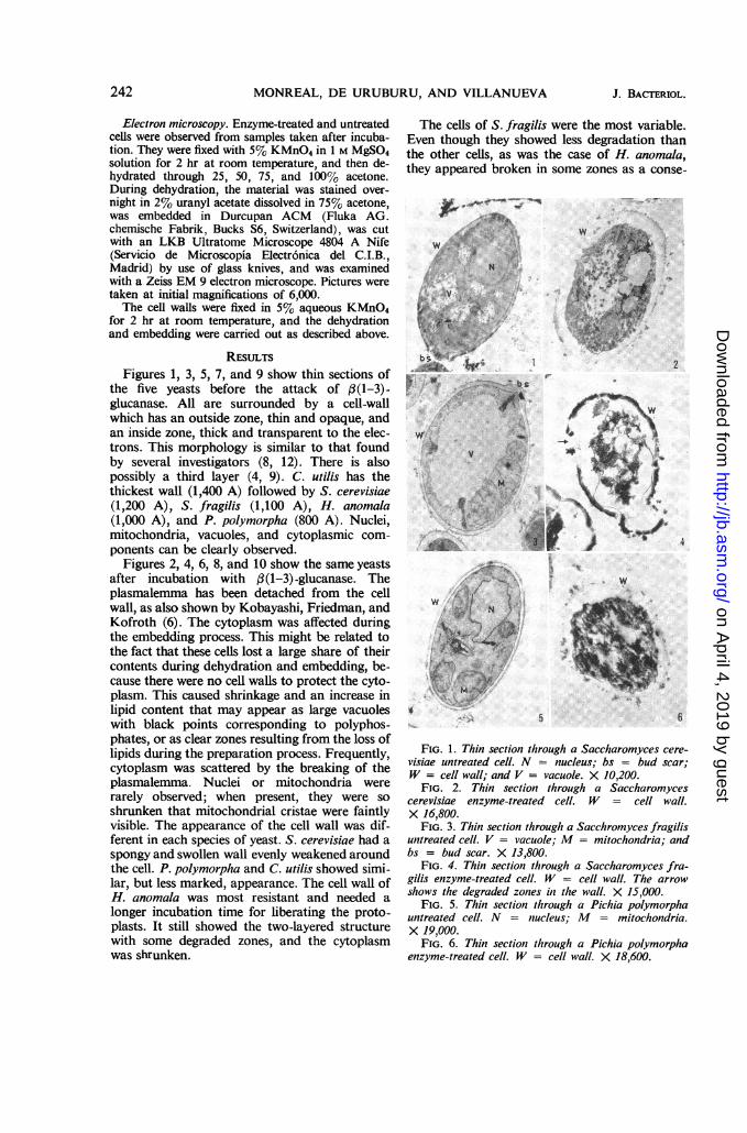
RESULTSFigures 1, 3, 5, 7, and 9 show thin sections of
the five yeasts before the attack of (3(1-3)-glucanase. All are surrounded by a cell-wallwhich has an outside zone, thin and opaque, andan inside zone, thick and transparent to the elec-trons. This morphology is similar to that foundby several investigators (8, 12). There is alsopossibly a third layer (4, 9). C. utilis has thethickest wall (1,400 A) followed by S. cerevisiae(1,200 A), S. fragilis (1,100 A), H. anomala(1,000 A), and P. polymorpha (800 A). Nuclei,mitochondria, vacuoles, and cytoplasmic com-ponents can be clearly observed.
Figures 2, 4, 6, 8, and 10 show the same yeastsafter incubation with 3(1-3)-glucanase. Theplasmalemma has been detached from the cellwall, as also shown by Kobayashi, Friedman, andKofroth (6). The cytoplasm was affected duringthe embedding process. This might be related tothe fact that these cells lost a large share of theircontents during dehydration and embedding, be-cause there were no cell walls to protect the cyto-plasm. This caused shrinkage and an increase inlipid content that may appear as large vacuoleswith black points corresponding to polyphos-phates, or as clear zones resulting from the loss oflipids during the preparation process. Frequently,cytoplasm was scattered by the breaking of theplasmalemma. Nuclei or mitochondria wererarely observed; when present, they were soshrunken that mitochondrial cristae were faintlyvisible. The appearance of the cell wall was dif-ferent in each species of yeast. S. cerevisiae had aspongy and swollen wall evenly weakened aroundthe cell. P. polymorpha and C. utilis showed simi-lar, but less marked, appearance. The cell wall ofH. anomala was most resistant and needed alonger incubation time for liberating the proto-plasts. It still showed the two-layered structurewith some degraded zones, and the cytoplasmwas shrunken.
The cells of S. fragilis were the most variable.Even though they showed less degradation thanthe other cells, as was the case of H. anomala,they appeared broken in some zones as a conse-
,Se-W.Ar.efA
..
S ..
W no;,
>"''ev
*S:.. ;}*rj ;
t.w.2
-
sm ..* .. :::r V .. ' ': ox .W sz/S-
Z.. , s .:w i"
,
*1L 5<'' ri't. :ei: J:
.
. sO
.4
41
I *uSt'<{ y.vv;we1 t: -siZi . ]! *t ,8)* t\ wf;f
.. ._ .e_.Y
4 5 6
FIG. 1. Thin section through a Saccharomyces cere-visiae untreated cell. N = nucleus; bs = bud scar;W = cell wall; and V = vacuole. X 10,200.
FIG. 2. Thin section through a Saccharomycescerevisiae enzyme-treated cell. W = cell wall.X 16,800.
FIG. 3. Thin section through a Sacchromyces fragilisuntreated cell. V = vacuole; M = mitochondria; andbs = bud scar. X 13,800.
FIG. 4. Thin section through a Saccharomyces fra-gilis enzyme-treated cell. W = cell wall. The arrowshows the degraded zones in the wall. X 15,000.
FIG. 5. Thin section through a Pichia polymorphauntreated cell. N = niucleus; M = mitochondria.X 19,000.
FIG. 6. Thin section through a Pichia polymorphaenzyme-treated cell. W = cell wall. X 18,600.
242 J. BACTERIOL.
on April 4, 2019 by guest
http://jb.asm.org/
Dow
nloaded from

LYTIC ACTION OF ,B(l-3)-GLUCANASE
quence of a preferential enzyme attack. Throughthese holes, protoplasts were liberated. This offersan explanation as to why we found intact cells,protoplasts on the way out, liberated protoplasts,and empty cell walls.
w
I, x.,INw.
.ssa,
N
.W..
" .:,i:
-!9
/
IC}.
.
FIG. 7. Thin section throughl a Cand,treated cell. W = cell wall; M = mitocV = vacuole. X 18,000.
FIG. 8. Thin section throughl a Candida,treated cell. W = cell wall. The arrows s
degraded zones of the cell wall. X 23,201FIG. 9. Thin section througlh a Hansei
untreated cell. N = nucleus; V = vacuo,FIG. 10. Thin section througlh a Hanse,
enzyme-treated cell. W = cell wall;X 19,800.
FIG. 11. Thin section through Candidaand untreated walls. Note the third layeiwall. X 12,000.
FIG. 12. Thin section through Candidaand enzyme-treated walls. Note the degrethe cell wall. X 15,000.
Figures 11 and 12 show enzyme-treated and un-treated cell walls of C. utilis. Isolated walls weresimilar to nonisolated ones, but they seemed tohave another inner layer (see arrows). Thus, cellsmay have a three-layered wall; the third layer hasnot been detected by microscopic observation ofthe cell, probably because it is attached to theprotoplasts.
DIscUSSION
Based on the photomicrographs, the attack ofJ /3(1-3)-glucanase on the cell walls of five yeasts
w varies greatly from S.fragilis to S. cerevisiae. The* other three yeasts examined show results inter-
mediate between those of these two yeasts.The cell wall of S. cerevisiae is homogeneously
* 8 degraded by the glucanase over the cell surface,leading us to suppose that glucan is evenly dis-tributed upon it.The localized attack on certain zones of the cell
wall of S.fragilis suggests that glucan of S. fragilisaccumulates in certain zones of the cell wall,namely, those that are attacked by the enzyme.
v On the other hand, it could be that, even if glucanis homogeneous over the cell surface, its structurediffers such that it is easily attacked only on theabove-mentioned zones. We must admit that,
J.whereas cell walls consist of accumulated fibers,we do not know the orientation of the fibers on
1 the cell-wall mesh. On the other hand, if the glu-can is uniformly located around the cell wall, thedifferences in behavior could depend on localized
/xt # mannan, the other polysaccharide of the cell wall(5).Most analyses for mannan were from cells
^ > older than our culture of 12 hr. It is possible thatthe 12-hr cells have not yet produced mannan andthe enzyme, not finding any mannan resistance,can homogeneously attack the S. cerevisiae cell
,,/ walls.|2 There are no reports about the chemical com-
position of the S. fragilis cell walls. We can as-
.ida utisu. sume they have mannan which is not evenly dis-hda utiis un- tributed all around the cell surface, such that thehlondria;and.
enzyme finds mannan resistance only at the un-
utilis enzyme- attacked zones.how the more On the other hand, there must be a relation be-0. tween yeast bud scars and attacked cell-wallinula anomala zones. We know these scars have a different ultra-kle. X 15,600. structure from the cell wall. For this reason, thefnula anomala S. fragilis cell walls are more readily degraded at= vacuole. the bud scars.
utili isolad The other three yeasts show an intermediaterutistheolaned behavior against /(1-3) glucanase. In this respect,
we can order them as follows: P. polymorpha,utilis isolated H. anomala, and C. utilis.aded zones of P. polymorpha is similar to S. cerevisiae. C.
utilis is more like S. fragilis, but in C. utilis the
243VOL. 94, 1967
on April 4, 2019 by guest
http://jb.asm.org/
Dow
nloaded from

MONREAL, DE URUBURU, AND VILLANUEVA
differences between degraded and undegradedzones are not so great. On isolated walls, thisdegradation is greater, and this may result froman attack on the inner layer, with the possibilitythat glucan accumulates in it.Of all yeasts tested, H. anomala is the least
sensitive to enzyme attack. It needed an incuba-tion period 7.5 times longer than the other yeaststo free the protoplasts. It showed the least cell-wall degradation of all the yeasts.
ACKNOWLEDGMENTS
We are indebted to E. T. Reese for correcting theEnglish manuscript and to the Servicio de Micros-copia Electronica del Instituto Cajal del CSICMadrid, for allowing us to use the electron micro-scope. We thank also Enrique Alvarez for helping usto translate the manuscript into English.
LITERATURE CITED
1. EDDY, A. A., AND D. H. WILLIAMSON. 1957. Amethod of isolating protoplasts from yeast.Nature 179:1252.
2. GARCIA-MENDOZA, C., AND J. R. VILLANUEVA.1963. Preparation of yeast cell walls. Can. J.Microbiol. 9:141-142.
3. GASCON, S., AND J. R. VILLANUEVA. 1965. Mag-nesium sulphate as stabilizer during liberationof yeast and mould protoplasts. Nature 205:822.
4. HEICK, H. M. C., AND H. B. STEWART. 1965.Mitochondria from Lipomyces lipofer. Can. J.Biochem. 43:561-571.
5. KESSLER, G., AND W. J. NICKERSON. 1959. Glu-
comannan-protein complexes from cell walls ofyeasts. J. Biol. Chem. 234:2281-2285.
6. KOBAYASHI, G. S., L. FRIEDMAN, AND J. F.KOFROTH. 1964. Some cytological and patho-genic properties of spheroplasts of Candidaalbicans. J. Bacteriol. 88:795-801.
7. NAGASAKI, S., N. P. NEUMANN, P. ARNOW, L. D.SCHANABLE, AND J. 0. LAMPEN. 1966. An en-zyme which degrades the walls of living yeast.Biochem. Biophys. Res. Commun. 25:158-164.
8. NORTHCOTE, D. H. 1963. The structure and or-ganization of polysaccharides of yeast. Proc.Symp. on the Chemistry and Biochemistry ofFungi and Yeast, p. 669-674. InternationalUnion of Pure and Applied Chemistry, Dublin.
9. SENTANDREU, R., AND J. R. VILLANUEVA. 1965.Electron microscopy of thin sections of Candidautilis. The structure of the cell wall. Arch.Mikrobiol. 50:103-110.
10. TANAKA, H., AND H. J. PHAFF. 1965. Enzymatichydrolysis of yeast cell walls. I. Isolation ofwall-decomposing organisms and separationand purification of lytic enzymes. J. Bacteriol.89:1570-1580.
11. VILLANUEVA, J. R. 1966. Protoplasts of fungi,p. 3. In G. C. Ainsworth and A. S. Sussman[ed.], The fungi, vol. 2. Academic Press, Inc.,New York.
12. VILLANUEVA, J. R., M. V. ELORZA, AND J.MONREAL. 1966. The use of purified lytic en-zymes to obtain yeast protoplasts. Proc.Intern. Symp. Yeast, 2nd, Bratislava, p. 85.
13. VIToLS, E., R. J. NORTH, AND A. W. LINNANE.1961. Studies on the oxidative metabolism ofSaccharomyces cerevisiae. I. Observations onthe fine structure of the yeast cell. J. Biophys.Biochem. Cytol. 9:689-699.
244 J. BACTERIOL.
on April 4, 2019 by guest
http://jb.asm.org/
Dow
nloaded from


![Isolation, Purification and Characterization of Glucanase ... · PDF fileIsolation, Purification and Characterization of Glucanase Enzyme from ... Mukesh Srivastava, ... et al. [26]](https://static.fdocuments.in/doc/165x107/5a84639b7f8b9a882e8b7c28/isolation-purification-and-characterization-of-glucanase-purification-and-characterization.jpg)


