LYSOSOMAL ENZYMES · • α-L-iduronidase 12 ± 15 1.6-69%(0.08-12 nmol/h) • Galactocerebrosidase...
32
LYSOSOMAL ENZYMES MARIE JACKSON, KEES SCHOONDERWOERD Clinical Genetics Leading the way in genetic issues
Transcript of LYSOSOMAL ENZYMES · • α-L-iduronidase 12 ± 15 1.6-69%(0.08-12 nmol/h) • Galactocerebrosidase...

LYSOSOMAL ENZYMES
MARIE JACKSON,KEES SCHOONDERWOERD
Clinical GeneticsLeading the way in genetic issues

Introduction
• Enzyme activity• Background & history of EQA scheme (ESGLD/
ERNDIM)• Current ERNDIM scheme.• Discussion• Diagnostic issues with fluorimetric substrates

ENZYME KINETICSEnzyme activity or how much enzyme is present.Two ways: disappearance of substrate or theappearance of product in time. (velocity)Measuring the appearance of product usuallymore accurate
Fixed-Timed or discontinuous assay With fixed-timed assays, one can measure many assayssimultaneously.Kinetic or continuous assay measure theappearance of product in real-time. Dis-advantage only one reaction can be measuredat a time, advantage is the convenience ofeasily measurable reaction rates.Initial rates measured. v0 must be bothreproducible and dependent on [E]t. Validassay conditions exist if the product hasconsumed less than 5% of the substrate.

ENZYME ACTIVITY MEASUREMENT: REQUIREMENTS• pH is constant: Enzymes have specific pH ranges of activity due to structural sensitivity to
proteins which cause enzymes to be sensitive to pH changes. A prepared buffer solution is usedto keep the pH constant so that the pH resist changes. Good buffers do not cross membranes; donot absorb light; are chemically stable; and are biochemically inert.
• V0 is proportional to the concentration of the enzyme, [E]t. An assay is only valid when a plotof V0 vs [E]t is linear because the enzyme must be the only limiting factor to the substrateconcentration. Additionally, when V0 and [E]t are proportional, the presence of effectors aremeasurable and one can determine if an inhibitor is competitive, noncompetitive, oruncompetitive.
• V0 is corrected for non-enzymatic conversion: V0(corrected) = V0(+enzyme) – V0(-enzyme). Acontrol factor must be measured while conducting enzyme assays in order to ensure accuratecalculations. For non-enzyme controls, buffers are used in place of enzymes.

ENZYME ACTIVITY• Salt Concentration: of importance for proper enzyme folding and substrate binding. This can effect the
enzyme's ability to catalyze a reaction.
• Enzyme and Substrate Concentration: Precise enzyme and substrate measurements must be calculated toensure optimum activity.
• pH Dependence: Most enzymes have an optimal pH at which their reaction is catalyzed the fastest. Oftenenzymes operate at a maximum activity when the pH is close to the pka of the enzyme's active site.
• Inhibition: Inhibitors can decrease reaction rates via binding to the enzyme in the active site, products of thereaction can often inhibit the enzyme by binding the active site.
• Activators: Activators increase the activity of an enzyme. Additional chemicals may be needed to achievenecessary concentrations of activators, reactants, and co-factors.
• Temperature dependence: Many enzymes have an optimal temperature that can be found by measuringreaction rates with varying temperatures. Reaction rates usually increase with temperature, however hightemperatures usually denature and give no activity of the enzyme.
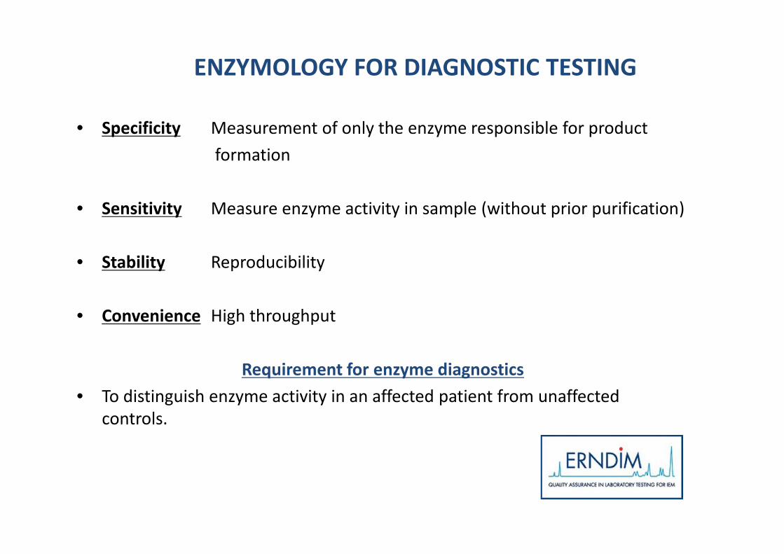
ENZYMOLOGY FOR DIAGNOSTIC TESTING
• Specificity Measurement of only the enzyme responsible for productformation
• Sensitivity Measure enzyme activity in sample (without prior purification)
• Stability Reproducibility
• Convenience High throughput
Requirement for enzyme diagnostics• To distinguish enzyme activity in an affected patient from unaffected
controls.

LYSOSOMAL ENZYME SCHEME HISTORY
Pilots “large scale” OvD ESGLD2006 (36) cultured fibroblasts2007 (46)2008 (55)Protein +MU standard2009 (59)EBV +leucocytes+ DBS
2010(58)EBV ERNDIM2011(61)EBV KS2012(64)Fibroblasts pellet+DBS2013(71)Fibroblasts2014(76)Fibroblasts+DBS2015(74)Fibroblasts2016(74)Fibroblasts2017(74)Fibroblasts
HISTORY
Complicated to send cultured flasksMany fibroblasts were not alive on arrival
Difficult to obtain enough blood from patientEBV easy to culture in bulk
Low enzyme activity EBVEBV not used in diagnostics and labs notfamiliar with the enzyme levels in thismaterial.

ACTIVITY ENZYMES CTRL EBV CELL LINEALL PARTICIPANTS 2010 SCHEME
– Act.EBV (% ctrl leuco) Range participants• α-L-iduronidase 12 ± 15 1.6-69%(0.08-12 nmol/h)• Galactocerebrosidase 16 ± 16 2-70%(0.4-15 nmol/17h)• Sphingomyelinase 206 ± 137 1.3-795%(2-127 nmol/17h)• α-N-acetylglucosaminidase 79 ± 32 26-284%(0.5-44 nmol/h)• β-Hexosaminidase A + B 41 ± 29 14-204%(49-2212 nmol/h)• β-Hexosaminidase A 42 ± 25 12-152%(5-1737 nmol/h)• Galactose-6S sulfatase 6 ± 5 0.6-19%(0.12-19 nmol/17h)• β-Galactosidase 25 ± 17 9-126%(0.06-136 nmol/h)• β-mannosidase 26 ± 12 3-56%(3-118 nmol/h)• β-Glucuronidase 36 ± 26 9.8-131%(6.6-369 nmol/h)

0
20
40
60
80
100
120
C F+P F 3w 37oC F+P 3w 37oC
Enzy
me
activ
ity(%
Cont
rol)
Condition
AIDU
BGAL
BHEX-T
FIBROBLAST HOMOGENATESSTABILITY OF LYOPHILISED LYSOSOMAL ENZYMES

• SELECTION OF ENZYMES IN SCHEME
• SELECTION OF SAMPLES IN SCHEME
• CULTURING ENOUGH FIBROBLASTS FORALL THE PARTICIPANTS (ABOUT 3 – 4months)
• HOMOGENISATION OF FIBROBLASTS
• SUBDIVISION AND LYOPHILISATION
• SHIPMENT TO PARTICIPANTS
ORGANISATION

LYSOSOMAL DISORDERS
• Sphingolipidosis Oligosaccharidosis Mucopolysaccharidosis• Gm1-gangliosidosis Mucolipidosis MPS-I• Gm2-gangliosidosis Fucosidosis MPS-II• Fabry α-Mannosidosis MPS-III A-D• Gaucher ß-Mannosidosis MPS-IV A-B• Niemann-Pick A/B/C Aspartyl-glucosaminuria MPS-VI• MLD Pompe MPS-VII• Krabbe Schindler• CLN• Wolman
• What enzymes in the scheme?

ERNDIM WEBSITE

USING LYOPHILISED FIBROBLASTS
6 samples:
– 75 laboratories– Measure activity of 10 enzymes + protein– Enter specific activity and % mean control.– Tickbox for diagnosis

% mean controlMany labs do not use fibroblasts.In 2017 scheme: 10 – 19% labshave not entered a value.
However, this probably moreuseful for inter lab comparison.
Lab ALL n
Lab specific activity (nmol/hr/mgptn)
All labs will have developed ownreference ranges or run assayswith simultaneous in-assaycontrols. Inter-lab variability andnot directly comparable.
RESULTS
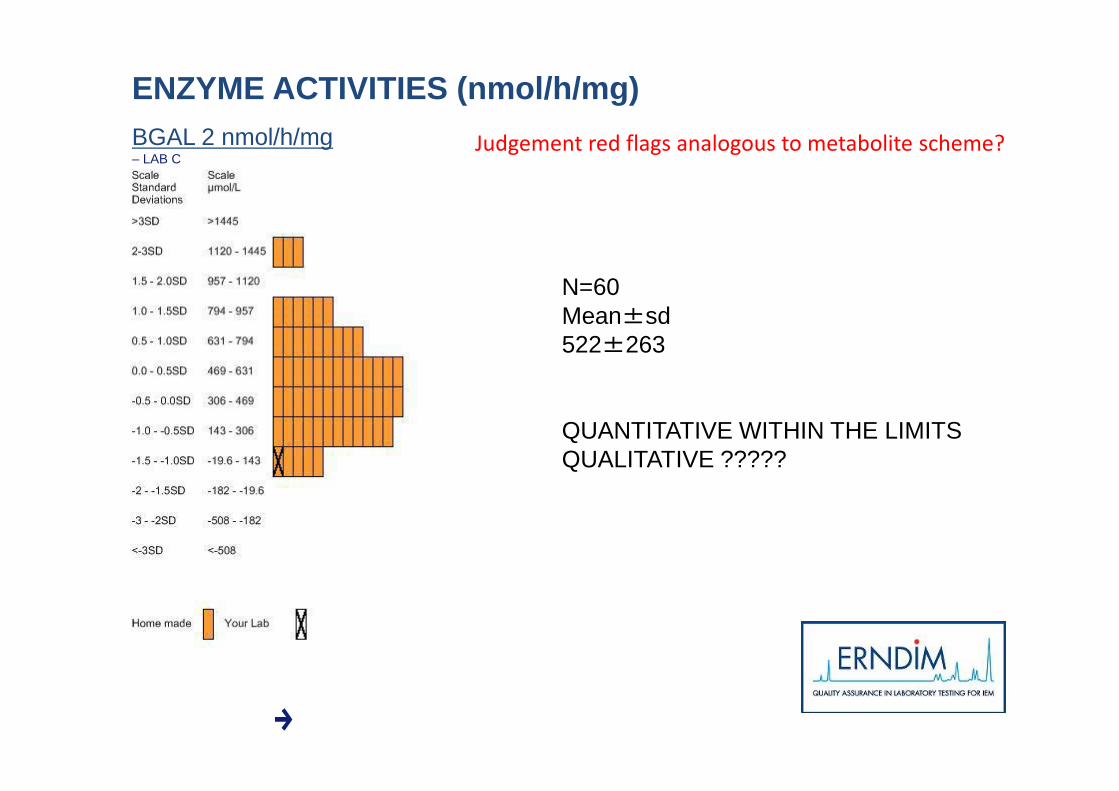
BGAL 2 nmol/h/mg– LAB C
N=60Mean±sd522±263
QUANTITATIVE WITHIN THE LIMITSQUALITATIVE ?????
ENZYME ACTIVITIES (nmol/h/mg)Judgement red flags analogous to metabolite scheme?

BGAL 0.6 %– LAB C N=60
Mean±sd98±67
QUALITATIVE FALSE POSITIVE
QUANTITATIVE NO RED FLAG
Red flags cannot be used in enzymescheme
RELATIVE ENZYME ACTIVITIES(%)
Diagnosis incorrect
• Enzyme activity control sample<15% mean control range
• Enzyme activity patient sample>30% mean control range

INDICATION OF DIAGNOSIS
• Useful for labs tosee correct results.

Discussion
• Are fibroblasts an appropriate material for theLSD scheme?
• Are the number of samples (6) andsubmissions (3) appropriate?
• Is the website appropriate for recording thedata?

SCORING SYSTEM
Nr.Enzymes ASB AGAL BGAL BGLU AGLU BHEX-T AIDU IDU2S GALC SM PROT Nr. satisfactory enzymes Score
5(22) 2 4 4 4 0 0 4 0 0 0 2 4 207(30) 4 4 4 4 4 4 0 4 0 0 2 7 306(26) 2 0 4 4 0 0 4 4 0 4 2 5 249(38) 4 4 4 4 4 4 4 4 0 4 2 9 3810(42) 4 4 4 4 4 4 4 4 4 4 4 10 429(38) 4 4 4 2 4 4 2 0 4 4 4 7 34
Quantitative Red flags+Qualitative (Reproducibility + Diagnosis)Reproducibility 2 pt for every enzyme and proteinCV>60% 0 pt; 35<CV<60 1 ptDiagnosis 2pt for every enzymeSample not measured, 6 samples for every sample -16% of totalpointsDiagnosis not correct 0 pt,Diagnosis correct but incorrect level measured enzyme activity 1ptDiagnosis not correct but correct level measured enzyme activity1 pt

Scoring scheme:
Diagnosis (2) and CV (2)Participants score >60% (average all enzymes) to besatisfactory performer.
Most important is to obtain correct diagnosis
Affected patient missed or unaffected patient given incorrectdiagnosis.
Score each enzyme individually?

Discussion
• Is the scoring system appropriate?• Is it more appropriate to mark enzymes
individually?• Are the enzymes offered satisfactory – should
other enzymes be included?• Is it more appropriate for the LSD enzyme
scheme to be a qualitative (diagnosticproficiency) scheme?

Qualitative scheme
• Enzyme results• Enzyme deficiency consistent with diagnosis of X• Normal result which would make diagnosis of Y
highly unlikely.• Any further comments to confirm diagnosis etc.
Other issues
• Labs have own reference ranges (or may quotesimultaneous controls).
• What are labs using to report % mean values (seemto be discrepancies!). ? Useful to report usingsample 1 as normal reference sample.
• Labs may be using different substrates (radiolabellednatural substrates/ fluorescent substrates).
This scheme needs to be simplified!
Qualitative scheme
• Enzyme results• Enzyme deficiency consistent with diagnosis of X• Normal result which would make diagnosis of Y
highly unlikely.• Any further comments to confirm diagnosis etc.

Discrepancies with artificialsubstrates.

NIEMANN PICK A/BQ292K MUTATION
• Affected patient with Q292K mutationincluded in scheme.
• Normal activity observed with artificialsubstrate.
• 50% of participants missed this diagnosis• Overcome by addition of lyso-sphingosine
(van Diggelen et al)

Discrepancies between natural/artificial substrates
• Traditionally diagnosis of Krabbe disease relied on measurement ofenzyme deficiency using a radiolabelled natural substrate (>30yrs).
• More convenient fluorimetric substrate implemented in some labs; (6-hexadecanoylamino-4-methylumbelliferyl- beta-D-galactoside availablefrom Moscerdam & Glycosynth).
• Potential for false positives – pseudodeficiencies.
• Potential for false negatives – normal GALC activity against artificialsubstrate.
• Similar situation with Niemann Pick A/B.

In 2017 ERNDIM Scheme:
• 3 participants measured a normal level ofgalactocerebrosidase activity in an affectedpatient sample.
• 6 participants measured a deficiency ofgalactocerebrosidase activity in an unaffectedsample.

CASE 1a: Diagnosis confirmed by molecular genetics
Patient
Index case is a child with biochemically diagnosed Krabbe disease (measuredIn leucocytes using the natural substrate for the enzyme):
Patient Normal rangeGalactocerebrosidase (nmol/h/mg) 0.01 0.4 – 4.0
Screening of the GALC gene showed the patient was homozygous for thec.621+2T>C pathogenic variant confirming the diagnosis.

CASE 1b: Pseudodeficiency
Patient’s unaffected motherEnzyme testing was also carried out for the patient’s unaffected mother:results were very low / deficient.
Patient’s mother Normal rangeGalactocerebrosidase (nmol/h/mg) 0.04 0.4 – 4.0
Screening of the GALC gene showed: in addition to being heterozygous forthe c.621+2T>C variant detected in her affected son, this individual wasalso heterozygous for the c.550C>T p.(Arg184Cys) variant.
This variant is present in up to 6% of alleles in the European population, isnot associated with enzyme deficiency when expressed in COS-1 cells and isgenerally regarded to be non-pathogenic.
As this individual is unaffected the observed enzyme deficiency is termed ‘pseudodeficiency’ andis due to reduced activity associated with c.550C>T.

CASE 2: Carrier identified by enzyme analysis
Patient with developmental delay who was referred for lysosomal enzymeanalysis. (Manchester Willink)
Galactocerebrosidase (nmol/h/mg) Patient Normal rangeFluorescent 0.34 0.8 – 4.0Natural 0.13 0.4 – 4.0
i.e. patient has reduced but clearly not deficient activity. Chitotriosidase normal
GALC gene showed the patient to be heterozygous for the common 30kbdeletion and also for the c.550C>T variant. NOT AFFECTED.
Note: The incidence of Krabbe disease in the general population is very low,equating to a carrier frequency of approx. 1/160. However as severalhundred patients undergo lysosomal enzyme screening in the larger UKbiochemical genetics labs per year we would occasionally expect to pick upcarriers.
Fluorescent substrate picking up these carriers and pseudodeficiencies.

Thank you – any questions?