Tumoricidal Response following Perfusion over Immobilized Protein A
Upload
robert-kellerCategory
view
213download
1
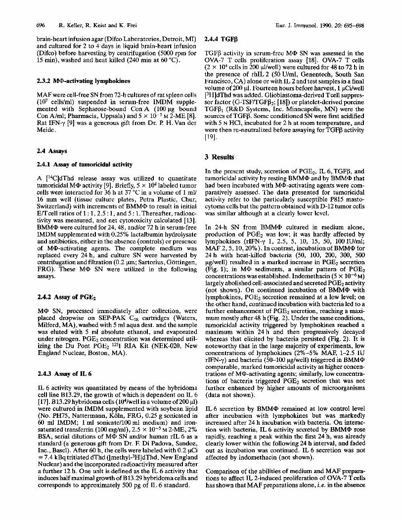
Eur. J. Immunol. 1990. 20: 695-698
Short paper
Macrophage secretion induced by lymphokines vs. bacteria 695
Robert KellerO, Ruth KeistO and Karl FreiA
Immunbiology Research Group, Institute for Immunology and Virologyo, University of Zurich and Section of Clinical Immunology and Department of NeurosurgeryA, University Hospital, Zurich
1 Introduction
Lymphokines and bacteria, tumoricidal activity, trigger response in macrophages*
that induce a different secretory
The abilities of various macrophage-activating agents to trigger tumoricidal activity and/or the secretion of prostaglandin E2 (PGEz), interleukin 6 (IL 6) and transforming growth factor P (TGFP) in bone marrow-derived mononuclear phagocytes (BMMQ) in vitro were comparatively assessed. Induction of tumo- ricidal activity by lymphokines, that is only short-lived, was not associated with enhanced secretion of these activities by BMMQ; in contrast, incubation with heat-killed facultative intracellular bacteria resulted in persisting tumoricidal activity and in marked enhancement of the secretion of IL 6 and PGE2, but not of TGFP activity. These findings support the concept that the pattern of the secretory response induced in macrophages by lymphokines differs from that triggered by bacteria and that the rapid decay of lymphokine-induced tumoricidal activity is not due to autocrine macrophage deactivation mediated by one of these agents alone.
MQ activation is essential for the capacity of the host to cope with infection and to contain tumor growth. Cyto- kines, notably IFN-y [l-31, and various bacteria andor their products [4-71 are particularly potent of eliciting tumoricidal andor bactericidal activity in MQ. In BM- derived mononuclear phagocytes (BMMQ) that are homo- geneous with respect to the cell lineage, tumoricidal activity triggered by lymphokines is short lived [ l , 8-11] whereas that elicited by heat-killed bacteria persists for weeks [9]. The causes responsible for the marked discrep- ancy in the antitumor potential of lymphokines and microbes are probably manifold. Earlier work has shown that secretion of IL 1 [12] and TNF-a* by MQ is differently affected by these two classes of MQ-activating agents. To evaluate this possibility further, the ability of these agents to trigger in BMMQ tumoricidal activity and/or the secre- tion of PGE2, IL 6, and TGFP was comparatively assessed in the present study.
[I 79601
* This study was supported by the Swiss National Science Foun- dation (grant 3.336.86) and the Canton Zurich.
Correspondence: Robert Keller, Arbeitsgruppe fiir Immunbiolog- ie, Institut fiir Immunologie und Virologie, Universitat Zurich, Birchstrasse 95, CH-8050 Zurich, Switzerland
Abbreviations: BMMQ: Bone marrow-derived mononuclear phagocytes CP: Corynebacterium parvum IMDM: Iscove's modified Dulbecco medium LM: Listeria monocytogenes MAF: Macrophage-activating lymphokines SE: Staphylococcus epidermidis TGFfk Transforming growth factor fi
* Keller, R., Wechsler, A., and Keist, R., in preparation.
2 Material and methods
2.1 Effector cells
Rat BM cells were obtained and cultured as previously described [9,13]. Briefly, BM cells (l@/ml) from the femurs of adult male DA rats were grown in Iscove's MDM (IMDM) supplemented with 10% FCS (Gibco, Grand Island, NY), 5% horse serum (Gibco), and antibiotics (50 IU penicillin/ml, 50 vg/ml streptomycin), and condi- tioned with SN (final concentration 10%) from strain L clone 929 cells (American Type Culture Collection, Rock- ville, h4D; ATCC CCL1). On day 6, the cells remaining adherent after washing were removed and resuspended in IMDM. These BMM", that were homogeneous with respect to the cell lineage [13, 141, were the source of effector cells. To enhance their functional activity, BMMQ were first interacted for 24 h with Ma-activating agents and then interacted with tumor targets in the presence of the same activating agent.
2.2 a m o r cells
D-12 tumor cells, originally induced by administration of dimethylbenz(a)anthracene to female DA rats [15], and DBA/2 murine mastocytoma cells P815 were cultured in IMDM supplemented with 5% FCS and antibiotics. Tumor cells were regularly checked to assure the absence of mycoplasma as previously described [ 161.
2.3 Reagents
2.3.1 Ma-activating agents
Corynebacterium parvum (CP; ATCC 6916), Listeria mon- ocytogenes (LM) and Staphylococcus epidermidis (SE) organisms were selected from a single colony on solid
0 VCH Verlagsgesellschaft mbH, D-6940 Weinheim, 1990 0014-2980/90/0303-0695$02.50/0
696 R. Keller, R. Keist and K. Frei Eur. J. Immunol. 1990.20: 695-698
brain-heart infusion agar (Difco Laboratories, Detroit, MI) and cultured for 2 to 4 days in liquid brain-heart infusion (Difco) before harvesting by centrifugation (5000 rpm for 15 min), washed and heat killed (240 min at 60 "C).
2.3.2 MQ-activating lymphokines
MAF were cell-free SN from 72-h cultures of rat spleen cells (lo7 cells/ml) suspended in serum-free IMDM supple- mented with Sepharose-bound Con A (100 pg bound Con A/ml; Pharmacia, Uppsala) and 5 x lop5 M 2-ME [8]. Rat IFN-y [9] was a generous gift from Dr. F? H.Van der Meide .
2.4 Assays
2.4.1 Assay of tumoricidal activity
A [14C]dThd release assay was utilized to quantitate tumoricidal MQ activity [9]. Briefly, 5 x 10⁴ labeled tumor cells were interacted for 36 h at 37 "C in a volume of 1 mY 16 mm well (tissue culture plates, Petra Plastic, Chur, Switzerland) with increments of BMMQ to result in initial Emcell ratios of 1 : 1,2.5 : 1, and 5 : 1. Thereafter, radioac- tivity was measured, and net cytotoxicity calculated [13]. BMMa were cultured for 24,48, and/or 72 h in serum-free IMDM supplemented with 0.25% lactalbumin hydrolysate and antibiotics, either in the absence (controls) or presence of Ma-activating agents. The complete medium was replaced every 24 h, and culture SN were harvested by centrifugation and filtration (0.2 pm; Sartorius, Gottingen, FRG). These MQ SN were utilized in the following assays.
2.4.2 Assay of PGE2
MQ SN, processed immediately after collection, were placed dropwise on SEP-PAK C18 cartridges (Waters, Milford, MA), washed with 5 ml aqua dest. and the sample was eluted with 5 ml absolute ethanol, and evaporated under nitrogen. PGE2 concentration was determined util- izing the Du Pont PGE2 1251 RIA Kit (NEK-020, New England Nuclear, Boston, MA).
2.4.3 Assay of IL 6
IL 6 activity was quantitated by means of the hybridoma cell line B13.29, the growth of which is dependent on IL 6 [ 171. B 13.29 hybridoma cells ( 104/well in a volume of 200 pl) were cultured in IMDM supplemented with soybean lipid (No. PH75, Nattermann, Koln, FRG, 0.25 g sonicated in 60 ml IMDM; 1 ml sonicate/100 ml medium) and iron- saturated transferrin (100 mg/ml), 2.5 X M 2-ME, 2% BSA, serial dilutions of MQ SN and/or human rIL 6 as a standard (a generous gift from Dr. E Di Padova, Sandoz, Inc., Basel). After 60 h, the cells were labeled with 0.2 pCi = 7.4 kBq tritiated dThd ([meth~l-~HIdThd, New England Nuclear) and the incorporated radioactivity measured after a further 12 h. One unit is defined as the IL 6 activity that induces half maximal growth of B13.29 hybridoma cells and corresponds to approximately 500 pg of IL 6 standard.
2.4.4 TGFB
TGFP activity in serum-free MQ SN was assessed in the OVA-7 T cells proliferation assay [18]. OVA-7 T cells (2 x lo4 cells in 200 pl/well) were cultured for 48 to 72 h in the presence of rhIL 2 (50 U/ml, Genentech, South San Francisco, CA) alone or with IL 2 and test samples in a final volume of 200 p1. Fourteen hours before harvest, 1 pCi/well [3H]dThd was added. Glioblastoma-derived Tcell suppres- sor factor (G-TSF/TGFP2; [ 181) or platelet-derived porcine TGFPl (R&D Systems, Inc. Minneapolis, MN) were the sources of TGFP. Some conditioned SN were first acidified with 5 N HC1, incubated for 2 h at room temperature, and were then re-neutralized before assaying for TGFP activity ~191.
3 Results
In the present study, secretion of PGE2, IL 6, TGFP, and tumoricidal activity by resting BMMQ and by BMMQ that had been incubated with Ma-activating agents were com- paratively assessed. The data presented for tumoricidal activity refer to the particularly susceptible P815 masto- cytoma cells but the pattern obtained with D-12 tumor cells was similar although at a clearly lower level.
In 24-h SN from BMMQ cultured in medium alone, production of PGE2 was low; it was hardly affected by lymphokines (rIFN-y 1, 2.5, 5, 10, 15, 50, 100IU/ml; MAF 2,5,10,20%). In contrast, incubation of BMMa for 24h with heat-killed bacteria (50, 100, 200, 300, 500 pg/well) resulted in a marked increase in PGE2 secretion (Fig. 1); in MQ sediments, a similar pattern of PGE2 concentrations was established. Indomethacin (5 x M) largely abolished cell-associated and secreted PGE2 activity (not shown). On continued incubation of BMMQ with lymphokines, PGE2 secretion remained at a low level; on the other hand, continued incubation with bacteria led to a further enhancement of PGEz secretion, reaching a maxi- mum mostly after 48 h (Fig. 2). Under the same conditions, tumoricidal activity triggered by lymphokines reached a maximum within 24 h and then progressively decayed whereas that elicited by bacteria persisted (Fig. 2). It is noteworthy that in the large majority of experiments, low concentrations of lymphokines (2%-5% MAF, 1-2.5 IU rIFN-y) and bacteria (50-100 pg/well) triggered in BMMQ comparable, marked tumoricidal activity as higher concen- trations of MQ-activating agents; similarly, low concentra- tions of bacteria triggered PGE2 secretion that was not further enhanced by higher amounts of microorganisms (data not shown).
IL6 secretion by BMMa remained at low control level after incubation with lymphokines but was markedly increased after 24 h incubation with bacteria. On interac- tion with bacteria, IL 6 activity secreted by BMMQ rose rapidly, reaching a peak within the first 24 h, was already clearly lower within the following 24 h interval, and faded out as incubation was continued. IL 6 secretion was not affected by indomethacin (not shown).
Comparison of the abilities of medium and MAF prepara- tions to affect IL 2-induced proliferation of OVA-7 Tcells has shown that MAF preparations alone, i. e. in the absence

Eur. J. Immunol. 1990. 20: 695-698
I
Macrophage secretion induced by lymphokines vs. bacteria 697
- 2000
- 1800
- 1600 - C - 1400 g .
- 1200 s an 0 4 . - 1000 g - L
0 - 800 V W Y)
- 600 $
- 400
- 200
100
90
80
70
W Y) 4
W LL
W r
Y 60
- 2 50 F 7 I
40 - I
+ Y
30
U 20
10
.I L CONTROL CP LR SE MAF lFNy
Figure 1. Comparative abilities of Ma-activating agents to trigger in BMM@ tumoricidal activity and/or to enhance PGE2 secretion. BMM@ were incubated for 24 h in medium alone (control) or in medium supplemented with Ma-activating agents before PGE2 secretion and tumoricidal activity (P815 mastocytoma cells at an initial effectorltarget cell ratio of 2.5 : 1) were assessed. BMMQ were derived from the same pool, and values are from three experiments, each performed in triplicate. PGE2 secretion by BhBla incubated with bacteria was statistically significantly different from controls (p < 0.001, Mann-Whitney U test). Con- centrations of Ma-activating agents: CP, LM and SE, 300 pg/well; MAF lo%;, rIFN-y 50 IU/well.
of BMMQ,, exhibited minor TGFP-like activity. In 24-h and 48-h SN from control BMMQ, and from B W Q , incubated with various concentrations of MAF or CP, TGFP-like activity remained at a low level (not shown). In conditioned SN that had been acidified, TGFP activity was at a similar, low level, i.e. acidification did not result in an increase in TGFP activity (not shown).
4 Discussion
Lymphokines and a variety of heat-killed bacteria are equally potent in triggering tumoricidal activity in a pure population of BMMQ, in vitro [ l , 3 , 7 , 201; however, that elicited by lymphokines is only short-lived whereas that induced by bacteria persists for a prolonged period of time [l, 8, 9, 111. In an in vivo correlate the tumor-protective effect of bacteria (BCG, CP, LM; [lo, 15, 211) was pronounced, but that of lymphokines weak [10].The causes responsible for the marked difference in the persistence of MQ,-mediated tumoricidal activity elicited by bacteria vs. lymphokines are still poorly understood. Tumoricidal activ- ity triggered by bacteria is maintained as long as the organisms are extracellularly present; after washing, tumo- ricidal activity rapidly decays although MQ, have engulfed large numbers of organisms [9]. Attempts to augment the
T 1600
1500
1400
1300
1200
1100
1000 f N
900 . 800 ~ $
5 700 ;
n
600 aE 0
500 5
- I-
W v)
400 WN z 300 4 200 i 100
0 24 48 72 h
Figure 2. Kinetics of PGE2 secretion and of tumoricidal activity triggered in Bh4MQ by lymphokines vs. bacteria. PGEl secretion was hardly affected by lymphokines (A, MAF 10% vs. resting BMMQ, O), but markedly enhanced by bacteria (B, CP 300 pg/well), reaching peak activity after 48 h incubation. Tumoricidal activity triggered by lymphokines (A, MAF 10%) and bacteria (0, CP 300 kg/well) was equally pronounced at 24 h, but that induced by lymphokines progressively decayed whereas that induced by bacteria persisted. Resting BMMQ did not manifest tumoricidal activity ([ 14C]dThd release C 2% ; not shown). Values are derived from three experiments, each performed in tripli- cate.
efficacy of lymphokines by their incorporation in liposomes andlor their repeated administration in vitro and in vivo had only little success [8, 101.
The present study explored the possibility that the various MQ,-activating agents might elicit in MQ, a different pattern of secretory response. Previous work has shown that IL 1 secretion by BMMQ, was considerably enhanced by lym- phokines but was suppressed rather than promoted by bacteria [12]; in contrast TNFa activity in BMMQ, SN was high after incubation with bacteria but remained at control levels on incubation with lymphokines*. The present find- ings, which show that secretion of I L 6 and PGEz by BMMQ, was similarly affected by Ma-activating agents as that of TNFa, i.e. considerably enhanced by bacteria but not by lymphokines, support the notion that lymphokines and bacteria induce in MQ, a different pattern of secretory response. The differences in the kinetics of IL 6 and PGE2 observed in the present study and the dependence of IL 6
* Keller, R., Wechsler, A. et al. (in preparation).
698 R. Keller, R. Keist and K. Frei Eur. J. Immunol. 1990.20: 695-698
production on TNF-a [22] raise the question whether induction of TNF-a, IL 6, and PGEz secretion by M a are correlated, sequential processes.
Persistent M a activation can have detrimental conse- quences even for normal tissues [23]. M a deactivation may therefore be in the vital interest of the host [24, 251. The mechanisms involved in M a deactivation are still poorly understood, but various secretory products of M a have been assigned to have deactivating potential. Noteworthy in this respect are PGEz, a major arachidonic metabolite of M a [2, 261, IL6, that appears to represent a major regulatory signal of cellular immunity [27,28], and TGFP, a family of peptides that may be potent in M a deactivation [24, 25, 29, 301.
The findings showing that bacteria, but not lymphokines, enhance the secretion of IL 6 and PGEz by BMMa, and that secretion of TGFP-like activity was always low and not affected by acidification, argue against the concept that the rapid decay of lymphokine-induced tumoricidal activity is mediated by one of these Ma-derived cytokines alone.
We thank Dr. Christine Siepl for TGFP assays, Drs. E Di Padova and M . Schreier for the B13.29 cell line and human rlL 6, Dr. l? H. Van der Meide for rat rlFN-y, Ms Claudia Hubmann for excellent technical assistance, and Drs M. Aguet and H . Hengartner for critical reading of the manuscript.
Received September 14, 1989; in revised form December 18, 1989.
5 References 1 Meltzer, M. S., Lymphokines 1981. 3: 319. 2 Schultz, R. M., Med. Hypotheses 1980. 6: 831. 3 Schreiber, R. D. and Celada, A., Lymphokines 1985. 11: 87. 4 Adams, D. 0. and Hamilton,T. A., Annu. Rev. Immunol. 1984.
5 Scott, M.T., Semin. Oncol. 1974. I : 367. 6 Laucius, J. F., Bodurtha, A. J., Mastrangelo, M. J. and Creech,
7 Woodruff, M. F. A., The Interaction of Cancer and Host, Grune
2: 283.
R. H., J. Reticuloendothel. SOC. 1974. 16: 347.
and Stratton, New York 1980.
8 Keller, R. and Keist, R., Cell. Irnrnunol. 1986. 101: 659. 9 Keller, R., Keist, R.,Van der Meide, P., Groscurth, €?, Aguet,
10 Keller, R., Keist, R. and Schwendener, R. A., Int. J. Cancer
11 Keller, R. and Keist, R., Biochem. Biophys. Res. Commun.
12 Keller, R. and Keist, R., Eur. J. Immunol. 1987. 17: 1665. 13 Keller, R. and Keist, R., Exp. Cell. Biol. 1982. 50: 255. 14 Keller, R., Joller, P.W. and Keist, R., Cell. Zmmunol. 1989.120:
15 Keller, R., J. Natl. Cancer Zmt. 1977. 59: 1751. 16 Koppel, P., Peterhans, E., Bertoni, G., Keist, R., Groscurth,
P., Wyler, R. and Keller, R., J. Immunol. 1984. 132: 2021. 17 Aarden. L., Lansdorp, P. and De Groot, E., Lymphokines
1985. 10: 175. 18 Siepl, C., Bodmer, S., Frei, K., MacDonald, H. R., De Martin,
R., Hofer, E. and Fontana, A., Eur. J. Zmmunol. 1988. 18: 593.
19 Assoian, R. K., Fleurdelys, B. E., Stevenson, H. C., Miller, €? J., Madtes, D. K., Raines, E. W., Ross, R. and Sporn, M. B., Proc. Natl. Acad. Sci. USA 1987. 84: 6020.
20 Keller, R., in Herberman, R. B. (Ed.), Natural Cell-Mediated Immunity Against Tumors, Academic Press, New York 1980, p. 1219.
21 Keller, R., in Van Furth, R. (Ed.), Mononuclear Phagocytes. Functional Ascpects, M. Nijhof, The Hague 1980, p. 1725.
22 McIntosh, J. K., Jablons, 0. M., MulC, J. J., Norden, R. P., Rudikoff, S., Lotze, M. T. and Rosenberg, S. A., J. Irnmunol. 1989. 143: 162.
M. and Leist, T. P., J. Immunol. 1987.138: 2366.
1989. 44: 512.
1989. 164: 968.
277.
23 Cross, C. E., Ann. Intern. Med. 1987. 107: 526. 24 Tsunawaki, S., Sporn, M., Ding, A. and Nathan, C., Nature
1988. 334: 260. 25 Tsunawaki, S . , Sporn, M. and Nathan, C. F., J. Immunol. 1989.
142: 3462. 26 Scott,W. A., Pawlowski, N. A., Murray, H. W., Andreach, M.,
Zrike, J. and Cohn, Z. A., J. Exp. Med. 1982. 155: 1148. 27 Tosato, G., Seamon, K. B., Goldman, N. D., Sehgal, F! B., May,
L.T.,Washington, G. C., Jones, K. D. and Pike, S. E., Science 1988. 239: 502.
28 Cleveland, M. G., Lane, R. G. and Klimpel, G. R., J. Zrnmu- nol. 1988. 141: 2043.
29 Espevik, T., Figari, I. S., Shalaby, M. R., Lackides, G. A., Lewis, G. D., Shepard, H. M. and Palladino, M. A., J. Exp. Med. 1987. 166: 571.
30 Wahl, S. M., McCartney-Francis, N. and Mergenhagen, S. E., Immunol. Today 1989. 10: 258.