Increased soluble phagocytic receptors sMer, sTyro3 and sAxl and ...
Upload
elijah-fullingtonCategory
view
217download
1
Lymphatic System
Chapter 20

An Overview
• One way system flowing towards heart• Functions
– Return fluid and proteins to venous blood– House phagocytic cells and lymphocytes– Carry absorbed fats from intestines to
blood• Components
– Lymphatic vessels– Lymph– Lymph nodes

Lymphatic Vessels• Capillaries
– Endothelial cells form minivalves– Inflammation increases permeability– Lacteals carry fat from intestines as chyle
• Collecting Vessels– Similar to veins– Varies between individuals
• Trunks– Lumbar– Bronchomediastinal– Subclavian– Jugular– Intestinal
• Ducts– Right lymphatic – Thoracic
• Cisterna chyli • Dump to venous blood

Lymph Transport
• Low pressure system w/o a pump– Similar return as veins– Arterial pulsations– Tunica media smooth muscle contraction
• Balances with blood fluid loss– Hydrostatic and colloid pressures (Chpt. 19)– ~ 3L every 24 hours
• Rate increases w/activity

Lymphocytes
• Primary fighters of immune response• Targets are antigens• T-cells – direct attack– Attack and destroy antigens
• B-cells – indirect attack– Produce antibodies from plasma cells to ‘flag’
antigens

Other Lymphoid Cells
• Macrophages– Phagocytic themselves– Activate T-cells
• Dendritic cells– Capture and move antigens to lymph nodes– Activate T-cells too
• Reticular cells– Fibroblast-like cells that form supportive network

Lymphoid Tissue
• Proliferation & surveillance sites• Primarily reticular CT (except thymus)– Diffuse lymphatic tissue• Sparse scatterings in all lymph organs,• Concentrated in lamina propria of mucus membranes
– Lymphoid follicles (nodules)• Spherically packed tissue w/o capsule
– Larger organs and few isolated patches
• Germinal centers where B cells proliferate– Enlarge w/ increased B cell division
Lymph Nodes
• Main lymphatic organs • Located along lymph vessel path– Concentrated near large collecting vessel junctions
• Inguinal region• Axillary region• Cervical region
• Functions– Filtration
• Macrophages prevent foreign molecule entrance to blood
– Immune system activation• Monitor for antigens to fight
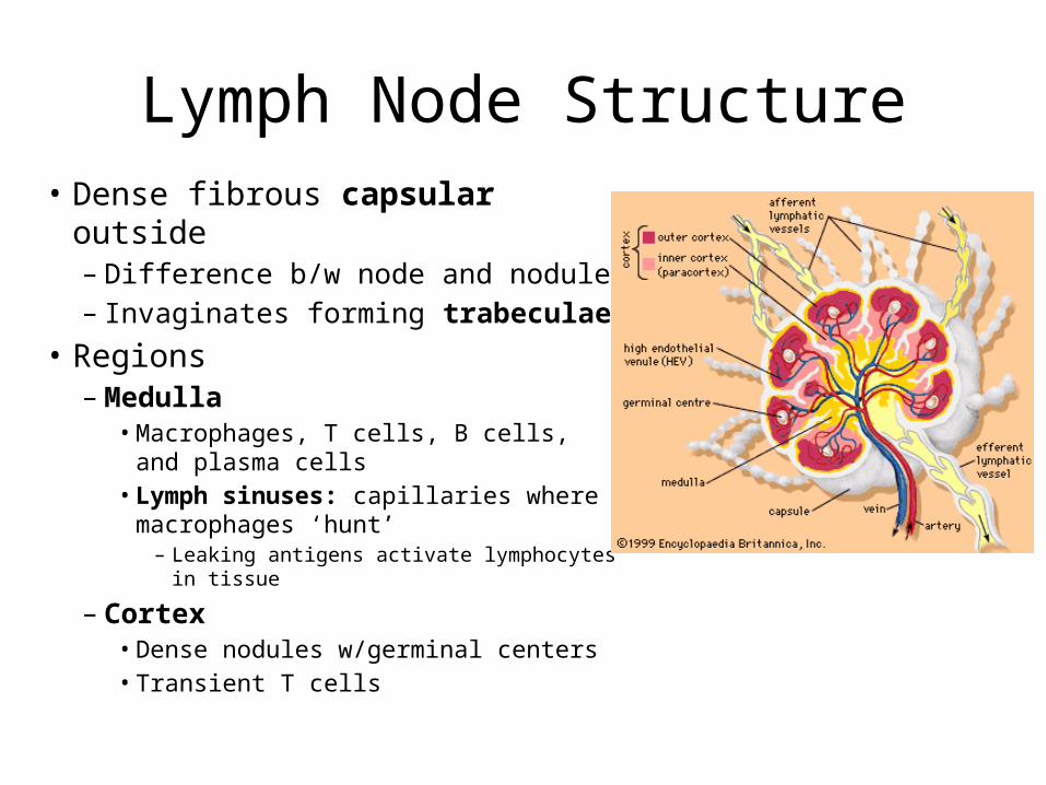
Lymph Node Structure• Dense fibrous capsular outside
– Difference b/w node and nodule– Invaginates forming trabeculae
• Regions– Medulla
• Macrophages, T cells, B cells, and plasma cells
• Lymph sinuses: capillaries where macrophages ‘hunt’– Leaking antigens activate lymphocytes in
tissue
– Cortex• Dense nodules w/germinal centers• Transient T cells

Lymphatic Circulation
• Enters node in afferent lymphatic vessels
• Large subscapular sinus to smaller, cortical sinuses
• Enter medulla• Exit at hilum via efferent
lymphatic vessels– Fewer slows flow– Allows lymphocytes &
macrophages to work
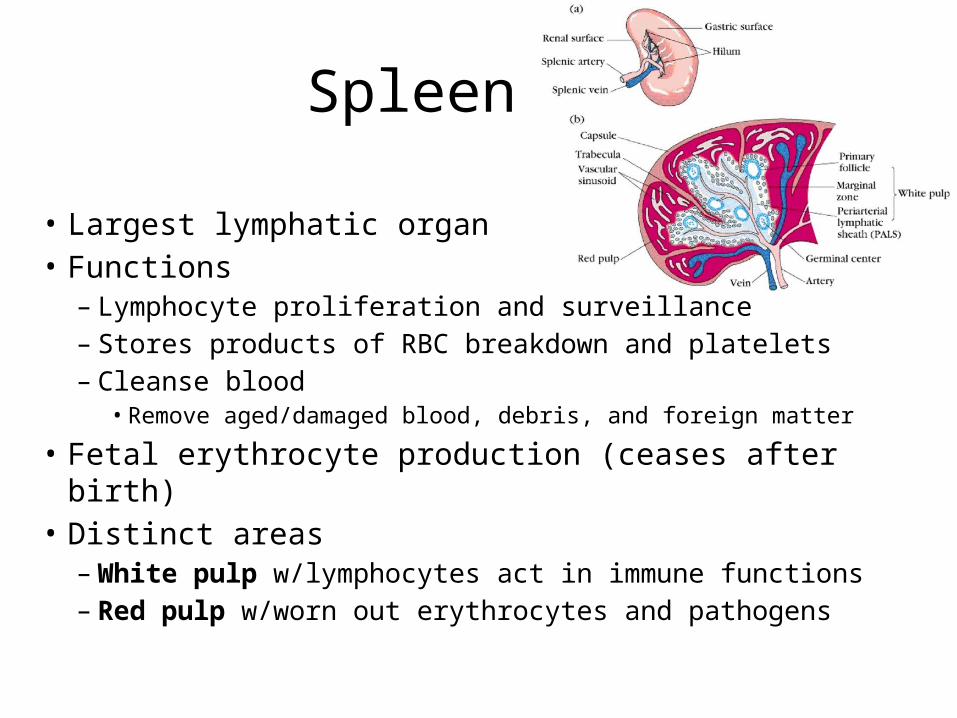
Spleen
• Largest lymphatic organ• Functions– Lymphocyte proliferation and surveillance – Stores products of RBC breakdown and platelets– Cleanse blood
• Remove aged/damaged blood, debris, and foreign matter
• Fetal erythrocyte production (ceases after birth)• Distinct areas– White pulp w/lymphocytes act in immune functions– Red pulp w/worn out erythrocytes and pathogens

Thymus
• Bilobed organ at base of neck– More pronounced when young– Corresponds w/importance of immune function
• T lymphocyte maturation only– Lacks B cells– Doesn’t directly fight antigens
• Thymocytes secrete thymosin and thymopoietin to signal T cell maturation

Tonsils
• Lymphatic tissue ring around pharynx– Palatine: largest and most likely infected– Lingual– Pharyngeal (adenoids)– Tubal
• Follicles w/germinal centers• Gather and remove pathogens from pharynx– Crypts are deep invaginations to trap and destroy
• Tonsil stones
– Produces ‘memory’ immune cells for future attacks

Mucosa-Associated Lymphatic Tissue (MALT)
• Collections of lymphatic tissue to protect external environment openings
• Peyer’s patches– In walls of small intestine– Destroy bacteria before it leaves intestines– Generate ‘memory’ lymphocytes
• Appendix– Junction of small and large intestine– Similar function as Peyer’s patches
• Lymphoid nodules– In walls of bronchi

Homeostatic Imbalances
• Tonsillitis: inflammation of tonsils• Lymphangitis: vasa vasorum of lymph vessels congested
w/blood• Lymphedema: blockage prevents return to blood• Buboes: inflamed lymph nodes • Splenectomy: removal of a ruptured spleen• Appendectomy: removal of appendix• Elephantiasis: lymph vessels clogged by worms causing
increased swelling• Hodgkin’s disease: malignant B-cells• Non-Hodgkin’s lymphoma: any lymphoma, but Hodgkin’s