Lymphangioleiomyomatosis, multifocal micronodular ... · Lymphangioleiomyomatosis, multifocal...
4
CASE REPORT Open Access Lymphangioleiomyomatosis, multifocal micronodular pneumocyte hyperplasia, and sarcoidosis: more pathological findings in the same chest CT, or a single pathological pathway? Fabiano Di Marco 1* , Giuseppina Palumbo 1 , Silvia Terraneo 1 , Gianluca Imeri 1 , Elena Lesma 2 , Nicola Sverzellati 3 , Angela Peron 4 , Lorenzo Gualandri 5 , Maria Paola Canevini 4 and Stefano Centanni 6 Abstract Background: Autoimmune hepatitis/primary biliary cirrhosis overlap syndrome, lymphangioleiomyomatosis/tuberous sclerosis complex (LAM-TSC), and sarcoidosis are three rare diseases. Here we present, to the best of our knowledge, the first description of a patient with the coexistence of these three diseases. Case presentation: A 47-year-old woman affected by LAM-TSC and primary biliary cirrosis/autoimmune hepatitis overlap syndrome. During her follow up a high resolution chest CT scan (HRTC) confirmed the presence of both multiple cysts and micronodular opacities consistent with multifocal micronodular pneumocytes hyperlasia (MMPH), and revealed multiple hilar-mediastinal symmetrical lymphadenopathies suggestive of sarcoidosis. Simultaneously, subcutaneous nodules appeared on her forearm bilaterally. Cutaneous biopsy showed granulomatous dermatitis with sarcoid-like granulomas. A diagnosis of stage I pulmonary sarcoidosis was made. No treatment for sarcoidosis was initiated since the patient had neither systemic involvement, nor respiratory impairment. Conclusions: The presence of more than one rare disease should challenge the concept of a potential common underlying mechanism, since the a priori probability of the concomitant presence of different conditions with different pathogenic mechanisms - especially if rare diseases - is low. We speculate that the dysregulation of the pathway involving mTOR and MAPK and their interaction might play a role in the pathogenesis of other diseases, including sarcoidosis. Keywords: LAM, TSC, Sarcoidosis, Thoracic images Background Tuberous sclerosis complex (TSC) is a rare genetic disorder, characterized by predominantly benign tumours developing potentially in all organ systems. Pulmonary in- volvement consists of Lymphangioleiomyomatosis (LAM) and Multifocal Micronodular Pneumocyte Hyperplasia (MMPH), which cause cystic and nodular diseases, re- spectively. Pneumothorax and chylothorax are common clinical presentations of LAM, whereas MMPH is usually asymptomatic. Here we describe a female with TSC and LAM with new pulmonary findings. Case presentation A 47-year-old woman, affected by TSC with a mutation identified in the TSC1 gene [c.682C > T (p.Arg228*)], was referred to the TSC Clinic of San Paolo Hospital (Milan, Italy). Family history was noticeable for Addison’ s disease and brain glioblastoma (in her mother) and idiopatic pul- monary fibrosis (in her father). Her daughter was affected by TSC. The patient suffered from primary biliary * Correspondence: [email protected] 1 Respiratory Unit, Ospedale San Paolo, Department of Health Sciences, Università degli Studi di Milano, Via A. di Rudinì, 8, 20142 Milan, Italy Full list of author information is available at the end of the article © The Author(s). 2017 Open Access This article is distributed under the terms of the Creative Commons Attribution 4.0 International License (http://creativecommons.org/licenses/by/4.0/), which permits unrestricted use, distribution, and reproduction in any medium, provided you give appropriate credit to the original author(s) and the source, provide a link to the Creative Commons license, and indicate if changes were made. The Creative Commons Public Domain Dedication waiver (http://creativecommons.org/publicdomain/zero/1.0/) applies to the data made available in this article, unless otherwise stated. Di Marco et al. BMC Pulmonary Medicine (2017) 17:107 DOI 10.1186/s12890-017-0447-x
Transcript of Lymphangioleiomyomatosis, multifocal micronodular ... · Lymphangioleiomyomatosis, multifocal...
Di Marco et al. BMC Pulmonary Medicine (2017) 17:107 DOI 10.1186/s12890-017-0447-x
CASE REPORT Open Access
Lymphangioleiomyomatosis, multifocalmicronodular pneumocyte hyperplasia, andsarcoidosis: more pathological findings inthe same chest CT, or a single pathologicalpathway?
Fabiano Di Marco1*, Giuseppina Palumbo1, Silvia Terraneo1, Gianluca Imeri1, Elena Lesma2, Nicola Sverzellati3,Angela Peron4, Lorenzo Gualandri5, Maria Paola Canevini4 and Stefano Centanni6Abstract
Background: Autoimmune hepatitis/primary biliary cirrhosis overlap syndrome, lymphangioleiomyomatosis/tuberoussclerosis complex (LAM-TSC), and sarcoidosis are three rare diseases. Here we present, to the best of our knowledge,the first description of a patient with the coexistence of these three diseases.
Case presentation: A 47-year-old woman affected by LAM-TSC and primary biliary cirrosis/autoimmune hepatitisoverlap syndrome. During her follow up a high resolution chest CT scan (HRTC) confirmed the presence of bothmultiple cysts and micronodular opacities consistent with multifocal micronodular pneumocytes hyperlasia(MMPH), and revealed multiple hilar-mediastinal symmetrical lymphadenopathies suggestive of sarcoidosis.Simultaneously, subcutaneous nodules appeared on her forearm bilaterally. Cutaneous biopsy showedgranulomatous dermatitis with sarcoid-like granulomas. A diagnosis of stage I pulmonary sarcoidosis was made. Notreatment for sarcoidosis was initiated since the patient had neither systemic involvement, nor respiratory impairment.
Conclusions: The presence of more than one rare disease should challenge the concept of a potential commonunderlying mechanism, since the a priori probability of the concomitant presence of different conditions withdifferent pathogenic mechanisms - especially if rare diseases - is low.We speculate that the dysregulation of the pathway involving mTOR and MAPK and their interaction mightplay a role in the pathogenesis of other diseases, including sarcoidosis.
Keywords: LAM, TSC, Sarcoidosis, Thoracic images
BackgroundTuberous sclerosis complex (TSC) is a rare geneticdisorder, characterized by predominantly benign tumoursdeveloping potentially in all organ systems. Pulmonary in-volvement consists of Lymphangioleiomyomatosis (LAM)and Multifocal Micronodular Pneumocyte Hyperplasia(MMPH), which cause cystic and nodular diseases, re-spectively. Pneumothorax and chylothorax are common
* Correspondence: [email protected] Unit, Ospedale San Paolo, Department of Health Sciences,Università degli Studi di Milano, Via A. di Rudinì, 8, 20142 Milan, ItalyFull list of author information is available at the end of the article
© The Author(s). 2017 Open Access This articInternational License (http://creativecommonsreproduction in any medium, provided you gthe Creative Commons license, and indicate if(http://creativecommons.org/publicdomain/ze
clinical presentations of LAM, whereas MMPH is usuallyasymptomatic. Here we describe a female with TSC andLAM with new pulmonary findings.
Case presentationA 47-year-old woman, affected by TSC with a mutationidentified in the TSC1 gene [c.682C > T (p.Arg228*)], wasreferred to the TSC Clinic of San Paolo Hospital (Milan,Italy). Family history was noticeable for Addison’s diseaseand brain glioblastoma (in her mother) and idiopatic pul-monary fibrosis (in her father). Her daughter was affectedby TSC. The patient suffered from primary biliary
le is distributed under the terms of the Creative Commons Attribution 4.0.org/licenses/by/4.0/), which permits unrestricted use, distribution, andive appropriate credit to the original author(s) and the source, provide a link tochanges were made. The Creative Commons Public Domain Dedication waiverro/1.0/) applies to the data made available in this article, unless otherwise stated.
Di Marco et al. BMC Pulmonary Medicine (2017) 17:107 Page 2 of 4
cirrosis/autoimmune hepatitis overlap syndrome. She wastreated with ursodeoxycholic acid (15 mg/kg/day).She received a diagnosis of LAM by chest CT scan,
which showed bilateral lung cysts randomly distributedthoughout the lungs. Chest CT scan revealed also the pres-ence of sclerotic bone lesions. Pulmonary function testswere normal, such as the 6-min walking test. Vascularendothelial growth factor (VEGF)-D, a lymphangiogenicgrowth factor proposed as a biomarker for LAM diagnosisand severity, was 582 pg/mL (normal limits 153–642 pg/mL). Dermatological examination showed hypomelanoticmacules, facial angiofibromas, xantelasma palpebrarum,periungual fibromas and erythematous plaque on the leftknee. She had a renal angiomyolipoma of 4.2 cm in the leftkidney. Brain magnetic resonance (MR) showed the pres-ence of cortical tubers. There was no ocular or heartinvolvement.One year later, the patient was adressed to our clinic. A
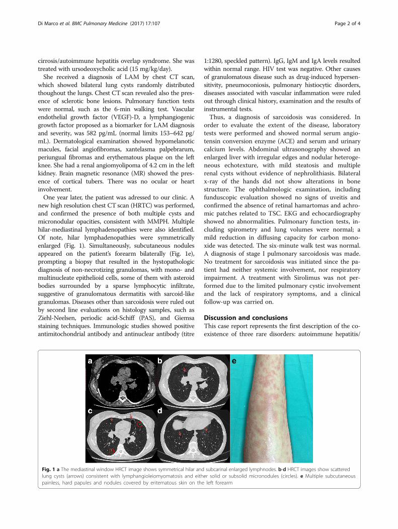
new high resolution chest CT scan (HRTC) was performed,and confirmed the presence of both multiple cysts andmicronodular opacities, consistent with MMPH. Multiplehilar-mediastinal lymphadenopathies were also identified.Of note, hilar lymphadenopathies were symmetricallyenlarged (Fig. 1). Simultaneously, subcutaneous nodulesappeared on the patient’s forearm bilaterally (Fig. 1e),prompting a biopsy that resulted in the hystopathologicdiagnosis of non-necrotizing granulomas, with mono- andmultinucleate epithelioid cells, some of them with asteroidbodies surrounded by a sparse lymphocytic infiltrate,suggestive of granulomatous dermatitis with sarcoid-likegranulomas. Diseases other than sarcoidosis were ruled outby second line evaluations on histology samples, such asZiehl-Neelsen, periodic acid-Schiff (PAS), and Giemsastaining techniques. Immunologic studies showed positiveantimitochondrial antibody and antinuclear antibody (titre
Fig. 1 a The mediastinal window HRCT image shows symmetrical hilar andlung cysts (arrows) consistent with lymphangioleiomyomatosis and eithpainless, hard papules and nodules covered by eritematous skin on the
1:1280, speckled pattern). IgG, IgM and IgA levels resultedwithin normal range. HIV test was negative. Other causesof granulomatous disease such as drug-induced hypersen-sitivity, pneumoconiosis, pulmonary histiocytic disorders,diseases associated with vascular inflammation were ruledout through clinical history, examination and the results ofinstrumental tests.Thus, a diagnosis of sarcoidosis was considered. In
order to evaluate the extent of the disease, laboratorytests were performed and showed normal serum angio-tensin conversion enzyme (ACE) and serum and urinarycalcium levels. Abdominal ultrasonography showed anenlarged liver with irregular edges and nodular heteroge-neous echotexture, with mild steatosis and multiplerenal cysts without evidence of nephrolithiasis. Bilateralx-ray of the hands did not show alterations in bonestructure. The ophthalmologic examination, includingfunduscopic evaluation showed no signs of uveitis andconfirmed the absence of retinal hamartomas and achro-mic patches related to TSC. EKG and echocardiographyshowed no abnormalities. Pulmonary function tests, in-cluding spirometry and lung volumes were normal; amild reduction in diffusing capacity for carbon mono-xide was detected. The six-minute walk test was normal.A diagnosis of stage I pulmonary sarcoidosis was made.No treatment for sarcoidosis was initiated since the pa-tient had neither systemic involvement, nor respiratoryimpairment. A treatment with Sirolimus was not per-formed due to the limited pulmonary cystic involvementand the lack of respiratory symptoms, and a clinicalfollow-up was carried on.
Discussion and conclusionsThis case report represents the first description of the co-existence of three rare disorders: autoimmune hepatitis/
subcarinal enlarged lymphnodes. b-d HRCT images show scattereder solid or subsolid micronodules (circles). e Multiple subcutaneousleft forearm
Di Marco et al. BMC Pulmonary Medicine (2017) 17:107 Page 3 of 4
primary biliary cirrhosis overlap syndrome, TSC/LAM,and sarcoidosis. The presence of more than one rare dis-ease should challenge the concept of a potential commonunderlying mechanism.Lymphadenopathy, both thoracic and abdominal, has
been described as another possible feature of LAM. Forinstance, a recent study on 138 patients with LAM, bothsporadic and associated with TSC, found a prevalence of9.4% for mediastinum and pulmonary hilum lymphaticlesions [1]. However, the presence of granulomatous skinlesions has not yet been described in LAM associatedwith TSC. Thus, the results of the skin biopsy togetherwith the presence of bilateral hilar lymphadenopathylead to the diagnosis of sarcoidosis.With respect to sarcoidosis, the importance of host
susceptibility and gene-environment interaction is widelyaccepted [2]. Although sarcoidosis does not meet thecriteria for autoimmune disease, it can coexist with a widerange of autoimmune disorders, including primary biliarycirrhosis, which is characterized by hepatic granulomasformation, connective tissue diseases (e.g. systemic scler-osis and Sjogren’s syndrome), Addison’s disease, and thy-roiditis [3]. It is noteworthy that the diagnosis ofsarcoidosis in case of granulomatous skin lesions ismade by exclusion criteria. Granulomatous lesions havebeen described in case of granulomatous-lymphocyticinterstitial lung disease (GLILD) associated with com-mon variable immunodeficiency (CVID), drug toxicity,or infections such as tuberculosis or fungal infections.All the aforementioned diagnoses were ruled out in ourpatient. As previously stated, the association betweengranulomatous lesions and autoimmune hepatitis/PBC,such as other immune-mediated and chronic inflamma-tory disease, has been previously described [4].TSC is an autosomal-dominant disease caused by
heterozygous loss-of-function mutations in the TSC1(chromosome 9q34) or TSC2 (chromosome 16p13)tumour suppressor genes coding for hamartin andtuberin, respectively. Tuberin and hamartin, togetherwith TBC1D7, form a complex that functions as anegative regulator of mammalian target of rapamycin(mTOR) through the inhibition of Rheb. Inactivationof TSC1 or TSC2 results in overactivation of mTORleading to abnormal cell growth, proliferation, meta-bolism, and angiogenesis. A common molecular mecha-nism for LAM/TSC and sarcoidosis is not known.However, a strong immunoreactivity for cathepsin-kwas demonstrated in spindle and epithelioid-shapedcells of lung LAM and in granulomas of sarcoidosiscases [5]. Since it is known that modulation of cathepsin-kmay occur through the mTOR pathway, it is possible tospeculate that the mTOR pathway might play a role in sar-coidosis [6]. Several evidences demonstrated an integrationof signaling of the mTOR pathway and mitogen-activated
protein kinases (MAPKs)-Erk activation. For instance, vas-cular endothelial growth factor (VEGF), a lymphangiogenicgrowth factor present at high levels in serum and urine ofpatients with LAM, induces phosphorylation of Akt,mTOR, S6 K, S6 and MAPK-Erk. Uncontrolled inflamma-tion and chronic inflammatory diseases may be caused bythe persistent activation of MAPKs, which can occur insarcoidosis [7]. This finding suggests an explanation for thepersistent production of several inflammatory cytokines,such as TNF-α and IL-12, in sarcoidosis. Interestingly,Linke et al. found that activation of mTORC1 due to TSC2deficiency causes granulomatous disease, including sar-coidosis, in both mice and humans. mTORC1 inhibitionresolves granulomas in TSC2-deficient mice, suggestingthat treatment with mTOR inhibitors, used to stop theprogression of benign tumors and LAM in TSC, might im-prove the signs of sarcoidosis as well [8]. However, the rea-son why not all patients with TSC develop sarcoidosis isstill unknown.Although at present it is not possible to demonstrate a
common mechanism underlying LAM/TSC, sarcoidosis,primary biliary cirrhosis and autoimmune hepatitis, andtheir coexistence could well occur by chance, we mightspeculate that the dysregulation of the pathway involvingmTOR and MAPK and their interaction may play a rolein the alteration of the diseases. Further reports areneeded to demonstrate our hypothesis.
AbbreviationsACE: Serum angiotensin conversion enzyme; AMA: Antimitochondrialantibody; HRTC: High resolution chest CT scan;LAM: Lymphangioleiomyomatosis; MAPK: Mitogen-activated protein kinases;MMPH: Multifocal micronodular pneumocyte hyperplasia; MR: Magneticresonance; MTOR: Mammalian target of rapamycin; TSC: Tuberous sclerosiscomplex; VEGF: Vascular endothelial growth factor
AcknowledgementsNot applicable.
FundingThis work was not funded.
Availability of data and materialsNot applicable.
Authors’ contributionsFDM, GP, ST, GI had full access to all of the patient’s clinical data and takesresponsibility for the integrity of the data. EL, NS, MPC, SC, AP contributedsubstantially to the data interpretation, and the writing of the manuscript. Allauthors read and approved the final manuscript.
Ethics approval and consent to participateNot applicable.
Consent for publicationWritten informed consent was obtained from the patient for publication ofthis Case report and all accompanying images.
Competing interestsFDM is currently an Associate Editor of this journal; other authors declarethat they have no competing interests.
Di Marco et al. BMC Pulmonary Medicine (2017) 17:107 Page 4 of 4
Publisher’s NoteSpringer Nature remains neutral with regard to jurisdictional claims in publishedmaps and institutional affiliations.
Author details1Respiratory Unit, Ospedale San Paolo, Department of Health Sciences,Università degli Studi di Milano, Via A. di Rudinì, 8, 20142 Milan, Italy.2Laboratories of Pharmacology, Università degli Studi di Milano, Milan, Italy.3Radiology, Department of Medicine and Surgery, University of Parma,Parma, Italy. 4Epilepsy Center, Ospedale San Paolo, Department of HealthSciences, Università degli Studi di Milano ASST Santi Paolo e Carlo, Milan,Italy. 5Dermatologic Clinic, Ospedale San Paolo, ASST Santi Paolo e Carlo,Milan, Italy. 6Respiratory Unit, Ospedale San Paolo, Department of HealthSciences, Università degli Studi di Milano, Milan, Italy.
Received: 13 February 2017 Accepted: 18 July 2017
References1. Tobino K, Johkoh T, Fujimoto K, et al. Computed tomographic features of
lymphangioleiomyomatosis: evaluation in 138 patients. Eur J Radiol. 2015;84:534–41. doi:10.1016/j.ejrad.2014.12.008.
2. Newman LS, Rose CS, Maier LA. Sarcoidosis. N Engl J Med. 1997;336:1224–34. doi:10.1056/NEJM199704243361706.
3. Sharma OP. Sarcoidosis and other autoimmune disorders. Curr Opin PulmMed. 2002;8:452–6.
4. Rajoriya N, Wotton CJ, Yeates DGR, et al. Immune-mediated and chronicinflammatory disease in people with sarcoidosis: disease associations ina large UK database. Postgrad Med J. 2009;85:233–7. doi:10.1136/pgmj.2008.067769.
5. Chilosi M, Pea M, Martignoni G, et al. Cathepsin-k expression in pulmonarylymphangioleiomyomatosis. Mod Pathol an Off J United States Can AcadPathol Inc. 2009;22:161–6. doi:10.1038/modpathol.2008.189.
6. Karbowniczek M, Spittle CS, Morrison T, et al. mTOR is activated in themajority of malignant melanomas. J Invest Dermatol. 2008;128:980–7. doi:10.1038/sj.jid.5701074.
7. Rastogi R, Du W, Ju D, et al. Dysregulation of p38 and MKP-1 in response toNOD1/TLR4 stimulation in sarcoid bronchoalveolar cells. Am J Respir CritCare Med. 2011;183:500–10. doi:10.1164/rccm.201005-0792OC.
8. Linke M, Pham HTT, Katholnig K, et al. Chronic signaling via the metaboliccheckpoint kinase mTORC1 induces macrophage granuloma formation andmarks sarcoidosis progression. Nat Immunol Published Online First: January.2017; doi:10.1038/ni.3655.
• We accept pre-submission inquiries
• Our selector tool helps you to find the most relevant journal
• We provide round the clock customer support
• Convenient online submission
• Thorough peer review
• Inclusion in PubMed and all major indexing services
• Maximum visibility for your research
Submit your manuscript atwww.biomedcentral.com/submit
Submit your next manuscript to BioMed Central and we will help you at every step: