
Luteolin Induces Apoptosis and Autophagy in Mouse...
10
Research Article Luteolin Induces Apoptosis and Autophagy in Mouse Macrophage ANA-1 Cells via the Bcl-2 Pathway Yuexia Liao , 1,2,3,5 Yang Xu, 4 Mengyao Cao, 4 Yuanyuan Huan, 3 Lei Zhu, 4 Ye Jiang, 3 Weigan Shen , 4 and Guoqiang Zhu 1,2,5 1 Jiangsu Co-Innovation Center for Important Animal Infectious Diseases and Zoonoses, Joint International Research Laboratory of Agriculture and Agri-Product Safety of the Ministry of Education of China, Yangzhou 225009, China 2 College of Veterinary Medicine, Yangzhou University, Yangzhou 225009, China 3 College of Nursing, Yangzhou University, Yangzhou, China 4 College of Medicine, Yangzhou University, Yangzhou 225009, China 5 Jiangsu Co-innovation Center for Prevention and Control of Important Animal Infectious Diseases and Zoonoses, Yangzhou 225009, China Correspondence should be addressed to Weigan Shen; [email protected] and Guoqiang Zhu; [email protected] Received 6 February 2018; Revised 5 June 2018; Accepted 14 June 2018; Published 30 August 2018 Academic Editor: Kai Wang Copyright © 2018 Yuexia Liao et al. This is an open access article distributed under the Creative Commons Attribution License, which permits unrestricted use, distribution, and reproduction in any medium, provided the original work is properly cited. Plants rich in luteolin have been used as Chinese traditional medicines for inflammatory diseases, hypertension, and cancer. However, little is known about the effect of luteolin on the apoptosis or autophagy of the macrophages. In this study, mouse macrophage ANA-1 cells were incubated with different concentrations of luteolin. The viability of the cells was determined by an MTT assay, apoptosis was determined by flow cytometric analysis, the level of cell autophagy was observed by confocal microscopy, and the expression levels of apoptotic or autophagic and antiapoptotic or antiautophagic proteins were detected by Western blot analysis. The results showed that luteolin decreased the viability of ANA-1 cells and induced apoptosis and autophagy. Luteolin induced apoptosis accompanied by downregulation of the expression of Bcl-2 and upregulation of the expression of caspase 3 and caspase 8. And luteolin increased FITC-LC3 punctate fluorescence accompanied by the increased expression levels of LC3-I, ATG7, and ATG12, while it suppressed the expression level of Beclin-1. Luteolin treatment resulted in obvious activation of the p38, JNK, and Akt signaling pathways, which is important in modulating apoptosis and autophagy. Thus, we concluded that luteolin induced the apoptosis and autophagy of ANA-1 cells most likely by regulating the p38, JNK, and Akt pathways, inhibiting the activity of Bcl-2 and Beclin-1 and upregulating caspase 3 and caspase 8 expression. These results provide novel insights into a therapeutic strategy to prevent and possibly treat macrophage-related diseases through luteolin-induced apoptosis and autophagy. 1. Introduction Macrophages fulfill a broad range of functions in phagocyto- sis, microbial killing, host defense, tissue homeostasis and repair, pathology and development, and their proliferation; migration and apoptosis have been emerged as important therapeutic targets for a variety of human diseases [1, 2]. Macrophages are critically involved in diseases that are caused by chronic inflammation (e.g., arthritis, multiple sclerosis, inflammatory bowel diseases, atherosclerosis, and cardiovascular disease) [3–6]. In addition, the apoptosis and autophagy dysfunction of the macrophage impact the development of chronic inflammation (e.g., atherosclerosis, tuberculosis, and sepsis) [7–10]. Plants rich in luteolin (3 ′ ,4 ′ ,5,7-tetrahydroxylavone), an active polyphenolic compound, have been used as Chinese traditional medicine to treat inflammatory diseases, hyper- tension, and cancer for a long period of time [11]. The phar- macological activities of luteolin, such as antioxidant, radical scavenging, cytoprotective, anti-inflammatory, antiallergic, and antitumor properties [12, 13], have been observed, suggesting that luteolin might possess diverse health benefits Hindawi Journal of Immunology Research Volume 2018, Article ID 4623919, 9 pages https://doi.org/10.1155/2018/4623919
Transcript of Luteolin Induces Apoptosis and Autophagy in Mouse...
Research ArticleLuteolin Induces Apoptosis and Autophagy in Mouse MacrophageANA-1 Cells via the Bcl-2 Pathway
Yuexia Liao ,1,2,3,5 Yang Xu,4 Mengyao Cao,4 Yuanyuan Huan,3 Lei Zhu,4 Ye Jiang,3
Weigan Shen ,4 and Guoqiang Zhu 1,2,5
1Jiangsu Co-Innovation Center for Important Animal Infectious Diseases and Zoonoses, Joint International Research Laboratory ofAgriculture and Agri-Product Safety of the Ministry of Education of China, Yangzhou 225009, China2College of Veterinary Medicine, Yangzhou University, Yangzhou 225009, China3College of Nursing, Yangzhou University, Yangzhou, China4College of Medicine, Yangzhou University, Yangzhou 225009, China5Jiangsu Co-innovation Center for Prevention and Control of Important Animal Infectious Diseases and Zoonoses,Yangzhou 225009, China
Correspondence should be addressed to Weigan Shen; [email protected] and Guoqiang Zhu; [email protected]
Received 6 February 2018; Revised 5 June 2018; Accepted 14 June 2018; Published 30 August 2018
Academic Editor: Kai Wang
Copyright © 2018 Yuexia Liao et al. This is an open access article distributed under the Creative Commons Attribution License,which permits unrestricted use, distribution, and reproduction in any medium, provided the original work is properly cited.
Plants rich in luteolin have been used as Chinese traditional medicines for inflammatory diseases, hypertension, and cancer.However, little is known about the effect of luteolin on the apoptosis or autophagy of the macrophages. In this study, mousemacrophage ANA-1 cells were incubated with different concentrations of luteolin. The viability of the cells was determined byan MTT assay, apoptosis was determined by flow cytometric analysis, the level of cell autophagy was observed by confocalmicroscopy, and the expression levels of apoptotic or autophagic and antiapoptotic or antiautophagic proteins were detected byWestern blot analysis. The results showed that luteolin decreased the viability of ANA-1 cells and induced apoptosis andautophagy. Luteolin induced apoptosis accompanied by downregulation of the expression of Bcl-2 and upregulation of theexpression of caspase 3 and caspase 8. And luteolin increased FITC-LC3 punctate fluorescence accompanied by the increasedexpression levels of LC3-I, ATG7, and ATG12, while it suppressed the expression level of Beclin-1. Luteolin treatment resultedin obvious activation of the p38, JNK, and Akt signaling pathways, which is important in modulating apoptosis and autophagy.Thus, we concluded that luteolin induced the apoptosis and autophagy of ANA-1 cells most likely by regulating the p38, JNK,and Akt pathways, inhibiting the activity of Bcl-2 and Beclin-1 and upregulating caspase 3 and caspase 8 expression. Theseresults provide novel insights into a therapeutic strategy to prevent and possibly treat macrophage-related diseases throughluteolin-induced apoptosis and autophagy.
1. Introduction
Macrophages fulfill a broad range of functions in phagocyto-sis, microbial killing, host defense, tissue homeostasis andrepair, pathology and development, and their proliferation;migration and apoptosis have been emerged as importanttherapeutic targets for a variety of human diseases [1, 2].Macrophages are critically involved in diseases that arecaused by chronic inflammation (e.g., arthritis, multiplesclerosis, inflammatory bowel diseases, atherosclerosis, andcardiovascular disease) [3–6]. In addition, the apoptosis
and autophagy dysfunction of the macrophage impact thedevelopment of chronic inflammation (e.g., atherosclerosis,tuberculosis, and sepsis) [7–10].
Plants rich in luteolin (3′,4′,5,7-tetrahydroxylavone), anactive polyphenolic compound, have been used as Chinesetraditional medicine to treat inflammatory diseases, hyper-tension, and cancer for a long period of time [11]. The phar-macological activities of luteolin, such as antioxidant, radicalscavenging, cytoprotective, anti-inflammatory, antiallergic,and antitumor properties [12, 13], have been observed,suggesting that luteolin might possess diverse health benefits
HindawiJournal of Immunology ResearchVolume 2018, Article ID 4623919, 9 pageshttps://doi.org/10.1155/2018/4623919
for treating some diseases, such as allergies, cancer, respira-tory diseases, and cardiovascular health [14]. Moreover, ithas been reported that luteolin can induce apoptosis in awide variety of cancer cells in vitro, including prostate cancercells, esophageal carcinoma cells, gastric cancer cells, andcolon cancer cells [15–17]. Despite the well-documentedantioxidant, anti-inflammatory, and antineoplastic potentialof luteolin, little is known about the effect of luteolin on theapoptosis or autophagy of the macrophages.
The results of this study revealed that luteolin inducedapoptosis and autophagy of the mouse macrophage ANA-1cells via altering the cell viability and DNA fragment. More-over, luteolin improved apoptosis and autophagy of themacrophage presumably through the Bcl-2- and MAPK-mediated signaling pathways. The results suggested that theunderstanding of the molecular mechanisms underlyingluteolin-induced apoptosis and autophagy may be useful incancer therapeutics, chemoprevention, and treating inflam-mation and neurodegenerative diseases, such as Parkinson’sdisease and Alzheimer’s disease [18].
2. Materials and Methods
2.1. Reagents and Antibodies. Luteolin (HPLC> 95%) waspurchased from Nanjing TCM Institute of Chinese MateriaMedica and dissolved in sodium carbonate (1mM) stocksolutions and stored at −20°C. Purified rabbit anti-Akt,anti-phospho-Akt, anti-phospho-ERK1/2, anti-ERK1/2,anti-phospho-p38, anti-p38, anti-β-actin, and HRP-linkedanti-rabbit IgG were purchased from Cell Signaling Technol-ogy (USA). Apoptosis/Necroptosis Antibody Sampler Kitand Autophagy Antibody Sampler Kit were purchased fromCell Signaling Technology (USA). Annexin V-FITC Apopto-sis Detection Kit was purchased from Beyotime Institute ofBiotechnology (Shanghai, China). In Situ Cell Death Kit,Fluorescein, was purchased from Roche Applied Science(Mannheim, Germany).
2.2. Cell Culture. ANA-1 macrophages, obtained from theCell Bank of Shanghai Institute of Biochemistry and CellBiology of the Chinese Academy of Sciences (Shanghai,China), were maintained in RPMI 1640 medium supple-mented with 10% fetal bovine serum (FBS), 100 units/mlpenicillin, and 100μg/ml streptomycin and cultured at 37°Cin a humidified atmosphere of 6% CO2.
2.3. Cell Viability Assay. Cell viability was assessed by usingthe MTT (3-(4,5-dimethylthiazol-2-yl)-2,5-diphenylterazo-lium bromide) assay as described previously [19]. In brief,200μl of 2× 10⁵ cells in RPMI 1640 medium with 0, 5, 10,20, 40, 80, or 160μM luteolin was seeded in 96-well cultureplates and cultured at 37°C in 6% CO2 for 24h or 48 h andwas incubated with 50μl of MTT solution (5mg/ml in PBS)for another 4 h at 37°C. Subsequently, the MTT solutionwas removed from the wells by aspiration and the forma-zan crystals were dissolved in 100μl of DMSO. The absor-bance at 570nm was measured with a Bio-Tek MQX 680(Bio-Tek Instruments Inc., Winooski, VT, USA). The data
are described as mean± standard deviations (SD) from atleast five independent experiments.
2.4. Analysis of Cell Apoptosis. For assays measuring theinduction of apoptosis of the ANA-1 cells by luteolin, theANA-1 cells were incubated with 0, 5, 10, 20, and 40μM ofluteolin at 37°C in 6% CO2 for 24 h or 48 h and then assayedfor apoptosis using the techniques listed below as describedpreviously [19]. For the quantification of the cells in thesub-G1 stages based on DNA content, cells were fixed inethanol (70%) at 4°C overnight and stained with PI(50μg/ml) and RNAse (10μg/ml) at 37°C in the dark for30 min and subsequently quantified with flow cytometry(BD Biosciences, San Jose, CA, USA). For detecting cellsurface phosphatidylserine, cells were dual stained withFITC-conjugated Annexin V and PI using an AnnexinV-FITC Apoptosis Detection Kit according to the manufac-turer’s instructions. Apoptosis was further confirmed bythe TUNEL assay, which detects the DNA fragmentationthat is the characteristic of apoptosis, using an In Situ CellDeath Kit, Fluorescein (Roche Applied Science, Mannheim,Germany), following the manufacturer’s guideline. TheTUNEL-positive cells were evaluated by flow cytometry. Inaddition, the levels of caspase 3, Bax, and Bcl-2 were deter-mined by Western blot analysis as described below.
2.5. Cell Autophagy Assay. To analyze autophagic flux, theANA-1 cells were seeded on glass cover slips in 24-well plateswith 0, 5, 10, 20, or 40μM luteolin for 48 h. Routinely, cellswere washed with PBS, fixed with ice cold 4% paraformalde-hyde for 30min at 4°C and blocked with 5% BSA in PBS for1 h. Cells were incubated with primary antibody to LC3(1 : 500 dilution anti-Rabit-LC3-pAb, MBL, Japan) overnightat 4°C and then incubated with FITC-goat-anti-rabbit-IgG(ZSJB-BIO, Beijing, China) for 30min at 37°C in the dark;DAPI (Beyotime Institute of Biotechnology, Shanghai,China) counterstain was used for nuclear staining. Afterextensive washing, the cover slips were then mounted onglass slides and the fluorescent images were captured withconfocal microscopy (TCS SP8 STED, Leica, German). Also,the averaged intracellular autophagosome was counted as theintracellular gathering FITC-LC3 spots divided the cell num-ber dyed with DAPI in 5 random fields at a magnification of×1000 in different cover slips. The levels of LC3, Beclin-1,ATG7, and ATG12 were determined by Western blot analy-sis as described below.
2.6. Western Blot Analysis. The cells were treated asmentioned above and lysed in cell lysis buffer (BeyotimeInstitute of Biotechnology, Shanghai, China) containing1μM phenylmethylsulfonyl fluoride, 1.5μM pepstatin A,and 0.2μM leupeptin. The samples were separated by 10%SDS-polyacrylamide gel electrophoresis and were then trans-ferred to polyvinylidene fluoride membranes, which wereblocked with 5% dried milk protein using the techniqueslisted below as described previously [19]. After blocking,the blots were incubated with primary antibodies. After beingwashed with TBST, the membranes were incubated at 37°Cfor 1 h in blocking buffer that contained the secondary
2 Journal of Immunology Research
antibodies and were visualized via a chemiluminescence ECLWestern blotting analysis system (BD, USA). The proteinlevels were quantified using ImageJ software (NIH, USA)and are expressed as percentages of the control after normal-ization to the housekeeping protein β-actin.
2.7. Statistical Analyses. The quantitative data are expressedas mean± SDs or SEM. The data were analyzed using SPSS19.0 software. Statistical analyses were performed withone-way analyses of variance (ANOVA) followed by Tukey’smultiple comparison post hoc tests. p values below 0.05 wereconsidered statistically significant.
3. Results
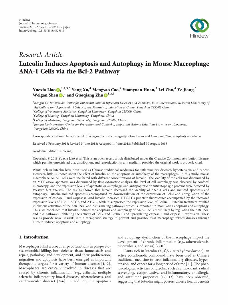
3.1. Luteolin Decreases ANA-1 Cell Viability. To examine theeffects of luteolin on cell viability, the ANA-1 cells wereexposed to different concentrations (0, 5, 10, 20, 40, 80, and160μM) of luteolin for 24 h or 48 h and cell viability wasmeasured by MTT assay. As shown in Figure 1, luteolinsignificantly suppressed the viability of ANA-1 cells in adose- and time-dependent manner. After 48 h of incubationwith 20μM luteolin, the viability of the cells was reduced by55% approximately. Moreover, 80μM luteolin reduced cellviability by 73% or more after 24 h or 48h of treatment.These results indicate that luteolin significantly decreasesthe viability of ANA-1 cells, and the half maximal inhibitoryconcentration (IC50) was about 40μM in the cell line. Thus,we chose 5μM to 40μM as experimental concentrations infurther experiments to avoid severe cytotoxic side effect.
3.2. Luteolin Induces ANA-1 Cell Apoptosis via Modulatingthe Expression of Caspase 3 and Bcl-2. To verify whether theobserved inhibition of cell viability induced by luteolin isthe consequence of the induction of apoptosis, we evaluated
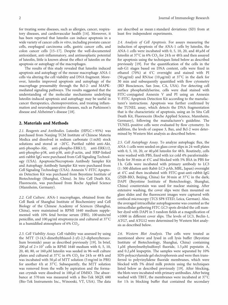
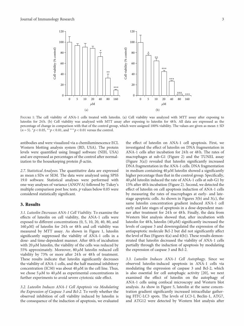
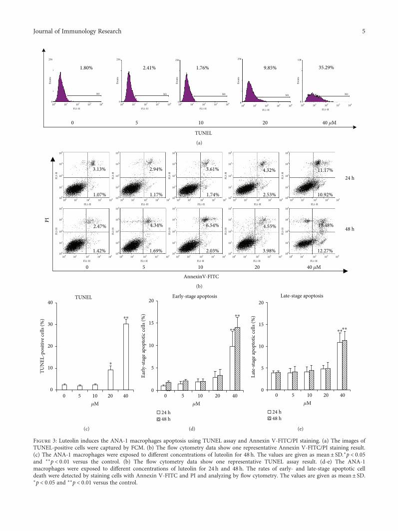
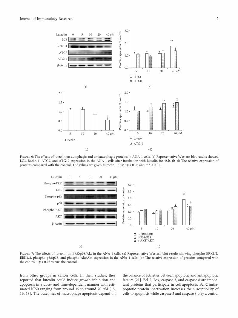
the effect of luteolin on ANA-1 cell apoptosis. First, weinvestigated the effect of luteolin on DNA fragmentation inANA-1 cells after incubation for 24h or 48 h. The rates ofmacrophages at sub-G1 (Figure 2) and the TUNEL assay(Figure 3(a)) revealed that luteolin significantly increasedDNA fragmentation in the ANA-1 cells. DNA fragmentationin medium containing 40μM luteolin showed a significantlyhigher percentage than that in the control group. Specifically,40μM luteolin induced the rate of ANA-1 cells at sub-G1 by15% after 48 h incubation (Figure 2). Second, we detected theeffect of luteolin on cell apoptosis induction of ANA-1 cellsby measuring the rates of macrophages at early- and late-stage apoptotic cells. As shown in Figures 3(b) and 3(c), thesame luteolin concentration gradient induced ANA-1 cellearly and late stages of apoptosis in a dose-dependent man-ner after treatment for 24 h or 48 h. Finally, the data fromWestern blot analysis showed that, after incubation withluteolin for 48 h, luteolin (40μM) significantly increased thelevels of caspase 3 and downregulated the expression of theantiapoptotic molecule Bcl-2 but did not significantly affectthe level of Bax (Figures 4(a) and 4(b)). These results demon-strated that luteolin decreased the viability of ANA-1 cellspartially through the induction of apoptosis by modulatingthe expression of caspase 3 and Bcl-2.
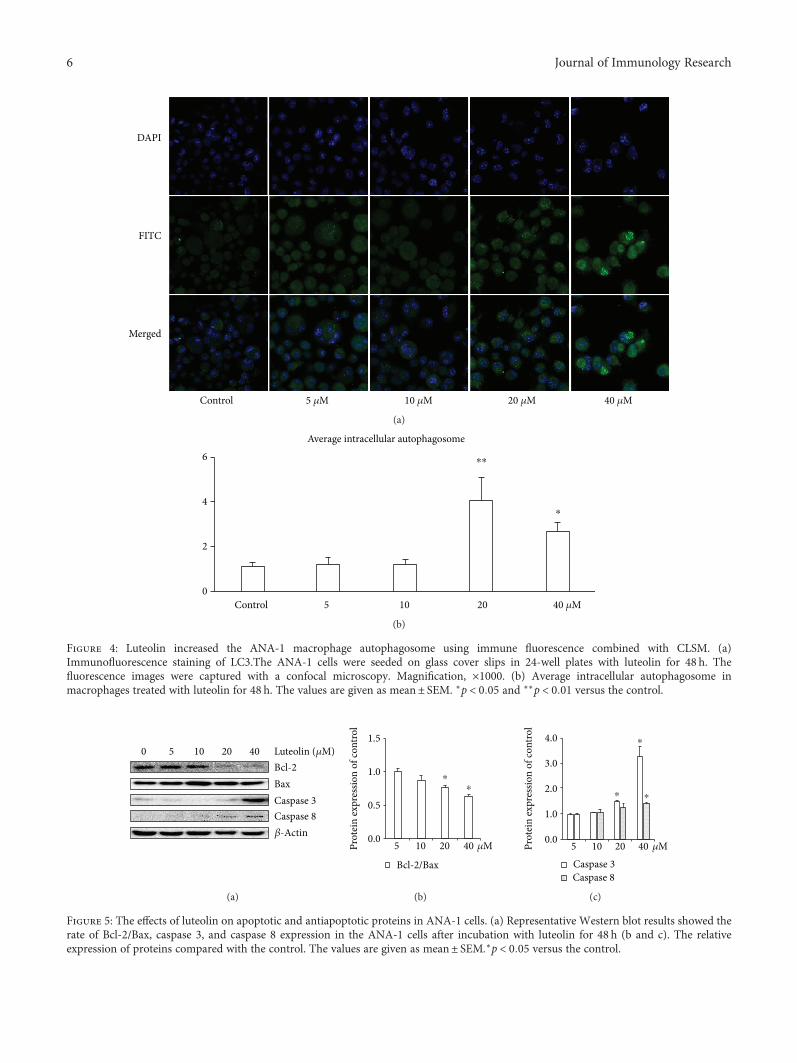
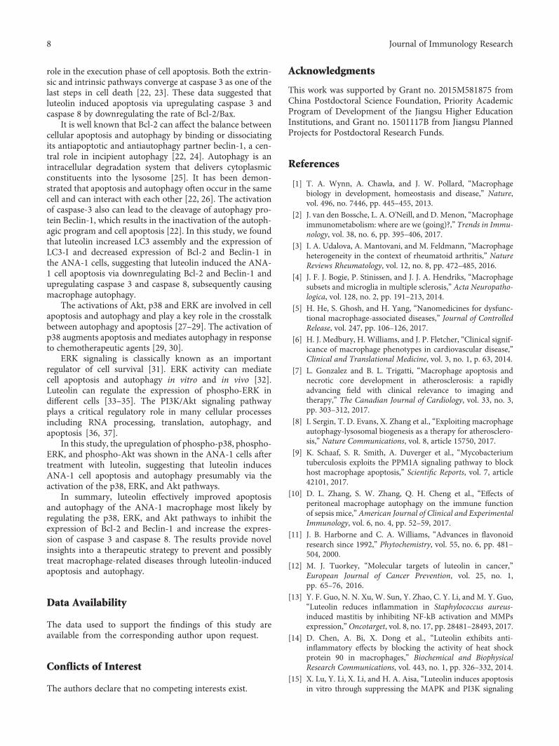
3.3. Luteolin Induces ANA-1 Cell Autophagy. Since weobserved luteolin-induced apoptosis in ANA-1 cells viamodulating the expression of caspase 3 and Bcl-2, whichis also essential for cell autophagic activity [20], we nextexamined the effect of luteolin on the autophagy ofANA-1 cells using confocal microscopy and Western blotanalysis. As show in Figure 5, luteolin at the same concen-tration gradient significantly increased intracellular gather-ing FITC-LC3 spots. The levels of LC3-I, Beclin-1, ATG7,and ATG12 were detected by Western blot analysis after
⁎
⁎⁎
⁎⁎⁎
0
20
40
60
80
100
120
Cel
l via
bilit
y (%
cont
rol)
50 20 804010 160�휇M
(a)
⁎
⁎⁎
⁎⁎⁎
⁎⁎⁎
⁎⁎⁎ ⁎⁎⁎0
20
40
60
80
100
120
Cel
l via
bilit
y (%
cont
rol)
50 20 804010 160�휇M
(b)
Figure 1: The cell viability of ANA-1 cells treated with luteolin. (a) Cell viability was analyzed with MTT assay after exposing toluteolin for 24 h. (b) Cell viability was analyzed with MTT assay after exposing to luteolin for 48 h. All data are expressed as thepercentage of change in comparison with that of the control group, which were assigned 100% viability. The values are given as mean ± SD(n = 5). ∗p < 0 05, ∗∗p < 0 01, and ∗∗∗p < 0 01 versus the control.
3Journal of Immunology Research
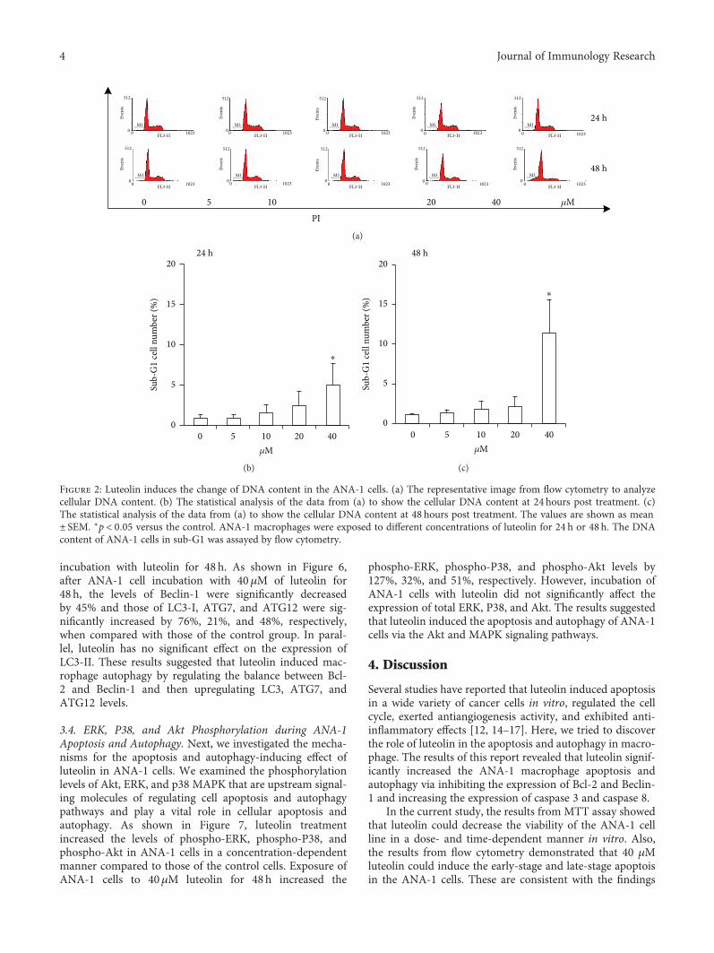
incubation with luteolin for 48 h. As shown in Figure 6,after ANA-1 cell incubation with 40μM of luteolin for48 h, the levels of Beclin-1 were significantly decreasedby 45% and those of LC3-I, ATG7, and ATG12 were sig-nificantly increased by 76%, 21%, and 48%, respectively,when compared with those of the control group. In paral-lel, luteolin has no significant effect on the expression ofLC3-II. These results suggested that luteolin induced mac-rophage autophagy by regulating the balance between Bcl-2 and Beclin-1 and then upregulating LC3, ATG7, andATG12 levels.
3.4. ERK, P38, and Akt Phosphorylation during ANA-1Apoptosis and Autophagy. Next, we investigated the mecha-nisms for the apoptosis and autophagy-inducing effect ofluteolin in ANA-1 cells. We examined the phosphorylationlevels of Akt, ERK, and p38 MAPK that are upstream signal-ing molecules of regulating cell apoptosis and autophagypathways and play a vital role in cellular apoptosis andautophagy. As shown in Figure 7, luteolin treatmentincreased the levels of phospho-ERK, phospho-P38, andphospho-Akt in ANA-1 cells in a concentration-dependentmanner compared to those of the control cells. Exposure ofANA-1 cells to 40μM luteolin for 48 h increased the
phospho-ERK, phospho-P38, and phospho-Akt levels by127%, 32%, and 51%, respectively. However, incubation ofANA-1 cells with luteolin did not significantly affect theexpression of total ERK, P38, and Akt. The results suggestedthat luteolin induced the apoptosis and autophagy of ANA-1cells via the Akt and MAPK signaling pathways.
4. Discussion
Several studies have reported that luteolin induced apoptosisin a wide variety of cancer cells in vitro, regulated the cellcycle, exerted antiangiogenesis activity, and exhibited anti-inflammatory effects [12, 14–17]. Here, we tried to discoverthe role of luteolin in the apoptosis and autophagy in macro-phage. The results of this report revealed that luteolin signif-icantly increased the ANA-1 macrophage apoptosis andautophagy via inhibiting the expression of Bcl-2 and Beclin-1 and increasing the expression of caspase 3 and caspase 8.
In the current study, the results from MTT assay showedthat luteolin could decrease the viability of the ANA-1 cellline in a dose- and time-dependent manner in vitro. Also,the results from flow cytometry demonstrated that 40 μMluteolin could induce the early-stage and late-stage apoptoisin the ANA-1 cells. These are consistent with the findings
PI
�휇M
24 h
48 h
0
00
512
Even
ts
1023FL3-H
00
512
Even
ts
1023FL3-H
00
512
Even
ts
1023FL3-H
00
512
Even
ts
1023FL3-H
00
512
Even
ts
1023FL3-H
00
512
Even
ts
1023FL3-H
00
512
Even
ts
1023FL3-H
00
512
Even
ts
1023FL3-H
00
512
Even
ts
1023FL3-H
00
512
Even
ts
1023FL3-H
5 10 20 40
M1
M1 M1
M1 M1
M1 M1
M1 M1
M1
(a)
24 h
⁎
0
5
10
15
20
Sub-
G1
cell
num
ber (
%)
50 20 4010�휇M
(b)
48 h
⁎
50 20 4010�휇M
0
5
10
15
20
Sub-
G1
cell
num
ber (
%)
(c)
Figure 2: Luteolin induces the change of DNA content in the ANA-1 cells. (a) The representative image from flow cytometry to analyzecellular DNA content. (b) The statistical analysis of the data from (a) to show the cellular DNA content at 24 hours post treatment. (c)The statistical analysis of the data from (a) to show the cellular DNA content at 48 hours post treatment. The values are shown as mean± SEM. ∗p < 0 05 versus the control. ANA-1 macrophages were exposed to different concentrations of luteolin for 24 h or 48 h. The DNAcontent of ANA-1 cells in sub-G1 was assayed by flow cytometry.
4 Journal of Immunology Research
1.80% 2.41% 1.76% 9.85% 35.29%
TUNEL
0 5 10 20 40 �휇M
1000
Even
ts
Even
ts
Even
ts
Even
ts
Even
ts
256
0
256
0
256
0
256
0
128
101 102
FL1-H
M1 M1 M1 M1 M1
103 104 100 101 102
FL1-H103 104 100 101 102
FL1-H103 104
100 101 102
FL1-H103 104 100 101 102
FL1-H103 104
(a)
PI
24 h
48 h
3.13%
1.07%
2.94%
1.17%
3.61%
1.74%
4.32%
2.53%
11.17%
10.92%
2.47%
1.42%
4.34%
1.69%
6.54%
2.03%
10.48%
12.27%
4.55%
3.98%
0 5 10 20 40 �휇M
AnnexinV-FITC
100100
101
102
103
104
101 102
FL1-H
FL3-
H
100
101
102
103
104
FL3-
H
100
101
102
103
104
FL3-
H
100
101
102
103
104
FL3-
H
100
101
102
103
104
FL3-
H
100
101
102
103
104
FL3-
H
100
101
102
103
104
FL3-
H
100
101
102
103
104
FL3-
H
100
101
102
103
104
FL3-
H
100
101
102
103
104
FL3-
H
103 104 100 101 102
FL1-H103 104 100 101 102
FL1-H103 104 100 101 102
FL1-H103 104 100 101 102
FL1-H103 104
100 101 102
FL1-H103 104 100 101 102
FL1-H103 104 100 101 102
FL1-H103 104 100 101 102
FL1-H103 104 100 101 102
FL1-H103 104
(b)
TUNEL
⁎
⁎⁎
5 10 20 400�휇M
0
10
20
30
40
TUN
EL-p
ositi
ve ce
lls (%
)
(c)
Early-stage apoptosis
24 h48 h
⁎⁎
⁎⁎
5 10 20 400�휇M
0
5
10
15
20
Early
-sta
ge ap
opto
tic ce
lls (%
)
(d)
Late-stage apoptosis
⁎⁎⁎⁎
5 10 20 400�휇M
0
5
10
15
20
Late
-sta
ge ap
opto
tic ce
lls (%
)
24 h48 h
(e)
Figure 3: Luteolin induces the ANA-1 macrophages apoptosis using TUNEL assay and Annexin V-FITC/PI staining. (a) The images ofTUNEL-positive cells were captured by FCM. (b) The flow cytometry data show one representative Annexin V-FITC/PI staining result.(c) The ANA-1 macrophages were exposed to different concentrations of luteolin for 48 h. The values are given as mean± SD.∗p < 0 05and ∗∗p < 0 01 versus the control. (b) The flow cytometry data show one representative TUNEL assay result. (d-e) The ANA-1macrophages were exposed to different concentrations of luteolin for 24 h and 48 h. The rates of early- and late-stage apoptotic celldeath were detected by staining cells with Annexin V-FITC and PI and analyzing by flow cytometry. The values are given as mean± SD.∗p < 0 05 and ∗∗p < 0 01 versus the control.
5Journal of Immunology Research
Control
Merged
FITC
DAPI
5 �휇M 10 �휇M 20 �휇M 40 �휇M
(a)
Control
Average intracellular autophagosome
0
2
4
6
5 10 20
⁎⁎
⁎
40 �휇M
(b)
Figure 4: Luteolin increased the ANA-1 macrophage autophagosome using immune fluorescence combined with CLSM. (a)Immunofluorescence staining of LC3.The ANA-1 cells were seeded on glass cover slips in 24-well plates with luteolin for 48 h. Thefluorescence images were captured with a confocal microscopy. Magnification, ×1000. (b) Average intracellular autophagosome inmacrophages treated with luteolin for 48 h. The values are given as mean± SEM. ∗p < 0 05 and ∗∗p < 0 01 versus the control.
Luteolin (�휇M)Bcl-2
0 5 10 20 40
BaxCaspase 3Caspase 8�훽-Actin
(a)
�휇MProt
ein
expr
essio
n of
cont
rol
1.5
Bcl-2/Bax
1.0
0.5
0.0 5 10 20 40
⁎⁎
(b)
Prot
ein
expr
essio
n of
cont
rol
4.0
2.0
3.0
1.0
0.0
Caspase 3Caspase 8
�휇M5 10 20 40
⁎
⁎
⁎
(c)
Figure 5: The effects of luteolin on apoptotic and antiapoptotic proteins in ANA-1 cells. (a) Representative Western blot results showed therate of Bcl-2/Bax, caspase 3, and caspase 8 expression in the ANA-1 cells after incubation with luteolin for 48 h (b and c). The relativeexpression of proteins compared with the control. The values are given as mean± SEM.∗p < 0 05 versus the control.
6 Journal of Immunology Research
from other groups in cancer cells. In their studies, theyreported that luteolin could induce growth inhibition andapoptosis in a dose- and time-dependent manner with esti-mated IC50 ranging from around 35 to around 70 μM [15,16, 18]. The outcomes of macrophage apoptosis depend on
the balance of activities between apoptotic and antiapoptoticfactors [21]. Bcl-2, Bax, caspase 3, and caspase 8 are impor-tant proteins that participate in cell apoptosis. Bcl-2 antia-poptotic protein inactivation increases the susceptibility ofcells to apoptosis while caspase 3 and caspase 8 play a central
Luteolin
β-Actin
ATG12
ATG7
Beclin-1
LC3�휇M40201050
(a)
Prot
ein
expr
essio
n of
cont
rol
0.0
1.0
2.0
3.0
LC3-ILC3-II
�휇M4020105
⁎⁎
(b)
0.0
0.5
1.0
1.5
2.0
Beclin-1
*
�휇M4020105
(c)
Prot
ein
expr
essio
n of
cont
rol
0.0
0.5
1.0
1.5
2.0
ATG7ATG12
�휇M4020105
⁎⁎
⁎⁎
⁎
(d)
Figure 6: The effects of luteolin on autophagic and antiautophagic proteins in ANA-1 cells. (a) Representative Western blot results showedLC3, Beclin-1, ATG7, and ATG12 expression in the ANA-1 cells after incubation with luteolin for 48 h. (b–d) The relative expression ofproteins compared with the control. The values are given as mean± SEM.∗p < 0 05 and ∗∗p < 0 01.
Luteolin
Phospho-ERK
β-Actin
ERK
Phospho-p38
p38
Phospho-AKT
AKT
�휇M40201050
(a)
0.0
0.5
1.0
1.5
2.0
2.5
3.0
p-ERK/ERKp-P38/P38p-AKT/AKT
Prot
ein
expr
essio
n of
cont
rol
�휇M4020105
⁎⁎
⁎
⁎⁎
(b)
Figure 7: The effects of luteolin on ERK/p38/Akt in the ANA-1 cells. (a) Representative Western blot results showing phospho-ERK1/2/ERK1/2, phospho-p38/p38, and phospho-Akt/Akt expression in the ANA-1 cells. (b) The relative expression of proteins compared withthe control. ∗p < 0 05 versus the control.
7Journal of Immunology Research
role in the execution phase of cell apoptosis. Both the extrin-sic and intrinsic pathways converge at caspase 3 as one of thelast steps in cell death [22, 23]. These data suggested thatluteolin induced apoptosis via upregulating caspase 3 andcaspase 8 by downregulating the rate of Bcl-2/Bax.
It is well known that Bcl-2 can affect the balance betweencellular apoptosis and autophagy by binding or dissociatingits antiapoptotic and antiautophagy partner beclin-1, a cen-tral role in incipient autophagy [22, 24]. Autophagy is anintracellular degradation system that delivers cytoplasmicconstituents into the lysosome [25]. It has been demon-strated that apoptosis and autophagy often occur in the samecell and can interact with each other [22, 26]. The activationof caspase-3 also can lead to the cleavage of autophagy pro-tein Beclin-1, which results in the inactivation of the autoph-agic program and cell apoptosis [22]. In this study, we foundthat luteolin increased LC3 assembly and the expression ofLC3-I and decreased expression of Bcl-2 and Beclin-1 inthe ANA-1 cells, suggesting that luteolin induced the ANA-1 cell apoptosis via downregulating Bcl-2 and Beclin-1 andupregulating caspase 3 and caspase 8, subsequently causingmacrophage autophagy.
The activations of Akt, p38 and ERK are involved in cellapoptosis and autophagy and play a key role in the crosstalkbetween autophagy and apoptosis [27–29]. The activation ofp38 augments apoptosis and mediates autophagy in responseto chemotherapeutic agents [29, 30].
ERK signaling is classically known as an importantregulator of cell survival [31]. ERK activity can mediatecell apoptosis and autophagy in vitro and in vivo [32].Luteolin can regulate the expression of phospho-ERK indifferent cells [33–35]. The PI3K/Akt signaling pathwayplays a critical regulatory role in many cellular processesincluding RNA processing, translation, autophagy, andapoptosis [36, 37].
In this study, the upregulation of phospho-p38, phospho-ERK, and phospho-Akt was shown in the ANA-1 cells aftertreatment with luteolin, suggesting that luteolin inducesANA-1 cell apoptosis and autophagy presumably via theactivation of the p38, ERK, and Akt pathways.
In summary, luteolin effectively improved apoptosisand autophagy of the ANA-1 macrophage most likely byregulating the p38, ERK, and Akt pathways to inhibit theexpression of Bcl-2 and Beclin-1 and increase the expres-sion of caspase 3 and caspase 8. The results provide novelinsights into a therapeutic strategy to prevent and possiblytreat macrophage-related diseases through luteolin-inducedapoptosis and autophagy.
Data Availability
The data used to support the findings of this study areavailable from the corresponding author upon request.
Conflicts of Interest
The authors declare that no competing interests exist.
Acknowledgments
This work was supported by Grant no. 2015M581875 fromChina Postdoctoral Science Foundation, Priority AcademicProgram of Development of the Jiangsu Higher EducationInstitutions, and Grant no. 1501117B from Jiangsu PlannedProjects for Postdoctoral Research Funds.
References
[1] T. A. Wynn, A. Chawla, and J. W. Pollard, “Macrophagebiology in development, homeostasis and disease,” Nature,vol. 496, no. 7446, pp. 445–455, 2013.
[2] J. van den Bossche, L. A. O'Neill, and D. Menon, “Macrophageimmunometabolism: where are we (going)?,” Trends in Immu-nology, vol. 38, no. 6, pp. 395–406, 2017.
[3] I. A. Udalova, A. Mantovani, and M. Feldmann, “Macrophageheterogeneity in the context of rheumatoid arthritis,” NatureReviews Rheumatology, vol. 12, no. 8, pp. 472–485, 2016.
[4] J. F. J. Bogie, P. Stinissen, and J. J. A. Hendriks, “Macrophagesubsets and microglia in multiple sclerosis,” Acta Neuropatho-logica, vol. 128, no. 2, pp. 191–213, 2014.
[5] H. He, S. Ghosh, and H. Yang, “Nanomedicines for dysfunc-tional macrophage-associated diseases,” Journal of ControlledRelease, vol. 247, pp. 106–126, 2017.
[6] H. J. Medbury, H. Williams, and J. P. Fletcher, “Clinical signif-icance of macrophage phenotypes in cardiovascular disease,”Clinical and Translational Medicine, vol. 3, no. 1, p. 63, 2014.
[7] L. Gonzalez and B. L. Trigatti, “Macrophage apoptosis andnecrotic core development in atherosclerosis: a rapidlyadvancing field with clinical relevance to imaging andtherapy,” The Canadian Journal of Cardiology, vol. 33, no. 3,pp. 303–312, 2017.
[8] I. Sergin, T. D. Evans, X. Zhang et al., “Exploiting macrophageautophagy-lysosomal biogenesis as a therapy for atherosclero-sis,” Nature Communications, vol. 8, article 15750, 2017.
[9] K. Schaaf, S. R. Smith, A. Duverger et al., “Mycobacteriumtuberculosis exploits the PPM1A signaling pathway to blockhost macrophage apoptosis,” Scientific Reports, vol. 7, article42101, 2017.
[10] D. L. Zhang, S. W. Zhang, Q. H. Cheng et al., “Effects ofperitoneal macrophage autophagy on the immune functionof sepsis mice,” American Journal of Clinical and ExperimentalImmunology, vol. 6, no. 4, pp. 52–59, 2017.
[11] J. B. Harborne and C. A. Williams, “Advances in flavonoidresearch since 1992,” Phytochemistry, vol. 55, no. 6, pp. 481–504, 2000.
[12] M. J. Tuorkey, “Molecular targets of luteolin in cancer,”European Journal of Cancer Prevention, vol. 25, no. 1,pp. 65–76, 2016.
[13] Y. F. Guo, N. N. Xu, W. Sun, Y. Zhao, C. Y. Li, and M. Y. Guo,“Luteolin reduces inflammation in Staphylococcus aureus-induced mastitis by inhibiting NF-kB activation and MMPsexpression,” Oncotarget, vol. 8, no. 17, pp. 28481–28493, 2017.
[14] D. Chen, A. Bi, X. Dong et al., “Luteolin exhibits anti-inflammatory effects by blocking the activity of heat shockprotein 90 in macrophages,” Biochemical and BiophysicalResearch Communications, vol. 443, no. 1, pp. 326–332, 2014.
[15] X. Lu, Y. Li, X. Li, and H. A. Aisa, “Luteolin induces apoptosisin vitro through suppressing the MAPK and PI3K signaling
8 Journal of Immunology Research
pathways in gastric cancer,” Oncology Letters, vol. 14, no. 2,pp. 1993–2000, 2017.
[16] P. Chen, J. Y. Zhang, B. B. Sha et al., “Luteolin inhibits cellproliferation and induces cell apoptosis via down-regulationof mitochondrial membrane potential in esophageal carci-noma cells EC1 and KYSE450,” Oncotarget, vol. 8, no. 16,pp. 27471–27480, 2017.
[17] K. A. Kang, M. J. Piao, Y. S. Ryu et al., “Luteolin inducesapoptotic cell death via antioxidant activity in human coloncancer cells,” International Journal of Oncology, vol. 51,no. 4, pp. 1169–1178, 2017.
[18] K. Kwon, Y. S. Kwon, S. W. Kim, K. Yu, K. H. Lee, and O. Y.Kwon, “Luteolin-induced apoptosis through activation ofendoplasmic reticulum stress sensors in pheochromocytomacells,”Molecular Medicine Reports, vol. 16, no. 1, pp. 380–386,2017.
[19] Y. Liao, W. Shen, G. Kong, H. Lv, W. Tao, and P. Bo, “Api-genin induces the apoptosis and regulates MAPK signalingpathways in mouse macrophage ANA-1 cells,” PLoS One,vol. 9, no. 3, article e92007, 2014.
[20] M. C. Maiuri, A. Criollo, and G. Kroemer, “Crosstalk betweenapoptosis and autophagy within the Beclin 1 interactome,” TheEMBO Journal, vol. 29, no. 3, pp. 515-516, 2010.
[21] I. Tabas and K. E. Bornfeldt, “Macrophage phenotype andfunction in different stages of atherosclerosis,” CirculationResearch, vol. 118, no. 4, pp. 653–667, 2016.
[22] S. Song, J. Tan, Y. Miao, M. Li, and Q. Zhang, “Crosstalk ofautophagy and apoptosis: involvement of the dual role ofautophagy under ER stress,” Journal of Cellular Physiology,vol. 232, no. 11, pp. 2977–2984, 2017.
[23] M. D'Amelio, V. Cavallucci, and F. Cecconi, “Neuronalcaspase-3 signaling: not only cell death,” Cell Death and Differ-entiation, vol. 17, no. 7, pp. 1104–1114, 2010.
[24] A. Vural and J. H. Kehrl, “Autophagy in macrophages: impact-ing inflammation and bacterial infection,” Scientifica,vol. 2014, Article ID 825463, 13 pages, 2014.
[25] T. Saitoh and S. Akira, “Regulation of inflammasomes byautophagy,” The Journal of Allergy and Clinical Immunology,vol. 138, no. 1, pp. 28–36, 2016.
[26] E. Wirawan, L. Vande Walle, K. Kersse et al., “Caspase-medi-ated cleavage of Beclin-1 inactivates Beclin-1-induced autoph-agy and enhances apoptosis by promoting the release ofproapoptotic factors from mitochondria,” Cell Death & Dis-ease, vol. 1, no. 1, article e18, 2010.
[27] L. S. Steelman, W. H. Chappell, S. L. Abrams et al., “Roles ofthe Raf/MEK/ERK and PI3K/PTEN/Akt/mTOR pathways incontrolling growth and sensitivity to therapy-implications forcancer and aging,” Aging, vol. 3, no. 3, pp. 192–222, 2011.
[28] E. Seki, D. A. Brenner, and M. Karin, “A liver full of JNK: sig-naling in regulation of cell function and disease pathogenesis,and clinical approaches,” Gastroenterology, vol. 143, no. 2,pp. 307–320, 2012.
[29] X. Sui, N. Kong, L. Ye et al., “p38 and JNK MAPK pathwayscontrol the balance of apoptosis and autophagy in responseto chemotherapeutic agents,” Cancer Letters, vol. 344, no. 2,pp. 174–179, 2014.
[30] K. Deacon, P. Mistry, J. Chernoff, J. L. Blank, and R. Patel, “p38mitogen-activated protein kinase mediates cell death and p21-activated kinase mediates cell survival during chemotherapeu-tic drug-induced mitotic arrest,” Molecular Biology of the Cell,vol. 14, no. 5, pp. 2071–2087, 2003.
[31] S. Tanimura and K. Takeda, “ERK signalling as a regulator ofcell motility,” Journal of Biochemistry, vol. 162, no. 3,pp. 145–154, 2017.
[32] S. Cagnol and J. C. Chambard, “ERK and cell death: mecha-nisms of ERK-induced cell death – apoptosis, autophagy andsenescence,” The FEBS Journal, vol. 277, no. 1, pp. 2–21, 2010.
[33] S. Kim, Y. W. Chin, and J. Cho, “Protection of cultured corti-cal neurons by luteolin against oxidative damage throughinhibition of apoptosis and induction of heme oxygenase-1,”Biological and Pharmaceutical Bulletin, vol. 40, no. 3,pp. 256–265, 2017.
[34] A. Y. Choi, J. H. Choi, H. Yoon et al., “Luteolin inducesapoptosis through endoplasmic reticulum stress and mito-chondrial dysfunction in Neuro-2a mouse neuroblastomacells,” European Journal of Pharmacology, vol. 668, no. 1-2,pp. 115–126, 2011.
[35] H. Yao, L. Zhou, L. Tang et al., “Protective effects of luteolin-7-O-glucoside against starvation-induced injury through upreg-ulation of autophagy in H9c2 cells,” Bioscience Trends, vol. 11,no. 5, pp. 557–564, 2017.
[36] H. Sun, Z. Wang, and J. S. Yakisich, “Natural products target-ing autophagy via the PI3K/Akt/mTOR pathway as anticanceragents,” Anti-Cancer Agents in Medicinal Chemistry, vol. 13,no. 7, pp. 1048–1056, 2013.
[37] N. Diehl and H. Schaal, “Make yourself at home: viral hijack-ing of the PI3K/Akt signaling pathway,” Viruses, vol. 5,no. 12, pp. 3192–3212, 2013.
9Journal of Immunology Research

Stem Cells International
Hindawiwww.hindawi.com Volume 2018
Hindawiwww.hindawi.com Volume 2018
MEDIATORSINFLAMMATION
of
EndocrinologyInternational Journal of
Hindawiwww.hindawi.com Volume 2018
Hindawiwww.hindawi.com Volume 2018
Disease Markers
Hindawiwww.hindawi.com Volume 2018
BioMed Research International
OncologyJournal of
Hindawiwww.hindawi.com Volume 2013
Hindawiwww.hindawi.com Volume 2018
Oxidative Medicine and Cellular Longevity
Hindawiwww.hindawi.com Volume 2018
PPAR Research
Hindawi Publishing Corporation http://www.hindawi.com Volume 2013Hindawiwww.hindawi.com
The Scientific World Journal
Volume 2018
Immunology ResearchHindawiwww.hindawi.com Volume 2018
Journal of
ObesityJournal of
Hindawiwww.hindawi.com Volume 2018
Hindawiwww.hindawi.com Volume 2018
Computational and Mathematical Methods in Medicine
Hindawiwww.hindawi.com Volume 2018
Behavioural Neurology
OphthalmologyJournal of
Hindawiwww.hindawi.com Volume 2018
Diabetes ResearchJournal of
Hindawiwww.hindawi.com Volume 2018
Hindawiwww.hindawi.com Volume 2018
Research and TreatmentAIDS
Hindawiwww.hindawi.com Volume 2018
Gastroenterology Research and Practice
Hindawiwww.hindawi.com Volume 2018
Parkinson’s Disease
Evidence-Based Complementary andAlternative Medicine
Volume 2018Hindawiwww.hindawi.com
Submit your manuscripts atwww.hindawi.com


![Research Paper Propranolol induced apoptosis and autophagy ... · autophagic cell apoptosis, which is known as type II PCD [16, 17]. Therefore, autophagy exerts dual effects of pro-survival](https://static.fdocuments.in/doc/165x107/60061dba135e2801d812a472/research-paper-propranolol-induced-apoptosis-and-autophagy-autophagic-cell-apoptosis.jpg)


