Lung & Thorax Exams - University of California, San...
33
Transcript of Lung & Thorax Exams - University of California, San...
Lung & Thorax Exams
Charlie Goldberg, M.D.
Professor of Medicine, UCSD SOM
Lung Exam
• Includes Vital Signs & Cardiac Exam
• 4 Elements (cardiac & abdominal too) – Observation
– Palpation
– Percussion
– Auscultation
Pulmonary Review of Systems
• All organ systems have an ROS
• Questions to uncover problems in area
• Need to know right questions & what the
responses might mean!
• An example:
http://meded.ucsd.edu/clinicalmed/ros.htm
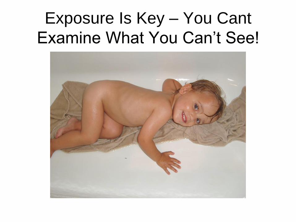
Exposure Is Key – You Cant
Examine What You Can’t See!
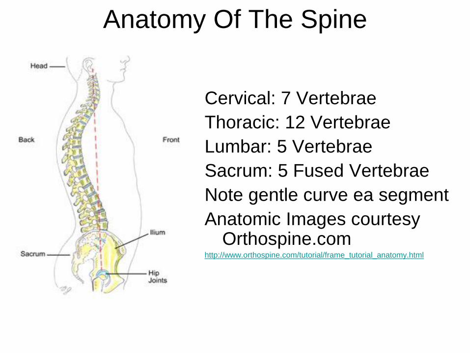
Anatomy Of The Spine
Cervical: 7 Vertebrae
Thoracic: 12 Vertebrae
Lumbar: 5 Vertebrae
Sacrum: 5 Fused Vertebrae
Note gentle curve ea segment
Anatomic Images courtesy Orthospine.com
http://www.orthospine.com/tutorial/frame_tutorial_anatomy.html
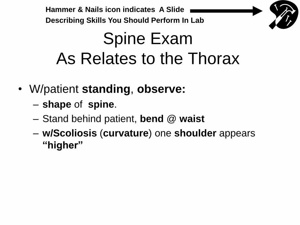
Spine Exam
As Relates to the Thorax
• W/patient standing, observe:
– shape of spine.
– Stand behind patient, bend @ waist
– w/Scoliosis (curvature) one shoulder appears
“higher”
Hammer & Nails icon indicates A Slide
Describing Skills You Should Perform In Lab
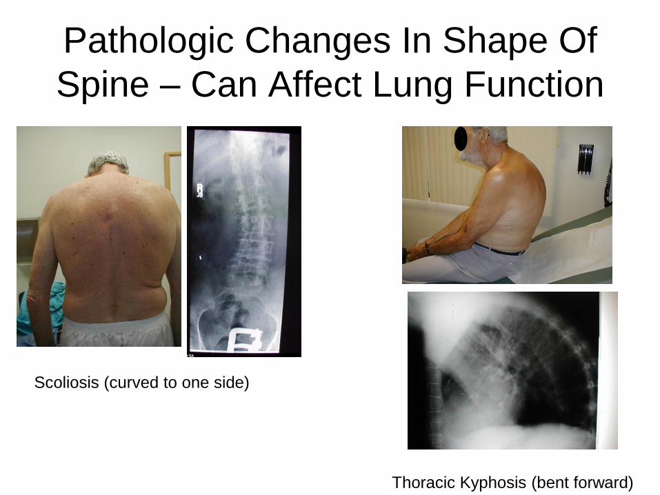
Pathologic Changes In Shape Of
Spine – Can Affect Lung Function
Thoracic Kyphosis (bent forward)
Scoliosis (curved to one side)
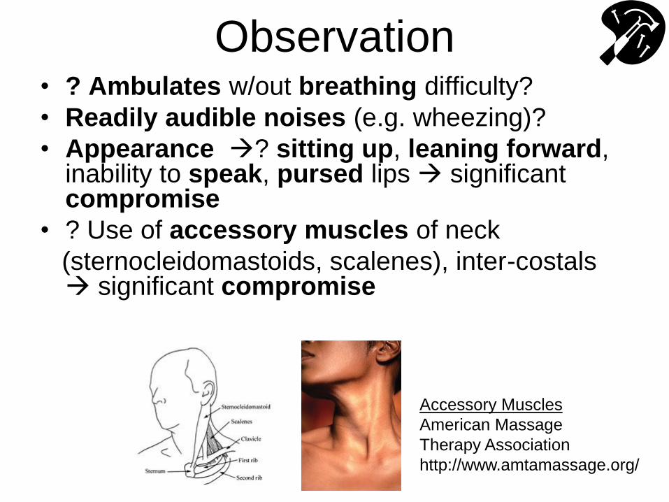
Observation
• ? Ambulates w/out breathing difficulty?
• Readily audible noises (e.g. wheezing)?
• Appearance ? sitting up, leaning forward, inability to speak, pursed lips significant compromise
• ? Use of accessory muscles of neck
(sternocleidomastoids, scalenes), inter-costals significant compromise
Accessory Muscles
American Massage
Therapy Association
http://www.amtamassage.org/
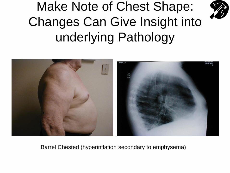
Make Note of Chest Shape:
Changes Can Give Insight into
underlying Pathology
Barrel Chested (hyperinflation secondary to emphysema)
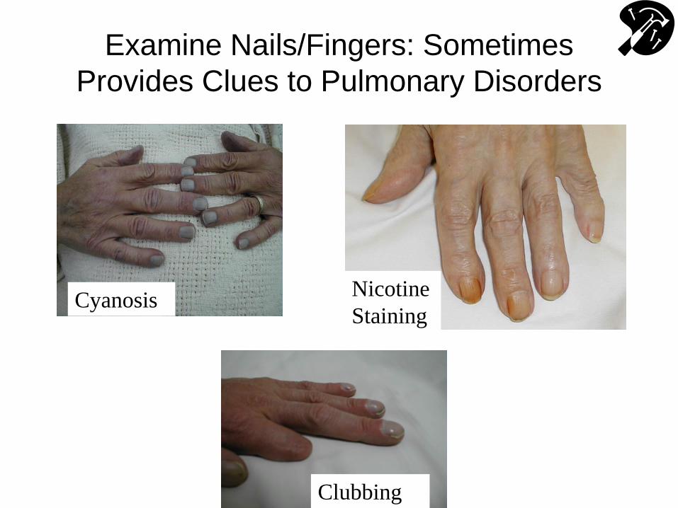
Examine Nails/Fingers: Sometimes
Provides Clues to Pulmonary Disorders
Cyanosis Nicotine
Staining
Clubbing
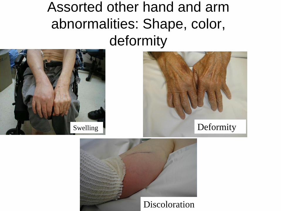
Assorted other hand and arm
abnormalities: Shape, color,
deformity
Swelling Deformity
Discoloration
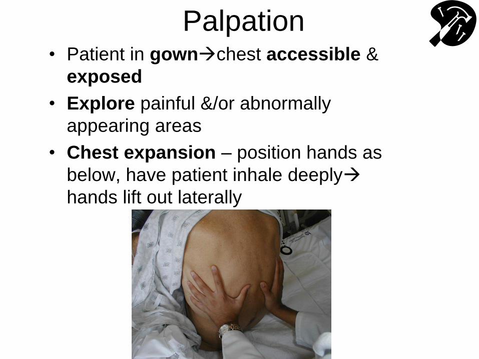
Palpation • Patient in gownchest accessible &
exposed
• Explore painful &/or abnormally
appearing areas
• Chest expansion – position hands as
below, have patient inhale deeply
hands lift out laterally
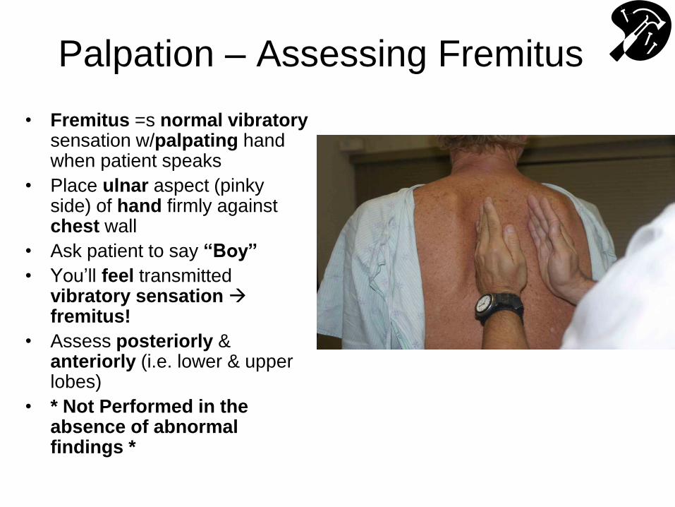
Palpation – Assessing Fremitus
• Fremitus =s normal vibratory sensation w/palpating hand when patient speaks
• Place ulnar aspect (pinky side) of hand firmly against chest wall
• Ask patient to say “Boy”
• You’ll feel transmitted vibratory sensation fremitus!
• Assess posteriorly & anteriorly (i.e. lower & upper lobes)
• * Not Performed in the absence of abnormal findings *
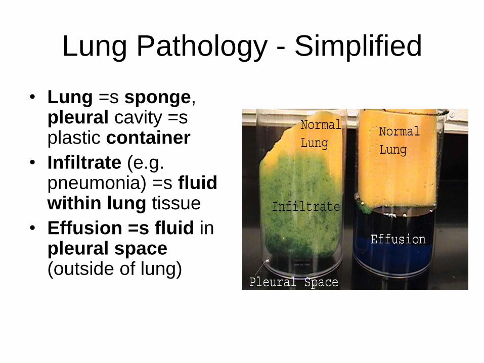
Lung Pathology - Simplified
• Lung =s sponge, pleural cavity =s plastic container
• Infiltrate (e.g. pneumonia) =s fluid within lung tissue
• Effusion =s fluid in pleural space (outside of lung)
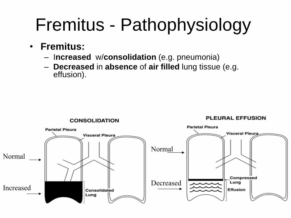
Fremitus - Pathophysiology • Fremitus:
– Increased w/consolidation (e.g. pneumonia)
– Decreased in absence of air filled lung tissue (e.g. effusion).
Normal
Increased
Normal
Decreased
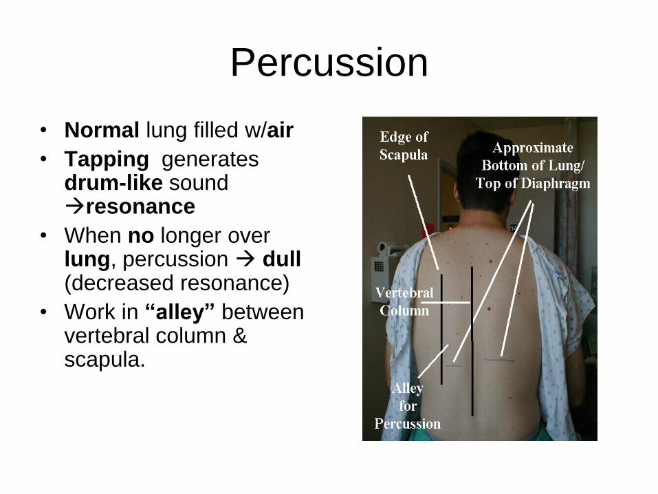
Percussion
• Normal lung filled w/air
• Tapping generates drum-like sound resonance
• When no longer over lung, percussion dull (decreased resonance)
• Work in “alley” between vertebral column & scapula.
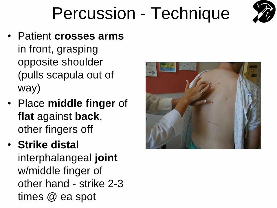
Percussion - Technique
• Patient crosses arms
in front, grasping
opposite shoulder
(pulls scapula out of
way)
• Place middle finger of
flat against back,
other fingers off
• Strike distal
interphalangeal joint
w/middle finger of
other hand - strike 2-3
times @ ea spot
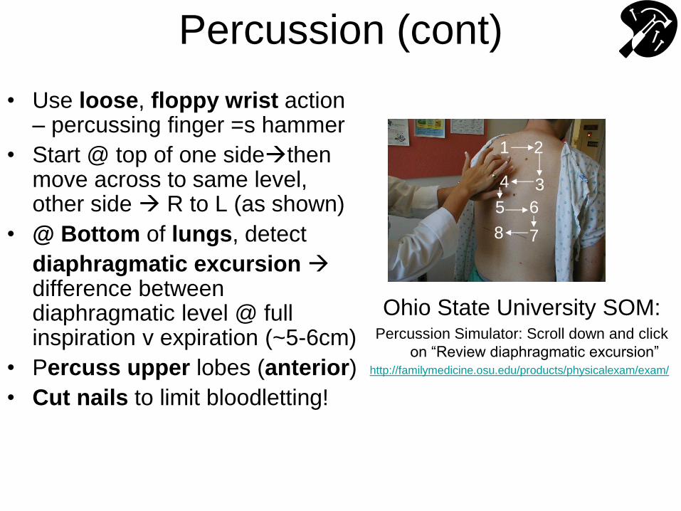
Percussion (cont)
• Use loose, floppy wrist action – percussing finger =s hammer
• Start @ top of one sidethen move across to same level, other side R to L (as shown)
• @ Bottom of lungs, detect
diaphragmatic excursion difference between diaphragmatic level @ full inspiration v expiration (~5-6cm)
• Percuss upper lobes (anterior)
• Cut nails to limit bloodletting!
Ohio State University SOM: Percussion Simulator: Scroll down and click
on “Review diaphragmatic excursion” http://familymedicine.osu.edu/products/physicalexam/exam/
1 2
3 4
5 6
7 8
Percussion (Cont)
• Difficult to master technique & detect tone
changes - expect to be frustrated!
• Practice – on friends, yourself (find your
stomach, tap on your cheeks, etc) • Detect fluid level in container
• Find studs in wall
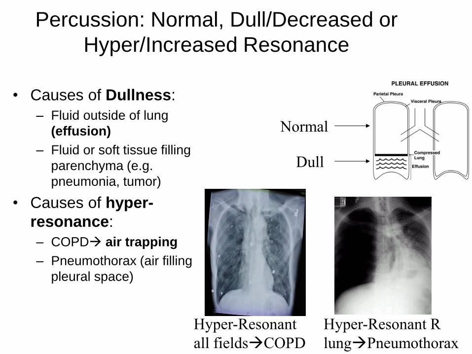
Percussion: Normal, Dull/Decreased or
Hyper/Increased Resonance
• Causes of Dullness:
– Fluid outside of lung
(effusion)
– Fluid or soft tissue filling
parenchyma (e.g.
pneumonia, tumor)
• Causes of hyper-
resonance:
– COPD air trapping
– Pneumothorax (air filling
pleural space)
Hyper-Resonant
all fieldsCOPD
Hyper-Resonant R
lungPneumothorax
Dull
Normal
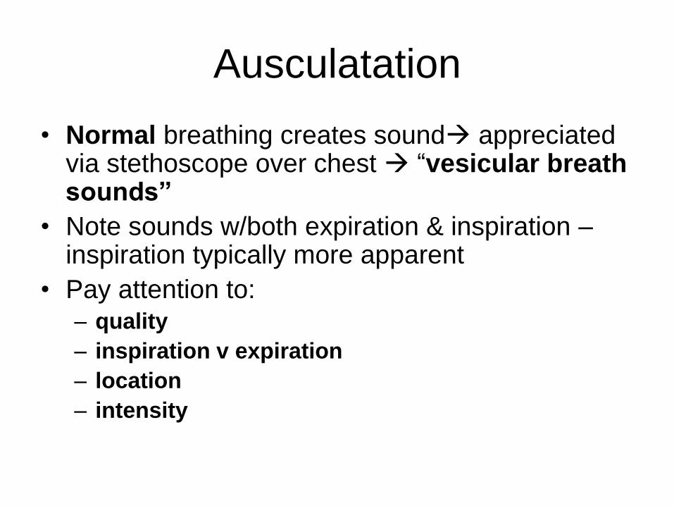
Ausculatation
• Normal breathing creates sound appreciated via stethoscope over chest “vesicular breath sounds”
• Note sounds w/both expiration & inspiration – inspiration typically more apparent
• Pay attention to: – quality
– inspiration v expiration
– location
– intensity
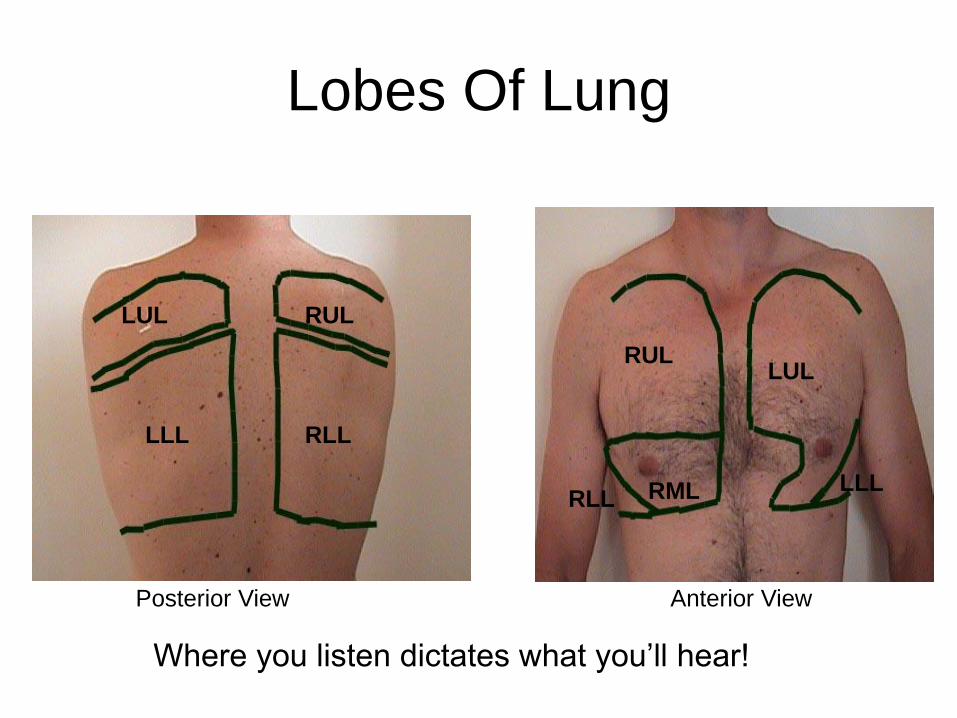
Lobes Of Lung
Where you listen dictates what you’ll hear!
LUL RUL
LLL RLL
RUL LUL
RML RLL LLL
Posterior View Anterior View
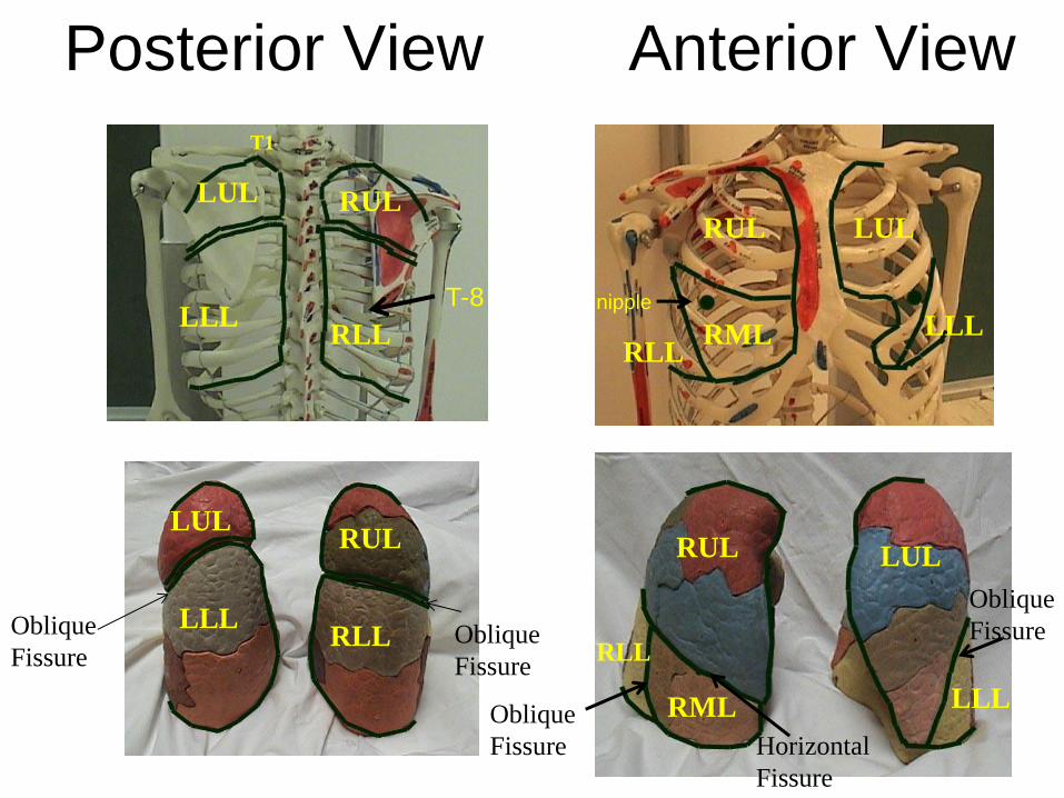
Posterior View Anterior View
LUL
LLL
RUL
RLL Oblique
Fissure Oblique
Fissure
LUL
LLL
RUL
RLL
T1
T-8
RUL
RML RLL
LUL
LLL nipple
RUL
RML
RLL
LUL
LLL
Oblique
Fissure
Oblique
Fissure Horizontal
Fissure
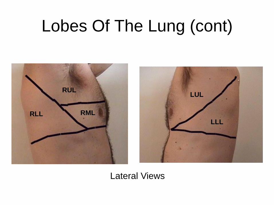
Lobes Of The Lung (cont)
RLL RML
RUL
LLL
LUL
Lateral Views
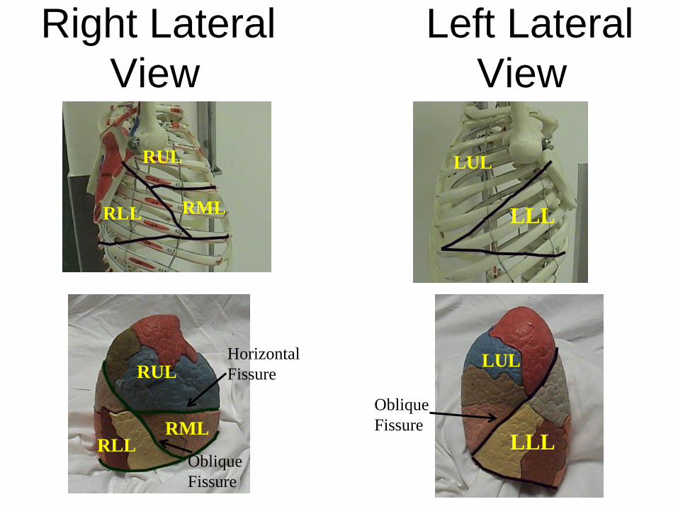
Right Lateral Left Lateral
View View
RUL
RML
RLL
RLL
RUL
RML
Oblique
Fissure
Horizontal
Fissure
LUL
LUL
LLL
LLL
Oblique
Fissure
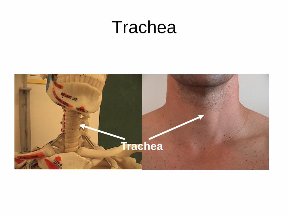
Trachea
Trachea
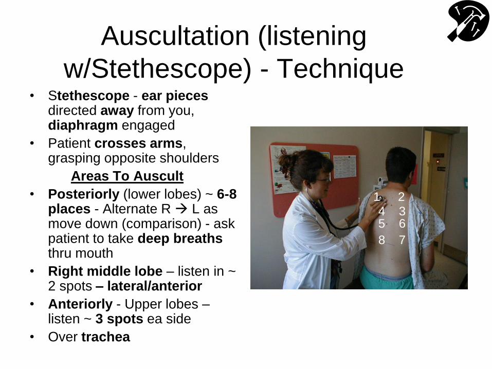
Auscultation (listening
w/Stethescope) - Technique • Stethescope - ear pieces
directed away from you, diaphragm engaged
• Patient crosses arms, grasping opposite shoulders
Areas To Auscult
• Posteriorly (lower lobes) ~ 6-8 places - Alternate R L as move down (comparison) - ask patient to take deep breaths thru mouth
• Right middle lobe – listen in ~ 2 spots – lateral/anterior
• Anteriorly - Upper lobes – listen ~ 3 spots ea side
• Over trachea
1 2
3 4 5 6
7 8
Pathologic Lung Sounds • Crackles (Rales): “Scratchy” sounds
associated w/fluid in alveoli & airways (e.g. pulmonary edema, pneumonia); finer crackles w/fibrosis
• Ronchi: “Gurgling” type noise, caused by fluid in large & medium sized airways (e.g. bronchitis, pneumonia)
• Wheezing: Whistling type noise, loudest on expiration, caused by air forced thru narrowed airways (e.g. asthma) – expiratory phase prolonged (E>>>I)
• Stridor: Inspiratory whistling type sound
due to tracheal narrowing heard best over trachea
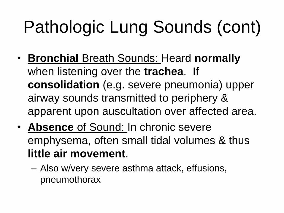
Pathologic Lung Sounds (cont)
• Bronchial Breath Sounds: Heard normally
when listening over the trachea. If
consolidation (e.g. severe pneumonia) upper
airway sounds transmitted to periphery &
apparent upon auscultation over affected area.
• Absence of Sound: In chronic severe
emphysema, often small tidal volumes & thus
little air movement.
– Also w/very severe asthma attack, effusions,
pneumothorax
Pathologic Lung Sounds (cont)
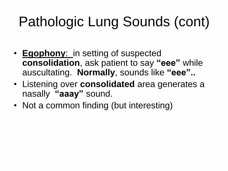
• Egophony: in setting of suspected consolidation, ask patient to say “eee” while auscultating. Normally, sounds like “eee”..
• Listening over consolidated area generates a nasally “aaay” sound.
• Not a common finding (but interesting)
Lung Sound Simulation
Lung Sound Simulation Sites (for practice):
1. Ohio State University http://familymedicine.osu.edu/products/physicalexam/exam/
2. R.A.L.E. Repository http://www.rale.ca/Recordings.htm
3. Bohadan A, et al. Fundamentals of Auscultation. NEJM
2014; 370: 744-51. Click on: Interactive Graphic -
Fundamentals of lung sound auscultation. http://www.nejm.org/doi/full/10.1056/NEJMra1302901
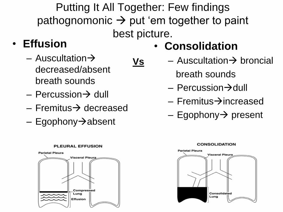
Putting It All Together: Few findings
pathognomonic put ‘em together to paint
best picture. • Effusion
– Auscultation
decreased/absent
breath sounds
– Percussion dull
– Fremitus decreased
– Egophonyabsent
• Consolidation
– Auscultation broncial
breath sounds
– Percussiondull
– Fremitusincreased
– Egophony present
Vs
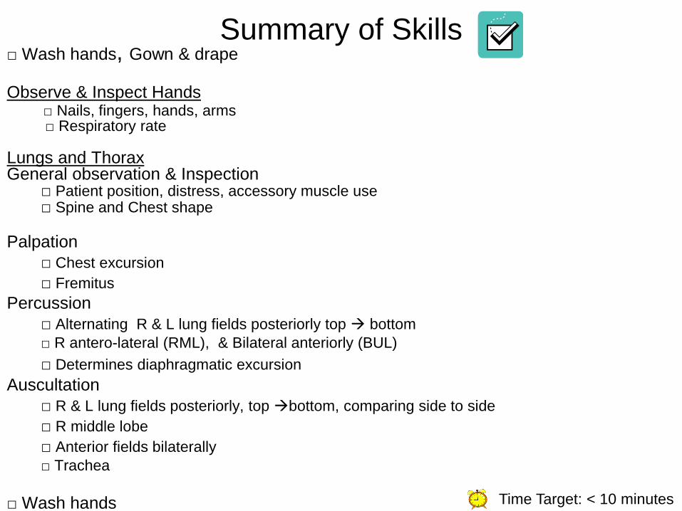
Summary of Skills □ Wash hands, Gown & drape Observe & Inspect Hands □ Nails, fingers, hands, arms □ Respiratory rate Lungs and Thorax General observation & Inspection
□ Patient position, distress, accessory muscle use □ Spine and Chest shape
Palpation
□ Chest excursion
□ Fremitus
Percussion
□ Alternating R & L lung fields posteriorly top bottom
□ R antero-lateral (RML), & Bilateral anteriorly (BUL)
□ Determines diaphragmatic excursion Auscultation
□ R & L lung fields posteriorly, top bottom, comparing side to side
□ R middle lobe
□ Anterior fields bilaterally
□ Trachea
□ Wash hands
Time Target: < 10 minutes