
Lung Isolation in Patients with Difficult Airways · 2014-08-06 · • Securing the airway first...
12
247 P. Slinger (ed.), Principles and Practice of Anesthesia for Thoracic Surgery, DOI 10.1007/978-1-4419-0184-2_17, © Springer Science+Business Media, LLC 2011 17 Lung Isolation in Patients with Difficult Airways Javier Campos Key Points Recognition of a difficult airway prior to the use of lung • isolation devices is essential. Radiological studies must be reviewed, including a poste- • rior–anterior chest radiograph and computer tomography scan of the chest. Securing the airway first is a must in patients with difficult • airways requiring lung isolation. The use of an independent bronchial blocker is the first-line • choice in patients who require nasotracheal intubation. The use of the airway exchange catheter is recommended • during placement of double-lumen endotracheal tubes in patients with difficult airways. Introduction One-lung ventilation (OLV) in the thoracic surgical patient can be achieved with the use of a double-lumen endotracheal tube (DLT) or an independent bronchial blocker [1]. A number of patients requiring lung isolation have a potentially difficult airway because of previous radiation to the neck or previous surgery to the tongue and larynx [2]. In order to manage these patients with difficult airways, it is important to understand the normal anatomy of the tracheobronchial tree as well as of the anatomical distances of the airway [3]. Normal Anatomy of the Airway and Tracheobronchial Tree The trachea is a cartilaginous and fibromuscular tubular structure that extends from the inferior aspect of the cricoid cartilage to the level of the carina [4]. The adult trachea is an average 15 cm long. The trachea is composed of 16–22 C-shaped cartilages. The cartilages compose the anterior and lateral walls of the trachea and are connected posteriorly by the membranous wall of the trachea, which lacks cartilage and is supported by the trachealis muscle. The average diameter in a normal trachea is 22 mm in men and 19 mm in women. In men, the coronal diameter ranges from 13 to 22 mm and the sagittal diameter ranges from 13 to 27 mm. In women, the average coronal diameter is 10–21 mm and the sagittal is 10–23 mm [4, 5]. The tracheal wall is about 3 mm in thickness in both men and women, with a tracheal lumen that is often ovoid in shape. The trachea is located in the midline position, but often can be deviated to the right at the level of the aortic arch, with a greater degree of displacement in the setting of an athero- sclerotic aorta, advanced age, or in the presence of severe chronic obstructive pulmonary disease (COPD). With COPD or aging, the lateral diameter of the trachea may decrease, with an increase in the anteroposterior diameter. Conversely, COPD may also lead to softening of the tracheal rings, with Introduction .................................................................................................................. 247 Normal Anatomy of the Airway and Tracheobronchial Tree ...................................... 247 Preoperative Evaluation of the Difficult Airway and Lung Isolation Techniques ....... 248 Difficult Airway and Lung Isolation: Securing the Airway First ................................ 249 Upper Airway Abnormalities and Lung Isolation ....................................................... 249 Lower Airway Abnormalities and Lung Isolation ....................................................... 255 Summary ...................................................................................................................... 256 Clinical Case Discussion.............................................................................................. 256
-
Upload
hoangkhuong -
Category
Documents
-
view
220 -
download
0
Transcript of Lung Isolation in Patients with Difficult Airways · 2014-08-06 · • Securing the airway first...

247P. Slinger (ed.), Principles and Practice of Anesthesia for Thoracic Surgery,DOI 10.1007/978-1-4419-0184-2_17, © Springer Science+Business Media, LLC 2011
17Lung Isolation in Patients with Difficult AirwaysJavier Campos
Key Points
Recognition of a difficult airway prior to the use of lung •isolation devices is essential.Radiological studies must be reviewed, including a poste-•rior–anterior chest radiograph and computer tomography scan of the chest.Securing the airway first is a must in patients with difficult •airways requiring lung isolation.The use of an independent bronchial blocker is the first-line •choice in patients who require nasotracheal intubation.The use of the airway exchange catheter is recommended •during placement of double-lumen endotracheal tubes in patients with difficult airways.
Introduction
One-lung ventilation (OLV) in the thoracic surgical patient can be achieved with the use of a double-lumen endotracheal tube (DLT) or an independent bronchial blocker [1]. A number of patients requiring lung isolation have a potentially difficult airway because of previous radiation to the neck or previous surgery to the tongue and larynx [2]. In order to manage these patients with difficult airways, it is important to understand the normal anatomy of the tracheobronchial tree as well as of the anatomical distances of the airway [3].
Normal Anatomy of the Airway and Tracheobronchial Tree
The trachea is a cartilaginous and fibromuscular tubular structure that extends from the inferior aspect of the cricoid cartilage to the level of the carina [4]. The adult trachea is an average 15 cm long. The trachea is composed of 16–22 C-shaped cartilages. The cartilages compose the anterior and lateral walls of the trachea and are connected posteriorly by the membranous wall of the trachea, which lacks cartilage and is supported by the trachealis muscle. The average diameter in a normal trachea is 22 mm in men and 19 mm in women. In men, the coronal diameter ranges from 13 to 22 mm and the sagittal diameter ranges from 13 to 27 mm. In women, the average coronal diameter is 10–21 mm and the sagittal is 10–23 mm [4, 5]. The tracheal wall is about 3 mm in thickness in both men and women, with a tracheal lumen that is often ovoid in shape.
The trachea is located in the midline position, but often can be deviated to the right at the level of the aortic arch, with a greater degree of displacement in the setting of an athero-sclerotic aorta, advanced age, or in the presence of severe chronic obstructive pulmonary disease (COPD). With COPD or aging, the lateral diameter of the trachea may decrease, with an increase in the anteroposterior diameter. Conversely, COPD may also lead to softening of the tracheal rings, with
Introduction .................................................................................................................. 247
Normal Anatomy of the Airway and Tracheobronchial Tree ...................................... 247
Preoperative Evaluation of the Difficult Airway and Lung Isolation Techniques ....... 248
Difficult Airway and Lung Isolation: Securing the Airway First ................................ 249
Upper Airway Abnormalities and Lung Isolation ....................................................... 249
Lower Airway Abnormalities and Lung Isolation ....................................................... 255
Summary ...................................................................................................................... 256
Clinical Case Discussion .............................................................................................. 256

248 J. Campos
a decrease in the anteroposterior diameter of the trachea [6]. The cricoid cartilage is the narrowest part of the trachea with an average diameter of 17 mm in men and 13 mm in women.
The trachea bifurcates at the carina into the right and left mainstem bronchus. An important fact is that the tracheal lumen narrows slightly as it progresses towards the carina. The tracheal bifurcation is located at the level of the sternal angle anteriorly and the 5th thoracic vertebra posteriorly. The right mainstem bronchus lies in a more vertical orientation related to the trachea, whereas the left mainstem bronchus lies in a more horizontal plane. The right mainstem bronchus continues as the bronchus intermedius after the takeoff of the right upper lobe bronchus. In men, the distance from the tra-cheal carina to the takeoff of the right upper lobe bronchus is an average of 2.0 cm, whereas it is approximately 1.5 cm in women. Also it is known that one in every 250 individuals from the general population may have an abnormal takeoff of the right upper lobe bronchus emerging from above the tracheal carina on the right side [7]. The diameter of the right main-stem bronchus is an average of 17.5 mm in men and 14 mm in women. The trifurcation of the right upper lobe bronchus consists of the apical, anterior, and posterior division. This is an important landmark to identify while performing fiberoptic bronchoscopy in order to distinguish the right from the left mainstem bronchus. The distance from the tracheal carina to the bifurcation of the left upper and lower lobes is approxi-mately 5.0 cm in men and 4.5 cm in women and is longer than the right mainstem bronchus (1.5–2 cm). The left upper lobe bronchus has a superior and inferior division. Figure 17.1 displays the anatomical distances of the airway.
Patients requiring OLV may be identified during the preop-erative evaluation to have a potentially difficult airway. Others present with airways that are unexpectedly difficult to intu-bate after induction of anesthesia. It is estimated that between 5 and 8% of patients with primary lung carcinoma also have a carcinoma of the pharynx, usually in the epiglottic area [2]. Many of these patients have had previous radiation therapy on the neck or previous airway surgery, such as hemimandibulec-tomy or hemiglossectomy, making intubation and achievement of OLV difficult due to distorted upper airway anatomy. Also, a patient who requires OLV might have distorted anatomy at or beyond the tracheal carina, such as a descending thoracic aortic aneurysm compressing the entrance of the left main-stem bronchus or an intraluminal or extraluminal tumor near the tracheobronchial bifurcation that makes the insertion of a left-sided DLT relatively difficult or impossible.
Preoperative Evaluation of the Difficult Airway and Lung Isolation Techniques
According to the ASA practice guideline for management of the difficult airway [8], an airway is termed difficult when conventional laryngoscopy reveals a grade III view (just the epiglottis is seen) or a grade IV view (just part of the soft
palate is seen). Once the airway is recognized as being poten-tially difficult, a careful examination of the patient ensues. Previous anesthesia records should be examined for a history of airway management. Patients should be asked to open their mouths as widely as possible and extend their tongues. The mandibular opening should be assessed and the pharyngeal anatomy observed. The length of the submental space should also be noted. Patients should be evaluated from side to side to assess any degree of maxillary overbite and their ability to assume the sniffing position. Also, the patency of the nostrils must be assessed in patients who cannot open their mouths, as a nasotracheal approach might be considered. In patients who had received radiation therapy to the neck, a palpation of the external surface of the neck is necessary to determine if this presents very hard, rigid-consistency tissue. Also, the neck range motion should be checked to determine the flexion and extension prior to laryngoscopy. For patients who have a
Fig. 17.1. The anatomical distances of the airway. The average length from the incisors to the vocal cords is approximately 15 cm, and the distance from the vocal cords to the tracheal carina is 12 cm. The average distance from the tracheal carina to the takeoff of the right-upper bronchus is 2.0 cm in men and 1.5 cm in women. The distance from the tracheal carina to the takeoff of the left upper and left lower lobe is approximately 5.0 cm in men and 4.5 cm in women. These anatomical distances apply to individuals with a height of 170 cm [3].

24917. Lung Isolation in Patients with Difficult Airways
tracheostomy cannula in place, the inlet of the stoma and the circumferential diameter must be assessed when considering replacing the tracheostomy cannula with a specific device to achieve OLV.
Another group of patients considered to have difficult air-ways during OLV are those who have distorted anatomy at the entrance of the mainstem bronchus. Such anomalies can be found by reviewing the chest radiographs and by review-ing the computer tomography scans of the chest regarding the mainstem bronchus diameter and anatomy, which can be distorted or compressed (see Fig. 17.2). Also, in some specific patients an examination with a flexible fiberoptic bronchoscope under local anesthesia and sedation will be necessary to assess a distorted area of the airway prior to the selection of a specific device to achieve OLV. Table 17.1 displays the patients at risk of having a difficult intubation during OLV [9].
Difficult Airway and Lung Isolation: Securing the Airway First
In patients who require OLV and present with the dilemma of a difficult airway, the primary goal after appropriate air-way anesthesia is achieved is to establish an airway with a single-lumen endotracheal tube placed orally with the aid of a flexible fiberoptic bronchoscope. In selected patients who seem easy to ventilate, this may be performed after induction of anesthesia with the use of a bronchoscope or with a video laryngoscope [10–12]. An alternative when securing the air-way prior to placing a lung isolation device is the use of a laryngeal mask airway; with the aid of a flexible fiberoptic
bronchoscope, a single-lumen endotracheal tube can be passed through the laryngeal mask airway [13].
Upper Airway Abnormalities and Lung Isolation
Patients requiring OLV can be identified during the preop-erative evaluation to have a potentially difficult airway. This is in part because of the distorted airway anatomy caused by previous surgeries, radiation therapy, or both. Distorted anatomy may be found in the upper airway (tongue, pharynx, larynx). Various methods are available to provide lung isola-tion under these circumstances. The first step is to establish an airway with a single-lumen endotracheal tube placed orally when the patient is awake.
Fig. 17.2. Computed tomography (CT) scans from just below the level of the carinal bifurcation. Left: CT from a patient with no compression of the airway. Right: CT from patient scheduled for left lung biopsy. The patient has a left-sided lung tumor and effusion, which compresses the left mainstem bronchus. This bronchial compression was not evident on the chest X-ray. It may be difficult to place a left-sided double-lumen endobronchial tube (DLT) in this patient. A right-sided DLT or a bronchial blocker would be the preferred method of lung isolation for this patient under fiberoptic bronchoscopy guidance.
Table 17.1. Characteristics of patients at risk of having a difficult intubation during one-lung ventilation.
Upper airway Lower airway
Short neck and increased neck circumference
Existing tracheostomy in place
Prominent upper incisors with a receding mandible
Distorted anatomy (trachea/bronchus)
Limited cervical mobility Compression at the entrance of left mainstem bronchus by a tumor or a descending thoracic aortic aneurysm
Limited jaw opening due to previous surgery
Radiation therapy of the neckHemiglossectomy/
hemimandibulectomyTumors (mouth, tongue, epiglottis)

250 J. Campos
Use of a Flexible Fiberoptic Bronchoscope During Awake Intubation
Patients undergoing an awake fiberoptic bronchoscopy must receive oxygen via nasal cannula and be monitored, includ-ing the use of pulse oximetry. All local anesthetics used via spray or aerosolizer should be quantified to avoid overdose or complications postlocal anesthetics administration, such as seizures or methemoglobinemia. Also, patients undergoing an awake intubation should receive an antisialog medication such as glycopyrrolate. A simple approach to anesthetize the posterior part of the tongue is to apply lidocaine 5% ointment to a tongue blade depressor and let the patient hold this in his or her mouth for about 5 min. After the tongue blade depres-sor is removed, the next step is to use a mucosal atomization device (MAD®) to spray the local anesthetic (lidocaine 4%, 10 mL) directly to the pharynx, larynx, and vocal cords. When the patient experiences a cough reflex, it is very likely that the anesthetic has entered the vocal cords. The next step is to suction all residual secretions that were accumulated in the airway. In order to test that the gagging reflex is abolished, a Berman® intubating pharyngeal airway impregnated with lidocaine 5% ointment at the posterior tip end of the canula is advanced in the middle of the tongue until it is completely inserted in the oral cavity. The advantage of using the Ber-man® canula is that it facilitates a view of the epiglottis and allows the direct passage of the fiberscope followed by a sin-gle-lumen endotracheal tube. Also, the cannula protects the fiberscope against damage from the patient’s teeth.
The fiberscope must be positioned in the midline such that the single-lumen endotracheal tube faces posteriorly during the attempt for intubation. In some cases, retraction of the sin-gle-lumen endotracheal tube and 90° counterclockwise rota-tion will facilitate passage of the tube through the vocal cords. Sometimes it is necessary to complement the local anesthetic with an additional dose of lidocaine 4% (3 mL) through the suction channel of the fiberscope to abolish the cough reflex during manipulation of the airway. The best indicator of proper placement of the fiberscope and the single-lumen endotracheal tube within the patient’s trachea is the direct visualization of the tracheal rings and tracheal carina with the fiberscope along with the view of the tip of the single-lumen endotracheal tube inside the trachea [14]. Once the patient is intubated with a single-lumen endotracheal tube, then an independent bron-chial blocker should be considered to achieve OLV.
Common independent bronchial blockers used through a single-lumen endotracheal tube include the following: a wire guided endobronchial Arndt® blocker sizes 5.0, 7.0, and 9.0 F [15], the Cohen® Flexitip blocker size 9.0 F [16], and the Fuji Uniblocker® sizes 4.5 and 9.0 F [17, 18] (see Chap. 16).
If a patient cannot open his/her mouth due to previous sur-gery and cannot be intubated orally, then an awake nasotra-cheal intubation can be performed taking all precautions of a nasal intubation, including the application of a vasoconstrictor followed by a local anesthetic and the passage of a single-lumen endotracheal tube. Once the airway is established, then
an independent bronchial blocker can be advanced [19, 20]. Figure 17.3 shows a patient with previous hemimandibulec-tomy requiring nasotracheal intubation with an 8.0 mm inter-nal diameter single-lumen endotracheal tube and an Arndt® blocker passed through the multiport connector.
When an independent bronchial blocker is used, specifically size 9.0 F (the smallest acceptable single-lumen endotracheal tube size recommended is 8.0 mm internal diameter), it is important to have enough space between the bronchial blocker and the flexible fiberoptic bronchoscope so that navigation can be achieved with the single-lumen endotracheal tube. Once the single-lumen endotracheal tube is secured in the patient’s trachea, an independent bronchial blocker can be advanced with the aid of a flexible fiberoptic bronchoscope. An advan-tage of the Cohen® or the Fuji Uniblocker® over the Arndt® wire-guided endobronchial blocker is that while advancing it to a desired bronchus, the distal tip of the blocker can be seen while entering a bronchus. With the Arndt blocker, the distal tip is looped into the fiberscope and cannot be seen until disen-gagement occurs. To achieve OLV the bronchial blocker must be advanced to the bronchus where lung collapse is required. One of the advantages of one-time intubation with a single-lumen endotracheal tube in a patient with a difficult airway is that it allows for the conversion to OLV with insertion of an independent bronchial blocker and simple removal of the blocker at the end of the procedure if postoperative ventilatory support is needed [21]. Once the blocker is within the targeted bronchus and the patient is turned into the lateral decubitus position, the endobronchial balloon is inflated. One of the multiple advantages of the newest bronchial blockers is that they have high-volume, low-pressure characteristics [22].
The amount of air needed to achieve a complete seal within the bronchus in an adult ranges between 5 and 8 mL of air. The optimal position of a bronchial blocker in the left or right bronchus is when the blocker balloon’s outer surface is seen at least 10 mm below the tracheal carina inside the blocked
Fig. 17.3. A patient with previous hemimandibulectomy requiring nasotracheal intubation with an 8.0-mm internal diameter single-lumen endotracheal tube and an Arndt® blocker.

25117. Lung Isolation in Patients with Difficult Airways
bronchus and a proper seal is achieved. Figure 17.4 shows the optimal position of an independent bronchial blocker through a single-lumen endotracheal tube.
Use of Laryngeal Mask Airway and a Bronchial Blocker During Difficult Airways
An alternative to achieve OLV in a patient with a difficult air-way is with the use of a laryngeal mask airway in conjunction with the use of an independent bronchial blocker. A modified laryngeal mask airway can be made in which the aperture bar of the mask is removed to facilitate passage and insertion of a flexible fiberoptic bronchoscope and an Arndt® blocker in a patient with a recent tracheostomy in whom the laryngeal mask airway was placed orally [23]. In addition, the use of a ProSeal laryngeal mask airway has been used with a bronchial blocker in patients in whom the airway was deemed difficult and who required OLV during thoracoscopic surgery [24, 25].
Use of a Double-Lumen Endotracheal Tube in Patients with Difficult Airways
Intubation with a DLT can be more difficult than intubation with a single-lumen endotracheal tube. The larger size, the rigidity, and the shape of the DLT, without a bevel at the tip of the tube, can obscure the view of the glottis. In practice there are three different ways to place a DLT in a patient with a difficult airway. The first involves the use of airway topical anesthesia and awake fiberoptic bronchoscopy with passage of the flexible fiberoptic bronchoscope through the bronchial lumen of the DLT, where the tube is advanced under bron-choscopy guidance [26] (see Fig. 17.5). The second technique involves the use of ancillary lighted devices or video laryngo-scopes that increase the visualization field of the epiglottis, vocal cords, and passage of the tube. The use of a malleable, lighted stylet (Mercury Medical, Clearwater, FL, USA) had been reported; it was used within the endobronchial lumen of the DLT, where the tip of the bulb was positioned distally at the tip of the DLT in patients with difficult airways [27].
Fig. 17.4. The optimal position of an independent bronchial blocker through a single-lumen endotracheal tube. (a) The bronchial blocker balloon fully inflated into the right mainstem bronchus. (b) Fully inflated balloon in the entrance of the left mainstem bronchus [14].
Fig. 17.5. (a) Pediatric fiberoptic bronchoscope passed through the endobronchial lumen of a double-lumen endotracheal tube (DLT) for fiberoptic intubation. Note that the actual work-ing length of the DLT beyond the distal bron-chial orifice is only approximately 20–25 cm. (b) Fiberoptic intubation of a mannequin with a left-sided DLT.

252 J. Campos
Others have reported the use of a fiberoptic laryngoscope, the WuScope (Pentax Precision Instruments, Orangeburg, NJ, USA) during placement of a DLT in patients with abnormal airway anatomy [28]. One of the advantages of the fiberoptic laryngoscope is that it protects against rupture of the endotra-cheal cuff during laryngoscopy because the DLT is enclosed with the laryngoscope blade. A disadvantage of this device is the need for smaller sized DLTs, such as 35–37 F.
The Glidescope® video laryngoscope (Saturn Biomedi-cal Systems, Burnaby, British Columbia, Canada) has been used in patients with a difficult airway during placement of a DLT [29]. Another alternative is to intubate the patient’s tra-chea with a single-lumen endotracheal tube during an awake fiberoptic bronchoscopy or after induction of anesthesia, and then a tube exchange technique can be used to replace the existing tube for a DLT after general anesthesia is induced [13] (see Fig. 17.6).
For an airway exchange catheter to function, it must have a hollow center channel and universal adapters to insufflate oxygen. The exchange catheter must have a flexible tip dis-tally to avoid airway lacerations, be long in length, and have outer markings to control the depth of insertion while in use. For a DLT, the exchange catheter should be at least 83 cm long. The airway Aintree tube exchanger (Cook® Critical Care, Bloomington, IN, USA) has a large internal diameter that allows fiberoptic bronchoscopy guidance. Also, a 14 F airway exchange catheter can be used to facilitate insertion of 39 and 41 F DLTs. For a 35 or 37 F DLT, an 11 F airway exchange catheter can be used [29] (see Fig. 17.7).
The airway exchange catheter, single-lumen endotracheal tube, and the DLT combination should be tested in vitro before the exchange [30]. A sniffing position will facilitate tube exchange. After the airway exchange catheter is lubricated, it is advanced through a single-lumen endotracheal tube. The airway catheter should not be inserted deeper than 24 cm from the lips to avoid accidental rupture of laceration of the trachea, bronchi, or lung [31, 32]. After cuff deflation, the single-lumen endotracheal tube is withdrawn. Then the endobronchial lumen
of the DLT is advanced over the exchange catheter. It is opti-mal to use a video laryngoscope during the tube exchange to guide the DLT through the glottis under direct vision [11, 33]. If a video laryngoscope is not available, then having an assis-tant perform a standard laryngoscopy during tube exchange partially straightens out the alignment of the oropharynx and glottis and facilitates the exchange. Proper final position of
Fig. 17.6. (a) GlideScope® (Verathon Corp., Bothell, WA) video-laryngoscope. Note the acute flexion of the laryngoscope blade. (b) View of the glottis from a GlideScope® during a tube exchange. A tube exchange catheter can be seen passing through the vocal cords. This clear view of the glottis facilitates manipulation of the airway device during replacement of a single-lumen endotracheal tube with a DLT or vice versa.
Fig. 17.7. The recently introduced Cook® airway catheter (Cook Criti-cal Care, Bloomington, IN) for exchange between DLTs and a single-lumen endotracheal tube. This recent modification has a soft distal (purple) tip to attempt to decrease the risk of distal airway injury during tube exchange (right). The proximal stiffer green end (left) has detach-able connectors for emergency ventilation. Shown is the standard 15-mm outside diameter breathing circuit connector. The exchange catheter also comes with a jet ventilation connector (not shown).
25317. Lung Isolation in Patients with Difficult Airways
the DLT is then achieved with auscultation, presence of end tidal carbon dioxide (ETCO
2) wave form, and a fiberoptic
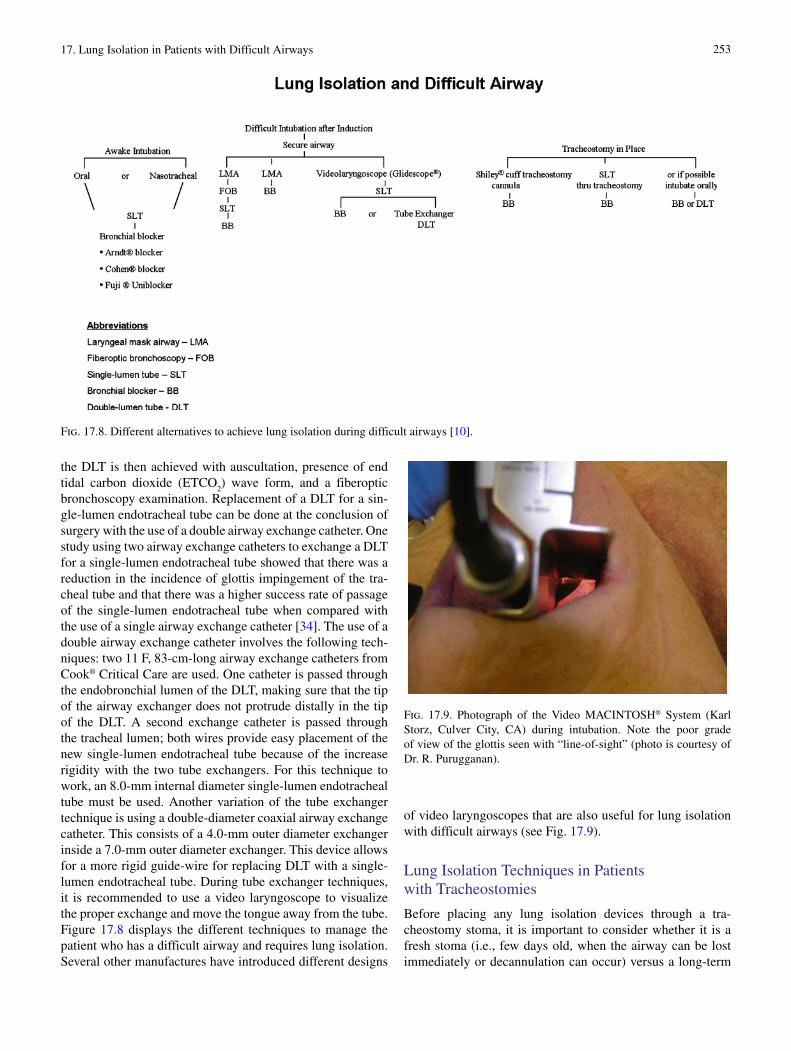
bronchoscopy examination. Replacement of a DLT for a sin-gle-lumen endotracheal tube can be done at the conclusion of surgery with the use of a double airway exchange catheter. One study using two airway exchange catheters to exchange a DLT for a single-lumen endotracheal tube showed that there was a reduction in the incidence of glottis impingement of the tra-cheal tube and that there was a higher success rate of passage of the single-lumen endotracheal tube when compared with the use of a single airway exchange catheter [34]. The use of a double airway exchange catheter involves the following tech-niques: two 11 F, 83-cm-long airway exchange catheters from Cook® Critical Care are used. One catheter is passed through the endobronchial lumen of the DLT, making sure that the tip of the airway exchanger does not protrude distally in the tip of the DLT. A second exchange catheter is passed through the tracheal lumen; both wires provide easy placement of the new single-lumen endotracheal tube because of the increase rigidity with the two tube exchangers. For this technique to work, an 8.0-mm internal diameter single-lumen endotracheal tube must be used. Another variation of the tube exchanger technique is using a double-diameter coaxial airway exchange catheter. This consists of a 4.0-mm outer diameter exchanger inside a 7.0-mm outer diameter exchanger. This device allows for a more rigid guide-wire for replacing DLT with a single-lumen endotracheal tube. During tube exchanger techniques, it is recommended to use a video laryngoscope to visualize the proper exchange and move the tongue away from the tube. Figure 17.8 displays the different techniques to manage the patient who has a difficult airway and requires lung isolation. Several other manufactures have introduced different designs
of video laryngoscopes that are also useful for lung isolation with difficult airways (see Fig. 17.9).
Lung Isolation Techniques in Patients with Tracheostomies
Before placing any lung isolation devices through a tra-cheostomy stoma, it is important to consider whether it is a fresh stoma (i.e., few days old, when the airway can be lost immediately or decannulation can occur) versus a long-term
Fig. 17.8. Different alternatives to achieve lung isolation during difficult airways [10].
Fig. 17.9. Photograph of the Video MACINTOSH® System (Karl Storz, Culver City, CA) during intubation. Note the poor grade of view of the glottis seen with “line-of-sight” (photo is courtesy of Dr. R. Purugganan).

254 J. Campos
tracheostomy. A standard DLT placed through a tracheostomy stoma can be prone to malposition because the upper airway has been shortened and a conventional DLT is too long.
When selecting a DLT as a replacement of the tracheostomy cannula, a specially designed short version of a DLT such as the Naruke DLT can be used through the tracheostomy stoma. In a report involving six patients with permanent tracheosto-mies, the Naruke tube was used with satisfactory results in patients requiring thoracic surgery and OLV [35].
An alternative to DLT placement to achieve OLV in a tra-cheostomized patient includes the following: insertion of a single-lumen endotracheal tube followed by an independent bronchial blocker through the tracheostomy stoma [36], or
if possible oral access to the airway for standard placement of a single-lumen endotracheal tube followed by a bronchial blocker. Another option is the use of a disposable cuff tracheo-stomy cannula with an independent bronchial blocker passed coaxially. In these cases, a small-size fiberoptic bronchoscope (i.e., 3.5-mm outer diameter) is recommended. Figure 17.10 shows lung isolation in a patient with a small tracheal stoma. The tracheostomy device has been replaced; a small laryngeal mask airway LMA #3 has been used to ventilate the patient through the stoma. Also shown in the picture is a patient with a tracheostomy stoma where a 7.0-mm internal diameter sin-gle-lumen endotracheal tube has been advanced through the stoma followed by an Arndt® bronchial blocker.
Fig. 17.10. Shows lung isolation in a patient with a small tracheal stoma. The tracheostomy device has been replaced; to the left a small laryngeal mask airway LMA #3 has been used to ventilate the patient through the stoma. To the right, is shown a patient with a stoma where a 7.0-mm internal diameter single-lumen endotracheal tube has been advanced through the stoma followed by an Arndt® bronchial blocker.

25517. Lung Isolation in Patients with Difficult Airways
Lower Airway Abnormalities and Lung Isolation
When dealing with the difficult airway and OLV, one impor-tant group of patients to consider include those patients who present with lower airway abnormalities, specifically dis-tal trachea or bronchial lesions. The common problems that will preclude or contraindicate the use of a left-sided DLT include an intraluminal tumor of the left mainstem bronchus or a descending thoracic aortic aneurysm that compresses the entrance of a left mainstem bronchus. One option in these cases is to use a right-sided DLT guided with flexible fiberop-tic bronchoscopy [37].
Another group of patients that has lower airway abnor-malities and require OLV are patients with previous lobec-tomy; sometimes in these cases the distorted anatomy may contribute to difficulties in recognizing the right and left bronchi because of the loss of anatomical landmarks [38]. In these patients, a complete fiberoptic bronchoscopy exam of the trachea and bronchi prior to placement of lung isolation device is required in order to properly identify the anatomy of the tracheobronchial tree. Another lower airway abnormality includes the patient that has a diagnosis of tracheal–esophageal fistula. In these cases, the use of two small endotracheal tubes as endobronchial tubes placed under flexible fiberoptic bron-choscopy guidance can be used to secure the airway under these circumstances as shown in Fig. 17.11.
Lung Isolation in Patients with Cervical Spine Abnormalities
Injuries to the cervical spine occur in only 2–3% of all patients with blunt trauma but are significant because of their high level
of associated mortality and morbidity [39, 40]. Some of these patients have experienced trauma to the chest with an injury to the descending thoracic aorta. In these trauma patients, the atlantoaxial region is the most common site of injury and the sixth and seventh vertebrae are involved in over one-third of all injuries [41]. In these patients, all precautions must be taken with regard to cervical spine injury [42]. When such patients arrive for emergency surgery (i.e., repair of a descending tho-racic aortic aneurysm), some are already intubated and have a Philadelphia cervical collar in place. If the patient requires lung isolation and is already intubated, a complete fiberoptic bronchoscopy exam must be done to assess injuries to the tra-chea or bronchus. Once the airway has been assessed, then the use of an independent blocker through the single-lumen endo-tracheal tube is recommended. A Fuji Uniblocker® or Cohen® blocker is desirable because during the insertion it is easier to observe the tip of the blocker as it is guided into the left mainstem bronchus. Once the surgery is completed and lung isolation is no longer needed, removal of the bronchial blocker at the conclusion of the case allows the patient to remain intu-bated with the existing single-lumen endotracheal tube.
Bronchial blockers are often the best option for lung isola-tion in patients with cervical spine instability due to medical conditions such as Rheumatoid Arthritis (see Fig. 17.12).
Extubation or Mechanical Ventilation After Surgery
Extubation at the completion of surgery in a patient who has a difficult airway represents a challenge. Factors to consider prior to extubation include any mucosal edema, bleeding or lacera-tions to the pharynx during intubation, the length of surgery, and the amount of fluid administered during the intraoperative
Fig. 17.11. (a) Fiberoptic bronchoscopic photograph of a tracheoesophageal fistula caused by esophageal cancer. The fistula is seen posteriorly at the level of carina at 5 o’clock. The left mainstem bronchus is at 9 o’clock, and the right mainstem bronchus is at 2 o’clock. (b) Fiberoptic-guided placement of bilateral endobronchial tubes (5-mm internal diameter microlaryngoscopy tubes) for repair of tracheoesophageal fistula in the same patient (photos are courtesy of Dr. R Grant).

256 J. Campos
period. Continuous access to the airway should be maintained in case reintubation is needed. The single-lumen endotra-cheal tube or the DLT can be removed with an airway catheter exchanger in place prior to extubation [43].
In some instances in a patient with a difficult airway and a DLT may require mechanical ventilation in the postoperative period. One option for extubation of these patients is to deflate both cuffs, withdraw the tube above the carina, then reinflate the tracheal cuff, and convert the DLT to two-lung ventilation [44], particularly if the conversion to exchange a DLT for a single-lumen endotracheal tube is considered too risky.
Another alternative technique is to exchange a DLT for a single-lumen endotracheal tube using an airway catheter exchanger under direct vision with a laryngoscope [45] or video laryngoscope [46–48].
Summary
In patients who require OLV, a key element during the pre-operative assessment is the recognition and identification of the potentially difficult airway. The safest way to establish an airway is by securing the airway with a single-lumen endotra-cheal tube placed orally or nasotracheally with the aid of flex-ible fiberoptic bronchoscopy. Lung isolation in these patients
is achieved best with the use of an independent bronchial blocker. An alternative can be the use of a DLT with an air-way catheter exchange technique. For the patient who has a tracheostomy in place, the use of an independent bronchial blocker through a single-lumen endotracheal tube or through a tracheotomy cannula in place is recommended. For all these devices, a flexible fiberoptic bronchoscopy examination is recommended prior, during placement, and at the conclusion of the use of lung isolation devices.
Clinical Case Discussion
Case: A 61-year-old male with a left-upper lobe lung mass is scheduled for a left-upper lobectomy. He is 175 cm tall and weights 93 kg. Relevant history includes right maxillary mucoepidermoid carcinoma resected by radical neck surgery in the past, with additional radiation therapy and reconstruc-tion using a right radial forearm flap; his tobacco history is that he smoked two packs of cigarettes per day for 40 years.
Airway exam reveals the following: full dentures. Mallam-pati score III, thyromental distance two finger breadths, neck range of motion very limited, and scarring from maxillary resection and radiation. Palpation of the anterior neck shows a hard and rigid consistency tissue. Also, the mouth is distorted and deviated towards the right side.
Questions
What technique would you select to intubate this patient?•What potential problems do you expect during an awake •fiberoptic bronchoscopy in a patient with a previous neck resection and extensive surgery?What device and size would you use to provide lung • isolation?What are the common problems in the intraoperative period •with the use of bronchial blockers?What are the complications associated with the bronchial •blocker?What are the advantages and disadvantages of using a •bronchial blocker during a case with a difficult airway that requires lung isolation?
The Key Is to Focus on Patient’s Anatomy in Order to Select the Lung Isolation Device
Review the chest radiograph to appreciate the distorted tra-•cheobronchial anatomy.Focus on lung isolation devices during difficult airways.•Familiarity with the use of independent blockers (Arndt• ®, Cohen®, and Fuji Uniblocker®) is mandatory.Skills with flexible fiberoptic bronchoscopy during awake •intubation, as well as during placement of bronchial block-ers, are essential.
Fig. 17.12. This patient with rheumatoid arthritis has a retroflexed odontoid process and associated inflammatory mass (pseudogout) causing compression of the cervical spinal cord just inferior to the foramen magnum. The patient also had intervertebral subluxations and osteophytes from C3 to C5 that cause cord compression. The anesthetic plan included an awake fiberoptic intubation with a sin-gle-lumen endotracheal tube.

25717. Lung Isolation in Patients with Difficult Airways
Expected Intraoperative Problems with the Use of Bronchial Blockers
High incidence of malpositions.•A balloon of the bronchial blocker can occlude the trachea •and produce cardiopulmonary collapse if dislodged.
Suggested Management
In patients requiring OLV who are identified during the preop-erative evaluation to have difficult airway as in the case pre-sented here, the main challenges include: (1) to safely secure the airway and (2) to select the proper bronchial blocker to achieve lung isolation during OLV. The main problem in this case is to safely establish an airway because of previous neck surgery that distorted his airway anatomy. An awake flexible fiberoptic bronchoscopy was used. After airway topical anes-thesia was achieved, a fiberoptic bronchoscope was passed orally with a guided 8.5-mm internal diameter single-lumen endotracheal tube. Bronchoscopy showed that the upper air-way was severely distorted; a deviation of the larynx towards the right side and bulging scar tissue from previous surgery.
A single-lumen endotracheal tube was advanced without difficulty. After the tube was secured, a complete fiberoptic bronchoscopy exam was achieved with general anesthesia. The second issue described in this case was to achieve suc-cessful lung isolation. It was done with a 9 F Arndt® blocker that was passed under direct fiberoptic bronchoscopy into the left-mainstem bronchus. The optimal position was confirmed in the lateral decubitus position. After completion of the left upper lobectomy, two-lung ventilation was reestablished, the bronchial blocker was withdrawn, and the patient was extu-bated without any complications.
References
1. Campos JH. Progress in lung separation. Thorac Surg Clin. 2005;15:71–83.
2. Hagihira S, Takashina M, Mori T, Yoshiya I. One-lung ventilation in patients with difficult airways. J Cardiothorac Vasc Anesth. 1998;12:186–8.
3. Campos JH. Update on tracheobronchial anatomy and flex-ible fiberoptic bronchoscopy in thoracic anesthesia. Curr Opin Anaesthesiol. 2009;22:4–10.
4. Boiselle PM. Imaging of the large airways. Clin Chest Med. 2008;29:181–93.
5. Seymour AH. The relationship between the diameters of the adult cricoid ring and main tracheobronchial tree: a cadaver study to investigate the basis for double-lumen tube selection. J Cardio-thorac Vasc Anesth. 2003;17:299–301.
6. Minnich DJ, Mathisen DJ. Anatomy of the trachea, carina and bronchi. Thorac Surg Clin. 2007;17:571–85.
7. Stene R, Rose M, Weigner MB, et al. Bronchial trifurcation at the carina complicating use of a double-lumen tracheal tube. Anes-thesiology. 1994;80:1162–4.
8. American Society of Anesthesiologists Task Force on Management of the Difficult Airway. Practice guidelines for management of the difficult airway: an updated report by the American Society of Anesthesiologists Task Force on Management of the Difficult Airway. Anesthesiology. 2003;98:1269–77.
9. Campos JH. Difficult airway and one-lung ventilation. Curr Rev Clin Anesth. 2002;22:199–205.
10. Campos JH. Lung isolation techniques for patients with difficult airways. Curr Opin Anaesthesiol. 2010;23:12–7.
11. Poon KH, Liu EH. The airway scope for difficult double-lumen tube intubation. J Clin Anesth. 2008;20:319.
12. Davis L, Cook-Sather SD, Schreiner MS. Lighted stylet tracheal intubation: a review. Anesth Analg. 2000;90:745–6.
13. Perlin DI, Hannallah MS. Double-lumen tube placement in a patient with a difficult airway. J Cardiothorac Vasc Anesth. 1996;10:787–8.
14. Campos JH. Fiberoptic bronchoscopy in anesthesia. Curr Rev Clin Anesth. 2008;29:61–72.
15. Arndt GA, Buchika S, Kranner PW, DeLessio ST. Wire-guided endobronchial blockade in a patient with a limited mouth open-ing. Can J Anaesth. 1999;46:87–9.
16. Cohen E. The Cohen flexitip endobronchial blocker: an alterna-tive to a double lumen tube. Anesth Analg. 2005;101:1877–9.
17. Campos JH. Which device should be considered the best for lung isolation: double-lumen endotracheal tube versus bronchial blockers. Curr Opin Anaesthesiol. 2007;20:27–31.
18. Narayanaswamy M, McRae K, Slinger P, et al. Choosing a lung isolation device for thoracic surgery: a randomized trial of three bronchial blockers versus double-lumen tubes. Anesth Analg. 2009;108:1097–101.
19. Campos JH. Use of the wire-guided endobronchial blocker for one-lung anesthesia in patients with airway abnormalities. J Car-diothorac Vasc Anesth. 2003;17:352–4.
20. Angie Ho CY, Chen CY, Yang MW, Liu HP. Use of the Arndt wire-guided endobronchial blocker via nasal for one-lung ven-tilation in patient with anticipated restricted mouth opening for esophagectomy. Eur J Cardiothorac Surg. 2005;28:174–5.
21. Cohen E. Pro: the new bronchial blockers are preferable to dou-ble-lumen tubes for lung isolation. J Cardiothorac Vasc Anesth. 2008;22:920–4.
22. Roscoe A, Kanellakos GW, McRae K, Slinger P. Pressures exerted by endobronchial devices. Pressures exerted by endo-bronchial devices. Anesth Analg. 2007;104:655–8.
23. Robinson III AR, Gravenstein N, Alomar-Melero E, Peng YG. Lung isolation using a laryngeal mask airway and a bronchial blocker in a patient with a recent tracheostomy. J Cardiothorac Vasc Anesth. 2008;22:883–6.
24. Ozaki M, Murashima K, Fukutome T. One-lung ventilation using the ProSeal laryngeal mask airway. Anaesthesia. 2004;59:726.
25. Tsuchihashi T, Ide S, Nakagawa H, Hishinuma N, et al. Differen-tial lung ventilation using laryngeal mask airway and a bronchial blocker tube for a patient with unanticipated difficult intubation. Masui. 2007;56:1075–7.
26. Patane PS, Sell BA, Mahla ME. Awake fiberoptic endobronchial intubation. J Cardiothorac Anesth. 1990;4:229–31.
27. O’Connor CJ, O’Connor TA. Use of lighted stylets to facilitate insertion of double-lumen endobronchial tubes in patients with difficult airway anatomy. J Clin Anesth. 2006;18:616–9.

258 J. Campos
28. Smith CE, Kareti M. Fiberoptic laryngoscopy (WuScope) for double-lumen endobronchial tube placement in two difficult-intubation patients. Anesthesiology. 2000;93:906–7.
29. Hernandez AA, Wong DH. Using a Glidescope for intuba-tion with a double lumen endotracheal tube. Can J Anaesth. 2005;52:658–9.
30. Benumof JL. Difficult tubes and difficult airways. J Cardiothorac Vasc Anesth. 1998;12:131–2.
31. Thomas V, Neustein SM. Tracheal laceration after the use of an airway exchange catheter for double-lumen tube placement. J Cardiothorac Vasc Anesth. 2007;21:718–9.
32. deLima LG, Bishop MJ. Lung laceration after tracheal extuba-tion over a plastic tube changer. Anesth Analg. 1991;73:350–1.
33. Chen A, Lai HY, Lin PC, Chen TY, Shyr MH. GlideScope-as-sisted double-lumen endobronchial tube placement in a patient with an unanticipated difficult airway. J Cardiothorac Vasc Anesth. 2008;22:170–2.
34. Suzuki A, Uraoka M, Kimura K, Sato S. Effects of using two air-way exchange catheters on laryngeal passage during change from a double-lumen tracheal tube to a single-lumen tracheal tube. Br J Anaesth. 2007;99:440–3.
35. Saito T, Naruke T, Carney E, et al. New double intrabronchial tube (Naruke tube) for tracheostomized patients. Anesthesiology. 1998;89:1038–9.
36. Tobias JD. Variations on one-lung ventilation. J Clin Anesth. 2001;13:35–9.
37. Campos JH, Ajax TJ, Knutson R, et al. Case conference 5–1990. A 76-year-old man undergoing an emergency descending thoracic aortic aneurysm repair has multiple intraoperative and postopera-tive complications. J Cardiothorac Anesth. 1990;4:631–45.
38. Campos JH. Update on selective lobar blockade during pulmonary resections. Curr Opin Anaesthesiol. 2009;22:18–22.
39. Hoffman JR, Schriger DL, Mower WR, et al. Low-risk criteria for cervical-spine radiography in blunt trauma: a prospective study. Ann Emerg Med. 1992;21:1454–60.
40. Roberge RJ, Wears RC, Kelly M, et al. Selective application of cervical spine radiography in alert victims of blunt trauma: a pro-spective study. J Trauma. 1988;28:784–8.
41. Goldberg W, Mueller C, Panacek E, et al. Distribution and pat-terns of blunt traumatic cervical spine injury. Ann Emerg Med. 2001;38:17–21.
42. Crosby ET. Airway management in adults after cervical spine trauma. Anesthesiology. 2006;104:1293–318.
43. Mort TC. Continuous airway access for the difficult extubation: the efficacy of the airway exchange catheter. Anesth Analg. 2007;105:1357–62.
44. Merlone SC, Shulman MS, Allen MD, Mark JB. Prolonged intu-bation with a polyvinylchloride double-lumen endobronchial tube. J Cardiothorac Anesth. 1987;1:563–4.
45. Benumof JL. Airway exchange catheters: simple concept, poten-tially great danger. Anesthesiology. 1999;91:342–4.
46. Merli G, Guarino A, Della Rocca G, et al. Recommendations for airway control and difficult airway management in thoracic anesthesia and lung separation procedures. Minerva Anestesiol. 2009;75:59–78.
47. Pott LM, Murray WB. Review of video laryngoscopy and rigid fiberoptic laryngoscopy. Curr Opin Anaesthesiol. 2008;21:750–8.
48. Thong SY, Lim Y. Video and optic laryngoscopy assisted tra-cheal intubation–the new era. Anaesth Intensive Care. 2009;37: 219–33.


