Lung Cancer Screening and COPD Update · 2020-01-01 · Lung Cancer Screening Uptake • Data from...
33
Lung Cancer Screening and COPD Update Michael T. Vest, DO, FACP, FCCP Assistant Professor of Medicine Pulmonary and Critical Care Medicine Christiana Care Healthcare System Newark, DE Sidney Kimmel Medical College Philadelphia, PA
Transcript of Lung Cancer Screening and COPD Update · 2020-01-01 · Lung Cancer Screening Uptake • Data from...

Lung Cancer Screening and COPD Update
Michael T. Vest, DO, FACP, FCCPAssistant Professor of Medicine
Pulmonary and Critical Care MedicineChristiana Care Healthcare System
Newark, DESidney Kimmel Medical College
Philadelphia, PA

Objectives
• Lung Cancer Screening
– Describe benefits of lung cancer screening
– List who should and should not be screened
• COPD Updates
– Understand use of beta blockers in COPD
– Describe role for inhaled steroids in COPD and discuss data on eosinophils as it relates to inhaled steroids
– Describe role of NIPPV in outpatients chronic hypercarbic respiratory failure

Nodule Versus Mass
• Nodule: less than 3 cm– The larger the nodule, the more likely malignant– The older the patient, the more likely malignant– The more lung CA risk factors: age, smoking, radon,
occupational (asbestos)– Pattern radiographically (upper v. lower lobe,
spiculation, eccentric calcification v. popcorn calcification)
– History of extrathorax malignancy
• Mass: greater than 3 cm– Malignant till proven otherwise

Nodules
• Most nodules less than 0.8 cm are benign
• Most nodules greater than 2 cm are malignant

Doubling Time in Lung Cancer
• Doubling time is the time it takes a group of cells to double in size
• 30 doublings are needed to get to 1 cm size
• Doubling time varies from 30 to >400 days (“2 year rule”)
• 26% increase in diameter corresponds to a doubling in volume (so more time may be needed before increase in size can be seen on CT)

Lung Cancer Screening
• NLST (NEJM 2011; 365: 395-409)
• Number Needed to Screen 320 to prevent one death
• 20% relative risk reduction
• Inclusion criteria
– Age 55-74, >30 pack year history of smoking, quit less than 15 years ago or current smoker
• 39% of patients in CT screening had nodules

NLST Survival Curves
• Overall mortality– “There were 1877
deaths in the low-dose CT group, as compared with 2000 deaths in the radiography group, representing a significant reduction with low-dose CT screening of 6.7% (95% CI, 1.2 to 13.6) in the rate of death from any cause (P=0.02).”

USPFT Recommendation
• Grade B Recommendation Applies to:– Asymptomatic Adults– 55-80 years old– ≥ 30 pack year history of smoking– Current smoker or quit smoking <15 years ago
• Recommendation– Annual low dose computer tomography– Stop screening once pt has stopped smoking for 15 years,
develops health condition that limits ability to get treatment or is no longer willing to undergo surgery or curative intent radiation
• Ann Intern Med 2014; 160: 330-338

Other organizations
• American Cancer Society, American College of Chest Physicians, American Society of Clinical Oncology, American Thoracic Society, American Cancer Society, International Association for the Study of Lung Cancer, American Lung Association– Ages 55-74 with ≥30 pack years, quit<15 years or still smoking– ACCP has changed age recommendation to 55-77
• National Comprehensive Cancer Network– Recommends same as above but also recommends screening
individuals with ≥ 20 pack year history and one additional risk factor (COPD, pulmonary fibrosis, other smoking related cancer)
• American Academy of Family Physician– The evidence is insufficient to recommend for or against lung
cancer screening by low dose CT

Lung Cancer Screening Uptake
• Data from National Health Interview Survey– 36,191 people questions about CT scan for lung
cancer screening, smoking status– Among high risk smokers, 5.8% on 2015 survey
reported screening CT scan– JAMA Internal Medicine 2017; 177: 439-441
• Editorial in American Journal of Public Health– “…the dismal uptake of low-dose CT screening
through physician-focused efforts suggests that the public health community needs to champion low-dose CT screening and embrace its cost-effectiveness.”
– AJPH Oct 2018; 108: 1292-1293

Lung Cancer versus other cancer screening
Screening intervention Benefit
Mammography, ages 40-49 3 lives saved per 10,000 screened
Mammography, ages 50-59 8 lives saved per 10,000 screened
Mammography, ages 60-69 21 lives saved per 10,000 screened
Mammography, ages 70-74 13 lives saved per 10,000 screened
Colonoscopy every 10 years 24 lives saved per 1000 screened
Lung cancer NLST population 3 live saved per 1000 screened
Lung: NEJM 2011; 365: 395-409 and Chest 2018; 153: 954-985Mammography: Ann Intern Med 2016; 164: 279-296Colonoscopy: JAMA 2016; 315: 2564-2575
Lung cancer screening out-performs mammography in terms of lives saved.
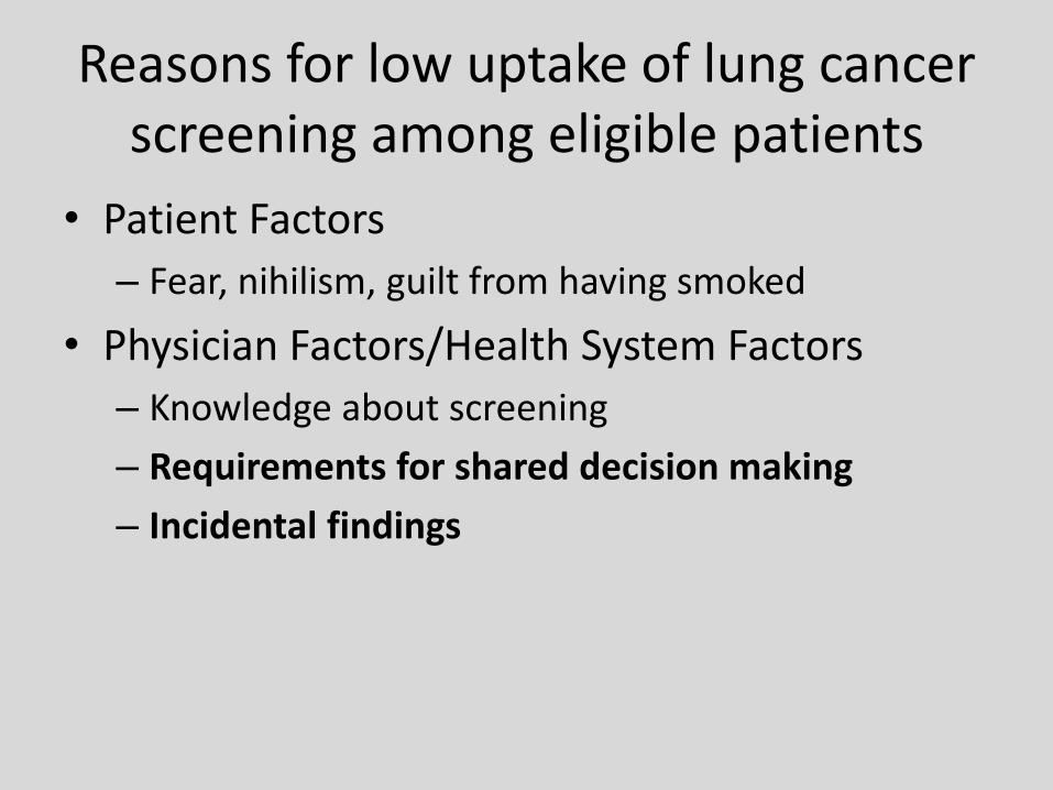
Reasons for low uptake of lung cancer screening among eligible patients
• Patient Factors
– Fear, nihilism, guilt from having smoked
• Physician Factors/Health System Factors
– Knowledge about screening
– Requirements for shared decision making
– Incidental findings

Do NOT screen patients with symptoms of lung cancer
• Symptoms might include otherwise unexplained: – Hemoptysis, coughing, shortness of breath
– Weight Loss, chest pain, hoarseness, bone pains
– Headaches, vision changes, paraneoplastic syndromes (confusion, nausea, constipation, weakness, clubbing)
• Symptomatic patients should get a diagnostic work-up– If CT scan is indicated as part of diagnostic evaluation
it should be diagnostic quality CT– Chest 2018; 153:954-985

Lung RADS
• Classification of Screening CT scan results– 0: incomplete
– 1: negative: no nodules or nodules with specific benign pattern (ex: granuloma)
– 2: Benign appearance: nodules with very low liklihoodof becoming clinically active cancer (<1% probability of malignancy)
– 3: Probably Benign: solid nodules less than 8 mm, ground glass less than 20 mm (1-2% probability of malignancy)
– 4: Suspicious for malignancy

Lung RADS Management
• Groups 1 and 2: low dose CT scan in one year• Group 3 (probably benign)
– Repeat CT scan in 6 months
• Group 4 (Suspicious)– 4A (subcentimeter nodules): CT scan in 3 months– 4B (>15 mm nodule, growing, GGO with solid
component>8mm): PET scan, tissue sampling– 4X: (above with additional features suggestive of
malignancy): tissue sampling
• At Christiana, refer to Cancer Center MDC for Group 4

Nonsurgical Biopsies
1. Bronchoscopic Approaches to lung nodule diagnosis
• Transbronchial biopsies• Yield greatly enhanced
with use of navigational bronchoscopy and radial probe endobronchial ultrasound
2. CT Guided Biopsies• High yield especially for
peripheral lesions, pneumothorax risk
3. SAMPLING ERROR• Biopsy are diagnostic
when cancer or a benign disease (i.e. Sarcoid) is diagnosed
• Otherwise they are nondiagnostic
• A nondiagnostic biopsy does not rule out cancer

Incidental Findings– Pulmonary findings (70%, n-223)
• Emphysema (n-162), vocal cord lesion (n-1), bronchial wall thickening (n-126)
– Cardiovascular (68%, n-216)
• Coronary artery calcification (n-182), Aortic calcification (n-66), aortic dilation (n-28)
– Genitourinary (4%, n-14)
• Renal cyst (n-8), renal stone (n-4), renal mass (n-2)
– GI (24%, n-79)
• Hiatal hernia (n-30), liver cyst (n-22), dilated esophagus (n-7), gallstone (n-6)
– Endocrine• Adrenal nodule (n-12), thyroid nodule (n-11)
– Other
• DJD (n-74), compression fx (n-3), breast nodule (n-1), splenic lesion (n-1)
Annals ATS 2017; 14: 1450-1456

Lung Cancer Screening Conclusion
• Screen: Asymptomatic Adults, 55-80 years old, ≥ 30 pack year history of smoking, Current smoker or quit smoking <15 years ago
• Don’t use screening CT scans for symptomatic patients
• Be prepared for incidental findings
• Consider developing screening program with multidisciplinary group

COPD Update

Diagnostic Instability of COPD
COPD
Normal
COPD
Normal
COPD
Normal

Diagnostic Instability of COPD
• Spirometry can change over time
• Particular around normal range (FEV1/FVC 0.6 to 0.8)
• Around FEV1/FVC 0.7 => 40% changed from obstructive to normal or vice versa
• 2018 GOLD guidelines update: repeat spirometry on pts with FEV1/FVC 0.6-0.8
• AJRCC 2017; 196: 306-314

GOLD COPD
• Group A
– No hospitalization, minimal symptoms
• Group B
– No hospitalization, some symptoms
• Group C
– 1 hospitalization or ≥2 exacerbation, symptoms controlled
• Group D
– 1 hospitalization, ≥2 exacerbations, worse symptoms

Treatment of Stable COPD
© 2017 Global Initiative for Chronic Obstructive Lung Disease

Role of ICS in COPD
• 4 trials with severe disease (GOLD group D)
– IMPACT (NEJM 2018; 378: 1671-1680)
– TRIBUTE (Lancet 2018; 391: 1076-1084)
– FLAME (NEJM 2016; 374: 2222-2234)
– KONOS (Lancet Respir Med 2018; 6: 747-758)
• FLAME showed LAMA/LABA to be better than LAMA/LABA/ICS but excluded patient with hx of asthma and high eosinopils
• The other 3 trials showed benefit with adding ICS

What’s new with ICS in COPD
• Eosinophils– Higher the eosinophils the more likely pts seem to
be to benefit from ICS
– KRONOS showed threshold for benefit of 100 (threshold met by 80% of patients)
– No guidelines on how to use eosinophil count• May be something to watch when withdrawing steroids
in stable severe patient
• Also if you have pt with high eosinophil count, may be more likely to benefit from starting steroids

Non-Pharmacologic Treatment
© 2017 Global Initiative for Chronic Obstructive Lung Disease
Interventional bronchoscopy and surgery
• In selected patients with heterogeneous or homogenous emphysema and significant hyperinflation refractory to optimized medical care, surgical or bronchoscopic modes of lung volume reduction (e.g., endobronchial one-way valves or lung coils) may be considered.
• In selected patients with a large bulla, surgical bullectomy may be considered.
• In selected patients with very severe COPD and without relevant contraindications, lung transplantation may be considered.

Bronchial Valves
• 2 trials this year:• LIBERATE (AJRCCM 2018 May 22. epub ahead of print)
• TRANSFORM (AJRCCM 2017; 196: 1535-1543)
• Compared valve versus standard of care– 48-55% improvement FEV1, QOL, NNT 2-3 – 1/3 had pneumothorax– All stayed overnight in hospital but some pneumothoraces
occurred after hospital discharge– 2.6% procedure related deaths
• Pros: improved quality of life, improved exercise function• Cons: pneumothorax and death

NIPPV in COPD
• Clear long-standing benefit for:– Acute exacerbation with hypercarbia
• Sweet spot: pH 7.25-7.35• <7.25 can be tried higher risk of failure
– Co-morbid sleep disorder breathing• GOLD guidelines indicate improvement of survival and reduced
hospitalization
• Newer in GOLD guidelines stable outpatient with COPD and hypercarbia– “NIPPV may improve hospitalization-free survival in
selected patients after recent hospitalization, particularly in those with pronounced daytime hypercapnia (PaCO2 ≥ 52 mm Hg)”

NIPPV in stable COPD
• Lancet Respir 2014
– RCT 116 COPD pts with paCO2 >53 after 2-4 weeks of hospital discharge
– Home oxygen versus nocturnal NIPPV and oxygen
– BMI>35, sleep disordered breathing, other causes of respiratory failure were excluded
– Less re-admissions in NIPPV group

Beta Blockers in COPD
• High incidence of comorbidities such as coronary artery disease, atrial fibrillation, congestive heart failure etc
• Beta blockade benefits these patients. Prefer selective beta-1 blockers (metoprolol, etc)
• Analysis of SUMMIT trial data showed that beta blocker did not increase incidence of COPD exacerbation and did not impact effectiveness of inhaled beta agonist therapy

Figure 1. Time to first exacerbation of chronic obstructive pulmonary disease (COPD) in the (A) absence or (B) presence of β-blocker therapy at
baseline. FF = fluticasone furoate, FF/VI = combination of fluticasone furoate and vilanterol; VI = vilanterol.
Annals ATS, 2018
https://www.atsjournals.org/doi/abs/10.1513/AnnalsATS.201708-626OC
Published in: Mark T. Dransfield; David A. McAllister; Julie A. Anderson; Robert D. Brook; Peter M. A. Calverley; Bartolome R. Celli; Courtney Crim; Natacha Gallot;
Fernando J. Martinez; Paul D. Scanlon; Julie Yates; Jørgen Vestbo; David E. Newby; Annals ATS 15, 608-614.
DOI: 10.1513/AnnalsATS.201708-626OC
Copyright © 2018 by the American Thoracic Society

COPD Update Conclusion
• Spirometric diagnosis of COPD may change – consider re-evaluation in some patients
• Inhaled steroids have role in COPD, particular for patients with high eosinophils, but only in those with significant symptoms and exacerbations (GOLD groups C and D)
• Refer patients with heterogenous emphysema on CT scans to centers with capability to do bronchial valves and lung volume reduction surgery for evaluation
• Outpatient nocturnal NIPPV benefits stable COPD patients with persistent hypercarbia (paCO2 ≥ 52)
• Beta blockers (beta 1 selective) are safe and effective in COPD patients with cardiovascular indication for their use

Thank You