luminescence in aqueous media Electronic … · Yiquan Wang,†, a Wei Zhou,†, a Feng Chen,a...
13
Electronic Supplementary Information Terbium complexes encapsulated in hierarchically organized hybrid MOF particles toward stable luminescence in aqueous media Yiquan Wang, †, a Wei Zhou, †, a Feng Chen, a Kaiyao Sun, a Jixi Zhang, a, * Ezgi Özliseli, b Jessica M. Rosenholm b, * a Key Laboratory of Biorheological Science and Technology, Ministry of Education, College of Bioengineering, Chongqing University, No. 174 Shazheng Road, Chongqing 400044, China. E- mail: [email protected]. b Pharmaceutical Sciences Laboratory, Faculty of Science and Engineering, Åbo Akademi University, Tykistökatu 6A, Turku 20520, Finland. E-mail: [email protected]. † The authors contributed equally to this work. 1. Experimental Section 1.1. Materials Unless otherwise noted, all reagent-grade chemicals were used as received, and distilled water was used for the preparation of all aqueous solutions. Ethanol (AR) and methanol (AR) were purchased from Fluka. Terbium (III) trifluoromethanesulfonate (AR) and polyvinylpyrrolidone (PVP, M.W. 29 k) were purchased from Aldrich. 2-Methylimidazole (HMIM, AR, 98%) and 5,6-Dimethylbenzimidazole (DMBIM, AR) were purchased from Adamas. Dopamine hydrochloride (AR), Zn(NO 3 ) 2 ·6H 2 O (AR), tetrahydrofuran (≥99%), and triethylamine (AR) were purchased from Aladdin Industrial Inc. All chemicals were used as received without further purification. The bis-tetrazolate-pyridine (H 2 pytz) ligand for sensitizing lanthanide complex is synthesized according to a procedure reported in the literature. 1 Electronic Supplementary Material (ESI) for CrystEngComm. This journal is © The Royal Society of Chemistry 2018
Transcript of luminescence in aqueous media Electronic … · Yiquan Wang,†, a Wei Zhou,†, a Feng Chen,a...
Electronic Supplementary Information
Terbium complexes encapsulated in hierarchically
organized hybrid MOF particles toward stable
luminescence in aqueous media
Yiquan Wang,†, a Wei Zhou,†, a Feng Chen,a Kaiyao Sun,a Jixi Zhang, a,* Ezgi Özliseli,b Jessica M. Rosenholm b,*
a Key Laboratory of Biorheological Science and Technology, Ministry of Education, College of Bioengineering, Chongqing University, No. 174 Shazheng Road, Chongqing 400044, China. E-mail: [email protected].
b Pharmaceutical Sciences Laboratory, Faculty of Science and Engineering, Åbo Akademi University, Tykistökatu 6A, Turku 20520, Finland. E-mail: [email protected].
† The authors contributed equally to this work.
1. Experimental Section
1.1. Materials
Unless otherwise noted, all reagent-grade chemicals were used as received, and distilled water
was used for the preparation of all aqueous solutions. Ethanol (AR) and methanol (AR) were
purchased from Fluka. Terbium (III) trifluoromethanesulfonate (AR) and polyvinylpyrrolidone
(PVP, M.W. 29 k) were purchased from Aldrich. 2-Methylimidazole (HMIM, AR, 98%) and
5,6-Dimethylbenzimidazole (DMBIM, AR) were purchased from Adamas. Dopamine
hydrochloride (AR), Zn(NO3)2·6H2O (AR), tetrahydrofuran (≥99%), and triethylamine (AR)
were purchased from Aladdin Industrial Inc. All chemicals were used as received without
further purification. The bis-tetrazolate-pyridine (H2pytz) ligand for sensitizing lanthanide
complex is synthesized according to a procedure reported in the literature.1
Electronic Supplementary Material (ESI) for CrystEngComm.This journal is © The Royal Society of Chemistry 2018

1.2. Preparation of Tb complex ([Tb(pytz)3](NEt3)3). The preparation of Tb complex was
based on a previous procedure with some modifications.2 Firstly, 43.3 mg of H2pytz was
dispersed in 0.8 mL of methanol, followed by the addition of 55.5 μL of triethylamine (NEt3).
After one hour of stirring, a solution of 30.3 mg of terbium (III) trifluoromethanesulfonate
dissolved in 0.5 mL of methanol was introduced to the mixture. The reaction mixture was stirred
at room temperature for 24 h, and then the product ([Tb(pytz)3](NEt3)3) was centrifuged down
(11000 rpm, 5 min). Tetrahydrofuran was used to wash the complexes. The white powder can
be obtained after drying in an oven at 60 °C overnight to remove the residual liquid for
subsequent use.
1.3. Synthesis of Tb complex encapsulated ZIF-8 (Tb complex@ZIF-8). The Tb
complex@ZIF-8 nanocrystals were prepared by an one-step synthesis method.3 Firstly, a
solution of Tb complex in methanol (100 μL, 5 mg mL-1) was first mixed with 223 μL of
methanol, followed by the addition of a methanol solution of HMIM (300 μL, 164 mg mL-1 in
methanol). After sonication, a solution of Zn(NO3)2·6H2O in methanol (57 μL, 297 mg mL-1)
was rapidly injected and the mixture was stirred at room temperature overnight. The molar ratio
used in the synthesis was 167770 MeOH : 7 Tb complex : 6000 HMIM : 560 Zn(NO3)2·6H2O.
The final luminescent product was centrifuged for further synthesis.
1.4. Synthesis of Tb complex encapsulated hybrid MOF particle (Tb complex@ZIF-
8@TIF-1Zn). Typically, 2 mg of freshly prepared Tb complex@ZIF-8 nanocrystals dispersed
in 572 μL of methanol were poured into a methanol (0.1 mL) solution of DMBIM (40 mg mL-
1), and then 28 μL of Zn(NO3)2·6H2O (297 mg mL-1 in methanol) was added into the mixture.
After stirring at room temperature for 6 h, the suspension was centrifuged (11000 rpm, 10 min),
and the obtained particles were washed with methanol for three times. For the investigation on
the contribution of DMBIM to the generation of the secondary and tertiary structures in the
hybrid MOF particles, less amounts of DMBIM (20%, 60% with respect to the original amount
in the standard synthesis (0.028 mmol)) were employed for the synthesis, while the molar ratio
of DMBIM/Zn was kept at 1. Particles without the addition of Zn(NO3)2·6H2O was also used

for the comparison of hydrodynamic diameters in the methanol solutions, to demonstrate the
aggregation of Tb complex@ZIF-8 nanoparticles under the influence of DMBIM.
1.5. Surface modification of Tb complex@ZIF-8@TIF-1Zn with polydopamine (PDA).
The modification of Tb complex@ZIF-8@TIF-1Zn particles with PDA was conducted by
utilizing DMBIM as the organic base to catalyze the polymerization of dopamine.7, 8 Typically,
4 mg of the as-prepared Tb complex@ZIF-8@TIF-1Zn particles was firstly dissolved in 562
μL of methanol, followed by the addition of DMBIM (0.028 mmol or 0.0056 mmol) in 0.1 mL
of methanol. After sonication, 28 μL of of Zn(NO3)2·6H2O (297 mg mL-1 in methanol) was
rapidly added. Then 10 μL of a methanol solution of dopamine (5 mg mL-1) was injected. The
mixture was stirred at room temperature overnight. The obtained particles (Tb complex@ZIF-
8@TIF-1Zn-PDA) were recovered by centrifugation (11000 rpm, 5 min) and washed with
ethanol for three times. The final luminescent product was suspended in ethanol for further use.
1.6. Photoluminescence stability study. For the luminescence stability study, Tb complex, Tb
complex@ZIF-8, Tb complex@ZIF-8@TIF-1Zn, and Tb complex@ZIF-8@TIF-1Zn-PDA
particles were respectively dispersed in PBS buffer at a concentration of 0.1 mg mL-1. A
fluorescence spectrophotometer (RF-6000, Shimadzu, Japan) was used to monitor the emission
luminescence change (543 nm) of aforementioned particles by measuring the intensities in the
same time intervals (20 min). The employed excitation wavelength was 304 nm.
2. Characterizations
The TEM (Transmission Electron Microscope) images were performed on a JEM-2010
instrument (JEOL, Japan). The SEM (Scanning Electron Microscope) images were obtained
on a field-emission scanning electron micro-probe (JSM7100, JEOL, Japan). The bulk and
surface chemical compositions of samples were analyzed with HAADF-STEM (high-angle
annular dark-field imaging, scanning transmission electron microscope) and elemental
mapping images via a JEM-6700F instrument (JEOL, Japan). XRD patterns were recorded on
a diffractometer (AXS, Bruker, Germany) with Cu target (40 kV, 40 mA, λ=1.54059Å). The
hydrodynamic size distributions of the samples were measured using dynamic light scattering

(DLS) techniques by a Zetasizer Nano instrument (Malvern, UK) at 25°C. Nitrogen sorption
isotherms were measured with an ASAP2010 analyzer (Micromeritcs, USA). The specific
surface areas were calculated by the Brunauer-Emmett-Teller (BET) method in a linear
relative pressure range between 0.05 and 0.25. The pore size distributions were derived from
the desorption branches of the isotherms by the non-localized density functional theory
(NLDFT) method using NLDFT kernel file. The Fourier transform infrared (FT-IR) spectra
were collected over the range of 4000-400cm on a Spectrum 100 infrared spectrophotometer
(PerkinElmer, USA) using a KBr technique. Fluorescence spectra for the particle suspensions
were measured by a RF-6000 fluorescence spectrometer (SHIMADZU, Japan).
Thermogravimetric analysis (TGA) was conducted with a TG 209 instrument (NETZSCH,
USA). The materials were tested under an air atmosphere from 30 to 900 °C at a heating rate
of 10°C min-1. The contact angles for water on the surfaces of the as-obtained particles films
were characterized at room temperature by a contact angle system (JY-PHa, JinHe Machine,
China). The results of contact angles were read within 20 s after depositing and drying 5 μL of
particle suspension (2 mg mL-1) on a mica substrate. Each data point represented an average of
five measurements on the same sample. Fluorescence quantum yields (QYs) were collected
using an integrating sphere (FS5, Edinburgh Instruments, UK). Luminescence lifetime
measurements are carried out on an FLS920 phosphorimeter (Edinburgh Instruments, UK)
using a microsecond pulse lamp as excitation source. The quantitative value of lifetime was
calculated by linear fitting.
3. Supporting figures.
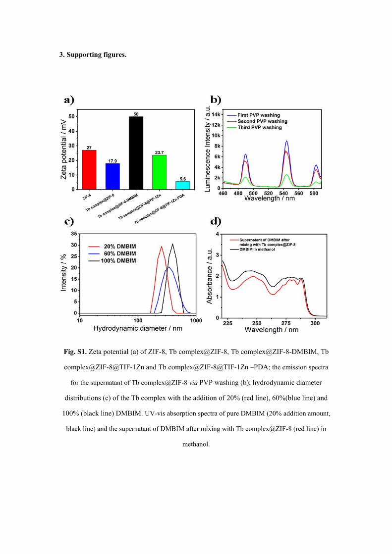
Fig. S1. Zeta potential (a) of ZIF-8, Tb complex@ZIF-8, Tb complex@ZIF-8-DMBIM, Tb
complex@ZIF-8@TIF-1Zn and Tb complex@ZIF-8@TIF-1Zn –PDA; the emission spectra
for the supernatant of Tb complex@ZIF-8 via PVP washing (b); hydrodynamic diameter
distributions (c) of the Tb complex with the addition of 20% (red line), 60%(blue line) and
100% (black line) DMBIM. UV-vis absorption spectra of pure DMBIM (20% addition amount,
black line) and the supernatant of DMBIM after mixing with Tb complex@ZIF-8 (red line) in
methanol.

Fig. S2. Typical SEM images of Tb complex@ZIF-8@TIF-1Zn prepared with 100% (a), 60%
(b), and 20% (c) DMBIM but a fixed DMBIM/Zn ratio (1) in the synthesis; comparison of
hydrodynamic diameter distributions (d) of the particles in (a-c). The average sizes from the
SEM images in (a), (b), and (c), were determined to be 404 47 nm, 336 82 nm, and
240 54 nm, respectively.

Fig. S3. FTIR spectra of (a) pure DMBIM and (b) pure PDA.
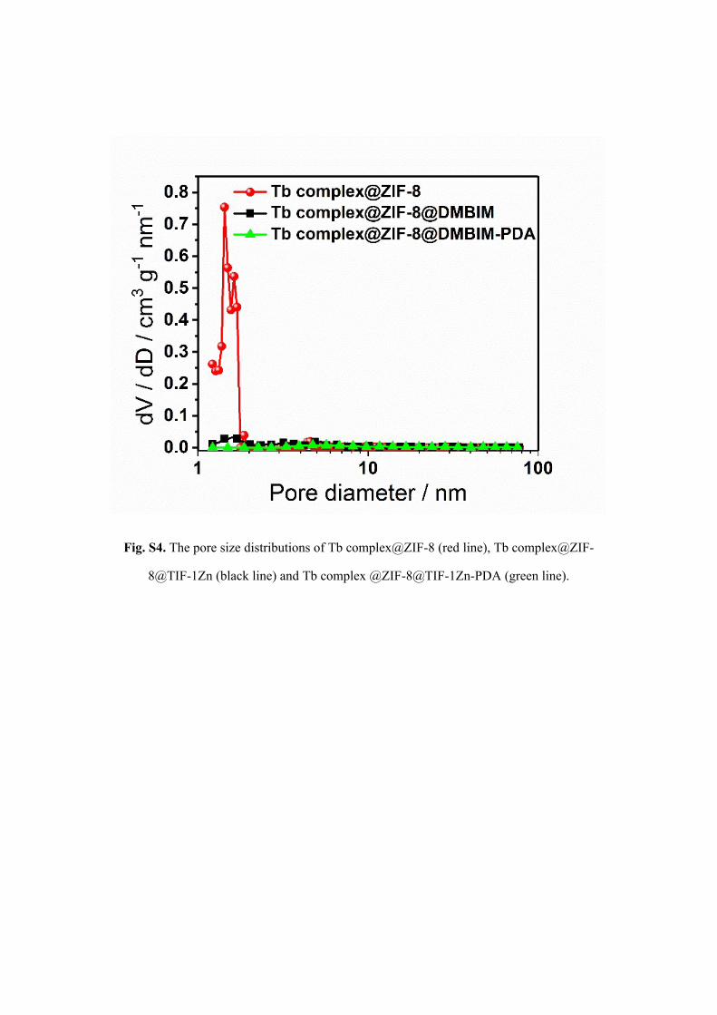
Fig. S4. The pore size distributions of Tb complex@ZIF-8 (red line), Tb complex@ZIF-
8@TIF-1Zn (black line) and Tb complex @ZIF-8@TIF-1Zn-PDA (green line).
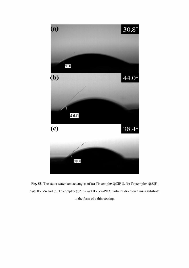
Fig. S5. The static water contact angles of (a) Tb complex@ZIF-8, (b) Tb complex @ZIF-
8@TIF-1Zn and (c) Tb complex @ZIF-8@TIF-1Zn-PDA particles dried on a mica substrate
in the form of a thin coating.
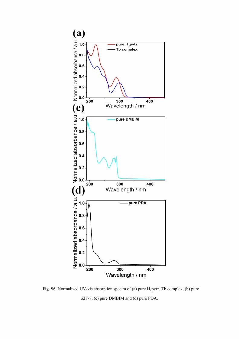
Fig. S6. Normalized UV-vis absorption spectra of (a) pure H2pytz, Tb complex, (b) pure
ZIF-8, (c) pure DMBIM and (d) pure PDA.
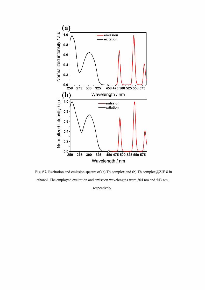
Fig. S7. Excitation and emission spectra of (a) Tb complex and (b) Tb complex@ZIF-8 in
ethanol. The employed excitation and emission wavelengths were 304 nm and 543 nm,
respectively.
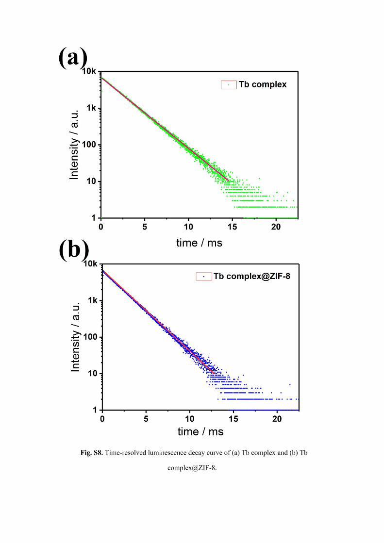
Fig. S8. Time-resolved luminescence decay curve of (a) Tb complex and (b) Tb
complex@ZIF-8.

Table S1. Summarized photophysical properties of the suspensions of Tb complex, Tb
complex@ZIF-8 and Tb complex@ZIF-8@TIF-1Zn-PDA NPs. Particles were dispersed in
PBS buffer (pH=7.4) at a fixed concentration of 1 mg mL-1.
Sample name σ[a] [×10-12 cm2] Φ[b]PBS / % τ[c]
PBS / ms
Tb complex - 15.7 2.2
Tb complex@ZIF-8 2.1 11.8 2.0
Tb complex@ZIF-8@TIF-1Zn-PDA 2.5 82.0 1.5
[a] Absorption cross-section for single particles; [b] absolute luminescence QY of particles
suspended in PBS buffer (pH=7.4); [c] lifetime τ of particles suspended in PBS buffer
(pH=7.4).
References
1. N. Wartenberg, O. Raccurt, E. Bourgeat‐Lami, D. Imbert and M. Mazzanti, Chem.Eur. J., 2013, 19, 3477-3482.
2. E. S. Andreiadis, D. Imbert, J. Pecaut, R. Demadrille and M. Mazzanti, Dalton Trans., 2012, 41, 1268-1277.
3. J. Zhuang, C.-H. Kuo, L.-Y. Chou, D.-Y. Liu, E. Weerapana and C.-K. Tsung, ACS Nano, 2014, 8, 2812-2819.
4. L.-Y. Chou, P. Hu, J. Zhuang, J. V. Morabito, K. C. Ng, Y.-C. Kao, S.-C. Wang, F.-K. Shieh, C.-H. Kuo and C.-K. Tsung, Nanoscale, 2015, 7, 19408-19412.
5. J. V. Morabito, L.-Y. Chou, Z. Li, C. M. Manna, C. A. Petroff, R. J. Kyada, J. M. Palomba, J. A. Byers and C.-K. Tsung, J. Am. Chem. Soc., 2014, 136, 12540-12543.
6. X. Liu, Y. Li, Y. Ban, Y. Peng, H. Jin, H. Bux, L. Xu, J. Caro and W. Yang, Chem. Commun., 2013, 49, 9140-9142.
7. Y. Liang, J. Wei, Y. X. Hu, X. F. Chen, J. Zhang, X. Y. Zhang, S. P. Jiang, S. W. Tao and H. T. Wang, Nanoscale, 2017, 9, 5323-5328.
8. X. Zhang, S. Wang, L. Xu, L. Feng, Y. Ji, L. Tao, S. Li and Y. Wei, Nanoscale, 2012, 4, 5581-5584.