Lumbar Spine Trauma Presented by M.A. Kaeser, DC Spring 2009.
36
Lumbar Spine Lumbar Spine Trauma Trauma Presented by M.A. Kaeser, Presented by M.A. Kaeser, DC DC Spring 2009 Spring 2009
-
Upload
clarence-clark -
Category
Documents
-
view
218 -
download
1
Transcript of Lumbar Spine Trauma Presented by M.A. Kaeser, DC Spring 2009.

Lumbar Spine TraumaLumbar Spine Trauma
Presented by M.A. Kaeser, DCPresented by M.A. Kaeser, DC
Spring 2009Spring 2009

Compression FracturesCompression Fractures
M/C fracture of the lumbar spineM/C fracture of the lumbar spine Result from combined flexion and axial Result from combined flexion and axial
compressioncompression M/C level is T12-L1M/C level is T12-L1 Degree of compression and Degree of compression and
comminution depends on severity fo the comminution depends on severity fo the force appliied and the strength of the force appliied and the strength of the vertebravertebra
Children – torus type fractureChildren – torus type fracture Incidence increases with ageIncidence increases with age

http://www.eorthopod.com/images/ContentImages/spine/spine_thoracic/compression_fx/thoracic_compression_fx_intro01.jpg
http://www.nature.com/sc/journal/v42/n2/images/3101546f1.jpg

Osteoporosis and Compression Osteoporosis and Compression FracturesFractures
Precipitates spontaneous compression Precipitates spontaneous compression fractures during everyday activitiesfractures during everyday activities
Classified as insufficiency fractures Classified as insufficiency fractures (“grandma fracture”)(“grandma fracture”)
Most commonly occur in womenMost commonly occur in women Up to 35% in female pts. Over the age of Up to 35% in female pts. Over the age of
45 years may be the result of early 45 years may be the result of early menopause and 30% to secondary menopause and 30% to secondary osteopenia (corticosteroids 15%, osteopenia (corticosteroids 15%, hyperthyroidism 8%, malignancy >2%)hyperthyroidism 8%, malignancy >2%)

http://api.ning.com/files/O9cKH-JobD4EbaBjcqs2c2RwKwqc51mgaV3nJ6kwGv5AnOKjq7OXspLL*FBSjq-RJdddXF432gr1MxZRI6RnV8-sgh*y3Fy5/compression_fracture.jpg

Symptoms/TreatmentSymptoms/Treatment
Acute symptoms of only 10-14 days Acute symptoms of only 10-14 days duration, if no dislocationduration, if no dislocation
Treatment is based on the nature of Treatment is based on the nature of the collapse and whether or not the collapse and whether or not there is associated neurological there is associated neurological symptomologysymptomology

StabilityStability
Post-fracture stability is determined based Post-fracture stability is determined based on the classification by Denison the classification by Denis• Anterior column – from ALL to the mid vertebral Anterior column – from ALL to the mid vertebral
bodybody• Middle column – from the mid vertebral body to Middle column – from the mid vertebral body to
the PLLthe PLL• Posterior column – from the PLL to the Posterior column – from the PLL to the
supraspinous ligamentsupraspinous ligament
If two or more compartments are disrupted the If two or more compartments are disrupted the fracture complex is unstable – neurological fracture complex is unstable – neurological injury is high and interventional surgery is likelyinjury is high and interventional surgery is likely

Radiographic Signs of Vertebral Radiographic Signs of Vertebral Compression FractureCompression Fracture
Lateral radiographs best Lateral radiographs best demonstrate fracture featuresdemonstrate fracture features
Include:Include:• Step defectStep defect• Wedge deformityWedge deformity• Linear zone of condensationLinear zone of condensation• Displaced endplateDisplaced endplate• Paraspinal hematomaParaspinal hematoma• Abdominal ileusAbdominal ileus
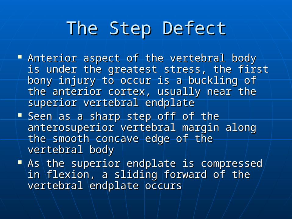
The Step DefectThe Step Defect
Anterior aspect of the vertebral body is Anterior aspect of the vertebral body is under the greatest stress, the first bony under the greatest stress, the first bony injury to occur is a buckling of the anterior injury to occur is a buckling of the anterior cortex, usually near the superior vertebral cortex, usually near the superior vertebral endplateendplate
Seen as a sharp step off of the Seen as a sharp step off of the anterosuperior vertebral margin along the anterosuperior vertebral margin along the smooth concave edge of the vertebral bodysmooth concave edge of the vertebral body
As the superior endplate is compressed in As the superior endplate is compressed in flexion, a sliding forward of the vertebral flexion, a sliding forward of the vertebral endplate occursendplate occurs

http://www.chiroweb.com/content/images/bassano02_1_2334.jpg

Wedge DeformityWedge Deformity Anterior depression of the vertebral body occurs, Anterior depression of the vertebral body occurs,
creating a triangular wedge shapecreating a triangular wedge shape The posterior vertebral height remains The posterior vertebral height remains
uncompromised, differentiating a traumatic uncompromised, differentiating a traumatic fracture from a pathologic fracturefracture from a pathologic fracture
May create angular kyphosis in the adjacent areaMay create angular kyphosis in the adjacent area Superior endplate is far more often involved than Superior endplate is far more often involved than
the inferior endplatethe inferior endplate Up to 30% or greater loss in anterior height may Up to 30% or greater loss in anterior height may
be required before the deformity is readily be required before the deformity is readily apparent on convention x-raysapparent on convention x-rays
Normal variant anterior wedging of 10-15% or 1-3 Normal variant anterior wedging of 10-15% or 1-3 mm is common thought the T/S and most marked mm is common thought the T/S and most marked at T11-L2.at T11-L2.

http://www.cascadewellnessclinic.com/GRAPHICS/2ARTGFX/00ARTGFX/COMPFRAC.GIF

Linear White Band of Condensation Linear White Band of Condensation (Zone of Impaction)(Zone of Impaction)
Band of raadiopacity may be seen just Band of raadiopacity may be seen just below the vertebral endplate that has below the vertebral endplate that has been fracturedbeen fractured
The band represents the early site of bone The band represents the early site of bone impaction following a forceful flexion injury impaction following a forceful flexion injury where the bones are driven togetherwhere the bones are driven together
Callus formation adds to the density of the Callus formation adds to the density of the radiopaque band later, in the healing radiopaque band later, in the healing stage of the fracture injurystage of the fracture injury
Band is not always apparent Band is not always apparent If present, denotes a fracture of recent If present, denotes a fracture of recent
origin (<2 months’ duration)origin (<2 months’ duration)

http://www.theamericanchiropractor.com/images/figure-1.jpg
Disruption in the Vertebral EndplateDisruption in the Vertebral Endplate
A sharp disruption in the fractured A sharp disruption in the fractured vertebral endplate may be seenvertebral endplate may be seen
May be difficult to perceive on plain May be difficult to perceive on plain films and tomographyfilms and tomography
CT provides the definitive means of CT provides the definitive means of identificationidentification
The edges of the disruption are often The edges of the disruption are often jagged and irregularjagged and irregular
Superior is more commonly fracturedSuperior is more commonly fractured

http://download.imaging.consult.com/ic/images/S1933033206711077/gr1-midi.jpg

Paraspinal EdemaParaspinal Edema
With extensive trauma, U/L or B/L With extensive trauma, U/L or B/L paraspinal masses may occur – these paraspinal masses may occur – these represent hemorrhagerepresent hemorrhage
Best seen in the thracic spine on the Best seen in the thracic spine on the AP projectionAP projection
In L/S edema creates asymmetrical In L/S edema creates asymmetrical densities or bulges in the psoas densities or bulges in the psoas marginsmargins

http://download.imaging.consult.com/ic/images/S1933033207730938/gr3-midi.jpg

Abdominal IleusAbdominal Ileus
An excessive amount of small or An excessive amount of small or large bowel has in a slightly large bowel has in a slightly distended lumendistended lumen
Occurs as a result of disturbance to Occurs as a result of disturbance to the visceral autonomic nerves or the visceral autonomic nerves or ganglia from pain, paraspinal soft ganglia from pain, paraspinal soft tissue injury, edema or hematomatissue injury, edema or hematoma
This sign warns that the trauma was This sign warns that the trauma was severe and fracture is likelysevere and fracture is likely

http://www.ganfyd.org/images/thumb/6/69/Axr_ileus.jpg/180px-Axr_ileus.jpg

Old vs New Compression FractureOld vs New Compression Fracture
Signs of a fracture <2 months old = soft tissue Signs of a fracture <2 months old = soft tissue hemorrhage, step defect and white band of hemorrhage, step defect and white band of condensationcondensation
Healing of compression fracture can take up to 3 Healing of compression fracture can take up to 3 months in the adult spinemonths in the adult spine
Presence of contiguous disc degeneration is common Presence of contiguous disc degeneration is common in old compression fractures owing to altered in old compression fractures owing to altered discovertebral mechanicsdiscovertebral mechanics
MRI reveals bone marrow edema with recent fractureMRI reveals bone marrow edema with recent fracture Bone scan shows increased uptake with recent Bone scan shows increased uptake with recent
fractures undergoing repair (they may remain active fractures undergoing repair (they may remain active for up to 18-24 months after injury, which diminishes for up to 18-24 months after injury, which diminishes its usefulness)its usefulness)

Burst FracturesBurst Fractures Specific form of a compression fractureSpecific form of a compression fracture Posterosuperior fragment is displaced into the spinal canalPosterosuperior fragment is displaced into the spinal canal Neurological injury may result in up to 50% of cases (best Neurological injury may result in up to 50% of cases (best
demonstrated by MRI or CT)demonstrated by MRI or CT) Most burst fractures are stable and can be treated Most burst fractures are stable and can be treated
adequately with conservative measuresadequately with conservative measures AP film – a vertical fracture line is often seen, AP film – a vertical fracture line is often seen, Widening of the interpediculate distance signifies a fracture Widening of the interpediculate distance signifies a fracture
within the neural archwithin the neural arch Acquired coronal cleft vertebra – coronally oriented fracture Acquired coronal cleft vertebra – coronally oriented fracture
the separates the vertebral body into anterior and posterior the separates the vertebral body into anterior and posterior halveshalves
Central depression of the superior and inferior endplates Central depression of the superior and inferior endplates occurs with comminution of the vertebral bodyoccurs with comminution of the vertebral body

http://www.spineuniverse.com/displaygraphic.php/2240/Fig-1c-BB.jpghttp://www.spineuniverse.com/displaygraphic.php/2240/Fig-1c-BB.jpg
http://www.ajronline.org/cgi/content-nw/full/187/4/859/FIG12

Posterior Apophyseal Ring Posterior Apophyseal Ring FracturesFractures
Separation of the posterior vertebral body ring Separation of the posterior vertebral body ring apophysis (posterior limbus bone) is a relatively apophysis (posterior limbus bone) is a relatively uncommon abnormalityuncommon abnormality
More common in adolescents and young adultsMore common in adolescents and young adults Clinical features include stiffness and spasm, Clinical features include stiffness and spasm,
numbness, weakness, neurogenic claudication and numbness, weakness, neurogenic claudication and occasionally cauda equina syndromeoccasionally cauda equina syndrome
Most common levels are L4/5 and L5/S1Most common levels are L4/5 and L5/S1 50% are caused by trauma (ie. weightlifting, MVAs, 50% are caused by trauma (ie. weightlifting, MVAs,
gymnastics)gymnastics) Surgery may be warrantedSurgery may be warranted Between 15% and 20% are visible on lateral Between 15% and 20% are visible on lateral
radiographsradiographs CT is definitive method of diagnosisCT is definitive method of diagnosis

http://www.scielo.br/img/revistas/aob/v10n1/a04fig05.gif

Kummel’s DiseaseKummel’s Disease
Delayed post-traumatic vertebral Delayed post-traumatic vertebral collapsecollapse
Occurs months after an episode of Occurs months after an episode of spinal traumaspinal trauma
Caused by complicating avascular Caused by complicating avascular necrosis resulting in progressive necrosis resulting in progressive compression deformitycompression deformity
Intravertebral vacuum phenomenon Intravertebral vacuum phenomenon may be evident on radiographsmay be evident on radiographs

http://download.imaging.consult.com/ic/images/S1933033206702300/gr10-midi.jpg

Fractures of the Neural ArchFractures of the Neural Arch TP fractures – 2TP fractures – 2ndnd M/C fracture of the L/S M/C fracture of the L/S Occur from avulsion of the paraspinal muscles, usually secondary to a Occur from avulsion of the paraspinal muscles, usually secondary to a
severe hyperextension and lateral flexion blow to the L/Ssevere hyperextension and lateral flexion blow to the L/S Large forces are usually involvedLarge forces are usually involved M/C segments are L2/3M/C segments are L2/3 Frequently missed on initial examinationFrequently missed on initial examination Usually occurring close to its point of origin from the vertebraUsually occurring close to its point of origin from the vertebra Frequently displaced inferiorlyFrequently displaced inferiorly If the fracture is horizontal, check for transverse or Chance fractureIf the fracture is horizontal, check for transverse or Chance fracture Fractures often occur at multiple levelsFractures often occur at multiple levels Fractures of the L5 TP are frequently found in association with pelvic Fractures of the L5 TP are frequently found in association with pelvic
fracturesfractures Loss of the psoas shadow may occur secondary to hemorrhageLoss of the psoas shadow may occur secondary to hemorrhage Always check for abdominal organ injuriesAlways check for abdominal organ injuries Associated renal damage may be associated with hematuriaAssociated renal damage may be associated with hematuria CT exam is recommended to check for fracture and integrity of the CT exam is recommended to check for fracture and integrity of the
abdominal contentsabdominal contents

http://www.medscape.com/content/2004/00/48/35/483518/art-ar483518.fig8b.jpg

Pars Interarticularis FracturesPars Interarticularis Fractures
True fractures, not stress fractures, are True fractures, not stress fractures, are uncommonuncommon
MOI – violent hyperextension of the L/S – MOI – violent hyperextension of the L/S – usually at the L4 or L5 levelusually at the L4 or L5 level
Not to be confused with spondylolysis of Not to be confused with spondylolysis of the pars, which is usually the result of a the pars, which is usually the result of a stress fracturestress fracture
Acute fractures are U/L, spondylolysis is Acute fractures are U/L, spondylolysis is B/LB/L
Acute fractures heal without residual Acute fractures heal without residual defects or anterior displacementdefects or anterior displacement

http://www.wheelessonline.com/image9/spdy1.jpg
http://www.sportsinjuryclinic.net/gallery/back/sponylolysis_large.jpg

Chance or Lap Seat Belt FractureChance or Lap Seat Belt Fracture
Horizontal splitting of the spine and neural archHorizontal splitting of the spine and neural arch Use of lap-type seat belts in the ’50s and ’60s Use of lap-type seat belts in the ’50s and ’60s
coincided with an increasing occurrence of coincided with an increasing occurrence of Chance fracturesChance fractures
Severe abrasions can be seen on the lower Severe abrasions can be seen on the lower anterior abdominal wall, outlining the position of anterior abdominal wall, outlining the position of the seat belt the seat belt
Internal visceral damage may occur – rupture of Internal visceral damage may occur – rupture of the spleen or pancreas and tears of the small the spleen or pancreas and tears of the small bowel and mesenterybowel and mesentery
Neurological deficits in 15% of casesNeurological deficits in 15% of cases M/C location is upper L/S (L1-L3)M/C location is upper L/S (L1-L3)

http://www.e-radiography.net/radpath/f/chance%20fracture/Chance-sagital_recon.jpg
http://www.ajronline.org/cgi/content-nw/full/183/4/959/FIG6

Fracture-DislocationFracture-Dislocation
M/C in the T/L area afer a vilent flexion injuryM/C in the T/L area afer a vilent flexion injury Avulsion fractures (teardrop) are commonly Avulsion fractures (teardrop) are commonly
found associated with dislocation of the L/Sfound associated with dislocation of the L/S Most dislocations are anterior in position, Most dislocations are anterior in position,
without lateral displacementwithout lateral displacement Shearing injuries with disc and ligament rupture Shearing injuries with disc and ligament rupture
and fractures of the posterior arch are commonand fractures of the posterior arch are common Naked facet sign – absence of apposed articular Naked facet sign – absence of apposed articular
facets – diagnostic for facet dislocationfacets – diagnostic for facet dislocation

http://www.ispub.com/ispub/ijns/volume_5_number_2_36/effect_of_pathology_and_gestational_age_on_the_management_of_neurosurgical_emergencies_in_pregnant_women/pregnant-fig9a.jpg

ReferencesReferences
Yokum TR, Rowe LJ. Essentials of Skeletal Radiology. Baltimore: Williams &Wilkins, 1996: 373–545.