LUMBAR SPINE SACRUM COCCYX SI JOINTS SCOLIOSIS
143
1 LUMBAR SPINE SACRUM COCCYX SI JOINTS SCOLIOSIS RT 124 2008-10 WEEK 7
description
LUMBAR SPINE SACRUM COCCYX SI JOINTS SCOLIOSIS. RT 124 2008-10 WEEK 7. LUMBAR SPINE. AP, AP AXIAL (HIBBS) BOTH OBLIQUES LAT, L5-S1 SPOT. LUMBAR SPINE SERIES SEQUENCE. AP AP AXIAL (HIBBS) BOTH OBLIQUES LAT, L5-S1 SPOT. AP. - PowerPoint PPT Presentation
Transcript of LUMBAR SPINE SACRUM COCCYX SI JOINTS SCOLIOSIS
1
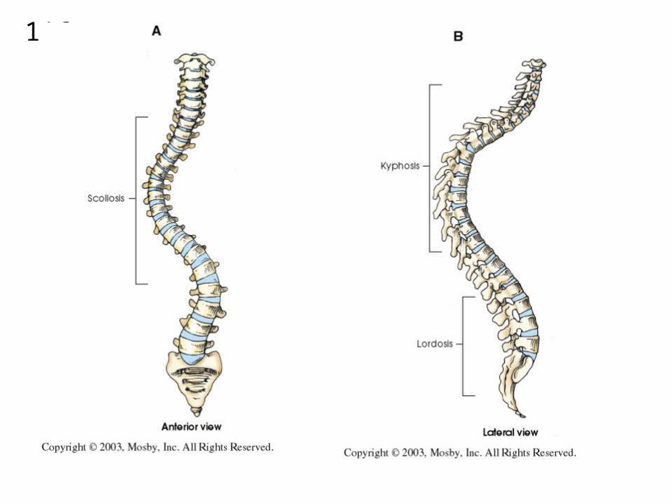
LUMBAR SPINESACRUM COCCYX
SI JOINTSSCOLIOSIS
RT 124 2008-10WEEK 7
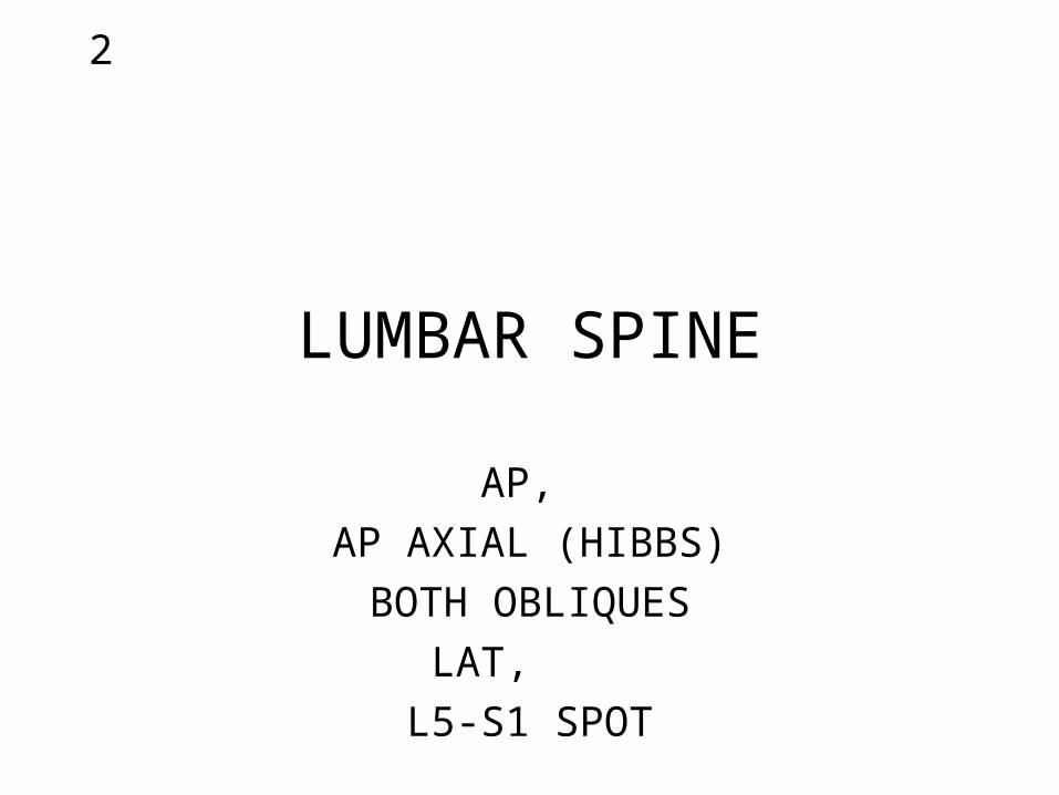
2
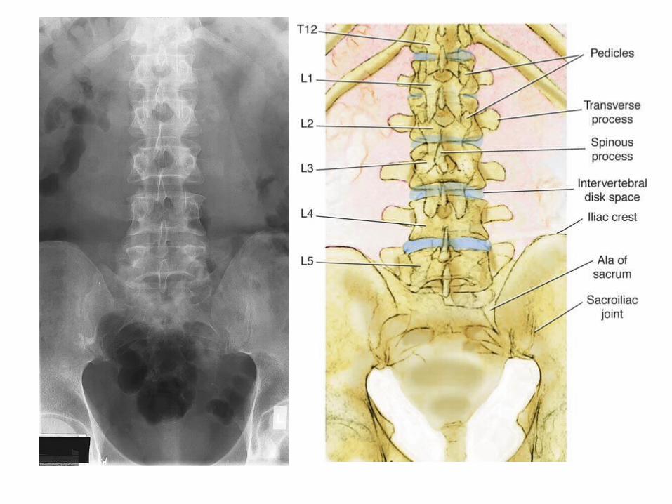
LUMBAR SPINE
AP,
AP AXIAL (HIBBS)
BOTH OBLIQUES
LAT,
L5-S1 SPOT
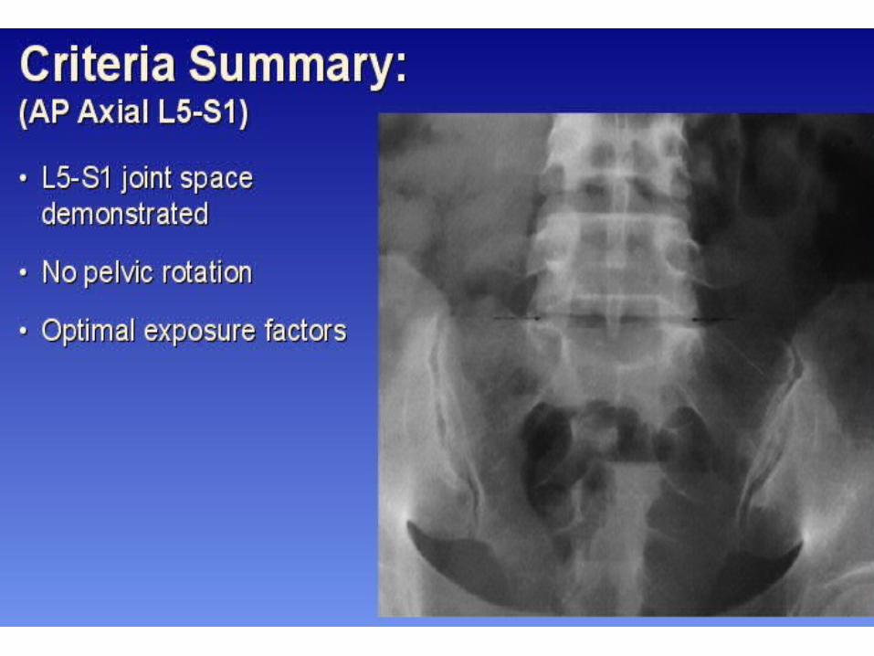
3
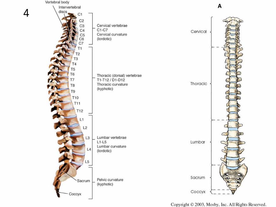
4

5

6
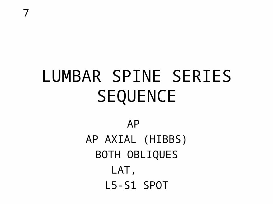
7
LUMBAR SPINE SERIESSEQUENCE
AP
AP AXIAL (HIBBS)
BOTH OBLIQUES
LAT,
L5-S1 SPOT
8
AP

9

10
11
12
13

14
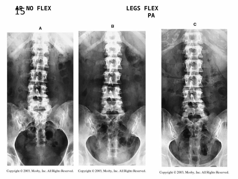
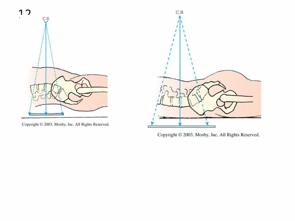
15 AP NO FLEX LEGS FLEX PA
16
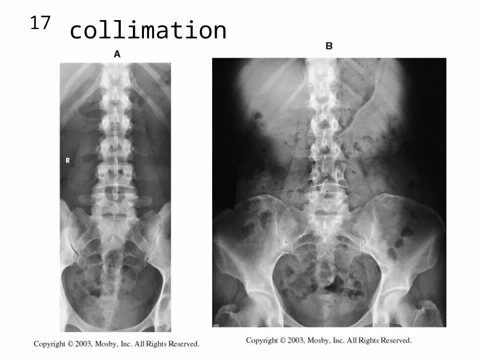
17 collimation
18
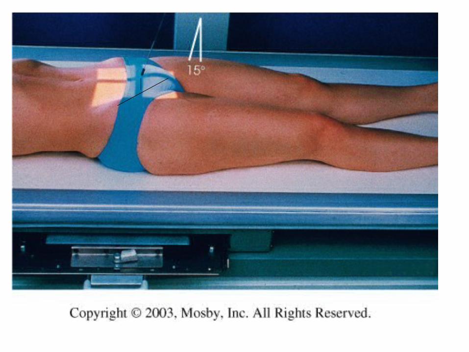
AP AXIAL HIBBS
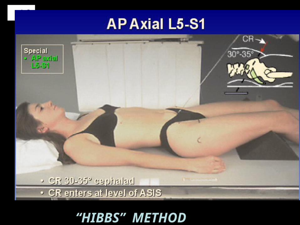
19
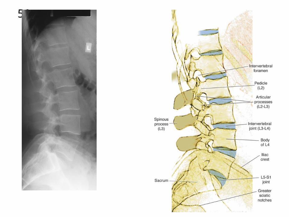
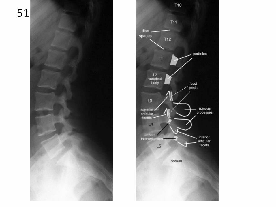
“HIBBS” METHOD
20

21

22
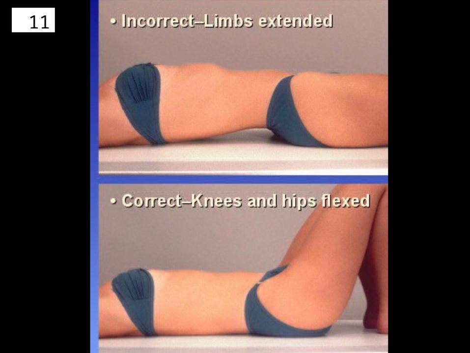
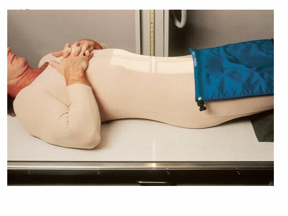
Flex legs
shield
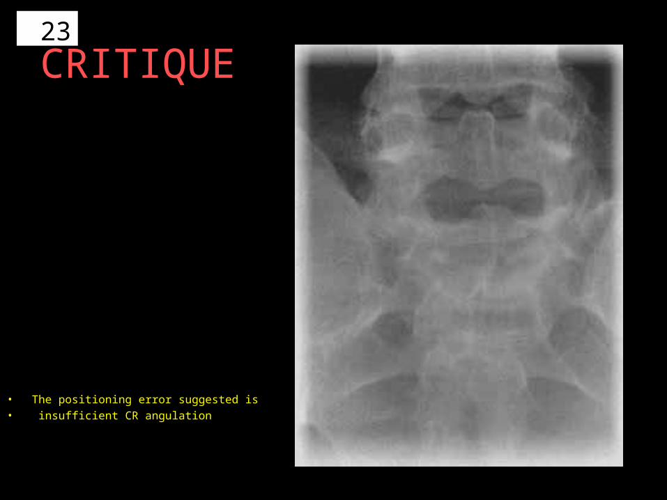
23
CRITIQUE
• The positioning error suggested is• insufficient CR angulation
24
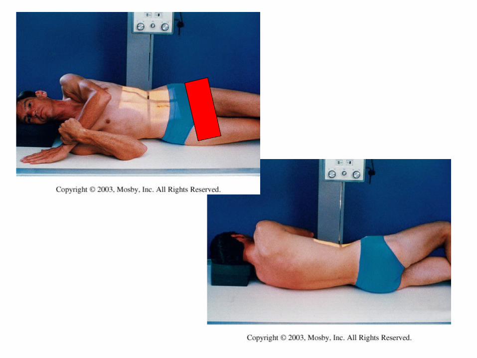
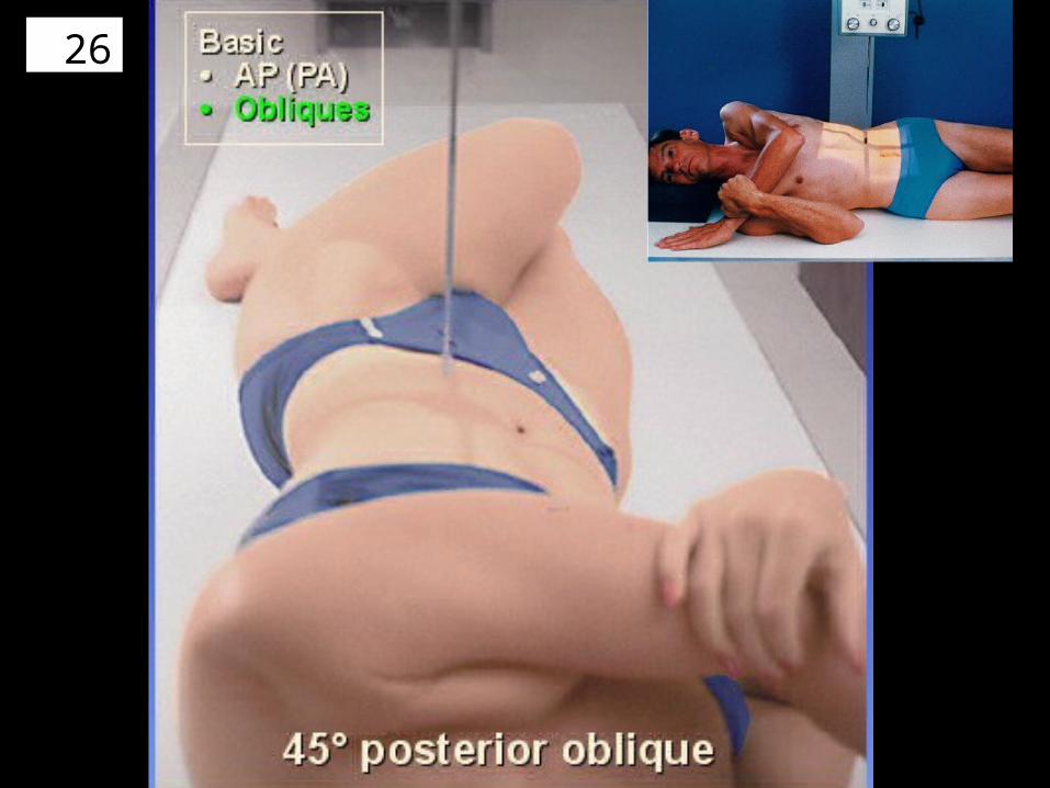
OBLIQUES
RPO & LPO
25
26
27

28

29
30
31

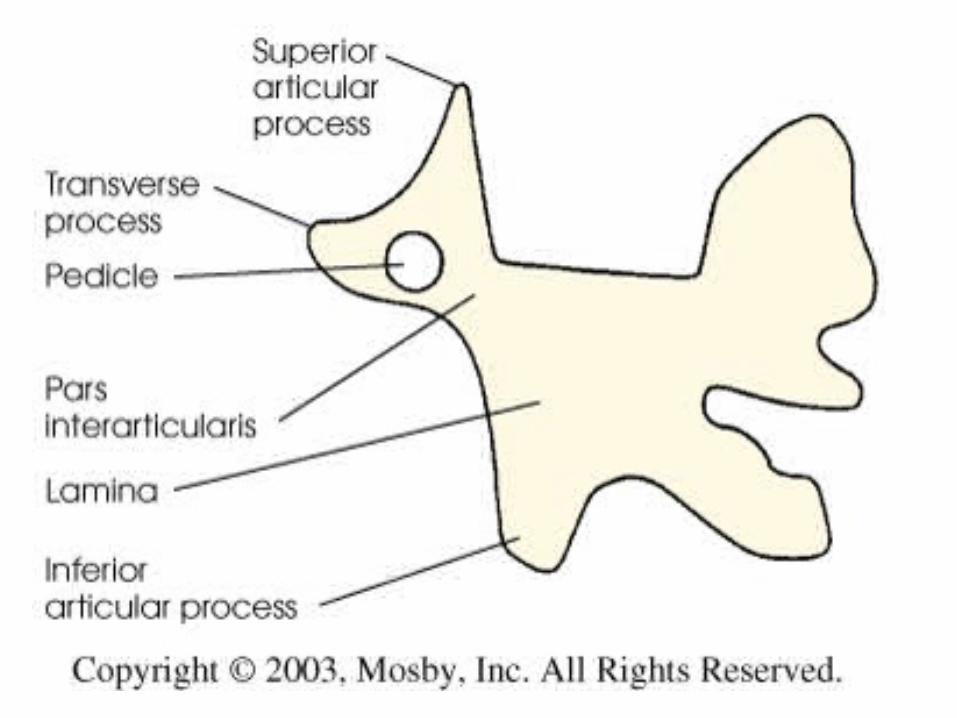
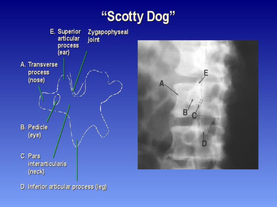
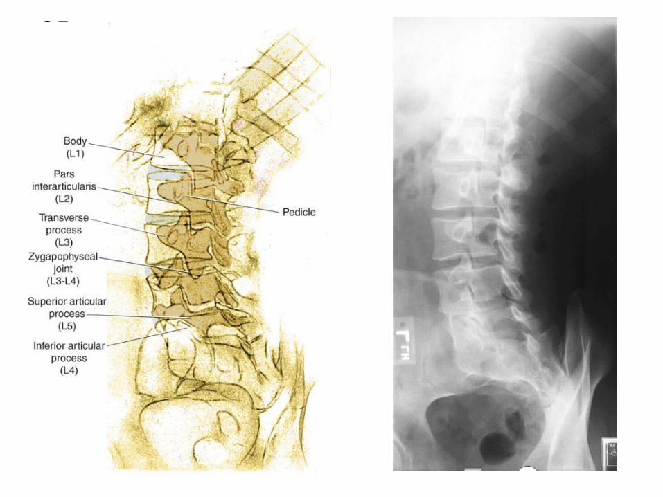
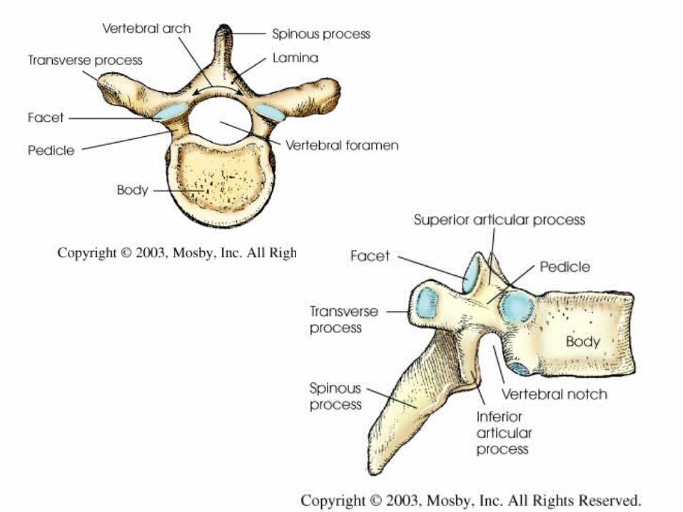
32• A body• E transverse process • D pedicle• O superior articular
facet, left• P pars
interarticularis, left• R inferior articular
facet, left• I apophyseal
(interfacetal) joint, left• V disk space
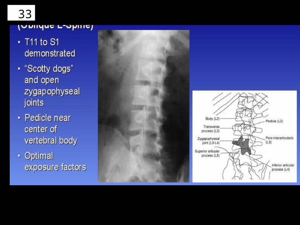
33

34
35
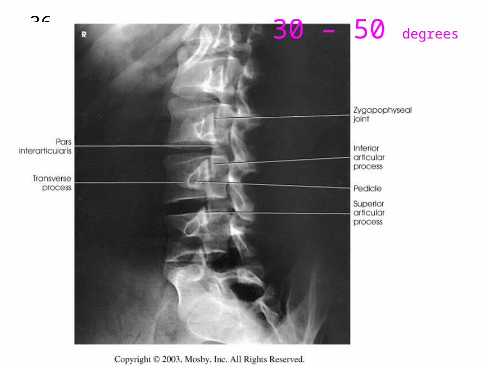
36 30 – 50 degrees
37
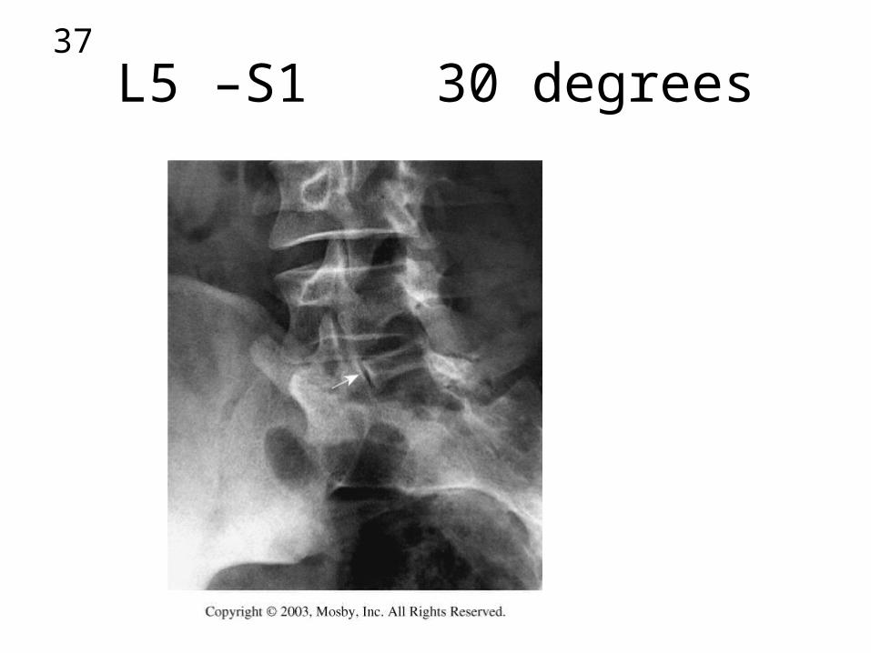
L5 –S1 30 degrees
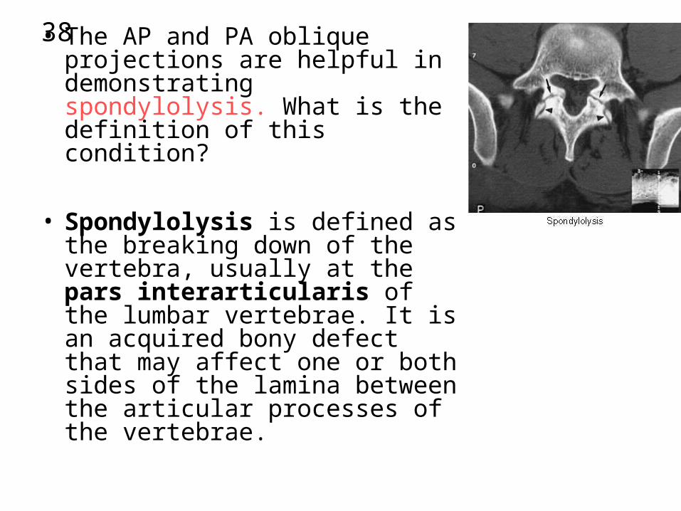
38• The AP and PA oblique projections are helpful in demonstrating spondylolysis. What is the definition of this condition?
• Spondylolysis is defined as the breaking down of the vertebra, usually at the pars interarticularis of the lumbar vertebrae. It is an acquired bony defect that may affect one or both sides of the lamina between the articular processes of the vertebrae.

39• Critique?• The positioning error
suggested is over-obliquity or excessive rotation of the patient.

40
• Zygapophyseal joints and pedicles are posterior to the vertebral body and indicate over-obliquity

41 • AP OBIQUE – CRITIQUE
• The positioning error suggested is insufficient obliquity or rotation of the patient
42
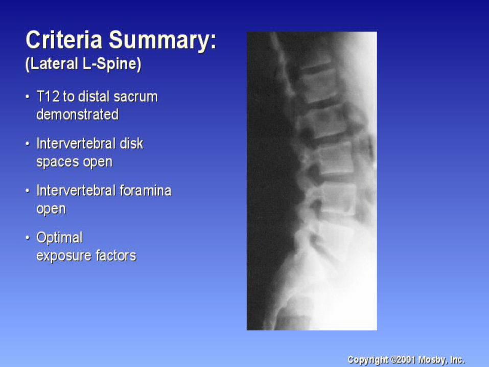
LUMBAR SPINE
LAT
43
44
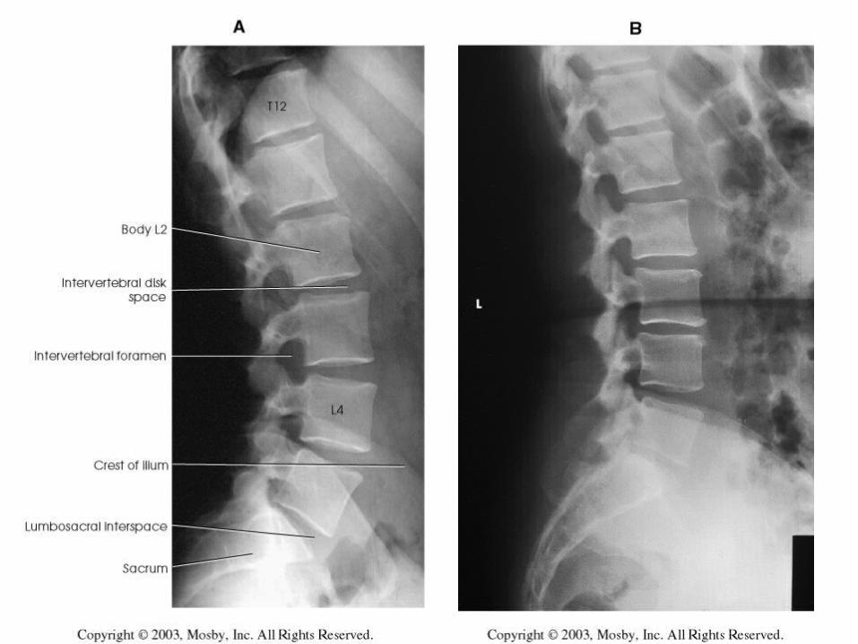
45
46

47
48
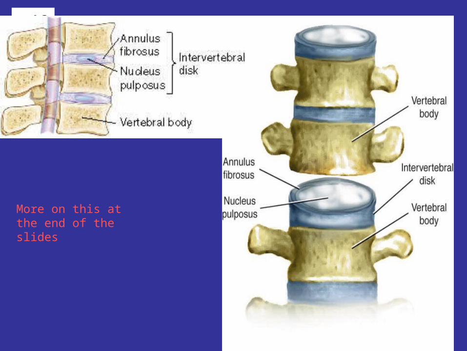
More on this at the end of the slides
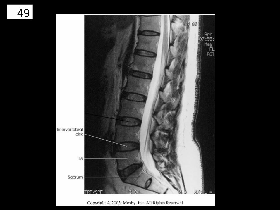
49
50
51
52
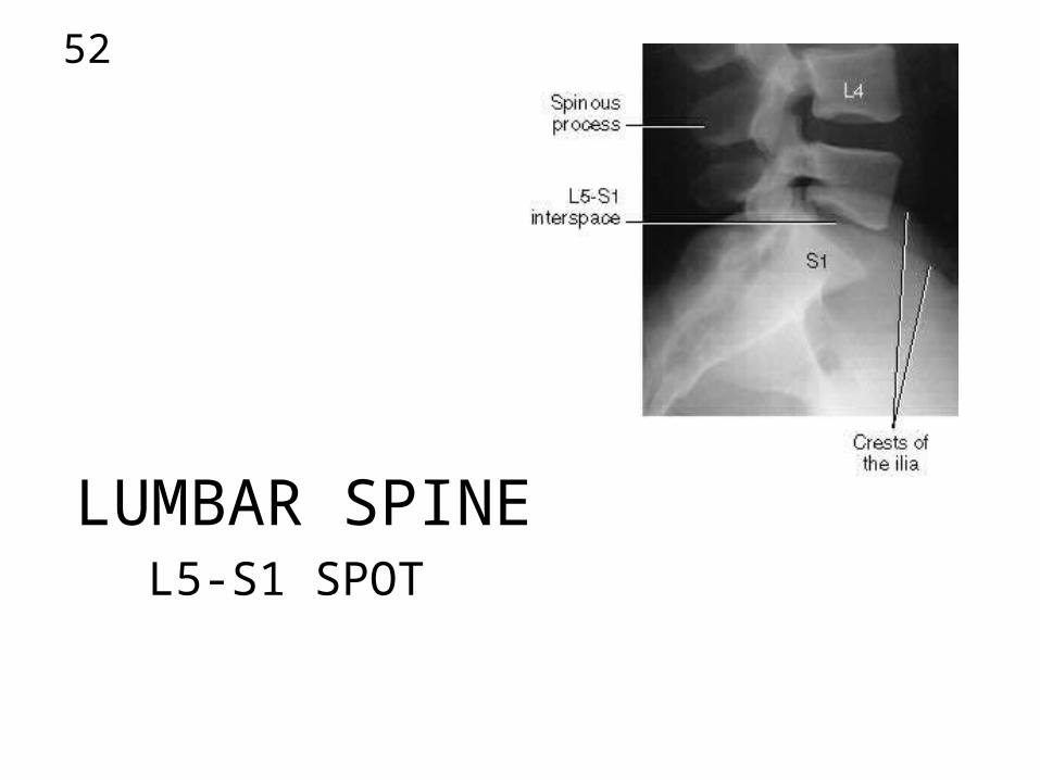
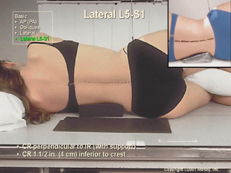
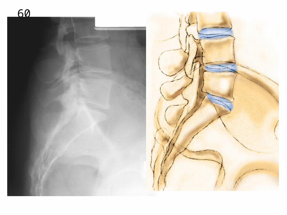
LUMBAR SPINEL5-S1 SPOT
53

54

55

56
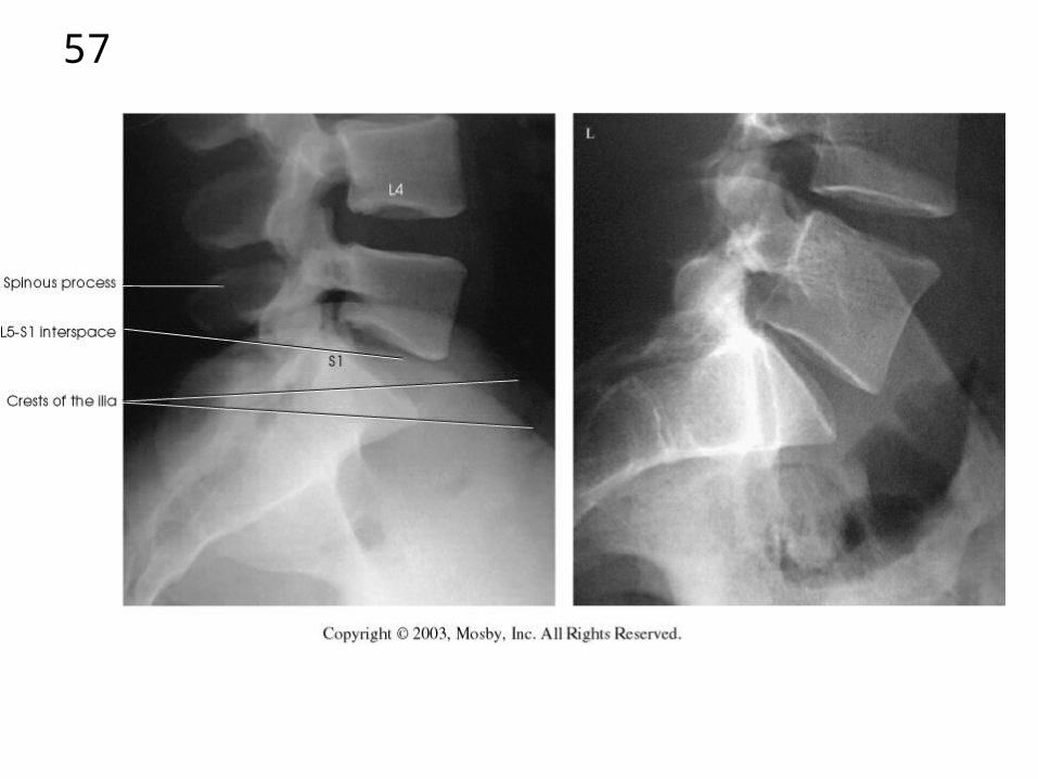
57
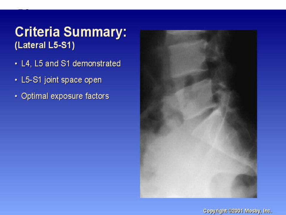
58
59
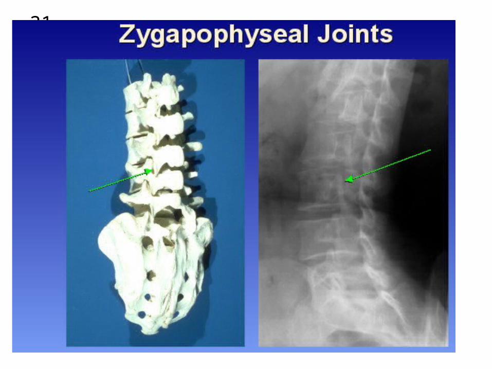
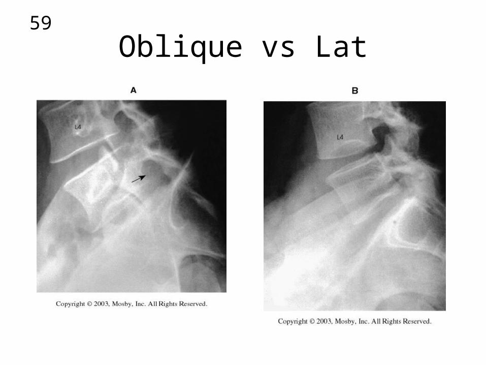
Oblique vs Lat
60
61
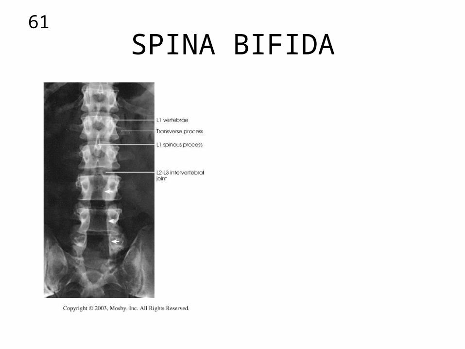
SPINA BIFIDA
62
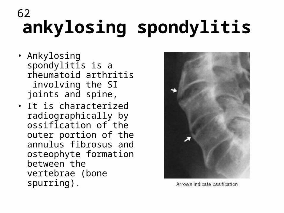
ankylosing spondylitis
• Ankylosing spondylitis is a rheumatoid arthritis involving the SI joints and spine,
• It is characterized radiographically by ossification of the outer portion of the annulus fibrosus and osteophyte formation between the vertebrae (bone spurring).
63
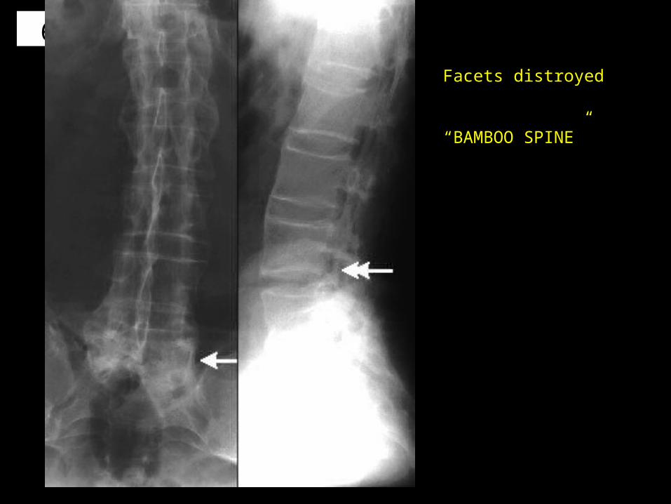
Facets distroyed
“BAMBOO SPINE”

64
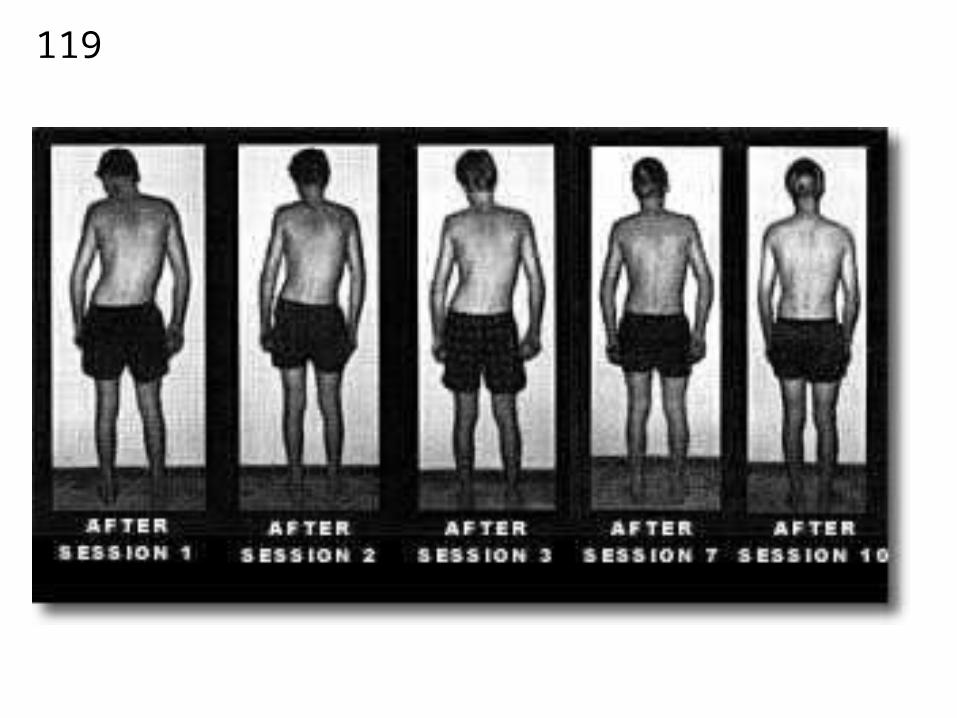
65
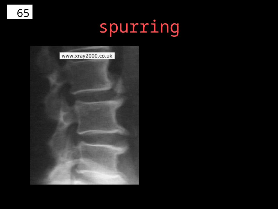
spurring
66
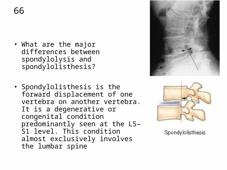
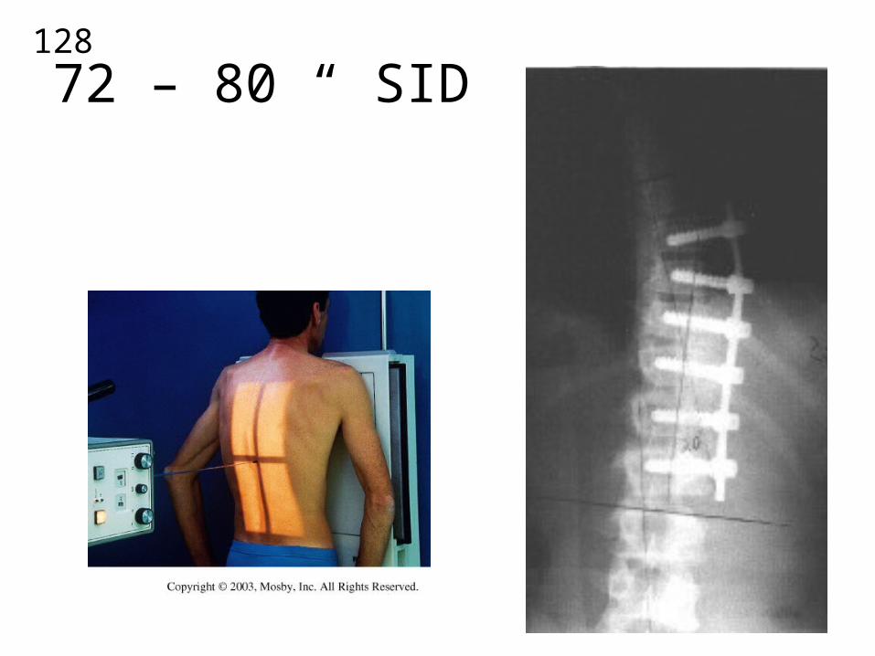
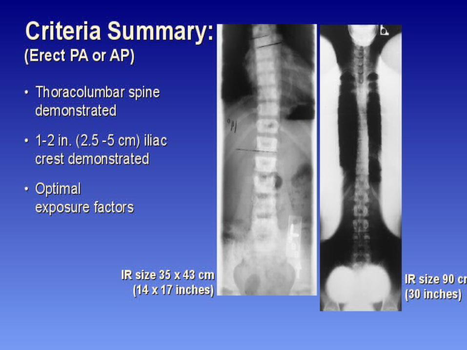
• What are the major differences between spondylolysis and spondylolisthesis?
• Spondylolisthesis is the forward displacement of one vertebra on another vertebra. It is a degenerative or congenital condition predominantly seen at the L5–S1 level. This condition almost exclusively involves the lumbar spine

67• The AP and PA oblique projections are helpful in demonstrating spondylolysis. What is the definition of this condition?
• Spondylolysis is defined as the breaking down of the vertebra, usually at the pars interarticularis of the lumbar vertebrae. It is an acquired bony defect that may affect one or both sides of the lamina between the articular processes of the vertebrae.
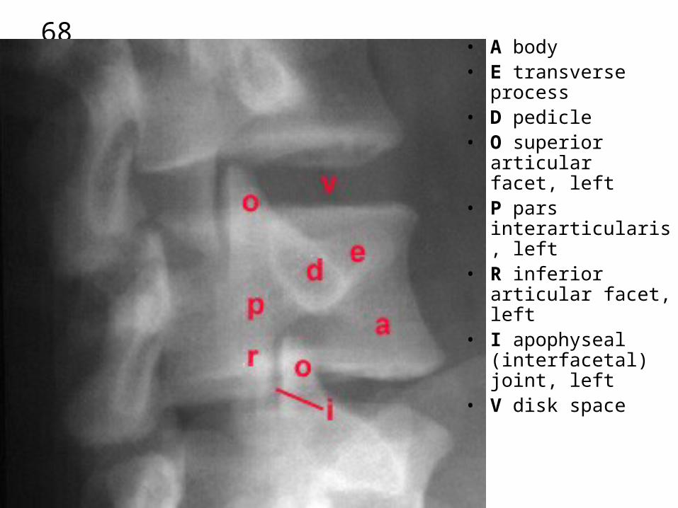
68• A body• E transverse process • D pedicle• O superior articular
facet, left• P pars
interarticularis, left• R inferior articular
facet, left• I apophyseal
(interfacetal) joint, left• V disk space
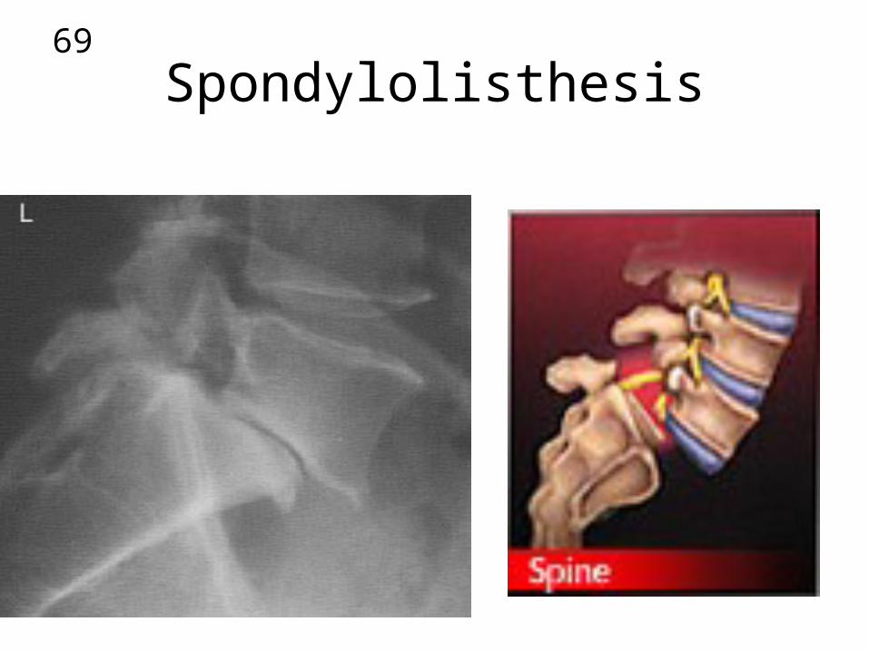
69
Spondylolisthesis
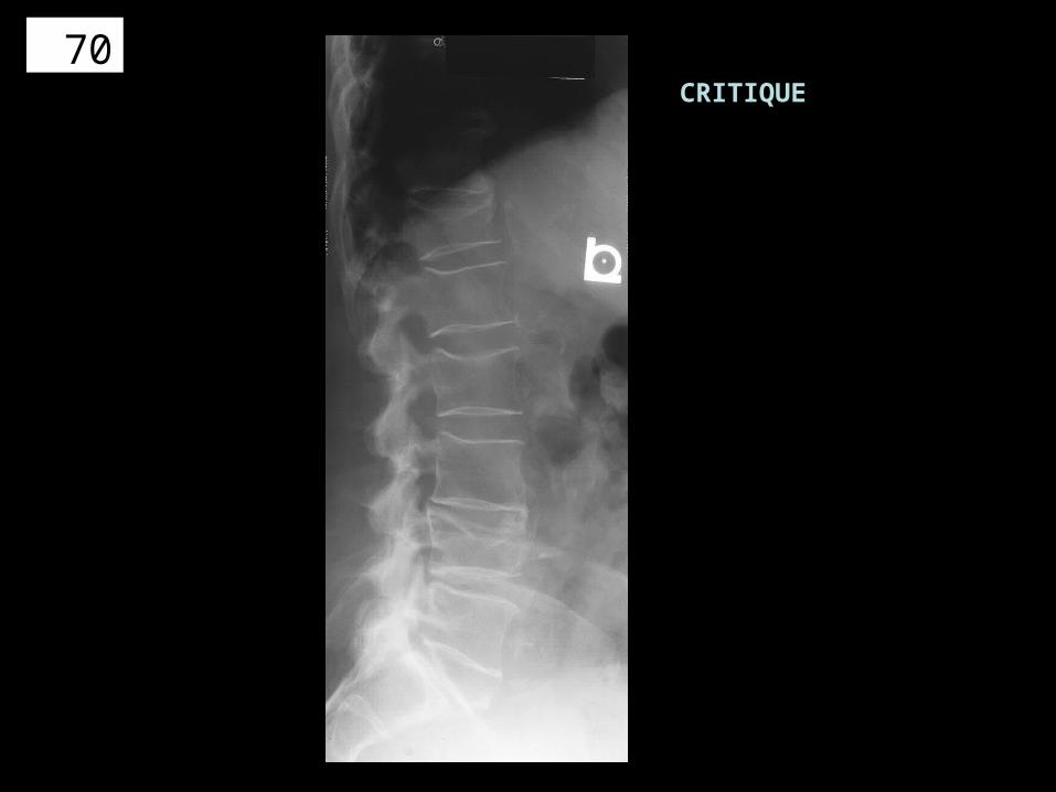
70CRITIQUE
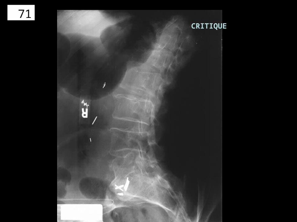
71CRITIQUE
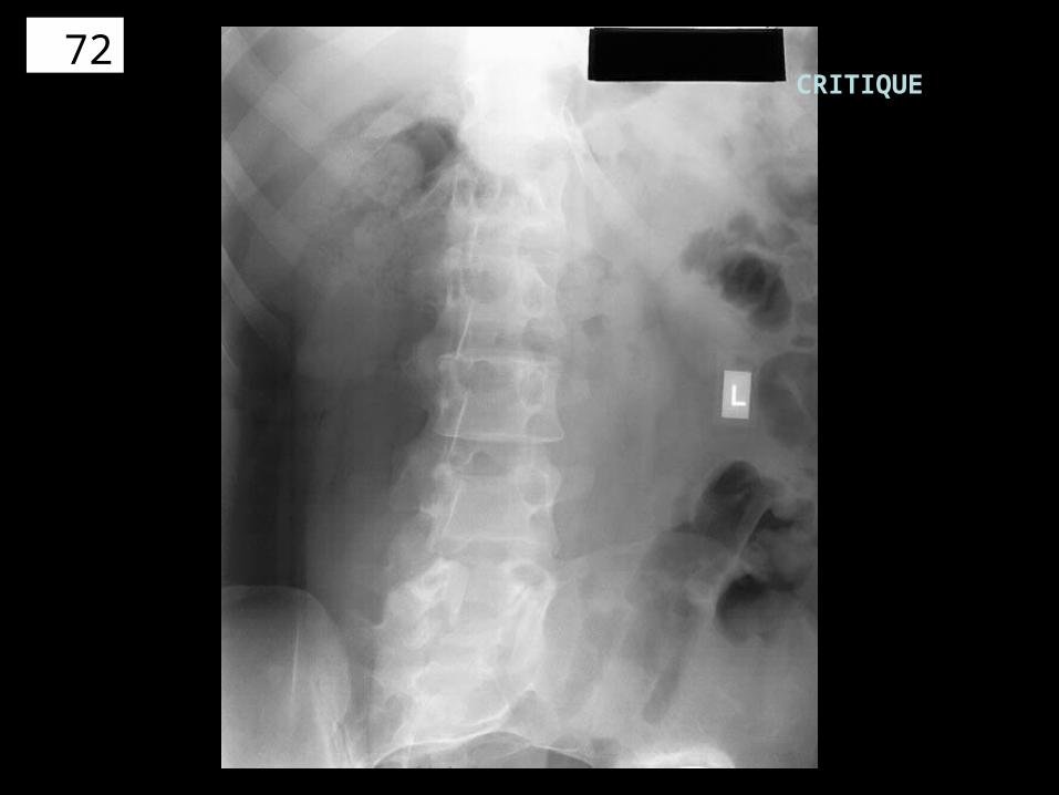
72CRITIQUE

73 CRITIQUE

74

75X-TABLE LATERAL
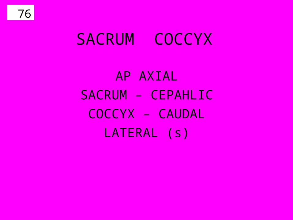
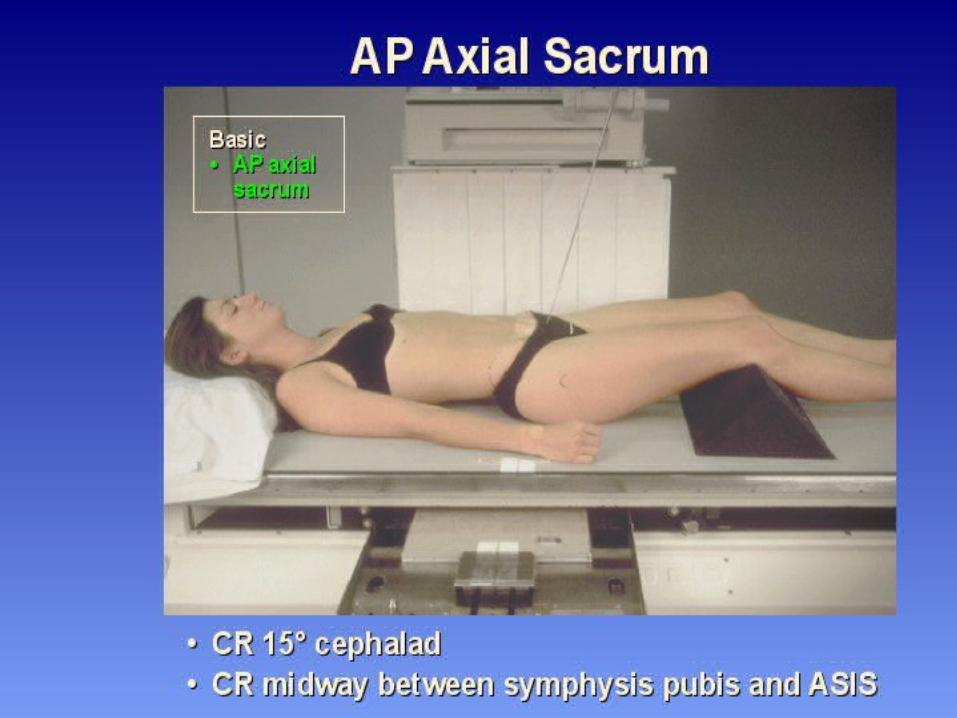
76
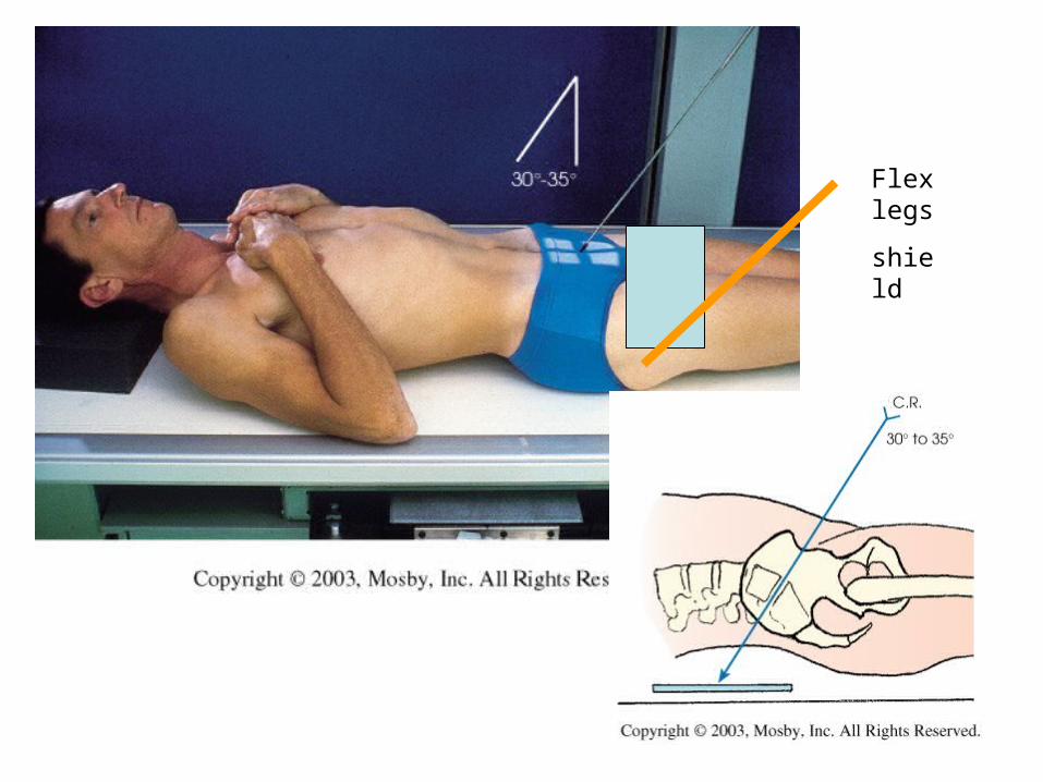
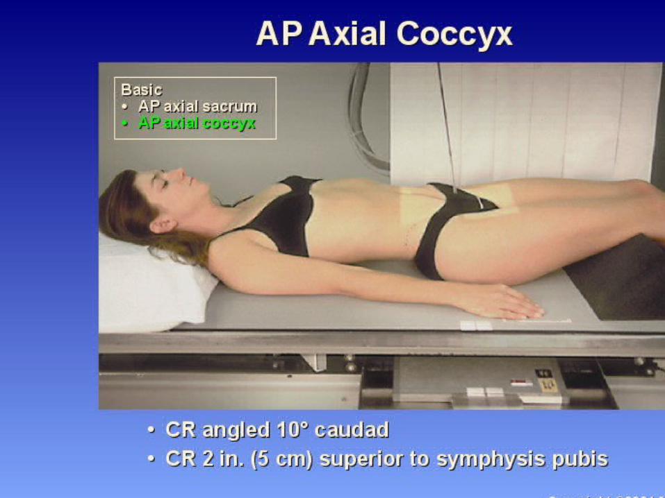
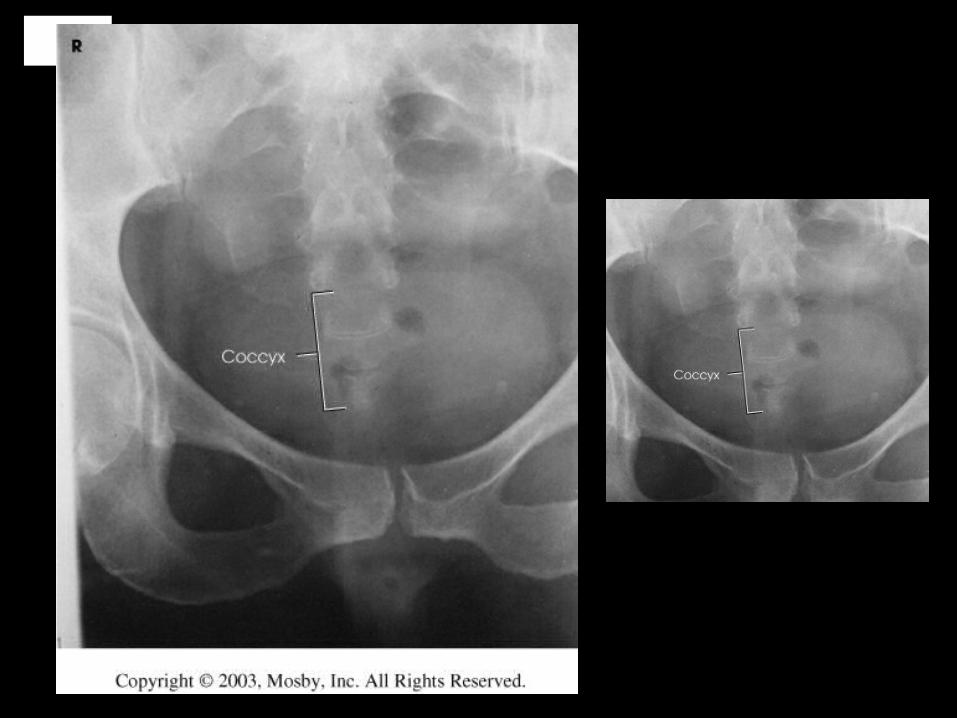
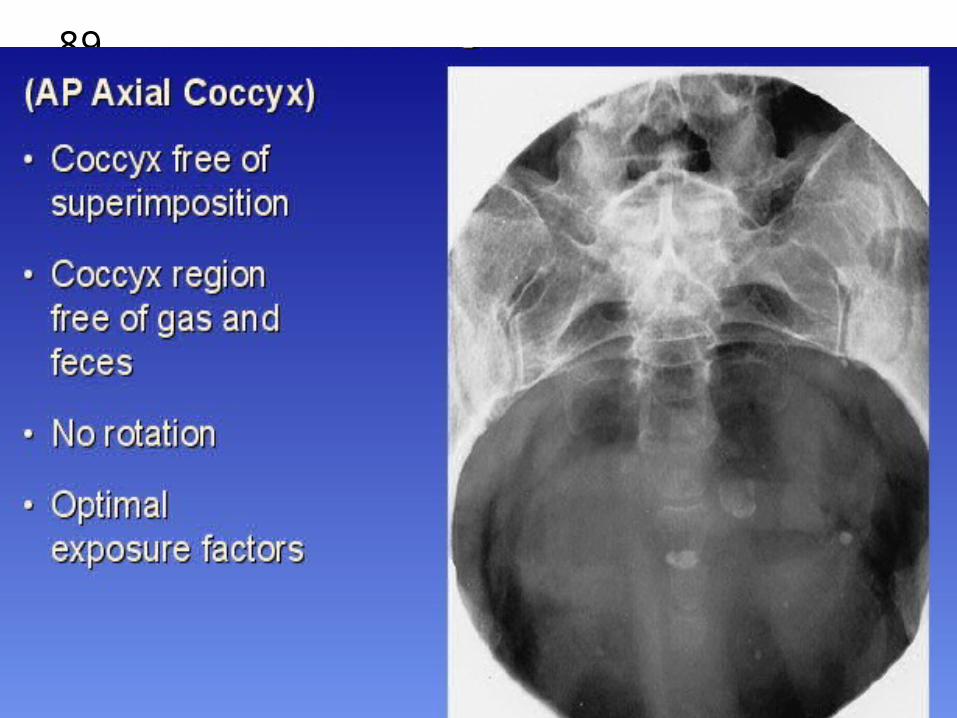
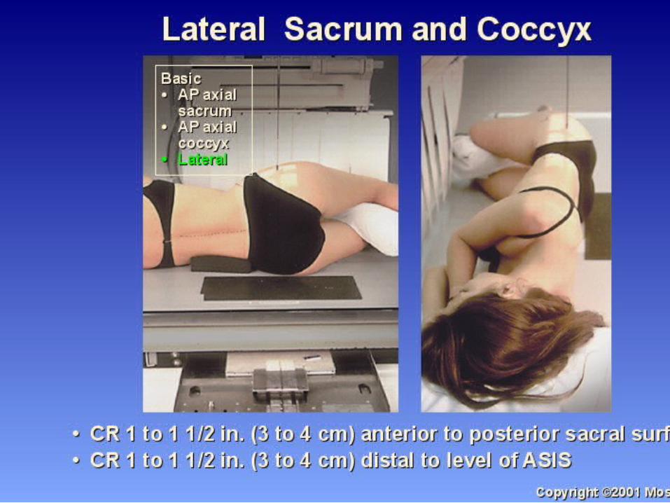
SACRUM COCCYX
AP AXIAL
SACRUM – CEPAHLIC
COCCYX – CAUDAL
LATERAL (s)

77
78
79
80
Flex legs
shield
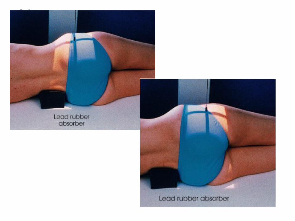
81
82

83
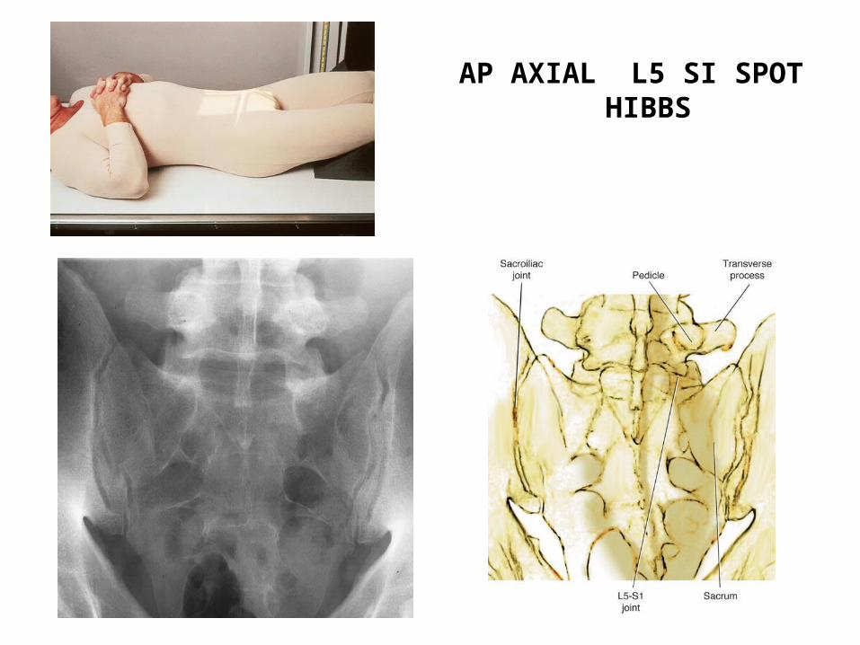
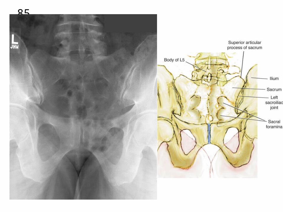
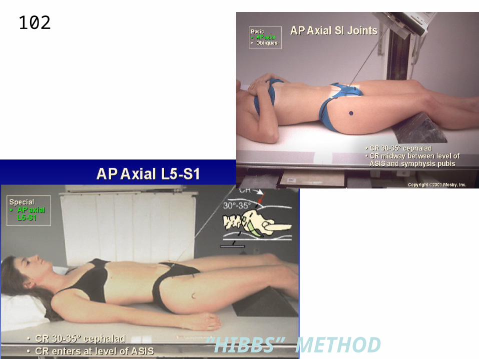
84AP AXIAL L5 SI SPOT
HIBBS
85
86

87
88
89
90

91
92

93
94
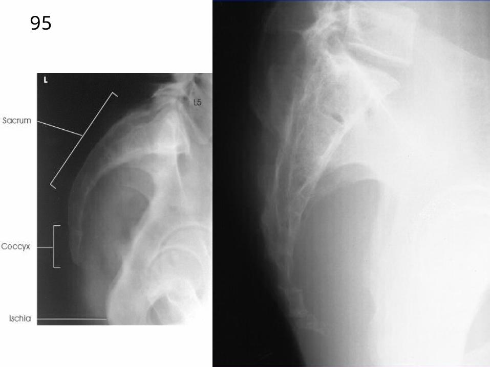
95
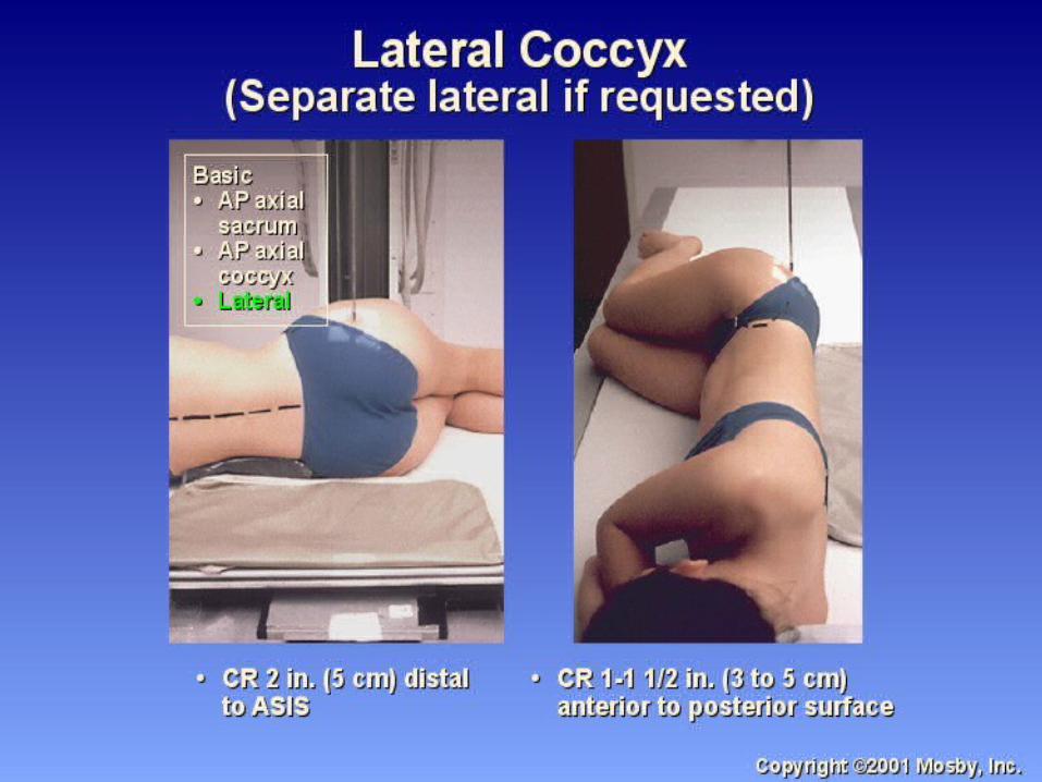
96
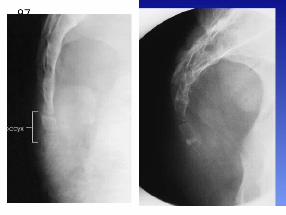
97
98
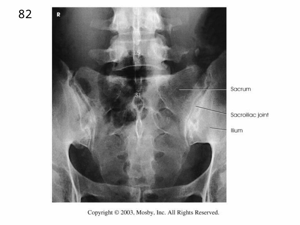
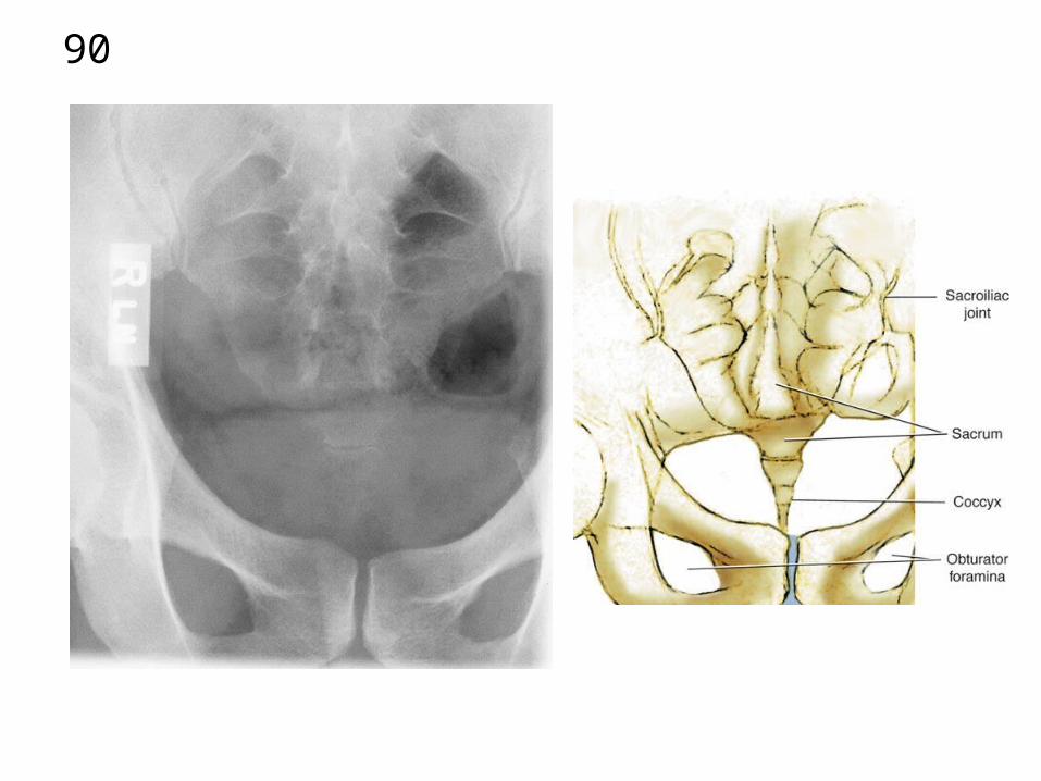
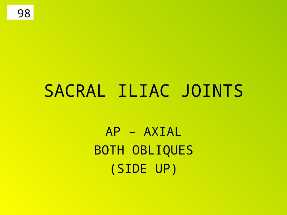
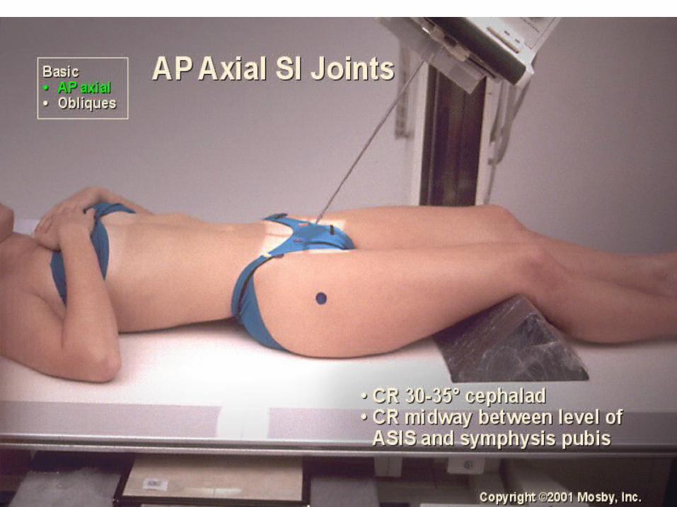
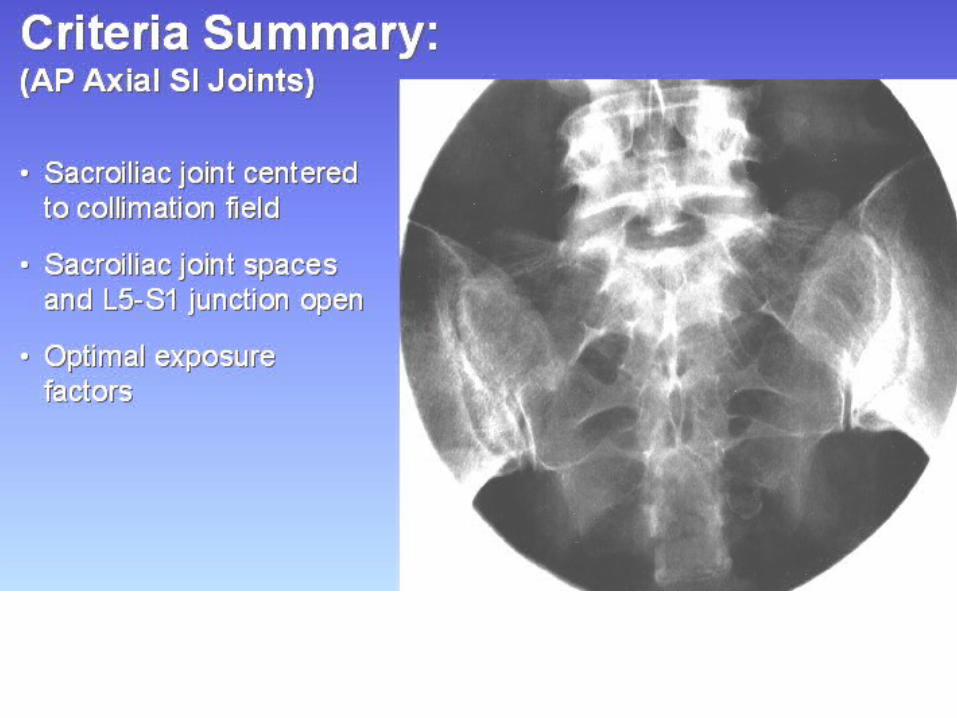
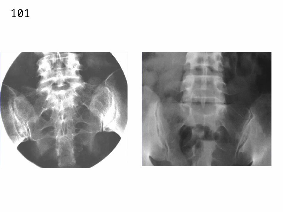
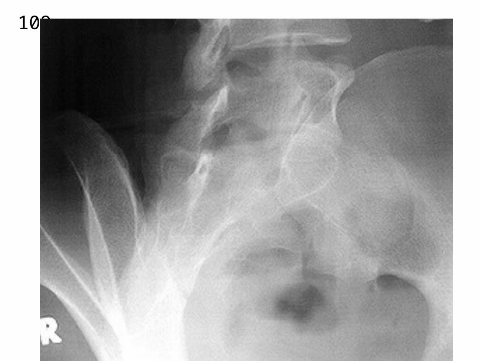
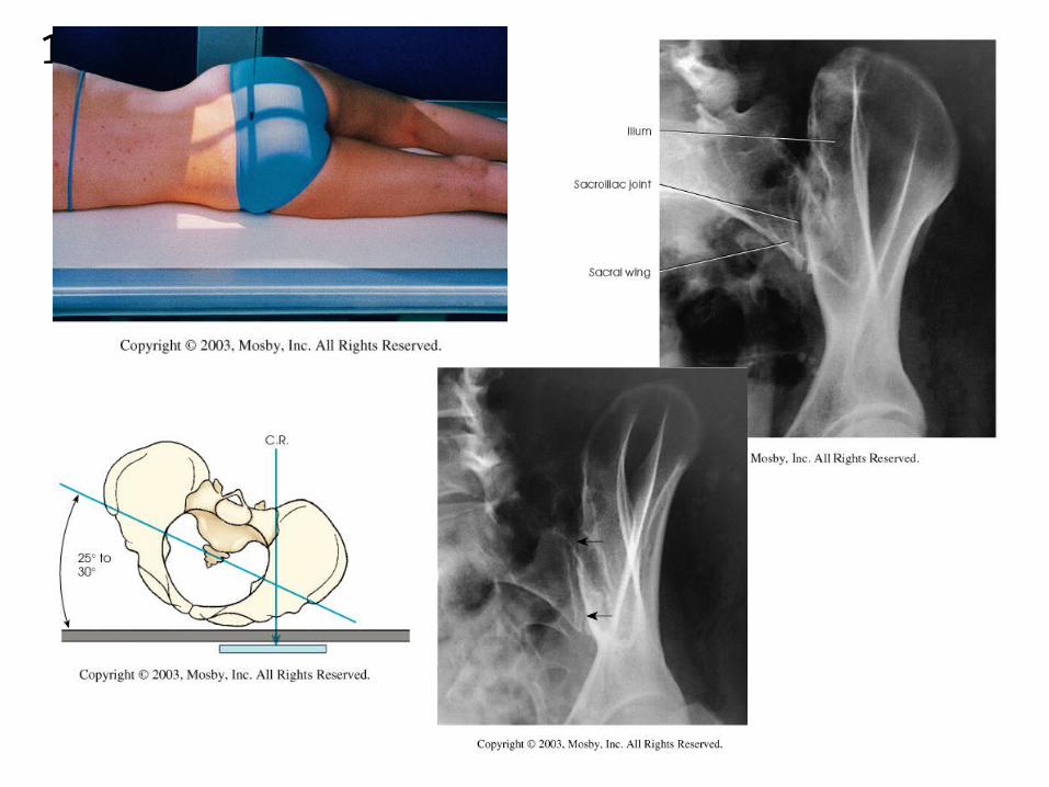
SACRAL ILIAC JOINTS
AP – AXIAL
BOTH OBLIQUES
(SIDE UP)
99
100
101
102
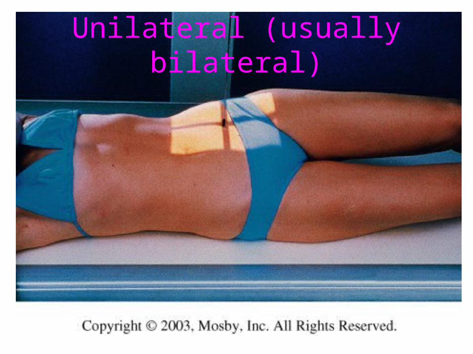
“HIBBS” METHOD
103
Unilateral (usually bilateral)

104

105
PA - With no rotation of patient

106

107

108
109

110
111

112
SIDE UP(SIDE UP)
SIDE DOWNSIDE UP
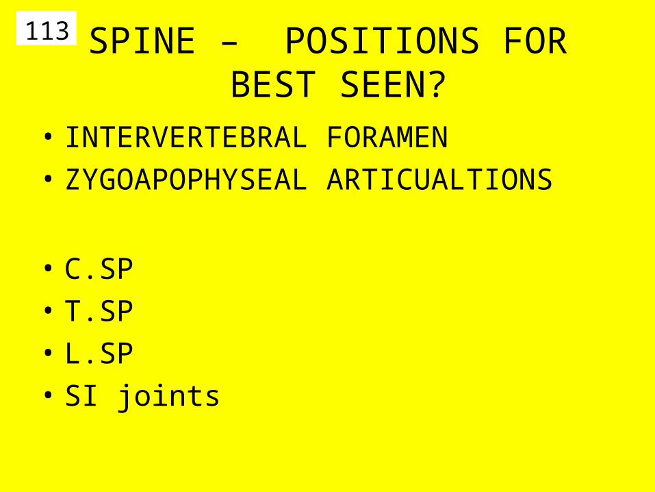
113 SPINE – POSITIONS FOR BEST SEEN?
• INTERVERTEBRAL FORAMEN
• ZYGOAPOPHYSEAL ARTICUALTIONS
• C.SP
• T.SP
• L.SP
• SI joints
114
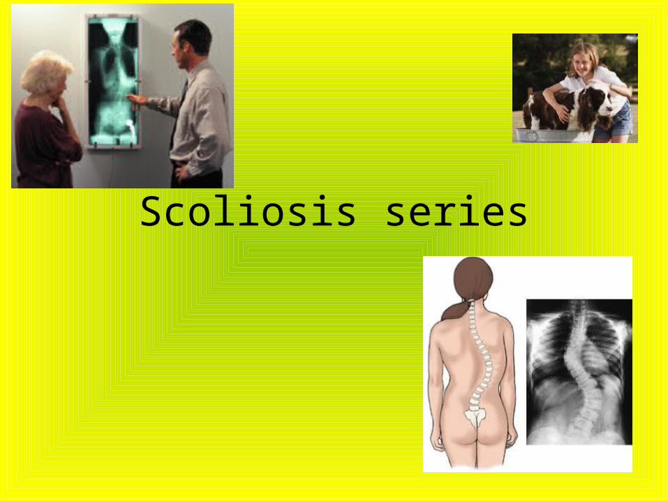
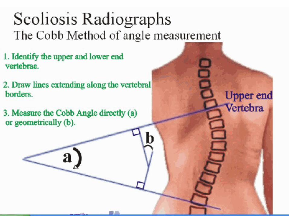
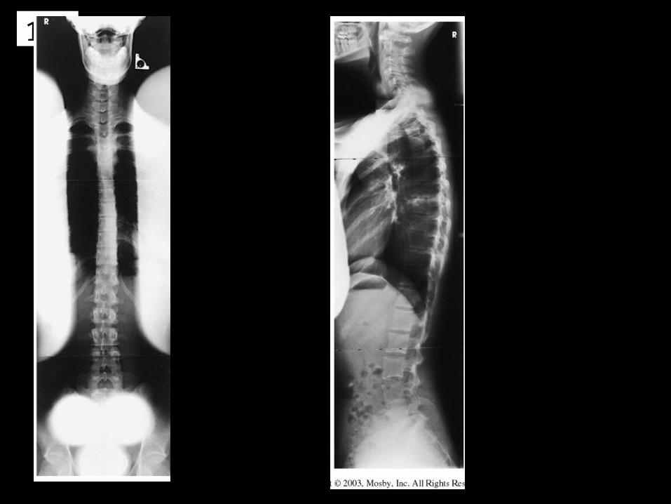
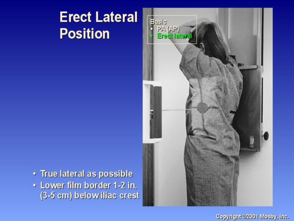
Scoliosis series

115
116
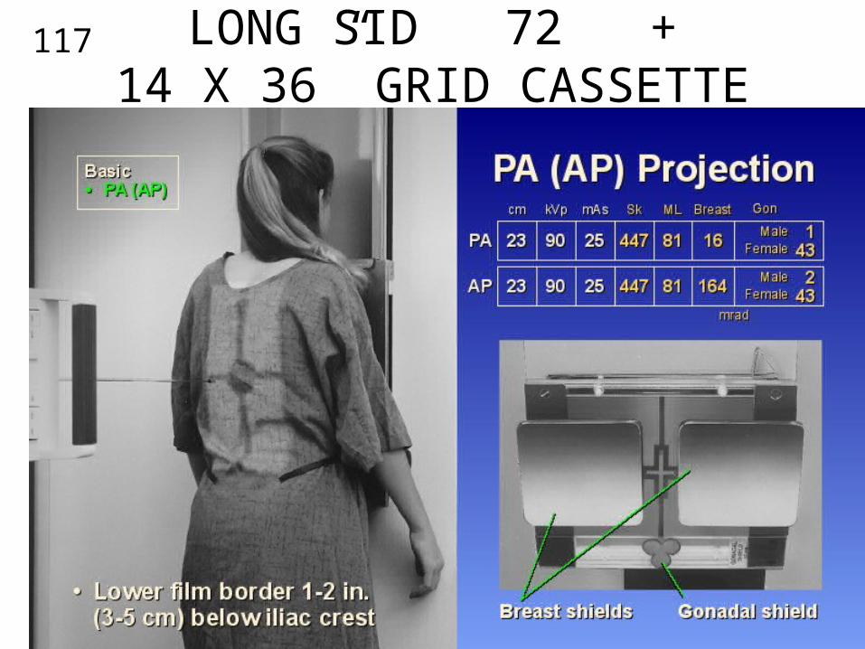
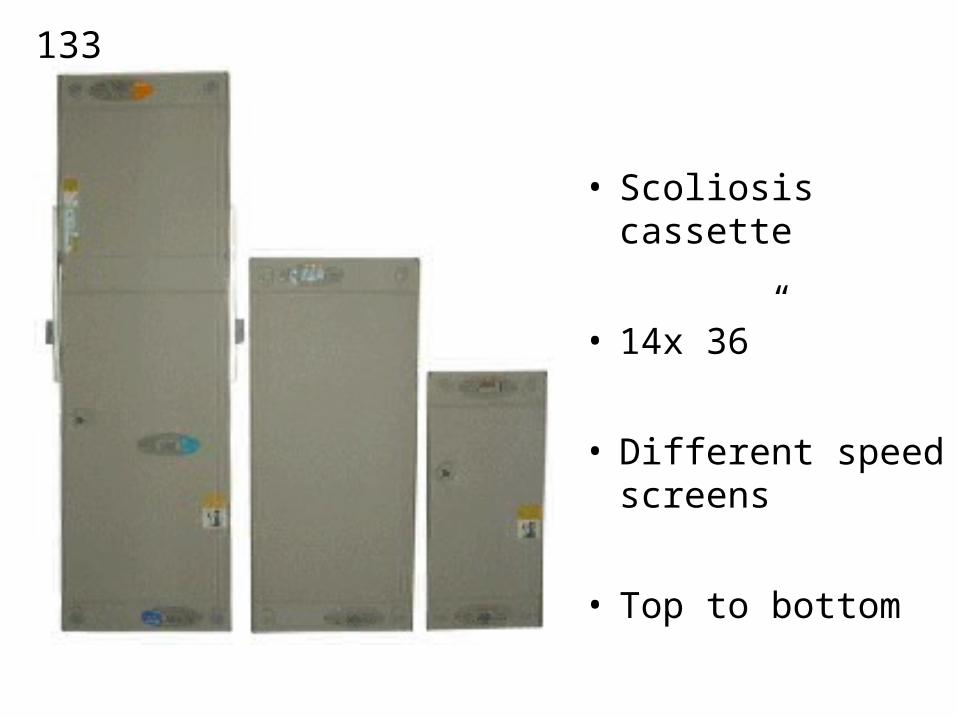
117 LONG SID 72” +14 X 36” GRID CASSETTE
118
119
120
121
122
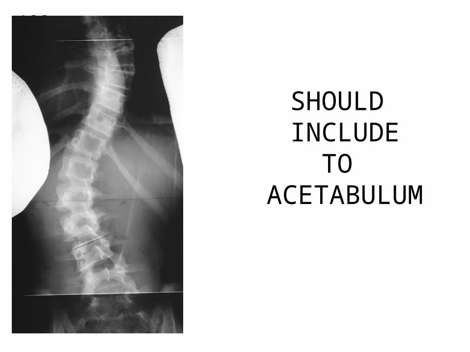
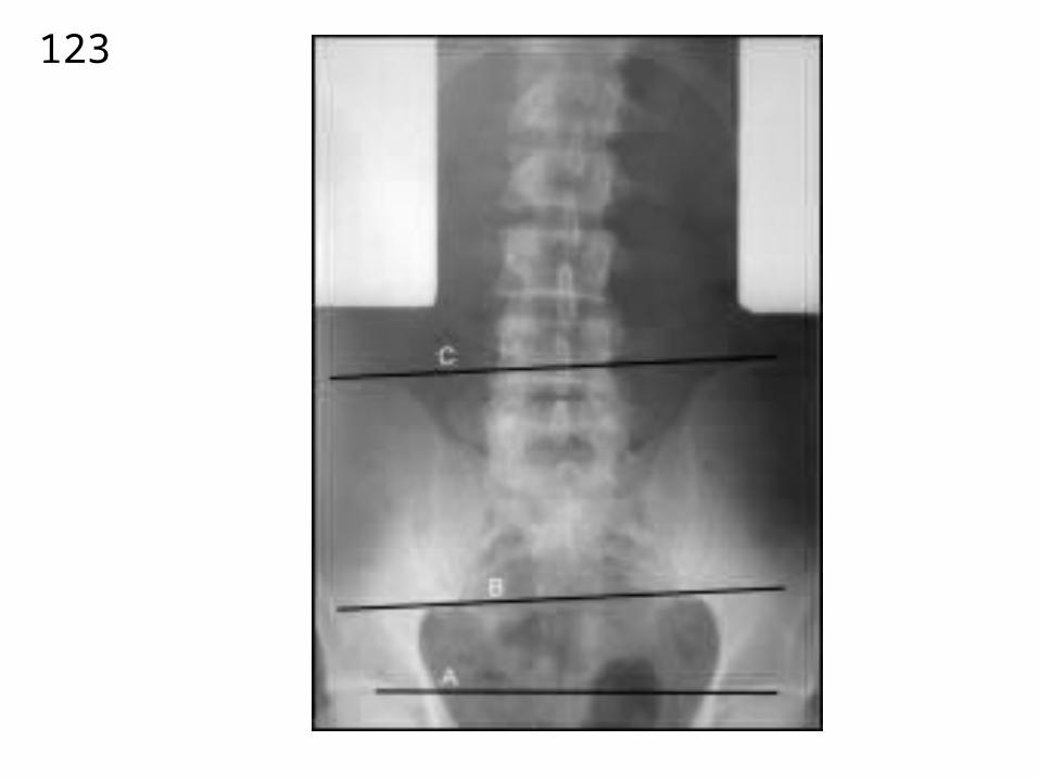
SHOULD INCLUDE
TO ACETABULUM
123
124

125
126
127
128
72 – 80 “ SID
129
130

131

132
133
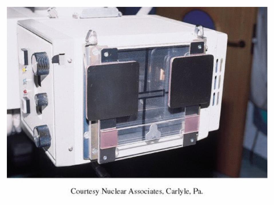
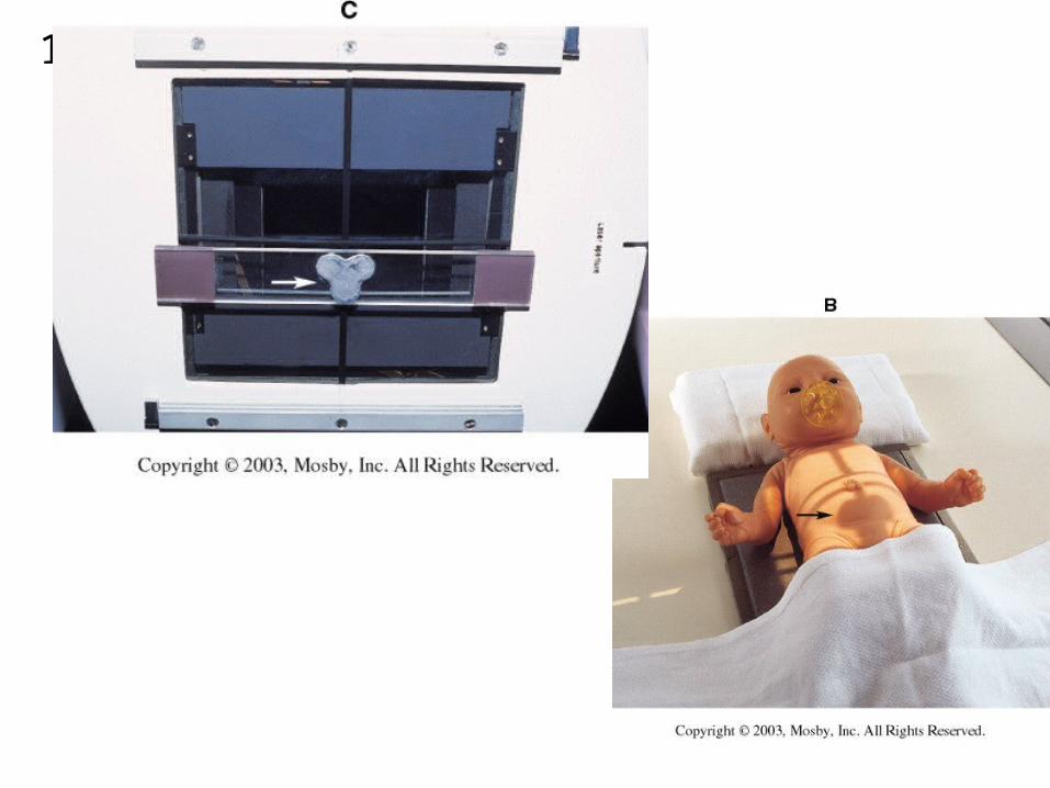
• Scoliosis cassette
• 14x 36”
• Different speed screens
• Top to bottom
134
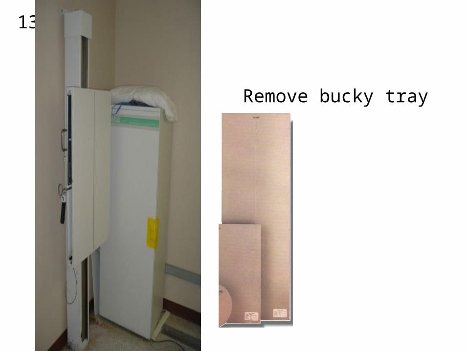
Remove bucky tray
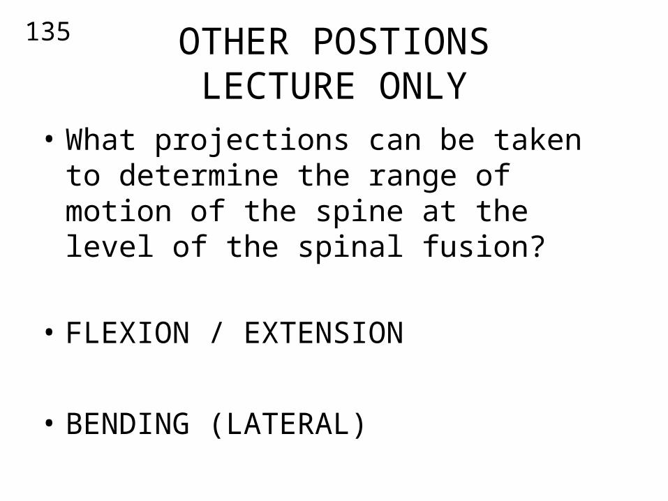
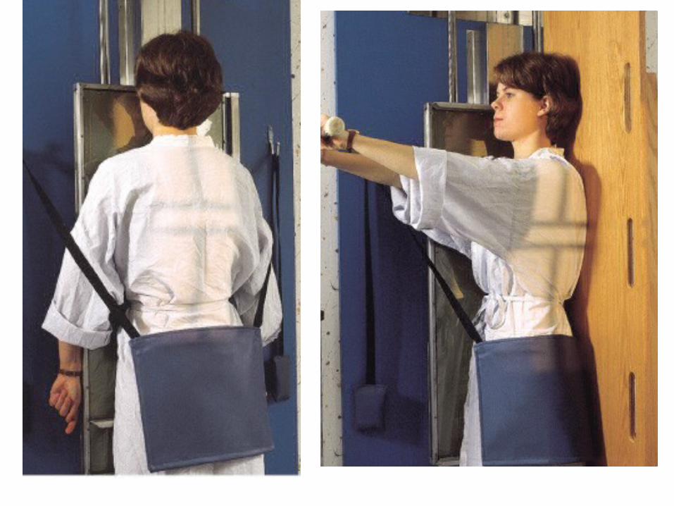
135 OTHER POSTIONSLECTURE ONLY
• What projections can be taken to determine the range of motion of the spine at the level of the spinal fusion?
• FLEXION / EXTENSION
• BENDING (LATERAL)

136
bending
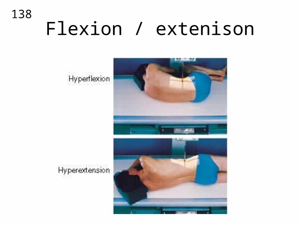
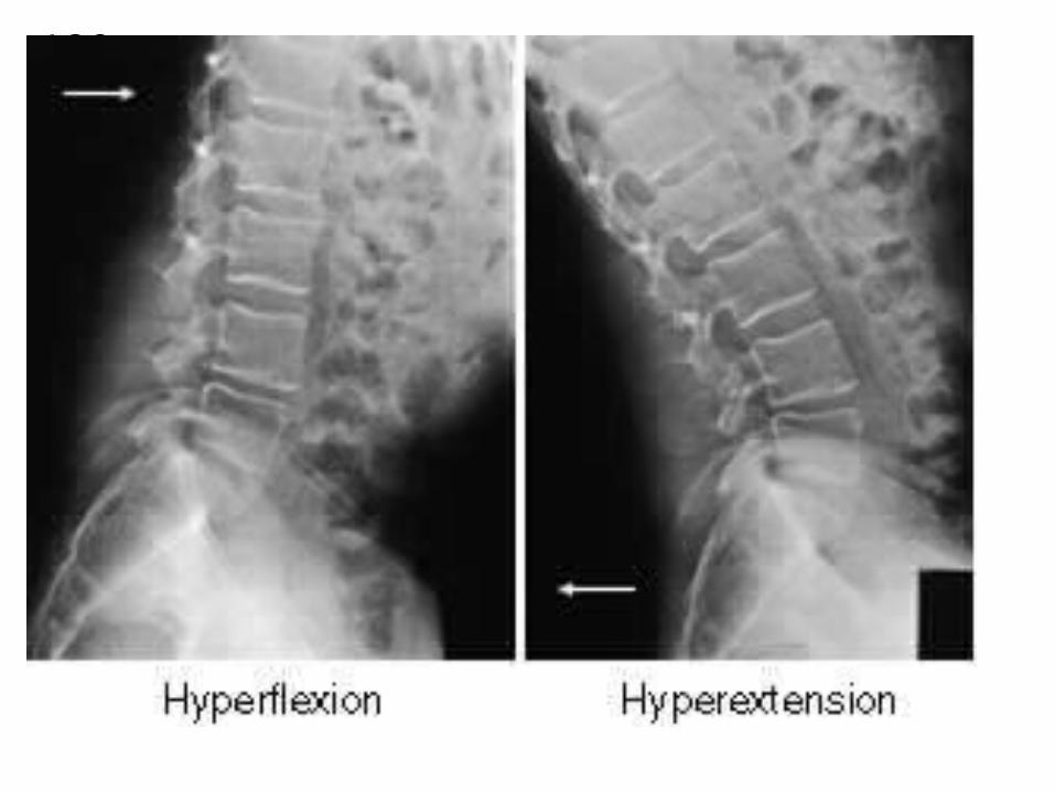
•These projections are functional studies of the lumbar spine to determine the range of motion of the spine at the point of spinal fusion.• Following spinal fusion surgery, the orthopedic physician or neurosurgeon may order a study to evaluate the flexibility of the spine.•These projections will require the patient to assume different positions to determine how much flexibility of the spine has returned.•Early signs of scoliosis and herniated intervertebral disk or HNP can be evaluated with these projections.

137
138
Flexion / extenison
139

140
What is HNP ?
• HNP refers to Herniated Nucleus Pulposus
• a situation in which the nucleus pulposus extrudes through an aspect of the annulus fibrosus of the intervertebral disk.
141
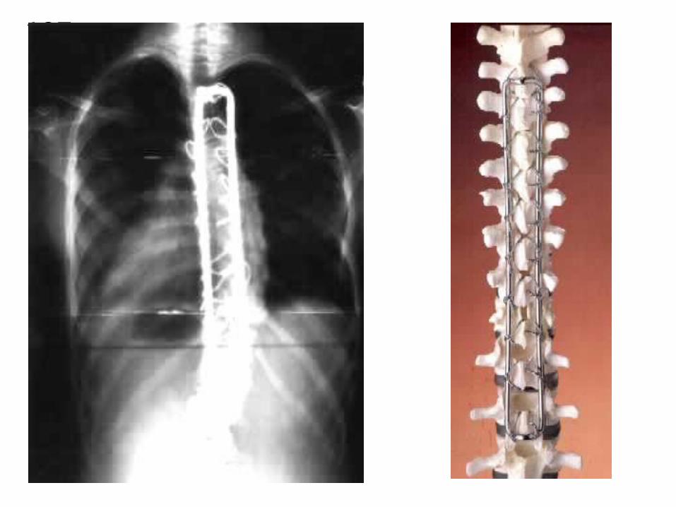
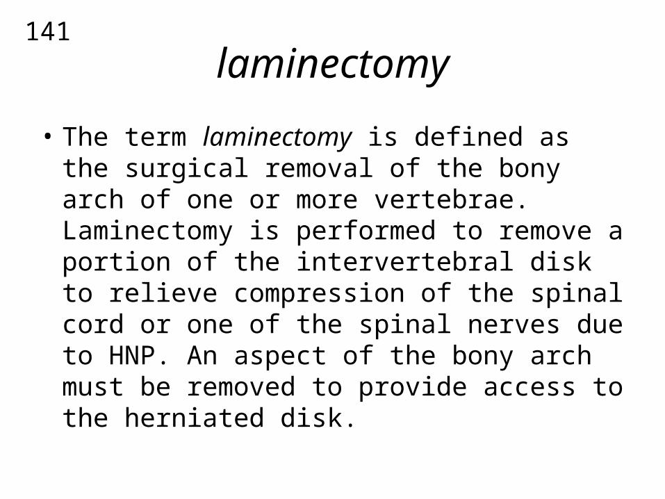
laminectomy
• The term laminectomy is defined as the surgical removal of the bony arch of one or more vertebrae. Laminectomy is performed to remove a portion of the intervertebral disk to relieve compression of the spinal cord or one of the spinal nerves due to HNP. An aspect of the bony arch must be removed to provide access to the herniated disk.
142

143
MORE IMAGE REVIEWNEXT WEEK
REVIEW ELSEIVER AS WELL!