Lues maligna in an HIV-infected patient - SciELO · paciente, com lesões cutâneas clássicas de...
4
181 Revista da Sociedade Brasileira de Medicina Tropical 38(2):181-184, mar-abr, 2005 RELATO DE CASO/CASE REPORT Lues maligna in an HIV-infected patient Lues maligna em um paciente com infecção pelo HIV Luiz Fernando Cabral Passoni 1 , Jacqueline Anita de Menezes 1 , Sayonara Rocha Ribeiro 1 and Érica Coutinho O. Sampaio 1 ABSTRACT We report such a case of malignant syphilis in a 42-year-old HIV-infected man, co-infected with hepatitis B virus, who presented neurolues and the classical skin lesions of lues maligna. The serum VDRL titer, which was 1:64 at presentation, increased to 1:2,048 three months after successful therapy with penicillin, decreasing 15 months later to 1:8. Key-words: Malignant syphilis. Lues maligna. Neurolues. HIV infection. RESUMO Descrevemos um caso de sífilis maligna em um paciente de 42 anos com infecção pelo HIV e pelo vírus da hepatite B. O paciente, com lesões cutâneas clássicas de lues maligna e VDRL positivo no soro e no líquor, teve uma resposta excelente ao tratamento com penicilina cristalina. O VDRL sérico, que no diagnóstico era de 1:64, aumentou três meses depois para 1:2.048 e diminuiu para 1:8 após 15 meses. Palavras-chaves: Sífilis maligna. Lues maligna. Neurolues. Infecção HIV. 1. Serviço de Doenças Infecciosas e Parasitárias do Hospital dos Servidores do Estado, Rio de Janeiro, RJ, Brasil. Address to: Dr. Luiz Fernando Cabral Passoni. R. Mal Mascarenhas de Morais 191/1007, Copacabana, 22030-040 Rio de Janeiro, RJ, Brasil. Tel: 55 21 2518-1594 e-mail: [email protected] Recebido para publicação em 21/5/2003 Aceito em 19/11/2004 Lues maligna is a rare ulcerative form of secondary syphilis characterized by papulopustular skin lesions that rapidly enlarge and evolve into round or oval ulcers with sharp borders, centrally covered by a dark, sometimes rupioid crust 6 . Lesions in various stages of development confer a pleomorphic picture. Mucous membranes of the mouth and nose may be involved, and prodromes of fever, headache, and myalgia are common 6 14 . Although its incidence had been decreasing since the beginning of the 20 th Century, the number of reported cases has increased after 1988, most occurring in patients with HIV infection. We describe a case of malignant syphilis in an HIV-infected patient, the third diagnosed at the Infectious Diseases Department of the Hospital dos Servidores do Estado, Rio de Janeiro, Brazil, from 1986-2002. Previously, in April 1989 and January 1993, respectively, a 43-year-old bisexual man and a 44-year-old homosexual man presented with lues maligna as their first manifestation of HIV-infection 12 . CASE REPORT In June 1999, a 42-year-old homosexual male presented with a two-week history of multiple erythematous papules on his face, trunk and extremities, which progressed to pustules and crusted ulcers. All lesions were painless and he denied having any systemic symptoms. He was advised that he was HIV-positive in June 1995, when he was seen for evaluation of spleen enlargement and anemia. At that time, laboratory investigations revealed a positive serology for both HIV (ELISA and Western-Blot) and HTLV I/II infection. Serology for hepatitis B showed the presence of HBsAg and HBcAb, whereas serology for hepatitis C was negative. A liver biopsy was refused and he was lost to follow-up until June 1996, when he sought medical assistance presenting clinical features of liver failure and hypersplenism. Six months later his CD4 + cell count was 242/mm 3 (20%), but he refused to use antiretroviral drugs, which were begun only in May 1998. At that time, his CD4 + cell count was 404/mm 3 (34%) and his HIV viral load 5,800 copies/ml (nucleic acid sequence based amplification, NASBA). Six weeks after starting stavudine and lamivudine, CD4 + count increased to 494/mm 3 (59%) and viral load dropped to undetectable levels (<400 copies/ml). He had been taking the medicines regularly until two months before the appearance of the skin lesions, when he discontinued the antivirals. On examination the patient appeared underweight. He was pale and afebrile. Skin lesions consisted of multiple erythematous
-
Upload
nguyenhanh -
Category
Documents
-
view
215 -
download
0
Transcript of Lues maligna in an HIV-infected patient - SciELO · paciente, com lesões cutâneas clássicas de...
181
Revista da Sociedade Brasileira de Medicina Tropical 38(2):181-184, mar-abr, 2005 RELATO DE CASO/CASE REPORT
Lues maligna in an HIV-infected patient
Lues maligna em um paciente com infecção pelo HIV
Luiz Fernando Cabral Passoni1, Jacqueline Anita de Menezes1, Sayonara Rocha Ribeiro1
and Érica Coutinho O. Sampaio1
ABSTRACT
We report such a case of malignant syphilis in a 42-year-old HIV-infected man, co-infected with hepatitis B virus, whopresented neurolues and the classical skin lesions of lues maligna. The serum VDRL titer, which was 1:64 at presentation,increased to 1:2,048 three months after successful therapy with penicillin, decreasing 15 months later to 1:8.
Key-words: Malignant syphilis. Lues maligna. Neurolues. HIV infection.
RESUMO
Descrevemos um caso de sífilis maligna em um paciente de 42 anos com infecção pelo HIV e pelo vírus da hepatite B. Opaciente, com lesões cutâneas clássicas de lues maligna e VDRL positivo no soro e no líquor, teve uma resposta excelenteao tratamento com penicilina cristalina. O VDRL sérico, que no diagnóstico era de 1:64, aumentou três meses depoispara 1:2.048 e diminuiu para 1:8 após 15 meses.
Palavras-chaves: Sífilis maligna. Lues maligna. Neurolues. Infecção HIV.
1. Serviço de Doenças Infecciosas e Parasitárias do Hospital dos Servidores do Estado, Rio de Janeiro, RJ, Brasil.Address to: Dr. Luiz Fernando Cabral Passoni. R. Mal Mascarenhas de Morais 191/1007, Copacabana, 22030-040 Rio de Janeiro, RJ, Brasil.Tel: 55 21 2518-1594e-mail: [email protected] para publicação em 21/5/2003Aceito em 19/11/2004
Lues maligna is a rare ulcerative form of secondary syphilischaracterized by papulopustular skin lesions that rapidly enlargeand evolve into round or oval ulcers with sharp borders, centrallycovered by a dark, sometimes rupioid crust6. Lesions in variousstages of development confer a pleomorphic picture. Mucousmembranes of the mouth and nose may be involved, andprodromes of fever, headache, and myalgia are common6 14.Although its incidence had been decreasing since the beginningof the 20th Century, the number of reported cases has increasedafter 1988, most occurring in patients with HIV infection. Wedescribe a case of malignant syphilis in an HIV-infected patient,the third diagnosed at the Infectious Diseases Department of theHospital dos Servidores do Estado, Rio de Janeiro, Brazil,from 1986-2002. Previously, in April 1989 and January 1993,respectively, a 43-year-old bisexual man and a 44-year-oldhomosexual man presented with lues maligna as their firstmanifestation of HIV-infection12.
CASE REPORT
In June 1999, a 42-year-old homosexual male presentedwith a two-week history of multiple erythematous papules on
his face, trunk and extremities, which progressed to pustulesand crusted ulcers. All lesions were painless and he deniedhaving any systemic symptoms.
He was advised that he was HIV-positive in June 1995, whenhe was seen for evaluation of spleen enlargement and anemia. Atthat time, laboratory investigations revealed a positive serologyfor both HIV (ELISA and Western-Blot) and HTLV I/II infection.Serology for hepatitis B showed the presence of HBsAg andHBcAb, whereas serology for hepatitis C was negative. A liverbiopsy was refused and he was lost to follow-up until June 1996,when he sought medical assistance presenting clinicalfeatures of liver failure and hypersplenism. Six months laterhis CD4+ cell count was 242/mm3 (20%), but he refused to useantiretroviral drugs, which were begun only in May 1998. At thattime, his CD4+ cell count was 404/mm3 (34%) and his HIV viralload 5,800 copies/ml (nucleic acid sequence based amplification,NASBA). Six weeks after starting stavudine and lamivudine, CD4+
count increased to 494/mm3 (59%) and viral load dropped toundetectable levels (<400 copies/ml). He had been taking themedicines regularly until two months before the appearance ofthe skin lesions, when he discontinued the antivirals.
On examination the patient appeared underweight. He waspale and afebrile. Skin lesions consisted of multiple erythematous
182
C
Passoni LFC et al
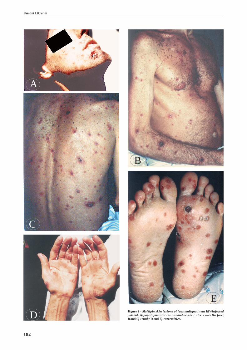
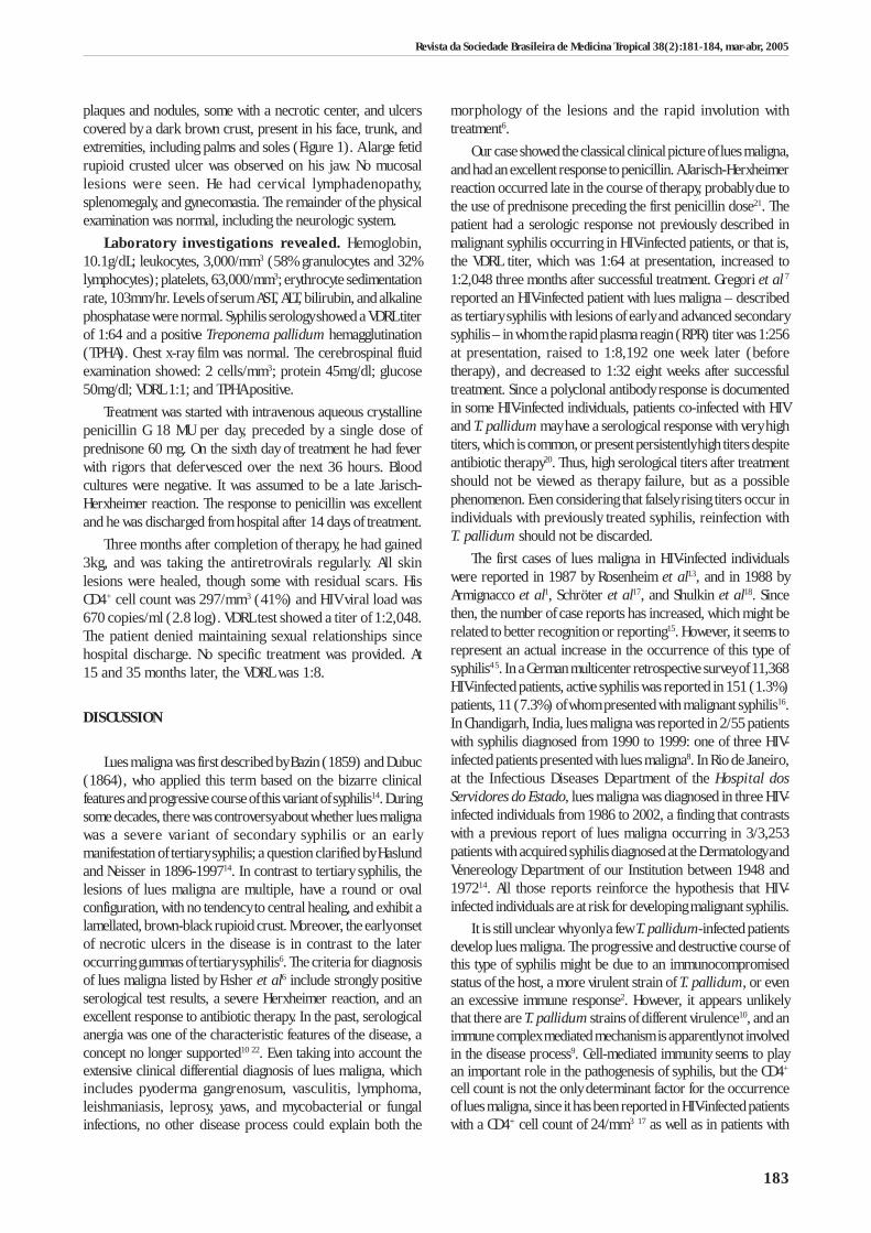
Figure 1 - Multiple skin lesions of lues maligna in an HIV-infectedpatient: A) papulopustular lesions and necrotic ulcers over the face;B and C) trunk; D and E) extremities.
A
D
E
B
183
Revista da Sociedade Brasileira de Medicina Tropical 38(2):181-184, mar-abr, 2005
plaques and nodules, some with a necrotic center, and ulcerscovered by a dark brown crust, present in his face, trunk, andextremities, including palms and soles (Figure 1). A large fetidrupioid crusted ulcer was observed on his jaw. No mucosallesions were seen. He had cervical lymphadenopathy,splenomegaly, and gynecomastia. The remainder of the physicalexamination was normal, including the neurologic system.
Laboratory investigations revealed. Hemoglobin,10.1g/dL; leukocytes, 3,000/mm3 (58% granulocytes and 32%lymphocytes); platelets, 63,000/mm3; erythrocyte sedimentationrate, 103mm/hr. Levels of serum AST, ALT, bilirubin, and alkalinephosphatase were normal. Syphilis serology showed a VDRL titerof 1:64 and a positive Treponema pallidum hemagglutination(TPHA). Chest x-ray film was normal. The cerebrospinal fluidexamination showed: 2 cells/mm3; protein 45mg/dl; glucose50mg/dl; VDRL 1:1; and TPHA positive.
Treatment was started with intravenous aqueous crystallinepenicillin G 18 MU per day, preceded by a single dose ofprednisone 60 mg. On the sixth day of treatment he had feverwith rigors that defervesced over the next 36 hours. Bloodcultures were negative. It was assumed to be a late Jarisch-Herxheimer reaction. The response to penicillin was excellentand he was discharged from hospital after 14 days of treatment.
Three months after completion of therapy, he had gained3kg, and was taking the antiretrovirals regularly. All skinlesions were healed, though some with residual scars. HisCD4+ cell count was 297/mm3 (41%) and HIV viral load was670 copies/ml (2.8 log). VDRL test showed a titer of 1:2,048.The patient denied maintaining sexual relationships sincehospital discharge. No specific treatment was provided. At15 and 35 months later, the VDRL was 1:8.
DISCUSSION
Lues maligna was first described by Bazin (1859) and Dubuc(1864), who applied this term based on the bizarre clinicalfeatures and progressive course of this variant of syphilis14. Duringsome decades, there was controversy about whether lues malignawas a severe variant of secondary syphilis or an earlymanifestation of tertiary syphilis; a question clarified by Haslundand Neisser in 1896-199714. In contrast to tertiary syphilis, thelesions of lues maligna are multiple, have a round or ovalconfiguration, with no tendency to central healing, and exhibit alamellated, brown-black rupioid crust. Moreover, the early onsetof necrotic ulcers in the disease is in contrast to the lateroccurring gummas of tertiary syphilis6. The criteria for diagnosisof lues maligna listed by Fisher et al6 include strongly positiveserological test results, a severe Herxheimer reaction, and anexcellent response to antibiotic therapy. In the past, serologicalanergia was one of the characteristic features of the disease, aconcept no longer supported10 22. Even taking into account theextensive clinical differential diagnosis of lues maligna, whichincludes pyoderma gangrenosum, vasculitis, lymphoma,leishmaniasis, leprosy, yaws, and mycobacterial or fungalinfections, no other disease process could explain both the
morphology of the lesions and the rapid involution withtreatment6.
Our case showed the classical clinical picture of lues maligna,and had an excellent response to penicillin. A Jarisch-Herxheimerreaction occurred late in the course of therapy, probably due tothe use of prednisone preceding the first penicillin dose21. Thepatient had a serologic response not previously described inmalignant syphilis occurring in HIV-infected patients, or that is,the VDRL titer, which was 1:64 at presentation, increased to1:2,048 three months after successful treatment. Gregori et al 7
reported an HIV-infected patient with lues maligna – describedas tertiary syphilis with lesions of early and advanced secondarysyphilis – in whom the rapid plasma reagin (RPR) titer was 1:256at presentation, raised to 1:8,192 one week later (beforetherapy), and decreased to 1:32 eight weeks after successfultreatment. Since a polyclonal antibody response is documentedin some HIV-infected individuals, patients co-infected with HIVand T. pallidum may have a serological response with very hightiters, which is common, or present persistently high titers despiteantibiotic therapy20. Thus, high serological titers after treatmentshould not be viewed as therapy failure, but as a possiblephenomenon. Even considering that falsely rising titers occur inindividuals with previously treated syphilis, reinfection withT. pallidum should not be discarded.
The first cases of lues maligna in HIV-infected individualswere reported in 1987 by Rosenheim et al13, and in 1988 byArmignacco et al1, Schröter et al17, and Shulkin et al18. Sincethen, the number of case reports has increased, which might berelated to better recognition or reporting15. However, it seems torepresent an actual increase in the occurrence of this type ofsyphilis4 5. In a German multicenter retrospective survey of 11,368HIV-infected patients, active syphilis was reported in 151 (1.3%)patients, 11 (7.3%) of whom presented with malignant syphilis16.In Chandigarh, India, lues maligna was reported in 2/55 patientswith syphilis diagnosed from 1990 to 1999: one of three HIV-infected patients presented with lues maligna8. In Rio de Janeiro,at the Infectious Diseases Department of the Hospital dosServidores do Estado, lues maligna was diagnosed in three HIV-infected individuals from 1986 to 2002, a finding that contrastswith a previous report of lues maligna occurring in 3/3,253patients with acquired syphilis diagnosed at the Dermatology andVenereology Department of our Institution between 1948 and197214. All those reports reinforce the hypothesis that HIV-infected individuals are at risk for developing malignant syphilis.
It is still unclear why only a few T. pallidum-infected patientsdevelop lues maligna. The progressive and destructive course ofthis type of syphilis might be due to an immunocompromisedstatus of the host, a more virulent strain of T. pallidum, or evenan excessive immune response2. However, it appears unlikelythat there are T. pallidum strains of different virulence10, and animmune complex mediated mechanism is apparently not involvedin the disease process9. Cell-mediated immunity seems to playan important role in the pathogenesis of syphilis, but the CD4+
cell count is not the only determinant factor for the occurrenceof lues maligna, since it has been reported in HIV-infected patientswith a CD4+ cell count of 24/mm3 17 as well as in patients with
184
1,200 CD4+ cells/mm3 4. Qualitative or functional defects of bothcell-mediated and humoral immunity are probably involved inthe pathogenesis of malignant syphilis: the pathogenic interactionbetween HIV and Treponema pallidum both leading to animmunodeficiency state may reduce the immunologic responseto treponemal infection through a decrease in cell-mediatedimmunity, macrophage functional defects, and possiblyimmunomodulation of the humoral immunity response19. Thelocal immune response to T. pallidum may be critical to thedevelopment of clinical manifestations as well as to the clearanceof spirochetes from infected tissues. However, relatively little isknown about the local immune responses to T. pallidum in skinand other body sites, and virtually nothing is known about thesubpopulations of immune cells in the lesions of patients withsyphilis co-infected with HIV11.
Although the clinical manifestations and the course of syphilismay be altered in the presence of HIV infection, most HIV-infectedpatients with syphilis have typical disease manifestations. Clinicalfeatures of syphilis are protean, and the diagnosis of lues malignashould be considered in all HIV-infected individuals withulceronodular skin lesions3 22. This approach might avoidunnecessary efforts or more complex investigations of otherdiseases occurring in these patients. As Witkowsky and Parish22
emphasized, a simple serologic test for syphilis can bring a caseto a speedier and less costly conclusion.
ACKNOWLEDGMENTS
We gratefully thank Dr. Elizabeth Machado, Dr. LeonClaude Sidi and Dr. Fernando Luiz Lopes Cardoso, colleaguesat the Infectious Disease Department, Hospital dos Servidoresdo Estado, for their comments.
REFERENCES
1. Armignacco O, Antonucci G, Croce GF, Grillo LR, Valenzano L. Lue edinfezione da HIV. Considerazioni su un caso di sifilide maligna. Recentiprogressi in medicina 79: 132-134, 1988.
2. Bahmer FA, Anton-Lamprecht L. Ultrastructural features of malignantsyphilis and demonstration of Treponema pallidum. International Journalof Dermatology 22: 165-170, 1983.
3. Belda-Jr W, Dias MC, Zolli CA, Santos-Jr MFQ, Siqueira LFG. Sífilis malignaprecoce. A propósito de um caso. Anais Brasileiros de Dermatologia65: 147-150, 1990.
4. Caumes E, Janier M, Janssen F, Feyeux C, Vignon-Pennamen MD, Morel P.Syphilis acquise au cours de l’infection par le virus de l’immunodéficiencehumaine. Six cas. La Presse Médicale 19: 369-371, 1990.
5. Don PC, Rubinstein R, Christie S. Malignant syphilis (Lues maligna) andconcurrent infection with HIV. International journal of dermatology34: 403-407, 1995.
6. Fisher DA, Chang LW, Tuffanelli DL. Lues maligna. Presentation of a caseand a review of the literature. Archives of Dermatology 99: 70-73, 1969.
7. Gregory N, Sanchez M, Buchness MR. The spectrum of syphilis in patientswith human immunodeficiency virus infection. Journal of the AmericanAcademy of Dermatology 22: 1061-1067, 1990.
8. Kumar B, Gupta S, Muralidhar S. Mucocutaneous manifestations ofsecondary syphilis in North Indian patients: a changing scenario? TheJournal of dermatology 28: 137-144, 2001.
9. Kumar B, Muralidhar S, Das A. Malignant syphilis: an immunological puzzle.International journal of STD & AIDS 9: 114-116, 1998.
10. Lejman K, Starzycki Z. Syphilis maligna praecox. A case report. The BritishJournal of Venereal Diseases 48: 194-199, 1972.
11. McBroom RL, Styles AR, Chiu MJ, Clegg C, Cockerell C, Radolf JD. Secondarysyphilis in persons infected with and not infected with HIV-1: a comparativeimmunohistologic study. The American Journal of Dermatopathology21: 432-441, 1999.
12. Menezes JA, Cunha RQ, Carvalho LM, Cruz MLS. Sífilis maligna precocecomo primeira manifestação de AIDS - relato de dois casos. In: Resumosdo XXXI Congresso da Sociedade Brasileira de Medicina Tropical, SãoPaulo, p. 302, 1995.
13. Rosenheim M, Brucker G, Leibowitch M, Niel G, Bournerias I, Duflo B,Gentilini M. Syphilis maligne chez un malade porteur d’anticorps anti-VIH. La Presse Médicale 16: 777, 1987.
14. Rutowitsch MS. Sífilis maligna precoce. Anais Brasileiros de Dermatologia55: 147-150, 1980.
15. Sands M, Markus A. Lues maligna, or ulceronodular syphilis, in a maninfected with human immunodeficiency virus: case report and review.Clinical infectious diseases 20: 387-390, 1995.
16. Schöfer H, Imhof M, Thoma-Greber E, Brockmeyer NH, Hartmann M,Gerken G, Pees HW, Rasokat H, Hartmann H, Sadri I, Emminger C, StellbrinkHJ, Baumgarten R, Plettenberg A, The German AIDS Study Group (GASG).Active syphilis in HIV infection: a multicentre retrospective survey.Genitourinary medicine 72: 176-181, 1996.
17. Schröter R, Näher H, Petzoldt D. Hautmanifestationen der syphilis malignabei HIV-infektion. Klinische Beobachtungen an drei Fällen. Der Hautarzt39: 463-466, 1988.
18. Shulkin D, Tripoli L, Abell E. Lues maligna in a patient with humanimmunodeficiency virus infection. The American Journal of Medicine85: 425-427, 1988.
19. Tosca A, Stavropoulos PG, Hatziolou E, Arvanitis A, Stavrianeas N,Hatzivassiliou M, Stratigos JD. Malignant syphilis in HIV-infected patients.International Journal of Dermatology 29: 575-578, 1990.
20. Tramont EC. Syphilis in adults: from Christopher Columbus to Sir AlexanderFleming to AIDS. Clinical Infectious Diseases 21: 1361-1371, 1995.
21. Tramont EC. Treponema pallidum (Syphilis). In: Mandell GL, Bennett JE,Dolin R (eds) Principles and practice of infectious diseases, 5th edition,Churchill Livingstone, Philadelphia, p. 2474-2490, 2000.
22. Witkowski JA, Parish LC. The great imitator: malignant syphilis withhepatitis. Clinics in Dermatology 20: 156-163, 2002.
Passoni LFC et al