LSUHSC Occupational Therapy Nerve Repair - LSUHSC-S Medical
34
LSUHSC Occupational Therapy Nerve Repair Carla Saulsbery LOTR, CHT Dr. A. Hollister MD Springer Images
Transcript of LSUHSC Occupational Therapy Nerve Repair - LSUHSC-S Medical

LSUHSC
Occupational Therapy
Nerve Repair
Carla Saulsbery LOTR, CHT
Dr. A. Hollister MD
Springer Images

Nerve Repairs
Primary nerve repair:
Indicated for clean, sharply cut nerves. Performed immediately after an
injury or within 1 to 2 weeks
Secondary nerve repair:
Usually indicated in the presence of a severely crushed or avulsed nerve.
Surgery involves resection of the damaged segment.
Nerve graft:
Performed when a direct repair cannot be done. Often done as a secondary
procedure. Nerve tubes may be used.
LSUHSC-Shreveport

• Appropriate post operative immobilization is important.
• The expected rate of recovery for nerve repairs is 1 mm per day or 1 inch per
month, after an initial period of 3 weeks when the axonal sprouts cross the repair
• Patient must be educated, especially with an insensate hand
• Care must be taken to avoid stretching of the repaired nerve ends. Tension
leads to scarring
• Motor retraining, sensory reeducation and desensitization programs should be
initiated as sensory and muscle re-innervation becomes evident
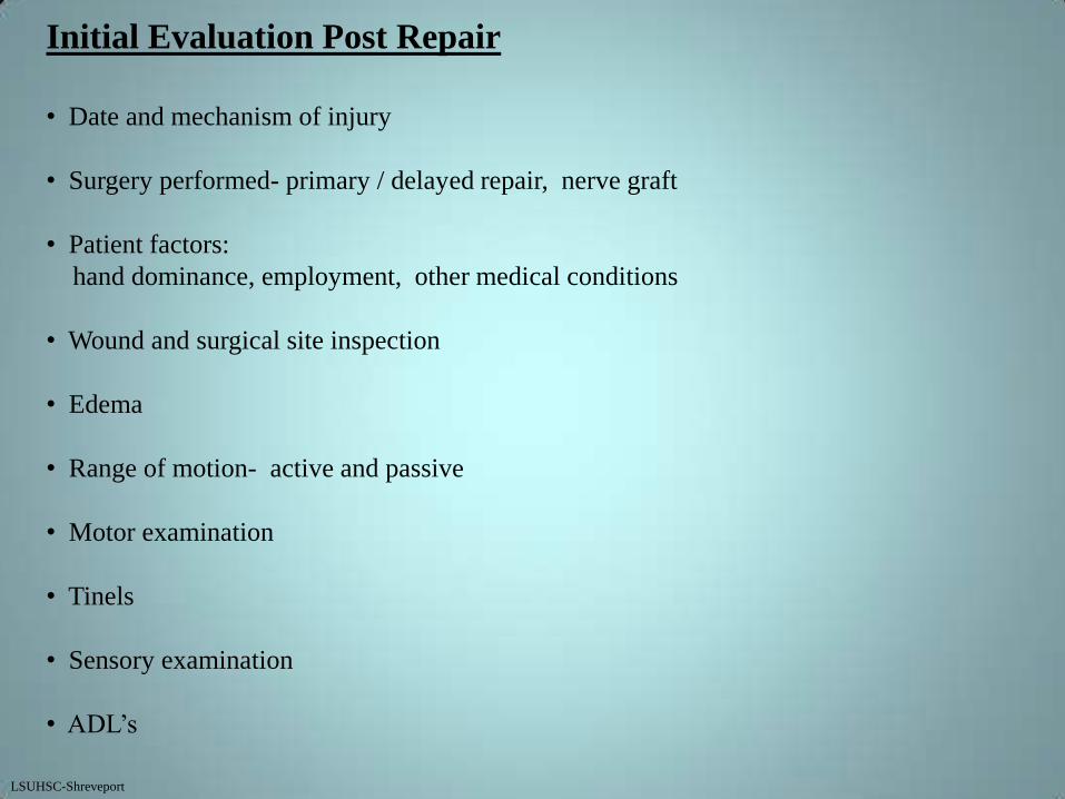
Initial Evaluation Post Repair
• Date and mechanism of injury
• Surgery performed- primary / delayed repair, nerve graft
• Patient factors:
hand dominance, employment, other medical conditions
• Wound and surgical site inspection
• Edema
• Range of motion- active and passive
• Motor examination
• Tinels
• Sensory examination
• ADL’s
LSUHSC-Shreveport

LSUHSC-Shreveport
Occupational Therapy
Upper Extremity Semmes Weinstein Right Left
Date :______________
OTR/L:_______________________
Volar Dorsal
Axillary nerve
Superior lateral
cutaneous (C5-6)
Radial nerve
Inferior lateral
cutaneous (C5-6)
Lateral cutaneous
nerve (C5-7)
Radial nerve
Median nerve
Palmar branch
(C6-7)
Intercosto-brachial nerve
(T2) and the medial
cutaneous nerve
(C8, T1-2)
Medial cutaneous nerve
(C8,T1)
Ulnar nerve
(C8, T1) Ulnar nerve
(C8, T1)
Radial nerve
Superficial branch
and dorsal digital
(C6-8)
Median nerve
Radial Nerve
Posterior cutaneous
(C5-7)
Inferior lateral
cutaneous
Posterior cutaneous
(C5-8)
Lateral cutaneous
(C5-7)
2.83 Green Normal
3.61 Blue Dim. light touch
4.31 Purple Dim protective
6(4.56) Red Loss of protective
6.6 Orange Deep pressure
Red Lined Untestable LSUHSC-Shreveport
Axillary nerve
Superior lateral
cutaneous (C5-6)

Re-evaluation
• Motor
re-innervation of musculature, assess for compensatory motions and
trick movements
• Range of motion– active and passive
• Tinels
• Sensation
• ADL’s
• Return of sympathetic function– sweat pattern
• Grip and pinch strength testing
• Note any contracture development
LSUHSC-Shreveport

Occupational Therapy Intervention
Acute management:
• Immobilization and protection of repaired structures with custom splinting
• Prevention of joint contractures
• Range of motion
• Prevention of injury secondary to decreased sensation
• Activities of daily living
• Patient education on insensate areas
• Begin scar massage
LSUHSC-Shreveport

Secondary Phase:
• Increase range of motion– active and passive
• Enhance function using appropriate splint
Radial nerve
wrist splint
dynamic splint
Median nerve
opponens splint
web space c-bar splint
Ulnar nerve
lumbrical bar splint
• Patient education, home program, risks related to loss of sensation
• Desensitization and scar massage
• ADL’s
Adaptive techniques
Assistive equipment
LSUHSC-Shreveport

Third Phase:
• Motor retraining
• Active and active-assistive exercise
• Strengthening
• Desensitization
• Sensory reeducation
• ADL’s
Adaptive techniques
Assistive equipment to increase independence
• Look at need for re-training to change hand dominance
LSUHSC-Shreveport

Long term follow up:
• Evaluate residual deficits
• Compensation
Assistive equipment for ADL independence
• Splinting to promote function
• Patient education
LSUHSC-Shreveport

Ulnar Nerve Innervation
Ulnar Nerve
Adductor pollicis
Palmar & dorsal interossei
3rd & 4th lumbricals
Abductor
Opponens Digiti quinti
Flexor
Palmaris brevis
Springer

Ulnar Nerve (median cord C8-T1)
MMS Flexor Carpi Ulnaris Forearm High Lesion
Flexor Dig. Profundus(RF/SF)
Abd. Digiti Minimi Hand Low Lesion
Opponens Digiti Minimi
Flexor Digiti Minimi
Lumbricals 3,4
Interossei (palmar/dorsal)
Flexor Pollicis Brevis (deep)
Adductor pollicis
Froments
Thumb IP flexion during lateral pinch
Positive Froments in hand B
LSUHSC-Shreveport
A B

Ulnar Nerve lesion at the wrist
* Loss of abduction and adduction due to paralysis of the interossei
* Hyperextension of the ring and small MCP joints with flexion of the IP
joints due to unopposed action of the extensor digitorum communis (EDC)
and the flexion of the flexor digitorum profundus
* Weak thumb adduction due to paralysis of the adductor pollicis
* Loss of opposition of the fifth finger due to paralysis of the abd. digiti quinti
* Weak thumb opposition due to paralysis of the AdP
* Weak MCP flexion due to paralysis of the third and fourth lumbricals
* Weak pinch due to paralysis of the AdP, deep head of the FPB and the
first dorsal interosseous
* Weak grasp due to paralysis of the interossei, third and fourth lumbricals, and
the FDP of the ring and small fingers
* Sensory loss of the volar and dorsal aspects of the medial third of the hand,
the small finger and the ulnar half of the ring finger
Ulnar Nerve lesions in proximal forearm involves these additional problems
* Weak flexion of IP joints of the ring and small fingers due to paralysis of the
ulnar half of the FDP
* Weak wrist flexion due to paralysis of the flexor carpi ulnaris (FCU)
LSUHSC-Shreveport

Median Nerve Innervation
Flexor digitorum sublimis
Flexor pollicis longus
Flexor digitorum profundus
Pronator quadratus
Abductor pollicis brevis
Opponens pollicis
Sup. Head FPB
1st & 2nd Lumbricals
Flexor carpi radialis
Palmaris longus
Pronator teres
Flexor digitorum profundus
CS

Median Nerve (medial cord C5-7 and lateral cords C8-T1 of
Brachial Plexus)
Muscle/Sensory Innervation
Pronator Teres Forearm Median Nerve High Lesion
Flexor Carpi Radialis
Palmaris Longus
Flexor Digitorum Superficialis
Palmar Cutaneous Branch
Flexor Digitorum Profundus (Index/Long) Anterior Interosseous Nerve
Flexor Pollicis Longus
Pronator Quadratus
Lumbricals (1,2) Carpal Tunnel Median Nerve Low Lesion
Opponens Pollicis
Abductor Pollicis Brevis
Flexor Pollicis Brevis (superficial)
Digital Cutaneous Branch
LSUHSC-Shreveport

Median Nerve Deficits (wrist level)
* Sensory loss of the central palm area and the palmar surfaces of the lateral
three and one-half digits
* Weak MCP joint flexion of the index and middle fingers due to paralysis
of the first two lumbricals
* Weak pinch due to paralysis of opponens pollicis. abductor pollicis brevis,
and the superficial head of the flexor pollicis brevis
* Loss of palmar abduction due to paralysis of the APB
Anterior Interosseous Nerve (AIN) (Proximal 1/3rd of forearm)
* Loss of DIP joint flexion of the index and middle fingers due to paralysis
of the FDP to each digit
* Loss of thumb IP flexion due to paralysis of the flexor pollicis longus (FPL)
* Weak forearm pronation due to paralysis of the pronator quadratus
Median Nerve lesion in the proximal forearm
* Weak forearm pronation due to paralysis of the pronator teres
* Weak wrist flexion due to paralysis of the flexor carpi radialis (FCR)
* Weak finger flexion due to paralysis of the flexor digitorum superficialis (FDS)
LSUHSC-Shreveport
Median or Ulnar Nerve Repair
Surgeon dictates wrist position in the OR and a dorsal plaster Kleinert splint applied
0-3 weeks post repair
* OT fabricates a thermoplastic Kleinert dorsal blocking splint. Same wrist
position as placed in OR.
If associated Flexor Tendon repair follow FTR protocol for splinting and PROM
If lesion is more proximal include a long arm splint with the elbow flexed to 90º
* If no tendon injury begin AROM/AAROM to digits
* Educate patient on insensate areas. ADL adaptations
* Monitor for thumb adduction contracture with Median nerve injury
* Begin scar massage once wound is healed
3 weeks post repair
* A volar wrist splint is molded with wrist in neutral
* OT performs baseline Semmes and motor examination. Assess Tinels
* Assess grip and pinch (wait until week 6 with tendon injury)
* AROM exercises
* Patient education on insensate hand
* Cocoa butter massage to hand for hydration, desensitization and massage to scar LSUHSC-Shreveport

4 weeks post repair
* Patient education on the expected sensations associated during
sensory return
* Serial splint the wrist into extension weekly. Check for complaints
of burning and tingling during extension of the wrist while forming splint.
Decrease extension and mold splint prior to this point
* Massage to entire hand for skin re-hydration / desensitization
* The hand should be kept warm during cold weather. Patient should use glove
or tube sock for warmth.
* Continue patient education on insensate hand. Use of visual compensation
* Continue AROM exercises. Continue PROM with associated tendon repair
* Assess Tinels. Document location
5 weeks post repair
* Lumbrical bar can be added to the volar wrist splint for Ulnar nerve lesion
* Continue AROM exercises. Continue PROM with associated tendon injury
* Continue patient education on insensate hand
* Massage for desensitization, skin re-hydration
* AROM exercises to the wrist
LSUHSC-Shreveport

6 weeks post repair
* Volar wrist splint is discontinued
* Lumbrical bar splint for ulnar nerve lesion
* C-bar splint and or opponens splint for median nerve lesion
* Re-evaluate sensation and motor exam. Assess grip and pinch
* Begin sensory reeducation and desensitization program when appropriate
* Continue education on insensate areas
Assess Tinels’s
7-8 weeks post repair
* Dynamic splinting can begin for ulnar nerve to decrease any extrinsic flexor
tightness
* Continue with Lumbrical bar for Ulnar nerve . Opponens splint for Med. N.
6-12 weeks post repair
* Motor retraining as appropriate
* OT repeats sensory and motor examinations every 3 to 4 weeks. Continue to
assess and document Tinel’s.
* Functional activities
* Strengthening
* Continue sensory reeducation
* Continue splinting as indicated LSUHSC-Shreveport
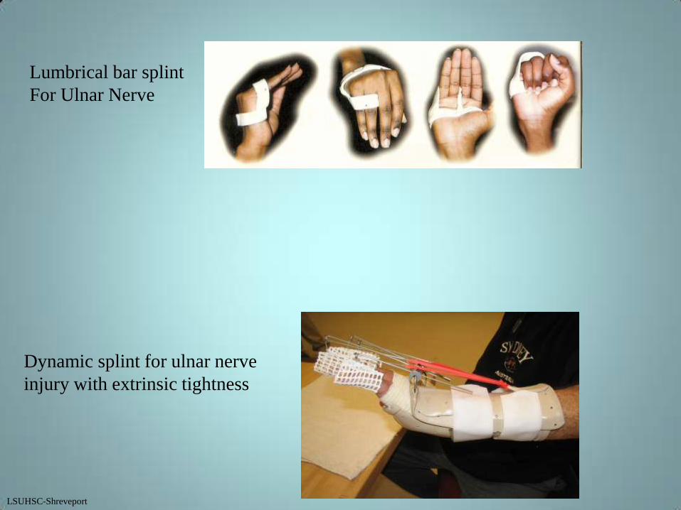
Dynamic splint for ulnar nerve
injury with extrinsic tightness
Lumbrical bar splint
For Ulnar Nerve
LSUHSC-Shreveport

Short opponens for Median Nerve
C-bar for correction of
a thumb adduction
contracture
LSUHSC-Shreveport

LSUHSC-Shreveport
Radial nerve
Triceps
Brachioradialis
Extensor carpi radialis longus
Lower lat cut. n. of arm
Post. cut. n. of FA
Post. Interosseous n.
Extensor carpi radialis brevis
Supinator
Extensor digitorum
Extensor digiti quinti
Extensor carpi ulnaris
Abductor pollicis longus
Extensor pollicis longus & brevis
Extensor indicis
Dorsal digital nerves
Radial nerve innervations
CS

MMS Triceps Radial Nerve High Lesion
Brachioradials
Wrist Extension
ECRL
ECRB Posterior Interosseous
Supinator Posterior Interosseous Low Lesion
Ext. Digitorum
Ext. Digiti Minimi
Ext. Carpi Ulnaris
Abd. Pollicis Longus
Ext. Pollicis Longus
Ext. Pollicis Brevis
Ext. Indicis Proprius
LSUHSC-Shreveport
Radial Nerve (Posterior cord) C 5, 6, 7, 8 and T1)

• Most frequently injured nerve in the upper extremity usually from humeral
shaft fractures
• 12 % of humeral shaft fractures are complicated by a radial nerve paralysis.
• In open humeral shaft fractures the incidence of Radial nerve laceration is
about 60%.
Can be complete or partial laceration.
• Radial nerve injuries result in a decrease in power grip and pinch related to the
loss of wrist extension.

High Radial Nerve Lesion
* Wrist drop due to paralysis of wrist extensors
* Diminished abduction and extension of the thumb due to paralysis of the
abductor pollicis longus (APL) and the extensor pollicis brevis (EPB)
* Inability to extend MCP joints due to paralysis of the long extensors
* Weak grasp and pinch due to inefficiency of the unopposed flexors
* Loss of sensation of the lateral two thirds of the dorsum of the hand,
a portion of the dorsum of the thumb and the dorsum of the proximal
phalanges of the lateral three and one-half digits
* Weakened supination due to paralysis of the supinator muscle
Posterior Interosseous Nerve Lesion
* Same effects as described except sensation is not lost and wrist extension is
present but weakened.
LSUHSC-Shreveport

Radial Nerve repair
Patient is placed in volar plaster post op splint by Surgeon in OR
0-3 weeks post repair
* OT fabricates a volar forearm based static wrist extension splint. Wrist extended 60º.
If lesion is more proximal the elbow should also be immobilized in 90º of elbow
flexion. Dynamic finger extension outriggers may be added
* AROM /AAROM of the digits
With associated extensor tendon repairs follow splinting protocol for zone of
injury.
* Patient education in ADL modifications
Patient education in wound care progressing to scar massage
3-6 weeks post repair
* Volar wrist splint is molded with wrist in 45º of extension
* Dynamic finger extension outriggers are added to daytime splint
* A volar wrist cock up splint is fabricated for night-time wear
* OT performs baseline motor and sensory evaluation. Assess Tinel’s
* Assess grip and pinch strengths at 6 weeks
* ADL modifications as needed
* Check for Tinel’s. Document advancement LSUHSC-Shreveport

Radial Nerve Repair
6-12 weeks post repair
* Continue with splinting as indicated for positioning and function
* Continue to monitor motor return
* Begin motor retraining when appropriate
Continue to re-assess sensation, grip, pinch and MMS
Assess Tinel’s. Continue to document advancement
Continue to advance ADL’s
LSUHSC-Shreveport

Splinting for Radial Nerve
Dynamic Splint, with active wrist
extension
Volar wrist splint
Begin with wrist extension at 60 and
serial splint towards neutral
Dynamic splint, no active wrist extension
LSUHSC-Shreveport

Long arm splint for more proximal Radial Nerve injury
LSUHSC-Shreveport
Digital Nerve
Week 1 post op
* Dorsal Blocking Kleinert splint. MCP’s flexed 60º and IP’s in full extension
* AROM exercises within splint 15 reps hourly
* Wound Care
Weeks 1-3 post op
* Dorsal Blocking Kleinert splint
* AROM exercises
* Scar massage
* Patient education on insensate area
* Baseline Semmes Weinstein
Weeks 3-6 post op
* AROM exercises
* Scar massage
* Desensitization/sensory re-education
* Begin ADL’s
Tinel’s
Patient education on insensate areas
LSUHSC-Shreveport

Digital Nerve
Weeks 6-8 post op
* Repeat Semmes Weinstein
* Extension splinting as needed for flexion contracture
* Continue patient education on insensate areas
* Continue desensitization
Week 8-10 post op
* Strengthening
Repeat Semmes Weinstein
Assess Tinel’s
Continue desensitization/sensory re-education
LSUHSC-Shreveport

Outcomes
1. Functional recovery can take up to 2 years, and improvement can occur from
nerve injuries proximal to the wrist for up to 4 years.
2. Some generalities that have been made:
-- distal nerve injuries do better than proximal ones
-- younger patients <20 years of age do better than older patients
-- younger patients generally regain a complete recovery of functional sensibility
however this is not the case in the adult
-- sensory or pure motor nerve repair does better than mixed nerve repair
-- guillotine injury does better than a crush injury or an avulsion injury
-- protective sensation usually occurs
3. According to a study by Schreaders
Median Nerve 61% have good motor return and 44% had good sensory return
Ulnar Nerve 45% good motor return and 41% had good sensory return
Combined Median/Ulnar had the worse prognosis
4. Sensory reeducation protocols greatly increase the functional outcome

5. Chronic neuropathic pain can be severe and directly influences patient outcome
6. Nerve regeneration seems to deteriorate with increasing distance to the innervated
organ. The more proximal the nerve injury the lower the chances for the axons
to re-innervate the adequate terminal receptors. (Lohmeyer, et al.)

References
Dr. A. Hollister, MD. Associate Professor Orthopaedics LSUHSC
Carla Saulsbery LOTR, CHT Chief Occupational Therapy, LSUHSC
Hunter; Mackin; Callahan. Rehabilitation of the Hand
Burke; Higgins; et.al. Hand and Upper Extremity Rehabilitation
Stanley; Tribuzi. Concepts in Hand Rehabilitation
Lohmeyer, et. al. Nerve Injuries of the UE. Plastic Surgical Nursing. Apr/June 2009