Clinical Significance Of Tibial Crest Width Following Tibial Pl
66
[REV. MED. CLIN. CONDES - 2010; 21(1) 66 - 75]
SUMMARYLower extremity reconstruction is an essential part of plastic surgery and focuses on the treatment of wounds and defects secondary to trauma, cancer, or chronic disease processes. During the last 25 years, advances in plastic surgical techniques such as free-tissue transfer and improved wound care technologies has revolutionized this field, allowing the salvage of limbs that would have otherwise been amputated. The following paper will review the field of lower extremity reconstruction focusing on the evaluation of leg defects and wounds and the various treatment options.
Key Words: Lower extremity reconstruction; open tibial fractures; free flaps; local flaps
INTRODUCTIONThe goal of lower extremity reconstruction is the coverage of defects and open wounds of the leg to give patients a healed wound and to let them resume their life, ambulate, and go back to work while preventing amputation. Open wounds and defects in the lower extremity result from trauma, tumor resection, and chronic diseases such as peripheral vascular disease and diabetes; these wounds need reconstruction for many reasons. First, any exposed bone that is not covered by vascularized soft tissue is at risk for osteomyelitis, bone necrosis, and sepsis. Osteomyelitis is a major cause of amputation in patients after leg trauma or patients with systemic diseases, most commonly diabetes (1-3). Second, open wounds cause chronic pain, inability to ambulate, significant medical expenses, and unemployment. Exposed tendons become dry and necrotic and exposed blood vessels are at risk for rupture.
The following paper will review the field of lower extremity reconstruction focusing on the repair of defects and wounds sustained
after trauma, the resection of tumors, or due to chronic illness. We will concentrate on defects of the lower leg, from the knee to the foot.
ANATOMY OF THE LOWER EXTREMITYThe lower leg is composed of 2 long bones arranged in parallel, the fibula and tibia. The fibula is thin and long and provides insertion to many of the muscles of the leg. The tibia is a long and thick bone responsible for over 80% of the weight bearing capacity of the lower leg. The tibia and fibula are connected along their length by a fibrous interosseus membrane. These 3 structures together divide the leg into an anterior and posterior compartment.
The anterior compartment is further divided into anterior and lateral compartments by an intermuscular fascia. The anterior compartment has 4 muscles: the extensor digitorum longus, extensor hallucis longus, peroneus tertius, and the tibialis anterior. These muscles are vascularized by anterior tibial vessels and innervated by the deep peroneal nerve.The lateral compartment has 2 muscles, the peroneus brevis and longus. They are supplied by the peroneal artery and innervated by the superficial peroneal nerve.
The posterior compartment is divided into superficial and deep compartments by a thin intermuscular fascia. The superficial compartment contains the gastrocnemius, soleus, and plantaris muscles. The gastrocnemius and soleus join together to become the Achilles tendon that inserts into the calcaneal bone. They are vascularized by the popliteal artery and innervated by the tibial nerve.
The deep compartment has four muscles: the popliteus, flexor digitorum longus, flexor hallucis longus, and tibialis posterior. The blood supply is provided by two arteries, the posterior tibial and the peroneal. The tibial nerve innervates the compartment.
Artículo recibido: 16-09-09Artículo aprobado para publicación: 10-11-09
BRIAN M. PARRETT, MD (1) (2), JULIAN J. PRIBAZ, MD (1)
1. Brigham and Women’s Hospital, Harvard Medical School, Boston, MA, USA2. The Buncke Clinic, San Francisco, CA, [email protected]
LOWER EXTREMITY RECONSTRUCTION
67
[LOWER EXTREMITY RECONSTRUCTION - BRIAN M. PARRETT ET AL.]
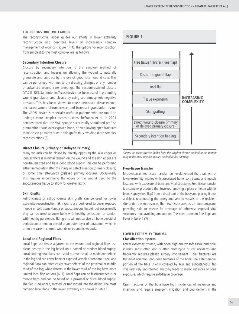
THE RECONSTRUCTIVE LADDERThe reconstructive ladder guides our efforts in lower extremity reconstruction and describes levels of increasingly complex management of wounds (Figure 1) (4). The options for reconstruction from simplest to the most complex are as follows:
Secondary Intention ClosureClosure by secondary intention is the simplest method of reconstruction and focuses on allowing the wound to naturally granulate and contract by the use of good local wound care. This can be performed with wet to dry dressing changes or any number of advanced wound care dressings. The vacuum-assisted closure (VAC®; KCI, San Antonio, Texas) device has been useful in promoting wound granulation and closure by using sub-atmospheric negative pressure. This has been shown to cause decreased tissue edema, decreased wound circumference, and increased granulation tissue. The VAC® device is especially useful in patients who are too ill to undergo more complex reconstructions. DeFranzo et al. in 2001 demonstrated that the VAC sponge successfully stimulated profuse granulation tissue over exposed bone, often allowing open fractures to be closed primarily or with skin grafts thus avoiding more complex reconstructions (5).
Direct Closure (Primary or Delayed Primary)Many wounds can be closed by directly opposing the skin edges as long as there is minimal tension on the wound and the skin edges are non-traumatized and have good blood supply. This can be performed either immediately after the injury or defect creation (primary closure) or some time afterwards (delayed primary closure). Occasionally this requires undermining the edges of the wound deep to the subcutaneous tissue to allow for greater laxity.
Skin GraftsFull-thickness or split-thickness skin grafts can be used for lower extremity reconstruction. Skin grafts are best used to cover exposed muscle or soft tissue (fascia or subcutaneous tissue), but occasionally they can be used to cover bone with healthy periosteum or tendon with healthy paratenon. Skin grafts will not survive on bone devoid of periosteum or tendon devoid of an outer layer of paratenon, which is often the case in chronic wounds or traumatic wounds.
Local and Regional FlapsLocal flaps use tissue adjacent to the wound and regional flaps use tissue nearby in the leg based on a named or random blood supply. Local and regional flaps are useful to cover small to moderate defects in the leg and can cover bone or exposed vessels or tendons. Local and regional flaps can more easily cover defects of the proximal or middle third of the leg; while defects in the lower third of the leg have more limited local flap options (6, 7). Local flaps can be fasciocutaneous or muscle flaps and can be based on a proximal or distal blood supply. The flap is advanced, rotated, or transposed into the defect. The most common local flaps in the lower extremity are shown in Table 1.
FIGURE 1.
Free tissue transfer (Free flap)
Distant, regional flap
Local flap
Tissue expansion
Skin grafting
Secondary intention healing
Direct wound closure (Primary or delayed primary closure)
INCREASING COMPLEXITY
Free-tissue Transfer Microvascular free tissue transfer has revolutionized the treatment of lower-extremity injuries with associated bone, soft tissue, and muscle loss, and with exposure of bone and vital structures. Free tissue transfer is a complex procedure that involves removing a piece of tissue with its blood supply (free flap) from a distal part of the body and placing it over a defect, reconnecting the artery and vein to vessels at the recipient site under the microscope. The new tissue acts as an autotransplant, providing skin or muscle for coverage of otherwise exposed vital structures, thus avoiding amputation. The most common free flaps are listed in Table 2 (7).
LOWER EXTREMITY TRAUMAClassification SystemLower extremity trauma, with open high-energy soft-tissue and tibial injuries, most often occurs after motorcycle or car accidents and frequently requires plastic surgery involvement. Tibial fractures are the most common long bone fractures of the body. The anteromedial portion of the tibia is only covered by skin and subcutaneous fat. This relatively unprotected anatomy leads to many instances of bone exposure, which require soft-tissue coverage.
Open fractures of the tibia have high incidences of malunion and infection, and require emergent irrigation and debridement in the
Shows the reconstructive ladder from the simplest closure method at the bottom rung to the most complex closure method at the top rung.
68
operating room to remove devitalized soft tissue and bone. Wounds are frequently left open and require repeated debridements, resulting in large soft-tissue defects. Open fractures have been assigned grades according to the Gustilo classification (Table 3), the most widely accepted method of categorizing open fractures (8).
Initial Treatment of Lower Extremity Traumatic WoundsManagement of the mangled lower extremity requires the combined input and treatment of the trauma, vascular, orthopedic and plastic surgeons. High-energy leg injuries are usually associated with other life-threatening injuries and the priorities are always to salvage the
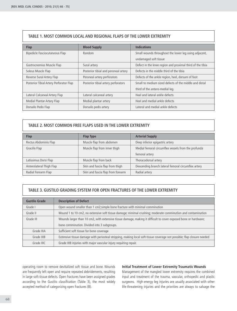
TABLE 1. MOST COMMON LOCAL AND REGIONAL FLAPS OF THE LOWER EXTREMITY
Flap
Bipedicle Fasciocutaneous Flap
Gastrocnemius Muscle Flap
Soleus Muscle Flap
Reverse Sural Artery Flap
Posterior Tibial Artery Perforator Flap
Lateral Calcaneal Artery Flap
Medial Plantar Artery Flap
Dorsalis Pedis Flap
Blood Supply
Random
Sural artery
Posterior tibial and peroneal artery
Peroneal artery perforators
Posterior tibial artery perforators
Lateral calcaneal artery
Medial plantar artery
Dorsalis pedis artery
Indications
Small wounds throughout the lower leg using adjacent,
undamaged soft tissue
Defect in the knee region and proximal third of the tibia
Defects in the middle third of the tibia
Defects of the ankle region, heel, dorsum of foot
Small to medium sized defects of the middle and distal
third of the antero-medial leg
Heel and lateral ankle defects
Heel and medial ankle defects
Lateral and medial ankle defects
TABLE 2. MOST COMMON FREE FLAPS USED IN THE LOWER EXTREMITY
Flap
Rectus Abdominis Flap
Gracilis Flap
Latissimus Dorsi Flap
Anterolateral Thigh Flap
Radial Forearm Flap
Flap Type
Muscle flap from abdomen
Muscle flap from inner thigh
Muscle flap from back
Skin and fascia flap from thigh
Skin and fascia flap from forearm
Arterial Supply
Deep inferior epigastric artery
Medial femoral circumflex vessels from the profunda
femoral artery
Thoracodorsal artery
Descending branch lateral femoral circumflex artery
Radial artery
TABLE 3. GUSTILO GRADING SYSTEM FOR OPEN FRACTURES OF THE LOWER EXTREMITY
Gustilo Grade
Grade I
Grade II
Grade III
Grade IIIA
Grade IIIB
Grade IIIC
Description of Defect
Open wound smaller than 1 cm2;simple bone fracture with minimal comminution
Wound 1 to 10 cm2, no extensive soft tissue damage; minimal crushing; moderate comminution and contamination
Wounds larger than 10 cm2, with extensive tissue damage, making it difficult to cover exposed bone or hardware;
bone comminution. Divided into 3 subgroups.
Sufficient soft tissue for bone coverage
Extensive tissue damage with periosteal stripping, making local soft-tissue coverage not possible; flap closure needed
Grade IIIB injuries with major vascular injury requiring repair.
[REV. MED. CLIN. CONDES - 2010; 21(1) 66 - 75]
69
[LOWER EXTREMITY RECONSTRUCTION - BRIAN M. PARRETT ET AL.]
life of the patient, not necessarily the salvage or treatment of the limb. Amputation of a mangled extremity in a clinically unstable patient may be more prudent than an extensive reconstructive course and should be considered in the initial evaluation of the patient.
Once the patient is stabilized, the initial assessment is to determine if the limb is salvageable. Does the extremity require revascularization and is this technically possible? Is the soft-tissue defect treatable with local or free tissue transfer? Is any bone loss reconstructible? Is there nerve injury and is this repairable or does the nerve injury preclude a functional limb? A complete injury of the neurologic function of the lower extremity may be a contraindication for extremity salvage, as nerve repair in the lower extremity has poor functional results and a below-knee amputation may be preferable to an insensate foot.
Loss of the posterior tibial nerve, with loss of sensation of the plantar aspect of the foot, is a relative contraindication for lower-extremity salvage. If the extremity is deemed unsalvageable, an amputation is indicated. If the extremity is salvageable, the reconstructive protocol is followed.
Reconstruction of Soft Tissue DefectsReconstruction of a traumatized lower extremity can be performed only after vascular injury has been addressed and repaired, bony fixation has been accomplished, and all contaminated and devitalized tissue has been debrided. The basic principle of debridement of all devitalized tissue is crucial to final success of any reconstruction and often requires serial operative debridements prior to final wound coverage.
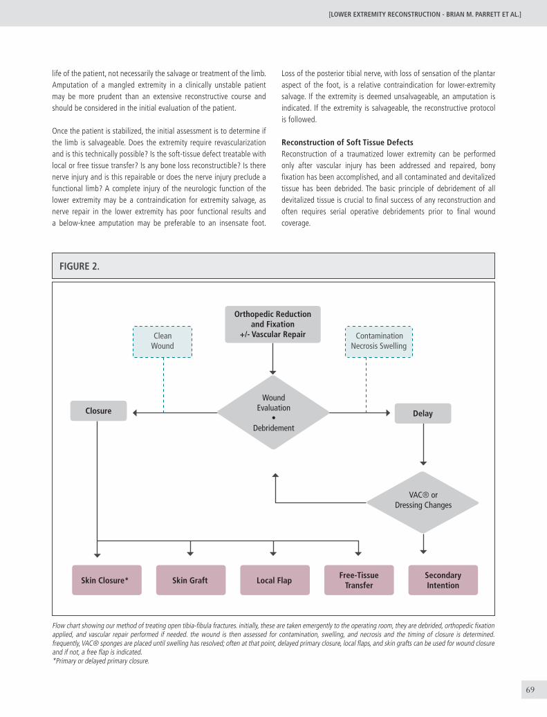
FIGURE 2.
Clean Wound
Contamination Necrosis Swelling
Orthopedic Reduction and Fixation
+/- Vascular Repair
Skin Closure* Skin Graft Local FlapFree-Tissue
TransferSecondary Intention
VAC® orDressing Changes
Closure Delay
WoundEvaluation
•Debridement
Flow chart showing our method of treating open tibia-fibula fractures. initially, these are taken emergently to the operating room, they are debrided, orthopedic fixation applied, and vascular repair performed if needed. the wound is then assessed for contamination, swelling, and necrosis and the timing of closure is determined. frequently, VAC® sponges are placed until swelling has resolved; often at that point, delayed primary closure, local flaps, and skin grafts can be used for wound closure and if not, a free flap is indicated. *Primary or delayed primary closure.
70
Early soft-tissue coverage is associated with a lower complication rate. The goal is to close wounds within 7 to 10 days to decrease the risk of infection, osteomyelitis, nonunion, and further tissue loss (9). Byrd et al. found that the overall complication rate of wounds closed within the first week of injury was 18% compared to a 50% complication rate for wounds closed in the subacute phase of 1 to 6 weeks (9-10). The reconstructive ladder guides soft tissue reconstruction (11).
Any wounds that can be closed by primary closure with minimal tension should be reconstructed by this method. This is an uncommon situation in severe lower extremity trauma.
Small areas of exposed bone or tendon can be treated successfully with secondary intention healing (5). This requires daily dressing changes or treatment with the VAC® device. The advantage of this method is that it does not require additional operations, is simple, and is especially useful in ill patients who cannot undergo more complex reconstructions (11). The disadvantage is that it often requires many weeks before definitive wound coverage.
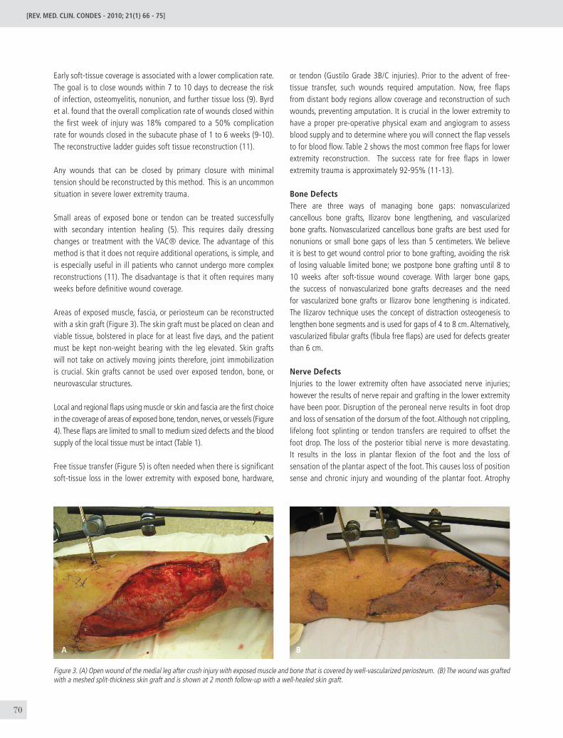
Areas of exposed muscle, fascia, or periosteum can be reconstructed with a skin graft (Figure 3). The skin graft must be placed on clean and viable tissue, bolstered in place for at least five days, and the patient must be kept non-weight bearing with the leg elevated. Skin grafts will not take on actively moving joints therefore, joint immobilization is crucial. Skin grafts cannot be used over exposed tendon, bone, or neurovascular structures.
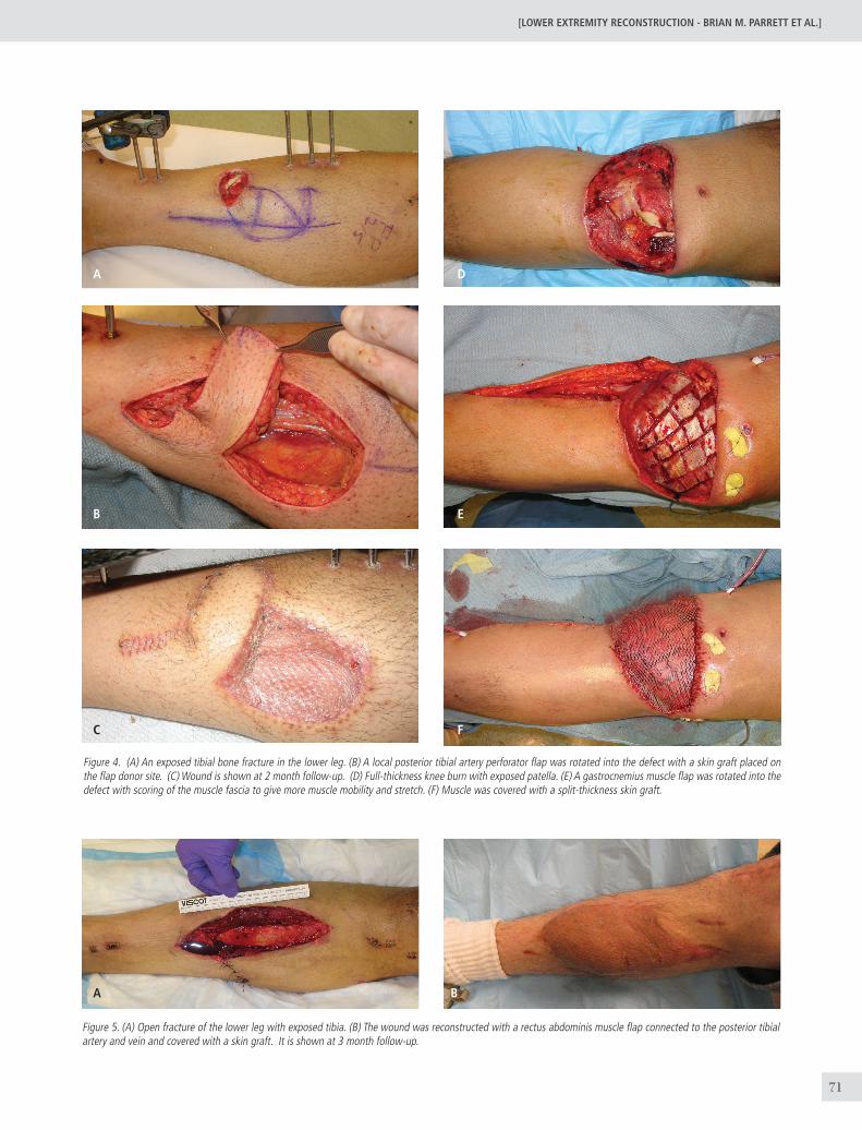
Local and regional flaps using muscle or skin and fascia are the first choice in the coverage of areas of exposed bone, tendon, nerves, or vessels (Figure 4). These flaps are limited to small to medium sized defects and the blood supply of the local tissue must be intact (Table 1).
Free tissue transfer (Figure 5) is often needed when there is significant soft-tissue loss in the lower extremity with exposed bone, hardware,
Figure 3. (A) Open wound of the medial leg after crush injury with exposed muscle and bone that is covered by well-vascularized periosteum. (B) The wound was grafted with a meshed split-thickness skin graft and is shown at 2 month follow-up with a well-healed skin graft.
or tendon (Gustilo Grade 3B/C injuries). Prior to the advent of free-tissue transfer, such wounds required amputation. Now, free flaps from distant body regions allow coverage and reconstruction of such wounds, preventing amputation. It is crucial in the lower extremity to have a proper pre-operative physical exam and angiogram to assess blood supply and to determine where you will connect the flap vessels to for blood flow. Table 2 shows the most common free flaps for lower extremity reconstruction. The success rate for free flaps in lower extremity trauma is approximately 92-95% (11-13).
Bone DefectsThere are three ways of managing bone gaps: nonvascularized cancellous bone grafts, Ilizarov bone lengthening, and vascularized bone grafts. Nonvascularized cancellous bone grafts are best used for nonunions or small bone gaps of less than 5 centimeters. We believe it is best to get wound control prior to bone grafting, avoiding the risk of losing valuable limited bone; we postpone bone grafting until 8 to 10 weeks after soft-tissue wound coverage. With larger bone gaps, the success of nonvascularized bone grafts decreases and the need for vascularized bone grafts or Ilizarov bone lengthening is indicated. The Ilizarov technique uses the concept of distraction osteogenesis to lengthen bone segments and is used for gaps of 4 to 8 cm. Alternatively, vascularized fibular grafts (fibula free flaps) are used for defects greater than 6 cm.
Nerve DefectsInjuries to the lower extremity often have associated nerve injuries; however the results of nerve repair and grafting in the lower extremity have been poor. Disruption of the peroneal nerve results in foot drop and loss of sensation of the dorsum of the foot. Although not crippling, lifelong foot splinting or tendon transfers are required to offset the foot drop. The loss of the posterior tibial nerve is more devastating. It results in the loss in plantar flexion of the foot and the loss of sensation of the plantar aspect of the foot. This causes loss of position sense and chronic injury and wounding of the plantar foot. Atrophy
[REV. MED. CLIN. CONDES - 2010; 21(1) 66 - 75]
a B
71
[LOWER EXTREMITY RECONSTRUCTION - BRIAN M. PARRETT ET AL.]
Figure 4. (A) An exposed tibial bone fracture in the lower leg. (B) A local posterior tibial artery perforator flap was rotated into the defect with a skin graft placed on the flap donor site. (C) Wound is shown at 2 month follow-up. (D) Full-thickness knee burn with exposed patella. (E) A gastrocnemius muscle flap was rotated into the defect with scoring of the muscle fascia to give more muscle mobility and stretch. (F) Muscle was covered with a split-thickness skin graft.
Figure 5. (A) Open fracture of the lower leg with exposed tibia. (B) The wound was reconstructed with a rectus abdominis muscle flap connected to the posterior tibial artery and vein and covered with a skin graft. It is shown at 3 month follow-up.
A
A
C
B
B
D
F
E
72
and vasomotor changes complicate the injury. Therefore, posterior tibial nerve injury is often an indication for amputation (14).
LOWER EXTREMITY TUMORSMalignant tumors arising from the skeleton frequently involve the tibia and are most frequently sarcomas. Tumors of the lower extremity are often treated with limb-sparing techniques, which require resection of the tumor coupled with adjuvant radiation therapy (15,16).This treatment regimen in the sarcoma literature has led to similar disease-free survival rates when compared with amputation and has often allowed better functional outcomes (16).
Soft-tissue reconstruction is an integral part of limb-sparing surgery resulting from wide radical resections and irradiation (17,18). Resection of the tumor with margins often results in large defects that cannot be closed primarily and have exposed bone, tendon or neurovascular structures. Irradiation makes successful closure of these wounds even more difficult, increasing acute and chronic wound complications (18,19). Thus, plastic surgery involvement in the treatment of these patients is crucial.
Reconstruction of Oncologic DefectsThe primary concern in the treatment of lower extremity tumors is curative resection of the malignant tumor. All tissues with appropriate margins are removed prior to reconstruction. Reconstruction should not be performed until the oncologic surgeon and pathologist have obtained negative margins. This can be performed via frozen sections. However, if the pathologist must review permanent slides to determine adequate resection, reconstruction should be delayed until this time. It is a shame to reconstruct a wound and then find out that more tissue needs to be removed. While waiting for final pathology, a VAC® sponge should be placed in the wound.
Reconstruction is guided by the reconstructive ladder but special concerns must be given to irradiated wounds as they have a high rate of breakdown. They must be reconstructed with well-vascularized, preferably non-radiated tissues (Figura 6). Primary, direct closure should not be performed if there is any wound tension or dead space beneath the closure. Flaps are often needed and should preferentially be taken from a distant, non-radiated site.
LOWER EXTREMITY CHRONIC WOUNDSEtiology and HistoryChronic wounds of the lower extremity often involve the foot and ankle and are the result of minor trauma in patients with systemic comorbidities including diabetes, peripheral vascular disease, and venous hypertension or stasis. These diseases are often accompanied by infection, ischemia, neuropathy, venous hypertension, hypercoagulability, and vasospasm (20, 21). These wounds are unrelenting, slow to heal, become infected easily and lead to prolonged hospitalizations, unemployment, and significant disability.
Evaluation of the patient with a foot wound or ulcer begins with a history and physical exam looking for nutritional deficits (body mass index, and albumin/prealbumin levels), osteomyelitis, excessive pressure points, and appropriate wound care. A magnetic resonance imaging (MRI) scan can detect osteomyelitis but a bone biopsy is the gold standard for diagnosis. It is also important to assess the patient’s current and anticipated level of activity. If the patient is using the leg in any way, including simple transfers, then salvage, if possible, is usually indicated. However, if the limb is not going to be used, then consideration should be given to an amputation.
The vascular supply to the foot is examined. If pulses are palpable (dorsalis pedis or posterior tibial artery), there is usually adequate blood supply for wound healing. If pulses are nonpalpable, then arterial Doppler studies are indicated with pulse volume recordings. Often, an arterial imaging study is obtained to evaluate whether a vascular bypass procedure is required.
Preparing the Chronic Wound for ReconstructionA chronic wound is a wound that is arrested in one of the wound-healing stages. It is important to convert a chronic wound to an acute wound; this requires correcting medical abnormalities (high blood sugar levels, coagulation abnormalities, and low albumin), restoring adequate blood flow, administering appropriate antibiotics if any infection or osteomyelitis is present, and debriding the wound aggressively.
The first step is to establish a clean and healthy wound base without any infection (22). If the wound is adequately vascularized, a clean base can be established with surgical debridement of dead or infected tissue and either immediate closure or covering the wound with a VAC® device or dressing changes for subsequent closure. Debridement should be considered complete only when normal bleeding tissue remains. If the wound has responded to this aggressive therapy, healthy granulation should appear, edema should decrease, and neoepithelialization should appear at the wounds edge. The VAC® device (7) is a useful post debridement dressing for the uninfected, well-vascularized wound because it decreases wound edema, helps keep the bacterial count down, and promotes granulation tissue.
Treatment OptionsCoverage of a wound should be performed as efficiently as possible. Once the wound is clean and well-vascularized, a reconstructive option is chosen from the reconstructive ladder (Figura 7) (22) The solution is guided by the patient’s health, the depth of the wound, the location of the wound, and the surgeon’s experience. Simple coverage (secondary intention, delayed primary closure, or skin graft) is indicated if there is no tendon, joint, or bone exposed. Even more complex wounds involving exposed tendon, joint, or bone that mandated flap reconstruction in the past can now be treated with simpler methods. For example, wounds over the Achilles tendon easily develop adequate granulation tissue with good wound care that can
[REV. MED. CLIN. CONDES - 2010; 21(1) 66 - 75]
73
[LOWER EXTREMITY RECONSTRUCTION - BRIAN M. PARRETT ET AL.]
then be simply covered with a skin graft. With the VAC® device, granulation tissue can form over tendon, bone, or joints that can then heal either by secondary intention or be skin grafted (7, 11). Regardless
A CB
D
F
E
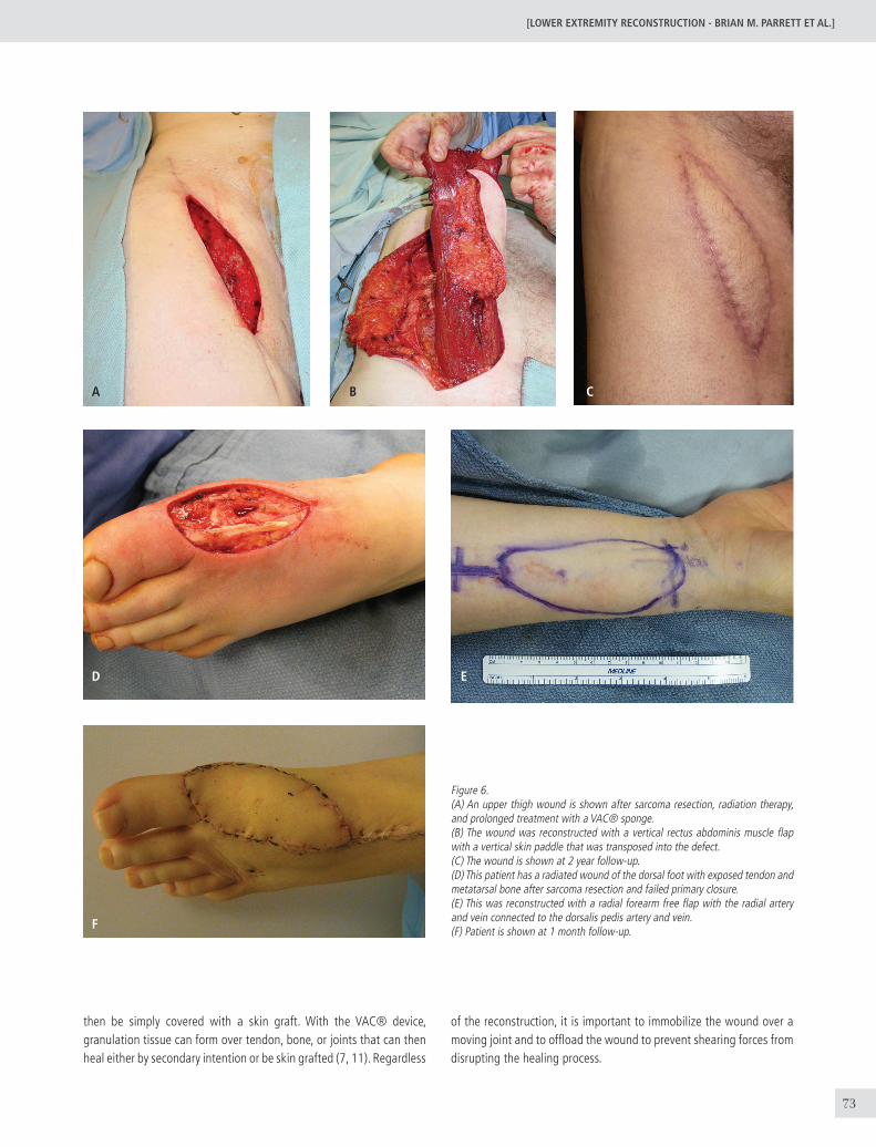
Figure 6. (A) An upper thigh wound is shown after sarcoma resection, radiation therapy, and prolonged treatment with a VAC® sponge. (B) The wound was reconstructed with a vertical rectus abdominis muscle flap with a vertical skin paddle that was transposed into the defect. (C) The wound is shown at 2 year follow-up. (D) This patient has a radiated wound of the dorsal foot with exposed tendon and metatarsal bone after sarcoma resection and failed primary closure. (E) This was reconstructed with a radial forearm free flap with the radial artery and vein connected to the dorsalis pedis artery and vein. (F) Patient is shown at 1 month follow-up.
of the reconstruction, it is important to immobilize the wound over a moving joint and to offload the wound to prevent shearing forces from disrupting the healing process.
74
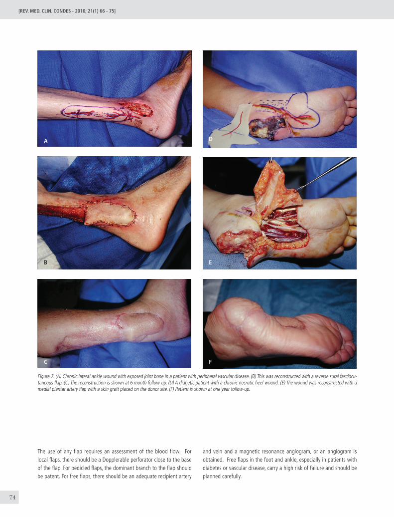
Figure 7. (A) Chronic lateral ankle wound with exposed joint bone in a patient with peripheral vascular disease. (B) This was reconstructed with a reverse sural fasciocu-taneous flap. (C) The reconstruction is shown at 6 month follow-up. (D) A diabetic patient with a chronic necrotic heel wound. (E) The wound was reconstructed with a medial plantar artery flap with a skin graft placed on the donor site. (F) Patient is shown at one year follow-up.
A
C
B
D
F
E
[REV. MED. CLIN. CONDES - 2010; 21(1) 66 - 75]
The use of any flap requires an assessment of the blood flow. For local flaps, there should be a Dopplerable perforator close to the base of the flap. For pedicled flaps, the dominant branch to the flap should be patent. For free flaps, there should be an adequate recipient artery
and vein and a magnetic resonance angiogram, or an angiogram is obtained. Free flaps in the foot and ankle, especially in patients with diabetes or vascular disease, carry a high risk of failure and should be planned carefully.
75
[LOWER EXTREMITY RECONSTRUCTION - BRIAN M. PARRETT ET AL.]
1. Harris AM, Althausen PL, Kellam J, Bosse MJ, Castillo R, Lower Extremity
Assessment Project (LEAP) Study Group. Complications following limb-
threatening lower extremity trauma. J Orthop Trauma. 2009;23:1-6.
2. Saddawi-Konefka D, Kim HM, Chung KC. A systematic review of
outcomes and complications of reconstruction and amputation for type IIIB
and IIIC fractures of the tibia. Plast Reconstr Surg. 2008;122:1796-805.
3. Scher KS, Steele FJ. The septic foot in patients with diabetes. Surgery.
1988;104:661-666.
4. Levin LS. The reconstructive ladder. An orthoplastic approach. Orthop
Clin North Am. 1993;24:393-409.
5. DeFranzo AJ, Argenta LC, Marks MW. The use of vacuum-assisted closure
therapy for the treatment of lower-extremity wounds with exposed bone.
Plast. Reconstr. Surg. 2001;108:1184-1190.
6. Parrett BM, Talbot SG, Pribaz JJ, Lee BT. A Review of Local and Regional
Flaps for Distal Leg Reconstruction. J Reconstr Microsurg. 2009 Jul 10.
[Epub ahead of print]
7. Mathes SJ, Nahai F. Classification of the vascular anatomy of muscles:
experimental and clinical correlation. Plast Reconstr Surg. 1981;67:177-187.
8. Gustilo RB, Simpson L, Nixon R, Ruiz A, Indeck W. Analysis of 511 open
fractures. Clin. Orthop. 1969;66:148-154.
9. Byrd HS, Cierny G III, Tebbetts JB. The management of open tibial
fractures with associated soft tissue loss: External pin fixation with early
flap coverage. Plast. Reconstr. Surg. 1981;68:73-82.
10. Cierny G, Byrd HS, Jones RE. Primary versus delayed soft tissue
coverage for severe open tibial fractures: A comparison of results. Clin.
Orthop. 1983;178:55-63.
11. Parrett BM, Matros E, Pribaz JJ, Orgill DP. Lower extremity trauma:
trends in the management of soft-tissue reconstruction of open tibia-fibula
fractures. Plast Reconstr Surg. 2006;117:1315-1322.
12. Khouri RK. Avoiding free flap failure. Clin Plast Surg. 1992;19:773-
781.
REFERENCES
13. Khouri RK, Shaw WW. Reconstruction of the lower extremity with
microvascular free flaps: a 10-year experience with 304 consecutive cases.
J Trauma. 1989;29:1086-1094.
14. Tomaino MM. Amputation or salvage of type 3B/3C tibial fractures:
what the literature says about outcomes. Am J Orthop. 2001;30:380-
385.
15. Mccarter MD, Jaques DP, Brennan MF. Randomized clinical trials in
soft tissue sarcoma. Surg. Oncol. Clin. North Am. 2002;11:11-22.
16. Talbert ML, Zagars GK, Sherman NE, Romsdahl MM. Conservative
surgery and radiation therapy for soft tissue sarcoma of the wrist, hand,
ankle, and foot. Cancer 1990;66:2482-2491.
17. Heller L, Kronowitz SJ. Lower extremity reconstruction. J. Surg. Oncol.
2006;94:479-489.
18. Parrett BM, Winograd JM, Garfein ES, Lee WP, Hornicek FJ, Austen
WG Jr. The vertical and extended rectus abdominis myocutaneous flap
for irradiated thigh and groin defects. Plast Reconstr Surg. 2008;122:171-
177.
19. Peat BG, Bell RS, Davis A, et al. Wound-healing complications after
soft-tissue sarcoma surgery. Plast. Reconstr. Surg. 1994;93:980-987.
20. Ndip A, Bowling F, Stickings D, Rayman G, Boulton AJ. The Diabetic
Foot in 2008: an update from the 12th Malvern Diabetic Foot Meeting. Int
J Low Extrem Wounds. 2008;7:235-8.
21. Searles JM Jr, Colen LB. Foot reconstruction in diabetes mellitus and
peripheral vascular insufficiency. Clin Plast Surg. 1991;18:467-483.
22. Brem H, Sheehan P, Rosenberg HJ, Schneider JS, Boulton AJ.
Evidence-based protocol for diabetic foot ulcers. Plast Reconstr Surg.
2006;117:193S-209S.
The authors declare no conflicts of interets with the article.