Loss of epithelial FAM20A in mice causes amelogenesis ...
12
OPEN ORIGINAL ARTICLE Loss of epithelial FAM20A in mice causes amelogenesis imperfecta, tooth eruption delay and gingival overgrowth Li-Li Li 1 , Pei-Hong Liu 1 , Xiao-Hua Xie 2 , Su Ma 1 , Chao Liu 3 , Li Chen 4 and Chun-Lin Qin 3 FAM20A has been studied to a very limited extent. Mutations in human FAM20A cause amelogenesis imperfecta, gingival fibromatosis and kidney problems. It would be desirable to systemically analyse the expression of FAM20A in dental tissues and to assess the pathological changes when this molecule is specifically nullified in individual tissues. Recently, we generated mice with a Fam20A-floxed allele containing the beta-galactosidase reporter gene. We analysed FAM20A expression in dental tissues using X-Gal staining, immunohistochemistry and in situ hybridization, which showed that the ameloblasts in the mouse mandibular first molar began to express FAM20A at 1 day after birth, and the reduced enamel epithelium in erupting molars expressed a significant level of FAM20A. By breeding K14-Cre mice with Fam20A flox/flox mice, we created K14-Cre;Fam20A flox/flox (conditional knock out, cKO) mice, in which Fam20A was inactivated in the epithelium. We analysed the dental tissues of cKO mice using X-ray radiography, histology and immunohistochemistry. The molar enamel matrix in cKO mice was much thinner than normal and was often separated from the dentinoenamel junction. The Fam20A-deficient ameloblasts were non-polarized and disorganized and were detached from the enamel matrix. The enamel abnormality in cKO mice was consistent with the diagnosis of amelogenesis imperfecta. The levels of enamelin and matrix metalloproteinase 20 were lower in the ameloblasts and enamel of cKO mice than the normal mice. The cKO mice had remarkable delays in the eruption of molars and hyperplasia of the gingival epithelium. The findings emphasize the essential roles of FAM20A in the development of dental and oral tissues. International Journal of Oral Science (2016) 8, 98–109; doi:10.1038/ijos.2016.14; published online 3 June 2016 Keywords: conditional knock out mice; enamel; FAM20A; gingival overgrowth; tooth eruption INTRODUCTION In mammals, the FAM20 family (family with sequence similarity 20) consists of the three following members: FAM20A, FAM20B and FAM20C. 1 Recent findings have led to great excitement about this small family of proteins, which have endoplasmic reticulum-entry signal sequences responsible for directing proteins into the secretory pathway and which appear to be involved in the post-translational modifications of secretory proteins. FAM20C is a kinase that phosphorylates many extracellular matrix proteins involved in biomineralization and other biological pro- cesses. 2–3 FAM20C is ubiquitously expressed, 1,4–5 and mutations in the human FAM20C gene cause Raine syndrome, an autosomal recessive disorder that demonstrates a broad spectrum of clinical manifestations. 6–10 Fam20C-deficient mice developed hypophospha- taemic rickets, along with severe dental defects. 11–12 FAM20B is a kinase that catalyses the attachment of phosphate to xylose, which is a step essential to the assembly of glycosaminoglycans during the synthesis of proteoglycans. 13 Defects in FAM20B have not been associated with human genetic disease. Constitutive inactivation of Fam20B in mice was embryonically lethal, 14 whereas conditional deletion of Fam20B in the rodent dental epithelium led to super- numerary incisors. 15 FAM20A was originally observed in the lungs and liver, and it displayed obvious differential expression in haematopoietic cells undergoing myeloid differentiation. 1 FAM20A is believed to be a pseudokinase that cannot independently catalyse the attachment of phosphate to other molecules, but it can form a functional complex with FAM20C and can enhance the capacity of the latter to phosphorylate extracellular proteins in their secretory pathways. 16 In vitro studies have shown that FAM20A potentiates the kinase 1 Harbin Medical University School of Stomatology, Harbin, China; 2 Department of Stomatology, the Second Affiliated Hospital of Harbin Medical University, Harbin, China; 3 Department of Biomedical Sciences and Center for Craniofacial Research and Diagnosis, Texas A&M University Baylor College of Dentistry, Dallas, USA and 4 Longjiang Scholar Laboratory, the First Affiliated Hospital of Harbin Medical University, Harbin, China Correspondence: Professor L Chen, Longjiang Scholar Laboratory, the First Affiliated Hospital of Harbin Medical University, 23 Youzheng Street, Harbin 150001, China E-mail: [email protected] Professor CL Qin, Department of Biomedical Sciences and Center for Craniofacial Research and Diagnosis, Texas A&M University Baylor College of Dentistry, 3302 Gaston Avenue, Room 400, Dallas, TX 75246, USA E-mail: [email protected] Accepted 25 February 2016 International Journal of Oral Science (2016) 8, 98–109 www.nature.com/ijos
Transcript of Loss of epithelial FAM20A in mice causes amelogenesis ...

OPEN
ORIGINAL ARTICLE
Loss of epithelial FAM20A in mice causesamelogenesis imperfecta, tooth eruption delay andgingival overgrowth
Li-Li Li1, Pei-Hong Liu1, Xiao-Hua Xie2, Su Ma1, Chao Liu3, Li Chen4 and Chun-Lin Qin3
FAM20A has been studied to a very limited extent. Mutations in human FAM20A cause amelogenesis imperfecta, gingival
fibromatosis and kidney problems. It would be desirable to systemically analyse the expression of FAM20A in dental tissues and
to assess the pathological changes when this molecule is specifically nullified in individual tissues. Recently, we generated mice
with a Fam20A-floxed allele containing the beta-galactosidase reporter gene. We analysed FAM20A expression in dental tissues
using X-Gal staining, immunohistochemistry and in situ hybridization, which showed that the ameloblasts in the mouse
mandibular first molar began to express FAM20A at 1 day after birth, and the reduced enamel epithelium in erupting molars
expressed a significant level of FAM20A. By breeding K14-Cre mice with Fam20Aflox/flox mice, we created K14-Cre;Fam20Aflox/flox
(conditional knock out, cKO) mice, in which Fam20A was inactivated in the epithelium. We analysed the dental tissues of cKO
mice using X-ray radiography, histology and immunohistochemistry. The molar enamel matrix in cKO mice was much thinner
than normal and was often separated from the dentinoenamel junction. The Fam20A-deficient ameloblasts were non-polarized
and disorganized and were detached from the enamel matrix. The enamel abnormality in cKO mice was consistent with the
diagnosis of amelogenesis imperfecta. The levels of enamelin and matrix metalloproteinase 20 were lower in the ameloblasts and
enamel of cKO mice than the normal mice. The cKO mice had remarkable delays in the eruption of molars and hyperplasia of
the gingival epithelium. The findings emphasize the essential roles of FAM20A in the development of dental and oral tissues.
International Journal of Oral Science (2016) 8, 98–109; doi:10.1038/ijos.2016.14; published online 3 June 2016
Keywords: conditional knock out mice; enamel; FAM20A; gingival overgrowth; tooth eruption
INTRODUCTION
In mammals, the FAM20 family (family with sequence similarity 20)consists of the three following members: FAM20A, FAM20B andFAM20C.1 Recent findings have led to great excitement about thissmall family of proteins, which have endoplasmic reticulum-entrysignal sequences responsible for directing proteins into the secretorypathway and which appear to be involved in the post-translationalmodifications of secretory proteins.FAM20C is a kinase that phosphorylates many extracellular matrix
proteins involved in biomineralization and other biological pro-cesses.2–3 FAM20C is ubiquitously expressed,1,4–5 and mutations inthe human FAM20C gene cause Raine syndrome, an autosomalrecessive disorder that demonstrates a broad spectrum of clinicalmanifestations.6–10 Fam20C-deficient mice developed hypophospha-taemic rickets, along with severe dental defects.11–12
FAM20B is a kinase that catalyses the attachment of phosphate toxylose, which is a step essential to the assembly of glycosaminoglycansduring the synthesis of proteoglycans.13 Defects in FAM20B have notbeen associated with human genetic disease. Constitutive inactivationof Fam20B in mice was embryonically lethal,14 whereas conditionaldeletion of Fam20B in the rodent dental epithelium led to super-numerary incisors.15
FAM20A was originally observed in the lungs and liver, and itdisplayed obvious differential expression in haematopoietic cellsundergoing myeloid differentiation.1 FAM20A is believed to be apseudokinase that cannot independently catalyse the attachment ofphosphate to other molecules, but it can form a functional complexwith FAM20C and can enhance the capacity of the latter tophosphorylate extracellular proteins in their secretory pathways.16
In vitro studies have shown that FAM20A potentiates the kinase
1Harbin Medical University School of Stomatology, Harbin, China; 2Department of Stomatology, the Second Affiliated Hospital of Harbin Medical University, Harbin, China;3Department of Biomedical Sciences and Center for Craniofacial Research and Diagnosis, Texas A&M University Baylor College of Dentistry, Dallas, USA and 4Longjiang ScholarLaboratory, the First Affiliated Hospital of Harbin Medical University, Harbin, ChinaCorrespondence: Professor L Chen, Longjiang Scholar Laboratory, the First Affiliated Hospital of Harbin Medical University, 23 Youzheng Street, Harbin 150001, ChinaE-mail: [email protected] CL Qin, Department of Biomedical Sciences and Center for Craniofacial Research and Diagnosis, Texas A&M University Baylor College of Dentistry, 3302 GastonAvenue, Room 400, Dallas, TX 75246, USAE-mail: [email protected] 25 February 2016
International Journal of Oral Science (2016) 8, 98–109www.nature.com/ijos

activity of FAM20C and promotes the FAM20C-catalysed attachmentof phosphate to enamel matrix proteins, such as enamelin.16 Thein vitro findings that FAM20A stimulates FAM20C-dependent phos-phorylation of certain enamel matrix proteins have provided somebasic information for understanding the molecular pathogenesisunderlying the enamel defects associated with the mutations in, anddeletion of, the FAM20A gene. The mutations in human FAM20Acause amelogenesis imperfecta with gingival fibromatosis syndrome(AIGFS, OMIM #614253) and enamel renal syndrome (ERS, OMIM#204690).17–21 Enamel defects have been described in mice withconstitutive loss of Fam20A.14 Related dental anomalies in humansubjects include generalized hypoplastic enamel, tooth eruption delay,intrapulpal calcification and fibrotic enlargement of the gingiva.20
Compared with FAM20C, FAM20A has been studied only to a limitedextent, and there is a need to profile systematically the expression ofFAM20A in dental tissues and to analyse the pathological andmolecular changes that occur when this molecule is specifically ablatedin individual tissues or cells. Recently, we created Fam20A-floxed mice,in which the floxed allele contained the beta-galactosidase (LacZ)reporter gene, allowing us to reveal the expression of FAM20A withX-Gal staining and to inactivate this molecule in the tissues or cells of
interest. In this study, we specifically ablated Fam20A from mouseepithelium by breeding Fam20Aflox/flox mice with K14-cre mice to createK14-Cre;Fam20Aflox/flox (K14-Cre;Fam20AΔ/Δ or conditional knock out(cKO)) mice. The cKO mice manifested enamel defects, tootheruption delay and gingival epithelium overgrowth.
MATERIALS AND METHODS
Generation of Fam20A-floxed miceMice with a Fam20A-floxed allele were generated by Biocytogen(Beijing, China). In the targeting vector used to create the floxed allele,exogenous elements were inserted into intron 4 and intron 8 of themouse Fam20A gene; the region from exon 5 through exon 8 wasflanked by two loxP sequences (Figure 1a). Both intron 4 and intron 8are sufficiently large, and insertion of loxP elements into these twointrons is not expected to interfere with mRNA splicing. Whenplanning the targeting strategy, we calculated that removal of exon 5though exon 8 would result in a frame shift for protein translation andcreate a truncated FAM20A with 247 amino acids, which could besubject to nonsense-mediated decay. In addition, the amino acidsequence encoded by the region of exon 5 through exon 8 in theFam20A gene is highly conserved among species, and it also shows
Figure 1 Targeting vector for creating Fam20A-mutant mice and genotyping strategy. (a) The mouse Fam20A gene contains 11 exons (green boxes). Intron4 is the largest among the 10 introns. In the targeting vector, an IRES-lacZ-Neo cassette flanked by two flippase recognition target (FRT) sites (red ovals)was inserted into intron 4, and one LoxP site (yellow triangle) was placed into intron 8, which is also relatively large. Recombination after Flp recombinasescission removed the IRES-lacZ-Neo cassette from the Fam20AlacZ-flox allele and produced the conditional allele Fam20Aflox, in which the region of exons 5through 8 was flanked by two LoxP sites (yellow triangles). (b) We used the primer set of a and b to distinguish the Fam20AlacZ-flox allele from the WT allele;PCR with these primers produced a 273-bp fragment for the mutant allele and 216-bp fragment for WT allele. (c) The primer set of c and d was used toidentify the Fam20Aflox allele after the IRES-lacZ-Neo cassette was removed by Flp recombinase. Note that the use of primers c and d was not expected togenerate any PCR products for the Fam20AlacZ-flox allele. (d) The primer set of c and b was used to identify the Fam20A-ablated (Fam20Δ) allele in theK14-Cre;Fam20Aflox/+ or the K14-Cre;Fam20Aflox/flox mice; PCR with these primers did not produce any fragment for the Fam20A-floxed (Fam20Aflox/+ andFam20Aflox/flox) or WT alleles. PCR, polymerase chain reaction; WT, wild type.
Loss of epithelial Fam20A in mice causes dental defectsLL Li et al
99
International Journal of Oral Science

strong similarity to the amino acid sequence of the kinase domain inthe Fam20C and Fam20B genes. Therefore, ablating the region fromexon 5 through exon 8 in the Fam20A gene should result in functionalloss of the FAM20A protein.Intron 4 of the targeting vector also contained an IRES-lacZ-Neo
cassette flanked by two FRT sites so that a conditional allele could begenerated after treatment with flippase (Flp) recombinase (Figure 1a).Before introducing Flp into the mice, the LacZ reporter gene encodingbeta-galactosidase was present, allowing for the utilization of X-Galstaining to visualize the expression pattern of FAM20A. After theintroduction of Flp recombinase, the IRES-lacZ-Neo cassette wasremoved, leaving the region of exons 5–8 flanked by two loxP sites(Figure 1a). We designated the mice (founders and their progenies)with one allele of Fam20A containing the IRES-lacZ-Neo cassette as“Fam20AlacZ-flox/+ mice” and referred to the mice with one allele ofFam20A that was floxed by two loxP elements but that contained noIRES-lacZ-Neo cassette as “Fam20Aflox/+ mice.” The Fam20AlacZ-flox/+
mice were used for X-Gal staining to reveal the expressionprofile of FAM20A. The Fam20Aflox/+ mice were inbred to createFam20Aflox/flox mice.DNA extracted from the mouse tails was analysed by polymerase
chain reaction (PCR) for genotyping to identify the Fam20AlacZ-flox/+
allele using the following set of primers: forward, 5′-GTCATTGAAGGAGTTGCCACTGTC-3′ (“a” in Figure 1a); and reverse,5′-CAGGTAGCCTCAAACATGCAACAT-3′ (“b” in Figure 1a). PCRwith these primers gave rise to a product of 273 base pairs (bp) for theFam20AlacZ-flox allele and a 216-bp fragment for the wild-type allele(Figure 1b).
Generation of K14-Cre;Fam20Aflox/flox
First, we mated K14-Cre mice, which express Cre recombinase in theepithelium of the oral mucosa approximately 10.5 days post coitum(d.p.c.),22–23 with Fam20Aflox/flox mice to create K14-Cre;Fam20Aflox/+
mice. Then, the K14-Cre;Fam20Aflox/+ mice were bred withFam20Aflox/flox mice to generate K14-Cre;Fam20Aflox/flox (that is, K14-Cre;Fam20AΔ/Δ) mice, which we refer to as cKO mice in this report.DNA samples from mouse tails were analysed by PCR for genotyping.A set of primers specific for the Cre transgene was adopted to identifythe Cre transgene. The sequences of primers specific for the Cretransgene were: forward, 5′-ATTTGCCTGCATTACCGGTC-3′; andreverse, 5′-ATCAACGTTTTCTTTTCGG-3′. PCR with the Cre-speci-fic primers produced a 350-bp fragment when the DNA samples frommice expressing the Cre recombinase were used as the template andproduced no PCR product for the wild-type allele (data not shown).One pair of primers with the forward sequence of 5′-GA
AACTTTGCAGTCCTTGTTCCC-3′ (“c” in Figure 1a, located inintron 4 and upstream of the first Frt element in the targeting vector)and the reverse sequence of 5′-GCACTATCAATGCCAAGTTTCC-3′(“d” in Figure 1a, located in intron 4 and downstream of the secondLoxP site) was used to distinguish the Fam20A-floxed (Fam20Aflox/+ orFam20Aflox/flox) alleles from the wild-type alleles; the use of theseprimers generated a 350-bp fragment for the Fam20A-floxed alleles inthe Fam20Aflox/+ or Fam20Aflox/flox mice and a 234-bp fragment for thewild-type alleles (Figure 1c).Another set of primers was designed to distinguish the Fam20A-
ablated (Fam20AΔ) allele in the K14-Cre;Fam20Aflox/+ or the K14-Cre;Fam20Aflox/flox mice from the Fam20A-floxed (Fam20Aflox/+ andFam20Aflox/flox) or wild-type alleles. The sequences of primers usedto identify the Fam20A-ablated allele were: forward, 5′-GAAACTTTGCAGTCCTTGTTCCC-3′ (“c” in Figure 1a); and reverse, 5′-CAGGTAGCCTCAAACATGCAACAT-3′ (“b” in Figure 1a); this set of
primers generated a 370-bp fragment for the Fam20A-null alleles andproduced no PCR band for the Fam20A-floxed or wild-type alleles(Figure 1d).The use of mice was approved by the Experimental Animal Ethics
Committee of Harbin Medical University, China, and was inaccordance with the recommendations in the Guide for Care andUse of Laboratory Animals published by the USA National Institutesof Health. All the mice analysed in this study were fed a normal (hard)diet after weaning. For each analysis at each time point, specimensfrom at least three mice were evaluated.
Analyses at the gross levelIn this investigation, we observed the incisors, molars and gingiva ofthe mice at different ages. The mice were weighed every week frombirth until postnatal 56 days to monitor animal growth. The averagevalues of the body weight calculated from three mice in each agegroup were used to generate growth curves.
X-Gal stainingFor X-Gal staining, the mandibles dissected from the Fam20AlacZ-flox/+
and wild-type (control) mice at the ages of 13.5, 14.5, 15.5 and 17.5days post coitum and 0, 1, 5, 7 and 11 days after birth, respectively,were fixed in 4% ice-cold paraformaldehyde for 1 h on a shaker andwere washed with phosphate-buffered saline (PBS) solution for45 min. Then, the mandibles were decalcified in 15% ethylenediaminetetraacetic acid (EDTA) solution (pH 7.4) at 4 °C for 1–7 days,depending on the ages of the animals. The samples were processed forsucrose infiltration, and 10-μm serial frozen sections were preparedwith a cryostat microtome. The frozen sections were incubated inX-Gal solution (Gold Biotechnology, St Louis, MO, USA) for 24 h at37 °C in darkness and were counterstained with Sirius Red andobserved under a microscope.
Haematoxylin and eosin and immunohistochemistry stainingFor haematoxylin and eosin (H&E) and immunohistochemistry (IHC)staining, the mouse mandibles were fixed with 4% paraformaldehydein PBS solution at 4 °C overnight and then were decalcified in 15%EDTA solution at 4 °C for 0–7 days, depending on the ages of theanimals. The samples were processed for paraffin embedding, andserial sections of 5 μm in thickness were cut for H&E and IHCanalyses.The polyclonal antibody against FAM20A (Abcam, Cambridge, MA,
USA) was used at a dilution of 1:200. The polyclonal antibodiesagainst enamelin (ENAM), ameloblastin (AMBN) and matrix metal-loproteinase 20 (MMP20) were purchased from Santa Cruz (SantaCruz, CA, USA) and were used at dilutions of 1:200, 1:800 and 1:100,respectively. The specimens of the normal and cKO mice from thesame litters were stained in the same batch of experiments to ensurebetter comparability. Normal rabbit serum at the same concentrationsas the polyclonal antibodies was used to replace the polyclonal anti-FAM20A, anti-ENAM and anti-AMBN antibodies and served as anegative control. Goat serum was used to replace the anti-MMP20antibody, acting as a negative control for this polyclonal antibody. Allof the IHC experiments were performed using the ABC kit forpolyclonal antibodies (Vector Laboratories, Burlingame, CA, USA).The 3,3′-diaminobenzidine (DAB) kit (Vector Laboratories, Burlin-game, CA, USA) was used for colour development, according to themanufacturer’s instructions. The sections were counterstained withmethyl green. Each set of the IHC experiments was repeated at leastthree times.
Loss of epithelial Fam20A in mice causes dental defectsLL Li et al
100
International Journal of Oral Science

In situ hybridizationFor in situ hybridization (ISH) analyses, diethylpyrocarbonate-treatedsolutions were used for processing the mouse mandibles and for thehybridization experiments to ensure RNase-free conditions. Wegenerated a 681-bp RNA probe that was complementary to the mouseFAM20A mRNA, using the same method as we previouslydescribed.11,24 Briefly, a primer set with 5′-ACAATTCAACCT-TACCTCCTTGG-3′ (in exon 2 of the mouse Fam20A gene) forforward and 5′-CTTTTCCTGACAGCGAGTAGG-3′ (in exon 8) forreverse was used to generate a cDNA fragment using PCR, with totalRNA extracted from the molars of 16.5-day-old mice as the template.Next, the PCR products were cloned into the pCRII-TOPO vector(Promega, Madison, WI, USA) and were transformed into competentEscherichia coli. The plasmid DNA was isolated from E. coli andsequenced. Digoxigenin-labelled single-stranded RNA probes weresynthesized and labelled with digoxigenin using an RNA labelling kit(Roche, Indianapolis, IN, USA), and the DIG-labelled RNA probeswere detected by enzyme-linked immunoassay with a specific anti-DIG-AP antibody conjugate, as we previously described.11,24 TheseRNA probes were used to detect FAM20A mRNA in the paraffinsections containing tissues from the mouse mandible.
Plain X-ray and micro-computed tomographyMandibles of normal and cKO mice at the ages of 1, 2, 4 and6 weeks after birth were examined by a Faxitron X-ray RadiographySystem (MX-20; Faxitron X-ray, Wheeling, IL, USA) at a voltage of26 kVp and an exposure time of 8.5 s. The first molars dissectedfrom the mandibles of the 4- and 6-week-old mice were radio-graphed by a micro-computed tomography (μCT) system (μCT35;Scanco Medical AG, Bassersdorf, Switzerland) with a 12-μm voxelsize using the following parameters: 114 mA (current), 70 kVp(voltage) and 300 μs (exposure time). The scanning process lastedfor approximately 1 h per sample and generated approximately 600images for each sample.
RESULTS
Expression of FAM20A in the dental tissues of miceBecause the development of the mouse first molar resembles and isusually used to represent human teeth, the current study focused onthe expression of FAM20A in the mandibular first molar. We wereparticularly interested in the presence or absence of FAM20A signals inameloblasts at different developmental stages of the mouse molarbecause the cKO mice had enamel defects with relatively normaldentin. In addition, representative images from the X-Gal staining ofmandibular incisor regions are presented to demonstrate the expres-sion of FAM20A in incisor ameloblasts at different stages.X-Gal staining was not observed in the molars of wild-type mice,
confirming the high specificity of this approach. X-Gal staining showedthat, at 17.5 days post coitum, FAM20A signals were present in theodontoblasts (arrowhead) of the mandibular incisor but not in any cellsof the first molar in the Fam20AlacZ-flox/+ mice (Figure 2a). At postnatalday 0 (birth), FAM20A signals were absent from the ameloblasts butwere present in the odontoblasts in the mandibular first molars ofFam20AlacZ-flox/+ mice (Figure 2b); at this time point, FAM20A signalswere also observed in both the ameloblasts and odontoblasts of themandibular incisor; in the incisor, FAM20A was localized in thesecretory stage ameloblasts (Figure 2b and 2b1). At postnatal day 1,although FAM20A was observed in both the ameloblasts and odonto-blasts in the mandibular first molar, the X-Gal staining of the formercells was weaker than previously (Figure 2c). At postnatal day 1,FAM20A was primarily observed in the ameloblasts of the cusp tips inthe mandibular first molar. At postnatal day 5, the intensity of FAM20Asignals in the molar ameloblasts became stronger than at postnatal day 1and was comparable to that in the odontoblasts of the mandibular firstmolar (Figure 2d). At postnatal day 5, significant FAM20A signals werealso observed in some cells of the stellate reticulum (Figure 2d, asterisk).At postnatal day 7, the FAM20A signals in the molar ameloblasts weresimilar to those in the odontoblasts; intensive staining was observed innearly all of the ameloblasts and odontoblasts (Figure 2e). At postnatal day11, when the mandibular first molar was in its eruptive phase, a significantlevel of FAM20A was observed in the reduced enamel epithelium(Figure 2f, arrows). In the mandibular incisors of 5- and 7-day-old
a
c d e
b b1
M1
Inc
17.5 dpc: Fam20AlacZ-flox/+
P1: Fam20AlacZ-flox/+ P5: Fam20AlacZ-flox/+
*
P7: Fam20AlacZ-flox/+
M1
Inc
P0: Fam20AlacZ-flox/+
Loss of epithelial Fam20A in mice causes dental defectsLL Li et al
101
International Journal of Oral Science

Fam20AlacZ-flox/+ mice, X-Gal staining showed that FAM20A signalswere localized in the secretory stage ameloblasts and maturation stageameloblasts (Supplementary Figure 1a and 1b). X-Gal staining alsoshowed positive signals for FAM20A in the gingiva of postnatal 28-day-old mice (Supplementary Figure 1c). Throughout the observationperiod, the beta-galactosidase activity was either totally absent orbarely visible in the cells of the dental follicles and periodontalligaments. The X-Gal staining analyses demonstrated the absence ofFAM20A signals in the ameloblasts and odontoblasts of the molarsand incisors in the Fam20AlacZ-flox/+ mice at 13.5, 14.5 and 15.5 dayspost coitum (data not shown). These findings indicated that the
ameloblasts in the first molar of the mouse mandible began to expressFAM20A at 1 day after birth, and the stellate reticulum and reducedenamel epithelium expressed significant levels of FAM20A.IHC analyses (Figure 2h–2j) showed that, at postnatal day 1, the
anti-FAM20A immunoreactivity was weakly positive in the amelo-blasts and odontoblasts of the mandibular first molar (Figure 2h). Atday 5 after birth, FAM20A was observed in the ameloblasts (arrow),stellate reticulum (asterisk) and odontoblasts (arrowhead) of themandibular first molar (Figure 2i and 2i1), in agreement with theX-Gal staining results. In the ameloblasts, FAM20A protein wasuniformly located in the cytoplasm on the enamel matrix side of
f g h
ji1i
k l m
P11: Fam20AlacZ-flox/+ P1: WT (Ctrl)
P5: Anti-FAM20A IHC
*
P1: Anti-FAM20A IHC
P5: IHC Ctrl
P11: ISHP1: ISH17.5 dpc: ISH
Figure 2 Expression of FAM20A in the mouse dental tissues. (a) X-Gal staining revealed the presence of FAM20A signals in the odontoblasts (arrowhead) ofthe mandibular incisors and its total absence from the first molars (M1) of the Fam20AlacZ-flox/+ mice at 17.5 days post coitum. (b) At postnatal day 0 (P0),FAM20A signals were observed in the odontoblasts (arrowhead) but not in the ameloblasts (arrow) of the mandibular first molar (M1); at this stage, FAM20Asignals were also observed in the secretory stage ameloblasts (green arrow) in the incisor; (b1) higher magnification view of the box area in b. (c) At P1, theFAM20A signals in the odontoblasts (arrowhead) were stronger than in the ameloblasts (arrow). (d) At P5, the FAM20A signals in the ameloblasts (arrow) ofsome regions were stronger than in the odontoblasts (arrowhead); relatively strong FAM20A signals were also observed in some cells of the stellate reticulum(asterisk). (e) At P7, the intensity of FAM20A signals in the ameloblasts (arrow) was similar to that in the odontoblasts (arrowhead).(f) At P11, FAM20A signals were present in the reduced enamel epithelium (arrows) and odontoblasts (arrowhead) in the Fam20AlacZ-flox/+ mice. (g) Wild-typemice did not show any X-Gal-positive staining. (h) Immunohistochemistry showed weak anti-FAM20A immunoreactivity in the ameloblasts (arrow) and theodontoblasts (arrowhead) of the mandibular first molar at P1. (i) At P5, strong anti-FAM20A immunoreactivity was observed in the first molar ameloblasts(arrow), odontoblasts (arrowhead) and stellate reticulum (asterisk); (i1) higher magnification view of the box area in i revealed that, in the ameloblasts,FAM20A protein was almost exclusively located in the cytoplasm on the enamel matrix side of the nuclei. (j) Immunohistochemistry staining with the firstantibody-free control showed no FAM20A signal in the ameloblasts (arrow) or odontoblasts (arrowhead). (k) In situ hybridization analyses showed that, at17.5 days post coitum, FAM20A mRNA was not observed in any of the tooth germs but was observed in the tongue root region (arrow). (l) At P1, FAM20AmRNA was observed in the ameloblasts (arrow) and in the odontoblasts (arrowhead) of the mandibular first molar. (m) At P11, FAM20A mRNA was presentin the reduced enamel epithelium (arrows) and odontoblasts (arrowhead) of the first molar. (a–g) X-Gal staining of the mouse mandible; (h–j) anti-FAM20AIHC staining of the first molar region in the mouse mandible; (k–m) in situ hybridization staining of mouse head and jaw bones. Scale bars, 200 μm (a andb); 50 μm (b1); 10 μm (i1); 500 μm (k); 200 μm (l and m); 100 μm (all other images). P0, P1, P5, P7 and P11 are abbreviations of postnatal day 0, 1, 5,7 and 11, respectively. Ctrl, control; d.p.c., days post coitum; IHC, immunohistochemistry; Inc, incisor; ISH, in situ hybridization.
Loss of epithelial Fam20A in mice causes dental defectsLL Li et al
102
International Journal of Oral Science

the nuclei (that is, towards the dentinoenamel junction direction). Inthe odontoblasts, FAM20A protein was primarily located in thecytoplasm on the predentin side of the nuclei. It should be notedthat there are currently only a few anti-FAM20A antibodies commer-cially available, and we tried two of them in this study. Although theanti-FAM20A antibody described in this report recognized FAM20A,its titre and specificity were low; thus, the IHC analyses in thisinvestigation were not as sensitive as the X-Gal staining in assessingthe expression of FAM20A. Nevertheless, the IHC experiments werenecessary because such approaches could assess the actual localizationof FAM20A protein in the cells, constituting an advantage over theX-Gal staining used to monitor the expression of this molecule in theFam20AlacZ-flox/+ mice, which contained beta-galactosidase in thenuclei of FAM20A-expressing cells.ISH analyses demonstrated that, at 17.5 days post coitum, FAM20A
mRNA was present in the lingual root region of the embryo but wasabsent from the ameloblasts or the odontoblasts of any tooth germs(Figure 2k). At postnatal day 1, FAM20A mRNA was detected in boththe ameloblasts and odontoblasts of the mandibular first molar; thesignals in the former were weaker than in the latter (Figure 2l),consistent with the X-Gal staining results. At postnatal day 11, whenthe mandibular first molar was in its eruptive phase, and the outer andinner enamel epithelia were fused to become reduced enamelepithelium, a relatively strong level of FAM20A mRNA was observedin the reduced enamel epithelium (Figure 2m, arrows). The ISH datalent further support to the conclusion drawn from the X-Gal stainingthat the ameloblasts in the mouse mandibular first molar started toexpress FAM20A at postnatal day 1 and that the reduced enamelepithelium expressed a significant level of FAM20A.
Overall assessment of Fam20A-mutant miceThe body sizes, teeth and gingivae of the Fam20Aflox/+ mice, Fam20A-flox/flox mice or K14-Cre;Fam20Aflox/+ mice were same as the age-matched wild-type mice, indicating that insertion of the loxP elements
or Fam20A haploinsufficiency did not cause developmental abnorm-alities. Because the Fam20Aflox/flox mice were completely normal, weused as normal control (Ctrl) mice from the same litters as the cKOmice created during the crossbreeding regime. Utilizing Fam20Aflox/flox
littermates of cKO mice as normal controls not only reduced thenumber of utilized mice, but it also prevented potential variances thatmight result from comparing animals from different litters.The cKO mice were apparently smaller than the normal mice at
postnatal 28 and 35 days. As the mice aged, this difference in body sizebetween the cKO and normal mice became decreasingly remarkable;the body sizes of 56-day-old cKO mice were close to those of thenormal control mice at the same age (Figure 3a).
Enamel defects in cKO miceThe labial sides of the upper and lower incisors of the cKO mice had achalky-white and opaque appearance, which was in clear contrast to those ofthe normal mice, which showed a yellow-brown colour and a transparentglossy appearance (Figure 3b and 3b1). Compared with the mandibularmolars of the normal mice, the molars of the cKOmice had a rough surfaceand appeared yellow, likely caused by rapid, abrasive loss of enamel andexposure of the underlining yellow dentin (Figure 3b3 and 3c3).Because plain X-ray analyses could not distinguish the enamel layer
from the dentin, the radiopaque area on X-ray radiography reflectedthe whole mineralized walls of pulp chamber and root canals. PlainX-ray examination showed that, at postnatal day 7, the mineralizedwalls of pulp chambers in the crowns of the mandibular first molars ofthe cKO mice were thinner than in the normal control mice(Figure 4a), likely due to a reduction in the thickness of the enamellayer. At postnatal day 14, the pulp chamber walls were much thinnerin the molar crowns of the cKO mice than in the control mice(Figure 4b). At postnatal day 28 and day 42, the thin wall defects in themandibular first molars of the cKO mice became even moreremarkable (Figure 4c and 4d), which could be attributed to the lossof the hypomineralized enamel layer after the mice started to chew
30
25
20
15
Bod
y w
eigh
t /g
10
5
00 7 14 21
Age (days after birth)
Ctrl
ba b1 b2 b3
c c1 c2 c3
cKO
28 35 42 49 56
P42 Ctrl
P42 cKO
Figure 3 Assessment of normal and cKO mice at the gross level. (a) Growth curve of mice from postnatal day 0 to 56. The 28- and 35-day-old cKO micewere smaller than the control mice, whereas the body weights of the 56-day-old cKO were close to those of the normal (control) mice. (b) Gross photographof incisors in the 42-day-old control mice; (b1) the upper incisor (with gingiva removed) in 42-day-old control mice; (b2) the lower incisor (with gingivaremoved) in 42-day-old control mice; (b3) the mandibular molars (with gingiva removed) in 42-day-old control mice. (c) Gross photograph of incisors in the42-day-old cKO mice; (c1) the upper incisor (with gingiva removed) in 42-day-old cKO mice; (c2) the lower incisor (with gingiva removed) in 42-day-old cKOmice; (c3) the mandibular molars (with gingiva removed) in 42-day-old cKO mice. Note the differences in the colour and surface smoothness between theteeth of normal and cKO mice. Ctrl, normal control group; cKO, conditional knockout group.
Loss of epithelial Fam20A in mice causes dental defectsLL Li et al
103
International Journal of Oral Science
a
CtrlM1 M2
M1 M2
P7
cKO
b
CtrlM1 M2
M1 M2
P14
cKO
c
e
g g1 h h1
f f1e1
Ctrl
Ctrl
Ctrl
M1 M2
M1 M2
P28
P28 P28
P42P42
cKO
d
CtrlM1 M2
M1 M2
P42
cKO
cKO
cKO
Figure 4 X-ray radiography analyses. (a–d) The plain X-ray radiographic analyses of the mandibles from the 7-day-old (P7), 14-day-old (P14), 28-day-old (P28)and 42-day-old (P42) mice. (e–h) μCT analyses of mandibular first molars from 28-day-old (P28) and 42-day-old (P42) mice. (a) At P7, the mineralized walls ofpulp chambers in the mandibular first molars of the cKO mice appeared thinner than in control mice; the first molar in the cKO mice was covered by a largeamount of overtopping bone (arrow) on the oral cavity side, whereas no obvious bone tissue was observed on the oral cavity side of the first molar in the normalmice. (b) At P14, the pulp chamber walls in cKO mice were thinner compared with control mice, and a significant amount of bone (arrow) was still presentbetween the mandibular first molar and the oral cavity in the cKO mice. (c, d) In the P28 and P42 cKO mice, the pulp chamber walls were much thinner, and thecrowns were shorter than in the control mice. These X-ray observations indicated that the eruption of the mandibular first and second molars in the cKO mice wasdramatically delayed. M1, mandibular first molar; M2, mandibular second molar; arrows indicate the bone tissues overlying the oral cavity side of the mandibularfirst molars. (e, f) Compared with the control mice, the occlusal surface of the mandibular first molar in the P28 cKO mice was rougher. (e1, f1) On section views,an enamel layer (arrow) was clearly visible in the molars of the normal mice, whereas no enamel or enamel-like structures could be identified in the molars of thecKO mice. (g, h) At P42, the occlusal surface of the first molar in the cKO mice was nearly flat, and the tooth crown was much shorter than in the control mice.(g1, h1) An enamel layer (arrow) was clearly visible in the control mice but not in the cKO mice. e, f, g and h: full views; e1, f1, g1 and h1: longitudinal-section views. cKO, conditional knock out; Ctrl, control; μCT, micro-computed tomography.
Loss of epithelial Fam20A in mice causes dental defectsLL Li et al
104
International Journal of Oral Science

food. In addition, the crowns of the mandibular first molars of thecKO mice were much shorter than in the normal mice. The plainX-ray examination also revealed a dramatic delay in the eruption ofthe mandibular first and second molars in the cKO mice (see below).The μCT analyses could clearly distinguish the enamel layer from
the dentin in the mandibular first molars of the normal mice, whereasin the mandibular first molars of the cKO mice, μCT radiography didnot reveal any structure in the pulp chamber wall with a mineraliza-tion level differing from that of the molar dentin (Figure 4e–4h1).These observations indicated that either the mineralization level of theFam20A-deficient enamel was too low to make it distinguishable fromthe dentin, or perhaps the very thin layer of enamel that formed in thecKO mice was rapidly lost after the tooth began to function. Atpostnatal day 28, the occlusal surface of the mandibular first molar inthe cKO mice appeared rougher than in the normal mice (Figure 4eand 4f). At postnatal day 42, the mandibular first molars in the cKOmice lost nearly all of their cusps and became flattened, in clearcontrast to those in the normal mice, which had distinct cusps and aglossy surface (Figure 4g and 4h). The crown of the mandibular firstmolar in the 42-day-old cKO mice was much shorter than in thenormal mice at the same age. The shortened crown of the mandibularfirst molar in the 42-day-old cKO mice was likely attributed to therapid loss of coronal hard tissue due to attrition. The mice start to
chew food after weaning at approximately 21 days postnatally. The lossof mineralized tissue in the first molar crowns of 28-day-old cKOmice, which had been in occlusion for approximately 7 days, wasmuch less than in the molar crowns of 42-day-old cKO mice, whichhad been in function for approximately 21 days.Histology analyses with H&E staining showed that, at postnatal day
1, the overall morphology of the mandibular first molars in the cKOmice was similar to that of the normal mice (Figure 5a and 5b).Although the ameloblasts in the normal mice were highly columnar,polarized and well organized (Figure 5a1, arrow), those in the cKOmice were low columnar or cuboid, non-polarized and disorganized(Figure 5b1, arrow). At postnatal day 5, the mandibular first molars inthe normal mice formed a significant amount of enamel matrix(Figure 5c and 5c1, asterisk in 5c1), whereas those of the cKO micehad very little enamel matrix (Figure 5d and 5d1, asterisk in 5d1). Theameloblast layer in the mandibular first molars of the normal micewas closely attached to the enamel matrix (Figure 5c and 5c1), whereasthat in the cKO mice was detached from the enamel matrix (Figure 5dand 5d1). The separation of ameloblasts from the enamel matrix in thecKO mice created a void space (“VS” in Figure 5d), in which acertain amount of liquid seemed to accumulate. The ameloblasts inthe mandibular first molars of the cKO mice (arrow in Figure 5d1)were “pushed” away from the enamel matrix and became flattened
a
P1: Ctrl
a1
c
P5: Ctrl
c1
b
P1: cKO
*
P5: cKO
** VS
b1
d1d
e
P5: Ctrl
e1
P7: cKO
**De
VS
Def1f
Figure 5 H&E staining of the mandibular first molars from 1-day-old (P1), 5-day-old (P5) and 7-day-old (P7) mice. (a) H&E staining of the mandibular firstmolar region from 1-day-old normal (control) mice. (a1) Higher magnification view of the box area in a showed that the ameloblasts (arrow) in control micewere high columnar, polarized and well aligned. (b) H&E staining of the mandibular first molar region from 1-day-old cKO mice. (b1) Higher magnificationview of the box area in b demonstrated that the ameloblasts (arrow) in cKO mice were cuboid, non-polarized and disorganized. (c, c1) In the 5-day-oldcontrol mice, the mandibular first molar formed a significant amount of enamel matrix (asterisk in c1), and high columnar ameloblasts (arrow in c1) weretightly attached to the enamel matrix. (d, d1) In the 5-day-old cKO mice, very little enamel matrix was formed, and the thin layer of enamel matrix (asteriskin d1) was also often detached from the dentinoenamel junction. The flattened and clustered ameloblasts (arrow in d1) seemed to be “pushed” away fromthe enamel matrix, and the separation of ameloblasts from the enamel matrix in the cKO mice created a void space. (e, e1) H&E staining of the mandibularfirst molar region from the 7-day-old control mice showed that the enamel matrix (asterisk in e1) beneath the well-developed ameloblasts (arrow in e1) wastightly attached to dentin. (f, f1) The void space between the ameloblasts (arrow in f1) and enamel matrix (asterisk in f1) became more prominent. Themolar dentin was not much different between the control and cKO groups. Scale bars, 100 μm (a, b, c, d, e and f); 20 μm (a1, b1, c1, d1, e1 and f1).cKO, conditional knock out; Ctrl, control; De, dentin; H&E, Haematoxylin and eosin; VS, void space.
Loss of epithelial Fam20A in mice causes dental defectsLL Li et al
105
International Journal of Oral Science
and clustered, in contrast to the high columnar, well-polarized andwell-organized ameloblasts in the normal mice (arrow inFigure 5c1). At postnatal day 7, the void space between theameloblast mass and enamel matrix in the cKO mice (“VS” inFigure 5f) became more prominent. At either postnatal day 5 or day7, the dentin in the cKO mouse molar was not much different fromthat in the normal mice.IHC analyses were performed on the paraffin sections from
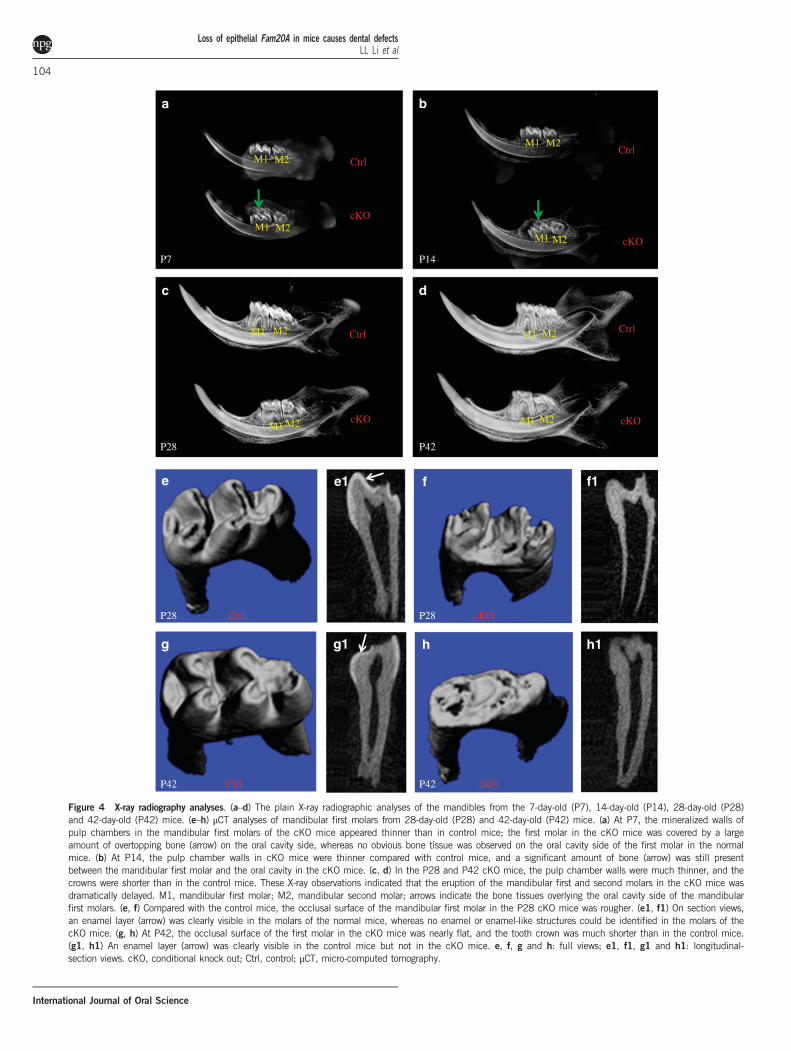
postnatal day 1 and day 7 mice. The immunostaining differences inENAM, AMBL and MMP20 between the 7-day-old cKO and normalmice were similar to those observed in the 1-day-old samples. Thus,only the IHC results from the 1-day-old mice are presented in thisreport. In the normal mice, ENAM protein was highly concentrated inthe distal region of the ameloblasts close to the basal lamina zone,although its signal was also observed in the other regions of theameloblast cytoplasm (Figure 6a). The anti-ENAM signal was remark-ably weaker in the Fam20A-deficient ameloblasts (Figure 6b) than inthe normal cells. The distribution of ENAM in the Fam20A-deficientameloblasts also appeared to be more diffused than in the normalcells. In the 7-day-old normal mice, the enamel matrix close to thebasal lamina zone and ameloblasts showed very strong anti-ENAMimmunoreactivity, whereas in the 7-day-old cKO mice, the anti-ENAM signal was hardly visible in the enamel matrix (data notshown). The anti-AMBL signal was stronger in the ameloblasts of cKOmice than in the normal mice (Figure 6c and 6d). The immunor-eactivity for MMP20 in the ameloblasts of cKO mice was remarkablyweaker than in the normal mice (Figure 6e and 6f).
Tooth eruption delay in cKO micePlain X-ray radiography showed that, at postnatal day 7, the mesialand middle cusps of the mandibular first molars in the normal micewere close to the mucosa of the oral cavity, and there was very little
or no bone tissue overlying the oral cavity side of the tooth, whereasthe cusps of the mandibular first molars in the cKO mice were faraway from the oral cavity, and a large amount of the overtoppingbone (arrow in Figure 4a) was covering the tooth. At postnatal day14, there were still significant amounts of bone present between themandibular first molar and the oral cavity in the cKO mice(Figure 4b). Histology analyses revealed that the reduced enamelepithelium of the mandibular first molar had fused with the oralmucosa, and the tooth was about to emerge into the oral cavity inthe 14-day-old normal mice (Figure 7a). In the 14-day-old cKOmice, there was still a significant distance between the crown of themandibular first molar and the oral mucosa (Figure 7b). Atpostnatal day 14, a large void space (“VS” in Figure 7b) was presentbetween the reduced enamel epithelium and the crown of themandibular first molar in the cKO mice, and the void spacecontained tissue/cellular debris (asterisks in Figure 7b) that appearedto have fallen off from the surrounding soft tissue. At postnatal day28, the mandibular second molar in the normal mice had fullyemerged in the oral cavity, whereas the mandibular second molar inthe cKO mice was still covered by the jawbone on the oral cavityside (Figure 4c). On the bais of our X-ray and histology observa-tions, we estimated that the eruption of the mandibular first molarin the cKO mice was delayed by approximately 10 days.
Gingival overgrowth in cKO miceThe cKO mice showed noticeable gingival overgrowth, which was moreremarkable on the lingual side than on the buccal side. The gingivalovergrowth was clearly visible on gross observation (Figure 8a–8d).Histology analyses of H&E-stained sections revealed that the gingivalepithelium in the cKO mice was obviously thicker than in the normalmice at postnatal day 28 (Figure 8e and 8f). The basal cells in thegingival epithelium formed a single and uniform layer in the normal
a
Ctrl Anti-ENAM
c e
f
Ctrl Anti-AMBL Ctrl Anti-MMP20
b
Anti-ENAM
d
cKOcKO Anti-AMBL cKO Anti-MMP20
Figure 6 Immunohistochemistry analyses of ENAM, AMBL and MMP20 in the mandibular first molars from 1-day-old (P1) mice. (a) In the normal (control)mice, ENAM protein was highly concentrated in the region of the ameloblast cytoplasm, close to the basal lamina zone. (b) The anti-ENAM signal wasremarkably weaker and more diffused in the Fam20A-deficient ameloblasts than in the normal cells. (c, d) The AMBL signal was stronger in the Fam20A-nullameloblasts than in the normal mice. (e, f) The immunoreactivity for MMP20 was weaker in the ameloblasts of cKO mice than in the normal mice. Arrowsindicate ameloblasts. Scale bars=100 μm in all the images. cKO, conditional knock out; Ctrl, control.
Loss of epithelial Fam20A in mice causes dental defectsLL Li et al
106
International Journal of Oral Science

a bCtrl cKO
P14 P14
VS * *
Figure 7 Haematoxylin and eosin staining of the first and second molar regions in the mandibles from 14-day-old (P14) mice. (a) In the normal (control)mice, the reduced enamel epithelium in the cusp tip region of the mandibular first molar fused with the oral epithelium, and the cusp tips (arrows) wereabout to emerge in the oral cavity. (b) In the cKO mice, there was still a great distance between the crown of the first molar and the oral cavity; the voidspace (VS) contained tissue/cellular debris (yellow asterisks), which appeared to have fallen into the cavity from the surrounding soft tissues. Scale bars,200 μm in both images. cKO, conditional knock out; Ctrl, control; VS, void space.
a
P42: Ctrl
b
P42: cKO
c
P42: Ctrl Lingual Lingual
Lingual
d
f
P42: cKO
P28: cKO
f1
*
*
Linguale
P28: Ctrl
e1
*
*
Lingualh
P42: cKO
h1
*
*
Lingualg
P42: Ctrl
g1
*
*
Figure 8 Gingival overgrowth in the cKO mice. (a) Gross photograph on occlusal view of the mandibular molar region in the 42-day-old (P42) normal(control) mice. (b) Gross photograph on occlusal view of the mandibular molar region in the P42 cKO mice; arrows indicate the enlarged gingiva. (c) Grossphotograph on lingual view of the mandibular molar region in the P42 control mice. (d) Gross photograph on lingual view of the mandibular molar region inthe P42 cKO mice; arrows indicate the enlarged gingiva. (e) H&E staining of a buccal–lingual section from the mandibular first molar region showed thelingual side gingiva in the 28-day-old control mice; double arrow indicates the thickness of the epithelium. (e1) Higher magnification view of the box area ine revealed a single layer of basal cells (asterisks). (f) The epithelium thickness (double arrow) in the lingual side gingiva of P28 cKO mice was greater thanin the normal mice. (f1) Higher magnification view of the box area in f revealed multiple layers of basal cells (asterisks). (g) H&E stain image of the lingualside gingiva in P42 normal mice; double arrow indicates the thickness of the epithelium. (g1) Higher magnification view of the box area in g revealed asingle layer of basal cells (asterisks). (h) The epithelium thickness (double arrow) in the lingual side gingiva of P42 cKO mice was greater than in the normalmice. (h1) Higher magnification view of the box area in h revealed multiple layers of basal cells (asterisks). Scale bars, 50 μm (a, b, c and d); 20 μm (a1,b1, c1 and d1). cKO, conditional knock out; Ctrl, control; H&E, Haematoxylin and eosin.
Loss of epithelial Fam20A in mice causes dental defectsLL Li et al
107
International Journal of Oral Science

mice (asterisks in Figure 8e1), whereas in the cKO mice, the gingivalepithelium often had multiple layers of basal cells (asterisks inFigure 8f1). The basal cells or basal cell-like cells in the Fam20A-deficient gingival epithelium also appeared misaligned or disorganized.At postnatal day 42 (Figure 8g–8h1), the histological appearance of thehyperplastic gingiva in the cKO mice was similar to that observed inthe 28-day-old mice.
DISCUSSION
FAM20A is a member of a small gene family that includes threeproteins, all of which contain endoplasmic reticulum-entry signalpeptides that guide molecules into the secretory pathway.1 FAM20A ispostulated to be localized primarily in the Golgi apparatus, where it isbelieved to form a complex with FAM20C and to enhance the catalyticactivity of the latter in phosphorylating certain secretory proteins.16
Deficiency in FAM20A or FAM20C might alter the post-translationalmodifications of their substrate proteins, leading to pathologicalchanges in tissues in which the proper functions of these kinases ortheir substrates are essential. The critical roles of FAM20A andFAM20C in the development of dental tissues have been welldemonstrated in human and mouse genetic studies, which haveshown the associations of FAM20A or FAM20C deficiencies withinherited dental defects.6–10,14,17–21
Information about FAM20A and the available tools to study thismolecule are limited. Although a previous study showed the expres-sion of FAM20A in ameloblasts and odontoblasts,21 there has been alack of systematic profiling for the expression of FAM20A in dentaltissues. Multipronged approaches in the current study showed that theameloblasts in the first molars of mouse mandibles started to expressFAM20A at postnatal day 1, a time point when the morphogenesis ofthe tooth has completed. Our previous study showed that theFAM20C was expressed in the dental epithelium of the cap-stageenamel organ at approximately 14.5 days post coitum;4 the expressionof FAM20C in the dental tissue occurred much earlier than that ofFAM20A. In addition, FAM20C was also much more broadlyexpressed than FAM20A.1 The differences in the temporospatialdistribution between FAM20A and FAM20C suggested that the formermight not be always essential to the physiological function of the latter,although the two molecules are postulated to work as partners inphosphorylating secretory proteins.16 The expression of FAM20A at avery late stage is consistent with the finding that inactivating thismolecule in the dental epithelium did not cause significant morpho-logic changes in the tooth germ of the mandibular first molar atpostnatal day 1. A high level of FAM20A was also observed in thestellate reticulum of the mandibular first molar in the 5-day-old miceand in the reduced enamel epithelium in eruptive-phase molars. Suchexpression profiling data provided invaluable clues regarding thebiological roles of FAM20A in these tissues at different stages. Ineither the ameloblasts or odontoblasts, FAM20A protein is primarilylocated in the distal region of the cytoplasm towards the enamelmatrix or dentin matrix. This unidirectional localization pattern ofFAM20A on the secretory side of the cells supports the postulationthat it is a Golgi-enriched protein colocalized with FAM20C.16
The μCT radiography analyses revealed a lack of true enamel in themolars of the 28-day-old cKO mice. Histologic evaluation showed thatthe molars of the cKO mice formed very little enamel matrix atpostnatal day 5 or day 7, the ameloblast layer in the cKO mice wasdetached from the enamel matrix, and the ameloblasts in the cKOmice were low columnar or flattened, non-polarized and disorganized.The findings in the teeth of the cKO mice were consistent with thediagnosis of amelogenesis imperfecta (AI). The gross and radiographic
appearance and histological changes in the enamel in the cKO micewere similar to those observed in mice with the constitutive ablation ofFam20A.14 The enamel defects, such as the scarcity of enamel matrix,detachment of the ameloblast layer from the enamel matrix and non-polarized/disorganized ameloblasts in the Fam20A-cKO mice,resembled those observed in the Fam20C-deficient mice;12 thesesimilarities between the Fam20A-cKO and Fam20C-deficient micesupported the conclusions drawn from in vitro studies that FAM20A isrequired for the kinase function of FAM20C in phosphorylatingcertain enamel matrix proteins.16 Enamelin (ENAM) is believed to bea substrate of FAM20C, and the phosphorylation of this enamelmatrix protein by FAM20C requires the presence of FAM20A.16
Without FAM20A, ENAM might not be properly phosphorylated,which can subsequently lead to the loss of ENAM function. WhenENAM cannot be phosphorylated, its degradation is accelerated, whichcan lead to a reduction in its protein level. When ENAM cannot beproperly phosphorylated, it might not be transported to the correctsite, which could have led to the relatively diffused distribution patternobserved in the cKO mice, in contrast with the normal mice, in whichENAM was concentrated in the distal region of the ameloblasts closeto the basal lamina zone. Like the Fam20A-cKO mice, Enam-null micealso showed AI phenotypes.25–27 Thus, the lack of proper phosphor-ylation in and/or the reduction of ENAM might be among thecontributing factors causing the enamel defects in the Fam20A-cKOmice and in human patients with AIGFS and ERS. Further studies arewarranted to examine whether the phosphorylation of enamel matrixproteins, including ENAM, is defective in cKO mice.Tooth eruption delay associated with inactivating mutations in the
FAM20A gene has been described in human patients20 but has neverbeen reported in animal studies. Three components are essential to theeruption of teeth: reduced enamel epithelium, dental follicles andperiodontal ligaments. When any of these three tissues is lost ordysfunctional, tooth eruption will be disturbed, causing lack oferuption or eruption delay.28–29 In this study, the X-Gal and ISHanalyses revealed a relatively high level of FAM20A in the reducedenamel epithelium in eruptive-phase molars, whereas this moleculewas barely detectable in the dental follicles or periodontal ligaments.Thus, it is reasonable to postulate that FAM20A might be essential tothe normal function of the reduced enamel epithelium in the controlof tooth eruption. The tooth eruption delay might have hampered themasticatory function of the cKO mice shortly after they were weanedfrom their mothers, leading to mild malnutrition, which affected thegrowth of these mice for a short period of time. Thus, the cKO mice atpostnatal 28 and 35 days had smaller body sizes, and subsequently,their body weights gradually caught up with those of the normal mice;after postnatal 35 days, the cKO mice might have gradually overcomethe molar eruption delay and gained the proper mastication forcerequired for chewing food and obtaining nutrition.Gingival overgrowth has been observed in human patients suffering
from AIGFS, but it has never been reported in animal studies. Weobserved severe gingival overgrowth in the cKO mice. The gingiva ismade of two types of tissues: the epithelium on the surface and theconnective tissue underlying the epithelium. It was unclear whetherthe gingival overgrowth in human patients is caused by abnormalproliferation of the epithelium or by hyperplasia of the connectivetissue. That the K14-Cre;Fam20Aflox/flox mice, in which Fam20A isinactivated in the epithelium but is intact in the connective tissue,showed severe gingival epithelium overgrowth, which suggested thatthe gingival overgrowth in human patients suffering from AIGFSmight occur primarily due to proliferation of the epithelium. Theovergrowth of the gingival epithelium was not observed in Fam20C-
Loss of epithelial Fam20A in mice causes dental defectsLL Li et al
108
International Journal of Oral Science

deficient mice,11–12 and it has not been reported in human Rainesyndrome, which has been associated with FAM20C mutations.6–10
The observation that hyperplasia of the gingival epithelium waspresent in the Fam20A-cKO mice but not in the FAM20C-deficientsubjects suggested that FAM20A might perform biological roles in thisoral tissue independent of FAM20C, although in vitro studies haveindicated that these two molecules might function in a dependentmanner in phosphorylating certain secretory proteins.16 Furtherinvestigations are needed to elucidate the molecular mechanismsunderlining hyperplasia of the gingival epithelium in Fam20A-deficientsubjects.
ACKNOWLEDGEMENTS
This work was supported by the National Natural Science Foundation of China(Grant No. 81171744) and the Natural Science Foundation of HeilongjiangProvince of China (Grant H201418).
1 Nalbant D, Youn H, Nalbant SI et al. FAM20: an evolutionarily conserved family ofsecreted proteins expressed in hematopoietic cells. BMC Genomics 2005; 6: 11.
2 Tagliabracci VS, Engel JL, Wen J et al. Secreted kinase phosphorylates extracellularproteins that regulate biomineralization. Science 2012; 336(6085): 1150–1153.
3 Tagliabracci VS, Wiley SE, Guo X et al. A single kinase generates the majority of thesecreted phosphoproteome. Cell 2015; 161(7): 1619–1632.
4 Wang X, Hao J, Xie Y et al. Expression of FAM20C in the osteogenesis andodontogenesis of mouse. J Histochem Cytochem 2010; 58(11): 957–967.
5 Du EX, Wang XF, Yang WC et al. Characterization of Fam20C expression in odonto-genesis and osteogenesis using transgenic mice. Int J Oral Sci 2015; 7(2): 89–94.
6 Simpson MA, Hsu R, Keir LS et al. Mutations in FAM20C are associated with lethalosteosclerotic bone dysplasia (Raine syndrome), highlighting a crucial molecule in bonedevelopment. Am J Hum Genet 2007; 81(5): 906–912.
7 Simpson MA, Scheuerle A, Hurst J et al. Mutations in FAM20C also identified in non-lethal osteosclerotic bone dysplasia. Clin Genet 2009; 75(3): 271–276.
8 Ababneh FK, AlSwaid A, Youssef T et al. Hereditary deletion of the entire FAM20C genein a patient with Raine syndrome. Am J Med Genet A 2013; 161A(12): 3155–3160.
9 Takeyari S, Yamamoto T, Kinoshita Y et al. Hypophosphatemic osteomalacia and bonesclerosis caused by a novel homozygous mutation of the FAM20C gene in an elderlyman with a mild variant of Raine syndrome. Bone 2014; 67: 56–62.
10 Seidahmed MZ, Alazami AM, Abdelbasit OB et al. Report of a case of Raine syndromeand literature review. Am J Med Genet A 2015; 167A(10): 2394–2398.
11 Wang X, Wang S, Li C et al. Inactivation of a novel FGF23 regulator, FAM20C, leads tohypophosphatemic rickets in mice. PLoS Genet 2012; 8(5): e1002708.
12 Wang X, Wang S, Lu Y et al. FAM20C plays an essential role in the formation ofmurine teeth. J Biol Chem 2012; 287(43): 35934–35942.
13 Koike T, Izumikawa T, Tamura J et al. FAM20B is a kinase that phosphorylatesxylose in the glycosaminoglycan-protein linkage region. Biochem J 2009; 421(2):157–162.
14 Vogel P, Hansen GM, Read RW et al. Amelogenesis imperfecta and other biominer-alization defects in Fam20a and Fam20c null mice. Vet Pathol 2012; 49(6): 998–1017.
15 Tian Y, Ma P, Liu C et al. Inactivation of Fam20B in the dental epithelium of mice leadsto supernumerary incisors. Eur J Oral Sci 2015; 123(6): 396–402.
16 Cui J, Xiao J, Tagliabracci VS et al. A secretory kinase complex regulates extracellularprotein phosphorylation. Elife 2015; 4: e06120.
17 O'Sullivan J, Bitu CC, Daly SB et al. Whole-exome sequencing identifies FAM20Amutations as a cause of amelogenesis imperfecta and gingival hyperplasia syndrome.Am J Hum Genet 2011; 88(5): 616–620.
18 Cho SH, Seymen F, Lee KE et al. Novel FAM20A mutations in hypoplastic amelogenesisimperfecta. Hum Mutat 2012; 33(1): 91–94.
19 Jaureguiberry G, De la Dure-Molla M, Parry D et al. Nephrocalcinosis (enamel renalsyndrome) caused by autosomal recessive FAM20A mutations. Nephron Physiol 2012;122(1/2): 1–6.
20 Wang SK, Aref P, Hu Y et al. FAM20A mutations can cause enamel-renalsyndrome (ERS). PLoS Genet 2013; 9(2): e1003302.
21 Wang SK, Reid BM, Dugan SL et al. FAM20A mutations associated with enamel renalsyndrome. J Dent Res 2014; 93(1): 42–48.
22 Järvinen E, Salazar-Ciudad I, Birchmeier W et al. Continuous tooth generation in mouseis induced by activated epithelial Wnt/beta-catenin signaling. Proc Natl Acad Sci USA2006; 103(49): 18627–18632.
23 Liu F, Chu EY, Watt B et al. Wnt/beta-catenin signaling directs multiple stages of toothmorphogenesis. Dev Biol 2008; 313(1): 210–224.
24 Baba O, Qin C, Brunn JC et al. Colocalization of dentin matrix protein 1 and dentinsialoprotein at late stages of rat molar development.Matrix Biol 2004; 23(6): 371–379.
25 Hu JC, Hu Y, Smith CE et al. Enamel defects and ameloblast-specific expression inEnam knock-out/lacz knock-in mice. J Biol Chem 2008; 283(16): 10858–10871.
26 Smith CE, Wazen R, Hu Y et al. Consequences for enamel development andmineralization resulting from loss of function of ameloblastin or enamelin. Eur J OralSci 2009; 117(5): 485–497.
27 Hu JC, Hu Y, Lu Y et al. Enamelin is critical for ameloblast integrity and enamelultrastructure formation. PLoS One 2014; 9(3): e89303.
28 Nanci A. Ten Cate's oral histology. 8 Edn. St Louis: Elsevier Mosby, 2013: 233–249.29 Avery J, Steels P, Avery N. Oral development and histology. 3rd Edn. New York: Thieme,
2002: 123–139.
This work is licensed under a Creative Commons Attribution 4.0International License. The images or other third party material in this
article are included in the article’s Creative Commons license, unless indicated otherwisein the credit line; if thematerial is not includedunder theCreativeCommons license, userswill need toobtainpermission fromthe licenseholder to reproduce thematerial.Toviewacopy of this license, visit http://creativecommons.org/licenses/by/4.0/
Supplementary Information of this article can be found on the International Journal of Oral Science's website (http://www.nature.com/ijos)
Loss of epithelial Fam20A in mice causes dental defectsLL Li et al
109
International Journal of Oral Science