Long-TermHyperexcitabilityinthe ...njms2.njms.rutgers.edu/vija/documents/2001Annals-.pdf ·...
10
Long-Term Hyperexcitability in the Hippocampus After Experimental Head Trauma Vijayalakshmi Santhakumar, BS, 1,2 Anna D. H. Ratzliff, BS, 1 Jade Jeng, BS, 1 Zsolt Toth, MD, PhD, 1 and Ivan Soltesz, PhD 1 Head injury is a causative factor in the development of temporal lobe epilepsy. However, whether a single episode of concussive head trauma causes a persistent increase in neuronal excitability in the limbic system has not been unequiv- ocally determined. This study used the rodent fluid percussion injury (FPI) model, in combination with electrophysio- logical and histochemical techniques, to investigate the early (1 week) and long-term (1 month or longer) changes in the hippocampus after head trauma. Low-frequency, single-shock stimulation of the perforant path revealed an early granule cell hyperexcitability in head-injured animals that returned to control levels by 1 month. However, there was a persistent decrease in threshold to induction of seizure-like electrical activity in response to high-frequency tetanic stimulation in the hippocampus after head injury. Timm staining revealed both early- and long-term mossy fiber sprouting at low to moderate levels in the dentate gyrus of animals that experienced FPI. There was a long-lasting increase in the frequency of spontaneous inhibitory postsynaptic currents in dentate granule cells after FPI, and ionotropic glutamate receptor antagonists selectively decreased the spontaneous inhibitory postsynaptic current frequency in the head-injured animals. These results demonstrate that a single episode of experimental closed head trauma induces long-lasting alterations in the hippocampus. These persistent structural and functional alterations in inhibitory and excitatory circuits are likely to influence the development of hyperexcitable foci in posttraumatic limbic circuits. Ann Neurol 2001;50:708 –717 Head injury is an important risk factor in remote symptomatic epilepsy, and accounts for up to 13% of nonidiopathic epilepsy. 1,2 After head injury, there is a long-lasting increase in the incidence of epilepsy lasting for several years. 1,3,4 Although clinical studies have found an association between prior head trauma and temporal lobe epilepsy (TLE), 5–7 the mechanisms un- derlying epilepsy following head injury are not under- stood. One of the most important unresolved issues regard- ing posttraumatic epilepsy is whether a single episode of traumatic brain injury leads to a persistent decrease in seizure threshold. The rodent fluid-percussion injury (FPI) model of head trauma has been used to study the anatomical and physiological sequelae of concussive head injury. 8 After moderate (2–2.2 atmosphere [atm]) FPI in adult rats, there is hilar cell loss, 9,10 reminiscent of the pattern of histopathological changes accompany- ing end-folium sclerosis in TLE. 9 –12 However, the de- gree of hyperexcitability in limbic circuits after FPI has been studied only at the early time points (up to 1 week). The dentate granule cells at this early time point have been found to show hyperexcitable re- sponses to low-frequency (single shock) stimulation of the perforant path in animals that experienced moderate FPI. 9,10,13 In addition, 1 week after FPI, hippocampal-entorhinal cortex (HEnC) slices were re- ported to show a decreased threshold to self-sustaining, seizure-like electrical field activity in response to high-frequency, tetanic stimulation. 14 These studies demonstrated early dentate hyperexcitability after ex- perimental head trauma, both in response to low- and high-frequency stimulation. However, whether the post-FPI hyperexcitability in the hippocampus is per- sistent or transient on a time scale of weeks and months is not known. Long-term behavioral changes 15 and progressive cell loss 16,17 have been reported after severe (2.5–2.9atm) FPI with cortical cavitation. Fol- lowing moderate FPI, dentate hilar cell loss has been observed up to 5 months, 10,13 and the maintenance of synaptic plasticity is impaired for as long as 2 months. 18 However, the long-term posttraumatic changes in hippocampal excitability have not been in- vestigated. From the 1 Department of Anatomy and Neurobiology, and 2 Reeve- Irvine Research Center, University of California, Irvine, CA. Received May 7, 2001, and in revised form Jul 18. Accepted for publication Jul 18, 2001. Published online Sep 13, 2001; DOI 10.1002/ana.1230 Address correspondence to Ms Santhakumar, Department of Anat- omy and Neurobiology, University of California, Irivne, CA 92697- 1280. E-mail: [email protected] 708 © 2001 Wiley-Liss, Inc.
Transcript of Long-TermHyperexcitabilityinthe ...njms2.njms.rutgers.edu/vija/documents/2001Annals-.pdf ·...

Long-Term Hyperexcitability in theHippocampus After Experimental
Head TraumaVijayalakshmi Santhakumar, BS,1,2 Anna D. H. Ratzliff, BS,1 Jade Jeng, BS,1 Zsolt Toth, MD, PhD,1
and Ivan Soltesz, PhD1
Head injury is a causative factor in the development of temporal lobe epilepsy. However, whether a single episode ofconcussive head trauma causes a persistent increase in neuronal excitability in the limbic system has not been unequiv-ocally determined. This study used the rodent fluid percussion injury (FPI) model, in combination with electrophysio-logical and histochemical techniques, to investigate the early (1 week) and long-term (1 month or longer) changes in thehippocampus after head trauma. Low-frequency, single-shock stimulation of the perforant path revealed an early granulecell hyperexcitability in head-injured animals that returned to control levels by 1 month. However, there was a persistentdecrease in threshold to induction of seizure-like electrical activity in response to high-frequency tetanic stimulation inthe hippocampus after head injury. Timm staining revealed both early- and long-term mossy fiber sprouting at low tomoderate levels in the dentate gyrus of animals that experienced FPI. There was a long-lasting increase in the frequencyof spontaneous inhibitory postsynaptic currents in dentate granule cells after FPI, and ionotropic glutamate receptorantagonists selectively decreased the spontaneous inhibitory postsynaptic current frequency in the head-injured animals.These results demonstrate that a single episode of experimental closed head trauma induces long-lasting alterations in thehippocampus. These persistent structural and functional alterations in inhibitory and excitatory circuits are likely toinfluence the development of hyperexcitable foci in posttraumatic limbic circuits.
Ann Neurol 2001;50:708–717
Head injury is an important risk factor in remotesymptomatic epilepsy, and accounts for up to 13% ofnonidiopathic epilepsy.1,2 After head injury, there is along-lasting increase in the incidence of epilepsy lastingfor several years.1,3,4 Although clinical studies havefound an association between prior head trauma andtemporal lobe epilepsy (TLE),5–7 the mechanisms un-derlying epilepsy following head injury are not under-stood.
One of the most important unresolved issues regard-ing posttraumatic epilepsy is whether a single episodeof traumatic brain injury leads to a persistent decreasein seizure threshold. The rodent fluid-percussion injury(FPI) model of head trauma has been used to study theanatomical and physiological sequelae of concussivehead injury.8 After moderate (2–2.2 atmosphere [atm])FPI in adult rats, there is hilar cell loss,9,10 reminiscentof the pattern of histopathological changes accompany-ing end-folium sclerosis in TLE.9–12 However, the de-gree of hyperexcitability in limbic circuits after FPI hasbeen studied only at the early time points (up to 1week). The dentate granule cells at this early time
point have been found to show hyperexcitable re-sponses to low-frequency (single shock) stimulationof the perforant path in animals that experiencedmoderate FPI.9,10,13 In addition, 1 week after FPI,hippocampal-entorhinal cortex (HEnC) slices were re-ported to show a decreased threshold to self-sustaining,seizure-like electrical field activity in response tohigh-frequency, tetanic stimulation.14 These studiesdemonstrated early dentate hyperexcitability after ex-perimental head trauma, both in response to low- andhigh-frequency stimulation. However, whether thepost-FPI hyperexcitability in the hippocampus is per-sistent or transient on a time scale of weeks andmonths is not known. Long-term behavioral changes15
and progressive cell loss16,17 have been reported aftersevere (2.5–2.9atm) FPI with cortical cavitation. Fol-lowing moderate FPI, dentate hilar cell loss has beenobserved up to 5 months,10,13 and the maintenance ofsynaptic plasticity is impaired for as long as 2months.18 However, the long-term posttraumaticchanges in hippocampal excitability have not been in-vestigated.
From the 1Department of Anatomy and Neurobiology, and 2Reeve-Irvine Research Center, University of California, Irvine, CA.
Received May 7, 2001, and in revised form Jul 18. Accepted forpublication Jul 18, 2001.
Published online Sep 13, 2001; DOI 10.1002/ana.1230
Address correspondence to Ms Santhakumar, Department of Anat-omy and Neurobiology, University of California, Irivne, CA 92697-1280. E-mail: [email protected]
708 © 2001 Wiley-Liss, Inc.

In addition, apart from the characteristic loss of hilarcells, possible structural alterations that may contributeto posttraumatic epilepsy are also not well understood.It is not known whether granule cell axons (the mossyfibers) undergo a posttraumatic long-term structural re-organization (sprouting) in a manner similar to whattakes place in the dentate gyrus of epileptic pa-tients,19–22 or in the dentate gyrus of experimental an-imals in various models of temporal lobe epilepsy.23–26
Previous reports indicate that the dentate gyrus of ep-ileptic patients with a history of head trauma mayshow supragranular sprouting of mossy fibers27,28;however, there is no conclusive, direct experimental ev-idence for mossy fiber sprouting after traumatic braininjury.29,30
This study was performed to determine the existenceof long-term limbic hyperexcitability following a singleepisode of concussive head injury. Specifically, we fo-cused on three major areas: (1) Does FPI in the ratresult in persistent changes in the hippocampal re-sponse to low- and high-frequency stimulation; (2) Isthere evidence for mossy fiber reorganization in thefluid percussion injured animals; and (3) Are therelong-term alterations in the interactions of inhibitoryand excitatory neuronal networks after head injury?
Materials and MethodsLateral Fluid Percussion InjuryThe lateral fluid percussion head trauma was carried out asdescribed previously.8–10,13 All procedures described were ap-proved by the Institutional Animal Care and Use Commit-tee, University of California, Irvine, CA. The fluid percus-sion device (Department of Biomedical Engineering, VirginiaCommonwealth University, Richmond, VA; see Toth etal8–10,13 for detailed description) was used to deliver a brief(20msec), 2.0 to 2.2atm impact on the intact dura. This re-sulted in a moderate level of injury that has been shown tocause a highly reproducible pattern of more than 50% hilarcell loss.9,10 Injured and age-matched sham-operated controlanimals were euthanized at various time points for slice phys-iology or Timm staining.
Slice PreparationThe animals were anesthetized with sodium pentobarbital(65mg/kg ip) and decapitated. Horizontal brain slices(400mm) were cut using a vibratome tissue sectioner (Lancerseries 1000; TPI, St Louis, MO) as previously described31 forthe field and whole-cell recordings. The slices were sagittallybisected and the slices ipsilateral to the side of injury weresubmerged in 32°C artificial cerebral spinal fluid (ACSF) com-posed of 126mM NaCl, 2.5mM KCl, 2mM MgCl2, 26mMNaHCO3, 2mM CaCl2, 1.25mM NaH2PO4, and 10mM glu-cose for 1 to 4 hours.
HEnC slices in which the trisynaptic circuit is preservedwere prepared as previously described.14,32–34 Briefly, thebrains were incubated for 2 minutes in 4°C oxygenated(95%O2, 5% CO2) sucrose ACSF composed of 200mM su-crose, 3mM KCl, 0.9mM MgCl2, 26mM NaHCO3, 2mM
CaCl2, 1.25mM NaH2PO4, and 10mM glucose. The dorsalsurface of the brain was glued onto a 12-degree agar rampwith the rostral end pointed up, and 450mm brain sliceswere sectioned with a vibratome tissue slicer (LeicaVT1000S; Leica, Nussloch, Germany). The slices ipsilateralto the injury were preincubated in 32°C oxygenatedlow Mg21-ACSF containing 130mM NaCl, 3mM KCl,0.5mM MgCl2, 26mM NaHCO3, 2mM CaCl2, 1.25mMNaH2PO4, and 10mM glucose to promote polysynaptic in-teractions.33,34
In Vitro ElectrophysiologySlices were transferred to the recording chamber35,36 andperfused with oxygenated ACSF or low Mg21-ACSF (for theHEnC slices) at 36°C. In some experiments the perfusionwas switched to ACSF containing 20mM bicuculline methio-dide (BMI) or 20mM 2-amino-5-phosphovaleric acid (APV)and 5mM 6-cyano-7-nitroquinoxaline-2,3-dione (CNQX).(All salts were obtained from Fluka, Buchs, Switzerland;APV and CNQX obtained from Tocris, Avonmouth, UK;and BMI from RBI, Natick, MA).
“Blind” whole-cell recordings were obtained as previouslydescribed,31 using patch pipettes filled with internal solutionthat consisted of 140mM Cs-gluconate, 2mM MgCl2, and10mM N-2-hydroxyethylpiperazine-N9-2-ethane-sulfonicacid. Granule cell population responses were evoked byconstant-current stimuli (0.5–8mA, 50ms) applied at 0.1Hzthrough a bipolar tungsten stimulating electrode placed inthe perforant path at the junction of the dorsal blade and thecrest. The field responses in the granule cell layer were mea-sured at five predetermined points, including the tips of thedorsal and ventral blades, the middle of the dorsal and ven-tral blades, and the middle of the crest, and the largest re-sponse was studied further. The input-output relationship inboth control and injured animals was obtained by comparingthe amplitudes of the population spikes evoked at each stim-ulation intensity.10,13 Field recordings in the HEnC sliceswere obtained from the CA1 pyramidal cell layer. The te-tanic stimulation consisted of a 2-second, 60Hz train ofstimuli applied to the Schaeffer collaterals with a pulse widthof 0.1msec through a stimulating electrode in the CA1 stra-tum radiatum.14,33,34 Stimulus was delivered at four timesthe minimal intensity required to evoke a 0.5mV populationspike (4–6mA). A maximum of 10 stimuli were given at10-minute intervals (to avoid interference by postictal refrac-tory periods). Once sustained epileptiform activity devel-oped, recording was terminated after 30 minutes (time pointselected to correspond to the clinical definition of status epi-lepticus, see Coulter et al14).
Timm StainingOne week (2 controls and 3 FPI) and 3 months (3 controlsand 7 FPI) after injury, control and injured animals weredeeply anesthetized and perfused transcardially with an aque-ous solution of 0.4% (wt/vol) sodium sulfide followed by500ml 1.25% (wt/vol) gluteraldehyde and 500ml of theaqueous solution of 0.4% (wt/vol) sodium sulfide. The hip-pocampus was sectioned (30mm), and every 20th section wasmounted and developed in the dark for 30 to 60 minutes inTimm solution (40ml distilled water, 2.55g citric acid, 2.35g
Santhakumar et al: Long-Term Hyperexcitability After Head Injury 709

sodium citrate, 1.7g hydroquinone, 60ml (50% wt/vol) gumarabic, and 0.1g silver nitrate) at 56°C.34,37 Sections werewashed in distilled water, placed in 1% (wt/vol) sodiumthiosulphate, washed again, and counterstained with 1%creysl violet acetate. The entire inner molecular layer of thedentate gyrus (ie, from the tip of the dorsal blade to the tipof the ventral blade of the dentate gyrus) from individualsections was examined by a blinded observer. Timm scoresfor sprouting were assigned to the sections based on the 0 to5 scale of Cavazos and colleagues.25 Briefly, 0 5 no granules;1 5 sparse granules in the supragranular layer; 2 5 morenumerous granules in the supragranular layer in a continuousdistribution; 3 5 prominent granules in the supragranularlayer in a continuous pattern with patches of confluent gran-ules; 4 5 prominent granules in the supragranular regionthat form confluent dense laminar bands; and 5 5 confluentdense laminar band of granules in the supragranular regionextending into the inner molecular layer.
AnalysisRecordings were filtered at 3kHz, digitization at 20kHz us-ing Strathclyde Electrophysiology Software (courtesy of Dr J.Dempster, University of Strathclyde, Glasgow, UK) and Syn-apse software (courtesy of Dr Y. De Koninck, McGill Uni-versity, Montreal, Canada). The spontaneous inhibitorypostsynaptic current (sIPSC) interevent interval (IEI) was ob-tained by sampling 100 sIPSCs from each cell. The effi-ciency of inhibition was measured on the amplitude of theevoked population spike. It was calculated as the ratio of theamplitude difference between bicuculline and ACSF record-ings to the amplitude of bicuculline recordings.
Efficiency of inhibition (%) 5
Amplitude in bicuculline2 Amplitude in ACSF
Amplitude in bicucullinep 100
Statistical analyses were performed with SigmaPlot orSPSS for Windows. The significance of differences infield recordings from the dentate gyrus of control andinjured animals was evaluated using Student’s t test.The nonparametric Mann-Whitney U test was used toassess the significance of difference in the HEnC sliceexperiments and Timm scoring, as the data from theseexperiments are not normally distributed. Kolmogorov-Smirnov test was used to assess the difference in thedistribution of sIPSC IEI in control and injured ani-mals. The level of significance was set at p , 0.05.Data are presented as mean 6 standard error.
ResultsLong-Term Recovery of Early Hyperexcitability toLow-Frequency StimulationPrevious studies conducted 1 week after FPI in ratshave shown an increase in the evoked field potentialamplitude recorded in the granule cell layer in responseto perforant path stimulation both in vivo9 and invitro10,14 in control ACSF (ie, without the presence ofvarious neuotransmitter receptor blockers in the extra-
cellular medium). One week after FPI, the populationspike amplitude, recorded in ACSF in response tosingle-shock stimulation of the perforant path (stimu-lus intensities: 2–8mA), was significantly larger10 inthe head-injured animals compared to the age-matched, sham-operated controls (eg, at 4mV stimula-tion intensity, the population spike amplitudes were:control 5 0.16 6 0.11mV from 3 animals, n 5 9slices; FPI 5 1.08 6 0.22mV from 4 animals, n 5 12;for examples of complete input-output curves at mul-tiple stimulation intensities, see Toth et al10 and San-thakumar et al13). However, the evoked populationspike amplitude 1 month and 3 months after FPI wasnot different from controls at any stimulation intensity(0.5–8mA; Fig 1A: control 5 0.17 6 0.12mV at 1month, n 5 9; 0.47 6 0.03mV at 3 months, n 5 13;3 animals each; FPI 5 0.34 6 0.17mV at 1 month,n 5 10; 0.63 6 0.23mV at 3 months, n 5 12; 3animals each; 4mA stimulation intensity). These dataindicate a recovery of the early dentate posttraumatichyperexcitability in response to low-frequency stimula-tion.
Perturbed inhibition in the dentate gyrus38,39 hasbeen suggested to play a role in the early posttraumatichyperexcitability.10,36 Could the recovery of the gran-ule cell field responses to single-shock perforant pathstimulation be explained by recovery of inhibition? Toanswer this question, we determined whether there is arecovery of the early hyperexcitable response during theearly (1-week) to long-term (3-month) post-FPI periodeven when fast inhibition is blocked. Bicuculline(20mM) was included in the perfusing medium toblock the g-animobutyric acid (GABA)A-mediatedfeedforward inhibition.13,36 As expected, addition ofbicuculline increased the amplitude of the populationspike both in the post-FPI and control slices (see Fig 1Aand B). One week after FPI, as described previously,13
the population spike amplitude in bicuculline was largerin the head-injured animals compared to controls (atstimulation intensities from 1–8mA) (see Fig 1B; con-trol 5 2.33 6 0.66mV, n 5 9; FPI 5 5.75 6 0.77mV,n 5 12; stimulus intensity 5 4mA). However, there wasno difference in the field potential amplitude betweencontrol and head-injured animals at later time pointseven in the presence of bicuculline at any stimulationintensity (0.5–8mA; see Fig 1B and C; 1 month afterFPI: control 5 1.43 6 0.42mV, n 5 9; FPI 5 2.03 60.62mV, n 5 10; 3 months: control 5 1.45 60.24mV, n 5 13; FPI 5 1.23 6 0.26mV, n 5 12).These data indicate that the early posttraumatic hyper-excitabile response of the dentate glutametergic networkto low-frequency stimulation recovers by 1 month, andthat the recovery cannot be the result of a possible re-covery of the perturbed fast synaptic inhibitory controlof granule cells.
These field recording data, obtained both in ACSF
710 Annals of Neurology Vol 50 No 6 December 2001
and bicuculline, also made it possible to calculate theamount of granule cell firing that is blocked by GABAA
receptor-dependent fast inhibition, providing a mea-sure of the efficiency of inhibition in control and head-injured animals at the various time points. The differ-ence between the population spike amplitude inbicuculline and ACSF was normalized to the amplitudeof the field potential in bicuculline (see Materials andMethods). The efficiency of inhibition was significantlydepressed in the head-injured animals at 1 week (con-trol 5 94.9 6 3.7%, n 5 9; FPI 5 76.1 6 5.7%,n 5 12), but recovered to control levels by 1 month(control 5 93.1 6 4.6%, n 5 9; FPI 5 93.1 6 4.5%,n 5 10) and 3 months (control 5 56.3 6 6.8%; n 513; FPI 5 63.7 6 10.4%; n 5 12) (see Fig 1D).These data demonstrate long-term recovery of the earlyposttraumatic decrease in the efficiency of dentate in-hibition.
Persistent Decrease in Threshold to Evoke Self-Sustaining Seizure-Like Population DischargesAfter Fluid Percussion InjuryTaken together, the data described above indicated arecovery of the field responses in the dentate gyrus by1 month in response to single-shock stimulation. Is itpossible that this recovery is incomplete, and that whenthe neuronal network is challenged with stronger stim-uli, the system reveals a persistently decreased seizurethreshold? To answer this question, tetanic stimulationwas applied to excitatory pathways as a form of a morerobust stimulation paradigm to test the stability of thesystem. These experiments were performed at a timepoint when there was a full recovery of the early hy-perexcitable field responses to single-shock stimulation,ie, at 3 months after FPI. The experiments with tetanicstimulation were carried out in a preparation that pre-serves the trisynaptic hippocampal–entorhinal pathway,
Fig 1. Long-term recovery of the hyperex-citable granule cell responses to low-frequency stimuli. Dark bars indicate con-trols, light bars indicate fluid percussioninjury (FPI). (A and B) Summary data ofthe amplitude of the perforant path-evokedgranule cell population spike show that theenhanced excitability observed at 1 week isnot present at 1 month and 3 monthsafter FPI, both in control artificial cere-bral spinal fluid (ACSF; A); and in20mM bicuculline (BMI; B). Stimulationintensity 5 4mV. (C) Averages of repre-sentative granule cell field responses fromFPI and sham operated control animals 3months after injury illustrate the absenceof enhanced excitability in ACSF and in20mM BMI. The initial deflection of theevoked response is the truncated stimulusartifact. Stimulation intensity 5 6mA.(D) Efficiency of inhibition in the dentategyrus is significantly decreased 1 week afterinjury, but it is not statistically differentfrom control animals at later time points.Calculation of the efficiency of inhibition:the population spike amplitude suppressedby feedforward inhibition was obtainedfrom the difference between the amplitudein BMI (20mM) and in ACSF. This dif-ference was normalized to the populationspike amplitude in BMI (20mM) to ob-tain the efficiency of inhibition. Asterisksindicate significance (p , 0.05, Student’st test).
Santhakumar et al: Long-Term Hyperexcitability After Head Injury 711

namely, in the combined HEnC slice40 that allows thedevelopment of self-sustaining seizure-like events. Asdescribed before,33,34,40 field recordings in this prepa-ration are best studied in the CA1 region because ofthe high signal (population spike amplitude)-to-noiseratio and stability of recorded events. The HEnC slicesfrom control and head-injured rats did not displayspontaneous field discharges in the CA1 pyramidal celllayer (Fig 2A). Tetanic stimulation of the Schaeffer col-laterals (60Hz train with a pulse width of 100ms, last-ing 2 seconds) in slices from control animals induced aprimary after-discharge that lasted less than 120 sec-onds (see Fig 2B). Among slices from the head-injuredanimals, 44.4% showed self-sustaining, recurrent epi-leptiform activity following the first tetanic stimula-tion, and all slices (n 5 9) showed prolonged (.30minutes) seizure-like activity after the third tetanic
stimulation (see Fig 2B and C). In contrast, 85.7% ofslices from control animals showed no evidence of re-current seizure-like activity even after the third train oftetanic stimulation (see Fig 2C). Once the seizure-likeactivity developed, it was also observed in the CA3 re-gion and the dentate granule cell layer (not shown).Therefore, these data show that although there is nospontaneous seizure-like activity in the posttraumatichippocampus, there is a persistent decrease in thethreshold for generation of self-sustaining epileptiformactivity when the system is challenged with a strongstimulus long after a single head injury episode.
Posttraumatic Mossy Fiber ReorganizationTimm staining for the zinc-containing mossy fiber ter-minals25,37 was performed on hippocampal sections 1week and 3 months after head injury to determine the
Fig 2. Long-term decrease in threshold toseizure-like activity in response to high-frequency stimulation in combinedhippocampal-entorhinal cortex (HEnC)slices. (A) Representative field recordings ofbaseline background activity from the CA1pyramidal cell layer in control and FPIanimals show the absence of spontaneousdischarges. (B) After the first high-frequency stimulation (Tet #1) of theSchaeffer collaterals, representative tracefrom a control shows no lasting secondaryself-sustaining activity following the pri-mary after discharge. In contrast, the re-cording from a head-injured animal showsrecurrent, spontaneous, self-sustained fieldactivity lasting for over 30 minutes, fol-lowing the first tetanic stimulation episode(Tet #1). The segments of recordings onthe right were taken at the time pointsindicated by asterisks, and are presentedat an expanded time scale. (C) Plot ofstimulation train number versus the dura-tion of after-discharge shows an increase inthe duration of self-sustained, rhythmicactivity in HEnC slices from injured ani-mals (n 5 7) compared with controls(n 5 9). Because recordings were termi-nated 30 minutes after onset of seizure-like activity, the maximum duration ofresponse was 1,800 seconds. Asterisks in-dicate significance (Mann-Whitney U test,p , 0.01).
712 Annals of Neurology Vol 50 No 6 December 2001
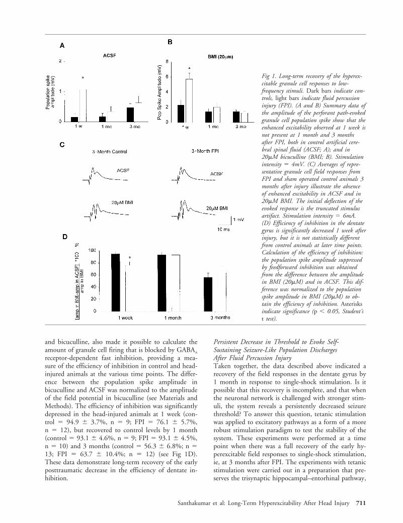
presence of aberrant mossy fiber sprouting in the innermolecular layer of the dentate gyrus. Timm scores25
were assigned by a blinded observer evaluating the en-tire supragranular region of the dentate gyrus (ie, theentire inner molecular layer from the tip of the dorsalblade to the tip of the ventral blade of the dentate gy-rus) to estimate the density of sprouting (see Materialsand Methods). None of the 32 sections from controls3 months after sham head injury had abnormal Timmstaining (none had a Timm score $1; Fig 3A, C, andE). In contrast, 3 months after trauma, 53% of thesections from the head-injured animals (37 of 70 sec-tions) had numerous Timm granules in the supra-granular region with occasional confluent patches(Timm score $2; see Fig 3B, D, and F). There was noevidence of confluent dense bands of Timm granules inthe supragranular layer (Timm score $4) in any of thesections from the head-injured animals, indicating thatthe degree of the mossy fiber sprouting was relativelylow. Interestingly, 25% of the sections from head-injured animals showed numerous Timm granules inthe supragranular region (Timm score .2; 18 of 73sections) even 1 week after FPI, indicating the rapidonset of supragranular mossy fiber sprouting (see Fig3G). The increase in sprouting observed in the sectionsfrom the head-injured compared to control animalswas significant both at 1 week (Timm scores: con-trol 5 0.45 6 0.08; FPI 5 1.27 6 0.08) and 3months (control 5 0.41 6 0.09; FPI 5 1.57 6 0.12)(see Fig 3G). Additionally, the post-injury increase insprouting was significantly higher at 3 months com-pared to 1 week after injury.
Prolonged Increase in the Frequency of sIPSCs inGranule Cells After Fluid Percussion InjuryThe posttraumatic presence of the moderate but signif-icant mossy fiber sprouting indicates an increase in theaxonal output from the excitatory principal cells of thedentate gyrus, most likely both to other granule cellsand to interneurons. Is there a similar increase in theoutput from posttraumatic dentate interneurons? Inlieu of a convenient histochemical marker such as theTimm stain, an alternative strategy was applied to an-swer this question. It has been shown that FPI at 2.0to 2.2atm impact strength in our laboratory results inthe loss of about 50% of the parvalbumin- andcholecytokinin-positive basket and axo–axonic cells,10
as well as other interneurons.13 The loss of interneu-rons was found to be accompanied by a permanent de-crease in the frequency of the spontaneous, actionpotential-independent miniature IPSCs,10 indicating aposttraumatic decrease in GABAergic release sites orprobability of GABA release. To determine whetherthe net synaptic output of the surviving inhibitory in-terneuronal network is altered in a persistent mannerafter head trauma, we examined the posttraumatic
Fig 3. Reorganization of the dentate granule cell axons follow-ing trauma. (A–F) Timm staining shows mossy fiber sproutingin the dentate gyrus of FPI animals 3 months after injury. Aand B are low-magnification images of the dentate gyrus; out-lined areas are shown at higher magnification in C throughF. Note the absence of black granules in the supragranularlayer of the control (A, C, and E) compared with the FPI (B,D, and F). C and D are from the crest, and E and F arefrom the suprapyramidal blade of the granule cell layer. (G)Summary data of Timm scores show a significant increase insprouting in the head-injured animals compared to age-matched, sham-operated controls. Asterisks indicate signifi-cance (Mann-Whitney U test, p , 0.001). ML 5 molecularlayer; GCL 5 granule cell layer. Bar 5 200mm (A and B)and 50mm (C–F).
Santhakumar et al: Long-Term Hyperexcitability After Head Injury 713
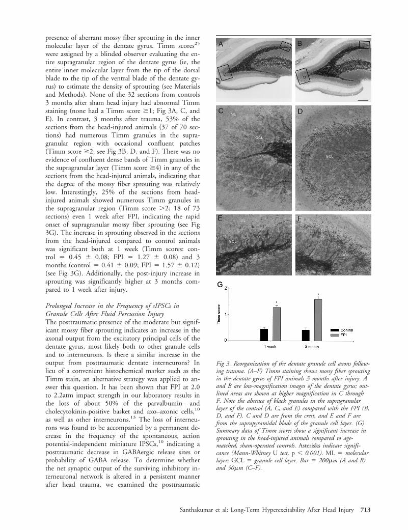
changes in the frequency of the spontaneous, actionpotential-dependent sIPSCs from granule cells, 1 weekand 5 to 6 months after head injury. The sIPSCs wererecorded at 0mV, with Cs-gluconate-filled patch pi-pettes, in ACSF. The sIPSC IEI was significantlysmaller (ie, the frequency was higher) in the head-injured animals compared with the age-matched con-trols at both the early (control 5 37.72 6 1.28 msec,n 5 9; FPI 5 18.20 6 0.05 msec, n 5 8) and late(control 5 109.22 6 4.86 msec, n 5 7; FPI 566.52 6 2.47 msec, n 5 12) post-FPI time points (Fig4A). The amplitude of the sIPSCs was significantlylarger in the head-injured animals (control 5 35.88 6
1.15pA; FPI 5 40.59 6 1.29pA, n 5 8 cells each) 1week after FPI. However, the rise time constants (con-trol 5 1.06 6 0.12ms; FPI 5 1.11 6 0.20ms, n 5 8each) and decay time constants (control 5 5.46 60.37ms; FPI 5 4.65 6 0.36ms, n 5 8 each) of thesIPSCs from head-injured animals were not differentfrom controls. Is the posttraumatic increase in the fre-quency of sIPSCs in granule cells influenced by theexcitatory drive to interneurons?41–45 To answer thisquestion, the frequency of sIPSCs from granule cells inthe control medium (ACSF) were compared to the fre-quency of sIPSCs in a medium containing ionotropicglutamate receptor antagonists (20mM APV and 5mMCNQX). As shown before,41–45 the sIPSC frequency isnot influenced significantly in control granule cells bythe presence of the ionotropic receptor blockers in theperfusate (sIPSC IEI in APV and CNQX, with respectto IEI in ACSF: 89.6 6 4.0% at 1 week, n 5 6; and86.6 6 4.1% at 5–6 months, n 5 6, after sham inju-ry). In contrast, APV and CNQX significantly de-creased the enhanced sIPSC frequency observed afterhead trauma (see Fig 4B; IEI in APV and CNQX, withrespect to IEI in ACSF at 1 week: 142.6 6 5.9%, n 56; 5–6 months: 117 6 5.5%, n 5 7). These findingsindicate that the glutamatergic excitatory drive to in-terneurons contributes to the early and long-term post-traumatic increase in the net output from the dentateinhibitory network.
DiscussionThis study demonstrates the permanent decrease inthreshold to seizure-like activity, and long-term struc-tural and functional hippocampal reorganization affect-ing both excitatory and inhibitory networks, after asingle episode of concussive head injury. Specifically,the data show that (1) the early hyperexcitability tolow-frequency stimulation recovers within 1 month;(2) there is a long-term decrease in the stability of theneuronal network, as determined by the decreasedthreshold for generation of seizure-like activity in re-sponse to high-frequency stimulation; (3) without te-tanic stimulation, there are no spontaneous epilepti-form discharges in the hippocampal circuit; (4) mossyfiber sprouting appears within 1 week after injury andincreases in density by 3 months; (5) the early increasein the frequency of granule cell sIPSCs persists at least5 months after FPI; and (6) ionotropic glutamate re-ceptor antagonists decrease the the posttraumatic po-tentiation of the sIPSC in dentate granule cells.
Alterations in Limbic Excitability After Head InjuryEach year an estimated 1.5 million people sustain trau-matic brain injury in the United States, and braintrauma is the leading cause of death and disabilityamong young adults.46 The annual economic burdenof traumatic brain injury is projected to exceed $40
Fig 4. Increase in granule cell spontaneous inhibitory postsyn-aptic current (sIPSC) frequency after FPI. (A) Summary dataof sIPSC interevent interval (IEI) in head-injured animals asa percent of sIPSC IEI in the age-matched controls. Note thedecrease in IEI (increased sIPSC frequency) after injury atboth time points. (B) Summary data show that the glutamatereceptor antagonists APV (20mM) and CNQX (5mM) in-creased the lower IEI of sIPSCs (decreased the elevated sIPSCfrequency) in granule cells from animals that experienced headinjury, but not in controls. Asterisks indicate significance(Kolmogorov-Smirnov test, p , 0.05).
714 Annals of Neurology Vol 50 No 6 December 2001

billion.46,47 Therefore, head injuries present an enor-mous medical and social problem. A major conse-quence of traumatic brain injury is epilepsy. The riskof epilepsy increases with the severity of the head in-jury. Immediate posttraumatic seizures are frequentlyfollowed by a latent period,6 during which epileptogen-esis can take place. However, the nature of the pro-cesses underlying the development of later unprovokedseizures is not understood. Clinical trials aimed at pre-vention of late posttraumatic epilepsy using antiepilep-tic drugs have been largely unsuccessful.48,49 A recentexperimental study, however, has shown that activity-dependent processes shortly after trauma are likely toplay a major role in the development of posttraumatichyperexcitability.50
Our data show that the early dentate hyperexcitabilityrecovers within a month after FPI. However, when chal-lenged with high-frequency stimulation, the injured hip-pocampal network exhibits a lower threshold to genera-tion of seizure-like activity even 3 months after injury.These findings indicate that, although there are no per-sistent spontaneous seizure-like events in the trisynapticcircuit after injury, there are underlying long-termchanges that predispose the limbic system to seizureswhen challenged with strong stimuli. In other words,following the initial impact, the hippocampal networkbecomes persistently hyperexcitable, but the neuronalsystem’s ability to handle incoming excitatory signalswithout generating self-sustaining epileptiform dis-charges undergoes a partial but not complete recovery.
What is the nature of the processes that underlie theapparent recovery to weaker excitation? The early en-hancement of the population spike amplitude evoked inthe presence of bicuculline recovered to control levels by1 month (see Fig 1B), indicating that the excitatory net-work undergoes some form of recovery and is at leastpartially responsible for the recovery. On the otherhand, the early decrease in the efficiency of inhibitionalso recovered by 1 month (see Fig 1D), suggesting thatthe recovery process may also involve the inhibitory sys-tem. The other side of the same issue concerns themechanism underlying the incompleteness of the recov-ery and the processes that contribute the latent period ofepileptogenesis found in human head injuries. Our datarevealed a progressive increase in the density of mossyfiber sprouting, which is often associated with enhancedrecurrent excitation and epileptiform discharges. The in-crease in the density of mossy fiber sprouting with timecould be one candidate underlying the late appearanceof hyperexcitability in human head injuries, and it maycontribute to the incompleteness of the recovery of thesystem’s stability in response to repetitive stimuli. Inter-estingly, supragranular mossy fiber sprouting is presentin tissue from temporal lobe epilepsy patients with a his-tory of head injury,27,28 and the sprouting of excitatoryaxons has also been reported in the cortical undercut
model of posttraumatic hyperexcitability.51 It is likelythat the degree of sprouting in animal models of headinjury depends on several factors, including strain differ-ences, injury paradigm, and impact strength. For exam-ple, robust mossy fiber sprouting has been reported afterweight-drop head injury,52 whereas no mossy fibersprouting29,30 was found after FPI that resulted in lowerlevels of hilar cell loss than what takes place under ourconditions following FPI at 2.0 to 2.2atm impactstrength. Our data also indicate that mossy fiber sprout-ing after FPI can be detected within 1 week after injury,similar to the early time course for sprouting reported inmodels of epilepsy.19,25 In light of the early occurrenceand progressive nature of the posttraumatic mossy fibersprouting, it is possible that sprouting plays a role inhippocampal hyperexcitabilty at both 1 week and 3months after FPI. It is also interesting to note that, al-though it was not a focus of this study, our data alsorevealed an age-dependent decrease in the efficiency ofinhibition (see Fig 1D) and a decrease in granule cellsIPSC frequency (increase in IEI), both in control andin post-FPI animals. Age-dependent alterations in func-tional synaptic connections taking place in cortical net-works53 are likely to contribute to these phenomena.
The recovery of the early posttraumatic decrease inthe efficiency of inhibition to age-matched control lev-els, as tested with low-frequency stimulation, tookplace without a complete recovery of the ability of theneuronal network to handle high-frequency excitatoryinputs. Furthermore, the increased spontaneous IPSCfrequency remained significantly elevated even 5 to 6months following FPI. As discussed elsewhere, in sev-eral models of epilepsy an increase in certain parame-ters of GABAA receptor mediated inhibition has beenreported, including increases in GABAA receptor num-bers,44,54,55 changes in GABAA receptor subunit com-position,55,56 and presynaptic enhancement of GABArelease.57 However, the relationship between enhancedinhibition and hyperexcitability is not fully understood.Zinc-dependent collapse of elevated inhibition,44 alteredratio of dendritic versus somatic inhibition,58 frequency-dependent conversion of increased inhibition to hyper-excitability caused by persistently modified h-channels inpostsynaptic cells,59 and a preferential collapse of Cl2
homeostasis in dendrites of principal cells60 may all playcomplex roles in determining the efficacy of potentiatedinhibitory processes in an ultimately hyperexcitable net-work. Increased sIPSC frequency that is sensitive toblockade of ionotropic glutamate receptors, similar toour data, has also been reported in kindred animals.44
Whether the increased excitatory drive to dentate inter-neurons, suggested by the data from kindred animalsand from our study, actually originates from thesprouted mossy fibers is not known. Shortly after FPI(within hours), there is also an elevated sIPSC frequencyin granule cells, which is associated with increased inter-
Santhakumar et al: Long-Term Hyperexcitability After Head Injury 715
neuronal spontaneous firing rates caused by a Na1
pump-dependent shift in the interneuronal resting mem-brane potential.61 However, the posttraumatic elevationof the interneuronal resting membrane potential is tran-sient, as it returns to nonsignificant levels by 4 days afterFPI.61 Therefore, it is likely that the elevated frequencyof the sIPSCs in granule cells from head-injured animalshas different underlying mechanisms at different timepoints following impact.
Taken together, the results demonstrate long-term al-terations in both the excitatory and inhibitory networksin the hippocampus after traumatic brain injury in ex-perimental animals. The persistent decrease in seizurethreshold indicates the possibility that in human headinjuries, particularly after severe injury, the neuronal net-works are persistently altered in the limbic system, evenif spontaneous behavioral or electrical seizures are notpresent. Future investigations aimed at understandingthe activity-dependent epileptogenic processes50 in vari-ous experimental models of head trauma will be neededto shed light on the interaction, importance, and thepro- versus antiepileptic nature of the cellular and syn-aptic alterations triggered by a brain injury episode.
I.S. was financially supported by the National Institutes of Health(NS35915).
We thank Dr O. Steward for his support; and R. Zhu for experttechnical assistance.
References1. Annegers JF, Grabow JD, Groover RV, et al. Seizures after
head trauma: a population study. Neurology 1980;30(7 Pt 1):683–689.
2. Annegers JF, Hauser WA, Coan SP, Rocca WA. A population-based study of seizures after traumatic brain injuries. N EnglJ Med 1998;338:20–24.
3. Caveness WF, Meirowsky AM, Rish BL, et al. The nature ofposttraumatic epilepsy. J Neurosurg 1979;50:545–553.
4. Angeleri F, Majkowski J, Cacchio G, et al. Posttraumatic epi-lepsy risk factors: one-year prospective study after head injury.Epilepsia 1999;40:1222–1230.
5. Jennet B. Epilepsy after nonmissile head injuries. London: Hei-nemann, 1975.
6. Salazar AM, Jabbari B, Vance SC, et al. Epilepsy after penetrat-ing head injury. I. Clinical correlates: a report of the VietnamHead Injury Study. Neurology 1985;35:1406–1414.
7. Willmore JL. Posttraumatic epilepsy: cellular mechanisms andimplications for treatment. Epilepsia 1990;31:S67–S73.
8. McIntosh TK, Vink R, Noble L, et al. Traumatic brain injuryin the rat: characterization of a lateral fluid-percussion model.Neuroscience 1989;28:233–244.
9. Lowenstein DH, Thomas MJ, Smith DH, McIntosh TK. Se-lective vulnerability of dentate hilar neurons following trau-matic brain injury: a potential mechanistic link between headtrauma and disorders of the hippocampus. J Neurosci 1992;12:4846–4853.
10. Toth Z, Hollrigel GS, Gorcs T, Soltesz I. Instantaneous per-turbation of dentate interneuronal networks by a pressure wave-transient delivered to the neocortex. J Neurosci 1997;17:8106–8117.
11. Margerison JH, Corsellis JA. Epilepsy and the temporal lobes.A clinical, electroencephalographic and neuropathological studyof the brain in epilepsy, with particular reference to the tem-poral lobes. Brain 1966;89:499–530.
12. Bruton C. The neuropathology of temporal lobe epilepsy. NewYork: Oxford University Press, 1988.
13. Santhakumar V, Bender R, Frotscher M, et al. Granule cellhyperexcitability in the early posttraumatic rat dentate gyrus:the ’irritable mossy cell’ hypothesis. J Physiol 2000;524(Pt 1):117–134.
14. Coulter DA, Rafiq A, Shumate M, et al. Brain injury-inducedenhanced limbic epileptogenesis: anatomical and physiologicalparallels to an animal model of temporal lobe epilepsy. EpilepsyRes 1996;26:81–91.
15. Pierce JE, Smith DH, Trojanowski JQ, McIntosh TK. Endur-ing cognitive, neurobehavioral and histopathological changespersist for up to one year following severe experimental braininjury in rats. Neuroscience 1998;87:359–369.
16. Bramlett HM, Dietrich WD, Green EJ, Busto R. Chronic his-topathological consequences of fluid-percussion brain injury inrats: effects of posttraumatic hypothermia. Acta Neuropathol1997;93:190–199.
17. Smith DH, Chen XH, Pierce JE, et al. Progressive atrophy andneuron death for one year following brain trauma in the rat.J Neurotrauma 1997;14:715–727.
18. Sanders MJ, Sick TJ, Perez-Pinzon MA, et al. Chronic failurein the maintenance of long-term potentiation following fluidpercussion injury in the rat. Brain Res 2000;861:69–76.
19. Sutula T, Cascino G, Cavazos J, et al. Mossy fiber synapticreorganization in the epileptic human temporal lobe. Ann Neu-rol 1989;26:321–330.
20. Houser CR, Miyashiro JE, Swartz BE, et al. Altered patterns ofdynorphin immunoreactivity suggest mossy fiber reorganizationin human hippocampal epilepsy. J Neurosci 1990;10:267–282.
21. Babb TL, Kupfer WR, Pretorius JK, et al. Synaptic reorganiza-tion by mossy fibers in human epileptic fascia dentata. Neuro-science 1991;42:351–363.
22. Zhang N, Houser CR. Ultrastructural localization of dynorphinin the dentate gyrus in human temporal lobe epilepsy: a studyof reorganized mossy fiber synapses. J Comp Neurol 1999;405:472–490.
23. Tauck DL, Nadler JV. Evidence of functional mossy fibersprouting in hippocampal formation of kainic acid-treated rats.J Neurosci 1985;5:1016–1022.
24. Sutula T, He XX, Cavazos J, Scott, G. Synaptic reorganizationin the hippocampus induced by abnormal functional activity.Science 1988;239:1147–1150.
25. Cavazos JE, Golarai G, Sutula TP. Mossy fiber synaptic reor-ganization induced by kindling: time course of development,progression, and permanence. J Neurosci 1991;11:2795–2803.
26. Okazaki MM, Evenson DA, Nadler JV. Hippocampal mossyfiber sprouting and synapse formation after status epilepticus inrats: visualization after retrograde transport of biocytin. J CompNeurol 1995;352:515–534.
27. Mathern GW, Pretorius JK, Babb TL. Influence of the type ofinitial precipitating injury and at what age it occurs on courseand outcome in patients with temporal lobe seizures. J Neuro-surg 1995;82:220–227.
28. Jeub M, Lie A, Blumcke I, et al. Loss of dynorphin-mediatedinhibition of voltage-dependent Ca21 currents in hippocampalgranule cells isolated from epilepsy patients is associated withmossy fiber sprouting. Neuroscience 1999;94:465–471.
29. Hill SJ, Barberese E, Gennarelli TA, McIntosh TK. The poten-tial role for reactive astrocytes in CNS reinnervation followingexperimental brain injury in rat. Soc Neurosci Abstr 1994;20:424.
716 Annals of Neurology Vol 50 No 6 December 2001

30. Shumate M, Rafiq A, Lyeth B, et al. Traumatic brain injuryproduces loss of neurons in hilus and CA3 but no significantmossy fiber sprouting in the hippocampus. Epilepsia 1995;36(S4):118.
31. Soltesz I, Mody I. Patch-clamp recordings reveal powerfulGABAergic inhibition in dentate hilar neurons. J Neurosci1994;14:2365–2376.
32. Rafiq A, DeLorenzo RJ, Coulter DA. Generation and propaga-tion of epileptiform discharges in a combined entorhinal cortex/hippocampal slice. J Neurophysiol 1993;70:1962–1974.
33. Rafiq A, Zhang YF, DeLorenzo RJ, Coulter DA. Long-durationself-sustained epileptiform activity in the hippocampal-parahippocampal slice: a model of status epilepticus. J Neuro-physiol 1995;74:2028–2042.
34. Dube C, Chen K, Eghbal-Ahmadi M, et al. Prolonged febrileseizures in the immature rat model enhance hippocampal excit-ability long term. Ann Neurol 2000;47:336–344.
35. Soltesz I, Smetters DK, Mody I. Tonic inhibition originatesfrom synapses close to the soma. Neuron 1995;14:1273–1283.
36. Hollrigel GS, Toth K, Soltesz I. Neuroprotection by propofolin acute mechanical injury: role of GABAergic inhibition.J Neurophysiol 1996;76:2412–2422.
37. Danscher G. Histochemical demonstration of heavy metals. Arevised version of the sulphide silver method suitable for bothlight and electronmicroscopy. Histochemistry 1981;71:1–16.
38. Halasy K, Somogyi P. Distribution of GABAergic synapsesand their targets in the dentate gyrus of rat: a quantitativeimmunoelectron microscopic analysis. J Hirnforsch 1993;34:299–308.
39. Freund TF, Buzsaki G. Interneurons of the hippocampus. Hip-pocampus 1996;6:347–470.
40. Dreier JP, Heinemann U. Regional and time dependent varia-tions of low Mg21 induced epileptiform activity in rat tempo-ral cortex slices. Exp Brain Res 1991;87:581–596.
41. Davies CH, Davies SN, Collingridge GL. Paired-pulse depres-sion of monosynaptic GABA-mediated inhibitory postsynapticresponses in rat hippocampus. J Physiol 1990;424:513–531.
42. Otis TS, Mody I. Modulation of decay kinetics and frequencyof GABAA receptor-mediated spontaneous inhibitory postsynap-tic currents in hippocampal neurons. Neuroscience 1992;49:13–32.
43. Buhl EH, Halasy K, Somogyi P. Diverse sources of hippocam-pal unitary inhibitory postsynaptic potentials and the numberof synaptic release sites. Nature 1994;368:823–828.
44. Buhl EH, Otis TS, Mody I. Zinc-induced collapse of aug-mented inhibition by GABA in a temporal lobe epilepsy model.Science 1996;271:369–373.
45. Prince DA, Jacobs K. Inhibitory function in two models ofchronic epileptogenesis. Epilepsy Res 1998;32:83–92.
46. Goldstein M. Traumatic brain injury: a silent epidemic. AnnNeurol 1990;27:327.
47. Max W, MacKenize EJ, Rice DP. Head injuries: costs and con-sequences. J Head Trauma Rehabil 1991;6:76–91.
48. Temkin NR, Dikmen SS, Anderson GD, et al. Valproate ther-apy for prevention of posttraumatic seizures: a randomized trial.J Neurosurg 1999;91:593–600.
49. Temkin NR, Dikmen SS, Winn HR. Clinical trials for seizureprevention. Adv Neurol 1998;76:179–188.
50. Graber KD, Prince DA. Tetrodotoxin prevents posttraumaticepileptogenesis in rats. Ann Neurol 1999;46:234–242.
51. Salin P, Tseng GF, Hoffman S, et al. Axonal sprouting in layerV pyramidal neurons of chronically injured cerebral cortex.J Neurosci 1995;15:8234–8245.
52. Golari G, Greenwood AC, Feeney DM, Connor JA. Traumaticbrain injury (TBI) induces sprouting and mossy fiber reorgani-zation of the mossy fiber pathway in the dentate gyrus of thehippocampus. Soc Neurosci Abstr 1998;24:1728.
53. Wong TP, Marchese G, Casu MA, et al. Loss of presynapticand postsynaptic structures is accompanied by compensatory in-crease in action potential-dependent synaptic input to layer Vneocortical pyramidal neurons in aged rats. J Neurosci 2000;20:8596–8606.
54. Otis TS, De Koninck Y, Mody I. Lasting potentiation of inhi-bition is associated with an increased number of gamma-aminobutyric acid type A receptors activated during miniatureinhibitory postsynaptic currents. Proc Natl Acad Sci U S A1994;91:7698–7702.
55. Nusser Z, Hajos N, Somogyi P, Mody I. Increased number ofsynaptic GABAA receptors underlies potentiation at hippocam-pal inhibitory synapses. Nature 1998;395:172–177.
56. Brooks-Kayal AR, Shumate MD, Jin H, et al. Selectivechanges in single cell GABAA receptor subunit expressionand function in temporal lobe epilepsy. Nat Med 1998;4:1166 –1172.
57. Chen K, Baram TZ, Soltesz I. Febrile seizures in the developingbrain result in persistent modification of neuronal excitability inlimbic circuits. Nat Med 1999;5:888–894.
58. Cossart R, Dinocourt C, Hirsch JC, et al. Dendritic but notsomatic GABAergic inhibition is decreased in experimental ep-ilepsy. Nat Neurosci 2001;4:52–62.
59. Chen K, Aradi I, Thon N, et al. Persistently modifiedH-channels after complex febrile seizures convert the seizure-induced enhancement of inhibition to hyperexcitability. NatMed 2001;7:331–337.
60. Staley KJ, Soldo BL, Proctor WR. Ionic mechanisms of neuronalexcitation by inhibitory GABAA receptors. Science 1995;269:977–981.
61. Ross ST, Soltesz I. Selective depolarization of interneurons inthe early posttraumatic dentate gyrus: involvement of theNa(1)/K(1)-ATPase. J Neurophysiol 2000;83:2916–2930.
Santhakumar et al: Long-Term Hyperexcitability After Head Injury 717