Long-term outcome of lumbar disc herniation surgery
64
Long-term outcome of lumbar disc herniation surgery Studies on different influencing factors Katarina Silverplats Department of Orthopaedics Institute of Clinical Science at Sahlgrenska Academy, University of Gothenburg Gothenburg, Sweden 2010
Transcript of Long-term outcome of lumbar disc herniation surgery

Long-term outcome of lumbar disc herniation surgery
Studies on different influencing factors
Katarina Silverplats
Department of Orthopaedics Institute of Clinical Science at
Sahlgrenska Academy, University of Gothenburg Gothenburg, Sweden 2010
© Katarina Silverplats 2010
Published articles have been reprinted with the permission of the respective copyright holder.
Katarina Silverplats
Dept. of Othopaedics
Sahlgrenska University Hospital
SE-413 45 Gothenburg
Sweden
Printed by Intellecta Infolog, Gothenburg, Sweden 2010
ISBN: 978-91-628-8056-9

“Du är borta mycket mamma,
men du finns alltid med mig i mitt hjärta”
Leo

Contents
1. List of publications 7
2. Abbreviations 8
3. Abstract 9
4. Summary in Swedish (sammanfattning på svenska) 11
5. Introduction 13
6. Background 14
6.1 Disc anatomy and pathology
6.2 Symptom
6.3 Diagnosis
6.4 Treatment
6.5 Outcome
7. Aims of the study 27
8. Summary of studies 28
8.1 Material and methods
8.2 Results
9. General discussion 47
10. Conclusions 52
11. Future 53
12. Acknowledgments 54
13. References 57
14. Papers I-IV

7
1. List of publications
This thesis is based on the following papers:
. Clinical factors of importance for outcome after lumbar disc
herniation surgery. Rönnberg K, Lind B, Halldin K, Zoëga B, Gellerstedt M, Brisby H
Submitted
. Patients´ satisfaction with provided care/information and
expectations on clinical outcome after lumbar disc herniation
surgery. Rönnberg K, Lind B, Halldin K, Zoëga B, Gellerstedt M, Brisby H
Spine (Philh Pa 1976). 2007 Jan 15:32(2):256-61
. Peridural scar and its relation to clinical outcome: a randomized
study on surgically treated lumbar disc herniation patients.Rönnberg K, Lind B, Gadeholt-Göthlin G, Halldin K, Zoëga B, Gellerstedt M,
Brisby H
Eur Spine J. 2008 Dec; 17(12):1714-20. Epub 2008 Oct 23.
V. Health-related quality of life in surgically treated lumbar disc
herniation patients- Long-term follow-up.Rönnberg K, Lind B, Halldin K, Zoëga B, Brisby H
Submitted
7
8
2. Abbreviations
CT Computed Tomography
EQ-5D The 5-dimensional scale of the EuroQol
HRQoL Health Related Quality of Life
LBP Low Back Pain
LDH Lumbar Disc Herniation
MRI Magnetic Resonance Imaging
ODI Oswestry Disability Index
PLL Posterior Longitudinal Ligament
SD Standard Deviation
SLR Straight Leg Raising
VAS Visual Analogue Scale
ZDS Zung Depression Scale
8
9
3. Abstract
Background: A majority of patients suffering from sciatica caused by lumbar disc
herniation experience a positive natural history and respond well to non-surgical
treatment. Patients who fail conservative treatment and are treated surgically have been
reported to get satisfactory result in about 70-90% in short-term (1-2 year) follow-up.
There are few long-term follow-up studies in this patient group. The surgical success of
treatment can be evaluated by different methods. Outcome based on patients’ satisfaction
with treatment and health related quality of life after surgery has gained increasing
interest in later years. Factors as age, sex, smoking, leg pain duration, working status,
type/level of disc herniation and psychosocial factors have been demonstrated to be of
importance for short-term results after lumbar disc herniation surgery. The effect of
epidural scar on the clinical outcome is still a controversy.
Aims: The aims of the present studies were to investigate the following factors in patients
undergoing lumbar disc herniation surgery in a prospective study design:
1) Possible predictive factors for short- and long-term result (2- and 5-10 years).
2) Patients satisfaction with care/preoperative information, if expectations on surgical
results and ability to return to work are related to baseline characteristics and/or can
predict self-reported outcome.
3) Scar development 6 and 24 months postoperatively on MRI, relationship between
postoperative peridural scar formation and clinical outcome, and the possible effect of
ADCON-L (a bioresorbable carbohydrate polymer gel) on scar size and patients’ outcome
4) Influence of preoperative factors on HRQoL and the postoperative change of HRQoL
(EQ-5D) over time.
Patients and methods: One-hundred-eighty-three patients undergoing lumbar disc
herniation surgery were recruited for the studies. Questionnaires to collect baseline data,
experienced preoperative information and care, expected and present work ability,
expectations on improvement of physical functions/symptoms (leg- and back pain,
sensibility and muscle function) and HRQoL were filled in preoperatively. The ZDS and
ODI were used to measure preoperative depression and disability. One-hundred-eight
patients underwent MRI at 6 and 24 month postoperatively and an independent radiologist
9
10
graded the size, location and development of the scar, by using a previously described
scoring system.
Outcomes were evaluated at 2 and 5-10 (7.3) years after surgery. At both follow-ups a
self-reported (subjective) outcome score was used. In addition an objective outcome
score, assessed by an independent neurologist was used at the 2-year follow-up.
Results and conclusions: In about 70 % of the patients excellent or good overall result
was reported at both the short and long-term follow-up, using objective as well as
subjective outcome measurements. Long preoperative sick leave predicted lower degree
of satisfaction with treatment at the 2-year follow-up. At the long-term follow-up long
duration of symptoms as well as time of sick leave preoperatively were identified as
negative predictors. A majority of patients undergoing lumbar disc herniation surgery
were satisfied with pre- and postoperative care, but to a lesser extent satisfied with given
information. Furthermore, patients with preoperative positive expectations on work return
and lower (realistic) expectations on pain and physical recovery had a greater chance to
experience satisfaction with the result of the surgical treatment. No significant association
between the size or localization of postoperative peridural scar formation and clinical
outcome could be detected. Furthermore no effects on scar formation using ADCON-L
were found.
Key words: Lumbar disc herniation surgery, clinical outcome, long-term follow-up, scar
formation, expectation, satisfaction, predictive factors, health related quality of life
(HRQoL)
10
11
4. Sammanfattning på svenska (summary in Swedish)
Bakgrund: De flesta som drabbas av diskbråck i ländryggen tillfrisknar spontant utan
kirurgisk behandling. De patienter som genomgår operation rapporterar i 70-90 % ett gott
resultat på kort sikt. Det finns hittills få studier gjorda med långtidsuppföljng på kirurgisk
behandling av diskbråck i ländryggen. Resultatet av en operation kan mätas på olika sätt
och på senare år har det blivit mer populärt att använda patientens egen bedömning av
resultatet samt att analysera patientens självskattade livskvalitet efter operationen. Även
patientens förväntningar på behandlingsresultatet har föreslagits kunna ha betydelse.
Faktorer som visat sig ha betydelse för det kirurgiska resultatet är ålder, kön, rökning,
smärt duration, arbetsförmåga/sjukskrivning, typ/nivå på diskbråcket, psykologiska
faktorer och funktionsstatus. Hos en del patienter kvarstår eller återkommer dock smärtan
i benet och betydelsen av ärrbildning runt nervroten har diskuterats.
Mål: Målet var att besvara följande frågor gällande patienter som genomgår operation av
ett lumbalt diskbråck i en prospektiv studie:
1) Finns det prediktiva faktorer som påverkar slutresultatet på kort respektive lång tid
efter operationen (2- och 5-10 år).
2) Är patienter som opereras för lumbalt diskbråck nöjda med det bemötande och den
information som ges preoperativt, är förväntningarna på operationsresultat samt återgång i
arbete relaterade till basfaktorer och/eller kan dessa faktorer prediktera patientens egen
bedömning av operationsresultatet.
3) Hur utvecklas det postoperativa ärret i ryggkanalen över tiden (6-24 månader).
Påverkar storleken av eventuell ärrbildning runt nervstrukturerna slutresultatet? Kan man
med hjälp av ADCON-L (bioresorberbar kolhydrat polymer gel) påverka graden av
ärrbildning.
4) Finns det någon faktor preoperativt som påverkar hälsorelaterad livskvalitet efter
operationen och hur förändras livskvaliteten över tid (efter 2- och 5-8 år) efter en
diskbråcksoperation.
Patienter och metoder:
Patienterna fyllde innan operation i formulär med uppgifter om demografiska basfakta,
11
1 patienter inkluderades till de olika studierna. 83
12
smärt duration, arbetskapacitet, sjukskrivningstid, psykologiskt och funktionellt status,
förväntningar på operationsresultatet och nöjdhet med preoperativt bemötande och given
information. 108 av patienterna genomgick magnetkamera undersökning (MRT) vid 6 och
24 månader, för att visualisera eventuell ärrbildningen samt följa utvecklingen av ärrets
storlek över tiden. Bilderna bedömdes av en oberoende radiolog. Uppföljning skedde 2 år
efter operationen med frågeformulär, kliniskt besök och bedömning av en oberoende
neurolog. Långtids uppföljningen bestod av ett validerat frågeformulär med frågor
avseende operations resultat, arbetsförmåga, smärta ben/rygg, psykologiskt och
funktionellt status vilket skickades hem till patienterna .
Resultat och slutsatser: Ca 70 % av patienterna bedöms objektivt och upplever själva
(subjektivt) att de är nöjda med operationsresultatet. Patienter med lång sjukskrivningstid
före operationen uppvisade ett sämre resultat vid både kort- och långtids uppföljningen,
medan lång smärt duration preoperativt påverkade 2 årsresultatet negativt. Vi fann inget
samband mellan förekomst av ärrbildning (lokalisation eller utbredning av ärret), och
operationsresultatet varken på kort eller på lång sikt. De flesta patienterna var nöjda med
bemötandet men endast hälften var nöjda med informationen de fick innan operationen.
Patienter som förväntade sig att återgå i arbete efter operationen samt hade realistiska
förväntningar på smärt- och funktionsförbättring skattade sig mer nöjda med
operationsresultatet. De flesta patienterna upplevde ökad livskvalitet efter operationen
både vid kort och vid långtids uppföljning. Vi kunde inte finna någon preoperativ faktor
som predikterade för bättre livskvalitet vid uppföljningarna. Det uppmätta värdet på
livskvalitet kom inte upp i samma nivå som friska personer i samma ålder vid någon av
uppföljningarna.
Nyckelord: Lumbalt diskbråck, kirurgi, kliniskt resultat, långtidsuppföljning, ärrbildning,
förväntningar, nöjd, prediktiva faktorer, Hälsorelaterad livskvalitet (HRQoL)
12
13
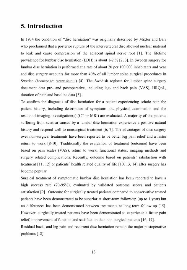
5. Introduction
In 1934 the condition of “disc herniation” was originally described by Mixter and Barr
who proclaimed that a posterior rupture of the intervertebral disc allowed nuclear material
to leak and cause compression of the adjacent spinal nerve root [1]. The lifetime
prevalence for lumbar disc herniation (LDH) is about 1-2 % [2, 3]. In Sweden surgery for
lumbar disc herniation is performed at a rate of about 20 per 100.000 inhabitants and year
and disc surgery accounts for more than 40% of all lumbar spine surgical procedures in
Sweden (homepage; www.4s.nu.) [4]. The Swedish register for lumbar spine surgery
document data pre- and postoperative, including leg- and back pain (VAS), HRQoL,
duration of pain and baseline data [5].
To confirm the diagnosis of disc herniation for a patient experiencing sciatic pain the
patient history, including description of symptoms, the physical examination and the
results of imaging investigation(s) (CT or MRI) are evaluated. A majority of the patients
suffering from sciatica caused by a lumbar disc herniation experience a positive natural
history and respond well to nonsurgical treatment [6, 7]. The advantages of disc surgery
over non-surgical treatments have been reported to be better leg pain relief and a faster
return to work [8-10]. Traditionally the evaluation of treatment (outcome) have been
based on pain scales (VAS), return to work, functional status, imaging methods and
surgery related complications. Recently, outcome based on patients’ satisfaction with
treatment [11, 12] or patients´ health related quality of life [10, 13, 14] after surgery has
become popular.
Surgical treatment of symptomatic lumbar disc herniation has been reported to have a
high success rate (70-95%), evaluated by validated outcome scores and patients
satisfaction [9]. Outcome for surgically treated patients compared to conservative treated
patients have been demonstrated to be superior at short-term follow-up (up to 1 year) but
no differences has been demonstrated between treatments at long-term follow-up [15].
However, surgically treated patients have been demonstrated to experience a faster pain
relief, improvement of function and satisfaction than non-surgical patients [16, 17].
Residual back- and leg pain and recurrent disc herniation remain the major postoperative
problems [18].
13
14
6. Background
6.1 Disc anatomy and pathology
6.1.1The intervertebral disc is the largest avascular structure in the human body and is mainly
composed of proteoglycans and collagen (type 1 and 2) [19]. Its major role is mechanical,
to bear and/or transmit loads arising from body weight and muscle activity. The
intervertebral disc consists of three anatomical structures; nucleus pulposus, annulus
fibrosus and the vertebral endplates. The nucleus pulposus is the water-rich (80%),
gelatineous center of the disc. Annulus fibrosus encircle the nucleus pulposus and is made
of collagen sheets, usually named lamellae. These are held together by proteoglycans and
help to maintain the fluid within the tissue of the disc. The proteoglycan aggregates within
the annulus fibrosus and the nucleus pulposus and gives the young disc its strengths and
pliability.
Both the top and the bottom of each vertebra are capped with a thin cartilaginous pad
called the vertebral endplates. The endplates are avascular and aneural in a healthy adult
disc [20].
6.1.2
Intervertebral disc degeneration visualized on MRI is commonly found in asymptomatic
individuals over 50 years of age [21]. Natural degeneration due to aging cause the nucleus
pulposus to become more fibrotic and less gel-like and initially the disc height is
preserved [22, 23].
Pathologic disc degeneration is also named “deteriorated disc” [23] and are believed to
start with a disruption or tear of the annulus fibrosus. A local inflammation thereafter
occurs and macrophages, mast cells and growth factors infiltrate the injured disc and try
to initiate repair of the annulus fibrosus [24]. However, there are no diagnostic methods
14
15
available to clearly differentiate between a normal aging of the disc and pathological disc
deterioration.
Various factors have been suggested to influence disc degeneration; mechanical, genetic,
shear stress or toxic factors [25, 26]. The most significant biochemical change and critical
factor in intervertebral disc degeneration is the loss of proteoglycans [20, 27] . The
decrease in proteoglycans leads to a lower water binding capacity of the disc. These
biochemical changes has a major effect on the load bearing and commonly cause a
degenerated lumbar discs to bulge posterior when the spine is loaded.
6.1.3
Herniation of an intervertebral disc occurs when the outer layer of the disc, the annulus
fibrosus, ruptures and allows disc material from the inner part, nucleus pulposus (NP) and
sometimes also annulus fibrosus and material from the end plate, to leak out. If the
leakage occurs posterior it leaks into the epidural space and may cause pressure on
nervous structures and thereby initiate symptoms including sciatic pain.
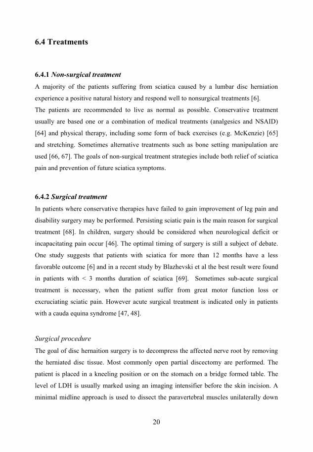
A disc bulge may be the first step towards a disc herniation but may also stay as a bulge
without any further rupture of the annulus fibrosus (figure 1a).
There is different ways to classify disc herniations and one of the most common
classifications separate between protrusion, extrusion or sequestration of the disc (figure
1b-d) [23].
Herniated discs can take the form of ”protrusion” or “extrusion” based on the shape of the
displaced material and they can be “contained” or “uncontained”. A contained disc
herniation has an intact outer annulus.
15
16
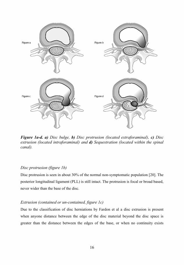
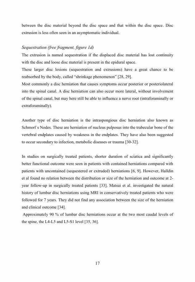
Disc bulge, Disc protrusion (located extraforaminal), Disc extrusion (located intraforaminal) and Sequestration (located within the spinal canal).
Disc protrusion (figure 1b)
Disc protrusion is seen in about 30% of the normal non-symptomatic population [20]. The
posterior longitudinal ligament (PLL) is still intact. The protrusion is focal or broad based,
never wider than the base of the disc.
Extrusion (contained or un-contained, figure 1c)
Due to the classification of disc herniations by Fardon et al a disc extrusion is present
when anyone distance between the edge of the disc material beyond the disc space is
greater than the distance between the edges of the base, or when no continuity exists
16
17
between the disc material beyond the disc space and that within the disc space. Disc
extrusion is less often seen in an asymptomatic individual.
Sequestration (free fragment, figure 1d) The extrusion is named sequestration if the displaced disc material has lost continuity
with the disc and loose disc material is present in the epidural space.
These larger disc lesions (sequestration and extrusions) have a great chance to be
reabsorbed by the body, called “shrinkage phenomenon” [28, 29].
Most commonly a disc herniation that causes symptoms occur posterior or posteriolateral
into the spinal canal. A disc herniation can also occur more lateral, without involvement
of the spinal canal, but may here still be able to influence a nerve root (intraforaminally or
extraforaminally).
Another type of disc herniation is the intraspongious disc herniation also known as
Schmorl´s Nodes. These are herniation of nucleus pulposus into the trabecular bone of the
vertebral endplates caused by weakness in the endplates. They have also been suggested
to occur secondary to infection, metabolic diseases or trauma [30-32].
In studies on surgically treated patients, shorter duration of sciatica and significantly
better functional outcome were seen in patients with contained herniations compared with
patients with uncontained (sequestered or extruded) herniations [6, 9]. However, Halldin
et al found no relation between the distribution or size of the herniation and outcome at 2-
year follow-up in surgically treated patients [33]. Matsui et al. investigated the natural
history of lumbar disc herniations using MRI in conservatively treated patients who were
followed for 7 years. They did not find any association between the size of the herniation
and clinical outcome [34].
Approximately 90 % of lumbar disc herniations occur at the two most caudal levels of
the spine, the L4-L5 and L5-S1 level [35, 36].
17
18
Incidence
The lifetime incidence for lumbar disc herniation (LDH) with nerve root
compression is about 1-2 % [2]. The incidence ranges from 1-10% [37]. For
sciatica the lifetime incidence is about 40% [3].
Risk factors A disc herniation occurs as a consequence of an annular rupture and can be viewed as a
special form of disc degeneration. As described earlier the etiology of disc degeneration
and thereby also disc herniation is multifactorial and related to factors as heredity [38-41],
lifting/carrying and extreme forward bending [42, 43], hard-working [40, 41], lumbar load
[40], cumulative physical workload [44], BMI over 25.7 [41], lack of sport activities and
night shift works [45]. The main factors associated with LDH in children are trauma or
sport activities with subsequent axial loading of the spine [46].
6.2 Symptoms
LBP is believed to often be the first symptom of a LDH and may also possibly be the only
symptom. Sciatica is the most classical symptom, characterized by radiating pain with a
dermatomal distribution, typically affecting one nerve root in the lumbar or sacral spine.
Sometimes it is associated with sensory and/or motor deficits in the leg in accordance to
the affected nerve root. The cauda equina syndrome is a special form of LDH where the
herniated disc material occupies most of the space in the spinal canal. This serious
condition involves sacral nerve roots and besides leading to uni- or bilateral sciatic pain,
can cause bowel and/or bladder disturbances, lowers extremity muscle weakness and loss
of sensibility and perineal or saddle paresthesia. This condition demands a more urgent
attention compared to when a single nerve root is affected [47-49].
The radiating pain, sciatica, is not always caused solely by compression of the nerve root.
It has been suggested to be a combination of chemical radiculopathy (neurotoxic agents)
[50-53] and/or discogenic sciatica [54-56].
18
19
The symptoms of LDH in children are more often limited to LBP than in the adult but can
also present as more classical radiculopathy. The diagnosis in young individuals may also
be delayed by their somewhat different description of the pain experience and the fact that
LDH is a rare diagnosis in children. The lumbar pain may only be present when coughing
or bending forward. Neurological examinations are often negative and it is rare with
bladder and genital dysfunctions [46, 57].
6.3 Diagnosis
The diagnosis is based on patient history (typically low back pain and/or
dermatomally distributed sciatica), clinical examination (positive findings involve
decreased tendon reflexes, sensory and/or motor deficits and positive SLR), and
imaging findings (computed tomography (CT) or magnetic resonance imaging
(MRI)). Most patients suffering from lumbar disc herniation have a positive straight
leg raising test (SLR) or Lasegue´s sign [58-61]. The MRI technique has
advantages compared to the CT with a better visualization of the soft tissue and the
neural structures. However, the relationship between a disc herniation visualized by
MRI (or CT) and the experience of sciatica are complex. Some patients have a disc
herniation with possible nerve influence on images, but experience no pain [20, 34,
62, 63]. In addition patients who completely recover from an episode of back pain
and sciatica, can demonstrate an unchanged disc herniation appearance on a follow-
up MRI [29].
19
20
6.4 Treatments
6.4.1 A majority of the patients suffering from sciatica caused by a lumbar disc herniation
experience a positive natural history and respond well to nonsurgical treatments [6].
The patients are recommended to live as normal as possible. Conservative treatment
usually are based one or a combination of medical treatments (analgesics and NSAID)
[64] and physical therapy, including some form of back exercises (e.g. McKenzie) [65]
and stretching. Sometimes alternative treatments such as bone setting manipulation are
used [66, 67]. The goals of non-surgical treatment strategies include both relief of sciatica
pain and prevention of future sciatica symptoms.
6.4.2 In patients where conservative therapies have failed to gain improvement of leg pain and
disability surgery may be performed. Persisting sciatic pain is the main reason for surgical
treatment [68]. In children, surgery should be considered when neurological deficit or
incapacitating pain occur [46]. The optimal timing of surgery is still a subject of debate.
One study suggests that patients with sciatica for more than 12 months have a less
favorable outcome [6] and in a recent study by Blazhevski et al the best result were found
in patients with < 3 months duration of sciatica [69]. Sometimes sub-acute surgical
treatment is necessary, when the patient suffer from great motor function loss or
excruciating sciatic pain. However acute surgical treatment is indicated only in patients
with a cauda equina syndrome [47, 48].
Surgical procedure
The goal of disc hernaition surgery is to decompress the affected nerve root by removing
the herniated disc tissue. Most commonly open partial discectomy are performed. The
patient is placed in a kneeling position or on the stomach on a bridge formed table. The
level of LDH is usually marked using an imaging intensifier before the skin incision. A
minimal midline approach is used to dissect the paravertebral muscles unilaterally down
20
21
to the laminae and thereafter the interlaminar ligament is resected. A partial laminotomy
is performed when necessary. When the ligamentum flavum is resected the neuronal
structures and the disc are visualized. Herniated disc material and loose fragments in the
disc is removed to decompress the affected neural structures. The surgery is performed
with or without microscope, due to the surgeons’ preference. It has previously been
demonstrated that the use of microscope does not effect the short-term (1-year) result
[70]. The duration of hospital stay depends on the patient’s mobility after surgery. The
aim is that patients should be ambulated already at the day of the surgery. In some centers
elective disc herniation surgery is planned as day surgery.
Results of surgical treatment
Reported early results of surgical discectomy have shown success rates of over 90% [9,
18, 71, 72]. Further, patients treated surgically have been reported to have better short-
term outcome than conservative treated patients [9, 73]. Long-term results have been less
positive with success rates of 40-79 % [74, 75]. Both short- and long-term studies have
demonstrated higher recovery rates and more complete relief of leg pain, higher
improvement of satisfaction with treatment and perceived recovery compared with
patients treated non-surgically [16, 17, 72].
However, in the study by Weber et al. no significant differences in clinical outcome
between surgically and non-surgically treated patients were found 4-10 years after surgery
[15].
6.5 Outcome
6.5.1 Many different parameters have been studied to identify predictive factors for outcome
after LDH surgery. Some of these factors are intensity and duration of leg pain, Physical
examination, gender, age, work and education level, social and psychological factors and
type of herniation [76] [77-83]. Factors that have been identified to predict a positive
21

22
outcome (leg pain relief and/or satisfaction with surgical result and/or return to work) are
short duration of preoperative leg pain [6, 84], no preoperative co-morbidity, male gender
[85], age [86] and short time to surgery [69, 87]. Longstanding preoperative leg pain has
been demonstrated to be a predictor for a less favorable outcome [6, 84, 88]. Heavy
manual work and low education level [74, 89], female gender [13, 85, 86, 90, 91]
contained herniation [9], disc protrusion [87] and central lumbar disc herniation [92] are
other factors that may affect the outcome negatively.
6.5.2 There are many different ways to evaluate the outcome after lumbar disc herniation
surgery. Traditionally the effect of treatment have been based on pain scales (VAS),
return to work, functional status, imaging measurements and surgery related
complications.
Outcome was earlier commonly assessed by the surgeon but in later year an independent
observer (objective) or the patient itself (subjective) has been introduced to evaluate the
outcome [93].
6.5.3
The risk for bias decrease when using an independent observer not involved in the
treatment of the patient, for assessment of surgical outcome. Objective outcome is
often classified by scales related to postoperative pain relief, work capacity/sick
leave, daily activity or analgesics consumption. These scales often use the scale;
excellent, good, fair or poor. Odom`s criteria [94] and Mcnab`s classification of
outcome [95, 96] are two such validated scales used in spinal surgery. They are
demonstrated to correlate well with other validated outcome scores based on
subjective outcome and patients´ satisfaction with treatment [12, 97].
22
23
6.5.4 There are several validated patient administered multi-item questionnaires used for
patients surgically treated for spinal disorders. Most of them are based on back- and leg
pain relief, daily living, physical activity, disability and social restriction.
The “Visual Analogue Scale” (VAS) is a pain scale used for visualizing the patients’ pain
in the clinical care of patients but also a well known outcome instrument for pain
assessment often used in orthopedic conditions (e.g. in spinal surgery). Clarke and Spear
introduced this instrument in medical science 1964, for assessment of wellbeing [98].
The “Oswestry Disability Index” (ODI) describes back-related disability with a
combination of physical and social restrictions [99]. It has emerged as the most
commonly recommended condition specific outcome for spinal disorders [100, 101].
ODI was developed by John O`Brian in 1976 and is based upon interviews by an
orthopaedic surgeon and an occupational therapist of back pain patients. Based on these
interviews they constructed a questionnaire made of 10 questions covering different
dimensions of daily living.
Another patient based outcome measure is the global assessment scale where patient rate
their satisfaction with the result or improvement of preoperative pain as satisfied, partly
satisfied or not satisfied, or grade their improvement/no improvement with treatment as
much better, better, unchanged or worse [11, 12, 102].
A good correlation has been demonstrated between patient based assessments and earlier
validated objective outcome scores [102].
Expectations Patients’ expectations on a given treatment have been demonstrated to be associated with
the success rate of the treatment and directly related to patients’ satisfaction with the
given treatment [12, 103-107]. Patients´ expectations appears to influence the
postoperative outcome [108, 109] . There are different factors that can influence
preoperative expectations, such as given information and care and influence from
relatives and friends [12, 110-112]. Patients with high or positive expectations on
surgical treatment of lumbar disc herniation have been demonstrated to have a better
outcome, based on pain relief and recovery time [103, 113].
23
24
To assess patient expectations it is common to use preoperative questionnaires about
expected improvement in physical function or expected ability to return to work and daily
living.
Health related quality of life
WHO´s definition of health is “a state of complete physical, mental and social well-being
and not merely the absence of infirmity” [114]. Quality of life is defined as “The
individuals´ perceptions of their position in life, in the context of the cultural and value
system in which they live and in relation to their goals, expectations, standards and
concerns” [115]. The concept of health status and quality of life in medicine are widely
used in different diagnoses [10, 13, 14, 80, 116-123]. In later year patients HRQoL has
gained an increasing interest in health care evaluation and is often used in studies of
patients undergoing spinal surgery [10, 13, 14, 74, 124, 125].
HRQoL measure an individual’s health in the aspects of physical, psychological, social
and spiritual role function as well as general well being [126]. These instruments are
validated and for the patient easily used self-completed questionnaires.
A number of instruments have been development to measure HRQoL during the last
years.
Disease specific instrument
Disease specific instruments are used for a specific disease or health problem.
Validated disease specific instrument commonly used in spinal disorders are the
Million Index [127], the Oswestry Disability Index [99], the Low Back Outcome
Score [128] and the Roland Morris Disability Index [129, 130].
Generic instrument These instruments focus on descriptions of health status and allow comparisons between
different study populations and disease groups.
The Medical Outcomes Survey 36-item Short-Form (SF-36) is one of the most used
HRQoL questionnaires. The SF-36 includes 8 health concepts of functional health and
well-being scores divided in equal amount of physical and mental components [131, 132].
24
25
Other instruments used in spinal research are Nottingham Health Profile [133] and the
Duke Health Profile [134, 135].
Utility instrument Utility instruments are designed to weight several dimensions into a single index which is
used as a score expressing the total health state. With non-disease specific HRQoL
instruments it is possibility to compare the effect of different medical conditions on the
quality of life and these instruments all allows comparisons between different treatments
on a specific condition.
Available instruments are, The Health Utility Index (HUI) [136], the Quality of Well-
Being [132] and the EQ-5D [137].
EQ-5D Questionnaire The EQ-5D is a non-disease specific instrument and thereby allows comparison between
different medical conditions. The instrument consists of two different parts, EQ-5D score
and EQ-VAS and is a patient self-administered multidimensional questionnaire [137].
The EQ-5D score comprises 5 dimensions; mobility, self-care, usual activities,
pain/discomfort and anxiety/depression. Each of the dimensions is divided into three
levels of severity; no problems, moderate problems or severe problems. This instrument
creates 243 health states that have been ranged as EQ-5D index scores by a large UK
population sample. The EQ-5D score thereby range from 0.00 (worse possible health
state) and 1.00 (best possible health state). Some states are considered as “worse than
death” and given negative values [138].
EQ-VAS is a global assessment of patient health, ranges from 0-100 representing “worst-
best imaginable health state”.
The result of the two-part questionnaire can be presented as a health profile or as a global
health index. It has good reliability and validity [139]
25
26
6.5.5 Magnetic Resonance Imaging (MRI) is the preferred investigations for spinal diseases.
The high resolution of MRI for soft tissues allows illustration of the intervertebral disc
morphology and the nervous structures[140]. MRI makes it possible to classify the LDH
preoperatively and to evaluate any eventual disc degeneration.
There are different ways to classify LDH by MRI. Komori et al graded the herniation
after continuity of nucleus tissue to the remaining disc [141].
The relationship between different type or size of the herniation and outcome following
lumbar disc herniation are well studied for both conservative and surgically treated
patients [76, 92, 142].
The postoperative period after lumbar disc herniation surgery use to be divided into early
(<6 months) and late stages. In the early stage it is difficult to interpret postoperative MRI
following lumbar spine surgery and it is hard to differentiate recurrent disc herniation
from post-surgical fibroses [143, 144].
The clinical significance of postoperative scar formation and outcome is debated.
Some authors have demonstrated a relationship between extensive peridural fibrosis
diagnosed by MRI and increasing low back pain and/or recurrent radicular pain [145-147]
whereas others have not [148]. To prevent or reduce postoperative peridural scar
formation numerous synthetic and natural materials have been evaluated in both animal
and human studies [149-155].
One of the materials is ADCON-L, a bioresorbable carbohydrate polymer gel. The
purpose of this material is to cover the dura and nerve root(s), to form a protective
membrane, until the fibrosis formation is completed [156-158].
26
27
7. Aims of the study
Overall the aim of the study was to study long-term result and different factors
influencing the result in patients undergoing surgical treatment for a lumbar disc
herniation using different outcome instruments.
More specific the aims of the study were to:
Investigate the long-term result after lumbar disc hernaition surgery and possible
predictive factors for surgical outcome, such as demographics, psychological,
social or physiological.
Investigate patients satisfaction with given care and given information in surgically
treated disc herniation patients. To study possible relationships between baseline
characteristics and expectations of surgical results and ability to return to work and
if these expectations are related to self- reported global assessment (subjective) 2
years after surgery. To compare the patients’ self-reported global assessed outcome
with the independent observer´s (objective) assessed outcome 2 years after surgery.
Study if there is any relationship between the size and/or location of peridural
formed scar and clinical outcomes 2-year after lumbar discectomy. Analyze scar
development between 6 and 24 months postoperatively by MRI and to study if
ADCON-L has effect on scar size or and/or patients’ outcome.
Study the influence of preoperative factors on HRQoL and the postoperative
development of HRQoL at 2-year and long-term follow-up, using the EQ-5D
instrument, in lumbar disc herniation surgery patients.
27

28
8. Summary of studies
8.1 Materials and methods Between September 1996 and March 2002, 183 consecutive patients surgically treated for
a CT or MRI verified one-level disc herniation on L4-L5 or L5-S1 level, were recruited
for the studies. Patients with other spinal disorders, previous spinal surgery, recurrent
herniation at the same level, extra foraminal herniations, perioperative negative
exploration or language difficulties were excluded.
The Regional Ethical Review Board approved all the studies and the patients gave their
informed consent for participation.
Surgery All patients were treated surgically at the department of Orthopaedics, Sahlgrenska
University Hospital in Gothenburg. Six different spine surgeons performed the surgery.
By using a midline approach the paravertebral muscles were dissected down to laminae
and the interlaminar ligaments resected. A partial laminotomy was performed when
necessary. Herniated disc material and loose fragments from the disc was removed to
decompress the affected neural structures. Postoperatively the patients were ambulated
already at the end of the day of surgery.
Study Ι: Patients From the initially 183 recruited patients, 171 were included in this study. Twelve patients
were excluded initially because of confusion of language, perioperative negative
exploration, other spinal disorders or previous spinal surgery. One-hundred fifty-four
(90%) of the patients completed the 2-year follow-up and 140 (82%) of the patients
completed the long-term follow-up. Surgery was performed at L4-L5 level in 77 (45%) of
the patients and 94 (55%) at L5-S1 level. The mean age was 39±11 years and 76 (44%)
were women. Mean time to long-term follow-up was 7.3± 1.0 (5.1-9.3) years.
28
29
Preoperative data
For baseline data preoperative questionnaires were used. Gender, age, smoking habits,
duration of leg pain, intensity of leg- and back pain (VAS), analgesics consumption, time
to sick leave, degree of depression/anxiety (ZDS), disability (ODI) and employment status
was recorded.
2-year follow-up data
Patients were followed-up by an independent observer, a neurologist, at the hospital. The
independent observer assessed outcome (objective) based on Mcnab´s classification of
postoperative outcome and graded the result as excellent, good, fair or poor.
Patients rated their satisfaction (subjective) with treatment as; satisfied, partly satisfied or
not satisfied. Questionnaire about improvement in pain intensity (VAS), analgesics
consumption, eventual sick leave, degree of depression/anxiety (ZDS), disability (ODI),
and employment status was recorded.
Long-term follow-up data (5.1-9.3 years) Follow-up questionnaires were sent by mail, and if the patient did not respond, up to two
reminders were sent by mail after telephone contact.
Again patients rated the satisfaction with treatment, filled in questionnaires including pain
intensity (VAS), sick leave, employment status, degree of depression/anxiety (ZDS) and
disability (ODI).
Primary outcomes at 2-year and long-term follow-upObjective outcome assessed by independent observer (at 2-year follow-up).
Subjective outcome based on patients´ satisfaction with treatment (at 2-year and long term
follow-up).
Secondary outcomes at 2-year follow-up Change in leg- and back pain (VAS), working capacity, analgesics consumption and need
for sleeping pills.
29
30
Predictive factors investigated
Gender, age, smoking habits, level of disc hernia, use of analgesics, time on sick leave,
duration of leg pain, baseline leg- and back pain (VAS), ZDS and ODI.
Statistical analysis Objective and subjective outcome were dichotomized in the analyses. Objective outcome
was categorized as excellent/good or fair/poor and subjective outcome was categorized as
satisfied or partly/not satisfied. Potential relationships with predictors were analyzed by
using chi-square test (categorical predictors), t-test (baseline data as a predictor) or the
Mann Whitney U test. For multivariate analyses logistic regression was used and analyzed
with a forward stepwise selection procedure to find the most influential predictor.
Study ΙΙ: Patients
This study included 172 patients. One-hundred forty-eight (86%) completed the 2-year
follow-up (study population). The study population had a mean age of 40 (18-66) years,
68 (46%) of the patients were women and 66 (45%) underwent surgery at the L4-L5
level.. The originally included 172 patients in study II included one more patient than
study I (171). This “extra” patient was a patient with negative exploration preoperatively.
However this was not noticed until the analyze for study I which was performed after
study II. The exclusion of this patient in study II would not in any way have changed any
of the results.
Preoperative information and care
Preoperative the patient received information from the surgeon regarding the planned
surgery about the surgical procedure, risks, expected time to sick leave and result.
Regarding the result the patient received information of expected improvement in pain
relief, especially leg pain and of sensibility and muscle function.
A physiotherapist gave instruction and information about daily living postoperatively.
Information was also given by spine educated nurses employed at the ward.
30
31
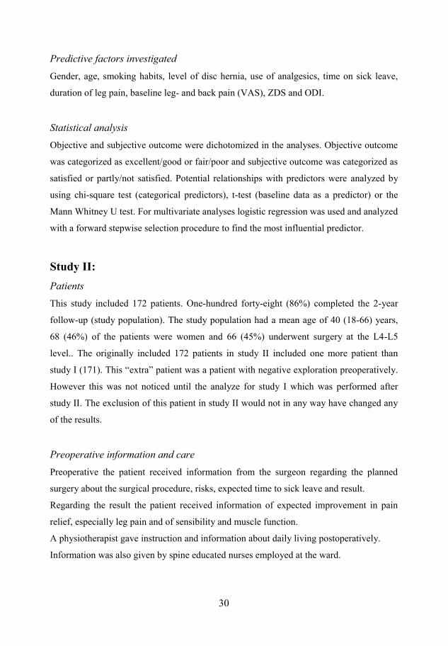
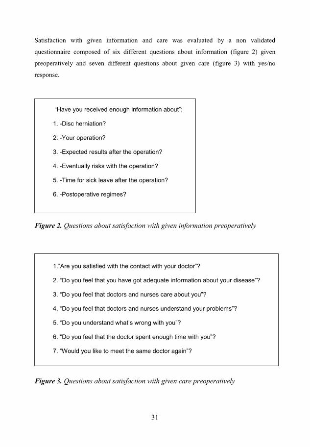
Satisfaction with given information and care was evaluated by a non validated
questionnaire composed of six different questions about information (figure 2) given
preoperatively and seven different questions about given care (figure 3) with yes/no
response.
“Have you received enough information about”;
30"/Fkue"jgtpkcvkqpA""
40"/[qwt"qrgtcvkqpA""
50"/Gzrgevgf"tguwnvu"chvgt"vjg"qrgtcvkqpA""
60"/Gxgpvwcnn{"tkumu"ykvj"vjg"qrgtcvkqpA""
70"/Vkog"hqt"ukem"ngcxg"chvgt"vjg"qrgtcvkqpA""
80"/Rquvqrgtcvkxg"tgikoguA""
Questions about satisfaction with given information preoperatively
1.”Are you satisfied with the contact with your doctor”?
2. “Do you feel that you have got adequate information about your disease”?
3. “Do you feel that doctors and nurses care about you”?
4. “Do you feel that doctors and nurses understand your problems”?
5. “Do you understand what’s wrong with you”?
6. “Do you feel that the doctor spent enqwij"vkog"with you”?
7. “Would you like to meet the same doctor again”?
Questions about satisfaction with given care preoperatively
31
32
Depending on how many “yes/no” answers the patient had they were deployed as
“satisfied” or “dissatisfied” and dichotomized into two groups. To be assessed as
“satisfied” with given information > 3 out of 6 questions must be answered as “yes”
and likewise >4 out of 7 for given care.
Expectations
Before surgery patients were asked about their expectations on surgical result regarding
pain intensity (leg- and back pain), sensibility and muscle function.
Expectations were assessed by using a non graded line with descriptions expected to
“become worse”, “stay the same” or “become better” at the left, middle and the right of
the line. Patient’s expectations were then graded as low, medium or high.
By using the same non graded line improvement at the two follow-up occasions was
assessed as “got worse”, “no change” or “got better”. Preoperatively questions were asked
about expectations on work ability postoperatively. Postoperatively the patients were
asked about if they had returned to their workplace (or a similar work) after surgery.
Subjective and objective outcome The patient rated their subjective outcome;”global assessment of satisfaction with
treatment” as satisfied, partly or not satisfied. An independent neurologist assessed
objective outcome based on Macnab´s classification on surgical outcome as excellent,
good, fair or poor.
Preoperative data
Baseline data and degree of preoperative depression (ZDS), leg- and back pain intensity
(VAS), duration of pain, disability (ODI), satisfaction with information and care,
expectations on surgical result and work ability was collected from questionnaires.
32
33
2-year follow-up data
Improvement in leg- and back pain, sensibility, muscle function and work capacity was
recorded. Patients rated their global assessment of satisfaction with treatment (subjective).
Objective outcome was assessed as in study I.
Statistical analysis All correlation coefficients (cc) presented are Spearman’s rank correlations. Cross-tables
were analyzed with standard chi-square tests or Fishers exact test. For analyses the
relationship between dichotomous variables Mann-Whitney-U´s rank-sum test was used.
Study ΙΙΙ:PatientsInitially 128, out of the total 183 patients, were recruited for this study. The patients were
peroperatively randomized into two groups, treated with ADCON–L (n=60) or not
(controls) (n=48). During the follow-up period 6 patients underwent surgery for a
recurrent herniation and 3 patients underwent spinal fusion surgery. Eleven were lost to
follow-up. Of the remaining 108 patients (ADCON–L (n=60), controls (n=48)) 103 (95%)
completed the MRI examination at 24 months, 99 (92%) filled in questionnaire about
satisfaction at 2-year follow-up and 102 (94%) was examined by the independent
observer.
The mean age was 39 years (18-66), 51 (47%) women and 48 (44%) underwent
surgery at the L4-L5 level.
Surgery All patients underwent the procedure of partial discectomy described above. Before
closure of the surgical site the patients were randomized by envelope to receive ADCON-
L (treated) or not (controls). For the treated group 3g of ADCON-L was applied to the
surgical site, surrounding the nerve root, the thecal sac and the posterior longitudinal
ligament, up to the lower surface of the lamina.
33
34
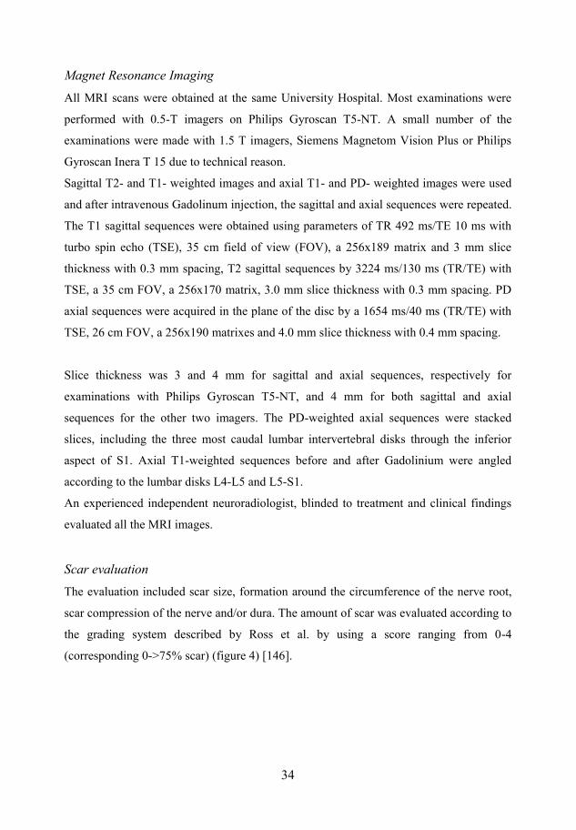
Magnet Resonance Imaging
All MRI scans were obtained at the same University Hospital. Most examinations were
performed with 0.5-T imagers on Philips Gyroscan T5-NT. A small number of the
examinations were made with 1.5 T imagers, Siemens Magnetom Vision Plus or Philips
Gyroscan Inera T 15 due to technical reason.
Sagittal T2- and T1- weighted images and axial T1- and PD- weighted images were used
and after intravenous Gadolinum injection, the sagittal and axial sequences were repeated.
The T1 sagittal sequences were obtained using parameters of TR 492 ms/TE 10 ms with
turbo spin echo (TSE), 35 cm field of view (FOV), a 256x189 matrix and 3 mm slice
thickness with 0.3 mm spacing, T2 sagittal sequences by 3224 ms/130 ms (TR/TE) with
TSE, a 35 cm FOV, a 256x170 matrix, 3.0 mm slice thickness with 0.3 mm spacing. PD
axial sequences were acquired in the plane of the disc by a 1654 ms/40 ms (TR/TE) with
TSE, 26 cm FOV, a 256x190 matrixes and 4.0 mm slice thickness with 0.4 mm spacing.
Slice thickness was 3 and 4 mm for sagittal and axial sequences, respectively for
examinations with Philips Gyroscan T5-NT, and 4 mm for both sagittal and axial
sequences for the other two imagers. The PD-weighted axial sequences were stacked
slices, including the three most caudal lumbar intervertebral disks through the inferior
aspect of S1. Axial T1-weighted sequences before and after Gadolinium were angled
according to the lumbar disks L4-L5 and L5-S1.
An experienced independent neuroradiologist, blinded to treatment and clinical findings
evaluated all the MRI images.
Scar evaluation The evaluation included scar size, formation around the circumference of the nerve root,
scar compression of the nerve and/or dura. The amount of scar was evaluated according to
the grading system described by Ross et al. by using a score ranging from 0-4
(corresponding 0->75% scar) (figure 4) [146].
34

35
Each MRI slice was divided into 4 spatial quadrants, 5 slices were available for evaluation (2 slices above-, 1 at- and 2 below the disc). For each patient this made 20 MRI quadrants available for evaluation. The quadrant with the most pronounced scarformation got the highest score and was used for calculation.
Preoperative Patients reported radicular pain by using VAS. Disc herniation was confirmed by a CT or
MRI evaluation.
6 months follow-up
All included patients were examined with MRI using the “ADCON-L” protocol.
2-year follow-up
Examination with MRI was done with the “ADCON-L” protocol. Radicular leg pain was
reported by using VAS. Satisfaction with treatment (subjective outcome) was rated by the
patients as satisfied, partly satisfied or not satisfied. Objective outcome was examined by
independent observer according to Macnab classification.
35
36
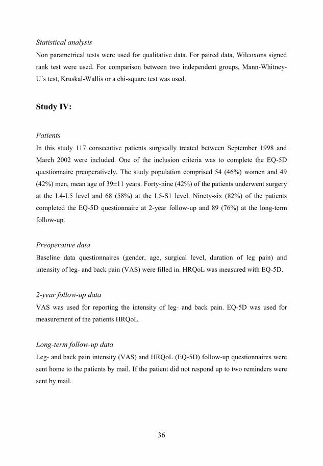
Statistical analysis
Non parametrical tests were used for qualitative data. For paired data, Wilcoxons signed
rank test were used. For comparison between two independent groups, Mann-Whitney-
U´s test, Kruskal-Wallis or a chi-square test was used.
Study ΙV:
Patients In this study 117 consecutive patients surgically treated between September 1998 and
March 2002 were included. One of the inclusion criteria was to complete the EQ-5D
questionnaire preoperatively. The study population comprised 54 (46%) women and 49
(42%) men, mean age of 39±11 years. Forty-nine (42%) of the patients underwent surgery
at the L4-L5 level and 68 (58%) at the L5-S1 level. Ninety-six (82%) of the patients
completed the EQ-5D questionnaire at 2-year follow-up and 89 (76%) at the long-term
follow-up.
Preoperative data Baseline data questionnaires (gender, age, surgical level, duration of leg pain) and
intensity of leg- and back pain (VAS) were filled in. HRQoL was measured with EQ-5D.
2-year follow-up data VAS was used for reporting the intensity of leg- and back pain. EQ-5D was used for
measurement of the patients HRQoL.
Long-term follow-up data Leg- and back pain intensity (VAS) and HRQoL (EQ-5D) follow-up questionnaires were
sent home to the patients by mail. If the patient did not respond up to two reminders were
sent by mail.
36
37
Statistical analysis
SPSS software was used for statistical analyzes. Paired Samples t-test was used for
analyzing differences in HRQoL at baseline and follow-ups. When comparing groups,
Independent Sample t-test or ANOVA was used. For correlations between different
variables nonparametric Spearman rank correlation test was used.
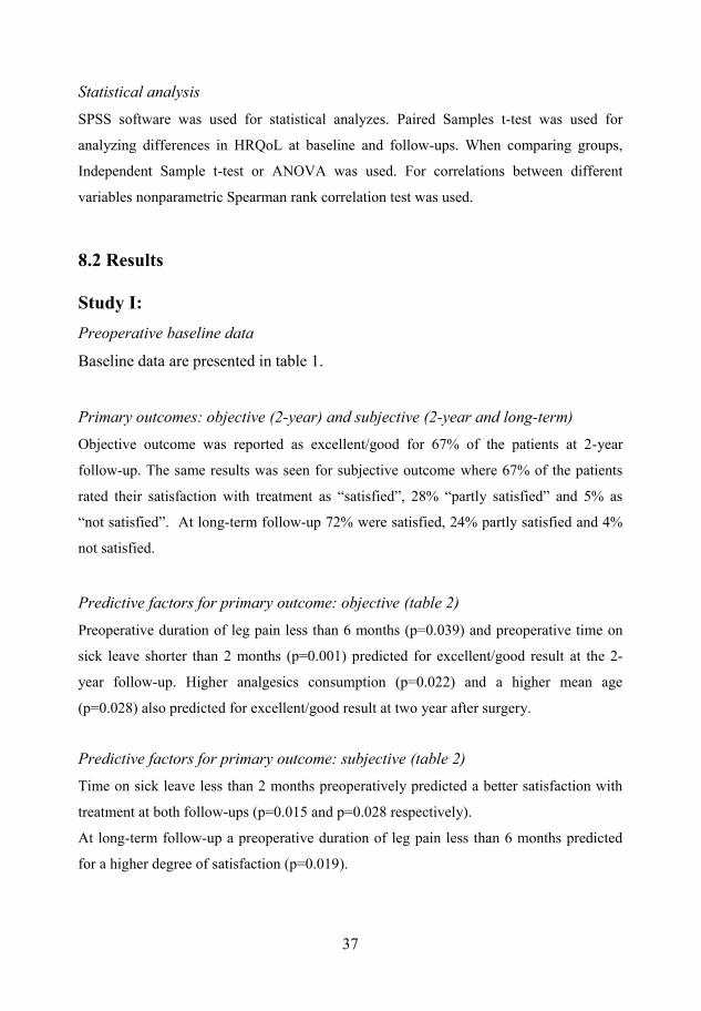
8.2 Results
Study Ι:Preoperative baseline data
Baseline data are presented in table 1.
Primary outcomes: objective (2-year) and subjective (2-year and long-term) Objective outcome was reported as excellent/good for 67% of the patients at 2-year
follow-up. The same results was seen for subjective outcome where 67% of the patients
rated their satisfaction with treatment as “satisfied”, 28% “partly satisfied” and 5% as
“not satisfied”. At long-term follow-up 72% were satisfied, 24% partly satisfied and 4%
not satisfied.
Predictive factors for primary outcome: objective (table 2)
Preoperative duration of leg pain less than 6 months (p=0.039) and preoperative time on
sick leave shorter than 2 months (p=0.001) predicted for excellent/good result at the 2-
year follow-up. Higher analgesics consumption (p=0.022) and a higher mean age
(p=0.028) also predicted for excellent/good result at two year after surgery.
Predictive factors for primary outcome: subjective (table 2) Time on sick leave less than 2 months preoperatively predicted a better satisfaction with
treatment at both follow-ups (p=0.015 and p=0.028 respectively).
At long-term follow-up a preoperative duration of leg pain less than 6 months predicted
for a higher degree of satisfaction (p=0.019).
37
38
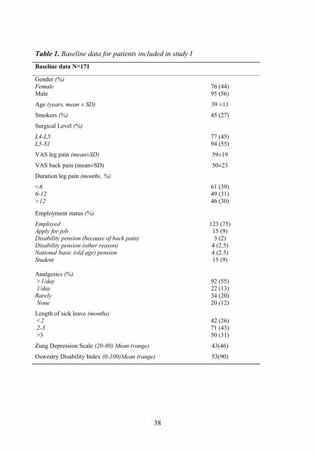
Baseline data for patients included in study I
Baseline data N=171
Gender (%)FemaleMale
76 (44)95 (56)
Age (years, mean ± SD) 39 ±11
Smokers (%) 45 (27)
Surgical Level (%)
L4-L5L5-S1
77 (45)94 (55)
VAS leg pain (mean±SD) 59±19
VAS back pain (mean±SD) 50±23
Duration leg pain (months, %)
<66-12>12
61 (39)49 (31)46 (30)
Employment status (%)
EmployedApply for jobDisability pension (because of back pain)Disability pension (other reason)National basic (old age) pensionStudent
123 (75)15 (9)3 (2)
4 (2.5)4 (2.5)15 (9)
Analgesics (%)>1/day1/day
RarelyNone
92 (55)22 (13)34 (20)20 (12)
Length of sick leave (months) <22-3>3
42 (26)71 (43)50 (31)
Zung Depression Scale (20-80) Mean (range) 43(46)
Oswestry Disability Index (0-100)Mean (range) 53(90)
38
39
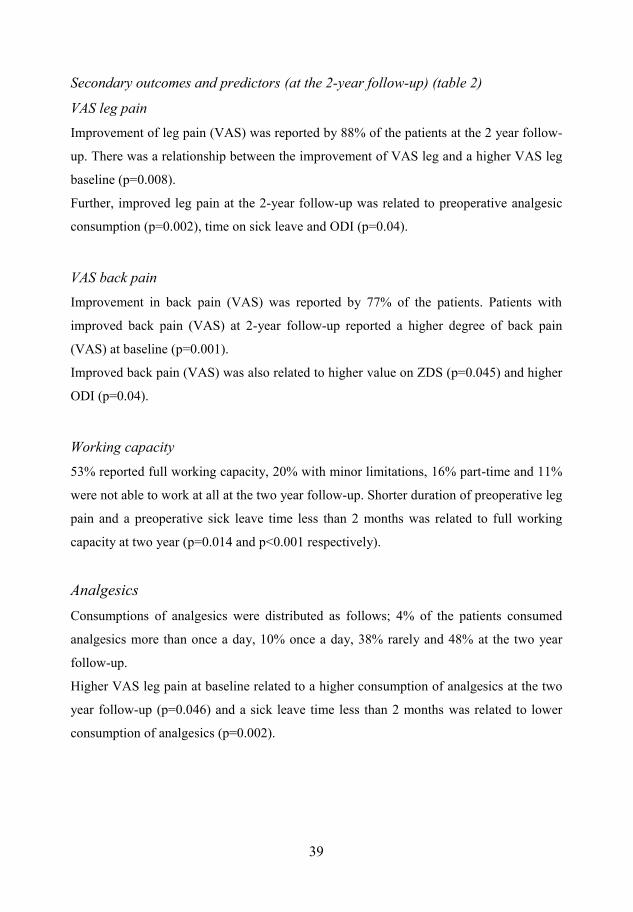
Secondary outcomes and predictors (at the 2-year follow-up) (table 2)
VAS leg pain Improvement of leg pain (VAS) was reported by 88% of the patients at the 2 year follow-
up. There was a relationship between the improvement of VAS leg and a higher VAS leg
baseline (p=0.008).
Further, improved leg pain at the 2-year follow-up was related to preoperative analgesic
consumption (p=0.002), time on sick leave and ODI (p=0.04).
VAS back pain Improvement in back pain (VAS) was reported by 77% of the patients. Patients with
improved back pain (VAS) at 2-year follow-up reported a higher degree of back pain
(VAS) at baseline (p=0.001).
Improved back pain (VAS) was also related to higher value on ZDS (p=0.045) and higher
ODI (p=0.04).
Working capacity 53% reported full working capacity, 20% with minor limitations, 16% part-time and 11%
were not able to work at all at the two year follow-up. Shorter duration of preoperative leg
pain and a preoperative sick leave time less than 2 months was related to full working
capacity at two year (p=0.014 and p<0.001 respectively).
Analgesics Consumptions of analgesics were distributed as follows; 4% of the patients consumed
analgesics more than once a day, 10% once a day, 38% rarely and 48% at the two year
follow-up.
Higher VAS leg pain at baseline related to a higher consumption of analgesics at the two
year follow-up (p=0.046) and a sick leave time less than 2 months was related to lower
consumption of analgesics (p=0.002).
39
40
Sleeping pills
Sex percent of the patients reported a regular consumption of sleeping pills At the two
year follow-up The use of sleeping pills regularly was related to time to sick leave longer
than 3 months (p=0.008) and higher baseline value on ZDS (p=0.02) and on ODI
(p=0.007).
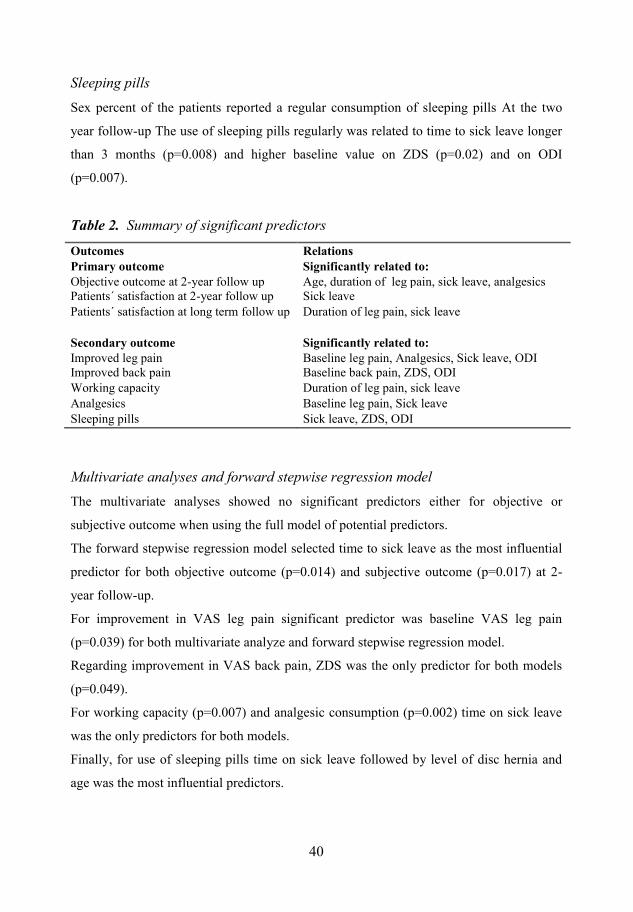
Summary of significant predictors
Outcomes RelationsPrimary outcome Significantly related to:Objective outcome at 2-year follow up Age, duration of leg pain, sick leave, analgesicsPatients´ satisfaction at 2-year follow up Sick leavePatients´ satisfaction at long term follow up Duration of leg pain, sick leave
Secondary outcome Significantly related to:Improved leg pain Baseline leg pain, Analgesics, Sick leave, ODIImproved back pain Baseline back pain, ZDS, ODIWorking capacity Duration of leg pain, sick leaveAnalgesics Baseline leg pain, Sick leaveSleeping pills Sick leave, ZDS, ODI
Multivariate analyses and forward stepwise regression model The multivariate analyses showed no significant predictors either for objective or
subjective outcome when using the full model of potential predictors.
The forward stepwise regression model selected time to sick leave as the most influential
predictor for both objective outcome (p=0.014) and subjective outcome (p=0.017) at 2-
year follow-up.
For improvement in VAS leg pain significant predictor was baseline VAS leg pain
(p=0.039) for both multivariate analyze and forward stepwise regression model.
Regarding improvement in VAS back pain, ZDS was the only predictor for both models
(p=0.049).
For working capacity (p=0.007) and analgesic consumption (p=0.002) time on sick leave
was the only predictors for both models.
Finally, for use of sleeping pills time on sick leave followed by level of disc hernia and
age was the most influential predictors.
40
41
Study ΙΙ:
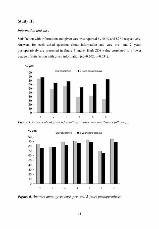
Information and care
Satisfaction with information and given care was reported by 46 % and 82 % respectively.
Answers for each asked question about information and care pre- and 2 years
postoperatively are presented in figure 5 and 6. High ZDS value correlated to a lower
degree of satisfaction with given information (cc=0.202, p=0.031).
Answers about given information, preoperative and 2-years follow-up.
Answers about given care, pre- and 2-years postoperatively.
232425262728292:2;2322
3 4 5 6 7 8
% yesrtgqrgtcvkxg 4/{gct"rquvqrgtcvkxg
2
32
42
52
62
72
82
92
:2
;2
322
3 4 5 6 7 8 9
% yesrtgqrgtcvkxg 4/{gct"rquvqrgtcvkxg
41

42
Expectations
A majority of the patients had high expectations on the surgical results, leg pain (94%),
back pain (81%), sensibility (71%) and muscle function (72%). High expectations
preoperatively were found to predict a better patient reported result 2 years
postoperatively.
Expectations on ability to return to previous or similar work after surgery had a good
correlation with the result at 2 year. 76% of the patients expected to return to work and
24% expected not to return and out of these respectively 78% and 26% returned to
work (p=0.021). Patients who expected to return to work were more often satisfied with
treatment (67%) than patients with low expectations where only 45% were satisfied with
treatment after 2 years (p=0.029).
Patients with high value on ZDS had a lower degree of expectations on leg pain recovery
(p=0.022) and ability to return to work (p=0.046).
Subjective and objective outcome
A high agreement between patients’ satisfaction (66% satisfied) with result and
independent assessment (67% excellent/good) was found (p<0.001). 86 % of the satisfied
patients were assessed as excellent/good. In the small group of patient (6%) “not
satisfied” no one was assessed as excellent/good by the independent observer.
There was a also a high agreement between satisfaction with treatment and recovery of all
physical functions and symptoms (p<0.001)
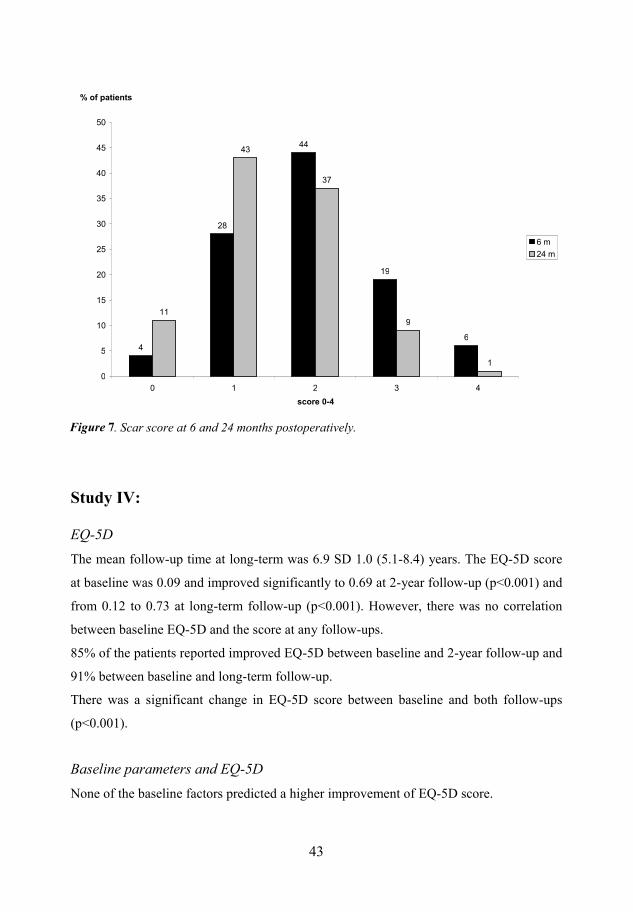
Study ΙΙΙ:There were no significant differences in baseline parameters between ADCON-L- and the
control group. The scar size decreased between 6- and 24 months in 49%, were
unchanged in 42% and increased in 9% of the patients. Scar score at 6- and 24 months are
presented in figure 7.
No relationship was found between scar size or scar location and clinical outcome (VAS
leg pain, subjective and objective).
In this study no positive effects on scar size, or outcome in ADCON-L treated patients
could be seen.
42
43
. Scar score at 6 and 24 months postoperatively.
Study ΙV:
EQ-5DThe mean follow-up time at long-term was 6.9 SD 1.0 (5.1-8.4) years. The EQ-5D score
at baseline was 0.09 and improved significantly to 0.69 at 2-year follow-up (p<0.001) and
from 0.12 to 0.73 at long-term follow-up (p<0.001). However, there was no correlation
between baseline EQ-5D and the score at any follow-ups.
85% of the patients reported improved EQ-5D between baseline and 2-year follow-up and
91% between baseline and long-term follow-up.
There was a significant change in EQ-5D score between baseline and both follow-ups
(p<0.001).
Baseline parameters and EQ-5DNone of the baseline factors predicted a higher improvement of EQ-5D score.
6
4:
66
3;
8
33
65
59
;
3
2
7
32
37
42
47
52
57
62
67
72
2 3 4 5 6
score 0-4
% of patients
8"o
46"o
43
7
44
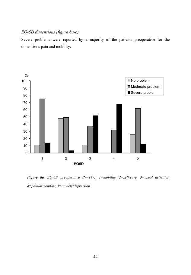
EQ-5D dimensions (figure 8a-c)
Severe problems were reported by a majority of the patients preoperative for the
dimensions pain and mobility.
EQ-5D preoperative (N=117). 1=mobility, 2=self-care, 3=usual activities,
4=pain/discomfort, 5=anxiety/depression
2
32
42
52
62
72
82
92
:2
;2
32
3 4 5 6 7
EQ5D
%Pq"rtqdngo
Oqfgtcvg"rtqdngo
Ugxgtg"rtqdngo
44

45
EQ-5D at 2-year follow-up (N=96). 1=mobility, 2=self-care, 3=usual activities,
4=pain/discomfort, 5=anxiety/depression
EQ-5D at long-term follow-up (N=89). 1=mobility, 2=self-care, 3=usual activities,
4=pain/discomfort, 5=anxiety/depression
0
32
20
52
40
72
60
70
80
90
100
1 2 3 4 5EQ5D
%
Pq"rtqdngoOqfgtcvg"rtqdngoUgxgtg"rtqdngo
2
32
42
52
62
72
82
92
:2
;2
322
3 4 5 6 7
EQ5D
%
Pq"rtqdngo
Oqfgtcvg"rtqdngo
Ugxgtg rtqdngo
2
32
42
52
62
72
82
92
:2
;2
322
3 4 5 6 7
EQ5D
%
Pq"rtqdngo
Oqfgtcvg"rtqdngo
Ugxgtg rtqdngo
45
46
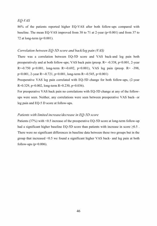
EQ-VAS
86% of the patients reported higher EQ-VAS after both follow-ups compared with
baseline. The mean EQ-VAS improved from 38 to 71 at 2-year (p<0.001) and from 37 to
72 at long-term (p<0.001).
Correlation between EQ-5D score and back/leg pain (VAS) There was a correlation between EQ-5D score and VAS back-and leg pain both
preoperatively and at both follow-ups; VAS back pain (preop. R= -0.338, p<0.001, 2-year
R=-0.750 p<0.001, long-term R=-0.692, p<0.001), VAS leg pain (preop. R= -398,
p<0.001, 2-year R=-0.721, p<0.001, long-term R=-0.545, p<0.001)
Preoperative VAS leg pain correlated with EQ-5D change for both follow-ups, (2-year
R=0.329, p=0.002, long-term R=0.230, p=0.036).
For preoperative VAS back pain no correlations with EQ-5D change at any of the follow-
ups were seen. Neither, any correlations were seen between preoperative VAS back- or
leg pain and EQ-5 D score at follow-ups.
Patients with limited increase/decrease in EQ-5D score
Patients (37%) with <0.5 increase of the preoperative EQ-5D score at long-term follow-up
had a significant higher baseline EQ-5D score than patients with increase in score ≥0.5 .
There were no significant differences in baseline data between these two groups but in the
group that increased <0.5 we found a significant higher VAS back- and leg pain at both
follow-ups (p=0.006).
46
47
9. General Discussion
In summary, the long-term result after lumbar disc herniation surgery is in our study
satisfactory in about two out of three patients, based on patient satisfaction with treatment.
Preoperative sick leave time was found to be a clinically important predictor for both
subjective and objective outcome in the investigated patients. No association between
peridural scar and clinical outcome was seen in this patient group. Neither could any
association between the localization of scar formation in relation to nervous structures and
clinical outcome be detected.
The patients in the present study were mostly satisfied with care pre- and postoperatively
but to a lesser extent satisfied with given information preoperatively. Patients with
preoperative positive expectations on work return and realistic expectations on pain and
physical recovery had a greater chance to be satisfied with the results after lumbar disc
herniation surgery compared to patients with negative and/or unrealistic expectations.
Based on HRQoL, more than 90% of the patients reported an improvement at both 2-year
and long-term follow-up. However, they do not, as a group, obtain the health related
quality of life level (as measured by EQ-5D) as a normal population at as long time as 5-8
years postoperatively.
Clinical outcome
In the present study 67% of the patient reported satisfaction with treatment after 2 year,
28% was partly satisfied and 5 % not satisfied. Objective assessment of the patients was
in accordance with these findings. Further, more than 90 % of the patients reported an
increase in EQ-5D score. The short-term results after surgical treatment of symptomatic
lumbar disc herniation has previously been reported to have a high success rate (70-95%),
evaluated by validated outcome scores, HRQoL and patients satisfaction [9, 10, 12, 18,
71]. The result from the present study is within the lower range of previous reports with
both a somewhat low satisfaction with treatment rate and excellent/good objective
assessed outcome frequency.
Long-term follow-up results were studied in study and V using subjective outcome,
measured as satisfaction with treatment and as change in EQ-5D score (HRQoL).
47
48
Satisfaction with treatment was here reported by 72% of the patients and more than 90%
of the patients reported improvement in HRQoL.
However, patients assigned to early surgery have previously been demonstrated to obtain
a faster pain relief and recovery in short-term but less in long-term [15, 17, 72, 159]. Our
study was in agreement with previously reported studies on long-term follow-up. Short-
term result was less positive with a quite low satisfaction and objective assessed outcome.
The explanation for the finding that our patient group, with a long mean waiting time for
surgery, reached similar outcome levels at long-term, but had somewhat less improvement
than in previous studies at the first follow-up, might be that the benefit from early surgery
seen in other studies decrease over time. Our short-term results are in agreement with
studies comparing conservative patients with surgically treated patients followed at long-
term.
Predictive factors
Patients who fail to recover from sciatic pain are at risk to develop chronic pain
syndromes which emphasizes the importance of identifying factors that can predict the
outcome, both regarding short-term and long-term results. Therefore possible predictive
factors for the surgical outcome are of interest.
In study we found that preoperative duration of leg pain less than 6 months were related
to excellent/good outcome at both 2-year and long-term follow-up. Longstanding
preoperative duration of leg pain has also previously been described as a predictor for bad
outcome after lumbar discectomy [6, 84, 85].
The length of preoperative sick leave was identified as the most influential predictive
factor, both for objective outcome and subjective outcome and for several of the
secondary outcomes as well, in our study. In patients with a sick leave period shorter than
2 months the satisfactory outcome frequency at 2 year follow-up was about 80% both
using objective and subjective assessments. Furthermore full working capacity was three
times more common among patients with the shortest sick leave (<2 months) compared to
patients with the longest sick leave period (>3 months). Analgesics consumption was also
related to time on sick leave.The need for analgesics was twice as high in the group with a
sick leave period >3 months compared to patients with sick leave time <2 months.
48
49
Finally, the chance to have an improvement of leg pain was higher (97% versus 78%) in
the group with <2 months of sick leave compared to sick leave time >3 months.
In a previous study patients with long preoperative sick leave time were found to have
less favorable outcome [160]. Further, in a study on work return after lumbar discectomy
long preoperative sick leave also were demonstrated to be a negative predictor for work
return [161].
Patients’ satisfaction
The overall level of patient satisfaction with treatment was found to be in accordance with
objective outcome assessed by the independent observer in the present study. Agreement
between patient satisfaction and other outcome instruments has previously been
demonstrated in studies on patients with chronic low back pain [102].
We found in study further that only 46% and 75% respectively of the patients were
satisfied pre- and 2-years postoperatively with the information regarding the procedure
(disc herniation surgery) that was given. It has previously also been reported that patients
are dissatisfied with the degree of information that they receive from their healthcare
provider [159].
As could be expected the amount of satisfaction with information increased over time
when the patients had obtained their own experience. This is in agreement with a study by
MgGregor at al.[110]. Almost 80% of the patients in the present study were satisfied with
given care both pre- and 2 years postoperatively.
The Quality theory suggests that, if an obtained result after an intervention is equal or
better than a customer expects he or she will be satisfied. This theory seems applicable for
medical treatments of patients where satisfaction has been demonstrated to be directly
related to patient expectations [104, 106, 107].
49
50
Expectations
Almost all patients in the study had high expectations on recovery regarding reduced leg
pain 2 years after surgery, which is in agreement with given information.
Different factors can influence expectations, such as known success rate with a given
treatment, given information and care, personality trait, health and mood status and
influence from relatives/friends [110, 111, 162]. In this study mood status was the only
baseline characteristics that influenced the expectation on clinical outcome. A high value
on ZDS was related to a low degree of expectations on leg pain recovery (p=0.022) and
expectations on ability to return to work (p=0.046). Preoperative high expectations on
reduced leg pain, improved sensibility and muscle function were all associated with better
postoperative subjective outcome. Further, positive expectations on work return were
associated with higher postoperative work return, increased physical function and
satisfaction with treatment. Positive expectations have also in earlier studies been
demonstrated to be associated with better health outcomes and have further been
suggested to influence clinical outcome independently of the treatment itself [103, 106,
113, 163].
Postoperative scar formation
In study we investigated the possible relation between postoperative scar formation
and clinical outcome. There was a reduction in the presence and amount of scar tissue
between MRI at 6 months compared to 24 months but no association between scar
formation (size or localization) and clinical outcome at any time point was demonstrated.
The relationship between the fibrotic tissues and the epidural structures (thecal sac and
nerve roots) and clinical symptoms have been extensively debated. Some previous studies
suggest that scar tissue is responsible for unfavorable outcome results after spinal surgery
[145, 146, 164]. Our study is in agreement with the findings by Nygaard et al where no
associations between the amounts of postoperative peridural scar formation or nerve root
displacement and outcome 1 year after microdiscectomy for lumbar disc herniation was
seen [148]. There has been an interest to investigate if anti-adhesion barriers would make
a difference if applied peroperatively in patients undergoing disc herniation surgery. An
effect of such a material would indirectly support the theory that the peridural scar causes
50

51
problems. One of the materials, ADCON-L, have been demonstrated to reduced scar
formation and improved clinical outcome[146, 165] In the present study we could not find
any positive effects of the anti-adhesion gel used, ADCON-L. It did not reduce the scar
size nor did it improve the clinical outcome. This is in agreement with some other studies
on ADCON-L [157, 166].
Health Related Quality of Life
A majority of the patients improved dramatically and significantly in HRQoL, measured
by EQ-5D after surgical treatment of lumbar discectomy. There was a large improvement,
expressed as a change in EQ-5D score, between baseline and the 2-year follow-up which
then stayed relatively stable until the long-term follow-up.
The mean preoperative EQ-5D score in our patients was only 0.09 which is lower than
been reported in previous studies on patients suffering from lumbar disc herniation [10,
13]. This could be related to the long waiting time for surgery. And when comparing the
EQ-5D score to a previously described age correlated normal population our study group
of operated disc herniation patients had a lower EQ-5D score at both follow-ups [167].
A strong correlation between EQ-5D and VAS back- and leg pain at baseline, 2-year and
long-term follow-up was seen. Our results suggests that despite a significant improvement
in EQ-5D score that the overall inferior result in this patients group are caused by a
subgroup of patients still suffering from pain many years postoperatively. We did not find
any correlation between the preoperative EQ-5D score and the postoperative score at any
of the follow-ups.
51
52
10. Conclusions
Long-term result after lumbar disc herniation surgery is in our study
satisfactory in about two out of three patients based on patient satisfaction with
treatment. Time on sick leave was an important predictor for outcome both at 2-
year and at long-term follow-up (5.1-9.3 years)
Dissatisfaction with preoperative information was found. Associations between
expectations and lower mood, experienced surgical result and subjective
outcome were found. Subjective outcome demonstrated high agreement with
objective outcome at 2-year follow-up
No association between peridural scar and clinical outcome were seen. No
effect on ADCON-L at scar size and/or patients outcome.
There was a strong correlation between EQ-5D and VAS back- and leg pain at
baseline, 2-year and long-term follow-up. More than 90% of the patient
reported an improvement in EQ-5D at both 2-year and long-term follow-up.
52
53
11. Future
Patient satisfaction is an important outcome after surgically treated lumbar disc
herniation and satisfaction is closely related to both expectations and given
information in this patient group. This highlights the importance of having the patient
in focus when evaluation a given treatment.
If a decision is made about surgery, when conservative treatment have failed it is
important to give disc herniation patients appropriate information which cause realistic
expectations. Patients of today themselves seek information from many sources e.g.
the internet and health-care providers need to be aware of this and advice and discuss
more around this than traditionally has been done.
The findings that both sick leave time and leg pain duration influence postoperative
results in this patient group indicates, what also previously has been suggested, that
surgery should be performed after a relative short waiting time.
This highlights the importance of a well functioning health care organization, where
time on the waiting list for clinical appointments as well as surgery do not delay the
treatment.
53
54
12. Acknowledgements
I wish to express my sincere appreciation to all those who have helped and supported me
in my research and with this thesis. Thanks especially to:
Helena Brisby, MD, Associate Professor, Department of Orthopaedics, Sahlgrenska
University Hospital at University of Gothenburg, my tutor and friend, for exceptional
encouragement and endless support in both clinical and scientific work.
Bengt Lind, MD, Associate Professor, Gothenburg Spine Center, co-tutor and former
tutor, for guidance and support. For making me interested in spinal research and letting
me start with my thesis.
Bjorn Zoëga, MD, PhD, Landspitali University Hospital, Reykjavik, Island, co-tutor, for
exceptional guidance and relevant points of views.
Klas Halldin, MD, PhD, Department of Orthopaedics, Sahlgrenska University Hospital at
University of Gothenburg, co-author, for sharing clinical and scientific experience and for
being a fun former room-mate.
Martin Gellerstedt, PhD, Trollhättan/Uddevalla, co-author and statistical analyzes. So
many times I wanted to give up with all these statistical “things” but you gave me so
much support and pushed me further.
Gro Gadehol-Göthlin, MD, PhD, Department of Radiology, Sahlgrenska University
Hospital at University of Gothenburg, co-author, thank you for making study
possible!
Lena Rutberg, MD, Department of Neurology, Sahlgrenska University Hospital at
University of Gothenburg, for assessment of patients at 2-year follow-up.
Björn Rydevik, MD, Professor, Department of Orthopaedics, Head Section of
Anesthesiology, Biomaterials and Orthopaedics, The Sahlgrenska Academy at University
of Gothenburg, for your kind help and support.
Jón Karlsson, MD, Professor and Chairman, Department of Orthopaedics, Sahlgrenska
University Hospital at University of Gothenburg, for good advice and support.
54

55
Jonas Thanner, MD, PhD and Peter Nyberg, MD, PhD as former Head of the
Department of Orthopaedics, Sahlgrenska University Hospital
All my colleagues at the Spine section, Department of Orthopaedics and all other
colleagues and friends at the Department of Orthopaedics, Sahlgrenska University
Hospital.
Linda Johansson, secretary for academic guidance and help.
Jill Fallénius, secretary for help with editorial work, miss you!
Eva Fermén, secretary, for help with editorial work at long-term follow-up and for
laughing and giving friendly support.
Pontus Andersson, for professional illustrations.
Barbro Rönnberg, my sister-in-law, for endless friendship and guidance through life, in
good times and in bad.
Erika Löfstedt, my dearest friend. I told you so!
Carina Floberg, my close friend, for wonderful friendship.
All my friends in the military, KS14 medical platoon, NBG08 surgical unit and FS17.
You are always on my mind!
Louise Lindblad, for everything we’ve been through during the last years, so many
happy moments!
Monica Hyttmark Niska, FS17 FST, my room-mate, we did it!
My mother, Ingrid, for endless love and for always being there!
My father, Arne, who is not with us anymore, you are in my mind and in my heart!
My sisters, Kicki, Ulla, Annika, Ulrica, Cilla and my brothers, Peter and Stefan, for all
your support in life.
My husband, Mattias, what would life be without you?
Finally to my dearest treasures, Hanna, Emma and Leo for all love and happy moments!
55

56
The studies were financially supported by grants from the Gothenburg Medical Society,
Doktor Felix Néubergh Foundation , Marianne and Marcus Wallenberg Foundation and
government grants under the LUA agreement.
56
57
13. References
1. Barr, M.W.a., Rupture of the intervertebral disc with involvement of the spinal canal. NEngl J Med, 1934: p. 210-205.
2. Deyo, R.A., Nonsurgical care of low back pain. Neurosurg Clin N Am, 1991. 2(4): p. 851-62.
3. Frymoyer, J.W., et al., Risk factors in low-back pain. An epidemiological survey. J Bone Joint Surg Am, 1983. 65(2): p. 213-8.
4. Stromqvist, B., et al., One-year report from the Swedish National Spine Register. Swedish Society of Spinal Surgeons. Acta Orthop Suppl, 2005. 76(319): p. 1-24.
5. Stromqvist, B., et al., The Swedish National Register for lumbar spine surgery: Swedish Society for Spinal Surgery. Acta Orthop Scand, 2001. 72(2): p. 99-106.
6. Ng, L.C. and P. Sell, Predictive value of the duration of sciatica for lumbar discectomy. A prospective cohort study. J Bone Joint Surg Br, 2004. 86(4): p. 546-9.
7. Awad, J.N. and R. Moskovich, Lumbar disc herniations: surgical versus nonsurgical treatment. Clin Orthop Relat Res, 2006. 443: p. 183-97.
8. Atlas, S.J., et al., Long-term disability and return to work among patients who have a herniated lumbar disc: the effect of disability compensation. J Bone Joint Surg Am, 2000. 82(1): p. 4-15.
9. Dewing, C.B., et al., The outcomes of lumbar microdiscectomy in a young, active population: correlation by herniation type and level. Spine (Phila Pa 1976), 2008. 33(1): p. 33-8.
10. Gerszten, P.C., W.C. Welch, and J.T. King, Jr., Quality of life assessment in patients undergoing nucleoplasty-based percutaneous discectomy. J Neurosurg Spine, 2006. 4(1): p. 36-42.
11. Hudak, P.L. and J.G. Wright, The characteristics of patient satisfaction measures. Spine (Phila Pa 1976), 2000. 25(24): p. 3167-77.
12. Ronnberg, K., et al., Patients' satisfaction with provided care/information and expectations on clinical outcome after lumbar disc herniation surgery. Spine (Phila Pa 1976), 2007. 32(2): p. 256-61.
13. Jansson, K.A., et al., Health-related quality of life in patients before and after surgery for a herniated lumbar disc. J Bone Joint Surg Br, 2005. 87(7): p. 959-64.
14. Kagaya, H., et al., Quality of life assessment before and after lumbar disc surgery. JOrthop Sci, 2005. 10(5): p. 486-9.
15. Weber, H., Lumbar disc herniation. A controlled, prospective study with ten years of observation. Spine (Phila Pa 1976), 1983. 8(2): p. 131-40.
16. Atlas, S.J., et al., Long-term outcomes of surgical and nonsurgical management of sciatica secondary to a lumbar disc herniation: 10 year results from the maine lumbar spine study. Spine (Phila Pa 1976), 2005. 30(8): p. 927-35.
17. Peul, W.C., et al., Surgery versus prolonged conservative treatment for sciatica. N Engl J Med, 2007. 356(22): p. 2245-56.
18. Hakkinen, A., et al., Does the outcome 2 months after lumbar disc surgery predict the outcome 12 months later? Disabil Rehabil, 2003. 25(17): p. 968-72.
19. Trout, J.J., J.A. Buckwalter, and K.C. Moore, Ultrastructure of the human intervertebral disc: II. Cells of the nucleus pulposus. Anat Rec, 1982. 204(4): p. 307-14.
20. Raj, P.P., Intervertebral disc: anatomy-physiology-pathophysiology-treatment. Pain Pract, 2008. 8(1): p. 18-44.
21. Jensen, M.C., et al., Magnetic resonance imaging of the lumbar spine in people without back pain. N Engl J Med, 1994. 331(2): p. 69-73.
57
58
22. Buckwalter, J.A., Aging and degeneration of the human intervertebral disc. Spine (Phila Pa 1976), 1995. 20(11): p. 1307-14.
23. Fardon, D.F. and P.C. Milette, Nomenclature and classification of lumbar disc pathology. Recommendations of the Combined task Forces of the North American Spine Society, American Society of Spine Radiology, and American Society of Neuroradiology. Spine (Phila Pa 1976), 2001. 26(5): p. E93-E113.
24. Peng, B., et al., Possible pathogenesis of painful intervertebral disc degeneration. Spine (Phila Pa 1976), 2006. 31(5): p. 560-6.
25. Videman, T., et al., Associations of 25 structural, degradative, and inflammatory candidate genes with lumbar disc desiccation, bulging, and height narrowing. Arthritis Rheum, 2009. 60(2): p. 470-81.
26. Hadjipavlou, A.G., et al., The pathophysiology of disc degeneration: a critical review. JBone Joint Surg Br, 2008. 90(10): p. 1261-70.
27. Podichetty, V.K., The aging spine: the role of inflammatory mediators in intervertebral disc degeneration. Cell Mol Biol (Noisy-le-grand), 2007. 53(5): p. 4-18.
28. Saal, J.A., J.S. Saal, and R.J. Herzog, The natural history of lumbar intervertebral disc extrusions treated nonoperatively. Spine (Phila Pa 1976), 1990. 15(7): p. 683-6.
29. Takada, E., M. Takahashi, and K. Shimada, Natural history of lumbar disc hernia with radicular leg pain: Spontaneous MRI changes of the herniated mass and correlation with clinical outcome. J Orthop Surg (Hong Kong), 2001. 9(1): p. 1-7.
30. Rodacki, M.A., C.E. Castro, and D.S. Castro, Diffuse vertebral body edema due to calcified intraspongious disk herniation. Neuroradiology, 2005. 47(5): p. 316-21.
31. Stabler, A., et al., MR imaging of enhancing intraosseous disk herniation (Schmorl's nodes). AJR Am J Roentgenol, 1997. 168(4): p. 933-8.
32. Pfirrmann, C.W. and D. Resnick, Schmorl nodes of the thoracic and lumbar spine: radiographic-pathologic study of prevalence, characterization, and correlation with degenerative changes of 1,650 spinal levels in 100 cadavers. Radiology, 2001. 219(2): p. 368-74.
33. Halldin, K., et al., Three-dimensional radiological classification of lumbar disc herniation in relation to surgical outcome. Int Orthop, 2009. 33(3): p. 725-30.
34. Masui, T., et al., Natural history of patients with lumbar disc herniation observed by magnetic resonance imaging for minimum 7 years. J Spinal Disord Tech, 2005. 18(2): p. 121-6.
35. Cheung, K.M., et al., Prevalence and pattern of lumbar magnetic resonance imaging changes in a population study of one thousand forty-three individuals. Spine (Phila Pa 1976), 2009. 34(9): p. 934-40.
36. Humphreys, S.C. and J.C. Eck, Clinical evaluation and treatment options for herniated lumbar disc. Am Fam Physician, 1999. 59(3): p. 575-82, 587-8.
37. Lawrence, R.C., et al., Estimates of the prevalence of arthritis and selected musculoskeletal disorders in the United States. Arthritis Rheum, 1998. 41(5): p. 778-99.
38. Matsui, H., et al., Familial predisposition for lumbar degenerative disc disease. A case-control study. Spine (Phila Pa 1976), 1998. 23(9): p. 1029-34.
39. Ala-Kokko, L., Genetic risk factors for lumbar disc disease. Ann Med, 2002. 34(1): p. 42-7.
40. Zhang, Y.G., et al., Risk factors for lumbar intervertebral disc herniation in Chinese population: a case-control study. Spine (Phila Pa 1976), 2009. 34(25): p. E918-22.
41. Saftic, R., et al., Case-control study of risk factors for lumbar intervertebral disc herniation in Croatian island populations. Croat Med J, 2006. 47(4): p. 593-600.
42. Seidler, A., et al., The role of cumulative physical work load in lumbar spine disease: risk factors for lumbar osteochondrosis and spondylosis associated with chronic complaints.Occup Environ Med, 2001. 58(11): p. 735-46.
58
59
43. Seidler, A., et al., Occupational risk factors for symptomatic lumbar disc herniation; a case-control study. Occup Environ Med, 2003. 60(11): p. 821-30.
44. Seidler, A., et al., Cumulative occupational lumbar load and lumbar disc disease--results of a German multi-center case-control study (EPILIFT). BMC Musculoskelet Disord, 2009. 10: p. 48.
45. Elfering, A., et al., Risk factors for lumbar disc degeneration: a 5-year prospective MRI study in asymptomatic individuals. Spine (Phila Pa 1976), 2002. 27(2): p. 125-34.
46. Haidar, R., et al., Lumbar disc herniation in young children. Acta Paediatr, 2010. 99(1): p. 19-23.
47. Ahn, U.M., et al., Cauda equina syndrome secondary to lumbar disc herniation: a meta-analysis of surgical outcomes. Spine (Phila Pa 1976), 2000. 25(12): p. 1515-22.
48. Kostuik, J.P., et al., Cauda equina syndrome and lumbar disc herniation. J Bone Joint Surg Am, 1986. 68(3): p. 386-91.
49. Shapiro, S., Cauda equina syndrome secondary to lumbar disc herniation. Neurosurgery, 1993. 32(5): p. 743-6; discussion 746-7.
50. Murata, Y., et al., The role of tumor necrosis factor-alpha in apoptosis of dorsal root ganglion cells induced by herniated nucleus pulposus in rats. Spine (Phila Pa 1976), 2008. 33(2): p. 155-62.
51. Olmarker, K. and K. Larsson, Tumor necrosis factor alpha and nucleus-pulposus-induced nerve root injury. Spine (Phila Pa 1976), 1998. 23(23): p. 2538-44.
52. Igarashi, A., et al., Inflammatory cytokines released from the facet joint tissue in degenerative lumbar spinal disorders. Spine (Phila Pa 1976), 2004. 29(19): p. 2091-5.
53. Brisby, H., et al., Markers of nerve tissue injury in the cerebrospinal fluid in patients with lumbar disc herniation and sciatica. Spine (Phila Pa 1976), 1999. 24(8): p. 742-6.
54. Milette, P.C., et al., Radiating pain to the lower extremities caused by lumbar disk rupture without spinal nerve root involvement. AJNR Am J Neuroradiol, 1995. 16(8): p. 1605-13; discussion 1614-5.
55. Ohnmeiss, D.D., H. Vanharanta, and J. Ekholm, Degree of disc disruption and lower extremity pain. Spine (Phila Pa 1976), 1997. 22(14): p. 1600-5.
56. Ohnmeiss, D.D., H. Vanharanta, and J. Ekholm, Relation between pain location and disc pathology: a study of pain drawings and CT/discography. Clin J Pain, 1999. 15(3): p. 210-7.
57. Zamani, M.H. and G.D. MacEwen, Herniation of the lumbar disc in children and adolescents. J Pediatr Orthop, 1982. 2(5): p. 528-33.
58. Jonsson, B. and B. Stromqvist, Symptoms and signs in degeneration of the lumbar spine. A prospective, consecutive study of 300 operated patients. J Bone Joint Surg Br, 1993. 75(3): p. 381-5.
59. Andersson, G.B. and R.A. Deyo, History and physical examination in patients with herniated lumbar discs. Spine (Phila Pa 1976), 1996. 21(24 Suppl): p. 10S-18S.
60. Vroomen, P.C., M.C. de Krom, and J.A. Knottnerus, Diagnostic value of history and physical examination in patients suspected of sciatica due to disc herniation: a systematic review. J Neurol, 1999. 246(10): p. 899-906.
61. Majlesi, J., et al., The sensitivity and specificity of the Slump and the Straight Leg Raising tests in patients with lumbar disc herniation. J Clin Rheumatol, 2008. 14(2): p. 87-91.
62. Boden, S.D., et al., Abnormal magnetic-resonance scans of the lumbar spine in asymptomatic subjects. A prospective investigation. J Bone Joint Surg Am, 1990. 72(3): p. 403-8.
63. Boos, N., et al., 1995 Volvo Award in clinical sciences. The diagnostic accuracy of magnetic resonance imaging, work perception, and psychosocial factors in identifying symptomatic disc herniations. Spine (Phila Pa 1976), 1995. 20(24): p. 2613-25.
59
60
64. Zou, R., Y. Xu, and H.X. Zhang, [Evaluation on analgesic effect of electroacupuncture combined with acupoint-injection in treating lumbar intervertebral disc herniation].Zhongguo Gu Shang, 2009. 22(10): p. 759-61.
65. McKenzie, The lumbar spine mechanical diagnoses and therapy. Waikanae, New Zeeland: Spinal Publicationa. 1981.
66. Lu, L.J., et al., [The observe of clinical effect of treating lumbar intervertebral disc herniation by bone setting manipulation of different directions]. Zhongguo Gu Shang,2009. 22(4): p. 255-8.
67. Zhang, W.B., et al., [Manipulative reduction for lumbar intervertebral disc herniation: a controlled clinical trial]. Zhongguo Gu Shang, 2008. 21(4): p. 273-5.
68. Peul, W.C., et al., Prolonged conservative care versus early surgery in patients with sciatica caused by lumbar disc herniation: two year results of a randomised controlled trial. BMJ, 2008. 336(7657): p. 1355-8.
69. Blazhevski, B., et al., Predictive value of the duration of sciatica for lumbar discectomy.Prilozi, 2008. 29(2): p. 325-35.
70. Tullberg, T., J. Isacson, and L. Weidenhielm, Does microscopic removal of lumbar disc herniation lead to better results than the standard procedure? Results of a one-year randomized study. Spine (Phila Pa 1976), 1993. 18(1): p. 24-7.
71. Asch, H.L., et al., Prospective multiple outcomes study of outpatient lumbar microdiscectomy: should 75 to 80% success rates be the norm? J Neurosurg, 2002. 96(1 Suppl): p. 34-44.
72. Weinstein, J.N., et al., Surgical versus nonoperative treatment for lumbar disc herniation: four-year results for the Spine Patient Outcomes Research Trial (SPORT). Spine (Phila Pa 1976), 2008. 33(25): p. 2789-800.
73. Atlas, S.J., et al., The Maine Lumbar Spine Study, Part II. 1-year outcomes of surgical and nonsurgical management of sciatica. Spine (Phila Pa 1976), 1996. 21(15): p. 1777-86.
74. Loupasis, G.A., et al., Seven- to 20-year outcome of lumbar discectomy. Spine (Phila Pa 1976), 1999. 24(22): p. 2313-7.
75. Naylor, A., Late results of laminectomy for lumbar disc prolapse. A review after ten to twenty-five years. J Bone Joint Surg Br, 1974. 56(1): p. 17-29.
76. Carragee, E.J., et al., Clinical outcomes after lumbar discectomy for sciatica: the effects of fragment type and anular competence. J Bone Joint Surg Am, 2003. 85-A(1): p. 102-8.
77. Graver, V., et al., Seven-year clinical follow-up after lumbar disc surgery: results and predictors of outcome. Br J Neurosurg, 1999. 13(2): p. 178-84.
78. Rothoerl, R.D., et al., Are there differences in the symptoms, signs and outcome after lumbar disc surgery in the elderly compared with younger patients? Br J Neurosurg, 1998. 12(3): p. 250-3.
79. Iglesias-Casarrubios, P., et al., [Lasegue's test as prognostic factor for patients undergoing lumbar disc surgery]. Neurocirugia (Astur), 2004. 15(2): p. 138-43.
80. Dvorak, J., et al., The outcome of surgery for lumbar disc herniation. II. A 4-17 years' follow-up with emphasis on psychosocial aspects. Spine (Phila Pa 1976), 1988. 13(12): p. 1423-7.
81. Kohlboeck, G., et al., Prognosis of multifactorial outcome in lumbar discectomy: a prospective longitudinal study investigating patients with disc prolapse. Clin J Pain, 2004. 20(6): p. 455-61.
82. Junge, A., et al., Predictors of bad and good outcome of lumbar spine surgery. A prospective clinical study with 2 years' follow up. Spine, 1996. 21(9): p. 1056-64; discussion 1064-5.
83. Hurme, M. and H. Alaranta, Factors predicting the result of surgery for lumbar intervertebral disc herniation. Spine, 1987. 12(9): p. 933-8.
60
61
84. Nygaard, O.P., R. Kloster, and T. Solberg, Duration of leg pain as a predictor of outcome after surgery for lumbar disc herniation: a prospective cohort study with 1-year follow up. J Neurosurg, 2000. 92(2 Suppl): p. 131-4.
85. Vucetic, N., et al., Diagnosis and prognosis in lumbar disc herniation. Clin Orthop Relat Res, 1999(361): p. 116-22.
86. Manniche, C., et al., Analysis of preoperative prognostic factors in first-time surgery for lumbar disc herniation, including Finneson's and modified Spengler's score systems. Dan Med Bull, 1994. 41(1): p. 110-5.
87. Hurme, M. and H. Alaranta, Factors predicting the result of surgery for lumbar intervertebral disc herniation. Spine (Phila Pa 1976), 1987. 12(9): p. 933-8.
88. Rothoerl, R.D., C. Woertgen, and A. Brawanski, When should conservative treatment for lumbar disc herniation be ceased and surgery considered? Neurosurg Rev, 2002. 25(3): p. 162-5.
89. Almeida, D.B., et al., Is preoperative occupation related to long-term pain in patients operated for lumbar disc herniation? Arq Neuropsiquiatr, 2007. 65(3B): p. 758-63.
90. Peul, W.C., et al., Influence of gender and other prognostic factors on outcome of sciatica. Pain, 2008. 138(1): p. 180-91.
91. Stromqvist, F., et al., Gender differences in lumbar disc herniation surgery. Acta Orthop, 2008. 79(5): p. 643-9.
92. Barlocher, C.B., J.K. Krauss, and R.W. Seiler, Central lumbar disc herniation. Acta Neurochir (Wien), 2000. 142(12): p. 1369-74; discussion 1374-5.
93. Graham, Chemonucleolysis. A preliminary report on a double blind study comparing chemonucleolysis and intradiscal administration of hydrocortisone in the treatment of backache and sciatica. . Orthop Clin North Am, 1975. 6: p. 259-263.
94. Odom, G.L. and F.V. Kristoff, Unilateral rupture of cervical disc. N C Med J, 1948. 9(3): p. 117-22.
95. Macnab, I., Negative disc exploration. An analysis of the causes of nerve-root involvement in sixty-eight patients. J Bone Joint Surg Am, 1971. 53(5): p. 891-903.
96. Macnab, I., Chapter 14. Pain and disability in degenerative disc disease. Clin Neurosurg, 1973. 20: p. 193-6.
97. Zoega, B., J. Karrholm, and B. Lind, Outcome scores in degenerative cervical disc surgery. Eur Spine J, 2000. 9(2): p. 137-43.
98. Clarke PRF, S., F.G., Reliability and sensitivity in the self-assessment of well-being. Bull Brit Psychol Soc 17, 1964.
99. Fairbank, J.C. and P.B. Pynsent, The Oswestry Disability Index. Spine, 2000. 25(22): p. 2940-52; discussion 2952.
100. Deyo, R.A., et al., Outcome measures for studying patients with low back pain. Spine (Phila Pa 1976), 1994. 19(18 Suppl): p. 2032S-2036S.
101. Turk, D.C. and D.A. Marcus, Assessment of chronic pain patients. Semin Neurol, 1994. 14(3): p. 206-12.
102. Hagg, O., et al., Simplifying outcome measurement: evaluation of instruments for measuring outcome after fusion surgery for chronic low back pain. Spine (Phila Pa 1976), 2002. 27(11): p. 1213-22.
103. Lutz, G.K., et al., The relation between expectations and outcomes in surgery for sciatica.J Gen Intern Med, 1999. 14(12): p. 740-4.
104. Nettleman, M.D., Patient satisfaction--what's new? Clin Perform Qual Health Care, 1998. 6(1): p. 33-7.
105. McGregor, A.H. and S.P. Hughes, The evaluation of the surgical management of nerve root compression in patients with low back pain: Part 2: patient expectations and satisfaction. Spine (Phila Pa 1976), 2002. 27(13): p. 1471-6; discussion 1476-7.
61
62
106. Iversen, M.D., et al., The prognostic importance of patient pre-operative expectations of surgery for lumbar spinal stenosis. Patient Educ Couns, 1998. 34(2): p. 169-78.
107. de Groot, K.I., S. Boeke, and J. Passchier, Preoperative expectations of pain and recovery in relation to postoperative disappointment in patients undergoing lumbar surgery. Med Care, 1999. 37(2): p. 149-56.
108. Yee, Do Patient Expectations of Spinal Surgery Relate to Funvtional Outcome? The Association of Bone and Joint Surgeons, 2008: p. 1154-1161.
109. Gepstein, Decompression surgery for lumbar spinal stenosis in the eldery: preoperative expectations and postoperative satisfaction. Spinal Cord, 2006. 44: p. 427-421.
110. McGregor, A.H. and S.P. Hughes, The evaluation of the surgical management of nerve root compression in patients with low back pain: Part 2: patient expectations and satisfaction. Spine, 2002. 27(13): p. 1471-6; discussion 1476-7.
111. Mancuso, C.A., et al., Patients' expectations of shoulder surgery. J Shoulder Elbow Surg, 2002. 11(6): p. 541-9.
112. Mancuso, C.A. and E.A. Salvati, Patients' satisfaction with the process of total hip arthroplasty. J Healthc Qual, 2003. 25(2): p. 12-8; quiz 18-9.
113. Mondloch, M.V., D.C. Cole, and J.W. Frank, Does how you do depend on how you think you'll do? A systematic review of the evidence for a relation between patients' recovery expectations and health outcomes. Cmaj, 2001. 165(2): p. 174-9.
114. WHO, The World Health Organization constirution, Geneva. 1947. 115. WHO, International classification of impairments, disabilities and handicaps: A manual
of classificationrelating to the consequences of disease. World Health Organisation, Geneva. 1980.
116. Greenfield, S. and S. Kaplan, Quality of life after myocardial infarction: Canada versus the United States. N Engl J Med, 1995. 332(7): p. 471; author reply 471-2.
117. Dvorak, J., M.H. Gauchat, and L. Valach, The outcome of surgery for lumbar disc herniation. I. A 4-17 years' follow-up with emphasis on somatic aspects. Spine (Phila Pa 1976), 1988. 13(12): p. 1418-22.
118. Scott, D.L. and T. Garrood, Quality of life measures: use and abuse. Baillieres Best Pract Res Clin Rheumatol, 2000. 14(4): p. 663-87.
119. Cleary, P.D., S. Greenfield, and B.J. McNeil, Assessing quality of life after surgery.Control Clin Trials, 1991. 12(4 Suppl): p. 189S-203S.
120. Heider, D., et al., Health-related quality of life in patients after lumbar disc surgery: a longitudinal observational study. Qual Life Res, 2007. 16(9): p. 1453-60.
121. Jansson, K.A., et al., Health-related quality of life (EQ-5D) before and one year after surgery for lumbar spinal stenosis. J Bone Joint Surg Br, 2009. 91(2): p. 210-6.
122. Rampersaud, Y.R., et al., Assessment of health-related quality of life after surgical treatment of focal symptomatic spinal stenosis compared with osteoarthritis of the hip or knee. Spine J, 2008. 8(2): p. 296-304.
123. Sanda, M.G., et al., Quality of life and satisfaction with outcome among prostate-cancer survivors. N Engl J Med, 2008. 358(12): p. 1250-61.
124. Saban, K.L., et al., Health-related quality of life of patients following selected types of lumbar spinal surgery: a pilot study. Health Qual Life Outcomes, 2007. 5: p. 71.
125. Hallberg, I., et al., Health-related quality of life after vertebral or hip fracture: a seven-year follow-up study. BMC Musculoskelet Disord, 2009. 10: p. 135.
126. Burstrom, B. and P. Fredlund, Self rated health: Is it as good a predictor of subsequent mortality among adults in lower as well as in higher social classes? J Epidemiol Community Health, 2001. 55(11): p. 836-40.
127. Million, R., et al., Assessment of the progress of the back-pain patient 1981 Volvo Award in Clinical Science. Spine (Phila Pa 1976), 1982. 7(3): p. 204-12.
62
63
128. Greenough, C.G. and R.D. Fraser, Assessment of outcome in patients with low-back pain.Spine (Phila Pa 1976), 1992. 17(1): p. 36-41.
129. Roland, M. and R. Morris, A study of the natural history of low-back pain. Part II: development of guidelines for trials of treatment in primary care. Spine (Phila Pa 1976), 1983. 8(2): p. 145-50.
130. Roland, M. and R. Morris, A study of the natural history of back pain. Part I: development of a reliable and sensitive measure of disability in low-back pain. Spine (Phila Pa 1976), 1983. 8(2): p. 141-4.
131. Ware, J.E., Jr. and C.D. Sherbourne, The MOS 36-item short-form health survey (SF-36). I. Conceptual framework and item selection. Med Care, 1992. 30(6): p. 473-83.
132. Kaplan, R.M., et al., The Quality of Well-Being Scale: critical similarities and differences with SF-36. Int J Qual Health Care, 1998. 10(6): p. 509-20.
133. Hunt, S.M., et al., The Nottingham Health Profile: subjective health status and medical consultations. Soc Sci Med A, 1981. 15(3 Pt 1): p. 221-9.
134. Parkerson, G.R., Jr., W.E. Broadhead, and C.K. Tse, The Duke Health Profile. A 17-item measure of health and dysfunction. Med Care, 1990. 28(11): p. 1056-72.
135. Parkerson, G.R., Jr., W.E. Broadhead, and C.K. Tse, Development of the 17-item Duke Health Profile. Fam Pract, 1991. 8(4): p. 396-401.
136. Horsman, J., et al., The Health Utilities Index (HUI): concepts, measurement properties and applications. Health Qual Life Outcomes, 2003. 1: p. 54.
137. group, T.E., EuroQol - a new facility for the measurment of health-related quality of life.Health Policy, 1990. 16: p. 199-208.
138. Dolan, P., Modeling valuations for EuroQol health states. Med Care, 1997. 35(11): p. 1095-108.
139. Petrou, S. and C. Hockley, An investigation into the empirical validity of the EQ-5D and SF-6D based on hypothetical preferences in a general population. Health Econ, 2005. 14(11): p. 1169-89.
140. Sheehan, Magnetic resonance imaging for low back pain: indications and limitation. Ann Rheum Dis, 2010. 69:: p. 7-11.
141. Komori, Factors Predicting the prognosis of lumbar radiculopathy due to disc herniation.J Orthop Sci, 2020. 7: p. 56-61.
142. Vucetic, N., E. de Bri, and O. Svensson, Clinical history in lumbar disc herniation. A prospective study in 160 patients. Acta Orthop Scand, 1997. 68(2): p. 116-20.
143. Babar, S. and A. Saifuddin, MRI of the post-discectomy lumbar spine. Clin Radiol, 2002. 57(11): p. 969-81.
144. Crocker, The clinical value of early postopoerative MRI after lumbar spine surgery.British Journal of Neurosurgery, 2010. 24(1): p. 46-50.
145. Maroon, J.C., A. Abla, and J. Bost, Association between peridural scar and persistent low back pain after lumbar discectomy. Neurol Res, 1999. 21 Suppl 1: p. S43-6.
146. Ross, J.S., et al., Association between peridural scar and recurrent radicular pain after lumbar discectomy: magnetic resonance evaluation. ADCON-L European Study Group.Neurosurgery, 1996. 38(4): p. 855-61; discussion 861-3.
147. Burton, C.V., et al., Causes of failure of surgery on the lumbar spine. Clin Orthop Relat Res, 1981(157): p. 191-9.
148. Nygaard, O.P., et al., No association between peridural scar and outcome after lumbar microdiscectomy. Acta Neurochir (Wien), 1997. 139(12): p. 1095-100.
149. Abitbol, J.J., et al., Preventing postlaminectomy adhesion. A new experimental model.Spine (Phila Pa 1976), 1994. 19(16): p. 1809-14.
150. Vakis, A., et al., Use of polytetrafluoroethylene dural substitute as adhesion preventive material during craniectomies. Clin Neurol Neurosurg, 2006. 108(8): p. 798-802.
63
64
151. Kim, K.D., et al., Reduction of leg pain and lower-extremity weakness for 1 year with Oxiplex/SP gel following laminectomy, laminotomy, and discectomy. Neurosurg Focus, 2004. 17(1): p. ECP1.
152. Jensen, T.T., et al., First-time operation for lumbar disc herniation with or without free fat transplantation. Prospective triple-blind randomized study with reference to clinical factors and enhanced computed tomographic scan 1 year after operation. Spine, 1996. 21(9): p. 1072-6.
153. Ivanic, G.M., et al., Prevention of epidural scarring after microdiscectomy: a randomized clinical trial comparing gel and expanded polytetrafluoroethylene membrane. Eur Spine J, 2006. 15(9): p. 1360-6.
154. Tatsui, C.E., et al., Evaluation of DuraGen in preventing peridural fibrosis in rabbits. Invited submission from the Joint Section Meeting on Disorders of the Spine and Peripheral Nerves, March 2005. J Neurosurg Spine, 2006. 4(1): p. 51-9.
155. Schimizzi, A.L., et al., High-molecular-weight hyaluronan inhibits macrophage proliferation and cytokine release in the early wound of a preclinical postlaminectomy rat model. Spine J, 2006. 6(5): p. 550-6.
156. Ross, J.S., MR imaging of the postoperative lumbar spine. Magn Reson Imaging Clin N Am, 1999. 7(3): p. 513-24, viii.
157. Richter, H.P., et al., Results of applying ADCON-L gel after lumbar discectomy: the German ADCON-L study. J Neurosurg, 2001. 95(2 Suppl): p. 179-89.
158. Robertson, J.T., et al., ADCON-L symposium. Round table discussion. Eur Spine J, 1996. 5 Suppl 1: p. S26-8.
159. Verbeek, J., et al., Patient expectations of treatment for back pain: a systematic review of qualitative and quantitative studies. Spine, 2004. 29(20): p. 2309-18.
160. Nygaard, O.P., B. Romner, and J.H. Trumpy, Duration of symptoms as a predictor of outcome after lumbar disc surgery. Acta Neurochir (Wien), 1994. 128(1-4): p. 53-6.
161. Kitze, K., et al., Preoperative predictors for the return to work of herniated disc patients.Zentralbl Neurochir, 2008. 69(1): p. 7-13.
162. Mancuso, C.A., et al., Patients' expectations and satisfaction with total hip arthroplasty. JArthroplasty, 1997. 12(4): p. 387-96.
163. Kalauokalani, D., et al., Lessons from a trial of acupuncture and massage for low back pain: patient expectations and treatment effects. Spine, 2001. 26(13): p. 1418-24.
164. Samy Abdou, M. and R.W. Hardy, Jr., Epidural fibrosis and the failed back surgery syndrome: history and physical findings. Neurol Res, 1999. 21 Suppl 1: p. S5-8.
165. de Tribolet, N., et al., Clinical assessment of a novel antiadhesion barrier gel: prospective, randomized, multicenter, clinical trial of ADCON-L to inhibit postoperative peridural fibrosis and related symptoms after lumbar discectomy. Am J Orthop, 1998. 27(2): p. 111-20.
166. Ganzer, D., et al., Two-year results after lumbar microdiscectomy with and without prophylaxis of a peridural fibrosis using Adcon-L. Arch Orthop Trauma Surg, 2003. 123(1): p. 17-21.
167. Burstrom, K., M. Johannesson, and F. Diderichsen, Swedish population health-related quality of life results using the EQ-5D. Qual Life Res, 2001. 10(7): p. 621-35.
64


![Percutaneous Endoscopic Lumbar Spine Surgery for …1.1. Lumbar Disc Herniation (LDH) Lumbar disc herniation [1][2] [3] (Figure 1(a)) is a medical condition affecting the spine in](https://static.fdocuments.in/doc/165x107/5f01e5a17e708231d4019244/percutaneous-endoscopic-lumbar-spine-surgery-for-11-lumbar-disc-herniation-ldh.jpg)


![Disc Herniation Mobilization Comparisons · Lumbar disc herniation (LDH) is a common condition which frequently affects the spine in young and middle-aged patients [1-3]; it is one](https://static.fdocuments.in/doc/165x107/5c9206dd09d3f21a578ca604/disc-herniation-mobilization-comparisons-lumbar-disc-herniation-ldh-is-a-common.jpg)