Long-Term Culture of Purified Postnatal Oligodendrocyte ...
14
The Rockefeller University Press, 0021-9525/2000/02/971/14 $5.00 The Journal of Cell Biology, Volume 148, Number 5, March 6, 2000 971–984 http://www.jcb.org 971 Long-Term Culture of Purified Postnatal Oligodendrocyte Precursor Cells: Evidence for an Intrinsic Maturation Program that Plays out over Months Dean G. Tang, Yasuhito M. Tokumoto, and Martin C. Raff MRC Laboratory for Molecular Cell Biology and the Biology Department, University College London, London, WC1E 6BT, United Kingdom Abstract. Oligodendrocytes myelinate axons in the vertebrate central nervous system (CNS). They develop from precursor cells (OPCs), some of which persist in the adult CNS. Adult OPCs differ in many of their properties from OPCs in the developing CNS. In this study we have purified OPCs from postnatal rat optic nerve and cultured them in serum-free medium con- taining platelet-derived growth factor (PDGF), the main mitogen for OPCs, but in the absence of thyroid hormone in order to inhibit their differentiation into oligodendrocytes. We find that many of the cells con- tinue to proliferate for more than a year and progres- sively acquire a number of the characteristics of OPCs isolated from adult optic nerve. These findings suggest that OPCs have an intrinsic maturation program that progressively changes the cell’s phenotype over many months. When we culture the postnatal OPCs in the same conditions but with the addition of basic fibro- blast growth factor (bFGF), the cells acquire these ma- ture characteristics much more slowly, suggesting that the combination of bFGF and PDGF, previously shown to inhibit OPC differentiation, also inhibits OPC maturation. The challenge now is to determine the molecular basis of such a protracted maturation pro- gram and how the program is restrained by bFGF. Key words: oligodendrocyte precursor cells • cell cycle • optic nerve • PDGF • bFGF Introduction In many cell lineages the behavior of precursor cells changes as development proceeds, but it is generally not known to what extent the changes reflect changes in the cells’ environment, an intracellular program for change built into the cells themselves, or both. We have been ad- dressing this problem in the oligodendrocyte cell lineage. Oligodendrocytes develop from proliferating precursor cells (OPCs) 1 that initially arise in germinal zones and then migrate throughout the CNS (Small et al., 1987; Le- Vine and Goldman, 1988; Hardy and Reynolds, 1991; Pringle and Richardson, 1993; Ono et al., 1997). The OPCs divide a limited number of times before they stop and ter- minally differentiate into postmitotic oligodendrocytes (Temple and Raff, 1986; Gard and Pfeiffer, 1990). Al- though OPCs can differentiate into astrocytes (type-2 as- trocytes) in culture and have therefore been called oligo- dendrocyte type-2 astrocyte (O-2A) progenitor cells (Raff et al., 1983), there is still no convincing evidence that they normally do so in vivo (Skoff, 1990; Fulton et al., 1992). Various growth factors can promote OPC proliferation in culture, including PDGF (Noble et al., 1988; Raff et al., 1988; Richardson et al., 1988), bFGF (Bögler et al., 1990; McKinnon et al., 1990), insulin-like growth factor I (IGF-I; McMorris and Dubois-Dalcq, 1988), ciliary neurotrophic factor (CNTF) and its relatives (Barres et al., 1996), neu- rotrophin-3 (NT-3; Barres et al., 1994a), and neuregulins (NRG; Canoll et al., 1996; Vartanian et al., 1999). How- ever, only PDGF can stimulate the proliferation of puri- fied OPCs on its own (Ibarrola et al., 1996), and it is prob- ably the most important mitogen in vivo (Calver et al., 1998; Fruttiger et al., 1999). OPC differentiation is tightly coupled to cell cycle with- drawal and, in cultures of OPCs isolated from the postna- tal rat optic nerve at least, it appears to be regulated by a cell-intrinsic timer (Temple and Raff, 1986; Gao et al., 1997). The timer consists of at least two components: a timing component that measures elapsed time and an ef- fector component that stops cell proliferation and initiates differentiation when time is up (Barres et al., 1994b; Bö- gler and Noble, 1994). Although the timer is cell intrin- Address correspondence to Dean G. Tang, MRC Laboratory for Molecu- lar Cell Biology, University College London, London, WC1E 6BT, The United Kingdom. Tel.: 44 171 419 3538. Fax: 44 171 380 7805. E-mail: [email protected] 1 Abbreviations used in this paper: BrdU, bromodeoxyuridine; CNS, central nervous system; CNTF, ciliary neurotrophic factor; GAPDH, glyc- eraldehyde 3-phosphate dehydrogenase; NRG, neuregulins; NT-3, neu- rotrophin 3; O-2A, oligodendrocyte type-2 astrocyte; OPCs, oligodendro- cyte precursor cells; PDL, poly-D-lysine; Tc, cell cycle time; TH, thyroid hormone. Downloaded from http://rupress.org/jcb/article-pdf/148/5/971/1290417/9911082.pdf by guest on 16 February 2022
Transcript of Long-Term Culture of Purified Postnatal Oligodendrocyte ...

The Rockefeller University Press, 0021-9525/2000/02/971/14 $5.00The Journal of Cell Biology, Volume 148, Number 5, March 6, 2000 971–984http://www.jcb.org 971
Long-Term Culture of Purified Postnatal Oligodendrocyte Precursor Cells: Evidence for an Intrinsic Maturation Program that Plays out over Months
Dean G. Tang, Yasuhito M. Tokumoto, and Martin C. Raff
MRC Laboratory for Molecular Cell Biology and the Biology Department, University College London, London, WC1E 6BT, United Kingdom
Abstract.
Oligodendrocytes myelinate axons in the vertebrate central nervous system (CNS). They develop from precursor cells (OPCs), some of which persist in the adult CNS. Adult OPCs differ in many of their properties from OPCs in the developing CNS. In this study we have purified OPCs from postnatal rat optic nerve and cultured them in serum-free medium con-taining platelet-derived growth factor (PDGF), the main mitogen for OPCs, but in the absence of thyroid hormone in order to inhibit their differentiation into oligodendrocytes. We find that many of the cells con-tinue to proliferate for more than a year and progres-sively acquire a number of the characteristics of OPCs isolated from adult optic nerve. These findings suggest
that OPCs have an intrinsic maturation program that progressively changes the cell’s phenotype over many months. When we culture the postnatal OPCs in the same conditions but with the addition of basic fibro-blast growth factor (bFGF), the cells acquire these ma-ture characteristics much more slowly, suggesting that the combination of bFGF and PDGF, previously shown to inhibit OPC differentiation, also inhibits OPC maturation. The challenge now is to determine the molecular basis of such a protracted maturation pro-gram and how the program is restrained by bFGF.
Key words: oligodendrocyte precursor cells • cell cycle • optic nerve • PDGF • bFGF
Introduction
In many cell lineages the behavior of precursor cellschanges as development proceeds, but it is generally notknown to what extent the changes reflect changes in thecells’ environment, an intracellular program for changebuilt into the cells themselves, or both. We have been ad-dressing this problem in the oligodendrocyte cell lineage.
Oligodendrocytes develop from proliferating precursorcells (OPCs)
1
that initially arise in germinal zones andthen migrate throughout the CNS (Small et al., 1987; Le-Vine and Goldman, 1988; Hardy and Reynolds, 1991;Pringle and Richardson, 1993; Ono et al., 1997). The OPCsdivide a limited number of times before they stop and ter-minally differentiate into postmitotic oligodendrocytes(Temple and Raff, 1986; Gard and Pfeiffer, 1990). Al-though OPCs can differentiate into astrocytes (type-2 as-
trocytes) in culture and have therefore been called oligo-dendrocyte type-2 astrocyte (O-2A) progenitor cells (Raffet al., 1983), there is still no convincing evidence that theynormally do so in vivo (Skoff, 1990; Fulton et al., 1992).
Various growth factors can promote OPC proliferationin culture, including PDGF (Noble et al., 1988; Raff et al.,1988; Richardson et al., 1988), bFGF (Bögler et al., 1990;McKinnon et al., 1990), insulin-like growth factor I (IGF-I;McMorris and Dubois-Dalcq, 1988), ciliary neurotrophicfactor (CNTF) and its relatives (Barres et al., 1996), neu-rotrophin-3 (NT-3; Barres et al., 1994a), and neuregulins(NRG; Canoll et al., 1996; Vartanian et al., 1999). How-ever, only PDGF can stimulate the proliferation of puri-fied OPCs on its own (Ibarrola et al., 1996), and it is prob-ably the most important mitogen in vivo (Calver et al.,1998; Fruttiger et al., 1999).
OPC differentiation is tightly coupled to cell cycle with-drawal and, in cultures of OPCs isolated from the postna-tal rat optic nerve at least, it appears to be regulated by acell-intrinsic timer (Temple and Raff, 1986; Gao et al.,1997). The timer consists of at least two components: atiming component that measures elapsed time and an ef-fector component that stops cell proliferation and initiatesdifferentiation when time is up (Barres et al., 1994b; Bö-gler and Noble, 1994). Although the timer is cell intrin-
Address correspondence to Dean G. Tang, MRC Laboratory for Molecu-lar Cell Biology, University College London, London, WC1E 6BT, TheUnited Kingdom. Tel.: 44 171 419 3538. Fax: 44 171 380 7805. E-mail:[email protected]
1
Abbreviations used in this paper:
BrdU, bromodeoxyuridine; CNS,central nervous system; CNTF, ciliary neurotrophic factor; GAPDH, glyc-eraldehyde 3-phosphate dehydrogenase; NRG, neuregulins; NT-3, neu-rotrophin 3; O-2A, oligodendrocyte type-2 astrocyte; OPCs, oligodendro-cyte precursor cells; PDL, poly-
D
-lysine; Tc, cell cycle time; TH, thyroidhormone.
Dow
nloaded from http://rupress.org/jcb/article-pdf/148/5/971/1290417/9911082.pdf by guest on 16 February 2022

The Journal of Cell Biology, Volume 148, 2000 972
sic, its normal function depends on extracellular signals:PDGF is required for the timing component to operate(Noble et al., 1988; Raff et al., 1988), while thyroid hor-mone (TH) regulates the effector component (Barres et al.,1994b). In the absence of PDGF, OPCs immediately stopdividing and prematurely differentiate into oligodendro-cytes (Noble and Murray, 1984; Temple and Raff, 1985); inthe presence of PDGF and the absence of TH, most OPCstend to keep dividing and do not differentiate for at least16 d (Barres et al., 1994b; Ahlgren et al., 1997; Gao et al.,1998). Bögler et al.
(1990) used a combination of PDGFand FGF, rather than PDGF in the absence of TH, to in-hibit oligodendrocyte differentiation, and showed inde-pendently that the timer in OPCs consists of separabletiming component and effector components. There is evi-dence that the cyclin-dependent kinase inhibitor P27
kip1
(Durand et al., 1997, 1998) and the
b
1 thyroid hormone re-ceptor (Gao et al., 1998) are both part of the intrinsic timer.
OPCs are present in the adult CNS (ffrench-Constantand Raff, 1986; Levine and Gard, 1987; Levine and Stall-cup, 1987; Wolswijk and Noble, 1989; Fulton et al., 1992;Wren et al., 1992; Shi et al., 1998), although they have dif-ferent properties from perinatal OPCs: OPCs isolatedfrom adult rat optic nerve, for example, proliferate, differ-entiate, and migrate more slowly in culture than do OPCsisolated from postnatal optic nerve (ffrench-Constant andRaff, 1986; Wolswijk and Noble, 1989; Shi et al., 1998).Noble and his colleagues provided evidence that cells withsome of the properties of adult OPCs can arise in culturefrom neonatal OPCs (Wren et al., 1992). Neonatal OPCsalso differ from embryonic OPCs (Gao and Raff; 1997;Gao et al.,
1998): embryonic cells have a simpler morphol-ogy and lower levels of TR
b
1; they proliferate for longerbefore they differentiate; and they divide and migratefaster. Moreover, when purified embryonic OPCs are cul-tured in PDGF without TH, they acquire the properties ofthe neonatal OPCs of the equivalent age, suggesting thatprogressive maturation is an intrinsic property of develop-ing OPCs (Gao et al., 1998).
In this study, we investigate how purified OPCs fromneonatal rat optic nerve behave when cultured continu-ously for many months in the presence of PDGF, orPDGF and bFGF, but in the absence of TH. We comparethese long-term cultured OPCs with OPCs freshly isolatedfrom adult optic nerves of different ages. The results sug-gest that the long-term cultured OPCs gradually acquireseveral of the characteristics of adult OPCs, suggestingthat OPCs have an intrinsic maturation program that pro-gressively changes the cells’ properties over many months.
Materials and Methods
Animals and Reagents
Sprague-Dawley rats were obtained from the Animal Facility at Univer-sity College London. Recombinant human PDGF-AA and NT-3 werepurchased from Peprotech, bFGF from Promega. NRG (glial growth fac-tor 2, or GGF2) was a gift from Cambridge NeuroScience. The followingantibodies were used: monoclonal anti-RAN-2 (Bartlett et al., 1981);A2B5 monoclonal (Eisenbarth et al., 1979); monoclonal anti-galactocere-broside (anti-GC; Ranscht et al., 1982); O1 and O4 monoclonals (Sommerand Schachner, 1981); monoclonal anti-myelin basic protein (anti-MBP;Boehringer Mannheim); affinity-purified rabbit anti-proteolipid protein
(anti-PLP) antibodies (gift from W. Stoffel); rabbit antiserum against glialfibrillary acidic protein (anti-GFAP, Pruss, 1979); goat antiserum againstvimentin (Santa Cruz Biotechnology); monoclonal anti-rat nestin (rat 401;Hockfield and McKay, 1985; PharMingen); monoclonal anti-bromode-oxyuridine (anti-BrdU; Maguad et al., 1988); and rabbit antiserum againstthe NG2 proteoglycan (PharMingen). Most of these antibodies have beendescribed in previous studies (Barres et al., 1992; Durand et al., 1997). Allother chemicals were purchased from Sigma unless indicated otherwise.
Purification and Culture of OPCs
OPCs were prepared from optic nerves of postnatal day zero (P0), P7, P9,or P14 rats and purified by sequential immunopanning as previously de-scribed (Barres et al., 1992; Gao et al., 1997). The purified OPCs (gener-ally
.
98% purity) were plated at either clonal (2,000 cells) or higher(15,000 cells) density into poly-
D
-lysine (PDL)-coated 25-cm
2
flasks (T25,Falcon) in 3 ml culture medium. In some cases they were cultured in PDL-coated slide flasks (Nunc) at a density of 500–1,000 cells/flask in 1.5 ml cul-ture medium. The serum-free culture medium was a modified B-S me-dium (Bottenstein and Sato, 1979), based on DME containing bovineinsulin (10
m
g/ml), NT-3 (5 ng/ml), human transferrin (100
m
g/ml), BSA(100
m
g/ml), progesterone (60 ng/ml), sodium selenite (40 ng/ml),
N
-acetyl-cysteine (60
m
g/ml), putrescine (16
m
g/ml), forskolin (5
m
M),trace minerals (GIBCO BRL), and penicillin-streptomycin (GIBCOBRL). Human PDGF (10 ng/ml) was added to one set of cultures, andbFGF and PDGF (both 10 ng/ml) were added to another set. TH was notadded unless indicated otherwise. In some experiments, mixed optic nervecells were directly plated onto PDL-coated glass coverslips and cultured inthe above medium without PDGF for 2–3 d. These cells were used to de-termine the optimal concentrations of antibodies for immunofluorescencestaining.
Cultures were maintained at 37
8
C in an 8% CO
2
incubator, and half ofthe culture medium was changed every 48 h. For high-density cultures,fresh PDGF was added every 8 h. Cells were passaged using 0.0125%trypsin (GIBCO BRL) when they reached 80–90% confluence, whichgenerally took 15–20 d. About 2,000 cells were plated into T25 flasks ateach passage. A total of eight independent sets of P7 cultures were initi-ated and followed through multiple passages. The morphology of the cellswas regularly observed by inverted phase microscopy and photography(Tmax 100 B/W film) and sometimes by time-lapse video recording (seebelow).
Purification and Culture of Adult OPCs
Adult OPCs were purified from P50, P300, P390, or P510 rat optic nerves,using a collagenase-trypsin protocol initially described by Wolswijk andNoble (1989) and later modified by Shi et al. (1998; the detailed protocolwas kindly provided by B. Barres). The dissection of adult optic nervesand subsequent enzymatic dissociation procedures were essentially thesame as described by Shi et al.
(1998). The OPCs were purified by incubat-ing the cell suspension first on an anti–RAN-2 antibody-coated dish for 30min to deplete type-1 astrocytes, and meningeal cells, and microglia, andthen on an A2B5-coated dish for 1 h to select the OPCs. Negative selec-tion on an anti-GC antibody-coated dish was omitted, as adult OPCs ex-press GC on their surface (Shi et al., 1998; this study). Finally, after exten-sive washing, adult OPCs were removed from the A2B5 plate with trypsinand plated on PDL-coated coverslips or slide flasks. The P50 OPCs werecultured in PDGF-supplemented B-S medium as described above for peri-natal precursor cells. For OPCs purified from older rats, we added NRG(50 ng/ml), as we found this was required for the cells to survive.
Determination of Cell Cycle Time byBrdU Incorporation
To label OPCs in S phase, cells that had been in culture for different peri-ods of time were plated at a density of 3,000 cells/13-mm PDL-coatedglass coverslip and cultured for 2 d in medium containing either PDGF orPDGF plus bFGF. Then 2.5
m
M BrdU (Boehringer Mannheim) wasadded to the culture medium for 2–144 h, as indicated in Results. Half ofthe media was changed every 3 d and fresh BrdU was added. Finally, thecells were fixed in 4% paraformaldehyde (5 min, room temperature), per-meabilized in 70% ethanol (in PBS,
2
20
8
C) for 10 min, incubated in 6NHCl and 1% Triton X-100 (15 min, room temperature) to denature nu-clear DNA, and then incubated in 0.1 M sodium borate (in PBS and 1%Triton X-100) for 10 min. The cells were subsequently blocked with 50%
Dow
nloaded from http://rupress.org/jcb/article-pdf/148/5/971/1290417/9911082.pdf by guest on 16 February 2022
Tang et al.
Long-Term Culture of Oligodendrocyte Precursor Cells
973
normal goat serum and 1% Triton X-100 for 30 min and then incubatedwith monoclonal anti-BrdU antibody (supernatant; used at 1:5 dilution)for 1 h, followed by a goat anti–mouse IgG-biotin and streptavidin-FITC(diluted 1:100, 30 min each; Amersham). Finally, the nucleus of all cellswas labeled with bisbenzamide (Hoechst 33342 dye). In some experi-ments, cells were first surface-labeled with the A2B5 monoclonal anti-body, before ethanol fixation (see below), using Texas red–coupled goatanti-mouse Ig antibodies. To label the proliferating OPCs freshly isolatedfrom adult animals, cells growing on coverslips for 2–3 d were pulsed with2.5
m
M BrdU for 8 h and then processed for immunofluorescence stainingas described above. All coverslips were mounted with Citifluor (Chemis-try Lab) on glass slides and sealed with nail vanish. Cells were examinedin a Zeiss Axioplan-2 fluorescence microscope and photographed on ei-ther Tmax-400 black and white film or Kodak Ektochrome 100 color slidefilm.
A total of 1,000–1,500 cells (identified by bisbenzamide nuclear stain-ing) were counted per coverslip to determine the proportion of BrdU
1
cells. Two coverslips were counted for each condition. The labeling indexwas plotted against the BrdU pulse time to obtain a cumulative labelingcurve. The estimate of cell cycle time (Tc) was determined from the cumu-lative labeling index (Nowakowski et al., 1989), using CricketGraph. Us-ing the formula y
5
ax
1
b, where y is the labeling index, a the slope, x thecumulative BrdU labeling time, and b the intercept on the y axis, we de-termined Tc
5
1/a and the rough S-phase time Ts
5
bTc. The linear re-gion of the curve (generally between 2 and 24 h) was used to deduce Tc. Inall cases, the linear regression coefficient
g
2
was
.
0.95. All the experi-ments were repeated at least twice.
Determination of Population Doubling Time, Cell Death, and Spontaneous Differentiation byClonal Analysis
Purified OPCs that were in culture for different periods of time wereplated at clonal density (2,000 cells/flask) into PDL-coated T25 flasks andcultured in either PDGF or PDGF plus bFGF for 8 d. Duplicate or tripli-cate flasks were set up for each time point. OPCs and oligodendrocyteswere identified by their characteristic morphologies (Temple and Raff,1986). On day 8, the numbers of live, dead, and differentiated cells werecounted for each clone; a total of 200–400 clones were randomly countedfor each time point. The proportions of cell death and spontaneous differ-entiation were determined by dividing the number of dead or differenti-ated cells by the total number of cells (including live, dead, and differenti-ated) in each clone. The approximate population doubling time wasdetermined by using the formula d
5
t/log
2
N, where d is the populationdoubling time, t is the time of cells in culture, and
N
is the total number ofcells in the clone.
Induced Differentiation
Differentiation of OPCs was induced by either the addition of TH (T3 at30 ng/ml; Barres et al., 1994b) or PDGF withdrawal (Noble and Murray,1984; Temple and Raff, 1985). Several approaches were employed toquantitate the differentiation response. For the majority of experiments,OPCs of different ages were plated at high density (15,000 cells/flask) intoPDL-coated T25 flasks and cultured for 2 d in either PDGF or PDGF plusbFGF. Then, either TH was added to the culture medium or cells wereswitched to medium without PDGF. Half of the culture medium was re-placed with fresh medium every 3 d. Differentiated oligodendrocytes werecounted, based on their characteristic morphology (Raff et al., 1978; Tem-ple and Raff, 1985). Duplicate flasks were set up for each time point andgenerally
z
1,000 cells were counted for each flask. At the end of the ex-periment, cells were harvested by scraping and used in RT-PCR assays todetermine the levels of mRNAs encoding the myelin proteins PLP andMBP. RT-PCR analysis of glyceraldehyde 3-phosphate dehydrogenase(GAPDH) mRNA was used as a control. mRNA was extracted using aQuickPrep Micro mRNA Purification kit (Pharmacia), according to themanufacturer’s instructions. cDNA synthesis was then carried out using0.5
m
g of mRNA, Superscript II MMLV reverse transcriptase, and 3
9
-PCRprimers (for MBP, 5
9
-GAAGAGACAGCCGCTCTG-3
9
; for PLP, 5
9
-GTATAGGCAGTCTCTGCGC-3
9
; for GAPDH, 5
9
-TCCACCACCCT-GTTGCTGTA-3
9
) in a 20-
m
l reaction under the conditions previously de-scribed (Tang et al., 1993). PCR was performed using 2
m
l of the respec-tive cDNA and corresponding PCR primers (the 3
9
-primers weredescribed above; the 5
9
-primers for MBP, PLP, and GAPDH were: 5
9
-AAGTACTTGGCCACAGCAAG-3
9
, 5
9
-CAGTATGTCATCTATGG-
AAC-3
9
, and 5
9
-ACCACAGTCCATGCCATCAC-3
9
, respectively). AllPCR primers were designed to span introns to distinguish PCR productsderived from RNA and the products derived from genomic DNA. PCRconditions were as previously described (Tang et al., 1993), and the cy-cling profile was 94
8
C
3
15 s, 53
8
C
3
30 s, and 72
8
C
3
1 min for 30 cycles.In some experiments cells were plated at clonal density (1,000 cells/
flask) in PDL-coated slide flasks and treated as described above. Thenumber of differentiated oligodendrocytes in each clone was counted ev-ery 2 d, and clones were scored as precursor clones or oligodendrocyteclones according to the predominant (i.e.,
.
50%) cell type present in theclone (Barres et al., 1993, 1994b). Duplicate flasks were set up for eachtime point and about 100 clones were counted for each flask. At the end ofthe experiment, the slides were stained for MBP. The experiments werecarried out twice.
Immunofluorescence Staining
Cells of different ages were plated onto either PDL-coated 13-mm glasscoverslips (3,000 cells/coverslip) or slide flasks (10,000 cells/flask) and cul-tured for 2–3 d before they were immunostained. For cell surface stainingwith A2B5, O1, O4, or anti-GC antibodies, cells were fixed in 3.7%paraformaldehyde for 5 min at room temperature. For staining for NG2antigen, MBP, and PLP, cells were fixed and permeabilized for 5 min inacid alcohol (95% ethanol and 5% glacial acetic acid) at room tempera-ture. For intracellular labeling of vimentin, nestin, and GFAP, cells werefixed and permeabilized for 10 min in methanol:acetone (1:1) at –20
8
C.Following washing, cells were blocked for nonspecific binding sites witheither 50% horse serum (for anti-vimentin antibody) or goat serum (forthe other antibodies) for 30 min at room temperature. Primary antibodieswere used at either 1:10 (for supernatants) or 1:100 (for the others), di-luted in 25% blocking serum in PBS. The incubations were generally 1 hat room temperature. After extensive washing, coverslips were incubated(30 min, room temperature) in goat anti-mouse IgG-biotin, goat anti–rab-bit IgG-biotin, or donkey anti–goat IgG-biotin, all diluted 1:100 in 25%blocking serum in PBS. Finally, cells were incubated with a mixture of ei-ther FITC-streptavidin or Texas red–streptavidin (both at 1:100) and ei-ther bisbenzamide (1:200) or propidium iodide (PI, 1
m
g/ml) for 30 min atroom temperature. Coverslips were mounted and observed as describedfor BrdU staining. The fluorescent and biotin-coupled reagents were allobtained from Amersham.
Time-Lapse Video Recording
OPCs of different ages were cultured at clonal density (1,000 cells/flask) inPDL-coated slide flasks in medium containing either PDGF or PDGFplus bFGF. After 2–3 d, the flask was placed on the stage of a Zeiss in-verted phase-contrast microscope and maintained at 37
8
C. Time-lapsevideo recordings were made using a Sony CCD black and white videocamera and a Sony video cassette recorder. Cell cycle times were deter-mined by measuring the time between mitotic telophases. Cell motilitywas measured by determining the distance that the cell body had movedduring a specific period of time, and the results were expressed as
m
m/h.Generally, clones of 2–10 cells were chosen for analysis.
Results
Establishment of Long-Term OPC Cultures and Changes in Morphology
When purified P7 OPCs were cultured in serum-free B-Smedium containing either PDGF or PDGF plus bFGF, butin the absence of TH, most of the cells initially had a bipo-lar or unipolar morphology with unbranched processes(Fig. 1, A and B). When cultured at high density (15,000–20,000 cells/T25 flask) in PDGF alone, the cells dividedrapidly and reached near confluence (
z
300,000 cells/flask)in 7–8 d; when cultured at clonal density (2,000 cells/T25flask), they reached near confluence in
z
2 wk. When cul-tured in PDGF plus bFGF, the cells reached near conflu-ence later (10–12 d at high density and
z
20 d at low den-sity), suggesting that bFGF inhibited PDGF-stimulatedOPC proliferation (see below).
Dow
nloaded from http://rupress.org/jcb/article-pdf/148/5/971/1290417/9911082.pdf by guest on 16 February 2022

The Journal of Cell Biology, Volume 148, 2000 974
We repeatedly passaged the cells when they reachednear confluence, replating 2,000 cells per T25 flask ateach passage. Many cells died in the initial few passages,and among the surviving cells the rate of spontaneousdifferentiation and apoptotic cell death, which occurredrandomly in most clones (Fig. 1, C and D; see below), in-creased progressively, as previously described (Gao etal., 1998). Even at its peak, however,
,
50% of the cellsdifferentiated and/or died (see below). Between 45 and60 d, the amount of spontaneous differentiation andcell death decreased, and the surviving OPCs could bereadily passaged (Fig. 1, E and F). The OPCs in PDGFalone progressively developed a more complex morphol-ogy (Fig. 1, E and G), whereas the OPCs in PDGF andbFGF maintained a simpler morphology (Fig. 1, F and
H). By 4–6 mo, the majority of OPCs in PDGF had alarge cell body and multibranched processes and thismorphology was maintained thereafter (Fig. 1 G). Incontrast, the majority of OPCs cultured in PDGF plusbFGF maintained a small cell body and a bipolar mor-phology for more than a year (Fig. 1 H), suggesting thatthe combination of PDGF and bFGF inhibits the mor-phological maturation of OPCs, as previously described(Bögler et al., 1990).
When OPCs cultured in PDGF alone for 75 d wereswitched to PDGF plus for bFGF for 60 d, they had a bipo-lar morphology. When OPCs cultured in PDGF alone for300 d were switched to PDGF plus bFGF for 30 d, celldeath increased, but most cells maintained a complex mor-phology (not shown).
Figure 1. Photomicrographsshowing the morphologies ofOPCs cultured without TH ineither PDGF or PDGF plusbFGF for different periods oftime. In C and D, the arrow-heads point to dead cells andthe arrows point to differenti-ated oligodendrocytes. In E,the arrow points to a dif-ferentiated oligodendrocyte.Bar, 10 mm.
Dow
nloaded from http://rupress.org/jcb/article-pdf/148/5/971/1290417/9911082.pdf by guest on 16 February 2022

Tang et al.
Long-Term Culture of Oligodendrocyte Precursor Cells
975
Analysis of Cell Cycle Time, Spontaneous Differentiation, and Cell Death
To quantitate the changes that occurred with time inculture, we carried out clonal analyses to determine popu-lation doubling times and the amount of spontaneous dif-ferentiation and cell death. We did BrdU labeling experi-ments to estimate Tc. As shown in Table I, P7 OPCscultured in PDGF for 8 d had a population doubling timeof
z
27 h, while cells cultured in PDGF plus bFGF for thesame time period had a population doubling time of
z
47 h.Using cumulative BrdU labeling to estimate Tc, we foundthat the inhibitory effect of bFGF on the population dou-bling time in the first 2–3 wk culture resulted at least partlyfrom an increase in Tc (Fig. 2): at 2 d, for example, OPCscultured in PDGF had a Tc of
z
25 h, whereas the samepreparation of cells cultured in PDGF plus bFGF had a Tcof
z
30 h (Fig. 2 C). bFGF also enhanced the rate of celldeath at 8 d: the rate was
z
7% in cells cultured in PDGFalone and
z
12% in cells cultured in PDGF plus bFGF(Table II). At these early times, the rate of spontaneousdifferentiation was not statistically different in cells cul-tured in PDGF plus bFGF compared with cells cultured inPDGF alone (Table II).
By 2–3 wk after initial plating, the population doublingtimes had increased and were still much higher in PDGFplus bFGF than in PDGF alone (Table I). CumulativeBrdU labeling experiments (Fig. 2, A and B) indicatedthat part of the increase with time was due to an increasein Tc, while clonal analyses indicated that increased spon-taneous differentiation and cell death also contributed(Table II). Nonetheless, the difference between PDGFalone and PDGF plus bFGF was mainly due to the differ-ence in Tcs (Fig. 2, A and B).
The increased rates of spontaneous differentiation andcell death at 2–3 wk were not related to the passaging ofthe cells, as cells cultured for 17 d with or without passag-ing had very similar rates of spontaneous differentiationand cell death (Table II). This observation raised the pos-sibility that the increase in Tc, spontaneous differentia-tion, and cell death may reflect the normal maturation ofOPCs. To test this possibility, we purified OPCs from P0,P9, and P14 rat optic nerves and performed cumulativeBrdU labeling experiments to estimate Tc and clonal anal-yses to determine population doubling time, spontaneousdifferentiation, and cell death. As shown in Fig. 3, P14OPCs had longer Tcs and produced smaller clones than P9OPCs, which in turn had longer Tcs and produced smallerclones than P0 OPCs; furthermore, P9 and P14 OPCs hadsignificantly higher rates of spontaneous differentiationand cell death than P0 OPCs. The results were remarkablysimilar to those obtained with cultured OPCs of equiva-lent ages (Fig. 2; Tables I and II), supporting the possibil-ity that the changes with time in culture reflect normalOPC cell maturation, rather than a cell culture artefact.
After 17 d in culture, spontaneous differentiation andcell death continued to increase (Table II), reaching pla-teau levels by 4–6 wk (Table II and data not shown). By 55 din culture, the Tcs stabilized at
z
35 h (Fig. 2). Spontane-ous differentiation and cell death rates stabilized at
,
10%,and now cells in PDGF plus bFGF showed lower sponta-neous differentiation and death rates than cells in PDGFalone (Table II and not shown). Once established after 40–60 d in culture, the OPCs appeared to self renew indefi-nitely. This was the case for eight of eight separate long-term experiments. In each case, the established OPCs ineither PDGF or PDGF plus bFGF had Tcs of
z
35 h (Fig. 2C). The cells in PDGF alone had longer population dou-bling times than the same set of cells in PDGF plus bFGF(Table I), mainly because the spontaneous differentiationand death rates were lower in bFGF (Table II).
Induced Differentiation
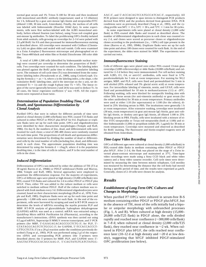
When cells in PDGF were induced to differentiate by theaddition of TH after various periods of time in culture, therate at which oligodendrocytes developed progressivelyslowed as the time before TH addition increased, althoughthe rate of oligodendrocyte development when PDGF was
Figure 2. Cell cycle proper-ties of OPCs in culture with-out TH for various periods oftime in either PDGF (A) orPDGF plus bFGF (B). Thecells were pulsed with BrdUfor the times indicated, andthe percentage of BrdU1
cells was determined by im-munofluorescence. The re-sults are expressed asmean 6 SEM of two inde-pendent experiments, eachwith duplicate cultures foreach time point. The calcu-lated Tcs (see Materials andMethods) are shown in C.
Table I. Population Doubling Times of OPCs in Culture
Days in culture
Population doubling time
PDGF PDGF
1
bFGF
h
8 27
6
4 (306) 47
6
4 (203)18 43
6
6 (332) 63
6
3 (455)90 40
6
4 (119) 36
6
5 (127)330 45
6
4 (335) ND390 45
6
7 (143) 37
6
3 (136)
Purified OPCs that had been in culture for various periods of time were cultured atclonal density (2,000 cells per T25 flask) in B-S media containing either PDGF orPDGF
1
bFGF for 8 d before the number of cells in each clone was counted. Thenumber of clones analyzed is indicated in parentheses. Days in culture refers to the totaltime that the cells were in culture before replating at clonal density for the assay. Thepopulation doubling time was determined as in Materials and Methods. Data are shownas mean
6
SEM.
Dow
nloaded from http://rupress.org/jcb/article-pdf/148/5/971/1290417/9911082.pdf by guest on 16 February 2022

The Journal of Cell Biology, Volume 148, 2000 976
withdrawn slowed much less. After 2 d in culture,
z
100%of P7 OPCs differentiated into morphologically typical oli-godendrocytes within 6 d when PDGF was withdrawn(Fig. 4 A). Although the response of such cells to TH addi-tion was slower than to PDGF withdrawal,
.
80% differ-entiated within 7 d (Fig. 4 A). By contrast, only
z
50% ofOPCs cultured in PDGF for 75 d differentiated within 1 wkwhen induced by TH, although their response to PDGFwithdrawal was comparable to that of cells cultured inPDGF for 2 d (not shown). After 150 d in PDGF, the re-
sponse to TH addition was even slower, although the re-sponse to PDGF withdrawal was still just as fast as after 2 din PDGF (Fig. 4 C). After 390 d, it took 18 d for 40% ofthe cells to differentiate after TH addition and 10 d beforealmost all of the cells differentiated after PDGF removal(Fig. 4 E). In all cases, differentiation was confirmed byRT-PCR analysis of MBP and PLP mRNAs (Fig. 5 andnot shown); for 390-d cultures, differentiation was alsoconfirmed by clonal analysis and MBP staining (notshown). In summary, the response of OPCs to TH de-creases with time in culture, whereas the response toPDGF withdrawal decreases much less.
OPCs cultured in PDGF plus bFGF showed much lessslowing in their differentiation response with time in cul-ture. Almost all of these cells differentiated within 1 wkwhen deprived of both PDGF and bFGF, even after theyhad been in culture for 390 d (Fig. 4, B, D, and F) or 510 d(not shown). The cells responded somewhat more slowlywhen deprived only of PDGF (Fig. 4, B, D, and F), sug-gesting that bFGF weakly suppresses PDGF withdrawal–induced differentiation. bFGF had a much stronger inhibi-tory effect on TH-induced differentiation: in the presenceof bFGF and PDGF, only
z
16% of OPCs that had been inculture for 2 d differentiated after 6 d of TH treatment(Fig. 4 B), but a similar rate of differentiation was seenwhen cells cultured in PDGF plus bFGF for 390 d weretreated with TH.
Expression of Antigenic Markers and Migration
We next used immunofluorescence to examine the expres-sion of a number of antigenic markers in OPCs after vari-ous times in culture. We stained cells for A2B5, O1, O4,GC, NG2, MBP, PLP, vimentin, nestin, and GFAP, mostof which are expressed by oligodendrocyte lineage cells atvarious developmental stages (Gard and Pfeiffer, 1990;Goldman and Vaysse, 1991; Hardy and Reynolds, 1991;Pfeiffer et al., 1993; Grinspan and Franceschini, 1995).Some of the results are shown in Fig. 6 and are summa-rized in Table III. Strong A2B5 staining was observed inall the cultures (Fig. 6, A–D), whereas MBP, PLP, orGFAP staining was never observed (not shown). Strong,filamentous vimentin staining was observed in OPCs after3 d in culture, whereas no such staining was seen after
.
50 d
Table II. Clonal Analysis of Spontaneous Differentiation and Cell Death in OPCs in Culture*
Days in culture(passage number)
Cell death
‡
Differentiation
‡
PDGF PDGF
1
bFGF PDGF PDGF
1
bFGF
%
8 (0) 7
6
2 (203) 12
6
4 (203)
§
10
6
2 (306) 9
6
0.3 (203)17 (0) 18
6
4 (289) 17
6
3 (356) 14
6
3 (289) 15
6
2 (356)17 (1) 18 6 4 (231) 15 6 5 (328) 14 6 2 (231) 16 6 1 (328)23 (1) 28 6 6 (101) 26 6 4 (127) 17 6 2 (101) 17 6 4 (127)90 (5) 6 6 3 (249) 3 6 1 (215)§ 8 6 1 (249) ,1 (215)§
374 (29) 5 6 2 (335) ND 3 6 0.1 (N 7 335) ND390 (30) 7 6 2 (143) 3 6 2 (205)§ 4 6 0.1 (143) ,1 (143)§
*Purified OPCs that had been in culture for various periods of time were cultured at clonal density (2,000 cells per T25 flask) in B-S media containing either PDGF or PDGF 1bFGF for 8 d before counting. Days in culture refers to the total time that the cells were in culture before replating at clonal density or the assay.‡The numbers of dead or differentiated (cells with a characteristic oligodendrocyte morphology) cells in each individual clone were counted. The number of clones analyzed isindicated in parentheses. Data are shown as mean 6 SD for all the clones analyzed.§Significant difference (P , 0.05) compared with PDGF only cultures, when analyzed by Student’s t test.
Figure 3. Analyses of Tc,clonal expansion, spontane-ous differentiation, and celldeath in cultures of OPCs pu-rified from P0, P9, and P14rat optic nerve. Cells werecultured in PDGF withoutTH. (A) After 2 d, BrdU wasadded for 2–96 h before cellswere stained for BrdU. Thecumulative BrdU labeling in-dices were used to calculatethe cell cycle time, as in Fig.2. The results are shown asmean 6 SEM of two inde-pendent experiments, eachwith triplicate cultures foreach time point. The resultsare significantly different(P , 0.01) between P0 andP9 and between P9 and P14.In B and C, the OPCs werecultured at clonal densityfor 7 d, and then the numberof cells per clone (B) and theproportions of oligodendro-cytes and dead cells per clone(C) were determined. The re-sults are shown as mean 6
SEM of two independent experiments. In B, the results are sig-nificantly different (P , 0.01) between P0 and P9 and betweenP9 and P14. In C, the results are significantly different (P , 0.01)between P0 and P9 and P14 for cell death, and also (P , 0.001)between P0 and P9 and P14 for differentiation.
Dow
nloaded from http://rupress.org/jcb/article-pdf/148/5/971/1290417/9911082.pdf by guest on 16 February 2022
Tang et al. Long-Term Culture of Oligodendrocyte Precursor Cells 977
in culture in either PDGF or PDGF plus bFGF (notshown).
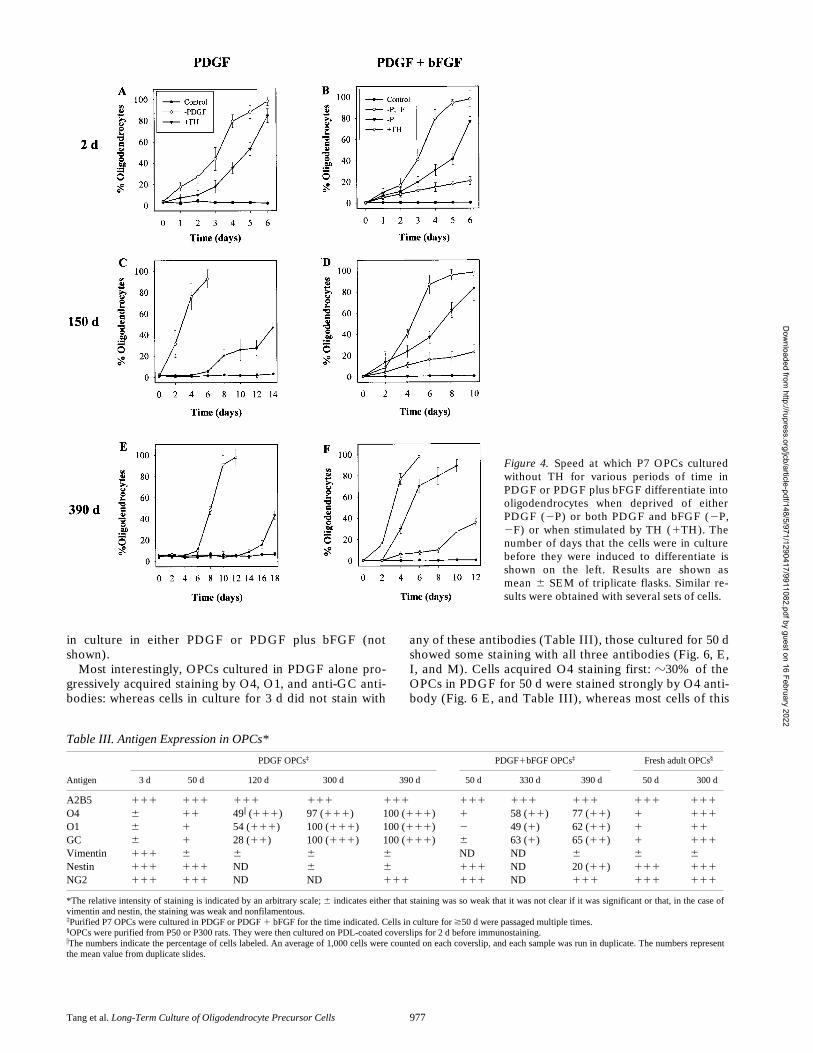
Most interestingly, OPCs cultured in PDGF alone pro-gressively acquired staining by O4, O1, and anti-GC anti-bodies: whereas cells in culture for 3 d did not stain with
any of these antibodies (Table III), those cultured for 50 dshowed some staining with all three antibodies (Fig. 6, E,I, and M). Cells acquired O4 staining first: z30% of theOPCs in PDGF for 50 d were stained strongly by O4 anti-body (Fig. 6 E, and Table III), whereas most cells of this
Figure 4. Speed at which P7 OPCs culturedwithout TH for various periods of time inPDGF or PDGF plus bFGF differentiate intooligodendrocytes when deprived of eitherPDGF (2P) or both PDGF and bFGF (2P,2F) or when stimulated by TH (1TH). Thenumber of days that the cells were in culturebefore they were induced to differentiate isshown on the left. Results are shown asmean 6 SEM of triplicate flasks. Similar re-sults were obtained with several sets of cells.
Table III. Antigen Expression in OPCs*
Antigen
PDGF OPCs‡ PDGF1bFGF OPCs‡ Fresh adult OPCs§
3 d 50 d 120 d 300 d 390 d 50 d 330 d 390 d 50 d 300 d
A2B5 111 111 111 111 111 111 111 111 111 111
O4 6 11 49i (111) 97 (111) 100 (111) 1 58 (11) 77 (11) 1 111
O1 6 1 54 (111) 100 (111) 100 (111) 2 49 (1) 62 (11) 1 11
GC 6 1 28 (11) 100 (111) 100 (111) 6 63 (1) 65 (11) 1 111
Vimentin 111 6 6 6 6 ND ND 6 6 6
Nestin 111 111 ND 6 6 111 ND 20 (11) 111 111
NG2 111 111 ND ND 111 111 ND 111 111 111
*The relative intensity of staining is indicated by an arbitrary scale; 6 indicates either that staining was so weak that it was not clear if it was significant or that, in the case ofvimentin and nestin, the staining was weak and nonfilamentous.‡Purified P7 OPCs were cultured in PDGF or PDGF 1 bFGF for the time indicated. Cells in culture for $50 d were passaged multiple times.§OPCs were purified from P50 or P300 rats. They were then cultured on PDL-coated coverslips for 2 d before immunostaining.iThe numbers indicate the percentage of cells labeled. An average of 1,000 cells were counted on each coverslip, and each sample was run in duplicate. The numbers representthe mean value from duplicate slides.
Dow
nloaded from http://rupress.org/jcb/article-pdf/148/5/971/1290417/9911082.pdf by guest on 16 February 2022

The Journal of Cell Biology, Volume 148, 2000 978
age showed only weak staining with O1 and anti-GC anti-bodies (Fig. 6, I and M; Table III). The staining of theseantibodies increased with time, and by 300 d, most of thecells showed strong staining with all three antibodies, com-parable to that observed for fully differentiated oligoden-drocytes (Fig. 6, F, J, and N).
OPCs cultured in PDGF and bFGF also acquired theseantigens but with a much slower time course. After 50 dthe cells did not stain with the O1 antibody (Fig. 6 K) andstained only very weakly with the O4 and anti-GC anti-bodies (Fig. 6, G and O; Table III). The staining with theanti-GC antibody (Ranscht et al., 1982; Fig. 6 O) resem-bled the polarized patch staining seen with this antibodyreported previously on OPCs treated briefly with TH (To-kumoto et al., 1999). Even after 390 d, OPCs cultured inPDGF plus bFGF showed only weak staining with theanti-GC and O1 antibodies and moderate staining with theO4 antibody, and some cells still did not stain at all (Fig. 6,H, L, and P; Table III).
Nestin is an intermediate filament protein characteristicof neural stem cells (Lendahl et al., 1990). OPCs culturedin PDGF alone showed strong, filamentous staining fornestin for up to 50 d (Fig. 6 Q), but by 300 d they showedonly weak, nonfilamentous nestin staining (Fig. 6 R). Incontrast, z25% of OPCs cultured for up to 390 d in PDGFplus bFGF still showed strong filamentous nestin staining(Fig. 6 T).
Although the long-term OPCs cultured in PDGF alonestrongly stained with O4, O1, and anti-GC antibodies (Fig.6, F, J, and N), the cells were still clearly precursors ratherthan oligodendrocytes as they also expressed NG2 (Fig. 7A), an antigen expressed on OPCs that is lost when OPCsdifferentiate into oligodendrocytes (Levine and Stallcup,1987; not shown), and they were still actively proliferatingas evidenced by BrdU incorporation (Fig. 7, B and C).Furthermore, OPCs that had been cultured in PDGF for
16 mo still differentiated into type-2 astrocytes whenswitched to medium containing 10% FCS (Raff et al.,1983; data not shown).
We also examined the migration of OPCs after varioustimes in culture. As shown in Table IV, the migration rateof OPCs cultured in PDGF alone progressively slowed,while that of OPCs cultured in PDGF plus bFGF slowedmuch less, further suggesting that the presence of bFGFslows down OPC maturation in culture.
Freshly Purified Adult OPCs
Taken together, our findings suggested that OPCs cul-tured in PDGF for an extended period of time came toresemble adult OPCs in a number of their properties(ffrench-Constant and Raff, 1986; Levine and Gard, 1987;Wolswijk and Noble, 1989; Fulton et al., 1992; Wren etal., 1992; Shi et al., 1998). To compare our long-termOPCs with adult OPCs, we purified OPCs from adult ratoptic nerves of different ages. With advancing age, it be-came more and more difficult to culture the adult OPCs.Whereas we could easily culture purified OPCs from P50rats, for example, we were unable to use the same mediumto culture purified OPCs from $P150 rats. The addition ofneuregulin greatly enhanced the survival of the older cells,as we report elsewhere (Fernandez, P.-A., D.G. Tang, L.Cheng, A.W. Mudge, A. Prochiantz, and M.C. Raff, manu-script submitted for publication).
We first compared the ability of purified adult OPCsand age-matched long-term cultured OPCs to incorporateBrdU after an 8-h pulse of BrdU. As shown in Fig. 8 A,z45% of OPCs maintained in PDGF for 50 d and z35%maintained for 300 d or 390 d incorporated BrdU, whereas,10% of the adult OPCs did so under the same condi-tions, and the proportion decreased with increasing age ofthe adult OPCs. The OPCs from P300 and P390 rats weremaintained in PDGF and NRG, but NRG did not enhanceBrdU incorporation, either by itself or with PDGF (notshown).
Although adult OPCs proliferated much more slowlythan long-term cultured OPCs, the two types of OPCsshared a number of other characteristics. Both populationsdifferentiated equally slowly in response to TH addition,although P50 OPCs also differentiated slowly in responseto PDGF withdrawal, whereas P7 OPCs that had been inculture for 50 d differentiated quickly when PDGF was
Figure 5. RT-PCR assays forMBP and PLP mRNAs fol-lowing the induction of dif-ferentiation in OPCs thathad been cultured in PDGFwithout TH for 7 or 150 d be-fore induction by eitherPDGF withdrawal or the ad-dition TH. Note that at leastthree bands are detected forboth MBP and PLP (arrow-heads), both of which areknown to have multiplemRNA species produced byalternative RNA splicing(Roach et al., 1983; Milner etal., 1985; Malotka and Dorn-mair, 1995). The left lane is a100-bp ladder of size mark-ers, and the arrows indicatethe 600-bp band. TheGAPDH mRNA was used asa control.
Table IV. Migration Rates of OPCs in Culture
Time
Migration rate
PDGF PDGF 1 bFGF
d* mm/h‡
5 23 6 3 24 6 260 21 6 3 22 6 2
150 13 6 2 19 6 2300 6 6 3 16 6 1420 5 6 1 15 6 3
*Purified P7 OPCs that had been in culture for various periods of time were culturedin B-S medium containing either PDGF or PDGF 1 bFGF for the days indicated.‡Cell migration was determined by measuring the distance that the cell body movedduring a randomly chosen period of time. Time-lapse video recordings were performedfor 48–96 h. Clones of 2–10 cells were chosen, and 15–20 cells from 3–5 differentclones were analyzed for each time point. The results are shown as mean 6 SEM.
Dow
nloaded from http://rupress.org/jcb/article-pdf/148/5/971/1290417/9911082.pdf by guest on 16 February 2022
Tang et al. Long-Term Culture of Oligodendrocyte Precursor Cells 979
withdrawn (Fig. 8 B). The two types of OPCs were also an-tigenically similar. Adult OPCs of all ages examined, justlike long-term cultured OPCs, stained strongly for A5B5(Fig. 9, A and B), but they did not stain for MBP, PLPGFAP, or filamentous vimentin (not shown). Moreover,P50 adult OPCs stained only weakly with O1, O4, andanti-GC antibodies, whereas P300 OPCs stained stronglywith all these antibodies (Fig. 9, C and D, and not shown).A notable difference, however, was that adult OPCs of allages stained strongly for filamentous nestin (Fig. 9, E andF), whereas the longterm cultured OPCs in PDGF pro-gressively lost such staining (see Fig. 6 R).
DiscussionMost OPCs in the developing optic nerve divide a lim-ited number of times before they stop and terminally dif-ferentiate into postmitotic oligodendrocytes (reviewed inBarres and Raff, 1994). Some of these cells, however,seem to follow a different developmental pathway (Wren
et al., 1992). Instead of differentiating into oligodendro-cytes, they develop into slowly dividing adult OPCs with adistinctive character, which, in culture at least, can still dif-ferentiate into either oligodendrocytes or type-2 astro-cytes (ffrench-Constant and Raff, 1986; Wolswijk and No-ble, 1989; Shi et al., 1998). The adult OPCs in the opticnerve have a complex morphology and appear to extendprocesses exclusively to nodes of Ranvier (Miller et al.,1989; Fulton et al., 1992; Butt et al., 1999). Similar cells arewidely distributed in the adult CNS (Levine and Gard,1987). The function(s) of these cells in the adult CNS re-mains uncertain. It is also uncertain why these cells do notdifferentiate in vivo, although it has been suggested thatthey are inhibited from differentiating by Notch signaling,which blocks differentiation of neonatal OPCs in culture(Wang et al., 1998; Shi et al., 1998).
By manipulating the culture medium appropriately onecan keep most perinatal OPCs dividing in culture beyondthe time when they would normally withdraw from the cellcycle and terminally differentiate. One way is to omit TH
Figure 6. Immunofluores-cence staining with A2B5,O1, O4, Ranscht anti-GC, oranti-nestin antibodies. P7OPCs were cultured withoutTH in either PDGF orPDGF plus bFGF for 50 or390 d. Antibody staining isshown in green. Nuclei werestained with propidium io-dide and are shown in red.Bar, 10 mm.
Dow
nloaded from http://rupress.org/jcb/article-pdf/148/5/971/1290417/9911082.pdf by guest on 16 February 2022

The Journal of Cell Biology, Volume 148, 2000 980
from the culture medium (Barres et al., 1994; Ibarrola et al.,1996; Ahlgren et al., 1997; Gao et al., 1998), another is touse a combination of PDGF and bFGF to stimulate prolif-eration (Bögler et al., 1990; McKinnon et al., 1990). Thequestion we address in this study is what happens if onemaintains purified neonatal OPCs in culture in the ab-sence of TH and in the presence of either PDGF or PDGFand bFGF. In particular, do OPCs acquire the propertiesto OPCs in the adult CNS?
Noble and his colleagues (Wren et al., 1992) showedthat adult-like OPCs develop in cultures of neonatal opticnerve cells that are passaged repeatedly on monolayers ofastrocytes. They also provided evidence that adult-likeOPCs can develop directly from neonatal OPCs: in time-
lapse microcinematographic studies, several cells with aneonatal phenotype were seen to give rise to cells with anadult-like phenotype, although the transition sometimesoccurred over two or more divisions. Here we provide evi-dence that the acquisition of some adult OPC propertiescan occur progressively over months in cultures of purifiedneonatal OPCs stimulated to divide by PDGF in the ab-sence of TH, suggesting that the changes reflect the opera-tion of an intrinsic maturation program in the OPCs thatplays out over many months.
Why Do So Many P7 OPCs Differentiate and/or Die in Our Cultures?
We showed previously that when purified P7 OPCs werecultured for weeks in PDGF without TH their prolifera-tion slowed, their rate of spontaneous differentiation intooligodendrocytes gradually increased, and by 30 d manycells had differentiated and/or died (Gao et al., 1998). Weattributed these changes to two intrinsic timing mecha-nisms operating in the OPCs. One mechanism was pro-posed to regulate the timing of normal oligodendrocytedifferentiation by progressively increasing the probabilitythat an OPC will stop dividing and terminally differenti-ate; whereas this probability is increased by TH, it is clearthat, both in vitro and in vivo, OPCs can terminally differ-entiate in the absence of TH, although the differentiationis delayed (Ibarrola et al., 1996; Ahlgren et al., 1997; Gaoet al., 1998). The second timing mechanism was proposedto be a timer that controls the onset of replicative cell se-nescence (Gao et al., 1998), which is thought to operate inmany kinds of dividing cells in culture (Hayflick, 1965;Smith and Pereira-Smith, 1996). An alternative explana-tion for why many OPCs spontaneously differentiate and/ordie in our cultures after several weeks is that the extracel-lular signals that the cells need to survive and/or dividemay change over time as the cells proliferate in culture, sothat the conditions, at least transiently, become subopti-mal for proliferation and survival.
We favor the second explanation for several reasons. Atthe time that our cells become difficult to maintain andpassage, they show few of the changes that are characteris-tic of replicative senescence: they do not stain (unpub-lished observations), at low pH, with senescence-associ-ated b-galactosidase (Dimri et al., 1995), for example, andthe cells differentiate and/or die rather than arrest and sur-vive. The survival signals required by OPCs change afterthe cells differentiate into oligodendrocytes: PDGF, for in-stance, promotes the survival of OPCs and newly formedoligodendrocytes (Barres et al., 1992), but oligodendro-cytes lose their PDGF receptors within days (Hart et al.,1989; McKinnon et al., 1990) and become insensitive toPDGF. It is perhaps not surprising therefore that the oli-godendrocytes that spontaneously differentiate in our cul-tures soon die. Moreover, the survival signals required byOPCs themselves seem to change as they mature. Weshow here that the rates of spontaneous differentiationand death are greater in clones of P9 or P14 OPCs than inclones of P0 OPCs in the same culture medium. We alsofind that NRG is required for the survival of OPCs iso-lated from $P150 rats but not from P50 rats; finally, bFGFincreases the death of neonatal OPCs, whereas it signifi-
Figure 7. NG2 expression and BrdU incorporation in OPCs cul-tured without TH in PDGF for 450 d. (A) Cell stained for NG2.(B and C) OPCs were pulsed with BrdU for 4 h before stainingfor A2B5 (green, in C) and BrdU (red in B and C). Nuclei werestained with Hoechst dye (blue in B). Bars, 10 mm.
Dow
nloaded from http://rupress.org/jcb/article-pdf/148/5/971/1290417/9911082.pdf by guest on 16 February 2022

Tang et al. Long-Term Culture of Oligodendrocyte Precursor Cells 981
cantly inhibits the spontaneous differentiation and deathof long-term cultured OPCs. If we are correct that many ofthe P7 OPCs die during the first few weeks in our culturebecause the culture conditions are suboptimal for oligo-dendrocytes and more mature OPCs, we should be ableto find conditions where most neonatal OPCs survivethrough this critical period in culture without differentia-tion. If this proves possible, we should then be able to de-termine whether most P7 OPCs can develop into adultOPCs or whether only a subpopulation of P7 OPCs havethis potential.
What Distinguishes the OPCs That Survive?
Despite the significant amount of cell death, many OPCsalways seem to survive and continue to proliferate indefi-nitely, or at least for 18 mo, which is as long as we have fol-lowed them. We do not know what is special about thesecells that enables them to live while many others differen-tiate and/or die. One possibility is that they preexist as asubpopulation of stem-like cells with exceptional capacityfor survival and self renewal. Another is that they arise asimmortalized mutants. Although we cannot exclude this
Figure 8. Comparison of BrdU labeling (A) andoligodendrocyte differentiation (B) in P7 OPCscultured without TH for various periods of timein PDGF alone (open bars and symbols) andfreshly isolated OPCs from adult optic nerve ofequivalent age (filled bars and symbols). In allcases, OPCs were plated into PDL-coated slideflasks and cultured without TH in PDGF for 2 d.In A, BrdU was added for the last 8 h of the cul-ture, and the cells were then fixed and stained forBrdU. An average of 1,000 cells were counted induplicate flasks, and the results are shown as
mean 6 SEM. In B, either the cells were switched after the 2 d to culture medium without PDGF (2PDGF) or TH (1TH) was addedfor the time indicated. The proportion of differentiated oligodendrocytes was determined using morphological criteria. The results areshown as mean 6 SEM of two experiments with duplicate flasks for each condition.
Figure 9. Immunofluores-cence staining with A2B5,O1, and anti-nestin antibod-ies of adult OPCs freshly iso-lated from P50 and P300 ratoptic nerve. The cells werecultured without TH in slideflasks in PDGF (P50 cells) orPDGF plus NRG (P300 cells)for 2 d before they werestained. In C, cell nuclei werestained with propidium io-dide (red). Bar, 10 mm.
Dow
nloaded from http://rupress.org/jcb/article-pdf/148/5/971/1290417/9911082.pdf by guest on 16 February 2022

The Journal of Cell Biology, Volume 148, 2000 982
latter possibility, we think it unlikely for four reasons: (a)OPCs with the same phenotype developed in eight out ofeight experiments. (b) The cells that survive seem to ma-ture over months and acquire at least some of the proper-ties of adult OPCs on much the same schedule as theirnormal counterparts in vivo, as we discuss below. (c) Evenafter 15 mo, the cells growing in either PDGF or PDGFplus bFGF show normal cell cycle checkpoint and apop-totic responses (Tang et al., manuscript in preparation).(d) The cells that survive during the critical period arefound in most clones.
Yet another possible explanation for what distinguishesthe OPCs that survive in our cultures is that they representthe statistical tail of the OPC population with the best sur-vival capabilities. Whatever their origins, it is clear thatthese cells have the ability for long-term self renewal inculture, in the absence of other cell types.
The Long-Term Survivors in PDGF Progressively Acquire Some of the Properties of Adult OPCs
The most important finding in this study is that in all eightexperiments where we established long-term cultures ofpurified P7 OPCs in PDGF, we find that the cells progres-sively acquire, over months, some of the properties thatare characteristic of OPCs in the adult optic nerve. Thecells become larger and their morphology becomes morecomplex, so that by 5–6 mo they have multiple branchingprocesses. One has to be cautious, however, in interpretingchanges in morphology, as cell morphology is very sensi-tive to environmental influence. Although OPCs in theadult optic nerve have a similar complex morphology(Miller et al., 1989; Fulton et al., 1992; Butt et al., 1999),for example, they can acquire a relatively simple morphol-ogy when cultured in TH on astrocyte monolayers (Wols-wijk and Noble, 1989, 1992; Wren et al., 1992).
More importantly, the OPCs cultured in PDGF progres-sively change their antigenic phenotype, mimicking manyof the antigenic changes that normally occur in vivo. Mostremarkably, the cells gradually acquire the glycolipids, in-cluding GC, that are recognized by the O1 (Sommer andSchachner, 1981) and Ranscht monoclonal (Ranscht et al.,1982) anti-GC antibodies. Although the cells do not stain,or stain only very weakly, with these antibodies after 50 din culture, most do stain after 300 d, and the staining iseven stronger at 390 d, when it is almost as intense as ondifferentiated oligodendrocytes. As reported previouslyby Shi et al. (1998) and confirmed here, although OPCsisolated from developing optic nerve do not stain withthese antibodies, OPCs freshly isolated from adult opticnerve do. We find weak staining in P50 cells and strongstaining in P300 cells. Thus, the acquisition of GC seems tooccur on a similar schedule in vitro and in vivo. Even after13–16 mo in vitro, however, the cultured P7 OPCs are stillproliferating, are intensely stained by the A2B5 and NG2antibodies, and do not express MBP or PLP mRNA de-tectable by RT-PCR, indicating that cells have not differ-entiated into oligodendrocytes. These findings make itclear that one cannot use anti-GC antibodies to identifydifferentiated oligodendrocytes in the adult CNS and mayexplain the previous finding that GC1 cells can be a sourceof remyelinating cells in the injured adult CNS (Wood and
Bunge, 1991). The expression of nestin, by contrast, differsin the long-term cultured cells and the freshly isolatedadult OPCs; whereas cultured cells lose expression, theadult cells retain it.
Noble and colleagues (Wolswijk and Noble, 1989, 1992;Wren et al., 1992) showed previously that the migrationrate of OPCs in neonatal optic nerve cell cultures becomesslower with time in vitro. Gao and Raff (1997) providedevidence that this slowing of migration is an intrinsic prop-erty of the developing OPCs. We show here that the slow-ing can continue for many months, compared with the mi-gration rate after 60 d in vitro, the rate is reduced twofoldat 150 d and fourfold at 300 d. By 300 d in culture, the mi-gration rate is z5 mm/h, which is very similar to the rate (4mm/h) reported by Wolswijk and Noble (1989) for OPCsin optic nerve cultures prepared from adult rats that wereat least 240 d old.
It was also shown previously that adult OPCs differenti-ate into oligodendrocytes in culture more slowly than doperinatal OPCs (ffrench-Constant and Raff, 1986; Wols-wijk and Noble, 1989; Shi et al., 1998), and we show herethat this is also the case for our long-term cultured cells. Inresponse to TH treatment, 150 d cells differentiate moreslowly than 7 d cells, and 390 d cells differentiate evenmore slowly than 150 d cells. Surprisingly, 150 d cells dif-ferentiate as fast in response to PDGF withdrawal as do7 d cells, although 390 d cells differentiate more slowly.This contrasts with freshly isolated adult OPCs, whicheven at P50 differentiate only slowly in response to PDGFwithdrawal.
Taken together, these findings strongly suggest that thesurviving OPCs in our cultures continue to mature in vitrofor many months, even though they are growing in isola-tion from other cell types. Gao and Raff (1997) showedpreviously that purified OPCs from the embryonic rat op-tic nerve differed in many of their properties from OPCspurified from P7 optic nerve. Embryonic cells were sim-pler in morphology, proliferated longer before they differ-entiated when cultured in PDGF and TH, and divided andmigrated more rapidly. Remarkably, the embryonic cellsacquired the properties of the P7 cells when they were pu-rified and cultured on their own for 10 d in PDGF in theabsence of TH, suggesting that OPCs have an intrinsicprogram that is responsible for their progressive matura-tion (Gao and Raff, 1997). Our present findings suggestthat the intrinsic maturation program continues to changecells for many months, raising the intriguing question ofwhat kind of molecular mechanism underlies such a pro-longed maturation program.
P7 OPCs cultured for many months in PDGF withoutTH do not acquire all of the characteristics of adult OPCs.They do not slow their proliferation rate to nearly thesame extent, for example. Whereas the cultured cells havea Tc of z35 h from 50 d onward, freshly isolated adultOPCs cultured in the same medium divide much moreslowly, at a rate that is inversely correlated with the age ofthe animal from which they are obtained. It is interestingthat adult OPCs can be induced to divide rapidly if theyare stimulated in culture with the right combination of fac-tors (Wolswijk and Noble, 1992; Shi et al., 1998), indicat-ing that the slowing of the cell cycle with maturation isreversible. It will be important to determine how the intra-
Dow
nloaded from http://rupress.org/jcb/article-pdf/148/5/971/1290417/9911082.pdf by guest on 16 February 2022
Tang et al. Long-Term Culture of Oligodendrocyte Precursor Cells 983
cellular cell cycle control system is altered with matura-tion.
Basic FGF Inhibits the Maturation of OPCs in Culture
Whereas the OPCs growing in PDGF progressively ma-ture and acquire a number of the properties of adultOPCs, those maintained in PDGF plus bFGF maturemuch more slowly and maintain many of their youthfulcharacteristics. They retain their simple bipolar morphol-ogy, their rapid migration rate, a relatively fast differentia-tion response, and acquire differentiation markers moreslowly. Thus, the combination of bFGF and PDGF clearlyinhibits OPC maturation in culture, just as it inhibits OPCdifferentiation into oligodendrocytes (Bögler et al., 1990;McKinnon et al., 1990). It remains to be determined whichintracellular signaling pathways activated by FGF recep-tors, which, like PDGF receptors, are receptor tyrosine ki-nases, are responsible for these inhibitory effects. It is ofinterest that bFGF has different effects on P7 OPCs thathave been in culture for a short time compared with P7OPCs that have been in culture for months. Whereas it in-creases the death and slows down the cell cycle of theyoung OPCs, it inhibits the spontaneous differentiationand/or death and enhances the cell cycle progression oflong-term cultured OPCs. This is further evidence that, asOPCs mature, they change their responsiveness to growthfactors.
In summary, we have shown that when purified P7OPCs are cultured in PDGF (or PDGF plus bFGF) with-out TH, many of the cells differentiate and/or die afterseveral weeks, but many others continue to proliferate forup to 16 months or more. In PDGF, the surviving cellsprogressively acquire a number of properties that arecharacteristic of adult OPCs, while in PDGF and bFGFthey retain many of their neonatal properties. These find-ings suggest that OPCs have an intrinsic maturation pro-gram that normally plays out over many months but canbe restrained by bFGF. The main challenge now is to de-termine the molecular basis of such a prolonged intracellu-lar program.
The authors are grateful to Mark Marcionni for NRG; Fen-Biao Gao forhis initial help in the project; Bill Richardson, Ben Barres, and AnneMudge for advice; Ben Barres for providing detailed immunopurificationprotocols for adult precursor cells; Paul Van Heyningen for help with cellcycle analysis; and Lili Cheng and the members of the Raff lab for discus-sion and support.
D.G. Tang is a recipient of a Hitchings-Elion Award from Burroughs-Wellcome Fund. Y.M. Tokumoto and M.C. Raff are supported by grantsfrom the British Medical Research Council.
Submitted: 17 November 1999Revised: 17 January 2000Accepted: 20 January 2000
References
Ahlgren, S.C., H. Wallace, J. Bishop, C. Neophytou, and M.C. Raff. 1997. Ef-fects of thyroid hormone on embryonic oligodendrocyte precursor cell de-velopment in vivo and in vitro. Mol. Cell. Neurosci. 9:420–432.
Barres, B.A., and M.C. Raff. 1994. Control of oligodendrocyte number in thedeveloping rat optic nerve. Neuron. 12:935–942.
Barres, B.A., I.K. Hart, H.S.R. Coles, J.F. Burne, J.T. Voyvodic, W.D. Richard-son, and M.C. Raff. 1992. Cell death and control of cell survival in the oligo-dendrocyte lineage. Cell. 70:31–46.
Barres, B.A., R. Schmid, M. Sendtner, and M.C. Raff. 1993. Multiple extracel-
lular signals are required for long-term oligodendrocyte survival. Develop-ment. 118:283–295.
Barres, B.A., M.C. Raff, F. Gaese, I. Bartke, G. Dechant, and Y.A. Barde.1994a. A crucial role for neurotrophin-3 in oligodendrocyte development.Nature. 367:371–375.
Barres, B.A., M.A. Lazar, and M.C. Raff. 1994b. A novel role for thyroid hor-mone, glucocorticoids, and retinoic acid in timing oligodendrocyte develop-ment. Development. 120:1097–1108.
Barres, B.A., J.F. Burne, B. Holtmann, H. Thoenen, M. Sendtner, and M.C.Raff. 1996. Ciliary neurotrophic factor enhances the rate of oligodendrocytegeneration. Mol. Cell. Neurosci. 8:146–156.
Bartlett, P.F., M.D. Noble, R.M. Pruss, M.C. Raff, S. Rattray, and C.A. Wil-liams. 1981. Rat neural antigen-2 (RAN-2): a cell surface antigen on astro-cytes, ependymal cells, Muller cells and leptomeninges defined by a mono-clonal antibody. Brain Res. 204:339–351.
Bögler, O., D. Wren, S.C. Barnett, H. Land, and M. Noble. 1990. Cooperationbetween two growth factors promotes extended self-renewal and inhibitsdifferentiation of oligodendrocyte-type-2 astrocyte (O-2A) progenitor cells.Proc. Natl. Acad. Sci. USA. 87:6368–6372.
Bögler, O., and M. Noble. 1994. Measurement of time in oligodendrocyte-type-2astrocyte (O-2A) progenitors is a cellular process distinct from differentia-tion or division. Dev. Biol. 162:525–538.
Bottenstein, J.E., and G.H. Sato. 1979. Growth of a rat neuroblastoma cell linein serum-free supplemented medium. Proc. Natl. Acad. Sci. USA. 76:514–517.
Butt, A.M., A. Duncan, M.F. Hornby, S.I. Kirvell, A. Hunter, J.M. Levine, andM. Berry. 1999. Cells expressing the NG2 antigen contact Nodes of Ranvierin adult CNS white matter. Glia. 26:84–91.
Calver, A.R., A.C. Hall, W.-P. Yu, F.S. Walsh, J.K. Heath, C. Betsholtz, andW.D. Richardson. 1998. Oligodendrocyte population dynamics and the roleof PDGF in vivo. Neuron. 20:869–882.
Canoll, P.D., M.A. Musacchio, R. Hardy, R. Reynolds, M.A. Marchionni, andJ.L. Salzer. 1996. GGF/neuregulin is a neronal signal that promotes the pro-liferation and survival and inhibits differentiation of oligodendrocyte pro-genitors. Neuron. 17:229–243.
Dimri, G.P., X. Lee, G. Basile, M. Acosta, G. Scott, C. Roskelley, E.E. Me-drano, M. Linskens, I. Rubelj, O. Pereira-Smith, M. Peacocke, and J. Camp-isi. 1995. A biomarker that identifies senescent human cells in culture and inaging skin in vivo. Proc. Natl. Acad. Sci. USA. 92:9363–9367.
Durand, B., F.-B. Gao, and M. Raff. 1997. Accumulation of the cyclin-depen-dent kinase inhibitor p27/Kip1 and the timing of oligodendrocyte differenti-ation. EMBO (Eur. Mol. Biol. Organ.) J. 16:306–317.
Durand, B., M.L. Fero, J.M. Roberts, and M. Raff. 1998. P27Kip1 alters the re-sponse of cells to mitogen and is part of a cell-intrinsic timer that arrests thecell cycle and initiates differentiation. Curr. Biol. 8:431–440.
Eisenbarth, G.S., F.S. Walsh, and M. Nirenburg. 1979. Monoclonal antibodiesto a plasma membrane antigen of neurons. Proc. Natl. Acad. Sci. USA. 76:4913–4916.
ffrench-Constant, C., and M.C. Raff. 1986. Proliferating bipotential glial pro-genitor cells in adult optic nerve. Nature. 319:499–502.
Fruttiger, M., L. Karisson, A.C. Hall, A. Abramsson, A.R. Calver, H. Bostrom,K. Willetts, C.-H. Bertold, J.K. Heath, C. Betsholtz, and W.D. Richardson.1999. Defective oligodendrocyte development and severe hypomyelinationin PDGF-A knockout mice. Development. 126:457–467.
Fulton, B.P., J.F. Burne, and M.C. Raff. 1992. Visualization of O-2A progenitorcells in developing and adult rat optic nerve by quisqualate-stimulated co-balt uptake. J. Neurosci. 12:4816–4833.
Gao, F.-B., B. Durand, and M.C. Raff. 1997. Oligodendrocyte precursor cellscount time but not cell divisions before differentiation. Curr. Biol. 7:152–155.
Gao, F.-B., and M. Raff. 1997. Cell size control and a cell-intrinsic maturationprogram in proliferating oligodendrocyte precursor cells. J. Cell Biol. 138:1367–1377.
Gao, F.-B., J. Apperly, and M. Raff. 1998. Cell-intrinsic timers and thyroid hor-mone regulate the probability of cell-cycle withdrawal and differentiation ofoligodendrocyte precursor cells. Dev. Biol. 197:54–66.
Gard, A.L., and S.E. Pfeiffer. 1990. Two proliferative stages of the oligodendro-cyte lineage (A2B51O42 and O41GalC2) under different mitogenic control.Neuron. 5:615–625.
Goldman, J.E., and P.J.-J. Vaysse. 1991. Tracing glial cell lineages in the mam-malian forebrain. Glia. 4:149–156.
Grinspan, J.B., and B. Franceschini. 1995. Platelet-derived growth factor is asurvival factor for PSA-NCAM1 oligodendrocyte pre-progenitors. J. Neu-rosci. Res. 41:540–551.
Hayflick, L. 1965. The limited in vitro lifetime of human diploid cell strains.Exp. Cell Res. 37:614–636.
Hardy, R., and R. Reynolds. 1991. Proliferation and differentiation potential ofrat forebrain oligodendroglial progenitors both in vitro and in vivo. Develop-ment. 111:1061–1080.
Hart, I.K., W.D. Richardson, C.H. Heldin, B. Westermark, and M.C. Raff.1989. PDGF receptors on cells of the oligodendrocyte-type-2 astrocyte(O-2A) cell lineage. Development. 105:595–603.
Hockfield, S., and R.D.G. McKay. 1985. Identification of major cell classes inthe developing mammalian nervous system. J. Neurosci. 5:3310–3328.
Ibarrola, N., M. Mayer-Proschel, A. Rodriguez-Pena, and M. Noble. 1996. Evi-dence for the existence of at least two timing mechanisms that contribute tooligodendrocyte generation in vivo. Dev. Biol. 180:1–21.
Dow
nloaded from http://rupress.org/jcb/article-pdf/148/5/971/1290417/9911082.pdf by guest on 16 February 2022
The Journal of Cell Biology, Volume 148, 2000 984
Lendahl, U., L.B. Zimmerman, and R.D.G. McKay. 1990. CNS stem cells ex-press a new class of intermediate filament protein. Cell. 60:585–595.
Levine, J.M., and J.P. Gard. 1987. Light and electron microscopic localizationof a cell surface antigen (NG2) in the rat cerebellum: association withsmooth protoplasmic astrocytes. J. Neurosci. 7:2711–2720.
Levine, J.M., and W.B. Stallcup. 1987. Plasticity of developing cerebellar cellsin vitro studied with antibodies against the NG2 antigen. J. Neurosci. 7:2721–2731.
LeVine, S.L., and J.E. Goldman. 1988. Embryonic divergence of oligodendro-cyte and astrocyte lineages in developing rat cerebrum. J. Neurosci. 8:3992–4006.
Maguad, J.P., I. Sargent, and D.Y. Mason. 1988. Detection of human white cellproliferative responses by immunoenzymatic measurement of bromode-oxyuridine uptake. J. Immunol. Methods. 106:95–100.
Malotka, J., and K. Dornmair. 1995. Alternative splicing and cDNA sequencingof myelin basic protein gene of the Lewis rat. Autoimmunity. 20:67–68.
McKinnon, R.D., T. Matsui, M. Dubois-Dalcq, and S.A. Aaronson. 1990. FGFmodulates the PDGF-driven pathway of oligodendrocyte development.Neuron. 5:603–614.
McMorris, F.A., and M. Dubois-Dalcq. 1988. Insulin-like growth factor I pro-motes cell proliferation and oligodendroglial commitment in rat glial pro-genitor cells developing in vitro. J. Neurosci. Res. 21:199–209.
Miller, R.H., B.P. Fulton, and M.C. Raff. 1989. A novel type of glial cell associ-ated with Nodes of Ranvier in rat optic nerve. Eur. J. Neurosci. 1:172–181.
Milner, R.J., C. Lai, K.-A. Nave, D. Lenoir, J. Ogata, and J.G. Sutcliffe. 1985.Nulceotide sequence of two mRNAs for rat myelin proteolipid protein. Cell.42:931–939.
Noble, M., and K. Murray. 1984. Purified astrocytes promote the in vitro divi-sions of a bipotential glial progenitor cell. EMBO (Eur. Mol. Biol. Organ.) J.3:2243–2247.
Noble, M., K. Murray, P. Stroobant, M.D. Waterfield, and P. Riddle. 1988.Platelet-derived growth factor promotes the division and motility and inhib-its premature differentiation of the oligodendrocyte/type 2 astrocyte progen-itor cell. Nature. 333:560–562.
Nowakowski, R.S., S.B. Lewin, and M.W. Miller. 1989. Bromodeoxyuridine im-munohistochemical determination of the lengths of the cell cycle and theDNA-synthetic phase for an anatomically defined population. J. Neurocytol.18:311–318.
Ono, K., Y. Yasui, U. Rutishauser, and R.H. Miller. 1997. Focal ventricular ori-gin and migration of oligodendrocyte precurors into the chick optic nerve.Neuron. 19:283–292.
Pfeiffer, S.E., A.E. Warrington, and R. Bansal. 1993. The oligodendrocyte andits many cellular processes. Trends Cell Biol. 3:191–197.
Pringle, N.P., and W.D. Richardson. 1993. A singularity of PDGF alpha-recep-tor expression in the dorsoventral axis of the neural tube may define the ori-gin of the oligodendrocyte lineage. Development. 117:525–533.
Pruss, R.M. 1979. Thy-1 antigen on astrocytes in long-term cultures of rat cen-tral nervous system. Nature. 280:688–690.
Raff, M.C., R. Mirsky, K.L. Fields, R.P. Lisak, S.H. Dorfman, D.H. Silberberg,N.A. Gregson, S. Leiboweitz, and M.C. Kennedy. 1978. Galactocerebrosideis a specific cell-surface antigenic marker for oligodendrocytes in culture.Nature. 274:813–816.
Raff, M.C., R.H. Miller, and M. Noble. 1983. A glial progenitor cell that devel-ops in vitro into an astrocyte or an oligodendrocyte depending on culture
medium. Nature. 303:390–396.Raff, M.C., L.E. Lillien, W.D. Richardson, J.F. Burne, and M. Noble. 1988.
Platelet-derived growth factor from astrocytes drives the clock that timesoligodendrocyte development in culture. Nature. 333:562–565.
Ranscht, B., P.A. Clapshaw, J. Price, M. Noble, W. Seifert. 1982. Developmentof oligodendrocytes and Schwann cells studied with a monoclonal antibodyagainst galactocerebroside. Proc. Natl. Acad. Sci. USA. 79:2709–2713.
Richardson, W.D., N. Pringle, M.J. Mosley, B. Westermark, and M. Dubois-Dalcq. 1988. A role for platelet-derived growth factor in normal gliogenesisin the central nervous system. Cell. 53:309–319.
Roach, A., K.B. Boylan, S. Horvath, S.B. Prusiner, and L.E. Hood. 1983. Char-acterization of cloned cDNA representing rat myelin basic protein: absenceof expression in brain of shiverer mutant mice. Cell. 34:799–806.
Shi, J., A. Marinovich, and B. Barres. 1998. Purification and characterization ofadult oligodendrocyte precursor cells from the rat optic nerve. J. Neurosci.18:4627–4636.
Skoff, R.P. 1990. Gliogenesis in rat optic nerve: astrocytes are generated in asingle wave before oligodendrocytes. Dev. Biol. 139:149–168.
Small, R.K., P. Riddle, and M. Noble. 1987. Evidence for migration of oligoden-drocyte–type-2 astrocyte progenitor cells into the developing rat optic nerve.Nature. 328:155–157.
Smith, J.R., and O.M. Pereira-Smith. 1996. Replicative senescence: implicationsfor in vivo aging and tumor suppression. Science. 273:63–67.
Sommer, I., and M. Schachner. 1981. Monoclonal antibodies (O1 to O4) to oli-godendrocyte cell surfaces: an immunocytochemical study in the central ner-vous system. Dev. Biol. 83:311–327.
Tang, D.G., Y. Chen, C.A. Diglio, and K.V. Honn. 1993. PKC-dependent ef-fects of 12(S)-HETE on endothelial cell vitronectin and fibronectin recep-tor. J. Cell Biol. 121:689–704.
Temple, S., and M.C. Raff. 1985. Differentiation of a bipotential glial progeni-tor cell in single cell microculture. Nature. 313:223–225.
Temple, S., and M.C. Raff. 1986. Clonal analysis of oligodendrocyte develop-ment in culture: evidence for a developmental clock that counts cell divi-sions. Cell. 44:773–779.
Tokumoto, Y.M., B. Durand, and M.C. Raff. 1999. An analysis of the earlyevents when oligodendrocyte precursor cells are triggered to differentiate bythyroid hormone, retinoic acid, or PDGF withdrawal. Dev. Biol. 213:327–339.
Vartanian, T., G. Fischbach, and R. Miller. 1999. Failure of spinal cord oligo-dendrocyte development in mice lacking neuregulin. Proc. Natl. Acad. Sci.USA. 96:731–735.
Wang, S., A.D. Sdrulla, G. diSibio, G. Bush, D. Nofziger, C. Hicks, G. Wein-master, and B.A. Barres. 1998. Notch receptor activation inhibits oligoden-drocyte differentiation. Neuron. 21:63–75.
Wolswijk, G., and M. Noble. 1989. Identification of an adult-specific glial pro-genitor cell. Development. 105:387–400.
Wolswijk, G., and M. Noble. 1992. Cooperation between PDGF and FGF con-verts slowly dividing O-2A adult progenitor cells to rapidly dividing cellswith the characteristics of perinatal O-2A progenitor cells. J. Cell. Biol. 118:889–897.
Wood, P.M., and R.P. Bunge. 1991. The origin of remyelinating cells in theadult central nervous system: the role of the mature oligodendrocyte. Glia.4:225–232.
Wren, D., G. Wolswijk, and M. Noble. 1992. In vitro analysis of the origin andmaintenance of O-2A adult progenitor cells. J. Cell. Biol. 116:167–176.
Dow
nloaded from http://rupress.org/jcb/article-pdf/148/5/971/1290417/9911082.pdf by guest on 16 February 2022