
Long non-coding RNA ANRIL down-regulates microRNA-7 to ... · cRNA ANRIL overexpression. miR-7...
10
3173 Abstract. – OBJECTIVE: Although the poten- tial involvements of INK4 locus reported in glau- coma, the effects of long non-coding RNA (ln- cRNA) antisense noncoding RNA in the INK4 locus (ANRIL) on trabecular meshwork (TM) cells remain unclear. In this study, we aimed to explore the effects of lncRNA ANRIL on the oxi- dative injury of human TM cells as well as the un- derlying mechanisms. MATERIALS AND METHODS: Oxidative injury of human TM cells was induced by H 2 O 2 stimula- tion. Cell viability, apoptotic cells, expression of proteins related to apoptosis, and reactive oxy- gen species (ROS) level were testified by CCK-8 assay, flow cytometry assay, Western blot anal- ysis, and DCFH-DA staining, respectively. Ln- cRNA ANRIL was overexpressed, and its effects on H2O2-injured TM cells were analyzed. After- ward, miR-7 expression in lncRNA ANRIL over- expressing-cells was determined by RT-qPCR. Moreover, it was verified whether lncRNA AN- RIL affected H 2 O 2 -treated TM cells via miR-7, fol- lowed by the measurements of the involved sig- naling pathways. RESULTS: H 2 O 2 -induced decrease of cell via- bility and the increases in apoptosis and ROS generation were significantly attenuated by ln- cRNA ANRIL overexpression. miR-7 expres- sion was down-regulated by lncRNA ANRIL, and miR-7 overexpression abrogated the effects of lncRNA ANRIL on H 2 O 2 -treated TM cells. Phos- phorylation levels of the key kinases in the mTOR and MEK/ERK pathways were enhanced by lncRNA ANRIL via down-regulating miR-7 in H2O2-treated TM cells. CONCLUSIONS: LncRNA ANRIL attenuated oxidative injury of human TM cells and activat- ed the mTOR and MEK/ERK pathways, possibly through down-regulating miR-7. Key Words: Glaucoma, Oxidative stress, LncRNA ANRIL, MicroR- NA-7, MTOR/MEK/ERK. Introduction Being a neurodegenerative disease of the eye, the glaucoma is the second leading cause of irreversible blindness that behind cataract and affects more than 70 million individuals globally 1,2 . In patients with glaucoma, retinal ganglion cells which convey vi- sual messages from the retina to the brain, as well as the visual field, are selectively lost, and the retinal ganglion cell axons and somas are degenerated 3-5 . Glaucoma is asymptomatic at the beginning, and it may not be diagnosed until a substantial loss of vi- sion has occurred. Therefore, the actual number of patients with glaucoma may be even higher than the numbers estimated, making the exploration of the effective therapies for glaucoma important 6 . Several risk factors may contribute to glauco- ma, including age, intraocular pressure (IOP), re- duced ocular perfusion pressure, and genetic mu- tations 7 . Among them, IOP is the only modifiable risk factor for the development and progression of glaucoma, and lowing IOP is the only therapy which can effectively reduce glaucomatous pro- gression 8,9 . IOP homeostasis is previously defined European Review for Medical and Pharmacological Sciences 2019; 23: 3173-3182 J. ZHAO 1 , H. SUN 2 , J.-M. ZHANG 1 , M. WANG 1 , X.-J. DU 3 , J.-L. ZHANG 4 1 Department of Ophthalmology, Linyi People’s Hospital, Linyi, Shandong, China 2 Department of Ophthalmology, The First People’s Hospital of Tancheng, Tancheng County, Shandong, China 3 Department of Ophthalmology, Eye Institute of Shandong University of Traditional Chinese Medicine, Affiliated Eye Hospital of Shandong University of TCM, Jinan, Shandong, China 4 Department of Ophthalmology, The People’s Hospital of Pingyi County, Pingyi County, Shandong, China Corresponding Author: Jinlian Zhang, MD; e-mail: [email protected] Long non-coding RNA ANRIL down-regulates microRNA-7 to protect human trabecular meshwork cells in an experimental model for glaucoma
Transcript of Long non-coding RNA ANRIL down-regulates microRNA-7 to ... · cRNA ANRIL overexpression. miR-7...

3173
Abstract. – OBJECTIVE: Although the poten-tial involvements of INK4 locus reported in glau-coma, the effects of long non-coding RNA (ln-cRNA) antisense noncoding RNA in the INK4 locus (ANRIL) on trabecular meshwork (TM) cells remain unclear. In this study, we aimed to explore the effects of lncRNA ANRIL on the oxi-dative injury of human TM cells as well as the un-derlying mechanisms.
MATERIALS AND METHODS: Oxidative injury of human TM cells was induced by H2O2 stimula-tion. Cell viability, apoptotic cells, expression of proteins related to apoptosis, and reactive oxy-gen species (ROS) level were testified by CCK-8 assay, flow cytometry assay, Western blot anal-ysis, and DCFH-DA staining, respectively. Ln-cRNA ANRIL was overexpressed, and its effects on H2O2-injured TM cells were analyzed. After-ward, miR-7 expression in lncRNA ANRIL over-expressing-cells was determined by RT-qPCR. Moreover, it was verified whether lncRNA AN-RIL affected H2O2-treated TM cells via miR-7, fol-lowed by the measurements of the involved sig-naling pathways.
RESULTS: H2O2-induced decrease of cell via-bility and the increases in apoptosis and ROS generation were significantly attenuated by ln-cRNA ANRIL overexpression. miR-7 expres-sion was down-regulated by lncRNA ANRIL, and miR-7 overexpression abrogated the effects of lncRNA ANRIL on H2O2-treated TM cells. Phos-phorylation levels of the key kinases in the mTOR and MEK/ERK pathways were enhanced by lncRNA ANRIL via down-regulating miR-7 in H2O2-treated TM cells.
CONCLUSIONS: LncRNA ANRIL attenuated oxidative injury of human TM cells and activat-
ed the mTOR and MEK/ERK pathways, possibly through down-regulating miR-7.
Key Words:Glaucoma, Oxidative stress, LncRNA ANRIL, MicroR-
NA-7, MTOR/MEK/ERK.
Introduction
Being a neurodegenerative disease of the eye, the glaucoma is the second leading cause of irreversible blindness that behind cataract and affects more than 70 million individuals globally1,2. In patients with glaucoma, retinal ganglion cells which convey vi-sual messages from the retina to the brain, as well as the visual field, are selectively lost, and the retinal ganglion cell axons and somas are degenerated3-5. Glaucoma is asymptomatic at the beginning, and it may not be diagnosed until a substantial loss of vi-sion has occurred. Therefore, the actual number of patients with glaucoma may be even higher than the numbers estimated, making the exploration of the effective therapies for glaucoma important6.
Several risk factors may contribute to glauco-ma, including age, intraocular pressure (IOP), re-duced ocular perfusion pressure, and genetic mu-tations7. Among them, IOP is the only modifiable risk factor for the development and progression of glaucoma, and lowing IOP is the only therapy which can effectively reduce glaucomatous pro-gression8,9. IOP homeostasis is previously defined
European Review for Medical and Pharmacological Sciences 2019; 23: 3173-3182
J. ZHAO1, H. SUN2, J.-M. ZHANG1, M. WANG1, X.-J. DU3, J.-L. ZHANG4
1Department of Ophthalmology, Linyi People’s Hospital, Linyi, Shandong, China2Department of Ophthalmology, The First People’s Hospital of Tancheng, Tancheng County, Shandong, China3Department of Ophthalmology, Eye Institute of Shandong University of Traditional Chinese Medicine, Affiliated Eye Hospital of Shandong University of TCM, Jinan, Shandong, China4Department of Ophthalmology, The People’s Hospital of Pingyi County, Pingyi County, Shandong, China
Corresponding Author: Jinlian Zhang, MD; e-mail: [email protected]
Long non-coding RNA ANRIL down-regulates microRNA-7 to protect human trabecular meshwork cells in an experimental model for glaucoma
J. Zhao, H. Sun, J.-M. Zhang, M. Wang, X.-J. Du, J.-L. Zhang
3174
as corrective adjustments of the aqueous humor outflow resistance in response to pressure chang-es10. IOP is regulated by the resistance to aqueous humor outflow since the aqueous humor inflow is relatively constant and pressure-insensitive. The trabecular meshwork (TM) is a series of fenestrat-ed beams and sheets of the extracellular matrix, through which aqueous humor leaves the anteri-or chamber of the eye11. Previous studies12,13 have pointed out that the probable site of the resistance to aqueous humor outflow is located within the juxtacanalicular and Schlemm’s canal. Therefore, the regulatory mechanism in TM cells isolated from the juxtacanalicular is of great importance for the clinical treatment of glaucoma.
Long non-coding RNAs (lncRNAs) are a group of RNAs which are not involved in the protein generation while they are involved in the regulatory processes14. Currently, an increasing number of lncRNAs are identified to participate in the development of the glaucoma, such as ln-cRNA-MEG3 and lncRNA-MALAT115,16. The INK4 locus at chromosome 9p21, which encodes three tumor suppressor genes and the lncRNA an-tisense noncoding RNA in the INK4 locus (AN-RIL), has been reported to be related to primary open angle glaucoma (POAG) and normal-tension glaucoma17,18. Therefore, we hypothesized that there might be a relationship between lncRNA ANRIL and glaucoma.
In this study, we induced in vitro oxidative in-jury model using human TM cells and studied the effects of lncRNA ANRIL on H2O2-treated human TM cells as well as the regulatory mech-anism. We aimed to explore the potential regula-tory mechanism of lncRNA ANRIL in glaucoma, assisting in the identification of novel therapies for glaucoma.
Materials and Methods
Cell Culture and Treatment Primary human TM cells, isolated from the
juxtacanalicular and corneoscleral regions of the human eye and cryopreserved at P0, were pur-chased from Sciencell Research Laboratories (Carlsbad, CA, USA). Cells were maintained in Trabecular Meshwork Cell Medium (TMCM) containing 2% fetal bovine serum (FBS), 1% tra-becular meshwork cell growth supplement and 1% penicillin/streptomycin solution (all from Scien-cell Research Laboratories, Carlsbad, CA, USA), and were plated in poly-L-lysine-coated surface
(2 μg/cm2). Cell maintaining was performed in a humidified incubator with 5% CO2 and 95% air at 37°C. Cells in passages 2-5 were used for experi-ments. Culture medium was refreshed every three days until the culture reached 70% confluence, whereas it was refreshed every other day when the confluence of cells was 70%-90%. For the stimulation with H2O2, the cells were cultured in TMCM containing 50-400 μM H2O2 (Sigma-Al-drich, St. Louis, MO, USA) for 24 h after they reached 80-85% confluence.
Cell Transfection Full-length human lncRNA ANRIL sequenc-
es were ligated into a pcDNA3.1 plasmid (In-vitrogen, Carlsbad, CA, USA), and the recom-bined plasmid was sequenced and referred to as pc-ANRIL. Either pcDNA3.1 or pc-ANRIL was transfected into TM cells using the lipofectamine 3000 reagent (Invitrogen, Carlsbad, CA, USA) following the instructions provided by the man-ufacturer. The stable transfected cell lines were harvested after selection in the culture medium containing 0.5 mg/mL G418 (Sigma-Aldrich, St. Louis, MO, USA) for approximately 4 weeks. For the transient transfection with microRNAs (miRs), the scramble miRs or miR-7 mimic, syn-thesized by GenePharma (Shanghai, China), were transfected into TM cells using the lipofectamine 3000 reagent.
Cell Viability Assay TM cell viability was examined using a Cell
Counting Kit-8 (CCK-8; Dojindo Molecular Tech-nologies, Gaithersburg, MD, USA). In brief, TM cells were seeded in poly-L-lysine-coated 96-well plates at 5 × 10³ cells per well. Then, the cells were incubated at 37°C overnight for growth. Af-ter cell transfection and/or stimulation with H2O2, the culture medium was replaced by TMCM con-taining 10% CCK-8 solution and cells were incu-bated at 37°C for an additional 1 h. Finally, the absorbance at 450 nm was detected by using a Mi-croplate Reader (Bio-Rad, Hercules, CA, USA).
Apoptosis Assay Early-stage apoptotic cells after cell transfec-
tion and/or stimulation with H2O2 were identified by using the FITC Annexin V/Dead Cell Apop-tosis Kit with FITC Annexin V and PI, for Flow Cytometry (Invitrogen, Carlsbad, CA, USA). In brief, after the desired treatments, TM cells were collected and washed in phosphate-buffered sa-line (PBS). Then, the cells were suspended in the
Role of lncRNA ANRIL in glaucoma
3175
binding buffer provided by the kit, followed by staining with 5 μL of FITC Annexin V and 100 ng of propidium iodide (PI) according to the man-ufacturer’s instructions. Finally, the stained cells were subjected to a FACS can (Beckman Coulter, Fullerton, CA, USA), and the percentage of apop-totic cells was analyzed using the FlowJo soft-ware (Tree Star, San Carlos, CA, USA).
Reactive Oxygen Species (ROS) Assay Intracellular ROS level in TM cells was mea-
sured by using a Reactive oxygen species Assay Kit (E004, Nanjing Jiancheng, Nanjing, China). In brief, after cell transfection and/or stimulation with H2O2, the cell culture was discarded, and the cells were washed with PBS two times. Then, the cells were incubated in TMCM containing 10 μM 2,7-dichlorofluorescein diacetate (DCFH-DA) for 30 min at 37°C in the dark. Subsequently, after being rinsed in PBS two times, the cells were col-lected by trypsin treatment (0.25% trypsin-ED-TA) and centrifugation. Cells were washed again using PBS and resuspended in 500 μL PBS, fol-lowed by measurements of fluorescent intensity by using a flow cytometer (485 nm excitation, 525 nm emission).
RNA Immunoprecipitation (IP) RNA IP was performed by using Magna RIP
RNA-Binding Protein Immunoprecipitation Kit (Millipore, Bedford, MA). The wild-type AN-RIL or the mutant ANRIL (mutant in the pre-dicted miR-7 binding site) was inserted into a pc-DNA3.1-MS2 plasmid (Addgene, Cambridge, USA). The constructed vectors were co-transfect-ed with pMS2-GFP (Addgene, Cambridge, USA) into the cell by using lipofectamine 3000 reagent (Invitrogen, Carlsbad, CA, USA). Then, the cell lysate was extracted and incubated with magnetic beads, GFP antibody (ab290, Abcam, Cambridge, UK) or IgG (ab190475, Abcam, Cambridge, UK). Reverse transcription-quantitative PCR (RT-qP-CR) was then performed to detect signals from miR-7 specifically binding to ANRIL.
RT-qPCR After cell transfection and/or stimulation with
H2O2, TRIzol reagent (Invitrogen, Carlsbad, CA, USA) was used for the extraction of total RNA according to the manufacturer’s instructions. For the quantification of lncRNA ANRIL expres-sion, the One Step SYBR® PrimeScript™ PLUS RT-RNA PCR Kit (TaKaRa Biotechnology, Da-lian, China) was used following the supplier’s
protocol. For quantification of miR-7 expression, cDNA was prepared from 500 ng RNA using the TaqMan MicroRNA Reverse Transcription Kit (Applied Biosystems, Foster City, CA, USA). The thermocycling program of reverse transcription was 30 min at 16°C, 30 min at 42°C, and 5 min at 85°C. Real-time PCR was performed using the TaqMan Universal Master Mix II (Applied Bio-systems, Foster City, CA, USA) on a Bio-Rad iQ5 Real-time PCR Detection System (Bio-Rad, Her-cules, CA, USA). The temperature profile was 1 cycle at 95°C for 10 min, followed by 40 cycles of 95°C for 15 s and 60°C for 1 min. Relative ex-pression fold of lncRNA ANRIL and miR-7 was analyzed on the basis of the 2-ΔΔCt method19. GAP-DH and U6 were used as housekeeping genes for relative quantification of lncRNA ANRIL and miR-7, respectively.
Western Blot Analysis After cell transfection and/or stimulation with
H2O2, the cells were collected and washed in ice-cold PBS. After centrifugation to the pellet, the cells were suspended in RIPA buffer (Beyo-time Institute of Biotechnology, Shanghai, Chi-na) supplemented with 1 mM PMSF (Beyotime Institute of Biotechnology, Shanghai, China). The whole cell lysates were centrifuged, and the protein content in the supernatant was mea-sured with the bicinchoninic acid protein assay kit (Pierce, Rockford, IL, USA). Proteins were separated by SDS-PAGE and electrotransferred to polyvinylidene difluoride (PVDF) membranes. Then, the PVDF membranes were blocked with 5% non-fat milk and incubated with primary an-tibody and HRP-conjugated goat anti-rabbit IgG (ab205718, Abcam, Cambridge, UK), succes-sively. Primary antibodies used in this study in-cluded anti-Bcl-2 antibody (ab196495), anti-Bax antibody (ab32503), anti-pro caspase-3 anti-body (ab90437), anti-cleaved caspase-3 antibody (ab2302), anti-pro caspase-9 antibody (ab32539), anti-cleaved caspase-9 antibody (ab2324), an-ti-p70S6K antibody (ab186753), anti-phospho (p)-p70S6K antibody (ab59208), anti-mTOR antibody (ab32028), anti-p-mTOR antibody (ab109268), anti-MEK antibody (ab178876), anti-p-MEK an-tibody (ab194754), anti-ERK antibody (ab115799), anti-p-ERK antibody (ab214036) and anti-β-actin antibody (ab8227; all from Abcam, Cambridge, UK). The proteins in the PVDF membranes were visualized using the chemiluminescence detec-tion (ECL Plus; GE Healthcare Bio-Science, Pis-cataway, NJ, USA).

J. Zhao, H. Sun, J.-M. Zhang, M. Wang, X.-J. Du, J.-L. Zhang
3176
Statistical Analysis Experiments were performed in triplicate with
three repeats. Data are presented as the mean ± standard deviation (SD). Statistical analysis was performed using GraphPad Prism 5 software (GraphPad, San Diego, CA, USA). The compar-ison was evaluated using the unpaired two-tailed t-test, one-way analysis of variance (ANOVA) or multiple t-tests. ANOVA was conducted and was followed by the Tukey post-hoc test. p < 0.05 was considered a significant difference.
Results
Stimulation With H2O2 Induced Oxidative Injury in Human TM Cells
Oxidative injury of human TM cells was induced by stimulation with H2O2. Cells were stimulated with increasing doses of H2O2, and CCK-8 results showed that cell viability was significantly reduced by 100 μM H2O2 (p < 0.05), 200-300 μM H2O2 (p <
0.01), and 400 μM H2O2 (p < 0.001, Figure 1A), as compared to the control group. Since cell viability was dropped by more than 50% when the concen-tration of H2O2 was 200 μM, the oxidative injury of TM cells was induced by 200 μM H2O2 in subse-quent experiments. Cell apoptosis and ROS gener-ation were analyzed afterward to evaluate whether the oxidative injury was successfully induced. In Figure 1B, the percentage of apoptotic cells in the H2O2 group was dramatically higher than the con-trol group (p < 0.001). Likewise, H2O2 stimulation up-regulated pro-apoptotic Bax expression while down-regulated anti-apoptotic Bcl-2 expression and expression of cleaved caspase-3 and cleaved caspase-9 was observed in the H2O2-treated cells (Figure 1C). The results indicated that H2O2 stim-ulation elevated cell apoptosis. Finally, we also found that ROS level in the H2O2 group was re-markably higher than the control group (p < 0.001, Figure 1D). the results collectively illustrated that oxidative injury of human TM cells was success-fully induced after H2O2 stimulation.
Figure 1. Stimulation with H2O2 induced oxidative injury in human TM cells. Human TM cells were stimulated with increas-ing doses of H2O2 for 24 h. A, Cell viability was measured by the CCK-8 assay. The cells were stimulated with 200 μM H2O2 for 24 h, and non-treated cells were acted as control. B, Percentage of apoptotic cells was determined by flow cytometry assay. C, Expression of proteins associated with apoptosis was evaluated by Western blot analysis. D, ROS generation was estimated by DCFH-DA staining. Data are presented as the mean ± SD of at least three independent experiments. *, p < 0.05; **, p < 0.01; ***, p < 0.001.

Role of lncRNA ANRIL in glaucoma
3177
Overexpression of LncRNA ANRIL Attenuated H2O2-Induced TM Cell Injury
Effects of lncRNA ANRIL on the oxidative injury of TM cells were analyzed. After cell transfection, lncRNA ANRIL level in cells trans-fected with pc-ANRIL was markedly higher than the cells transfected with pcDNA3.1 (p < 0.01), suggesting that lncRNA ANRIL was overex-pressed successfully after cell transfection (Fig-ure 2A). Then, transfected and untransfected cells were stimulated with 200 μM H2O2 for 24 h, and non-treated cells were acted as control. The CCK-8 assay showed that lncRNA ANRIL over-expression significantly elevated cell viability of H2O2-treated cells (p < 0.05, Figure 2B). Flow cytometry assay showed that lncRNA ANRIL overexpression observably reduced the percent-age of apoptotic cells in H2O2-treated cells (p < 0.05, Figure 2C). Meanwhile, lncRNA ANRIL overexpression could up-regulate Bcl-2 expres-sion while down-regulating the expression of Bax, cleaved caspase-3 and cleaved caspase-9 compared to the H2O2 + pcDNA3.1 group (Figure 2D). Besides, the ROS level in cells overexpress-ing lncRNA ANRIL was significantly lower than pcDNA3.1-transfected cells after H2O2 stimula-tion (p < 0.05, Figure 2E). The results collectively
indicated that lncRNA ANRIL overexpression could attenuate H2O2-induced oxidative injury in TM cells.
Overexpression of LncRNA ANRIL Down-Regulated MiR-7 Expression
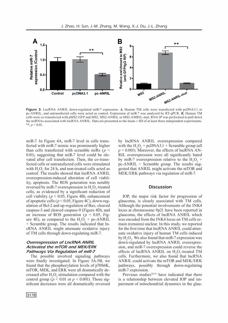
Effects of lncRNA ANRIL overexpression on the miR-7 level were testified. In Figure 3A, the miR-7 expression level in cells overexpressing lncRNA ANRIL was significantly lower than the pcDNA3.1-transfected cells (p < 0.01), indicating that lncRNA ANRIL might down-regulate miR-7 expression in TM cells. To further reveal the reg-ulatory relationship between lncRNA ANRIL and miR-7, anti-MS2 RNA IP was performed. RT-qP-CR data in Figure 3B showed that the ANRIL RNA IP in MS2-ANRIL group was significantly enriched for miR-7 compared to the MS2-ANRIL-mut group (p < 0.01). These results evidenced that lncRNA ANRIL down-regulated miR-7 expres-sion might be via directly binding with miR-7.
Overexpression of LncRNA ANRIL Alleviated H2O2-Induced Injury Via Down-Regulating MiR-7
We then studied whether lncRNA ANRIL af-fected H2O2-treated TM cells via regulation of
Figure 2. LncRNA ANRIL attenuated H2O2-induced TM cell injury. Human TM cells were transfected with pcDNA3.1 or pc-ANRIL, and untransfected cells were acted as control. A, Expression of lncRNA ANRIL was analyzed by RT-qPCR. Cells transfected with pcDNA3.1 or pc-ANRIL and untransfected cells were stimulated with 200 μM H2O2 for 24 h. Non-treated cells were acted as control. B, Cell viability was measured by the CCK-8 assay. C, Percentage of apoptotic cells was deter-mined by flow cytometry assay. D, Expression of proteins associated with apoptosis was evaluated by Western blot analysis. E, ROS generation was estimated by DCFH-DA staining. Data are presented as the mean ± SD of at least three independent experiments. *, p < 0.05; **, p < 0.01; ***, p < 0.001.
J. Zhao, H. Sun, J.-M. Zhang, M. Wang, X.-J. Du, J.-L. Zhang
3178
miR-7. In Figure 4A, miR-7 level in cells trans-fected with miR-7 mimic was prominently higher than cells transfected with scramble miRs (p < 0.01), suggesting that miR-7 level could be ele-vated after cell transfection. Then, the co-trans-fected cells or untransfected cells were stimulated with H2O2 for 24 h, and non-treated cells acted as control. The results showed that lncRNA ANRIL overexpression-induced alteration of cell viabil-ity, apoptosis. The ROS generation was notably reversed by miR-7 overexpression in H2O2-treated cells, as evidenced by a significant reduction of cell viability (p < 0.05, Figure 4B), enhancement of apoptotic cells (p < 0.05, Figure 4C), down-reg-ulation of Bcl-2 and up-regulation of Bax, cleaved caspase-3 and cleaved caspase-9 (Figure 4D), and an increase of ROS generation (p < 0.05, Fig-ure 4E), as compared to the H2O2 + pc-ANRIL + Scramble group. The results indicated that ln-cRNA ANRIL might attenuate oxidative injury of TM cells through down-regulating miR-7.
Overexpression of LncRNA ANRIL Activated the mTOR and MEK/ERK Pathways Via Regulation of miR-7
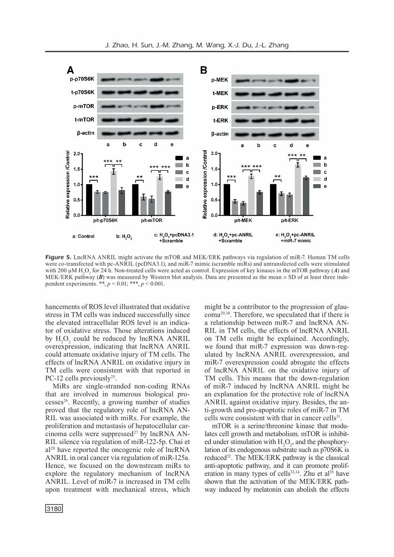
The possible involved signaling pathways were finally investigated. In Figure 5A-5B, we found that the phosphorylation levels of p70S6K, mTOR, MEK, and ERK were all dramatically de-creased after H2O2 stimulation compared with the control group (p < 0.01 or p < 0.001). Those sig-nificant decreases were all dramatically reversed
by lncRNA ANRIL overexpression compared with the H2O2 + pcDNA3.1 + Scramble group (all p < 0.001). Moreover, the effects of lncRNA AN-RIL overexpression were all significantly bated by miR-7 overexpression relative to the H2O2 + pc-ANRIL + Scramble group. The results sug-gested that ANRIL might activate the mTOR and MEK/ERK pathways via regulation of miR-7.
Discussion
IOP, the major risk factor for progression of glaucoma, is closely associated with TM cells. Although the potential involvements of the INK4 locus at chromosome 9p21 have been reported in glaucoma, the effects of lncRNA ANRIL which was encoded from the INK4 locus on TM cells re-main (remains) unclear. In this study, we reported for the first time that lncRNA ANRIL could atten-uate oxidative injury of human TM cells induced by H2O2. We also found that miR-7 expression was down-regulated by lncRNA ANRIL overexpres-sion, and miR-7 overexpression could reverse the effects of lncRNA ANRIL on H2O2-treated TM cells. Furthermore, we also found that lncRNA ANRIL could activate the mTOR and MEK/ERK pathways, possibly through down-regulating miR-7 expression.
Previous studies20,21 have indicated that there is a relationship between elevated IOP and im-pairment of mitochondrial dynamics in the glau-
Figure 3. LncRNA ANRIL down-regulated miR-7 expression. A, Human TM cells were transfected with pcDNA3.1 or pc-ANRIL, and untransfected cells were acted as control. Expression of miR-7 was analyzed by RT-qPCR. B, Human TM cells were co-transfected with pMS2-GFP and MS2, MS2-ANRIL or MS2-AMRIL-mut. RNA IP was performed to pull down the miRNAs associated with lncRNA ANRIL. Data are presented as the mean ± SD of at least three independent experiments. **, p < 0.01.

Role of lncRNA ANRIL in glaucoma
3179
comatous retina. Oxidative stress is an important pathophysiological mechanism in mitochondrial dysfunction and glaucoma22. Moreover, the oxi-dative injury of TM cells has been reported to ob-struct aqueous humor outflow and thereby to ele-vate IOP23. Therefore, we focused on the effects of lncRNA ANRIL on the oxidative injury of human
TM cells, aiming to explore the potential role of ln-cRNA ANRIL in the progression of glaucoma. In the literature by Kuespert et al24, oxidative stress in human TM cells was induced by the stimula-tion with H2O2. In the present study, cell viability was reduced, and apoptosis was enhanced after H2O2 stimulation. Meanwhile, H2O2-induced en-
Figure 4. LncRNA ANRIL affected H2O2-treated TM cells via regulation of miR-7. Human TM cells were transfected with scramble miRs or miR-7 mimic. A, Expression of miR-7 was analyzed by RT-qPCR. Cells co-transfected with pc-ANRIL (pcDNA3.1), and miR-7 mimic (scramble miRs) and untransfected cells were stimulated with 200 μM H2O2 for 24 h. Non-treat-ed cells were acted as control. B, Cell viability was measured by the CCK-8 assay. C, Percentage of apoptotic cells was deter-mined by flow cytometry assay. D, Expression of proteins associated with apoptosis was evaluated by Western blot analysis. E, ROS generation was estimated by DCFH-DA staining. Data are presented as the mean ± SD of at least three independent experiments. *, p < 0.05; **, p < 0.01; ***, p < 0.001.
J. Zhao, H. Sun, J.-M. Zhang, M. Wang, X.-J. Du, J.-L. Zhang
3180
hancements of ROS level illustrated that oxidative stress in TM cells was induced successfully since the elevated intracellular ROS level is an indica-tor of oxidative stress. Those alterations induced by H2O2 could be reduced by lncRNA ANRIL overexpression, indicating that lncRNA ANRIL could attenuate oxidative injury of TM cells. The effects of lncRNA ANRIL on oxidative injury in TM cells were consistent with that reported in PC-12 cells previously25.
MiRs are single-stranded non-coding RNAs that are involved in numerous biological pro-cesses26. Recently, a growing number of studies proved that the regulatory role of lncRNA AN-RIL was associated with miRs. For example, the proliferation and metastasis of hepatocellular car-cinoma cells were suppressed27 by lncRNA AN-RIL silence via regulation of miR-122-5p. Chai et al28 have reported the oncogenic role of lncRNA ANRIL in oral cancer via regulation of miR-125a. Hence, we focused on the downstream miRs to explore the regulatory mechanism of lncRNA ANRIL. Level of miR-7 is increased in TM cells upon treatment with mechanical stress, which
might be a contributor to the progression of glau-coma29,30. Therefore, we speculated that if there is a relationship between miR-7 and lncRNA AN-RIL in TM cells, the effects of lncRNA ANRIL on TM cells might be explained. Accordingly, we found that miR-7 expression was down-reg-ulated by lncRNA ANRIL overexpression, and miR-7 overexpression could abrogate the effects of lncRNA ANRIL on the oxidative injury of TM cells. This means that the down-regulation of miR-7 induced by lncRNA ANRIL might be an explanation for the protective role of lncRNA ANRIL against oxidative injury. Besides, the an-ti-growth and pro-apoptotic roles of miR-7 in TM cells were consistent with that in cancer cells31.
mTOR is a serine/threonine kinase that modu-lates cell growth and metabolism. mTOR is inhibit-ed under stimulation with H2O2, and the phosphory-lation of its endogenous substrate such as p70S6K is reduced32. The MEK/ERK pathway is the classical anti-apoptotic pathway, and it can promote prolif-eration in many types of cells33,34. Zhu et al35 have shown that the activation of the MEK/ERK path-way induced by melatonin can abolish the effects
Figure 5. LncRNA ANRIL might activate the mTOR and MEK/ERK pathways via regulation of miR-7. Human TM cells were co-transfected with pc-ANRIL (pcDNA3.1), and miR-7 mimic (scramble miRs) and untransfected cells were stimulated with 200 μM H2O2 for 24 h. Non-treated cells were acted as control. Expression of key kinases in the mTOR pathway (A) and MEK/ERK pathway (B) was measured by Western blot analysis. Data are presented as the mean ± SD of at least three inde-pendent experiments. **, p < 0.01; ***, p < 0.001.

Role of lncRNA ANRIL in glaucoma
3181
of H2O2 on cardiac microvascular endothelial cells. Ran et al25 also proved that myeloid cell leukemia 1 activated the MEK/ERK pathway and thereby attenuated H2O2-induced PC-12 cell injury. There-fore, we studied the alteration of the mTOR and MEK/ERK pathways in TM cells under stimulation with H2O2. Consistent with the literature described above, these two pathways were inhibited by H2O2 stimulation, and the inhibition was abrogated by ln-cRNA ANRIL overexpression. Interestingly, miR-7 overexpression reversed the activation of these two pathways induced by lncRNA ANRIL.
Conclusions
Preliminarily we explored the protective role of lncRNA ANRIL in human TM cells against oxi-dative stress as well as the regulatory mechanism. We reported for the first time that lncRNA AN-RIL could attenuate oxidative injury of TM cells via down-regulation of miR-7. Moreover, lncRNA ANRIL could activate the mTOR and MEK/ERK pathways through miR-7. The protective effects of lncRNA ANRIL on TM cells might provide innovative strategies for the treatments of glauco-ma. More experiments performed in animals are needed in the future to support these conclusions.
Conflict of InterestsThe Authors declare that they have no conflict of interests.
References
1) Kimura a, NameKata K, Guo X, Harada C, Harada t. Dock3-NMDA receptor interaction as a target for glaucoma therapy. Histol Histopathol 2017; 32: 215-221.
2) Williams Pa, Harder Jm, FoXWortH Ne, CoCHraN Ke, PHiliP Vm, PorCiatti V, smitHies o, JoHN sW. Vitamin B3 modulates mitochondrial vulnerability and pre-vents glaucoma in aged mice. Science 2017; 355: 756-760.
3) sauNders lJ, russell ra, KirWaN JF, mCNauGHt ai, Crabb dP. Examining visual field loss in pa-tients in glaucoma clinics during their predicted remaining lifetime. Invest Ophthalmol Vis Sci 2014; 55: 102-109.
4) maC Nair Ce, NiCKells rW. Neuroinflammation in glaucoma and optic nerve damage. Prog Mol Biol Transl Sci 2015; 134: 343-363.
5) Kim KY, PerKiNs Ga, sHim ms, busHoNG e, alCasid N, Ju s, ellismaN mH, WeiNreb rN, Ju WK. DRP1 inhibition
rescues retinal ganglion cells and their axons by preserving mitochondrial integrity in a mouse mod-el of glaucoma. Cell Death Dis 2015; 6: e1839.
6) miCHeal s, HoGeWiNd bF, KHaN mi, siddiqui sN, ZaFar sN, aKHtar F, qamar r, HoYNG Cb, deN HollaNder ai. Variants in the PRPF8 gene are associated with glaucoma. Mol Neurobiol 2018; 55: 4504-4510.
7) mCdoNNell F, irNateN m, ClarK aF, o’brieN CJ, Wal-laCe dm. Hypoxia-induced changes in DNA meth-ylation alter RASAL1 and TGFbeta1 expression in human trabecular meshwork cells. PLoS One 2016; 11: e0153354.
8) HeiJl a, lesKe mC, beNGtssoN b, HYmaN l, beNGtssoN b, HusseiN m. Reduction of intraocular pressure and glaucoma progression: results from the early manifest glaucoma trial. Arch Ophthalmol 2002; 120: 1268-1279.
9) HYsi PG, CHeNG CY, sPriNGelKamP H, maCGreGor s, baileY JNC, WoJCieCHoWsKi r, Vitart V, NaG a, HeWitt aW, HoHN r, VeNturiNi C, mirsHaHi a, ramdas Wd, tHorleiFssoN G, VitHaNa e, KHor CC, steFaNssoN ab, liao J, HaiNes Jl, amiN N, WaNG YX, Wild Ps, oZel ab, li JZ, FleCK bW, Zeller t, staFFieri se, teo YY, Cuellar-Partida G, luo X, alliNGHam rr, riCHards Je, seNFt a, KarsseN lC, ZHeNG Y, belleNGueZ C, Xu l, iGlesias ai, WilsoN JF, KaNG JH, VaN leeuWeN em, JoNssoN V, tHorsteiNsdottir u, desPriet ddG, eNNis s, moroi se, martiN NG, JaNsoNius Nm, YaZar s, tai es, amouYel P, KirWaN J, VaN KoolWiJK lme, Hauser ma, JoNassoN F, leo P, loomis sJ, FoGartY r, riVadeNeira F, KearNs l, laCKNer KJ, de JoNG P, simPsoN Cl, PeN-Nell Ce, oostra ba, uitterliNdeN aG, saW sm, loterY aJ, baileY-WilsoN Je, HoFmaN a, ViNGerliNG Jr, mau-baret C, PFeiFFer N, WolFs rCW, lemiJ HG, YouNG tl, Pasquale lr, delCourt C, sPeCtor td, KlaVer CCW, small Ks, burdoN KP, steFaNssoN K, WoNG tY, VisWa-NatHaN a, maCKeY da, CraiG Je, WiGGs Jl, VaN duiJN Cm, HammoNd CJ, auNG t. Genome-wide analysis of multi-ancestry cohorts identifies new loci influ-encing intraocular pressure and susceptibility to glaucoma. Nat Genet 2014; 46: 1126-1130.
10) aCott ts, KelleY mJ, Keller Ke, VraNKa Ja, abu-Has-saN dW, li X, aGa m, bradleY Jm. Intraocular pressure homeostasis: maintaining balance in a high-pressure environment. J Ocul Pharmacol Ther 2014; 30: 94-101.
11) VraNKa Ja, KelleY mJ, aCott ts, Keller Ke. Extra-cellular matrix in the trabecular meshwork: intra-ocular pressure regulation and dysregulation in glaucoma. Exp Eye Res 2015; 133: 112-125.
12) aCott ts, KelleY mJ. Extracellular matrix in the trabecular meshwork. Exp Eye Res 2008; 86: 543-561.
13) JoHNsoN m. ‘What controls aqueous humour out-flow resistance?’. Exp Eye Res 2006; 82: 545-557.
14) HaJJari m, salaVatY a. HOTAIR: an oncogenic long non-coding RNA in different cancers. Cancer Biol Med 2015; 12: 1-9.
15) WaNG Y, WaNG J, Wei lJ, ZHu dm, ZHaNG Js. Bio-logical function and mechanism of lncRNA-MEG3

J. Zhao, H. Sun, J.-M. Zhang, M. Wang, X.-J. Du, J.-L. Zhang
3182
in Tenon’s capsule fibroblasts proliferation: by MEG3-Nrf2 protein interaction. Biomed Pharma-cother 2017; 87: 548-554.
16) li Hb, You qs, Xu lX, suN lX, abdul maJid as, Xia Xb, Ji d. Long non-coding RNA-MALAT1 medi-ates retinal ganglion cell apoptosis through the PI3K/Akt signaling pathway in rats with glaucoma. Cell Physiol Biochem 2017; 43: 2117-2132.
17) burdoN KP, aWadalla ms, mitCHell P, WaNG JJ, WHite a, KeaNe mC, souZeau e, GraHam sl, GoldberG i, HealeY Pr, laNders J, mills rad, best s, HeWitt aW, sHarma s, CraiG Je. DNA methylation at the 9p21 glaucoma susceptibility locus is associated with normal-tension glaucoma. Ophthalmic Genet 2018; 39: 221-227.
18) VisHal m, sHarma a, KauraNi l, CHaKrabortY s, raY J, seN a, muKHoPadHYaY a, raY K. Evaluation of ge-netic association of the INK4 locus with primary open angle glaucoma in east indian population. Sci Rep 2014; 4: 5115.
19) liVaK KJ, sCHmittGeN td. Analysis of relative gene expression data using real-time quantitative PCR and the 2(-Delta Delta C(T)) method. Methods 2001; 25: 402-408.
20) Ju WK, Kim KY, liNdseY Jd, aNGert m, duoNG-PolK KX, sCott rt, Kim JJ, KuKHmaZoV i, ellismaN mH, PerKiNs Ga, WeiNreb rN. Intraocular pressure ele-vation induces mitochondrial fission and triggers OPA1 release in glaucomatous optic nerve. Invest Ophthalmol Vis Sci 2008; 49: 4903-4911.
21) Ju WK, Kim KY, duoNG-PolK KX, liNdseY Jd, ellis-maN mH, WeiNreb rN. Increased optic atrophy type 1 expression protects retinal ganglion cells in a mouse model of glaucoma. Mol Vis 2010; 16: 1331-1342.
22) Kim KY, PerKiNs Ga, sHim ms, busHoNG e, alCasid N, Ju s, ellismaN mH, WeiNreb rN, Ju WK. DRP1 inhibition rescues retinal ganglion cells and their axons by preserving mitochondrial integrity in a mouse mod-el of glaucoma. Cell Death Dis 2015; 6: e1839.
23) CHHuNCHHa b, siNGH P, stamer Wd, siNGH dP. Prdx6 retards senescence and restores trabecular meshwork cell health by regulating reactive oxy-gen species. Cell Death Dis 2017; 3: 17060.
24) KuesPert s, JuNGlas b, brauNGer bm, tamm er, FuCHsHoFer r. The regulation of connective tissue growth factor expression influences the viability of human trabecular meshwork cells. J Cell Mol Med 2015; 19: 1010-1020.
25) li r, YiN F, Guo Y-Y, ZHao K-C, ruaN q, qi Y-m. Knockdown of ANRIL aggravates H2O2-induced injury in PC-12 cells by targeting microRNA-125a. Biomed Pharmacother 2017; 92: 952-961.
26) tüFeKCi Ku, meuWisseN rl, GeNç s. The role of mi-croRNAs in biological processes. Methods Mol Biol 2014; 1107: 15-31.
27) ma J, li t, HaN X. Knockdown of LncRNA ANRIL suppresses cell proliferation, metastasis, and in-vasion via regulating miR-122-5p expression in hepatocellular carcinoma. J Cancer Res Clin On-col 2018; 144: 205-214.
28) CHai l, YuaN Y, CHeN C, ZHou J, Wu Y. The role of long non-coding RNA ANRIL in the carcinogene-sis of oral cancer by targeting miR-125a. Biomed Pharmacother 2018; 103: 38-45.
29) GoNZaleZ P, li G, qiu J, Wu J, luNa C. Role of mi-croRNAs in the trabecular meshwork. J Ocul Pharmacol Ther 2014; 30: 128-137.
30) baNaei-esFaHaNi a, moaZZeNi H, Nosar PN, amiN s, areFiaN e, soleimaNi m, YaZdaNi s, elaHi e. MicroR-NAs that target RGS5. Iran J of Basic Med Sci 2015; 18: 108-114.
31) Xu K, CHeN Z, qiN C, soNG X. miR-7 inhibits colorec-tal cancer cell proliferation and induces apoptosis by targeting XRCC2. Onco Targets Ther 2014; 7: 325-332.
32) oKa si, Hirata t, suZuKi W, Naito d, CHeN Y, CHiN a, YaGiNuma H, saito t, NaGaraJaN N, ZHai P, bHat s, sCHesiNG K, sHao d, HirabaYasHi Y, Yodoi J, sCiarretta s, sadosHima J. Thioredoxin-1 maintains mecha-nistic target of rapamycin (mTOR) function during oxidative stress in cardiomyocytes. J Biol Chem 2017; 292: 18988-19000.
33) Kim eK, CHoi eJ. Compromised MAPK signaling in human diseases: an update. Arch Toxicol 2015; 89: 867-882.
34) suN Y, liu WZ, liu t, FeNG X, YaNG N, ZHou HF. Signaling pathway of MAPK/ERK in cell prolif-eration, differentiation, migration, senescence and apoptosis. J Recept Signal Transduct Res 2015; 35: 600-604.
35) ZHu H, JiN q, li Y, ma q, WaNG J, li d, ZHou H, CHeN Y. Melatonin protected cardiac microvascu-lar endothelial cells against oxidative stress injury via suppression of IP3R-[Ca2+]c/VDAC-[Ca2+]m axis by activation of MAPK/ERK signaling path-way. Cell Stress Chaperones 2018; 23: 101-113.


![Research Paper Overexpression of lncRNA H19/miR-675 ...tumoriogenesis role of H19 may go through miR-675[14]. In human colon cell lines and cancer primary human colorectal , both and](https://static.fdocuments.in/doc/165x107/5f41e3b3db6474004f1cab52/research-paper-overexpression-of-lncrna-h19mir-675-tumoriogenesis-role-of-h19.jpg)


