Loma Linda University School of Dentistry - Barry Krall DDS · injury”. Although there are...
81
1 Local Anesthesia Local Anesthesia Manual Loma Linda University School of Dentistry 2011/2012 Barry Krall DDS
-
Upload
truongnhan -
Category
Documents
-
view
216 -
download
0
Transcript of Loma Linda University School of Dentistry - Barry Krall DDS · injury”. Although there are...

1
Loca
l An
esth
esia Local Anesthesia Manual
Loma Linda University School of Dentistry
2011/2012
Barry Krall DDS
Local Anesthesia Manual
3
Loca
l An
esth
esia
4 Course Objectives 5 History of Anesthesia and Sedation 6 Armamentarium 9 Fundamentals of injection technique 12 Pharmacology of local anesthetics 22 Pharmacology of vasoconstrictors 25 Patient evaluation 32 Dosages 36 Complications of local anesthetic injections 42 Supraperiosteal injection44 IA, lingual and buccal nerve blocks51 Infratemporal fossa 53 Posterior superior alveolar nerve block 57 Pterygopalatine fossa 58 Greater palatine and nasopalatine nerve blocks61 Mental/Incisive nerve blocks64 ASA “field block” injection66 Supplemental injections68 Miscellaneous injections77 Bacterial endocarditis prophylaxis regimen78 Table of blocks79 Dosage guidelines 81 References
Table of Contents
4
LOCAL ANESTHESIA MANUALCOURSE OBJECTIVES
To learn how to administer local anesthetics effectively, safely and painlessly.
To do this you need to know how and be able to do 5 things:
1. What you’re giving? Therefore we will discuss the pharmacology of local anesthetics.
2. Who you’re giving it to? We will discuss how to evaluate patients physically and emotionally. This will involve some physiology. Emergency prevention is emphasized.
3. Where to place it? The anatomy of respective nerves and adjacent structures must be learned.
4. How to place it there? Painlessly, in the right amount at the proper rate (slowly). The technique is both an art and a science.
5. How to handle emergencies? Some may occur in your office or elsewhere. As health professionals we should know what to do, especially if we precipitated the event!

5
Loca
l An
esth
esia
HISTORY & DEVELOPMENT OF ANESTHESIA & SEDATION
BEGINNINGS 1. Genesis 2:21 2. For centuries man had only alcohol and opium (morphine) to control pain. 3. Compression of arteries and/or nerves. 4. Refrigeration 5. Bleeding 6. Hypnosis - Anton Mesmer, 1786
1772 Joseph Priestley prepared O2 and N2O. He was a priest in England.1789 Humphrey Davy observed the analgesic effect of N2O on his own painful erupting “wisdom tooth”.1823 Henry Hill Hickman anesthetized dogs with CO2, thus demonstrating anesthesia with a gas. He was a physician in England.1841 Jayne patented his syringe with a nozzle tapering to a sharp point eliminating the necessity of making a skin incision first.1842 Crawford Long - a physician in Georgia. Removed a tumor after the patient “smelled” ether. He did not
publish this.1844 Horace Wells DDS. Inhaled N2O for extraction of his aching “wisdom tooth”. He is credited with the
discovery of general anesthesia.1846 W.T.G. Morton DDS administered ether for a patient at Massachusetts General Hospital surgeon Dr.
Warren.1847 Simpson - Chloroform for childbirth in Scotland. Used on Queen Victoria.1853 Pravez of France first employed a separate needle with a slip joint.1868 Andrew, Chicago added O2 to N2O. Better and safer. Showed N2O effect was not due to hypoxia.1884 Karl Koller a Viennese Ophthalmologist demonstrated the analgesic effect of cocaine.1884 Halstead blocked the inferior alveolar nerve with cocaine. This showed that the injection of a nerve
trunk in any part of its course is followed by local anesthesia in its entire peripheral distribution.1905 Einhorn synthesized procaine (Novocaine).1943 Lofgren synthesized lidocaine (xylocaine).1945 Braun added adrenaline to the procaine solution.1945 Jorgensen began using pentobarbital for IV sedation.1948 Xylocaine available (First amide)1958 Mepivacaine (Carbocaine)1965 Prilocaine (Citanest)1983 Bupivacaine (Marcaine) in dental cartridges2000 Articaine (Septocaine)-FDA approved

6
Loca
l An
esth
esia
ARMAMENTARIUM
The basic setup for delivery of local anesthetic requires 3 basic items: 1. a sterile, disposable needle 2. aspirating syringe 3. local anesthetic cartridge
A. Needle
Significant improvements have been made to the local anesthetic needle since its development. In dentistry today, needles used for local anesthesia delivery are: 1. sterile 2. disposable 3. manufactured out of stainless steel.
This reduces the risk of: 1. cross contamination 2. needle breakage 3. tissue trauma.
Needles are manufactured in various diameters and lengths. There are 3 diameters commonly utilized in dental intraoral injections: 1. 25 gauge (red) 2. 27 gauge (yellow) 3. 30 gauge (Blue) A smaller gauge number represents a larger diameter needle, whereas a larger gauge number reflects a finer needle.
There are 3 lengths of needles commonly used for intraoral injections in dentistry. 1. The first is a long needle, which depending on the manufacturer, may vary in length from 30 to 35 mm. 2. Secondly, a short needle ranges from 20 to 25 mm in length 3. And lastly, a seldom used ultrashort needle, which averages about 10 mm in length.
There is considerable variation in the preference of dental practitioners when selecting the length and gauge of a needle for local anesthetic administration. However, to simplify the selection of the proper needle diameter and length, two considerations should be addressed: 1. The depth of needle penetration necessary 2. And whether block or infiltration anesthesia is to be performed.
Most intraoral injections can be administered using a short needle. The length must be sufficient to reach the target area and at the same time allow for adequate exposure of the needle shaft in the event of a broken needle, which allows for the easy retrieval of the fragmented needle. The infraorbital, Gow-Gates and Akinosi injections are exceptions to the above and almost always require selection of a long needle.
Successful local anesthesia relies on the ability to deposit local anesthetic in close approximation to the nerve or nerves to be anesthetized. Therefore, when performing block anesthesia, it must be appreciated that a 27 or 30 gauge needle will result in significant deflection from the intended target area than the more rigid 25 gauge needle. The advantages of a 25 gauge needle are the following:

7
Loca
l An
esth
esia
1. it is rigid enough to be guided directly to the target without deviation. 2. it is less likely to penetrate the smaller vessels. 3. aspiration is much easier and certain through the larger lumen. 4. it is safer, as breakage is less likely to occur. A common misconception is that a larger diameter needle will result in more discomfort to the patient. However, this has not been demonstrated clinically when proper injection technique is utilized. Therefore, a 25 gauge needle is suggested for all block injections. With the exception of palatal nerve blocks, the use of a 30 gauge needle is discouraged. When performing multiple injections on the same patient, the needle must always be inspected for integrity and imperfections. This is especially important if bone has been contacted with the tip of the needle. Penetration of tissues with a barbed needle results in significant tissue damage and patient discomfort. A barbed needle is easily discovered by pulling the needle through a sterile 2x2 gauze pad. The imperfection is easily noted as the barb will catch on the fibers in the gauze. The barbed needle should be discarded and a new needle placed before proceeding with the planned injection.
B. Syringes
There are multiple varieties of syringes on the market today. However, there are certain criteria which must be met when selecting a syringe: 1. the syringe must either be disposable or autoclavable 2. must have aspiration capabilities and designed so that blood can be easily observed in the cartridge
There are two basic types of syringes available for the delivery of local anesthetic: a. cartridge type 1. breech-loading 2. self aspirating 3. peripress or ligmaject 4. needleless (Syrjet) 5. CCLAD b. luer-lok 1. used in medicine 2. used for IV sedation
A typical dental syringe consists of: 1. the barrel which holds the cartridge of anesthetic 2. the hub which serves to attach the needle to the syringe 3. and the piston which engages the plunger of the anesthetic cartridge
C. Cartridge Local anesthetic is delivered in single dose cartridges that are constructed of either glass or plastic. Each cartridge consists of a tube with a rubber stopper on one end and a metal cap enclosing a rubber diaphragm on the other. The harpoon on the plunger of the aspirating syringe engages the rubber stopper located at one end of the cartridge. The rubber diaphragm allows for the penetration of the needle into the anesthetic cartridge. Surrounding the cartridge is a thin plastic mylar strip which protects the patient in case of glass rupture and also provides information regarding cartridge contents. Local anesthetic cartridges should be stored at room temperature in the dark. Exposure of the cartridge to heat and light results in degradation of the vasoconstrictor.
The term Carpule is frequently used interchangeably with cartridge: However, Carpule is a registered trade name by Cook-Waite. A dental cartridge in the USA. has a volume of 1.8 ml.
1. Advantages of dental anesthetic cartridges compared to multi-dose vials a. convenient to use

8
Loca
l An
esth
esia
b. maintain sterility c. constant dosage d. does not contain parabens (antimicrobials) 2. Disadvantage - more expensiveContents: a. local anesthetic b. NaCl to make solution isotonic c. distilled H2O d. pH adjusted with NaOH or HCL e. sodium bisulfite/metabisulfite - antioxidant to stabilize vasoconstrictor (anesthetics
containing vasoconstrictor) f. vasoconstrictor (anesthetics containing vasoconstrictor) i. the least stable component ii. acid medium required for stability iii. broken down by heat, light, O2, alkalinity Care of Cartridges: a. They are considered clean as they come from the container (don’t autoclave) b. Wipe diaphragm end with alcohol before loading syringe (optional). c. Do not soak in alcohol - it will penetrate diaphragm. d. Store at room temperature in a dark place. e. Avoid freezing.
D. Assembly
Assembly of the anesthetic syringe: 1. retract the syringe piston 2. place the anesthetic cartridge into the syringe with the rubber stopper being placed first 3. gently engage the harpoon into the rubber stopper. The harpoon should remain embedded in the
stopper when the piston is pulled back gently. 4. attach the needle to the syringe, remove the cap and express a few drops to test for proper assembly.
Changing the cartridge while needle is in place. 1. Make sure needle is recapped. 2. Pull back the plunger. 3. Remove and replace cartridge. 4. Engage harpoon.
Unloading the cartridge. 1. Pull back the plunger. 2. Remove cartridge. 3. Remove needle.
NOTE: CARTRIDGE MUST BE REMOVED BEFORE NEEDLE IS REMOVED TO PREVENT CARTRIDGE FROM IMPLODING.
E. Recapping the needle/discarding sharps
After injection is complete, the needle should be recapped immediately to avoid the unfortunate “needle stick injury”. Although there are multiple devices available for this maneuver, the accepted technique at Loma Linda University School of Dentistry is the single handed “scoop” technique. The needle cap is placed on the tray and the uncapped needle is slipped back into the sheath. Only after the needle has fully entered the cap should the free hand be allowed to grasp the cap and secure it more tightly. When unloading the syringe pull back on the piston until the harpoon disengages from the rubber stopper. The cartridge can then be removed from the syringe and along with the needle be placed directly into the sharps container.
9
Loca
l An
esth
esia
FUNDAMENTALS OF INJECTION TECHNIQUE
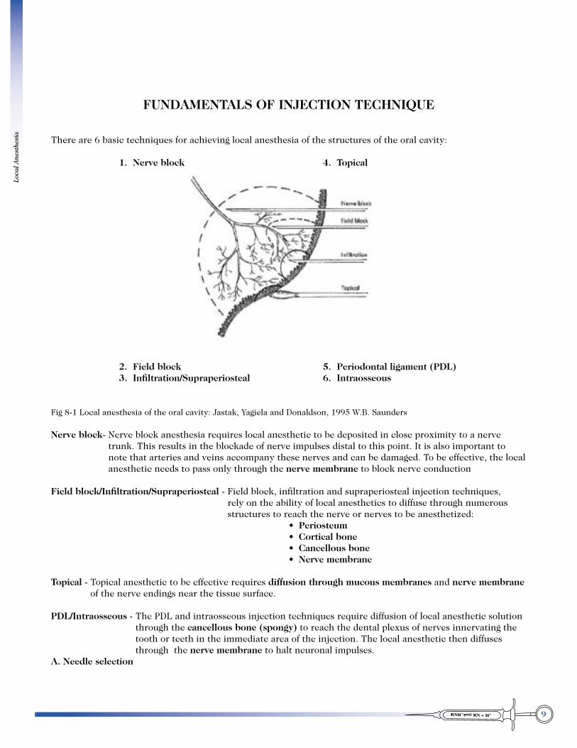
There are 6 basic techniques for achieving local anesthesia of the structures of the oral cavity:
1. Nerve block 4. Topical
2. Field block 5. Periodontal ligament (PDL) 3. Infiltration/Supraperiosteal 6. Intraosseous
Fig 8-1 Local anesthesia of the oral cavity: Jastak, Yagiela and Donaldson, 1995 W.B. Saunders
Nerve block- Nerve block anesthesia requires local anesthetic to be deposited in close proximity to a nerve trunk. This results in the blockade of nerve impulses distal to this point. It is also important to note that arteries and veins accompany these nerves and can be damaged. To be effective, the local anesthetic needs to pass only through the nerve membrane to block nerve conduction
Field block/Infiltration/Supraperiosteal - Field block, infiltration and supraperiosteal injection techniques, rely on the ability of local anesthetics to diffuse through numerous structures to reach the nerve or nerves to be anesthetized:
PeriosteumCortical boneCancellous boneNerve membrane
Topical - Topical anesthetic to be effective requires diffusion through mucous membranes and nerve membrane of the nerve endings near the tissue surface.
PDL/Intraosseous - The PDL and intraosseous injection techniques require diffusion of local anesthetic solution through the cancellous bone (spongy) to reach the dental plexus of nerves innervating the tooth or teeth in the immediate area of the injection. The local anesthetic then diffuses through the nerve membrane to halt neuronal impulses.
A. Needle selection
10
Loca
l An
esth
esia
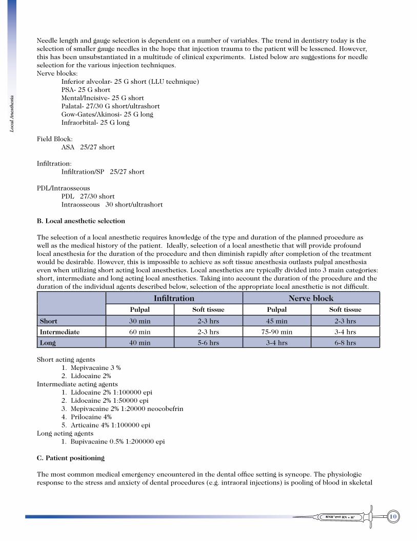
Needle length and gauge selection is dependent on a number of variables. The trend in dentistry today is the selection of smaller gauge needles in the hope that injection trauma to the patient will be lessened. However, this has been unsubstantiated in a multitude of clinical experiments. Listed below are suggestions for needle selection for the various injection techniques.Nerve blocks: Inferior alveolar- 25 G short (LLU technique) PSA- 25 G short Mental/Incisive- 25 G short Palatal- 27/30 G short/ultrashort Gow-Gates/Akinosi- 25 G long Infraorbital- 25 G long Field Block: ASA 25/27 short
Infiltration: Infiltration/SP 25/27 short
PDL/Intraosseous PDL 27/30 short Intraosseous 30 short/ultrashort
B. Local anesthetic selection
The selection of a local anesthetic requires knowledge of the type and duration of the planned procedure as well as the medical history of the patient. Ideally, selection of a local anesthetic that will provide profound local anesthesia for the duration of the procedure and then diminish rapidly after completion of the treatment would be desirable. However, this is impossible to achieve as soft tissue anesthesia outlasts pulpal anesthesia even when utilizing short acting local anesthetics. Local anesthetics are typically divided into 3 main categories: short, intermediate and long acting local anesthetics. Taking into account the duration of the procedure and the duration of the individual agents described below, selection of the appropriate local anesthetic is not difficult.
Infiltration Nerve blockPulpal Soft tissue Pulpal Soft tissue
Short 30 min 2-3 hrs 45 min 2-3 hrs
Intermediate 60 min 2-3 hrs 75-90 min 3-4 hrs
Long 40 min 5-6 hrs 3-4 hrs 6-8 hrs
Short acting agents 1. Mepivacaine 3 % 2. Lidocaine 2%Intermediate acting agents 1. Lidocaine 2% 1:100000 epi 2. Lidocaine 2% 1:50000 epi 3. Mepivacaine 2% 1:20000 neocobefrin 4. Prilocaine 4% 5. Articaine 4% 1:100000 epiLong acting agents 1. Bupivacaine 0.5% 1:200000 epi
C. Patient positioning
The most common medical emergency encountered in the dental office setting is syncope. The physiologic response to the stress and anxiety of dental procedures (e.g. intraoral injections) is pooling of blood in skeletal

11
Loca
l An
esth
esia
muscle. This leads to a drop in blood pressure which results in cerebral ischemia which leads to the undesirable symptoms of lightheadedness, pallor and possible loss of consciousness. With this in mind, it is always appropriate to place patients in the supine or semi-supine position to improve venous return and cerebral blood flow provided that the position is tolerated by the patient and is appropriate for their medical condition. D. Tissue preparation
The preparation of tissue involves the removal of debris, excess saliva and placement of topical anesthetic. Preparation of the injection site with an antiseptic is not necessary due to substantial bacterial flora present in the mouth. The tissue however, should be cleaned with a 2x2 gauze to remove debris. Before topical is placed the tissue is dried to prevent dilution and mobilization of the topical anesthetic by the patient’s saliva. Topical anesthetic should remain in contact with the tissues a minimum of 1-2 minutes.
E. Operator position/Finger rests
Most dental injections can be administered from one of two operator positions. For the right-handed operator, the 8 and 10 o’clock position and for left-handed operators, the corresponding 2 and 4 o’clock position almost always allows for optimal visualization of the injection field.
Finger rests should be established to allow for stabilization of the syringe and back support for the operator. Finger rests are unique for each injection and will vary with practitioners depending on individual preference and hand size. It is appropriate to use some parts of the patient’s anatomy for finger or elbow support (i.e. chin, shoulder) but discretion should be used.
F. Injection Before the initiation of the injection, a thorough understanding of anatomy is essential. A mental picture of the nerve or nerves to be anesthetized in relationship to identifying landmarks is crucial to the success of the injection. Proper orientation of the syringe before penetration of the tissue is important to avoid multiple reorientations of the syringe once the needle has entered the tissue. This lessens the amount of damage to the tissue and leads to less post-op discomfort as well as trauma to important structures such as muscles and nerves.
1. Topical anesthetic is placed for 1-2 minutes 2. The anesthetic syringe, needle and selected local anesthetic are then prepared and a drop of
anesthetic is expressed to assure proper assembly. 3. The needle is then aligned so that the bevel is oriented towards the periosteum (if applicable for the
injection). 4. The tissue should be retracted taut but gently and is oftentimes accomplished with 2x2 gauze. This
allows for a more comfortable needle insertion and also decreases the depth of tissue penetration to the target area.
5. Initial penetration of the mucosa should be done expeditiously, but with control, to a depth of a few millimeters. At this point, the operator should pause and deposit a few drops of local anesthetic.
6. After pausing 5-10 seconds, the needle is then advanced in incremental steps toward the target area. The needle should only be advanced into tissue that has been anesthetized ahead of the needle by the advancing pool of anesthetic.
7. When the final target area has been reached, aspiration is performed to ensure that the tip of the needle is not located in a blood vessel. If blood enters the cartridge, the syringe should be reoriented and aspiration performed again. It is also good practice to aspirate several times as the anesthetic is deposited to avoid inadvertent intravascular administration of local anesthetic.
8. The anesthetic solution should be administered slowly at a rate of 1 ml/min or less. This is especially true when injecting into dense palatal tissues and for the apprehensive patient. Slow administration of local anesthetic is equally important for avoiding complications in case of inadvertent intravascular injection. Rapid injection into a blood vessel results in transiently high plasma levels of local anesthetic leading to systemic toxicity.
9. In most cases deposition of 1 milliliter (or less) of local anesthetic at the final target area is all that is

12
Loca
l An
esth
esia
necessary for profound anesthesia to be obtained. 10. When the injection has been completed, the needle is withdrawn carefully and the needle is
recapped using proper technique. 11. Profound anesthesia can be expected in 3-5 minutes for most conventional injection techniques.
The Gow-Gates and Akinosi are examples of injection techniques that require 5-10 minutes for profound anesthesia due to the extensive diffusion that is necessary to reach the nerves to be anesthetized.
12. After completion of the local anesthetic injection, the local anesthetic and amount given should be recorded in the chart. It is very important to never leave the patient alone after local anesthetic administration. A majority of medical emergencies in the dental office occur either during or shortly after the delivery of the local anesthetic injection
PHARMACOLOGY OF LOCAL ANESTHETICS
A. Desirable Characteristics
1. Block axon conduction (nerve impulse) when applied locally in appropriate concentrations. 2. Local anesthetic action must be completely reversible; however, the duration of the anesthetic block
should be of sufficient length to allow completion of the planned treatment. 3. Produce minimal local toxic effects such as nerve and muscle damage as well as minimal systemic
toxic effects of organ systems such as the cardiovascular and central nervous system.
B. Source 1. Cocaine - 1st local anesthetic - isolated in the 1860’s from leaves of Erythroxylon coca plant. 2. Hundreds of local anesthetics have been synthesized in the laboratory but few have been marketed
due to toxicity, irritation, insolubility and unstable properties. 3. Local anesthetics used in dentistry today are synthetic:
- Articaine, bupivacaine, lidocaine, mepivacaine and prilocaine.C. Chemistry
1. Major features
a. Lipophilic aromatic group - Enhances the ability of the local anesthetic molecule to penetrate various anatomical structures between the site of injection and the target site - the sodium channel in the nerve axon.
b. Intermediate chain - Important for two main reasons:
local anesthetic molecule.
anesthetics into two groups: amides and esters c. Hydrophilic amino group - Imparts water solubility to the molecule ensuring that on
injection into the tissue, the local anesthetic will not precipitate.

13
Loca
l An
esth
esia
2. Classification
a. Esters 1. Formed from an aromatic acid and an amino alcohol. 2. Examples of ester type local anesthetics: Procaine Chloroprocaine Tetracaine Cocaine Benzocaine- topical applications only b. Amides
1. Formed from an aromatic amine and an amino acid. 2. Examples of amide type local anesthetics: Articaine Mepivacaine Bupivacaine Prilocaine Etidocaine Ropivacaine Lidocaine

14
Loca
l An
esth
esia
3. Chemical Properties
a. All commonly used local anesthetics are weak basic tertiary amines (acceptors of hydrogen ion).
Synthesized in the laboratory, anesthetic bases are of little use, as the compound is poorly soluble in water and unstable when exposed to air. This amine (local anesthetic base) is a weak base and therefore readily binds with acids to form a salt.
Example RN + HCLJ RNH+
base salt
This local anesthetic salt is quite soluble in water and relatively stable. Because of this property, local anesthetics are dispensed as salts (HCL) for injections dissolved in sterile water or saline. In sterile water or saline, the local anesthetic salt ionizes to form a quaternary amine cation and an acid anion.
Example
RNH+ J RNH+ + Cl–
salt cation acid anion
The cation [ionized] then, is in an dissociation equilibrium with the base[non-ionized].
Example RNH+ ' RN + H+
cation base The proportion of the ionized form (RNH+), to the free base (nonionized) form (RN) in any given solution depends upon the pH (H+ concentration) of the solution and the surrounding tissues as well as the pKa (dissociation constant) of each specific local anesthetic.
In the presence of a high concentration of hydrogen ions (low pH), the equilibrium shifts to the left and most of the anesthetic molecules are found in the ionized form. E.G. RNH+ > RN + H+
As the hydrogen ion concentration decreases (higher pH) more of the free base form of the anesthetic is formed. E.G. RNH+ < RN + H+
pKa (dissociation constant), simply put, is the affinity of a compound for hydrogen ions (H+). When the pH of a solution and the pKa of a particular local anesthetic are the same, 50% of the anesthetic drug exists in the ionized (cation) form and 50% in the nonionized (base) form.
Henderson-Hasselbach equation
cation Log ( –––––– ) = pKa - pH base

15
Loca
l An
esth
esia
The pKa of amides ranges from 7.6 to 8.1. At physiologic pH (7.4), most of the local anesthetic is in the ionized state (a charged base). For example, lidocaine has a pKa of 7.9. The above formula determines that at physiologic pH, lidocaine exists in a ratio of 3:1 ionized to non-ionized: log [ionized/non-ionized]= 7.8 - 7.4 log [ionized/non-ionized]= 0.4 ionized/non-ionized = 100.4
3 Ionized ~ — = —————— 1 non-ionized
To expand the example, the ester procaine has a pKa of 8.9. The higher pKa value means that at physiologic pH, procaine exists in a ratio of approximately 32:1, ionized to non-ionized. Because lidocaine has a relatively greater proportion of the non-ionized form than does procaine, it could therefore be expected to have a more rapid onset of action. This expectation is confirmed clinically, where lidocaine has been shown to have an onset range of 2 to 3 minutes and procaine to have an onset range of 6 to 12 minutes.
Daniel Haas DDS Septodont 2002
Since the pKa is a constant for any given local anesthetic, it can be determined from the above equation that the base/cation ratio is solely determined by the pH. When the pH and pKa coincidently happen to be equal, the ratio of base (nonionized) to cation (ionized) is 1:1. Because most local anesthetics have pKa values that range from 7.7 to 9.0, it is immediately apparent that the ionized form of the local anesthetic molecule is more abundant than the nonionized form at physiologic pH.
Dissociation constants
Local anesthetic pKa % of base(RN) at pH 7.4 onset of action(min)
Lidocaine 7.8 29 2-4
Bupivacaine 8.1 17 5-8
Mepivacaine 7.7 33 2-4
Prilocaine 7.9 25 2-4
Articaine 7.8 29 2-4
Procaine 9.1 2 14-18
Benzocaine 3.5 100 -
D. Mechanism of Action
When a local anesthetic is injected, it is the ionized [cation] form of the local anesthetic that actually binds to anionic channel receptors in the sodium channel, thus blocking the influx of sodium ions which are responsible for lowering the -70mv resting potential towards the firing threshold of -55mv which then results in depolarization of the nerve membrane. However, only the lipid soluble nonionized [base] form of the local anesthetic can penetrate the various barriers [e.g., nerve membrane, fibrous tissue] between the site of injection and the targeted destination which is the sodium channel.

16
Loca
l An
esth
esia
E. Entry Into Nerve Fibers
1. Entry into nerve fibers of injected local anesthetic is strongly influenced by the ratio of the nonionized to the ionized form since the nonionized form readily crosses the lipo-proteins of the
nerve sheath and axon membrane while the ionized form does not.
a. Events involved in injection and entry are illustrated below.
b. The injected solution of pH 6 has only 1% nonionized lidocaine but when mixed with the buffered
tissue fluid at pH 7.4 the nonionized form will rise to 26%, provided adequate buffering (bicarbonate) is present. Initially in some cases the proportion of nonionized form will be less (e.g. 1 ml of pH 6 solution could temporarily lower the pH of 1 ml of ECF to 7.1, thus decreasing the nonionized form to 15%). As fresh bicarbonate and other buffers diffuse from the blood and adjacent tissues the pH will rise back to 7.4 and the percent of nonionized form will rise.
c. Injection of relatively large volumes of acidic local anesthetic solution into a small area may lower pH of tissue so much that the anesthesia will be less effective than with smaller volumes (e.g. infiltration of 2-3 ml over the roots of the anterior or premolar maxillary teeth).
d. Reduction in the pH of ECF by infection will also reduce the percent of nonionized form and thus the effectiveness of anesthesia.
e. Both alkalinization and carbonation of local anesthetic solution may enhance onset as well as be less irritating on injection.
2. Topical Anesthetics
a. Uses 1. Anesthetize soft tissue before needle insertion. 2. Helpful in reducing gag reflex; e.g., x-rays, impressions. 3. Helpful in management of alveolar osteitis [dry socket]. 4. Reduces sensitivity during periodontal probing, gingival curettage and orthodontic

17
Loca
l An
esth
esia
band placement. b. Most injectable local anesthetics are inappropriate candidates for topical anesthetic preparations.
In order to be effective as topical agents, the concentrations would have to be so high that over-dosage and local tissue toxicity would be of concern.
c. Local anesthetics in solution as HCL salt do not penetrate intact skin appreciably. Small amounts spilled on hands, etc. in course of use are not likely to cause toxic effects but may induce the allergic state.
d. Spray preparations applied to membranes of the oropharynx may be rapidly and extensively absorbed. These preparations are potentially hazardous due to toxic overdose and should not be used without metered valve dispensers. These are considered no more effective than application by a cotton applicator.
F. Neurophysiology
1. Nerve fibers exhibit wide range of sensitivity to nerve blockade-in order of increasing resistance to block are the sensations of pain, cold, warmth, touch, pressure, proprioception and motor function.
Nerve Fibers: Types Size Speed (M./Sec.) Occurrence A (_) 20 µm 80 - 120 Myelinated (Primarily for muscular activity). (`) 8 - 15 µm Myelinated (Touch and pressure) (a) 4 - 8 µm Myelinated (Muscle spindle tone) (b) 3 - 4 µm 10-15 Myelinated (Pain and temperature sensation) B 4 µm 10-15 Myelinated (Preganglionic autonomic) C 1-2 µm 1 - 2 Unmyelinated (Pain and temperature sensation) Myelinated = faster conducting Unmyelinated = slower conducting 2. Small non-myelinated fibers (C- pain fibers) and smaller myelinated pre-ganglionic B fibers are more
readily blocked than are larger myelinated fibers responsible for muscle activity and touch [A-alpha and A-beta]. Clinically, a person would notice complete lack of sensation to a pinprick, while at the same time still be able to move their fingers.
3. The clinical phenomenon of differential nerve block is thought to result from the differences in the internodal distance seen between nerve fibers of varying sizes. It is generally accepted that to halt neuronal traffic, 3 successive nodes must be bathed with the local anesthetic solution. The internodal distance of small Ab fibers ranges from 0.3-0.7mm whereas the internodal distance of the larger A_ fibers range from 0.8-1.4mm. Knowing this, the “pool” of anesthetic covering the 3 nodes of the larger A_ nerve fiber would need to be at least twice the size of the “pool” covering the three smaller nodes of the Ab fiber. It therefore seems that differential sensitivity of fibers results from variations in the “critical length” that must be exposed to a local anesthetic for conduction to be blocked.
G. Effects and Toxic Actions on Organ Systems
1. Local anesthetics (dose dependent) interfere with transmission in any excitable tissue (e.g. CNS and CVS).
2. CNS effects a. Central neurons very sensitive. b. Excitatory-dizziness, visual and auditory disturbances, apprehension, disorientation and
muscle twitching more common with ester type agents. c. Depression manifested as slurred speech, drowsiness and unconsciousness more common
with amide type agents (e.g. lidocaine). d. Higher concentrations of local anesthetic may eventually produce tonic-clonic[grand mal]

18
Loca
l An
esth
esia
convulsions. e. Very large doses may produce respiratory depression which can be fatal. Artificial respiration
may be life-saving. 3. CVS effects a. Local anesthetics have direct action on the myocardium and peripheral vasculature by
closing the sodium channel, thereby limiting the inward flux of sodium ions. b. Myocardium usually depressed both in rate and force of contraction. Depression of ectopic
pacemakers useful in treating cardiac arrhythmias. c. Concentrations employed clinically usually cause vasodilation in area of injection. d. Vasoconstrictors such as epinephrine may counteract these effects on myocardium and
vasculature.
4. Local Tissue Responses a. Occasionally focal necrosis in skeletal muscle at injection site, decreased cell motility and
delayed wound healing. b. Tissue hypoxia may be produced by action of excessive amounts of vasoconstrictors.
H. Pharmacokinetics [Absorption, Distribution, Metabolism, Elimination]
1. Absorption a. Ideally, local anesthetic molecules would remain at the neural target interrupting neural
traffic. The local anesthetic molecule, having both hydrophilic and hydrophobic properties, does not remain confined to the injected site for long. A significant portion of the injected local anesthetic diffuses away from the injection site and is absorbed into the circulation.
b. In order to localize the site of action and decrease absorption, transport and/or destruction in the blood stream following injection, vasoconstrictor agents are sometimes used in conjunction with local anesthetics. (See pharmacology of vasoconstrictors for
further information). 2. Distribution a. Some agents such as procaine and lidocaine are active vasodilators and may enhance their
own absorption into the blood. b. Most agents once absorbed into the circulation are readily distributed from blood to well
perfused tissues (e.g. brain, heart, liver, etc.) c. As time progresses, local anesthetic partitions into less well perfused tissues[i.e. muscle, fat]
as well as into clearance organs[i.e., liver and kidney] e. Can cross placenta and occasionally may depress heart of fetus.
3. Metabolism a. Esters inactivated by hydrolysis in blood and/or liver by pseudocholinesterase; products
may be conjugated and/or excreted. Individuals with genetically based defects in pseudocholinesterase are unusually sensitive to procaine and other esters.
b. Amides metabolized in liver by oxidation (e.g. N-demethylation), and hydrolysis by liver amidase; products conjugated and excreted. Inactivation of prilocaine, a secondary amine, is relatively rapid because dealkylation not required before hydrolysis. Severe hepatic disease may reduce metabolism and lead to toxic reactions. Some metabolites of amides still have pharmacologic activity (e.g. metabolites of lidocaine are sedative).
4. Excretion a. Both esters and amides are excreted in the urine mainly as metabolites with only a few
percent as the unchanged agent.
19
Loca
l An
esth
esia
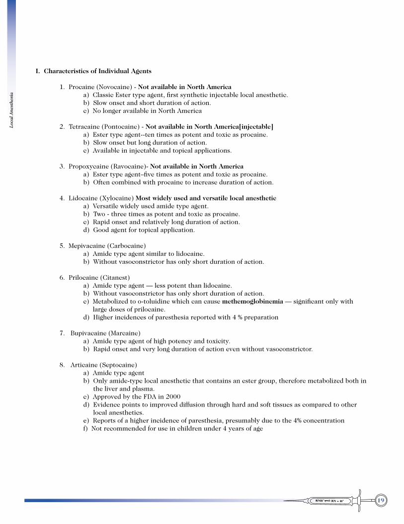
I. Characteristics of Individual Agents
1. Procaine (Novocaine) - Not available in North America a) Classic Ester type agent, first synthetic injectable local anesthetic. b) Slow onset and short duration of action. c) No longer available in North America 2. Tetracaine (Pontocaine) - Not available in North America[injectable] a) Ester type agent--ten times as potent and toxic as procaine. b) Slow onset but long duration of action. c) Available in injectable and topical applications.
3. Propoxycaine (Ravocaine)- Not available in North America a) Ester type agent–five times as potent and toxic as procaine. b) Often combined with procaine to increase duration of action.
4. Lidocaine (Xylocaine) Most widely used and versatile local anesthetic a) Versatile widely used amide type agent. b) Two - three times as potent and toxic as procaine. c) Rapid onset and relatively long duration of action. d) Good agent for topical application.
5. Mepivacaine (Carbocaine) a) Amide type agent similar to lidocaine. b) Without vasoconstrictor has only short duration of action.
6. Prilocaine (Citanest) a) Amide type agent — less potent than lidocaine. b) Without vasoconstrictor has only short duration of action. c) Metabolized to o-toluidine which can cause methemoglobinemia — significant only with
large doses of prilocaine. d) Higher incidences of paresthesia reported with 4 % preparation
7. Bupivacaine (Marcaine) a) Amide type agent of high potency and toxicity. b) Rapid onset and very long duration of action even without vasoconstrictor. 8. Articaine (Septocaine) a) Amide type agent b) Only amide-type local anesthetic that contains an ester group, therefore metabolized both in
the liver and plasma. c) Approved by the FDA in 2000 d) Evidence points to improved diffusion through hard and soft tissues as compared to other
local anesthetics. e) Reports of a higher incidence of paresthesia, presumably due to the 4% concentration f) Not recommended for use in children under 4 years of age

20
Loca
l An
esth
esia
TOPICAL ANESTHETICS
Agent Chemistry Property / Uses
Lidocaine (Xylocaine) AmideAmide Intermediate Potency, toxicity. Ointment (5%), solution, spray (10%) (also by injection)
Tetracaine (Pontocaine) Ester
Tetracaine is a potent agent and should be used with great caution to avoid systemic toxicity. Topically applied tetracaine as opposed to benzocaine has a prolonged duration of action. High potency & toxicity Solution, ointment, spray (Citacaine 2%) (also by injection)
Benzocaine Ester
Relatively weak (20-22%). Ointment, Solution, Powder. Used on oral lesions, dry sockets. In many over the counter remedies.
EMLA[eutectic mixture of local anesthetics] Combination of prilocaine 2.5% and lidocaine 2.5%
AmideSurface anesthesia of intact skin, must be applied for 1 hour to be effective.
Cocaine EsterSolution, used in ENT procedures. Toxic, addicting, a controlled substance.
Topical Anesthetics
BenzocaineBenzocaine is a derivative of procaine, an ester type local anesthetic, and is poorly soluble in water and is available only as a topical anesthetic. Localized allergic reactions are sometimes encountered when using benzocaine multiple times on the same patient. Overdosing is unlikely as benzocaine is poorly absorbed into the blood, which decreases the likelihood of systemic toxicity. However, there have been reports of methemoglobinemia with excessive doses of benzocaine. The onset of surface anesthesia is rapid requiring less than one minute. But, more profound anesthesia is achieved with longer application.
Benzocaine is available in several formulations including aerosol, gel, ointment and solution. Topical anesthetics that are delivered by pressurized spray should not be used without a metered dose dispenser as overdosing can rapidly occur.
Trade names - Anbesol, Hurricaine, Numzident, Orajel, Topex
TetracaineTetracaine is an ester type local anesthetic which is available both as an injectable and topical preparation. Tetracaine is a potent agent and should be used with great caution to avoid systemic toxicity. Topically applied tetracaine as opposed to benzocaine has a prolonged duration of action.

21
Loca
l An
esth
esia
CocaineCocaine is a ester type anesthetic that is used exclusively as a topical agent. Cocaine is unique among topical and injectable anesthetics in that it has vasoconstrictive as well as anesthetic properties. It is used sparingly because of its abuse potential but is still used when hemostasis of mucous membranes is essential. Cocaine is generally available in concentrations of 2-10 % solution.
Lidocaine
Lidocaine is an amide local anesthetic that is available in injectable and topical formulations. It is available in gel, viscous solution, ointment and aerosol preparations in concentrations ranging from 2-10 %. The onset of anesthesia is slower relative to benzocaine but, the duration is about the same. Absorption into the bloodstream is greater than benzocaine providing a greater risk of systemic toxicity.
REVIEW OF LOCAL ANESTHETIC AGENTS
1. Definition Agent which reversibly depress nerve conduction
2. Chemistry a. Weak bases, poorly water soluble - form HCL salts for injections b. Esters of BA, PABA, MABA c. Amides of xylidine or toluidine
3. Absorption a. Penetrate nerve fiber best as lipophilic free base b. vasoconstrictors slow absorption, prolong action, reduce toxicity, hemostasis
4. Mechanism of Action Blocks depolarization of nerve axons by preventing sodium ion influx
5. Metabolism a. Esters - hydrolysis by plasma and liver esterase’s. Don’t use these if patient is deficient in plasma cholinesterase
b. Amides - oxidation by liver enzymes
6. Side Action & a. CNS Toxicity 1. Variable - from stimulation to depression 2. Respiratory depression is usual cause of death. Treat with oxygen. b. Allergy - No cross-allergy between esters and amides c. CVS - Depress myocardium - Xylocaine used to treat cardiac arrhythmias
7. Rate of Injection The maximum recommended rate - 1 ml / minute 8. Aspirate To prevent intravascular injection which increases toxicity by 16x Aspiration test is not 100% dependable so inject SLOWLY
9. Benadryl 1% up to 5 ml. can be used if patient is sensitive to both esters and amides
10. Topical Application Caution with topical anesthetics and topical epinephrine in gingipak etc.
11. Epinephrine 1:50,000 In small amounts and usually only for hemostasis - then with caution due to possible sloughing, toxicity, less profound anesthesia as buffering capacity of interstitial fluid
22
Loca
l An
esth
esia
is decreased in complete ischemia
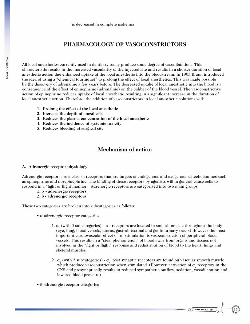
PHARMACOLOGY OF VASOCONSTRICTORS
All local anesthetics currently used in dentistry today produce some degree of vasodilatation. This characteristic results in the increased vascularity of the injected site and results in a shorter duration of local anesthetic action due enhanced uptake of the local anesthetic into the bloodstream. In 1903 Braun introduced the idea of using a “chemical tourniquet” to prolong the effect of local anesthetics. This was made possible by the discovery of adrenaline a few years before. The decreased uptake of local anesthetic into the blood is a consequence of the effect of epinephrine (adrenaline) on the caliber of the blood vessel. The vasoconstrictive action of epinephrine reduces uptake of local anesthetic resulting in a significant increase in the duration of local anesthetic action. Therefore, the addition of vasoconstrictors in local anesthetic solutions will: 1. Prolong the effect of the local anesthetic 2. Increase the depth of anesthesia 3. Reduces the plasma concentration of the local anesthetic 4. Reduces the incidence of systemic toxicity 5. Reduces bleeding at surgical site
Mechanism of action
A. Adrenergic receptor physiology
Adrenergic receptors are a class of receptors that are targets of endogenous and exogenous catecholamines such as epinephrine and norepinephrine. The binding of these receptors by agonists will in general cause cells to respond in a “fight or flight manner”. Adrenergic receptors are categorized into two main groups: 1. _ - adrenergic receptors 2. `�- adrenergic receptors
These two categories are broken into subcategories as follows:
_-adrenergic receptor categories
1. _� (with 3 subcategories) – _
� receptors are located in smooth muscle throughout the body
(eye, lung, blood vessels, uterus, gastrointestinal and genitourinary tracts) However the most important cardiovascular effect of _
� stimulation is vasoconstriction of peripheral blood
vessels. This results in a “steal phenomenon” of blood away from organs and tissues not involved in the “fight or flight” response and redistribution of blood to the heart, lungs and skeletal muscles.
2. _2 (with 3 subcategories) - _2 post synaptic receptors are found on vascular smooth muscle which produce vasoconstriction when stimulated. (However, activation of _2 receptors in the CNS and presynaptically results in reduced sympathetic outflow, sedation, vasodilatation and lowered blood pressure)

23
Loca
l An
esth
esia
1. `1 receptors- mainly found in the heart resulting in increased heart rate and contractility 2. `2 receptors- relaxes smooth muscle resulting in vasodilatation (most significantly skeletal
muscle) and bronchodilation (lungs). 3. `3 receptors- found in adipose tissue
Receptor potencies of adrenergic vasoconstrictors
Drug alpha-1 alpha-2 beta-1 beta-2
Epinephrine ++ ++ +++ ++
Norepinephrine ++ ++ ++ 0Levonordefrin + ++ ++ +
Potency: +++ = high, ++ = intermediate, + = low
Therefore, in summary local anesthetics containing epinephrine produce: 1. Localized
i. Hemostasis at surgical site ii. Ischemia of localized tissue 2. Systemic
i. Increased heart rate (`1) ii. Increased force and rate of contraction (`1) iii. Increased cardiac output iv. Increases oxygen demand v. Dilation of coronary arteries vi. Decreases threshold for arrhythmias
i. Bronchodilation (`2 )
i. Predominately vasodilation (fight or flight response) (`2 )
i. Minimal direct effect due to difficulty in crossing the blood-brain barrier. Most effects on the CNS are manifestations of the vasoconstrictor on other organs such as the heart.
B. Concentrations of vasoconstrictors
1. Epinephrine Vasoconstrictors such as epinephrine are prepared as diluted solutions of various strengths depending
on the intended use. Epinephrine solutions are typically present in the following strengths:
1:100000 (0.01mg/ml)(10 mcg/ml)1:200000 (0.005mg/ml)(5 mcg/ml)
The most commonly used epinephrine dilution in dentistry today is 1:100000. However it appears that a 1:200000 concentration is comparable in effect to the 1:100000 concentration.
2. Levonordefrin (Neocobefrin) Levonordefrin is a synthetic compound very similar in structure to epinephrine. It is the only
alternate choice of vasoconstrictor to epinephrine presently available in the USA. It is prepared as a 1:20000 (0.05mg/ml)(50 mcg/ml) concentration with 2 % mepivacaine.
24
Loca
l An
esth
esia
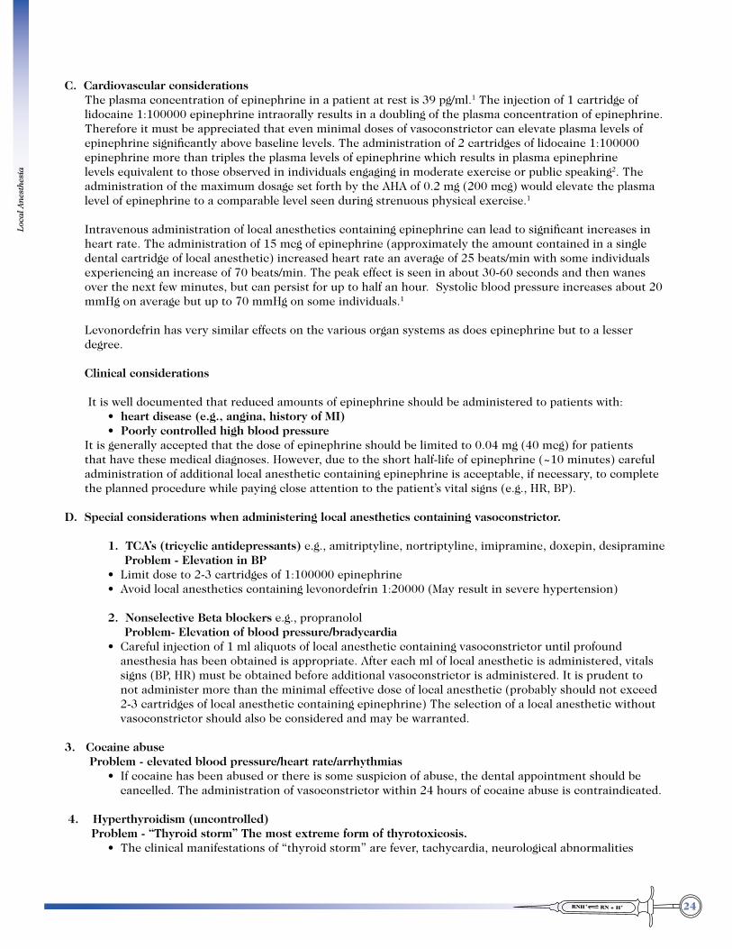
C. Cardiovascular considerations The plasma concentration of epinephrine in a patient at rest is 39 pg/ml.1 The injection of 1 cartridge of
lidocaine 1:100000 epinephrine intraorally results in a doubling of the plasma concentration of epinephrine. Therefore it must be appreciated that even minimal doses of vasoconstrictor can elevate plasma levels of epinephrine significantly above baseline levels. The administration of 2 cartridges of lidocaine 1:100000 epinephrine more than triples the plasma levels of epinephrine which results in plasma epinephrine levels equivalent to those observed in individuals engaging in moderate exercise or public speaking2. The administration of the maximum dosage set forth by the AHA of 0.2 mg (200 mcg) would elevate the plasma level of epinephrine to a comparable level seen during strenuous physical exercise.1
Intravenous administration of local anesthetics containing epinephrine can lead to significant increases in heart rate. The administration of 15 mcg of epinephrine (approximately the amount contained in a single dental cartridge of local anesthetic) increased heart rate an average of 25 beats/min with some individuals experiencing an increase of 70 beats/min. The peak effect is seen in about 30-60 seconds and then wanes over the next few minutes, but can persist for up to half an hour. Systolic blood pressure increases about 20 mmHg on average but up to 70 mmHg on some individuals.1
Levonordefrin has very similar effects on the various organ systems as does epinephrine but to a lesser degree.
Clinical considerations
It is well documented that reduced amounts of epinephrine should be administered to patients with:
It is generally accepted that the dose of epinephrine should be limited to 0.04 mg (40 mcg) for patients that have these medical diagnoses. However, due to the short half-life of epinephrine (~10 minutes) careful administration of additional local anesthetic containing epinephrine is acceptable, if necessary, to complete the planned procedure while paying close attention to the patient’s vital signs (e.g., HR, BP).
D. Special considerations when administering local anesthetics containing vasoconstrictor.
1. TCA’s (tricyclic antidepressants) e.g., amitriptyline, nortriptyline, imipramine, doxepin, desipramine Problem - Elevation in BP
2. Nonselective Beta blockers e.g., propranolol Problem- Elevation of blood pressure/bradycardia
anesthesia has been obtained is appropriate. After each ml of local anesthetic is administered, vitals signs (BP, HR) must be obtained before additional vasoconstrictor is administered. It is prudent to not administer more than the minimal effective dose of local anesthetic (probably should not exceed 2-3 cartridges of local anesthetic containing epinephrine) The selection of a local anesthetic without vasoconstrictor should also be considered and may be warranted.
3. Cocaine abuse Problem - elevated blood pressure/heart rate/arrhythmias
cancelled. The administration of vasoconstrictor within 24 hours of cocaine abuse is contraindicated.
4. Hyperthyroidism (uncontrolled) Problem - “Thyroid storm” The most extreme form of thyrotoxicosis.
25
Loca
l An
esth
esia
and hypertension followed by hypotension and shock. Epinephrine administration to these patients is absolutely contraindicated. Signs and symptoms of patients with uncontrolled hyperthyroidism include:
1. Agitation, confusion 2. Tachycardia, hypertension 3. Excessive sweating, elevated temperature
5. Hypertension Problem - Exacerbation of high blood pressure
as follows:
1. <180/110 - Treatment may proceed with stress reduction protocol. However, this is assuming that the patient has been shown to have a blood pressure range within normal limits and the elevated blood pressure is a direct result of the patient’s pretreatment dental anxiety. Patient’s with a diagnosis of
a. coronary artery disease b. cerebrovascular disease c. angina d. and a history of MI/CVA less than 6 months ago should not receive treatment and require a
consultation with their physician.
2. >180/110 - don’t administer local anesthetic containing vasoconstrictor. In actuality, all elective dental treatment should be aborted until the patient has been evaluated and treated for hypertension and blood pressure brought under control by their physician.
ASA IV patients - Vasoconstrictor contraindicated (No elective dental treatment)
PATIENT EVALUATION
A. Introduction A pre-treatment evaluation should be completed on every patient scheduled for treatment in the dental office. This is especially important for those individuals who will receive a local anesthetic with or without sedation (Nitrous Oxide, Oral and IV). In the ambulatory setting (i.e., dental office), it is fortunate that medical emergencies are only occasionally encountered during dental procedures. However, the incidences of medical emergencies are greatly increased in a small segment of the patient population. Therefore, a thorough medical work-up consisting of a
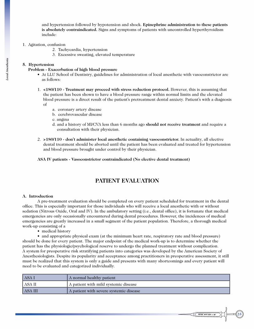
should be done for every patient. The major endpoint of the medical work-up is to determine whether the patient has the physiologic/psychological reserve to undergo the planned treatment without complication. A system for preoperative risk stratifying patients into categories was developed by the American Society of Anesthesiologists. Despite its popularity and acceptance among practitioners in preoperative assessment, it still must be realized that this system is only a guide and presents with many shortcomings and every patient will need to be evaluated and categorized individually.
ASA I A normal healthy patient
ASA II A patient with mild systemic disease
ASA III A patient with severe systemic disease

26
Loca
l An
esth
esia
ASA IV A patient with severe systemic disease that is a constant threat to life
ASA V A moribund patient who is not expected to survive without the operation
ASA VI A declared brain-dead patient whose organs are being removed for donor purposes
Exercise tolerance correlation ASA I No dyspnea (or undue fatigue or precordial pain) with normal activity. Normal activity is defined as: 1. climbing one flight of stairs 2. walking 2 level blocks at a normal pace 3. mowing lawn for 5 - 10 minutes ASA II Mild dyspnea after normal activity; may rest at top of flight of stairs = dyspnea after normal
activity. Both good risks if other points negative. If Class II patient is apprehensive use pre-op sedation.
ASA III Dyspnea during normal activity. Comfortable at rest in any position. May tend toward orthopnea (has to sit up to breathe). Rests before reaching top of a flight of stairs. Ankles swell as day progresses.
ASA IV Dyspnea and orthopnea at rest (all the time). The patient will rest several times when climbing a flight of stairs if he can climb them at all).
Treatment modifications ASA I - Psychological/procedural stress - well tolerated by the patient; no treatment modification
necessary (stress reduction?) ASA II - Psychological/procedural stress - tolerated by the patient; no treatment modification (stress
reduction?) ASA III - Psychological/procedural stress - don’t tolerate well
ASA IV - Psychological stress/procedural- may decompensate the patient
temporary filling etc).
maxillofacial surgery for removal in a hospital setting. Medical consultation required
B. Medical history
1. Cardiovascular
a. HTN Hypertension is very prevalent in our society. The clinician should seek to understand the
significance and extent of the hypertensive end organ disease (heart, kidney, cerebral) as well compliance with proscribed medical regimens. At LLUSD, it is appropriate to provide elective treatment for a patient that has a BP reading of <180/110 provided that this patient does not have a co-diagnosis of cardiac and cerebrovascular disease and is asymptomatic (chest pain, headache etc). However, it is assumed that the patient has controlled hypertension and the high blood pressure reading is the direct result of the patient’s anxiety.
Management: 1. Stress reduction protocol (gentle technique, profound local anesthesia, short appointments) 2. Minimize use of vasoconstrictors (2 cartridges of 1:100000 epinephrine) 3. Sedation (Nitrous Oxide, oral, IV)
b. Ischemic heart disease/angina

27
Loca
l An
esth
esia
Ischemic heart disease or angina is a condition in which there is a myocardial oxygen supply and demand imbalance. Atherosclerotic plaque is the most common cause of this condition. Anginal attacks (chest pain) are the clinical manifestations of this disease. It is important to question the patient with regards to the following:
In other words, is the angina brought on by strenuous exercise, while climbing one flight of stairs or at rest (sitting on the couch).
When the anginal episode occurs, what is done to relieve the pain? Rest, oxygen, nitrates?
If the patient experiences chest pain while doing minimal exercise (washing dishes) or at rest, the patient is considered to have unstable angina. Also, if the patient requires multiple doses of nitrates before symptoms resolve places them at risk for a myocardial infarction. All of these clinical situations are poor risks for elective dental care and require medical consultation and follow-up before care is provided to them.
Management: 1. Stress reduction protocol (gentle technique, profound local anesthesia, short appointments) 2. Sedation (Nitrous Oxide, Oral, IV) 3. Pre-op nitroglycerin sublingually 5 min (1 tablet) before starting local anesthesia. Use
patient’s tablets if possible. 4. Consider administering oxygen (nasal cannula) during treatment 5. Minimize use of vasoconstrictors (2 cartridges of 1:100000 epinephrine) 6. Remember patient is a definite risk. (ASA Class III) c. Myocardial infarction Myocardial infarction is the actual death and necrosis of myocardial cells. The long term prognosis
of these patients hinges on the extent and location of the damaged cardiac muscle. Management: 1. No elective dental treatment for 6 months. 2. Consultation with patient’s cardiologist. 3. Routine dental care can then be provided utilizing the same protocol as the patient with
ischemic heart disease.
d. Congestive heart failure
Congestive heart failure defined in its simplest terms is a pump (heart) that is failing due to any number of underlying causes (i.e., ischemia, infarction, valvular disease, cardiomyopathy). This condition results in decreased cardiac output and a backing up of blood behind the failing heart. This manifests clinically as peripheral edema (swollen ankles) and pulmonary edema (fluid filled lungs) for right and left sided heart failure respectively.
Management: 1. No treatment until patient has been optimized medically (minimal evidence of edema, good
exercise tolerance etc) 2. Stress reduction protocol (gentle technique, profound local anesthesia, short appointments) 3. Position patient semi-supine or upright to avoid fluid overload in patient’s lungs 4. Minimize use of vasoconstrictors (2 cartridges of 1:100000 epinephrine)
e. Valvular heart disease Patients with a history of valvular heart disease must be evaluated for the nature and
hemodynamic significance of the condition. Basically, patients with a positive history of valvular heart disease have as an underlying pathology the inability to either open or close the heart valve properly which results in impeded forward blood flow or a significant regurgitation (backflow) of blood. In the cases of severe stenosis or regurgitation cardiovascular hemodynamics (cardiac output, BP) can be significantly altered. Also, valvular heart disease carries the risk of bacterial

28
Loca
l An
esth
esia
endocarditis.
Management: 1. Medical consultation with the patient’s physician to determine the need for antibiotic
prophylaxis and the hemodynamic significance of the valvular disease 2. Maintain patient’s heart rate and blood pressure as close to baseline as possible to avoid
hemodynamic derangements 3. Provide antibiotic prophylaxis if indicated (see SBE dosing chart at back of manual)
f. Cardiac pacemaker, implanted cardioverter/defibrillator Cardiac pacemakers are most commonly placed in patients with symptomatic arrhythmias
and heart blocks that are unresponsive to medical therapy. The device is implanted under the skin and leads are fed through blood vessels into the right atrium or ventricle. Implanted cardioverter/defibrillator (ICD’s) is placed in a similar fashion to pacemakers. These devices detect life threatening arrhythmias and deliver a shock in these susceptible patients.
Management: 1. Consultation with the patient’s physician to determine if the patient is medically optimized
and evaluation of the device to check for proper function is vital 2. There is no need for antibiotic prophylaxis as the device is not placed into the heart 3. Extreme caution when using electrocautery units 4. No contraindication for administration of local anesthetics
2. Pulmonary a. Asthma Asthma is an inflammatory condition of smooth muscle of the tracheobronchial tree. An acute
attack is brought on by intrinsic and extrinsic stimuli such as environmental allergens (pollen) as well as exercise, colds, foods, food preservatives and emotional stress. However, most asthmatic attacks are brought on by inhaled allergens, but attacks brought on by food and food preservatives can be life-threatening.
Management: 1. Stress reduction protocol 2. Bronchodilator (B-2 agonist) such as albuterol (preferably the patient’s) must be
immediately available 3. Avoid local anesthetics containing bisulfite antioxidants in susceptible patients 4. Nitrous oxide/oxygen is suggested if sedation is needed 5. Patients must not have signs and symptoms of an acute asthmatic attack (wheezing,
dyspnea) on day of treatment
b. COPD (bronchitis, emphysema) Patients with a diagnosis of COPD (chronic obstructive pulmonary disease) have lung disease
in which the lungs are damaged making it difficult to breathe. The airways are partially obstructed (bronchitis) and some of the alveolar walls are destroyed (emphysema) making it difficult to move air in and out of the lungs. Treatment options include bronchodilators and inhaled steroids and patients with severe disease are often maintained on supplemental oxygen. In advanced cases of some COPD diseases (bronchitis), patients retain high levels of CO2 due to the obstructive nature of the disease. The primary drive for respiration in the normal population is increased levels of CO2, however, in these patients who have chronic elevated levels of CO2 the primary stimulus for breathing is decreased levels of oxygen (hypoxia). Therefore, theoretically, high levels of oxygen may depress the drive for respiration. Therefore, oxygen flow levels of 4L/minute or less are acceptable to maintain the hypoxic drive.
Management: 1. Consultation with physician to determine if patient is medically optimized
29
Loca
l An
esth
esia
2. Avoid nitrous oxide/oxygen sedation 3. Position semi-supine or upright 4. Avoid bilateral mandibular blocks and local anesthesia of the soft palate 5. If supplemental oxygen is given, keep oxygen flow levels below 4L/min 3. Endocrine a. Diabetes Mellitus Diabetes is a metabolic disorder in which there is a derangement in carbohydrate metabolism.
This results from either insufficient or a complete absence of insulin secretion or there is a lack of receptor response to circulating insulin. Patients are typically classified as type 1 or 2 diabetics. Type 2 diabetics are usually managed with diet modifications or by an oral medication regimen (some require insulin). Type 1 diabetics have an absolute lack of insulin and therefore require exogenous insulin. Patients with diabetes develop long term complications such as cardiovascular disease, blindness (retinal damage), renal failure, nerve damage (neuropathy) and gangrene. Diabetics that have tight control over their blood glucose levels have less long term complications and can lead a relatively normal life. On the other hand, those with poor control of their plasma glucose levels have more serious end organ disease.
Management: 1. Avoid hypoglycemia 2. Stress reduction protocol 3. No treatment modification necessary provided there is no evidence of end organ disease
(cardiac, kidney, neuropathy). In other words, it is okay to administer routine doses of local anesthetic with vasoconstrictor to patients with controlled disease
4. Elective dental treatment is contraindicated in the poorly controlled diabetic (brittle diabetes)
b. Hyperthyroidism (thyrotoxicosis) Patients with a diagnosis of hyperthyroidism that are not being treated are at risk for developing
a thyrotoxic crisis (“thyroid storm”) the most severe form of thyrotoxicosis. Infection, trauma, dental procedures and stress may precipitate the crisis.
Management: 1. No elective treatment until medical consultation and treatment of hyperthyroidism
completed 2. Important to manage dental infections 3. Avoid epinephrine and other sympathomimetic drugs in the uncontrolled hyperthyroid
patient
c. Adrenal gland insufficiency (Addison’s disease) Normal function of the adrenal gland allows for the body to cope with stress. Glucocorticoids
(cortisol), mineralocorticoids (aldosterone) and epinephrine are produced by the adrenal gland for this purpose. Therefore, in scenarios where the adrenal gland is suppressed either by primary causes (Addison’s) or secondary causes (corticosteroid therapy), there is a theoretical chance that under certain conditions (extreme stress) the patient could suffer cardiovascular collapse. However, it is unlikely that routine dental treatment with local anesthesia will precipitate a cardiovascular collapse. There is considerable controversy in management of the patient with adrenal insufficiency and whether it is appropriate to increase the steroid dose or not and if so, for how long.
Management: 1. No additional dosing of steroid for minor procedures (dental treatment), however, should
take daily dose of steroid 2. Consider additional steroid coverage for major surgical procedures (abdominal, thoracic
surgery) for patients taking steroids for systemic inflammatory diseases (rheumatoid arthritis, asthma, lupus)
30
Loca
l An
esth
esia
3. Consult with physician if unsure how to manage steroid coverage
4. Cerebrovascular a. Stroke (cerebrovascular accident) A stroke is the end result of a disruption of oxygenated blood flow to a part or parts of
the brain. The most common causes of stroke are hemorrhage and occlusion of a vessel (thromboembolism). The outcome of a stroke is at the worst death and if the patient survives there is a high probability that the patient will suffer long term neurologic and motor deficits depending on the area of the brain affected. As in myocardial infarction, the most common cause of a stroke is hypertension and atherosclerosis.
Management: 1. No elective dental care for 6 months (post CVA) 2. Consultation with physician to determine patient’s recovery and to manage anticoagulants
(see hematology section) 3. Avoid treatment in patient experiencing transient ischemic attacks (TIA’s) 4. Limit local anesthetic with vasoconstrictor (2-3 cartridges of 1:100000 epinephrine) 5. Stress reduction protocol
5. Hematology a. Hemophilia Patients at risk for bleeding during a dental procedure have either an inherited defect in the
coagulation pathway, an acquired form of hemophilia observed in patients taking anticoagulation medications (coumadin, aspirin, NSAIDS, heparin) or patients with some types of cancer (leukemia). In the dental office, bleeding abnormalities are usually elicited from the health history and should raise concern on the part of the practitioner and requires further questioning and consultation with the patient’s physician.
Management: 1. Consultation with the patient’s physician to determine how to manage anticoagulants. 2. The target INR (international normalized ratio) should be less than 3.0 before dental
treatment. This corresponds to a PT (prothrombin time) of about 1.5- 2.0 of normal value. 3. If a patient is on coumadin and the physician elects to reduce the anticoagulant, then
a period of 3 days is required before a change in the INR will be reflected. A repeat INR should be done on the day of surgery to determine if the desired therapeutic level has been reached.
4. Infiltration, PDL and intraosseous injection techniques are suggested when administering local anesthesia to any patient with an increased risk for bleeding. Avoid block anesthesia if possible due to risk of damaging blood vessels.
5. Local measures such as pressure packs, sutures, gelfoam etc should be used if postop bleeding is encountered.
6. Allergic Reactions Allergic reactions to local anesthetics have reduced dramatically since development of amide local
anesthetics (1948) and the removal of antimicrobials (methylparaben) from single dose cartridges of local anesthetic. Most “allergic reactions” to local anesthetics today are in reality adverse reactions and not true hypersensitivity reactions to the anesthetic. The more dramatic the adverse reaction (syncope, palpitations etc.) the more likely a patient will be labeled as allergic. However, there have been documented cases of hypersensitivity reactions to amide local anesthetics including anaphylaxis. A cartridge of local anesthetic contains the following ingredients:

31
Loca
l An
esth
esia
Local anestheticSodium bisulfite (only in LA with vasoconstrictor)
The only ingredients contained in a local anesthetic cartridge that can potentially cause an allergic response are in bold type. Asthmatics can be particularly sensitive to sodium bisulfite which functions as an antioxidant in local anesthetic cartridges. When encountering a patient with a positive history of “allergy” to local anesthetics, a detailed history and account of the episode must be elicited. Three main questions must be addressed when interviewing these patients:
1. What precipitated the event (Where/when)? 2. Describe the “allergic” event. 3. What emergency treatment was given?
What precipitated the event? It is important to determine if the episode occurred during administration of local anesthetic or
immediately after. It is also important to ask if the patient remembers the type and volume of local anesthetic administered and if it contained a vasoconstrictor. The patient should also be questioned if other medications were also administered concurrently (within the hour) such as antibiotics or analgesics. It is also helpful if the patient can remember when and where this event occurred so consultation can be done with the dentist or physician involved.
Describe the allergic event? A detailed description of the event is useful to determine the true nature of the episode. Patients can
generally be placed in 3 categories according to the description of the event: Psychogenic reactions - These type of reactions are most common and are described as palpitations, headache, sweating, hyperventilation, loss of consciousness (syncope). These are adverse reactions and not allergic in nature. Overdose/intravascular reactions - Seizure type symptoms are described and are usually transitory in nature and again are adverse reactions. Allergic reactions - Descriptive words such as itching, rash, watery eyes, wheezing, dyspnea, and laryngeal swelling raises considerable suspicion of a true allergic response.
What emergency treatment was given? This simple question gives much insight into the significance of the event. If dental treatment was
continued that day or the patient was treated with nonspecific medications/therapies (oxygen, ammonia inhalant etc) and the patient was discharged home, it is very unlikely that the patient was experiencing a true allergic reaction. However, if the patient was administered drugs (epinephrine, benadryl) and/or transported to a hospital then the level of suspicion should once again be raised.
Management: The management of these patients begins with convincing the patient and the practitioner that the patient did or did not have a true allergic response to the local anesthetic in question. Practical suggestions for management of these patients are as follows:
event.
there appears to be no cross allergenicity between the amide type anesthetics.
anesthetic used.
7. Pregnancy The pregnant patient presenting for dental treatment is a common occurrence. Dental treatment
32
Loca
l An
esth
esia
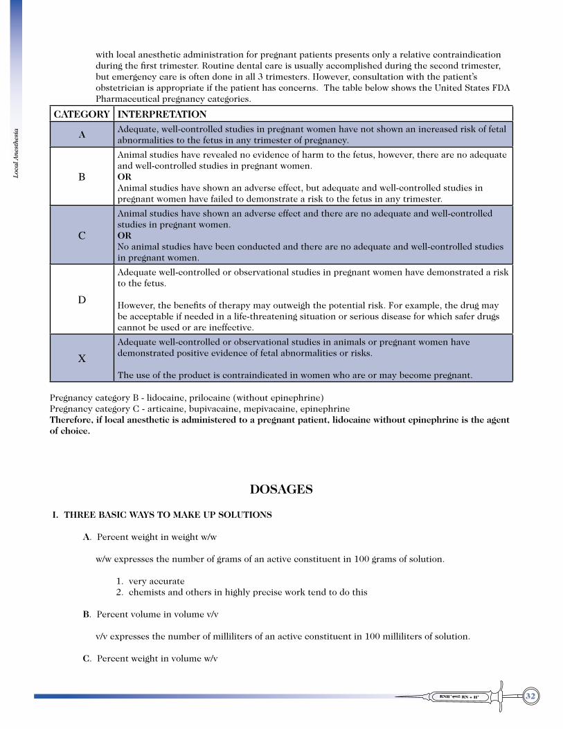
with local anesthetic administration for pregnant patients presents only a relative contraindication during the first trimester. Routine dental care is usually accomplished during the second trimester, but emergency care is often done in all 3 trimesters. However, consultation with the patient’s obstetrician is appropriate if the patient has concerns. The table below shows the United States FDA Pharmaceutical pregnancy categories.
CATEGORY INTERPRETATION
A Adequate, well-controlled studies in pregnant women have not shown an increased risk of fetal abnormalities to the fetus in any trimester of pregnancy.
B
Animal studies have revealed no evidence of harm to the fetus, however, there are no adequate and well-controlled studies in pregnant women. OR Animal studies have shown an adverse effect, but adequate and well-controlled studies in pregnant women have failed to demonstrate a risk to the fetus in any trimester.
C
Animal studies have shown an adverse effect and there are no adequate and well-controlled studies in pregnant women. OR No animal studies have been conducted and there are no adequate and well-controlled studies in pregnant women.
D
Adequate well-controlled or observational studies in pregnant women have demonstrated a risk to the fetus.
However, the benefits of therapy may outweigh the potential risk. For example, the drug may be acceptable if needed in a life-threatening situation or serious disease for which safer drugs cannot be used or are ineffective.
XAdequate well-controlled or observational studies in animals or pregnant women have demonstrated positive evidence of fetal abnormalities or risks.
The use of the product is contraindicated in women who are or may become pregnant.
Pregnancy category B - lidocaine, prilocaine (without epinephrine)Pregnancy category C - articaine, bupivacaine, mepivacaine, epinephrine Therefore, if local anesthetic is administered to a pregnant patient, lidocaine without epinephrine is the agent of choice.
DOSAGES I. THREE BASIC WAYS TO MAKE UP SOLUTIONS A. Percent weight in weight w/w w/w expresses the number of grams of an active constituent in 100 grams of solution. 1. very accurate 2. chemists and others in highly precise work tend to do this B. Percent volume in volume v/v v/v expresses the number of milliliters of an active constituent in 100 milliliters of solution. C. Percent weight in volume w/v
33
Loca
l An
esth
esia
w/v expresses the number of grams of an active constituent in 100 milliliters of solution. (mg./ml.) Used in medicine and dentistry for injectable solutions II. BASIC EXPLANATION OF WEIGHT TO VOLUME A. Basic unit of volume - liter; cube 10 cm/side (The terms milliliter (ml.) and cubic centimeter (cc) are interchangeable). B. Basic unit of weight - kilogram; weight of liter of H2O at 4oC. therefore, 1 ml. of water weighs 1 gram. C. List of prefixes used: micro - one millionth milli - one thousandth centi - one hundredth kilo - times one thousand Examples: 1,000 mcg (micrograms) = 1 mg. (milligram) 1,000 mg. (milligram) = 1 Gm. (gram) 1,000 Gm. (grams) = 1 Kg. (kilogram) 1,000 ml. (milliliters) = 1 L. (liter) 1,000 ml. (cubic centimeters) = 1 L. (liter) E. Concentration of solutions used in medicine and dentistry are expressed two ways: percent and ratio. Percent represents local anesthetic concentration contained in one milliliter!! Ratio represents vasoconstrictor concentration in one milliliter!! Example: Percent - 2% Mepivacaine HCl (Carbocaine) Ratio - 1:100,000 epinephrine Concentration is a statement of the amount of a solute present in a unit volume of solution. 1. 2% solution of Mepivacaine = 2 Gm. = 2000 mg. = 20 mg./ml. 100 ml. 100 ml. 2. epinephrine 1:100,000 = 1 Gm. = 1000 mg. = 1 mg. = .01 mg./ml. 100,000 ml 100,000 ml. 100 ml. F. CLINICAL EXAMPLES 1. If the maximum allowable dosage of Xylocaine is 200 mg., about how many cartridges of 2% Xylocaine
would that represent? a. Find how many milligrams are in each ml. first, then multiply that by l.8 to give you milligrams in
each cartridge. 2 Gm. = 2,000 mg. = 20 mg. in each ml. 100 ml 100 ml. 20 mg / ml x 1.8 ml / cartridge = 36 mg. in each cartridge

34
Loca
l An
esth
esia
b. Since there are 36 mg. in each cartridge, find out how many of these “36 mg. units” are in the 200 mg. max. dosage.
36 mg / cartridge divided into 200 mg = 5 + cartridges
2. If the maximum dosage of epinephrine is 0.2 mg. (as stated by the New York Heart Association) - for a healthy patient, how many cartridges of 2% Xylocaine with 1:100,000 epinephrine does that represent?
Step 1. 1:100,000 = 1 Gm = 1000 mg = 1 mg = 0.01 mg in each ml. 100,000 ml. 100 ml Step 2. 0.01 mg / ml x 1.8 ml / cartridge = 0.018 mg in each cartridge Step 3. 0.018 mg / cartridge divided into 0.2 mg = 11 cartridges However, for a cardiac patient the maximum dose is 0. 04 mg of Epinephrine. This would be .018 mg / cartridge divided into 0. 04 mg or about 2 cartridges 1:100,000 or
about 4 cartridges of 1:200,000 3. Suppose you wished to use 1:50,000 epinephrine how many cartridges could you use not to exceed
0.2 mg of epinephrine? Step 1. 1 Gm = 1,000 mg = 1 mg = 0.02 mg in each ml. 50,000 ml 50,000 ml 50 ml Step 2. .02 mg / ml x 1.8 ml / cartridge = .036 mg. in each cartridge Step 3. .036 mg / cartridge divided into 0.2 mg = 5.5 cartridges 4. How many ml. of a 4 % solution of Articaine to reach a total dosage limit of 200 mg? Step 1. 4 Gm = 4000 mg. = 40 mg. in each ml. 100 ml 100 ml. Step 2 40 mg / ml divided into 200 mg = 5 ml contains 200 mg of Articaine
5. The maximum allowable dosage of lidocaine for pediatric patients at LLU School of Dentistry is 4 mg/kg (2mg/lb). Calculate the maximum dosage and total number of cartridges of 2 % Lidocaine 1:100000 epinephrine that could be safely administered to a 3 year old patient who weighs 18 kg.
Step 1. 4 mg/kg x 18 kg = 72 mg (total allowable dose) Step 2. 2 Gm = 2000 mg = 20 mg/ml x 1.8 ml/cartridge = 36 mg/ml

35
Loca
l An
esth
esia
100 ml 100 ml
Step 3. 72 mg = 2 cartridges (maximum allowable dose) 36 mg/cartridge 6. If all else fails memorize this: By definition: A saturated solution (100%) contains 1 Kg / Liter 1000 gms / 1000 ml 1 gm / ml 1000 mg/ ml All concentrations in a row are equal All concentrations in a column decrease by 10%
Percent Ratio milligrams per ml micrograms per ml100 % 1:1 1000 mg / ml 1,000,000 mcg/ ml
10 % 1:10 100 mg / ml 100,000 mcg/ ml
1% 1:100 10 mg / ml 10,000 mcg/ ml
0.1% 1:1,000 1 mg / ml 1,000 mcg/ ml
0.01% 1:10,000 0.1 mg / ml 100 mcg/ ml
0.001% 1:100,000 0.01 mg / ml 10 mcg/ ml
0.0001% 1:1,000,000 0.001 mg / ml 1 mcg/ ml
Shortcut: Percentage x 10 = mg / ml Ratio x 1,000,000 = mcg / ml Examples: 2.5% x 10 = 25 mg./ml. 1:20,000 x 1,000,000 = 50 mcg / ml 2 % x 10 = 20 mg./ml. 1:50,000 x 1,000,000 = 20 mcg / ml 1 % x 10 = 10 mg./ml. 1:100,000 x 1,000,000 = 10 mcg / ml 3 % x 10 = 30 mg./ml. 1:200,000 x 1,000,000 = 5 mcg / ml
Figures are not available for Neo-Cobefrin but 1:20,000 Neo-Cobefrin may be quite comparable to 1:100,000 Epinephrine.

36
Loca
l An
esth
esia
COMPLICATIONS OF LOCAL ANESTHETIC INJECTIONS
LOCAL
1. Needle Breakage
Cause
needles. Malamed reported 34 instances of litigation involving a broken needle retained in a patient’s tissue. In 33 of these cases the inferior alveolar nerve block injection was being administered using a 30 gauge needle. In another report by Malamed from a needle manufacturer over a 5 year period revealed 27 incidences of needle breakage. In all instances a 30 gauge needle was used.3
thus weakening the needle or from the patient moving unexpectedly during the injection process. Most needles break at the hub, which emphasizes the point of not penetrating to the hub when administering injections unless it is absolutely essential for the particular injection to be successful (i.e., Akinosi). Also, bending the needle should only be done if absolutely necessary and then only one time to avoid weakening the needle.
Prevention
Management
1. Inform the patient 2. Record the exact sequence of events in the chart 3. Refer to a oral and maxillofacial surgeon for evaluation- may be prudent to leave needle
indefinitely 4. Save the remainder of the hub and needle 5. Contact your malpractice insurance carrier immediately 2. Hematoma - Hematoma formation occurs with some frequency during local anesthetic intraoral injections. Cause
Hematoma formation most likely to occur during the posterior superior alveolar nerve block.
occur with any injection)Pterygoid plexus of veins, posterior superior alveolar vessels,
inferior alveolar vessels, and mental vessels. Prevention Understanding the anatomy of the area where the injection is being administered and use atraumatic

37
Loca
l An
esth
esia
technique (i.e., avoid multiple attempts). Aspiration does not prevent hematoma formation; it only indicates where the needle lies at the time of aspiration. A positive aspiration does not imply that a hematoma will result.
Management
3. Infections - extremely rare since the development of the disposable sterile needle
Cause
immunocompromised patients
Prevention
Management
7-10 day course of antibiotics should be initiated. Penicillin would be the first choice of antibiotic
4. Soft Tissue injury
Cause
groups of patients such as children, mentally challenged, or elderly demented patients.
Prevention
Management
5. Sloughing/mucosal irritation Cause
local anesthetic solutions. Prevention

38
Loca
l An
esth
esia
tissues.Management
6. Prolonged anesthesia/paresthesia
Prolonged anesthesia and/or paresthesia is a condition most likely to occur after administration of the inferior alveolar nerve block, but can occur with any block injection. Nerve injury, especially the lingual nerve, is one of the most frequent causes of malpractice litigation.
Based on differences in patient responses to medications, it is not uncommon for some patients to experience prolonged anesthesia following local anesthetic administration. However, anesthesia or paresthesia that persists for several days or longer is not normal. The incidence of nerve injury solely resulting from needle trauma is about 1 % with the majority of cases of nerve damage resulting from surgical procedures. With the small needles used in dentistry, nerve damage is usually limited to a few fibers which results in minor sensory deficits rather than a total severing of the nerve.
Cause
Prevention (Virtually impossible to avoid nerve injury)
immediate vicinity of the nerve Management
usually temporary and most often resolves in 2 months or less but may remain indefinitely.
2 months, referral to an oral surgeon or neurologist who specializes in this type of injury may be appropriate for some patients. It should be understood that resolution of symptoms may take up to a year to resolve. (Microsurgical repair may be an option)
alternate technique for providing local anesthesia.
7. Trismus (Defined as the inability to open the mouth)
Cause
if injected supramuscularly
Prevention
to the medial pterygoid muscle (orienting the needle to medial) during the inferior alveolar nerve block injection

39
Loca
l An
esth
esia
tissues. Make sure that the needle is sharp and is free of imperfections (barbs)
Management (the symptoms of trismus usually manifest anywhere from 1-6 days post injection) Mild
Severe
of antibiotics
8. Facial nerve paralysis (Seventh cranial nerve - motor function to muscles of facial expression) Cause
block injection, results in deposition of anesthetic in the parotid gland. Prevention
Management
9. Unanticipated anesthesia Unanticipated anesthesia is for the most part of no concern, but can be alarming to the patient and
clinician.
Cause
Examples
distributed to the orbit resulting in dizziness, diploplia and temporary blindness.
posterior superior alveolar and maxillary nerve blocks.
Also, anesthesia of the auriculotemporal nerve sometimes occurs after a Gow-Gates injection as well as other inferior alveolar injection techniques.
10. Postanesthetic lesions Cause
traumatic procedure. These lesions usually appear several days after treatment usually around the site
40
Loca
l An
esth
esia
of injection, and are usually associated with pain.
Prevention
Management
SYSTEMIC
Millions of local anesthetic injections are administered in the United States each year. Fortunately, serious morbidity and/or mortality are rarely encountered due to the high dosages that usually must be administered before complications develop. However, this is not the case with the pediatric patient and close attention to dosing is mandatory to avoid toxic doses in a child. However, it must be appreciated that systemic toxic effects can occur with small amounts of local anesthetic. Inadvertent administration of local anesthetic intravascularly results in distribution to all systems of the body which then can interfere with Na+ conductance in all excitable tissues (i.e., CNS and CVS). Systemic toxic effects can usually be grouped into one of the following categories:
1. Psychogenic/anxiety reaction 2. Toxic overdose 3. Allergic reaction 4. Methemoglobinemia
1. Psychogenic/anxiety reaction – (most common - before, during or after injection) Local anesthetic administration (needle insertion) frequently described by patients as the most feared
procedure in dental treatment. Manifestations of psychogenic/anxiety reactions include:
a. Syncope (fainting) - Loss of consciousness is experienced as a direct result of inadequate cerebral perfusion. Most commonly caused by fear and anxiety (emotional stress). However, other causes of syncope are possible and should be considered.
b. Nausea c. Pallor d. Palpitations e. Sweating f. Hyperventilation
Management – Typically easily managed, stop procedure/injection and reassure patient. If patient becomes unconscious, proceed to steps outlined below:
a. Position - supine, feet elevated b. Airway - head tilt, chin lift c. Breathing - administer 100 % O2 d. Circulation - monitor BP and pulse e. Drugs - usually none (aromatic ammonia) 2. Toxic overdose – (second most common)
Systemic toxic effects are manifested when the plasma concentration of the local anesthetic are elevated sufficiently to result in adverse events. There are many factors that can contribute to excessively high plasma levels of local anesthetic and are listed below: a. Age/weight of the patient b. Injection technique c. Medical status of the patient
41
Loca
l An
esth
esia
d. Inadvertent venous/arterial injection e. Properties of local anesthetic (concentration %/presence of vasoconstrictor)
CNS - Very sensitive to the effects of local anesthetics and as the plasma concentration rises; vital areas
of the brain are depressed.
CVS - More resistant to the effects of local anesthetics than the CNS. Fortunately, with most anesthetics (exceptions being bupivacaine and etidocaine), the dose required to produce cardiac arrest experimentally is several times greater then the dose required to produce respiratory arrest, therefore, profound myocardial depression, vascular dilatation and cardiovascular collapse are unlikely to occur as long as the patient’s ventilation is adequately supported.4
Plasma concentration of lidocaine after intraoral injection of 40-160 mgs(1 to 4 cartridges of 2% of lidocaine result in blood levels of approximately 1 mcg/ml (average range of 0.5-2 mcg/ml depending on the individual).3
Plasma concentration of lidocaine after antiarrhythmic dose of 1-1.5mg/kg = 4.5 mcg/ml2(intravascular administration).
< 5 mcg/ml therapeutic (antiarrhythmic, analgesia, anticonvulsant and sedation)
5 -10 mcg/ml lightheadedness, slurred speech, drowsiness, muscle twitching
10-15 mcg/ml Respiratory depression, tonic-clonic seizures (CVS instability)
15-20 mcg/ml Coma, respiratory arrest
>20 (CV collapse) 3. Allergic (see Allergy section in patient evaluation)
True allergic reactions are rare in dentistry today due to the abandonment of ester type local anesthetics. Also, the development of the single dose cartridge eliminated the need for the inclusion of an antimicrobial agent, thus further reducing the potential for allergic type reactions. Patients, however, frequently report allergy to the “caine” anesthetics on their medical history forms. This is very often an erroneous diagnosis (psychogenic reactions often mimic allergic reactions) which is unfortunate as this diagnosis interferes with the delivery of appropriate dental care to the patient. However, antioxidants such as bisulfites are included in local anesthetics containing vasoconstrictors to prevent oxidation and therefore the potential for allergic reactions must be considered in patients with allergies to sulfites. Local anesthetics containing bisulfites are not contraindicated in patients with history of allergy to sulfa drugs.
Manifestations and treatment of allergic type reactions a. Localized skin reaction (hives/urticaria) - usually no treatment necessary, may consider oral
antihistamine (benadryl)
b. Generalized skin reaction – generalized pruritis (itching), hives, rash. Treatment - Antihistamine IM/ (benadryl 50 mg), monitoring of vitals, consultation with
physician (allergist) c. Angioedema - swelling of laryngeal tissues, tongue, lips etc. Results in dyspnea. Treatment - Oxygen, epinephrine IM/SC (adult-0.3 mg, pediatric-0.15 mg) - call 911!
d. Anaphylaxis- Marked airway obstruction, bronchospasm, cardiovascular collapse
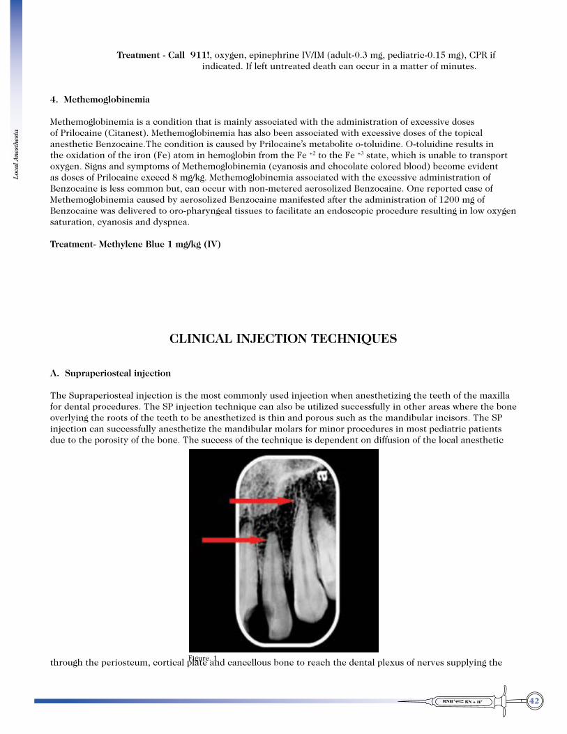
42
Loca
l An
esth
esia
Treatment - Call 911!, oxygen, epinephrine IV/IM (adult-0.3 mg, pediatric-0.15 mg), CPR if indicated. If left untreated death can occur in a matter of minutes.
4. Methemoglobinemia Methemoglobinemia is a condition that is mainly associated with the administration of excessive doses of Prilocaine (Citanest). Methemoglobinemia has also been associated with excessive doses of the topical anesthetic Benzocaine.The condition is caused by Prilocaine’s metabolite o-toluidine. O-toluidine results in the oxidation of the iron (Fe) atom in hemoglobin from the Fe +2 to the Fe +3 state, which is unable to transport oxygen. Signs and symptoms of Methemoglobinemia (cyanosis and chocolate colored blood) become evident as doses of Prilocaine exceed 8 mg/kg. Methemoglobinemia associated with the excessive administration of Benzocaine is less common but, can occur with non-metered aerosolized Benzocaine. One reported case of Methemoglobinemia caused by aerosolized Benzocaine manifested after the administration of 1200 mg of Benzocaine was delivered to oro-pharyngeal tissues to facilitate an endoscopic procedure resulting in low oxygen saturation, cyanosis and dyspnea. Treatment- Methylene Blue 1 mg/kg (IV)
CLINICAL INJECTION TECHNIQUES
A. Supraperiosteal injection
The Supraperiosteal injection is the most commonly used injection when anesthetizing the teeth of the maxilla for dental procedures. The SP injection technique can also be utilized successfully in other areas where the bone overlying the roots of the teeth to be anesthetized is thin and porous such as the mandibular incisors. The SP injection can successfully anesthetize the mandibular molars for minor procedures in most pediatric patients due to the porosity of the bone. The success of the technique is dependent on diffusion of the local anesthetic
through the periosteum, cortical plate and cancellous bone to reach the dental plexus of nerves supplying the Figure. 1

43
Loca
l An
esth
esia
targeted tooth to be anesthetized. The point of injection for the SP injection is in the alveolar mucosa just above the mucogingival line. However, visualizing the root length on the x-ray is recommended to account for patient variation in root length.(Figure. 1)
Technique: 1. The mucous membrane is dried and topical anesthetic placed in the vestibule of the area to be
injected.
2. The tissues are then pulled taut to expose the injection site above the targeted injection area. 3. The needle is inserted through the alveolar mucosa just above the mucogingival line at the height of
the muccobuccal fold with the bevel of the needle oriented toward bone. (Figure. 2)
4. After initial penetration of the needle, pause and deposit a few drops of anesthetic solution and wait a minimum of 5 seconds before advancing the needle.
5. The needle is advanced slowly in small incremental steps parallel to the long axis of the targeted
tooth injecting a few drops of local anesthetic at each step. 6. The depth of injection is usually only a few millimeters. When the tip of the needle approximates the
apical 1/3 of the tooth, the needle should be reoriented horizontally so that the periosteum is lightly contacted. (Figure. 3)
7. Aspiration is performed and ! milliter (1/3 of a cartridge) of local anesthetic is injected slowly over 1-2
minutes so as to avoid discomfort and dispersion of the anesthetic solution. 8. The syringe is then withdrawn and treatment can begin almost immediately, as pulpal and soft tissue
anesthesia is already complete.
MODIFIED APPROACH 1. Use this when routine approach is ineffective due to anatomical variation.
Figure. 2
Figure. 3
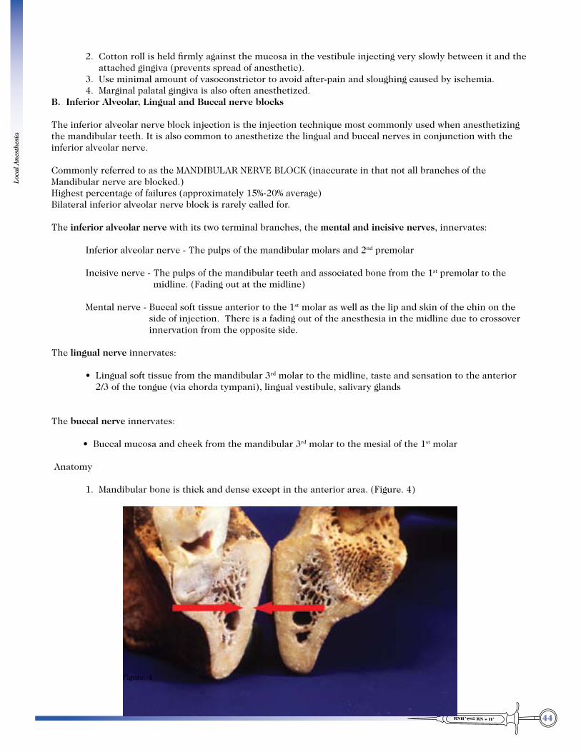
44
Loca
l An
esth
esia
2. Cotton roll is held firmly against the mucosa in the vestibule injecting very slowly between it and the attached gingiva (prevents spread of anesthetic).
3. Use minimal amount of vasoconstrictor to avoid after-pain and sloughing caused by ischemia. 4. Marginal palatal gingiva is also often anesthetized.B. Inferior Alveolar, Lingual and Buccal nerve blocks
The inferior alveolar nerve block injection is the injection technique most commonly used when anesthetizing the mandibular teeth. It is also common to anesthetize the lingual and buccal nerves in conjunction with the inferior alveolar nerve.
Commonly referred to as the MANDIBULAR NERVE BLOCK (inaccurate in that not all branches of the Mandibular nerve are blocked.)Highest percentage of failures (approximately 15%-20% average)Bilateral inferior alveolar nerve block is rarely called for.
The inferior alveolar nerve with its two terminal branches, the mental and incisive nerves, innervates: Inferior alveolar nerve - The pulps of the mandibular molars and 2nd premolar
Incisive nerve - The pulps of the mandibular teeth and associated bone from the 1st premolar to the midline. (Fading out at the midline)
Mental nerve - Buccal soft tissue anterior to the 1st molar as well as the lip and skin of the chin on the side of injection. There is a fading out of the anesthesia in the midline due to crossover innervation from the opposite side.
The lingual nerve innervates:
rd molar to the midline, taste and sensation to the anterior 2/3 of the tongue (via chorda tympani), lingual vestibule, salivary glands
The buccal nerve innervates:
rd molar to the mesial of the 1st molar
Anatomy
1. Mandibular bone is thick and dense except in the anterior area. (Figure. 4)
Figure. 4
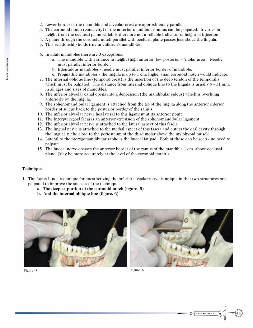
45
Loca
l An
esth
esia
2. Lower border of the mandible and alveolar crest are approximately parallel. 3. The coronoid notch (concavity) of the anterior mandibular ramus can be palpated. It varies in
height from the occlusal plane which is therefore not a reliable indicator of height of injection. 4. A plane through the coronoid notch parallel with occlusal plane passes just above the lingula. 5. This relationship holds true in children's mandibles.
6. In adult mandibles there are 3 exceptions: a. The mandible with variance in height (high anterior, low posterior - (molar area). Needle
must parallel inferior border. b. Edentulous mandibles - needle must parallel inferior border of mandible. c. Prognathic mandibles - the lingula is up to 1 cm. higher than coronoid notch would indicate. 7. The internal oblique line (temporal crest) is the insertion of the deep tendon of the temporalis
which must be palpated. The distance from internal oblique line to the lingula is usually 9 - 11 mm. in all ages and sizes of mandibles.
8. The inferior alveolar canal opens into a depression (the mandibular sulcus) which is overhung anteriorly by the lingula.
9. The sphenomandibular ligament is attached from the tip of the lingula along the anterior inferior border of sulcus back to the posterior border of the ramus.
10. The inferior alveolar nerve lies lateral to this ligament at its anterior point. 11. The Interpterygoid facia is an anterior extension of the sphenomandibular ligament. 12. The inferior alveolar nerve is attached to the lateral aspect of this fascia. 13. The lingual nerve is attached to the medial aspect of this fascia and enters the oral cavity through
the lingual niche close to the periosteum of the third molar above the mylohyoid muscle. 14. Lateral to the pterygomandibular raphe is the buccal fat pad. Both of these can be seen - no need to
palpate. 15. The buccal nerve crosses the anterior border of the ramus of the mandible 1 cm. above occlusal
plane. (May be more accurately at the level of the coronoid notch.)
Technique
1. The Loma Linda technique for anesthetizing the inferior alveolar nerve is unique in that two structures are palpated to improve the success of the technique;
a. The deepest portion of the coronoid notch (figure. 5) b. And the internal oblique line (figure. 6)
Figure. 5 Figure. 6
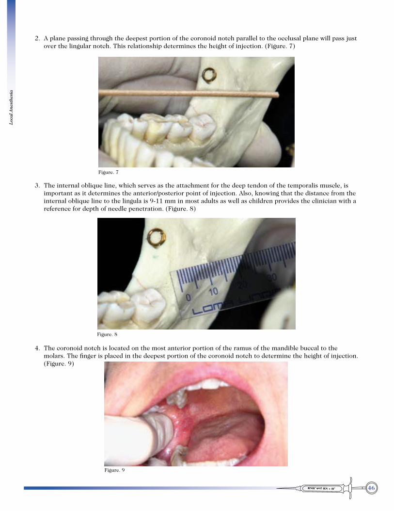
46
Loca
l An
esth
esia
2. A plane passing through the deepest portion of the coronoid notch parallel to the occlusal plane will pass just over the lingular notch. This relationship determines the height of injection. (Figure. 7)
3. The internal oblique line, which serves as the attachment for the deep tendon of the temporalis muscle, is important as it determines the anterior/posterior point of injection. Also, knowing that the distance from the internal oblique line to the lingula is 9-11 mm in most adults as well as children provides the clinician with a reference for depth of needle penetration. (Figure. 8)
4. The coronoid notch is located on the most anterior portion of the ramus of the mandible buccal to the molars. The finger is placed in the deepest portion of the coronoid notch to determine the height of injection. (Figure. 9)
Figure. 7
Figure. 8
Figure. 9
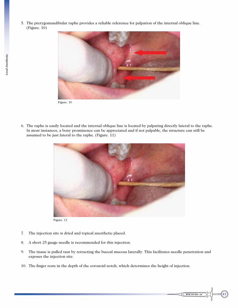
47
Loca
l An
esth
esia
5. The pterygomandibular raphe provides a reliable reference for palpation of the internal oblique line. (Figure. 10)
6. The raphe is easily located and the internal oblique line is located by palpating directly lateral to the raphe. In most instances, a bony prominence can be appreciated and if not palpable, the structure can still be assumed to be just lateral to the raphe. (Figure. 11)
7. The injection site is dried and topical anesthetic placed.
8. A short 25 gauge needle is recommended for this injection.
9. The tissue is pulled taut by retracting the buccal mucosa laterally. This facilitates needle penetration and exposes the injection site.
10. The finger rests in the depth of the coronoid notch, which determines the height of injection.
Figure. 10
Figure. 11

48
Loca
l An
esth
esia
11. With the barrel of the syringe positioned from the contralateral premolar region, the needle is inserted lateral and slightly posterior to the raphe. (Figure. 12)
12. After penetration of the tissue, pause and deposit a few drops of local anesthetic and wait 5-10 seconds before advancing the needle.
13. Contact is made with the medial surface of the ramus of the mandible medial just posterior to the internal oblique line, but anterior to the lingula. (Figure. 13-A, 13-B)
14. From this point it is reassuring to know that the lingula is approximately 10 mm from this point. Contact is maintained with the medial surface of the ramus as the needle is incrementally advanced toward the lingula (Figure. 14). To advance the needle after contact with the ramus, the needle is withdrawn 1 mm and the barrel of the syringe is moved slightly toward the midline and the needle advanced until contact is again made with ramus. (Figure. 15)
Figure. 12
Figure. 13-A Figure. 13-B
Figure. 14 Figure. 15

49
Loca
l An
esth
esia
15. After advancing the needle to a point halfway (a depth of 5mm) between the internal oblique line and the lingula, aspiration is performed and ! cc of local anesthetic is injected for the lingual nerve.
16. The needle is then advanced posteriorly toward the lingula maintaining contact at all times with the ramus.
17. The needle loses contact with the ramus as the needle slides over the lingular notch into the mandibular sulcus. Aspiration is performed and " to 1 cc of local anesthetic is deposited slowly over one minute (Figure. 16-A, 16-B)
18. Altered sensation of the lip and chin can be expected in 30-60 seconds and profound anesthesia will be
obtained in 3-5 minutes. The effectiveness of the inferior alveolar block can be confirmed by insertion of an explorer into the gingival papilla between the cuspid and the 1st premolar. The absence of pain confirms effectiveness of the block.
19. Buccal nerve block is accomplished by injecting 1 cm above the occlusal plane midway between the anterior border of the ramus and the internal oblique line. The depth of injection is only a few millimeters before periosteum is contacted. Aspiration is performed and 1/8th - 1/4 milliliter of anesthetic is deposited. (Figure. 17)
Summary: The puncture point is:
a. at the vertical level of the coronoid notch. b. just medial to the deep tendon of temporalis. c. lateral to the pterygomandibular raphe. d. medial and posterior to the buccal fat pad.
Figure. 16-A Figure. 16-B
Figure. 17

50
Loca
l An
esth
esia
The needle will penetrate:
a. oral mucosa, b. glandular tissue (containing minor salivary glands) and c. the buccinator muscle before touching periosteum. (Not a muscle of mastication).
Try to visualize the final position and location of the needle:
a. Just medial to deep tendon of the temporalis b. Just lateral to the lingual nerve (thus avoiding injury to it) c. Just above the lingular notch (ideal position) d. Just lateral to the inferior alveolar nerve. e. If the needle is slightly below the lingular notch it will slide past the medial side of the lingula, through the sphenomandibular ligament and be in the sulcus on the medial side of the
inferior alveolar nerve. d. Tip is in the pterygomandibular space Lateral wall = ramus of mandible Medial wall = medial pterygoid muscle Roof = lateral pterygoid muscle
Bent needle technique
Some of us find it advantageous to bend the needle slightly (20o - 30o) toward the bevel. This makes it easier to contact the periosteum without "fighting" the contralateral corner of the mouth with the barrel of the syringe. It is especially helpful if the ramus of the mandible flares laterally or the patient has a small oral aperture (opening). Some say it may contribute to needle breakage to bend the needle. The manufacturer, because of medico-legal reasons, will not say it is O.K. to bend them.
Direct thrust technique- not recommended
1. Palpate coronoid notch to determine height 2. Place syringe over contralateral premolars 3. Insert needle lateral to raphe 4. Advance to floor of mandibular sulcus, deposit 1 - 1.5 ml. 5. Block lingual nerve with 0.5 ml. as needle is withdrawn.
Four major disadvantages:
1. More painful 2. Less accurate 3. May penetrate and injure lingual or inferior alveolar nerves 4. May be too far back or medial if mandible flares much
Buccal Nerve Block - 3 Methods:
1. Inject 1 cm. above occlusal plane 3 - 4 mm. medial to anterior border of the ramus of the mandible between superficial and deep tendons of temporalis. (Sloman's Technique). This technique may also anesthetize aberrant nerve fibers to the molar teeth pulps.
2. In buccal vestibule opposite molars. (near mucogingival junction) this will give best blanching of buccal gingiva.)
3. Submucosally 1 cm. below parotid duct progressing distally. This technique is seldom used.

51
Loca
l An
esth
esia
POSTERIOR SUPERIOR ALVEOLAR NERVE BLOCK ANATOMY
Infratemporal Fossa
Infratemporal fossa - the open space behind the maxilla (an arbitrary space)
Boundaries: Superior - infratemporal surface of greater wing of sphenoid Anterior - posterior (infratemporal) surface of maxilla Medial - lateral pterygoid lamina (plate) Lateral - zygomatic arch and ramus of mandible Posterior - articular tubercle of temporal bone and spine of sphenoid
Communicates with: Temporal fossa - via wide opening deep to zygomatic arch Orbit - via inferior orbital fissure Pterygopalatine fossa - via pterygomaxillary fissure Cranial cavity - via foramen ovale and foramen spinosum Pterygomandibular space - via open inferior border
Contents: Maxillary nerve and posterior superior alveolar nerves Lateral pterygoid muscle Sphenomandibular ligament Interpterygoid fascia Maxillary artery (1st and 2nd parts) with branches Pterygoid venous plexus Communicating chorda tympani nerve
Otic ganglion - on medial surface of mandibular nerve just below foramen ovale. Mandibular nerve with branches:
a. Above lateral pterygoid muscle 1) Auriculotemporal - - behind neck of condyle, ant. to ear. 2) Masseteric 3) Posterior deep temporal
b. Between two heads of lateral pterygoid muscle 1) Anterior deep temporal 2) Buccal - crosses external oblique line 1 cm above occlusal plane (where it can be
blocked) c. Below (medial to) lateral pterygoid 1) Lingual 2) Inferior alveolar

52
Loca
l An
esth
esia
Foramina: Foramen ovale - 1. Mandibular nerve with 2. Accessory meningeal artery Foramen spinosum - 1. Middle meningeal artery 2. Meningeal branch of mandibular nerve Posterior superior alveolar foramina - Posterior superior alveolar nerves.

53
Loca
l An
esth
esia
C. Posterior superior alveolar nerve block (PSA, Tuberosity) Purpose and Indications
1. Provides profound anesthesia of pulpal and buccal soft tissues of the 3 maxillary molar teeth except the mesiobuccal root of the first molar.
2. Effective when supraperiosteal may fail.3. To avoid areas of infection or inflammation.

54
Loca
l An
esth
esia
Morphology
The target area for the PSA injection is the PSA foramina located on the posterior surface of the maxilla. The foramina are located midway between the upper and lower posterior part of the maxilla on the posterolateral aspect. (In the infratemporal fossa). (Figure. 18)
Maxillary height may vary from 27 to 66 mm on adult skulls.
The anterior height is the same as the posterior height of each maxilla. The anterior height can be determined by measuring the distance from the infraorbital margin to the gingiva of the bicuspids.
Technique1. The landmarks for the PSA injection are the zygomatic process and the maxillary 2nd molar, if present. The point of needle penetration will be distal to the zygomatic process and above the maxillary 2nd molar.
(Figure. 19)
Figure. 18
Figure. 19
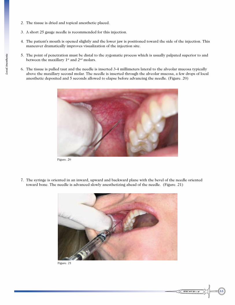
55
Loca
l An
esth
esia
2. The tissue is dried and topical anesthetic placed. 3. A short 25 gauge needle is recommended for this injection. 4. The patient’s mouth is opened slightly and the lower jaw is positioned toward the side of the injection. This
maneuver dramatically improves visualization of the injection site. 5. The point of penetration must be distal to the zygomatic process which is usually palpated superior to and
between the maxillary 1st and 2nd molars. 6. The tissue is pulled taut and the needle is inserted 3-4 millimeters lateral to the alveolar mucosa typically
above the maxillary second molar. The needle is inserted through the alveolar mucosa, a few drops of local anesthetic deposited and 5 seconds allowed to elapse before advancing the needle. (Figure. 20)
7. The syringe is oriented in an inward, upward and backward plane with the bevel of the needle oriented toward bone. The needle is advanced slowly anesthetizing ahead of the needle. (Figure. 21)
Figure. 20
Figure. 21

56
Loca
l An
esth
esia
8. Periosteum is soon contacted and the needle is advanced carefully, maintaining contact with the periosteum. Maintaining contact with the periosteum all but eliminates the possibility of damage to the pterygoid plexus of veins and the lateral pterygoid muscle. As the foramina are approached which corresponds to a depth of about 20 mm, the tip of the needle will lose contact with the posterior surface of the maxilla as it curves medially. The shaft of the needle however, maintains contact with bone and gives a different tactile sense. The tip of the needle is now in close proximity to the PSA foramina. (Figure. 22-A, 22-B)
9. Aspiration is performed and " to 1 ml of local anesthetic solution is injected slowly over 1-2 minutes. (For the median maxillary height, the angulation of the needle relative to both the occlusal and sagittal planes is 45o.) In a high maxilla, the vertical angulation of the needle is greater, up to 70o. In the low maxilla of some adults and most children, the angulation lies between 20 and 30 degrees from the occlusal plane.
THE MAIN PRINCIPLE IS TO KEEP THE NEEDLE CONTACTING PERIOSTEUM!
CAUTIONS
1. Angulation is important – misdirection of the needle tip can cause injury.2. Penetrating too deep (high and posteriorly) may injure the maxillary artery. A HEMATOMA could result. If
so, treat with pressure and ice.3. Penetrating too far laterally may injure pterygoid plexus of veins, or lateral pterygoid muscle (pain and
trismus).4. Anesthesia is often incomplete in first molar due to mesiobuccal root being supplied by the middle superior
alveolar nerve. Supraperiosteal injection, anterior to zygomatic process or infraorbital block will be needed.
Bent-Needle Technique
Advantages:
1. Needle is inserted to a measured depth2. Depth of penetration into tissues is less.
Disadvantages:
1. Needle must be bent sharply - approaching 90o.2. Have to measure height of maxilla.3. 2 needles needed if injecting both left and right sides to have bevel toward bone
Figure. 22-A Figure. 22-B

57
Loca
l An
esth
esia
Technique:
1. Measure distance from infraorbital rim to gingival margin of 2nd bicuspid.2. Bend a 25 ga. needle at half of this measured distance from the point. Bend to nearly a right angle so that the
bevel is toward the bone as the bent portion points upward for the injection.3. The puncture point is high in the vestibule opposite the 3rd molar or posterolateral aspect of maxillary
tuberosity.4. The needle is advanced in a direction almost parallel to the long axis of the 2nd molar. The needle point must
be touching periosteum when the bend in the needle is level with the gingival margin of the second molar. Aspirate, inject 1 ml.
PALATINE NERVE BLOCK ANATOMY
Pterygopalatine Fossa and its contents
Definition:
1. The vertical triangular gap between the pterygoid process of the sphenoid bone & the infratemporal surface of the maxilla.
2. Distribution center for maxillary nerve.
Boundaries:
Medial – vertical lamina of the palatine bone Lateral – pterygomaxillary fissure. Superior – body of sphenoid, orbital process of palatine. Anterior – Maxilla (infratemporal surface) (same as infratemporal fossa.) Posterior – pterygoid process of sphenoid. Inferior – apex of the triangle – Pterygomaxillary junction. greater palatine canal
Communicates with: ----------- via------------------------------- which transmits-------------
Cranial cavity foramen rotundum (directly) Maxillary Nerve Pterygoid canal (indirectly) Nerve & vessels of pterygoid canal.
Infratemporal fossa Pterygomaxillary fissure Maxillary A & N Posterior Superior Alveolar. N.
Nasal cavity Sphenopalatine foramen Nasopalatine N. Posterior. Superior lateral nasal Ns. Sphenopalatine vessels.
Orbit Inf. Orbital fissure Infraorbital N. & vessels.
Nasopharynx Pharyngeal canal Pharyngeal N. & vessels.
Oral cavity Greater palatine canal Greater & lesser (Pterygopalatine canal) palatine Ns. Descending palatine A.

58
Loca
l An
esth
esia
Contents:
Maxillary Nerve with Posterior Superior Alveolar branches
Maxillary Artery (3rd Part) with branches & corresponding veins
Sphenopalatine Ganglion & its branches D. The Greater Palatine nerve block (also called Anterior Palatine)
Purpose: The Greater Palatine Nerve Block injection anesthetizes unilateral palatal gingiva and bone from the
maxillary 3rd molar to cuspid and extends to the midpalatal suture. It is not supposed to anesthetize pulps of teeth, but clinically it sometimes completes pulpal anesthesia.
Anatomy: The greater palatine foramen is typically located lingual to the maxillary 3rd molar. The injection target
is anterior to the greater palatine foramen in an attempt to minimize anesthesia of the soft palate which can be disconcerting to the patient. This method generally does not anesthetize the middle and posterior palatine nerves which supply the soft palate. (Figure. 23)
Target: The point of injection for the greater palatine nerve block is just distal to the maxillary 1st molar and at a
point where the palate curves upward toward the teeth. This effectively anesthetizes the greater palatine nerve as it travels anteriorly in the funnel like depression in the hard palate. (Figure. 24)
Figure. 23
Figure. 24

59
Loca
l An
esth
esia
Technique:
1. A 30 gauge needle is acceptable for this injection. 2. Pressure anesthesia is applied using a cotton tip applicator or other similar device. The needle is
inserted into the blanched tissue adjacent to the applicator. (Figure. 25)
3. After initial penetration into the tissue, a few drops of local anesthetic are deposited and a sufficient amount of time is allowed to elapse before advancement of the needle. The needle should be advanced slowly in increments of 1mm, anesthetizing ahead of the needle.
4. There will be considerable resistance to deposition of the anesthetic in the fibrous tissues of the palate. However, once this tissue layer is traversed, the needle will lie in loose connective tissue and there will be little resistance to local anesthetic flow.
5. Aspiration is performed and ! ml of local anesthetic is deposited slowly. (Figure. 26)
E. Nasopalatine nerve block
Purpose: The nasopalatine nerve block injection anesthetizes bilateral palatal gingiva and bone from cuspid
to cuspid. The nasopalatine nerve block can be used to supplement pulpal anesthesia in difficult to anesthetize maxillary anterior teeth.
Figure. 25
Figure. 26
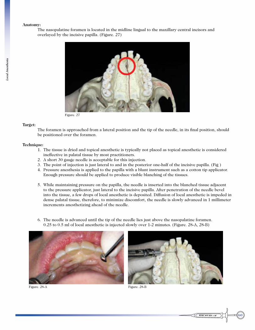
60
Loca
l An
esth
esia
Anatomy: The nasopalatine foramen is located in the midline lingual to the maxillary central incisors and
overlayed by the incisive papilla. (Figure. 27)
Target: The foramen is approached from a lateral position and the tip of the needle, in its final position, should
be positioned over the foramen.
Technique: 1. The tissue is dried and topical anesthetic is typically not placed as topical anesthetic is considered
ineffective in palatal tissue by most practitioners. 2. A short 30 gauge needle is acceptable for this injection. 3. The point of injection is just lateral to and in the posterior one-half of the incisive papilla. (Fig ) 4. Pressure anesthesia is applied to the papilla with a blunt instrument such as a cotton tip applicator.
Enough pressure should be applied to produce visible blanching of the tissues.
5. While maintaining pressure on the papilla, the needle is inserted into the blanched tissue adjacent to the pressure applicator, just lateral to the incisive papilla. After penetration of the needle bevel into the tissue, a few drops of local anesthetic is deposited. Diffusion of local anesthetic is impeded in dense palatal tissue, therefore, to minimize discomfort, the needle is slowly advanced in 1 millimeter increments anesthetizing ahead of the needle.
6. The needle is advanced until the tip of the needle lies just above the nasopalatine foramen. 0.25 to 0.5 ml of local anesthetic is injected slowly over 1-2 minutes. (Figure. 28-A, 28-B)
Figure. 27
Figure. 28-A Figure. 28-B
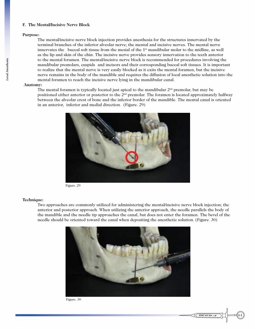
61
Loca
l An
esth
esia
F. The Mental/Incisive Nerve Block
Purpose: The mental/incisive nerve block injection provides anesthesia for the structures innervated by the
terminal branches of the inferior alveolar nerve; the mental and incisive nerves. The mental nerve innervates the buccal soft tissue from the mesial of the 1st mandibular molar to the midline, as well as the lip and skin of the chin. The incisive nerve provides sensory innervation to the teeth anterior to the mental foramen. The mental/incisive nerve block is recommended for procedures involving the mandibular premolars, cuspids and incisors and their corresponding buccal soft tissues. It is important to realize that the mental nerve is very easily blocked as it exits the mental foramen, but the incisive nerve remains in the body of the mandible and requires the diffusion of local anesthetic solution into the mental foramen to reach the incisive nerve lying in the mandibular canal.
Anatomy: The mental foramen is typically located just apical to the mandibular 2nd premolar, but may be
positioned either anterior or posterior to the 2nd premolar. The foramen is located approximately halfway between the alveolar crest of bone and the inferior border of the mandible. The mental canal is oriented in an anterior, inferior and medial direction. (Figure. 29)
Technique: Two approaches are commonly utilized for administering the mental/incisive nerve block injection; the
anterior and posterior approach. When utilizing the anterior approach, the needle parallels the body of the mandible and the needle tip approaches the canal, but does not enter the foramen. The bevel of the needle should be oriented toward the canal when depositing the anesthetic solution. (Figure. 30)
Figure. 29
Figure. 30
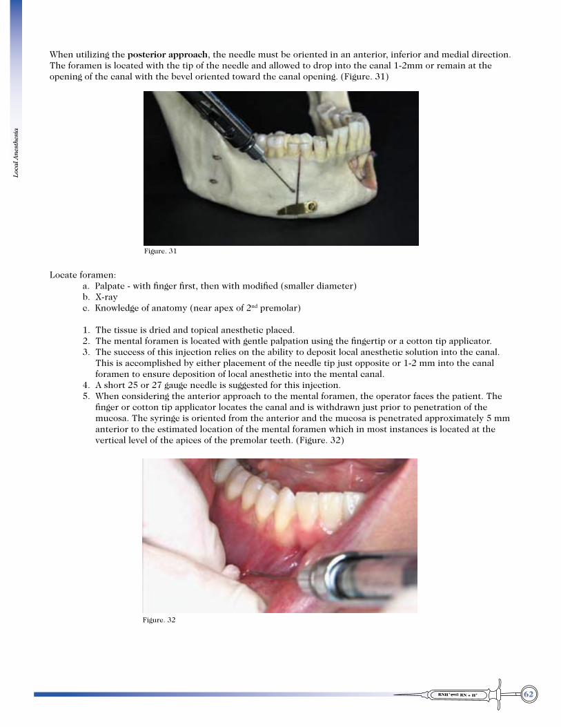
62
Loca
l An
esth
esia
When utilizing the posterior approach, the needle must be oriented in an anterior, inferior and medial direction. The foramen is located with the tip of the needle and allowed to drop into the canal 1-2mm or remain at the opening of the canal with the bevel oriented toward the canal opening. (Figure. 31)
Locate foramen: a. Palpate - with finger first, then with modified (smaller diameter) b. X-ray c. Knowledge of anatomy (near apex of 2nd premolar)
1. The tissue is dried and topical anesthetic placed. 2. The mental foramen is located with gentle palpation using the fingertip or a cotton tip applicator. 3. The success of this injection relies on the ability to deposit local anesthetic solution into the canal.
This is accomplished by either placement of the needle tip just opposite or 1-2 mm into the canal foramen to ensure deposition of local anesthetic into the mental canal.
4. A short 25 or 27 gauge needle is suggested for this injection. 5. When considering the anterior approach to the mental foramen, the operator faces the patient. The
finger or cotton tip applicator locates the canal and is withdrawn just prior to penetration of the mucosa. The syringe is oriented from the anterior and the mucosa is penetrated approximately 5 mm anterior to the estimated location of the mental foramen which in most instances is located at the vertical level of the apices of the premolar teeth. (Figure. 32)
Figure. 31
Figure. 32

63
Loca
l An
esth
esia
6. The tip of the needle is advanced toward the foramen and when positioned directly opposite the foramen opening, aspiration is performed and ! ml of anesthetic is deposited. (Figure. 33)
7. The needle is withdrawn and pressure is placed around the foramen to facilitate diffusion of local anesthetic into the canal and to prevent dispersion of anesthetic into the surrounding tissues. (Figure. 34)
8. In the posterior approach, the operator position is behind the patient. The canal is located in a similar fashion and the tissue pulled taut in a lateral and inferior direction. The needle is oriented so that it points downward, forward and medially to facilitate entry into the foramen.
9. After initial penetration of the tissue, a few drops of anesthetic are deposited and a minimum of 5 seconds should elapse before advancement of the needle.
Figure. 33
Figure. 34

64
Loca
l An
esth
esia
10. The needle is then directed toward the foramen, anesthetizing ahead of the needle. The needle is advanced until the tip of the needle lies at the orifice or just slightly into the foramen. Aspiration is performed and ! ml of local anesthetic is deposited into the canal. The onset of anesthesia is rapid and profound and the planned procedure can be initiated almost immediately. The success of this block approaches 100%. (Figure. 35)
G. Anterior Superior Alveolar “Field Block”
Purpose: The anterior superior alveolar field block injection, also called the ASA nerve block injection, is used
when multiple dental procedures are planned on maxillary anterior teeth as well as their associated buccal hard and soft tissues. The efficacy of this injection technique lies in the convergence of the nerves that innervate these teeth. Therefore, precise deposition of anesthetic in this region provides local anesthesia to these three teeth and their surrounding structures. However, it is important to realize that there may be cross-over nerve innervation to the central incisor from the contralateral anterior superior alveolar nerve. Therefore, a supplemental supraperiosteal injection should be considered at the apex of the central or a bilateral ASA field block should be given to complete anesthesia.
Structures anesthetized: 1. Pulps of the maxillary central, lateral and canine (premolars and MB root of 1st molar in some
cases) 2. Buccal hard/soft tissue of these same teeth 3. Lower eyelid, lateral aspect of the nose and upper lipAnatomy: The maxillary sinus posteriorly and nasal aperture medially, form anatomical barriers which result in
the convergence of these nerve fibers near the apex of the maxillary canine. Therefore, local anesthetic solution placed just medial and superior to the apex of the maxillary canine will result in anesthesia of the maxillary anterior teeth on that side. (Figure. 36)
Figure. 35
Figure. 36

65
Loca
l An
esth
esia
Technique: 1. A short 25 or 27 gauge needle is recommended for this injection. 2. The tissue is dried and topical anesthetic placed. 3. The lip is retracted and the tissue pulled taut over the maxillary canine to be anesthetized. 4. The point of needle penetration is high in the vestibule just above and medial to the apex of the
maxillary canine. After initial penetration of the alveolar mucosa, a few drops of local anesthetic are deposited and 5 seconds allowed to elapse before advancing the needle. (Figure. 37)
5. The needle is advanced incrementally toward the medial aspect of the canine apex, depositing local anesthetic ahead of the needle. Periosteum will be contacted after a few millimeters. Aspiration is performed, and 1 milliliter of local anesthetic solution is deposited slowly over 1-2 minutes. (Figure. 38)
6. Supplemental local anesthetic should also be considered for the maxillary central incisor if unilateral ASA “field block” injection was administered. Conversely, if bilateral ASA “field block” injection was administered, supplementation is not necessary.
Figure. 37
Figure. 38
66
Loca
l An
esth
esia
SUPPLEMENTAL INJECTIONS
It is not uncommon for different techniques and/or supplementation to conventional injection techniques to be necessary. Difficulty in anesthetizing a particular tooth due to pulpal inflammation or a short procedure on a single tooth where prolonged soft tissue anesthesia would be undesirable are examples of these type of scenario. The four supplemental injections that are most useful are: 1. Intraosseous 2. Periodontal ligament (PDL) 3. Intraseptal 4. Intrapulpal
The intraosseous, PDL and the intraseptal techniques all rely on local anesthetic either diffusing or being deposited into the cancellous bone surrounding the tooth or teeth to be treated which effectively anesthetizes the dental plexus of nerves innervating these teeth. When using these techniques, it must be appreciated that only short procedures should be considered as the duration of pulpal anesthesia is short.
1. Intraosseous Intraosseous anesthesia has been around for more than 100 years. This technique, until the 1990’s,
involved perforation of the cortical bone with a small round bur and then inserting a needle through this perforation into the cancellous bone. Today however, intraosseous systems such as stabident and X-tip, use a perforator that is placed in a slow speed contra-angle handpiece which penetrates the cortical plate of bone. This is followed by delivery of anesthetic through the small perforation with an ultrashort 27 gauge needle.
Indications: 1. Failed anesthesia (i.e., “hot tooth”) 2. Can be used when pulpal anesthesia is desired without accompanying soft tissue anesthesia 3. Short procedures on 1 or 2 teeth 4. As an alternative to bilateral inferior alveolar nerve block 5. Use in areas of thick cortical bone
Disadvantages: 1. Inadequate interseptal bone between teeth 2. Special equipment/cost ( $1/delivery) 3. Inability to find “pilot hole” with needle 4. Short duration of anesthesia 5. Pronounced tachycardia if vasoconstrictor used
Technique: 1. Ideally the injection site should be just distal to the tooth being treated 2. Place topical (???) and then inject a minimal amount of anesthetic to anesthetize the gingiva 3. Penetrate the tissue (2-3 mm below the facial gingival sulcus) with the pilot perforator until bone is
contacted. Carefully activate handpiece and penetrate cortical plate of bone and resistance is lost. 4. Insert 27 gauge needle into pilot hole and administer no more than 1/3 to 1/2 cartridge slowly 2. Periodontal Ligament Injection (PDL)
The PDL injection is a technique that has been utilized since the early 1900’s. Since the introduction of the pressure syringes (Peripress, Ligmaject) in the 1980’s, the technique has gained in popularity. However, complications are more likely with these syringes as extremely high pressures and excessive volumes can be delivered. May be used for any tooth but may be more effective where the PDL space is wide such as: recently erupted teeth or those with vertical bone loss due to chronic causes. Example: extracting mandibular bicuspids for young orthodontic patients.

67
Loca
l An
esth
esia
Indications:
Technique: 1. Preliminary supraperiosteal injection is optional. 2. A 27 ga needle penetrates gingival sulcus into the periodontal ligament space with the general
direction of the needle parallel with the long axis of the tooth and the bevel toward the tooth. (separate injection for each root on multi-rooted teeth)
3. Advance the needle to the depth of the sulcus where resistance will be felt 4. Slowly deposit 0.2 ml of local anesthetic. Considerable resistance should be felt as the solution is
expressed. If this is not the case, then the needle should be redirected and another attempt made. There should be minimal solution oozing out into the sulcus which would indicate poor needle placement.
5. The onset of anesthesia is very rapid and the procedure can begin almost immediately. 3. Interdental (Intraseptal)
The Intraseptal injection technique is very similar to the PDL in principle, the difference being the site of administration
Indications:
Technique: 1. A short 27 gauge needle is inserted into the center and 2 mm below the apex of the papilla adjacent
to the tooth to be anesthetized. 2. The needle should be angled 45 degrees to the long axis of the tooth and perpendicular to the frontal
plane. 3. After initial penetration of the tissue a few drops of local anesthetic are deposited before advancing
the needle. 4. The needle is then advanced until bone is contacted. The needle is advanced another 1-2 mm into
the interseptal bone. 5. 0.2 ml of local anesthetic is deposited slowly. As with the PDL injection, considerable resistance to
injection must be encountered.
4. Intrapulpal
Indications:
Technique: 1. A small 30 gauge needle is suggested 2. A small exposure into the pulp chamber is ideal as injected local anesthetic is more likely to flow
apically. 3. Once the needle is introduced into the pulp chamber, a few drops of local anesthetic are expressed
slowly into the pulp. After waiting a few seconds, 0.2-0.4 ml of local anesthetic can be administered and treatment can commence immediately.

68
Loca
l An
esth
esia
MISCELLANEOUS INJECTIONS
A. Middle superior alveolar nerve block "field block"
The middle superior alveolar nerve is present in only about 20% of the population; therefore, this nerve block has only limited usefulness. However, when the infraorbital nerve block fails to achieve pulpal anesthesia distal to the maxillary canine, the MSA nerve block is indicated for procedures on the premolars and for the mesiobuccal root of the maxillary first molar. This technique has a high success rate.Indications:
molar.
Technique: 1. 25-gauge short needle recommended. (27 ga. is O.K.). 2. Area of insertion: height of mucobuccal fold above maxillary second premolar. 3. Target area: maxillary bone superior to apex of maxillary second premolar. 4. Landmarks: mucobuccal fold above maxillary second premolar. 5. Orientation of bevel: facing toward bone. 6. Pull upper lip to make tissues taut and to gain visibility. 7. Penetrate mucous membrane and slowly advance needle until tip is located above second premolar
against periosteum. 8. Slowly deposit 1 to 1.25 ml. of solution. 9. Wait approximately 2 to 3 min. before commencing dental treatment. 10. Few symptoms: patient has difficulty determining extent of anesthesia: signs: no pain during
dental therapy.
B. Incisive Fossa Block
Technique: 1. The operator sits or stands behind the patient. 2. The efficacy of this injection relates to the small nutrient canals found in the cortical bone in the
bottom of the incisive fossa. The puncture point is in depth of the vestibule between the central and lateral incisor.
3. By depositing the solution in this region slowly and not in excess of 1 ml, pulpal and surgical anesthesia of the incisors usually is obtained.
4. If the lingual mucosa requires anesthesia, a small amount of the solution may be injected at the mucogingival junction.
5. Caution! Injections should never be made at the fornix of the lingual vestibule. The needle may enter into the sublingual gland and an infection may result.
6. (If the labial bone is thin, the supraperiosteal injection will usually provide pulpal anesthesia of the 6 mandibular anterior teeth).
C. Infraorbital nerve block injection
The infraorbital nerve and its branches provide pulpal innervation of the maxillary central, lateral, canine, premolars and MB root of the maxillary 1st molar and their associated buccal hard and soft tissues. In a majority of cases, the middle superior alveolar nerve is absent and innervation of the premolars and MB root of the 1st molar is more than likely provided by a branch of the ASA or PSA nerve. If innervation is derived from the PSA nerve, anesthesia will not be appreciated in the premolars and the MB root of the 1st molar. The infraorbital nerve block injection should be considered for longer, more complex procedures in this region and when infection and inflammation preclude the supraperiosteal injection technique. This injection technique is also useful when the supraperiosteal injection has failed due to dense maxillary bone.
69
Loca
l An
esth
esia
Technique: 1. The infraorbital foramen lies about 1 cm below the infraorbital rim and directly below the suture
line between the maxillary and zygomatic bone. This suture line can be palpated as a roughness on the infraorbital rim.
2. To prevent premature contact with bone the syringe is oriented parallel to the long axis of the 2nd bicuspid and 5-10 mm lateral to the alveolar mucosa.
3. The tissue is dried and topical anesthetic placed. 4. A 25 gauge long needle is recommended for this injection. 5. The suture between the zygomatic and maxillary bone is palpated in reference to a vertical line 1 to
4 mm medial to the pupil of the eye with the patient staring directly ahead. 6. The finger is then used to palpate the infraorbital foramen 1 cm below the infraorbital rim, which is
felt as a depression. The bulb of the index finger remains on the foramen and serves as a target for needle placement.
7. The thumb is used to retract the lip and cheek laterally. The syringe is oriented parallel with the axis of the 2nd bicuspid tooth and the barrel of the syringe lies gently against the lower lip. The needle penetrates the mucosa about 5-10 mm lateral to the alveolar mucosa in the height of the vestibule opposite the bicuspid teeth.
8. After needle penetration, a few drops of anesthetic are deposited and 5 seconds are allowed to elapse before advancing the needle.
9. Keeping the barrel of the syringe against the lower lip, the needle is advanced incrementally toward the foramen, using the index finger as a guide. It is important to remember that the lip and cheek must be continually retracted outward and upward to avoid inadvertent penetration of these tissues.
10. The needle is slowly advanced, anesthetizing ahead of the needle. 11. As the needle approaches the target area, local anesthetic is deposited slowly to anesthetize the
periosteum surrounding the foramen. The welling up of the tissue should be felt under the index finger.
12. After waiting 5-10 seconds, the needle is advanced gently toward the foramen. With proper orientation of the syringe, the needle will not be able to advance into the foramen and will contact the infraorbital rim overlying the foramen. Aspiration is performed and ! to " ml of local anesthetic is slowly deposited over 1 minute. If the needle is properly placed, the anesthetic solution will readily flow into the infraorbital canal without causing a welling up of the tissues surrounding the canal.
13. Paralysis of the inferior rectus muscle of the eye may occasionally occur temporarily while the area is anesthetized. This may cause diplopia (double vision) or other visual and sensory changes.
D. Akinosi nerve block
The Akinosi injection technique is one of the methods available for anesthetizing the inferior alveolar nerve. The Akinosi technique is unique in several ways: the maxillary teeth are used as a guide for syringe orientation, needle contact with bone is not necessary, and the patient’s mouth is closed throughout the procedure. The final position of the tip of the needle is in the mid to upper portion of the pterygomandibular space and in close relation to the mandibular nerve. The inferior alveolar, lingual, mylohyoid and in some instances the buccal nerve are anesthetized with this block. This technique is useful in anxious patients, patients with limited mouth opening secondary to trismus or trauma, and for the patient who is unable to maintain mouth opening for the conventional and Gow-Gates technique for anesthetizing the inferior alveolar nerve.

70
Loca
l An
esth
esia
Anatomy: 1. The relevant landmarks for administering the Akinosi injection are a line parallel to the occlusal
plane of the maxillary molars and penetration of the needle medial to the ramus of the mandible at the height established by mucogingival line of the maxillary molars. (Figures. 39, 40)
2. The needle penetrates the tissue medial to the ramus and is advanced the full length of a long needle. The tip of the needle now lies in the mid to upper portion of the pterygomandibular space.
Technique: 3. A 25 gauge long needle is recommended for this injection. 4. The tissue is dried and topical anesthetic placed. 5. The patient is positioned semi supine and instructed to just barely occlude their teeth together. (Figure. 41)
Figure. 39
Figure. 40
Figure. 41

71
Loca
l An
esth
esia
6. The lip and cheek are retracted taut with the finger resting on the anterior border of the ramus of the mandible. The syringe is oriented parallel to the maxillary occlusal plane at the height of or just above the mucogingival junction of the maxillary molars. The barrel of the syringe must be positioned gently against the maxillary alveolus throughout the injection process. (Figure. 42)
7. At this vertical height, the needle penetrates the mucosa so that the needle passes about 3-5 mm medial to the anterior border of the ramus of the mandible. Small amounts of local anesthetic are deposited ahead of the needle as it passes through the tissues. However, the bulk of the anesthetic in the cartridge should be preserved for deposition at the final target site to ensure an adequate volume of local anesthetic in the pterygomandibular space. (Figure. 43)
Figure. 42
Figure. 43
Anterior border
of the ramus
Needle penetration
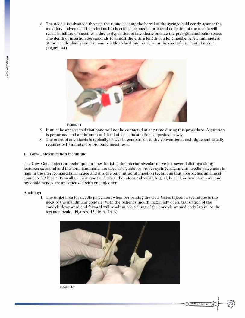
72
Loca
l An
esth
esia
8. The needle is advanced through the tissue keeping the barrel of the syringe held gently against the maxillary alveolus. This relationship is critical, as medial or lateral deviation of the needle will result in failure of anesthesia due to deposition of anesthetic outside the pterygomandibular space. The depth of insertion corresponds to almost the entire length of a long needle. A few millimeters of the needle shaft should remain visible to facilitate retrieval in the case of a separated needle. (Figure. 44)
9. It must be appreciated that bone will not be contacted at any time during this procedure. Aspiration is performed and a minimum of 1.5 ml of local anesthetic is deposited slowly.
10. The onset of anesthesia is typically slower in comparison to the conventional technique and usually requires 5-10 minutes for profound anesthesia.
E. Gow-Gates injection technique
The Gow-Gates injection technique for anesthetizing the inferior alveolar nerve has several distinguishing features: extraoral and intraoral landmarks are used as a guide for proper syringe alignment; needle placement is high in the pterygomandibular space and it is the only intraoral injection technique that approaches an almost complete V3 block. Typically, in a majority of cases, the inferior alveolar, lingual, buccal, auriculotemporal and mylohoid nerves are anesthetized with one injection.
Anatomy: 1. The target area for needle placement when performing the Gow-Gates injection technique is the
neck of the mandibular condyle. With the patient’s mouth maximally open, translation of the condyle downward and forward will result in positioning of the condyle immediately lateral to the foramen ovale. (Figures. 45, 46-A, 46-B)
Figure. 44
Figure. 45

73
Loca
l An
esth
esia
2. The syringe is positioned from the opposite cuspid and in a plane that will result in contact with the neck
of the condyle. It must be noted that this plane will vary according to the divergence of the mandible. In the widely divergent mandible, the barrel of the syringe will be oriented more posteriorly over the opposite premolar/molar region, and in the mandible with less flare, the barrel of the syringe will be positioned over the cuspid region. (Figures. 47, 48, 49)
Figure.46-A Figure. 46-B
Figure. 47
Figure. 48 (widely divergent mandible)
Figure. 49 (mandible with less flare)

74
Technique: 3. The patient is positioned semi-supine with the operator facing the patient. 4. A 25 gauge long needle is required for this injection. 5. The patient is instructed to open as widely as possible, which translates the mandibular condyle
to a more anterior position and in a closer relationship to the mandibular nerve as it exits foramen ovale. (Figure. 50)
6. The syringe is oriented along a plane from the opposite corner of the mouth to the tragus of the ear. The index finger can be placed posterior to the tragus of the ear to give a tactile target for needle orientation. Advancement of the needle along this plane of orientation results in contact of the neck of the condyle by the advancing needle. (Figure. 51)
Figure. 50
Figure. 51

75
Loca
l An
esth
esia
7. The point of needle insertion is just lateral to the pterygomandibular raphe at the height of the pterygomandibular depression. (Figure. 52)
8. The barrel of the syringe is positioned either anterior or posterior to the cuspid so as to point the needle to the tragus. This orientation ensures the contact of the needle with the neck of the condyle and accommodates for variations in flare of the mandible. (Figure. 53)
9. The needle is advanced slowly along this plane, depositing minimal amounts of anesthetic as the needle is advanced. This is done to preserve the bulk of the anesthetic for deposition at the final target location and because of the relative lack of sensory fibers in this region.
Figure. 52
Figure. 53

76
Loca
l An
esth
esia
10. Contact with the neck of the condyle is essential and local anesthetic should not be administered until bone is contacted. The depth of penetration of the needle is usually 25-30 mm. Once bone is contacted, the needle is carefully withdrawn 1-2 mm and aspiration is performed. Anesthetic is deposited slowly over 1 minute. It is recommended that a minimum of 1.5 ml of local anesthetic be delivered to ensure success of the injection. The success of the injection is dependent on the anterior, medial and inferior diffusion of the anesthetic in the pterygomandibular space. (Figure. 54)
11. The needle is withdrawn and the patient is instructed to keep the mouth open for approximately 1 minute to facilitate diffusion of the local anesthetic. (Figure. 55)
12. The onset of anesthesia is typically slower than the conventional technique due to the need for diffusion of local anesthetic through the pterygomandibular space.
Figure. 54
Figure. 55

77
Loca
l An
esth
esia
BACTERIAL ENDOCARDITIS PROPHYLAXIS
Recommended Standard Prophylactic Regimens for Dental Procedures:
A: For patients able to take Amoxicillin/Penicillin:
Amoxicillin 2.0 grams orally one hour before procedure.
B: For patients allergic to Amoxicillin/Penicillin: Order of antibiotic preference for patients allergic to Amoxicillin/Penicillin:
1. Clindamycin (Cleocin) - 600 mg (Peds 20 mg/kg) 1 hour before treatment. Don’t use for patients with inflammatory/irritable bowel syndrome 2. Azithromycin - 500 mg (Peds 15 mg/kg) 1 hour before treatment. Use as first choice for Penicillin allergic patient with irritable bowel/inflammatory bowel
syndrome 3. Clarithromycin (Biaxin) - 500 mg (Peds 15 mg/kg) 1 hour before treatment 4. Cephalexin (Keflex) - 2 grams (Peds 50 mg/kg) 1 hour before treatment 5-8 % cross allergenicity with Cephalosporin’s and Penicillin’s, higher if patient has
experienced a type 1 hypersensitivity reaction to Penicillin’s.
Total pediatric dose should not exceed total adult dose. Alternate Prophylactic Regimens for Dental Procedure
A: For standard risk patients unable to take oral medications: Ampicillin 2.0 grams (Peds 50 mg/kg) or Cefazolin 1 gram (Peds 25 mg/kg) IV or IM 30 minutes
before procedure. B: For standard risk patients allergic to Ampicillin/Amoxicillin/Penicillin unable to take oral
medications. Clindamycin 600 mg (Peds 20 mg/kg) IV 30 minutes before a procedure.
Note: Total pediatric dose should not exceed total adult dose.
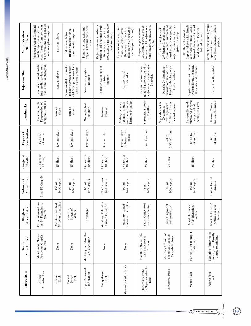
78
Loca
l An
esth
esia
Adm
inis
trat
ion
Tec
hniq
ue
Post
erio
r ar
ea o
f Cor
onoi
d no
tch
finge
r on
dee
p te
n-do
n. I
nser
t fr
om p
rem
olar
of
con
tral
ater
al s
ide
para
llel
to o
cclu
sal p
lane
. Asp
irat
e
sam
e as
abo
ve
M
ove
need
le fr
om
Infe
rior
alv
eola
r o
r re
-en
ter
at s
ite.
Asp
irat
e
Para
llel t
o lo
ng a
xis,
and
an
gle
in t
owar
d bo
ne n
ear
apex
.
30 g
a: P
ress
ure
anes
thes
ia
wit
h to
pica
l onc
otto
n sw
ab.
Rem
ove
and
inje
ct im
-m
edia
tely
.25
ga: s
eal n
eedl
e be
vel t
o ti
ssue
Pres
sure
ane
sthe
sia
wit
h to
pica
l on
cott
on s
wab
. R
emov
e an
d in
ject
im-
med
iate
ly. C
an u
se 2
5ga
(tec
hniq
ue d
iffer
ent)
Step
alo
ngpe
rios
teum
lo
se c
onta
ct w
ith
curv
eof
tube
rosi
ty. 4
5 de
gree
s up
-w
ard,
inw
ard,
& b
ackw
ard.
A
spir
ate
Para
llel t
o lo
ng a
xis
of
2nd b
icus
pid.
spa
n ca
nine
fo
ssae
to
ente
r in
frao
rbit
al
cana
l whi
ch is
cov
ered
by
finge
r on
the
ski
n. S
yrin
ge
agai
nst
low
er li
p.
Mou
th p
artl
y cl
osed
. D
epre
ss c
heek
wit
h th
umb
to e
xpos
e ve
stib
ule.
Nee
dle
dire
cted
dow
nwar
d in
war
d an
d m
edia
lly O
pera
tor
behi
nd p
atie
nt.
Con
tact
per
iost
eum
bey
ond
apic
es o
f inc
isor
s in
inci
-si
ve fo
ssa.
Ope
rato
r be
hind
pa
tien
t
Inje
ctio
n Si
te
Lev
el o
f cor
onoi
d no
tch:
ju
st m
edia
l to
deep
ten
-do
n la
tera
l to
pter
ygoi
d
sam
e as
abo
ve
3 m
m m
edia
l to
ante
rior
bo
rder
bet
wee
n su
perfi
-ci
al &
dee
p te
ndon
s 1
cm
abov
e o
cclu
sal p
lane
Nea
r m
uco-
gin
giva
ju
ncti
on
Post
erio
r 1/
3 at
sid
e of
In
cisi
vePa
pilla
e
At
Junc
tion
of
Lan
dmar
ks
3 -
4 m
m a
bove
muc
o-gi
ngiv
al ju
ncti
on D
ista
l to
zygo
mat
ic p
roce
ss a
bove
2nd
mol
ar.
Opp
osite
2nd
bic
uspi
d as
vi
ewed
from
ant
erio
r 1 c
m
late
ral t
o al
veol
ar p
roce
ss
high
in v
estib
ule
Palp
ate
fora
men
with
cot
ton
swab
unt
il re
ady
to in
ject
. D
eep
in la
tera
l ves
tibul
e
In th
e de
pth
of th
e ve
stibu
le
Lan
dmar
ks
Cor
onoi
d no
tch
Dee
p te
ndon
of
tem
pora
lis m
uscl
e
sam
e as
abo
ve
sam
e as
abo
ve
Muc
o-gi
ngiv
al
junc
tion
Inci
sive
Pap
illae
Mid
way
bet
wee
n gi
ngiv
al m
argi
n an
d ro
of o
f mou
th.
Dis
tal t
o 1st
mol
ar.
Zygo
mat
ic P
roce
ss
of M
axill
ae
Supr
aorb
italn
otch
. Zy
gom
atic
o-m
axill
ary
Sutu
re.
2nd B
icus
pid
4 m
m
med
ial t
o pu
pil
Betw
een
Bicu
spid
s m
idw
ay b
/n g
ingi
val
mar
gin
and
inf.
bord
er. C
k x-
rays
Be
twee
n Ce
ntra
l an
d L
ater
al In
ciso
rs
Dep
th o
f P
enet
rati
on
1/2
to 3
/4 o
f an
Inch
few
mm
dee
p
few
mm
dee
p
few
mm
dee
p
few
mm
dee
p
few
mm
dee
p t
o be
neat
har
d t
issu
e
3/4
of a
n In
ch
3/4
to 1
3/8
of a
n In
ch
1/4
to 1
/2 o
f an
Inch
1/2
of a
n In
ch
Gua
ge o
f N
eedl
e
25 S
hort
or
25 L
ong
25 S
hort
25 S
hort
25 S
hort
or
30 S
hort
25 S
hort
or
30 S
hort
25 S
hort
or
30 S
hort
25 S
hort
25 L
ong
25 S
hort
25 S
hort
Vol
ume
of A
nest
heti
c
1 m
l 1/2
Car
pule
1/2
ml
1/4
Car
pule
1/4
ml
1/8
Car
pule
1/2
ml
1/4
Car
pule
1/2
ml o
r le
ss 1
/4 C
arpu
le
1/2
ml
1/4
Car
pule
1 m
l 1
/2 C
arpu
le
3/4
ml
1/3
Car
pule
1/2
ml
1/4
Car
pule
1 m
l or
less
1/2
C
arpu
le
Gin
giva
e A
nest
heti
zed
Faci
al o
f man
dibu
-la
r 2nd
Pre
mol
ar t
o M
idlin
e
Man
dibl
e: L
ingu
al
3rd m
olar
to
mid
line
Man
dibl
e B
ucca
l of
Mol
ars
Any
whe
re
Max
illar
y: P
alat
al o
f C
uspi
d to
Cus
pid
Max
illar
y pa
lata
l m
olar
s to
bic
uspi
ds
Faci
al G
ingi
vae
of
teet
h an
esth
etiz
ed
Faci
al G
ingi
vae
of
teet
h an
esth
etiz
ed.
Man
dibl
e: B
ucca
l of
2nd
Bic
uspi
d to
m
idlin
e.
M
andi
ble
Labi
al o
f A
nter
iors
in a
rea
inje
cted
.
Teet
h A
nest
heti
zed
Man
dibu
lar:
Mol
ars
Prem
olar
s C
uspi
ds
Inci
sors
Non
e
Non
e
Max
illar
y: A
ll M
andi
bu-
lar:
6 A
nter
ior
Non
e
Non
e
Max
illar
y:M
olar
s E
X-
CE
PT M
B r
oot
of
1st m
olar
Max
illar
y M
B r
oot
of
1st
mol
ar B
icus
pids
C
uspi
ds I
ncis
ors
Man
dibl
e: 1
st B
icus
pid
to
Mid
line
Man
dibl
e: A
nter
iors
in
area
inje
cted
. Usu
ally
cu
spid
to
mid
line.
Inje
ctio
n
Infe
rior
A
lveo
larB
lock
Lin
gual
Blo
ck
Buc
cal
Ner
ve B
lock
Sup
ra-P
erio
stea
l I
nfilt
rati
on
Nas
o-pa
lati
ne B
lock
Gre
ater
Pal
atin
e B
lock
Tube
rosi
ty P
oste
-ri
or S
uper
ior
Alv
eola
r B
lock
Infra
orbi
tal B
lock
Men
tal B
lock
Inci
sive
foss
a B
lock
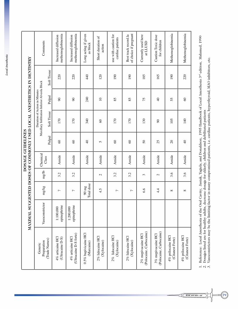
79
Loca
l An
esth
esia
DO
SAG
E G
UID
EL
INE
S
MA
XIM
AL
SU
GG
EST
ED
DO
SES
OF
CO
MM
ON
LY U
SED
LO
CA
L A
NE
STH
ET
ICS
IN D
EN
TIS
TR
Y
Com
men
ts
Incr
ease
d di
ffusio
nm
ethe
mog
lobi
nem
ia
Incr
ease
d di
ffusio
nm
ethe
mog
lobi
nem
ia
Long
act
ing
if gi
ven
as b
lock
Shor
t dur
atio
n of
actio
n
use
with
cau
tion
for
card
iac
patie
nts
Best
track
reco
rd.la
of
cho
ice
if pr
egna
nt
Curre
ntly
use
d he
re
at L
LUSD
Caut
ion
Toxi
c do
sefo
r chi
ldre
n
Met
hem
oglo
bine
mia
Met
hem
oglo
bine
mia
1. R
efer
ence
: L
ocal
Ane
sthe
sia
of t
he O
ral C
avit
y, J
asta
k, Y
agie
la, a
nd D
onal
dson
, 19
95 H
andb
ook
of L
ocal
Ane
sthe
sia
3rd e
diti
on,
Mal
amed
, 199
02.
Dos
ages
list
ed a
re fo
r he
alth
y ad
ults
, dec
reas
e do
sage
for
elde
rly,
chi
ldre
n an
d de
bilit
ated
pat
ient
s3.
Vas
ocon
stri
ctor
s m
ay b
e th
e lim
itin
g fa
ctor
in m
any
com
prom
ised
pat
ient
s, i.
e. c
ardi
ac p
atie
nts,
hyp
erth
yroi
d, M
AO
inhi
bito
rs, e
tc.
Dur
atio
n of
Act
ion
in M
inut
es0D[LOODU\�,QÀOWUDWLRQ�,QIHULRU�$
OYHROD�%ORFN
Pulp
al
Sof
t Tiss
ue
P
ulpa
l
Soft
Tiss
ue
220
220
440
120
190
190
185
165
190
220
90 90 240
10 85 85 75 40 55 60
170
170
340
60 170
170
130
90 105
140
60 60 40 5 60 60 50 25 20 40
Chem
ical
Clas
s
Am
ide
Am
ide
Am
ide
Am
ide
Am
ide
Am
ide
Am
ide
Am
ide
Am
ide
Am
ide
mg/
lb
3.2
3.2 2 3.2
3.2 3 2 3.6
3.6
mg/
kg 7 7
90 m
gTo
tal d
ose
4.5
7
7 6.6
4.4 8 8
Vaso
cons
trict
or
1:10
0,00
0ep
inep
hrin
e
1:20
0,00
0ep
inep
hrin
e
Gen
eric
Prep
arat
ion
(Tra
de N
ames
)
4% a
rtica
ine
HCl
(Ultr
acai
ne D
-S)
4% a
rtica
ine
HCl
(Ultr
acai
ne D
-S fo
rte)
0.5%
bup
ivic
aine
HCl
(Mar
cain
e)
2% li
doca
ine
HCl
(Xyl
ocai
ne)
2% l
idoc
aine
HCl
(Xyl
ocai
ne)
2% li
doca
ine
HCl
(Xyl
ocai
ne)
2% m
epiv
acai
ne H
Cl(P
oloc
aine
, Car
boca
ine)
3% m
epiv
acai
ne H
Cl(P
oloc
aine
, Car
boca
ine)
4% p
riloc
aine
HCl
(Cita
nest
Forte
)
4% p
riloc
aine
HCl
(Cita
nest
Forte
)

80
Loca
l An
esth
esia

81
Loca
l An
esth
esia
References
1. Yagiela 2. Troullos ES, Goldstein DS, Hargreaves KM, Dionne RA. ANESTH PROG 1987;34(1):10-3. 3. Malamed: Handbook of Local anesthesia Fifth edition. 4. Dionne, Phero and Becker: Management of pain and anxiety in the dental office, 2002. WB Sanders