Locomotion in Complex Fluids: Experiments
43
Locomotion in Complex Fluids: Experiments Josu´ e Sznitman and Paulo E. Arratia Recently, there has been renewed interest in the swimming of microorganisms for applications that include artificial swimmers, novel materials, drug delivery, and micro-robotics. Due to small length scales, the fluid mechanics of swimming of mi- croorganisms are governed by low Reynolds number (Re) hydrodynamics. In such a regime, linear viscous forces dominate over nonlinear inertial forces. While our cur- rent understanding of locomotion at low Re is derived mainly from investigations in simple, Newtonian fluids (e.g. water), many of the fluids in which locomotion occurs contain solids or (biological) polymers that are instead not Newtonian. Ex- amples include wet soils, human mucus, and fluids in the cervix and female repro- ductive tract amongst other. A major challenge remains to understand and determine the propulsion mechanisms in fluids that display both solid- and fluid-like behavior (i.e., viscoelastic media). 1 Introduction Microorganisms are surrounded by fluids. They cope and take advantage of water or wind currents to move, feed, and reproduce. Many, if not most, living organ- isms evolve in the realm of low Reynolds numbers (Re) [112], usually defined as Re= ρUL/μ , where U is a characteristic speed, L a characteristic length (e.g., body size), and ρ and μ are the fluid’s density and dynamic viscosity, respectively. This is specially true for mili- and micro-organisms due to their small length scales L spanning approximately O (10 −6 ) m to O (10 −3 ) m. Examples include include var- Josu´ e Sznitman Department of Biomedical Engineering, Technion - Israel Institute of Technology, Haifa 32000, Israel, e-mail: [email protected] Paulo E. Arratia Department of Mechanical Engineering & Applied Mechanics, University of Pennsylvania, Philadelphia, PA - 19104, USA, e-mail: [email protected] 1
Transcript of Locomotion in Complex Fluids: Experiments
Locomotion in Complex Fluids: Experiments
Josue Sznitman and Paulo E. Arratia
Recently, there has been renewed interest in the swimming of microorganisms for
applications that include artificial swimmers, novel materials, drug delivery, and
micro-robotics. Due to small length scales, the fluid mechanics of swimming of mi-
croorganisms are governed by low Reynolds number (Re) hydrodynamics. In such a
regime, linear viscous forces dominate over nonlinear inertial forces. While our cur-
rent understanding of locomotion at low Re is derived mainly from investigations
in simple, Newtonian fluids (e.g. water), many of the fluids in which locomotion
occurs contain solids or (biological) polymers that are instead not Newtonian. Ex-
amples include wet soils, human mucus, and fluids in the cervix and female repro-
ductive tract amongst other. A major challenge remains to understand and determine
the propulsion mechanisms in fluids that display both solid- and fluid-like behavior
(i.e., viscoelastic media).
1 Introduction
Microorganisms are surrounded by fluids. They cope and take advantage of water
or wind currents to move, feed, and reproduce. Many, if not most, living organ-
isms evolve in the realm of low Reynolds numbers (Re) [112], usually defined as
Re= ρUL/µ , where U is a characteristic speed, L a characteristic length (e.g., body
size), and ρ and µ are the fluid’s density and dynamic viscosity, respectively. This
is specially true for mili- and micro-organisms due to their small length scales L
spanning approximately O(10−6) m to O(10−3) m. Examples include include var-
Josue Sznitman
Department of Biomedical Engineering, Technion - Israel Institute of Technology, Haifa 32000,
Israel, e-mail: [email protected]
Paulo E. Arratia
Department of Mechanical Engineering & Applied Mechanics, University of Pennsylvania,
Philadelphia, PA - 19104, USA, e-mail: [email protected]
1

2 Josue Sznitman and Paulo E. Arratia
ious single-cell eukaryotic protozoa (e.g., sperm cells [18, 52, 53]), prokaryotes
(e.g., bacteria [26]), and multi-cellular organisms (e.g., nematodes [26, 54]). By
contrast, humans when swimming in the ocean or water pools can reach Re of ap-
proximately 104. This means that humans can rely on inertial forces for propulsion
due to relatively large length scales, while microorganisms simply cannot. Small
living organisms instead have to overcome the linear viscous forces and drag aris-
ing from the fluid in order to achieve any appreciable net motion. The picture that
emerges is that moving (and living) at low Reynolds number is drastically different
from what we are accustomed to; namely, the nonlinear forces that we humans rely
on for locomotion, that is inertia, are entirely absent in the realm of low Re. For
the case of swimming microorganisms in simple fluids such as water, the equations
of fluid motion become time-reversible. As a result, net locomotion can only be
generated from non-reciprocal kinematics in order to break time-reversal symmetry
[18, 85, 89]; this phenomenon is also known as the “scallop theorem” [112] which
states that organisms that rely on reciprocal motion for locomotion cannot achieve
net motion in the limit of vanishing Re.
Microorganisms have developed diverse strategies to break time-symmetry and
create non-reciprocal motion (Fig. 1). Such strategies include body undulations and
the presence of moving flagella. For example, the motility of various multi-cellular
organisms including the worm nematode Caenorhabditis elegans originates from
the propagation of undulatory waves from head to tail as a result of patterns of mus-
cle activation and neuromuscular control [29, 154, 157]. In the specific case of C.
elegans, body bending is generated by alternating contractions and relaxations of
dorsal and ventral muscle groups running along the worm’s body length [104, 149].
Locomotion may also result from flagellar motility where one or several bundled
appendages protrude from the cell body of certain prokaryotic and eukaryotic cells.
Although there exist many interspecies variations within flagellar motility, one can
typically distinguish between bacterial flagella that are helical filaments (e.g., Es-
cherichia coli [10]), each with a rotary motor at its base which can turn clockwise
or counterclockwise [8, 9, 11], and eukaryotic flagella that are flexible filaments
undergoing “whip-like” motions resulting from the action of molecular motors dis-
tributed along the filament length; this latter mode of flagellar actuation is seen
for example in many sperm cells [17, 98, 153]. Other eukaryotic organisms (e.g.,
Paramecium), have instead their body surface covered with thousands of small hair-
like protrusions (cilia) that beat in a coordinated manner [17, 49].
Beyond the vast world of self-propelled microorganisms, we note a number of
other cellular environments that are characterized by low Re locomotion. In mam-
mals, for instance, cells featuring motile cilia include the epithelium of the female
Fallopian tubes, where rhythmic beating guarantees translocation of the ovum from
the ovary into the uterus [93, 94, 102]. Motile cilia are also found in the epithelial
lining of the tracheobronchial airways of the lungs [120]. There, the ciliated epithe-
lium prevents mucus accumulation in the airway lumen and serves importantly as an
immune barrier against pathogens and foreign particulate matter by action of the so-
called mucociliary escalator [76, 107]. Such synchronous waves of ciliary beating

Locomotion in Complex Fluids: Experiments 3
Fig. 1 Examples of micro-organisms. (a) The green alga Chlamydomonas reinhardtii, a model
eukariotic organism, (b) sperm cells moving next to boundaries, and (c) the bacterium E. coli, one
the most widely studied prokaryotic model organism
effectively transport mucus secretions towards the laryngopharynx for expectoration
or swallowing to the stomach.
It is clear from everyday observation and from the few examples cited above that
nature has found many fascinating ways to break time-reversibility and achieve net
motion at the microscopic scale. One can of course spend a lifetime attempting to
map out in detail the various motility strategies found in nature. Here rather, we
seek to deliver a physical understanding of how microorganisms move (or swim) in
fluids at low Re. To do so, we first turn our attention to the equations of fluid motion
that govern the motility of microorganisms at low Re.
2 Basic Principles: Fluid Dynamics of Swimming at Low Re
We begin here by briefly discussing the underlying hydrodynamic principles of
swimming at low Re; a more thorough review of the subject can be found elsewhere
[18, 26, 58, 85]. The general form of the equation of motion for fluids is given as
ρ
(
∂u
∂ t+u ·∇u
)
=−∇p+∇ · τ+ρg, (1)
where u is the velocity vector, ρ is the fluid density, p is the pressure, τ is the
deviatoric component of the total stress tensor, g is the gravity field, and ∇ is
the divergence operator. Here, we restrict our discussion to incompressible fluids
(ρ = constant) such that
∇ ·u = 0. (2)
We will now make a series of simplifications to the above equations by consid-
ering the propulsion of a bacterium such as E. coli in water. Because the fluidic
medium considered is a Newtonian liquid, the shear-stress τ is linearly proportional
to the fluid strain-rate γ , the constant of proportionality being the fluid shear vis-
cosity µ such that τ = µγ = µ(∇u+∇uT ), where the superscript T represents the
transpose of the vector. Let us now estimate the representative Reynolds number of
an E. coli bacterium swimming in water. The shear viscosity (µ) of water is 1 mPa·s

4 Josue Sznitman and Paulo E. Arratia
(or 1 cP) and independent of shear-rate γ . The characteristic size L of E. coli is
approximately 2 µm, and the bacterium is known to achieve net swimming speeds
U of approximately 25 µm/s [22]. Following these parameters, we can estimate
the Reynolds number for E. coli to be approximately Re= ρUL/µ = 5×10⁻⁵ ≪ 1.
Such low value of Re implies that linear viscous forces dominate over inertial forces,
and the nonlinear convective term (u ·∇u) in Eq. (1) can be safely ignored.
Additionally, one can compute the frequency-based Reynolds number, typically
defined as Re f req = ρL2ω/µ , to assess unsteady flow effects (i.e., ∂u/∂ t), where
ω is the frequency at which the bacterium flagella rotate (∼ 100 Hz). We find again
for E. coli that Re f req ≪ 0.1, and thus one can assume the flow to be steady. Using
the above arguments, Eq. (1) effectively reduces to
∇p = ∇ · τ. (3)
The above equation is often referred to as the Stokes equation (named after the
mathematician Sir George Stokes) or alternatively as the creeping equations of fluid
motion. For a Newtonian fluid, the stress tensor is given as τ = µγ = µ(∇u+∇uT )and Eq. (3) reduces to
∇p = µ∇2u. (4)
It is important to note that the above result is instantaneous in the sense that
Eq. (4) has no dependence on time other than via boundary conditions. Equation (4)
is also linear in both velocity and pressure. Furthermore, it is time-reversible in the
sense that any time-reversed Stokes flow solves the same equations as the original
Stokes flow. This time-reversibility, or kinematic reversibility, forms the hydrody-
namic basis of the “scallop theorem” introduced earlier [112]. These hydrodynamic
properties illustrate that swimming at low Re can seem at first as a highly con-
fined phenomenon, yet microorganisms have found a variety of ways to overcome
the constraints of the “scallop theorem” including the semi-flexible, helical-shaped
flagella of E. coli or body undulations in the form of traveling waves for the nema-
tode C. elegans. In what follows, we briefly review some of the classical theories
that have shed light on the hydrodynamic mechanisms leading to net propulsion at
low Re.
Over half a century ago, G.I. Taylor demonstrated in his seminal work that a
slender body such as an (infinite) waving sheet (Fig. 2a) could swim in an incom-
pressible, Newtonian fluid by generating traveling waves in the absence of inertia
[137, 138]; that is, at vanishing values of Re. In Taylor’s work, the planar sheet os-
cillates in time in a prescribed form according to y(x, t) = asin(kx−ωt), where a
is the traveling wave amplitude, λ = 2π/k is the wavelength, c = ω/k is the trav-
eling wave speed and k is the wavenumber. For vanishing Re, Taylor found that the
sheet oscillations induce a forward velocity U = ωa2k/2+O(ka)4 [137], where the
sheet is propelled in the direction opposite to that of the propagating wave (Fig. 2a).
Many important investigations followed Taylor’s landmark contribution. Of partic-
ular relevance, we highlight the well-known resistive force theory (RFT) introduced
by Gray and Hancock in analyzing the locomotion of sperm cells [52]. There, the
authors assumed that the hydrodynamic forces experienced by the organism would

Locomotion in Complex Fluids: Experiments 5
be approximately proportional to the local body velocity such that the force exerted
by a body or flagellar segment is given by F =CNUN+CT UT, where C corresponds
to the local drag coefficient per unit length (dependent on geometry and fluid vis-
cosity), and N and T are the normal and tangential components, respectively (see
Fig. 2b). Hence, the total thrust can then be obtained by integrating the propulsive
force over the entire body or flagellum length. It is namely the anisotropy between
the normal and tangential drag coefficients, with CN >CT , that lies at the origin of
drag-based thrust.
Fig. 2 (a). Two-dimensional waving sheet in a viscous fluid illustrating the traveling wave of ve-
locity c progressing in the x-direction and the forward swimming speed (U) in opposite direction.
(b) Resistive Force Theory (RFT) diagram illustrating the normal and tangential components of
the velocity U and force F , and the resulting net propulsive force.
Using RFT, Gray and Hancock obtained for the case of large amplitude displace-
ments a closed-form solution for the swimming speed of an undulating filament
given by the expression U = πc(a/λ )2(CN/CT − 1)/ (1 + 2π2(CN/CT )(a/λ )2).Here, CN = 2CN = 4πµ/ ln(L/a) for a straight rod of length L such that the ra-
tio of normal to tangential drag coefficients yields CN/CT = 2 for a sine wave of
wavelength λ . For example, more recent experiments using the nematode C. ele-
gans estimated this ratio at CN/CT = 1.4 [133]; such value lies closely with earlier
estimates reported by Gray and Lissmann [54] who dropped thin wires into viscous
fluids (CN/CT = 1.4-1.6).
With the emergence of resistive force theory, Lighthill [89] later recognized the
importance of long-range hydrodynamics interactions and improved RFT the by in-
corporating slender-body approximations. Such improvements led to CN/CT = 1.5for the case of an undulating filament swimming in an infinite fluid medium. When
incorporating wall-effects into the analysis, a significantly larger value of the drag
coefficient ratio (CN/CT = 4.1) was subsequently obtained using the corrections of
Katz et al. [66].
As noted earlier, the flow disturbances driven by the kinematic motion of a swim-
ming microorganism in a Newtonian fluid will depend linearly upon the stresses ex-
erted by the moving body on the fluid (see Eq. (4)). These boundary-driven flows are
known to decay very slowly with the distance r away from the body [18, 85, 119].
Most often, such flow disturbances are mathematically cast as linear superpositions

6 Josue Sznitman and Paulo E. Arratia
of fundamental solutions of the Stokes equation and decay with inverse powers of
r. The first solution, referred to as a “Stokeslet”, arises from the net force on the
fluid, and has a velocity field that decays as 1/r. The next solution, also known
as a “stresslet” flow, is induced by the first force moment exerted by the body on
the fluid and decays more rapidly (1/r2); higher-order solutions decay even more
rapidly (1/r3). As a result, linear combinations of basic solutions of the creeping
equations of fluid motion can generate a multitude of complex flow fields, exhibit-
ing contrasting near- and far-field behaviors [119].
3 Experiments in Newtonian Fluids
Experimental studies on low Reynolds number locomotion in Newtonian fluids have
undoubtedly complimented early theories on the topic [52, 53, 54, 89, 137, 138].
Many of these works have aimed at addressing the validity of classical theoretical
models at least qualitatively, including RFT. In the section below, we briefly review
a number of relevant experimental efforts that have helped over time characterize
low Re propulsion in Newtonian media.
3.1 From scaled-up models to live microorganisms
Experiments with live microorganisms are generally challenging due to difficulties
with imaging and optical setups, as well as the lack of control over the organisms
themselves. One attractive experimental approach to circumvent some of these is-
sues relies on leveraging scaled-up systems, often designed to mimic the organism’s
main swimming kinematics (Fig. 3). Scaled-up models provide much valuable in-
sight into the main physical mechanisms governing microswimming phenomena;
they have brought valuable insight in understanding the net motion resulting from
traveling waves along elastic tails [74, 113, 163], helical flagella [118, 165] and flag-
ellar bundles [70, 71], as well as in uncovering the motility resulting from surface
traveling waves along cylindrical shells [122].
Beyond fundamental research into microorganism locomotion, a broad range of
scaled-up designs has been employed in the context of artificial swimming strate-
gies at low Re [85], including Purcell’s seminal “three-link swimmer” which pos-
sesses two hinges actuated with both time and phase differences [151] and a flapping
body performing reciprocal motions near a deformable free surface [143]. Smaller
mechanical systems have also been investigated. For example, the shapes of an os-
cillating passive actin filament have been experimentally probed [155] and more
recently, a three-sphere design has been implemented using colloidal beads and op-
tical tweezers [87].
Despite challenges in working with live microorganisms, microscopy imaging
of bacterial flagella has gained tremendous traction following the pioneering work

Locomotion in Complex Fluids: Experiments 7
Fig. 3 Example of scaled-up model of flagellar bundling dynamics [70]. (a) Image sequence of
semi-coiled helices shown at various instances in time. (b) Same helices viewed from the side. The
scale bars are 100 mm long; the helices are 310 mm long (from chuck to tip), 4.0 mm in diameter,
and turning at 0.1 Hz (Re f req ≈ 3×10⁻⁵). Copyright (2003) National Academy of Sciences, U.S.A.
of H.C. Berg [6, 7, 8, 10, 12]. Of utmost relevance, flagellar kinematics of indi-
vidual bacteria have been visualized in real time using fluorescent staining of both
cells and flagellar filaments [146]. In more recent years, these initial microscopy
techniques have been further developed to obtain time-resolved imaging of flagellar
motility using setups with high-speed cameras [88] as well as to track swimming
microorganisms three-dimensionally (3D), for instance in a fluid far from surfaces
[36].
Since, however, microorganisms evolve constantly near solid boundaries (e.g.,
migration of infectious bacteria through tissues), a growing number of experiments
has shown that the presence of surfaces is crucial to consider in low Re locomotion;
namely, surfaces and wall effects drastically alter the kinematics of live swimming
microorganisms relative to ideal unbounded swimming conditions. A best-known
example is perhaps illustrated for helical flagella (e.g., E. coli): swimming trajecto-
ries are modified from straight to circular in the vicinity of boundaries, clockwise
when the wall is rigid [84] and anticlockwise near a free surface [31]. In particular,
solid surfaces not only lead to the reorientation of microorganisms in the direction
parallel to the surfaces, they also attract the organism to the closest wall [14].
Uncovering the fundamental hydrodynamic interactions between surfaces and
swimming microorganisms has helped shed light on experimental observations of
the accumulation of confined spermatozoa on boundaries [28, 117, 158]. It was

8 Josue Sznitman and Paulo E. Arratia
recently observed [62] that ciliated Paramecium swimming in capillary tubes ex-
ecute helical trajectories that slowly transition to straight lines as the tube diame-
ter decreases (Fig. 4). Further experimental studies mimicking bio-locomotion in
confined environments (e.g., female reproductive tract) have also revealed that the
migration of motile spermatozoa in 3D microchannels is strongly influenced by spe-
cific wall shapes, including the turning angles at corners [30]. Beyond single-cell eu-
karyotic and prokaryotic microorganisms, it has also been noted that multi-cellular
organisms, such as C. elegans, may display some finite attraction to the presence of
boundaries [51] and certainly exhibit changes in their swimming kinematics under
confinement conditions (e.g., setups with parallel-wall cells) [86].
Fig. 4 Swimming of Paramecium in tubes of different diameters [62]. Here, Λ denotes the wave-
length, c denotes the radius of the organism and A the amplitude of the helical trajectory traced by
the organism in tubes of different diameter. (a) Paramecium swimming in large tube (R/c = 2.63)
where the trajectory of the motion is helical. (b) Small wavelength helices are seen inside tubes of
intermediate diameters (R/c = 1.67). (c) In very small tubes (R/c = 0.9), Paramecium swims in a
straight line. Figure reproduced with permission from the American Institute of Physics.
3.2 Propulsive force & flow measurements
Beyond microscopy visualizations of low Re motility including measurements of
swimming kinematics, experimental efforts have strived to deliver measurements of
the propulsive forces (i.e., thrust) directly at the scale of single organisms. For in-
stance, optical traps have been used to measure the forces required to tether sperm
cells and bacteria [22, 75, 134]. In parallel, atomic force microscopy (AFM) has en-
abled the measurements of forces exerted by mucus-propelling cilia that lie on the
order of < 1 nN per cilium during the effective stroke [139]. Recently, force mea-
surements using optical tweezers have been obtained on individual bacteria to test
the validity of RFT and determine the swimming efficiency of E. coli [23]. These

Locomotion in Complex Fluids: Experiments 9
latter measurements have revealed for the first time that long-range hydrodynamic
interactions are indeed critical in capturing accurately single-cell propulsion; in con-
trast, relying on RFT assumes a stationary background fluid while ignoring local
flows induced from the other moving parts of the cell. Such observations have been
most recently corroborated in scaled-up models of helical flagella, where the valid-
ity of RFT breaks down for increasing pitch angles of the helix [165]; a property
attributed to the hydrodynamic interactions among helical loops at higher pitch an-
gles. Concurrently, experiments on macroscopic swimmers for the range of helical
wavelengths λ , radii R, and lengths L relevant to bacterial flagella have also high-
lighted the qualitative and quantitative discrepancies with RFT predictions [116].
Despite the tremendous experimental progress brought on low Re locomotion
in Newtonian fluids, there are still few available data on the dynamics of the fluid
flows surrounding individual swimming microorganisms. One limiting reason re-
mains the difficulty to resolve the microflows generated by individual swimmers
such as E. coli bacteria and spermatozoa. In turn, scaled-up studies have offered
an attractive path to obtain time-resolved quantitative flow visualizations of the ve-
locity fields using particle image velocimetry (PIV) techniques [71, 165]. For live
organisms, perhaps the most well-known flow visualizations of individual undula-
tory swimming microorganisms date back historically to Gray and Lissmann [54],
where the authors presented qualitative pathlines of freely swimming nematodes in
water seeded with starch grains.
Since these seminal flow visualizations, the implementation of modern micro-
PIV (µPIV) techniques [152] has begun paving the way toward quantitative flow
measurements at the level of individual microorganisms. Most recently, pathlines of
particles seeded in flows generated by an individual ciliated Paramecium have been
imaged [62]. However, it is only by combining high-speed microscopy imaging with
µPIV that the planar (2D) flows generated by individual unicellular microorganisms
[88] and multi-cellular nematodes [133] have been resolved. In the latter case, the
authors demonstrated that velocity magnitudes of fluid motion follow closely an ex-
ponential decay of the form exp(−2πr/λ ) as a function of the distance r away from
the nematode body (see Fig. 5); this analytical solution was originally derived by
Lighthill [89] for an undulating sheet of wavelength λ in Stokes flow. Further exten-
sion of high-speed imaging techniques using for example tomographic PIV, where
multiple cameras image simultaneously the interrogation volume from different an-
gles [101], has enabled measurements of 3D time-resolved flow fields surrounding
millimeter-sized copepods (Calanus finmarchicus).
It is worthwhile noting that experiments have gone beyond highlighting the lim-
itations of classic analytical models such as RFT. Indeed, experiments resolving
flow fields surrounding freely swimming microalgae have demonstrated that lo-
cal fluid motions are much more complex than analytical models would initially
suggest [35, 57]. This observation is particularly true in the near field, where the
largest flow velocities occur. For example, Drescher et al. [35] detailed quantitative
measurements of time-averaged flows using µPIV for two different types of mi-
croalgae: Volvox carteri, a ciliated multicellular spherical alga, and Chlamydomonas
reinhardtii, a unicellular alga featuring two flagella that beat in a breaststroke-like

10 Josue Sznitman and Paulo E. Arratia
Fig. 5 Flow behavior surrounding a swimming C. elegans. Adapted from Sznitman et al. [133]. (a)
Color-coded particle pathlines are shown via the trace of dispersed fluorescent particles. Particle
trajectories are tracked over ten consecutive frames (∼ 0.06 s). Colors are associated with the
lengths of trajectories. Two dominant recirculation regions are resolved along the nematode body.
Scalebar represents 200 µm. (b) Normalized fluid velocity magnitude (|u|/|u|max) as a function
of the dimensionless distance (r/L) away from the nematode body, where L is the nematode body
length. Data points of different colors correspond to fluids of different viscosities. The solid line
corresponds to exp (−2πr/λ ) [89]. Inset: representative velocity magnitude field at a given instant
in time surrounding a nematode (marked with a black line). Figure reproduced with permission
from the American Institute of Physics.
fashion. Guasto et al. [57] resolved rather the oscillatory nature of the flow field
driven by Chlamydomonas reinhardtii over one period of motion using high-speed
imaging, where the swimming microorganisms were confined in thin liquid films.
Both studies have emphasized how distinct species are likely to drive qualitatively
different disturbance flows, as recently highlighted by Saintillan [119]. This flow
feature remains true in the far field as well, where it is commonly assumed that
there, flow fields can be described in terms of a stresslet. As a final word, we note
that the representation of these driven flows using time-averaged velocity fields fall
short of capturing the true nature of the flow, since time fluctuations can be of the
same order as the mean.
3.3 Individual organisms & collective behavior
Until now, we have stressed the unique swimming features of individual microor-
ganisms. Beyond such individual motion, a number of experimental studies have
focused in recent years on collective behavior phenomena that arise at low Re
[3, 85, 148]. Here, we highlight collective motions that result in flows affecting

Locomotion in Complex Fluids: Experiments 11
the dynamics of entire populations in contrast to single microorganisms, as seen for
instance in sperm cells [100, 114, 132]. Moreover, collective behavior may also be
noted in the non-reciprocal motion of cilia that is at the origin of the mucociliary
escalator and guarantees net fluid flow towards the trachea [40]. Most noticeably, ex-
periments using flow velocimetry and tracking techniques in bacterial suspensions
have demonstrated the existence of transient patterns of coherent locomotion with
correlation lengths much larger than the size of individual organisms (i.e., from a
few tens to several hundreds of microns) and the presence of recurring swirls and
jets [25, 33, 129, 159].
In analogy to analysis methods dedicated for high-Reynolds-number fluid tur-
bulence, the dynamics of so-called ‘bacterial turbulence’ have been recently char-
acterized in terms of kinetic energy, dissipation (quantified by the enstrophy, or
mean-squared flow vorticity) and spatio-temporal correlation functions due to the
transient, large-scale nature of such collective motions [38]. In this latter study, the
authors successfully probed for the first time the fluid and bacterial dynamics inde-
pendently by applying bright-field microscopy to analyze the movement of the bac-
teria whereas micron-sized fluorescent markers were seeded to visualize the flow
of the suspending fluid (see Fig. 6). From a practical standpoint, collective motion
has drawn attention as a strategy to achieve controlled mixing and pumping ac-
tion in microfluidic devices using bacterial suspensions [72, 73] and artificial cilia
[21, 126].
Fig. 6 Collective dynamics of suspensions of bacteria. Adapted from Dunkel et al. [38]. (a)
Streamlines and normalized vorticity field determined from PIV in a very dense homogeneous sus-
pension of B. subtilis. (b) Turbulent “Lagrangian” flow of fluorescent tracer particles (false-color)
in the same suspension, obtained by integrating emission signals over 1.5 s. Scalebar represents
70 µm. Figure reproduced with permission from the American Physical Society.

12 Josue Sznitman and Paulo E. Arratia
4 From Newtonian to Complex Fluids
Much of our understanding of low Re locomotion arises from considerations in
simple, Newtonian fluids like water [18, 52, 53, 89]. But such fluids do not cap-
ture the diversity of habitats characterizing locomotion in the low Re realm. In-
deed, many microorganisms evolve within complex fluids that contain particulates
and/or biopolymers [39, 131, 145]. But, what are complex fluids? Here, we define
complex fluids as a broad class of materials that are usually homogeneous at the
macroscopic scale and disordered at the microscopic scale, but possess structure at
an intermediate scale (typically, a few sizes of its particles). Examples include col-
loidal suspensions, foams and emulsions, polymeric fluids, gels, human mucus, and
blood. In colloidal crystals, for example, the intermediate scale is set by the size
of the organized crystalline structure; that is, if one considers a cup of a cornstarch
suspension, then the microscopic scale is the matrix that includes both the water
molecules and the cornstarch grains (∼ 1µm), and the macroscopic scale is the size
of the cup (∼ 10 cm), while the intermediate scale is set by the structural length
scale (if any) of the cornstarch grains. The cornstarch suspension will respond quite
differently to an applied stress, depending on the grain size, concentration, and the
grain arrangement in the suspending liquid. Thus, the macroscopic flow behavior,
otherwise known as the rheology of complex fluids, is a strong function of the fluid
microstructure.
Shear-Thinning Viscosity: Complex fluids such as human mucus, blood, wet soil,
and gels often display non-Newtonian rheological behavior such as strain-rate de-
pendent viscosity (e.g., shear-thinning) and viscoelasticity. For example, one of the
most common non-Newtonian behavior found in polymeric solutions and colloidal
suspensions is rate-dependent viscosity in which the fluid viscosity is a function of
the shear-rate; viscoelasticity can also be found in such fluids. This non-Newtonian
viscosity behavior can be captured by an empirical power law model of the type
η = η0|γ|n−1, where η is the non-Newtonian viscosity, η0 is the viscosity factor,
and n is the power law index. If n > 1, the fluid is shear thickening whereas if
n < 1 the fluid is shear-thinning. For the particular case n = 1, the model reduces
to Newtonian. Recall that for Newtonian fluids, the stress is linearly proportional to
the strain-rate and µ is a constant independent of γ; note that we use the symbol
µ for Newtonian viscosity and η for non-Newtonian viscosity. Another common
empirical rheological model is the Carreau viscosity model usually given as:
η = η0[1+λc|γ|2](n−1)/2, (5)
where λc is a time-scale associated with the shear-rate (1/s) at which the fluid vis-
cosity departs from Newtonian behavior. The Carreau number Cr = λcγ is often
used to characterize such transition; Cr = 1 marks the departure from low shear-rate
Newtonian viscosity. This model offers some advantages over the power law model
discussed above. The most significant perhaps is that the Carreau model is able to
capture the frequently observed viscosity transition from a low-shear-rate Newto-
nian plateau to the power-law region as the shear-rate γ is gradually increased. A

Locomotion in Complex Fluids: Experiments 13
drawback of the Carreau model, however, is that it assumes the fluid infinite-shear-
viscosity η∞ to be effectively zero. That is, the transition from the power-law region
to this high-shear, Newtonian plateau is not captured by Eq. (5). An alternative is
to use the more general Carreau-Yassuda model [15]. Nevertheless, Eq. (5) offers a
great improvement over the power-law model.
The nonlinear relationship between τ and γ seen in shear-thinning fluids can have
significant consequences to locomotion at low Re including (i) a breakdown of the
“scallop” theorem, (ii) kinematic changes in the organism’s swimming motion, and
(iii) changes in the drag forces experienced by the organism. In fact, it was recently
shown that shear-thinning viscosity may enhance both the efficiency and the swim-
ming speed of swimming microorganisms [99, 147]. Using Taylor’s waving sheet
with prescribed kinematics along with a Carreau fluid model, Velez-Cordero and
Lauga [147] showed that the swimming speed for an inextensible sheet is the same
for shear-thinning and Newtonian fluids, but the swimmer (i.e., sheet), however, is
more efficient in the shear-thinning fluid. A numerical simulation by Montenegro-
Johnson, Smith, and Loghin [99] showed that for large amplitude waves the swim-
ming speed increases in shear-thinning fluids compared to that in Newtonian fluids.
These recent studies have shown that even relatively simple non-Newtonian fluid
behavior can have a significant impact on the swimming kinematics of microorgan-
isms.
4.1 Viscoelasticity
A common fluid rheological behavior is viscoelasticity and is typically found in
liquids containing polymers or bio-polymers, gels, mud, intestinal fluid, and mucus.
Such viscoelastic fluids possess both fluid-like and solid-like behavior, as well as
time-dependent rheology. A very simple (and linear) constitutive model to describe
fluid viscoelasticity is the Maxwell model which is represented by a purely viscous
damper and a purely elastic spring connected in series [41, 81]. The Maxwell model
is usually expressed as
τ +λ∂τ
∂ t= µγ, (6)
where λ = µ/G is the fluid relaxation time and G is the elastic modulus (of the
spring). This simple model predicts that the stress relaxes exponentially in time,
which is accurate for many liquids containing polymers. However, Eq. (6) predicts
that stress will increase linearly with time under constant stress, a trend not observed
in real polymeric solutions. In addition, the Maxwell model is only valid for small
deformations. For larger deformations, one should instead turn to nonlinear models
such as the Oldroyd-B and the finite extensibility non-linear elasticity (FENE) mod-
els [81]. Such models are known to capture many nontrivial viscoelastic phenomena
such as the development of hoop-stresses and hydrodynamic instabilities [123].
14 Josue Sznitman and Paulo E. Arratia
Despite its simplicity and limited applicability, the Maxwell model (Eq. 6) is
useful in illustrating one of the main features of fluid viscoelasticity, namely that
the shear-stress is time dependent and that it depends on the history of deformation.
Such features give rise to flow behavior that is markedly different from that of New-
tonian fluids even at low Re [15, 81, 123], and they can even lead to the breakdown
of the scallop theorem [69]. Indeed, flow instabilities and nonlinear dynamics have
long been observed in viscoelastic fluids [4, 123, 160], particularly those contain-
ing polymers. Most of the nonlinear flow behavior observed in these studies arises
from the extra elastic stresses due to the presence of polymer molecules in the fluid.
Mechanical stresses in these fluids are history-dependent and depend namely on a
characteristic time λ that in dilute solutions is proportional to the relaxation time of
a single polymer molecule. In semi-dilute solutions, λ depends also on molecular
interactions.
As mentioned earlier, many microorganisms live in complex fluid environments
that are viscoelastic. The bulk and local nonlinear behavior response of such vis-
coelastic fluids is expected to play a significant role on small swimmers. Consider
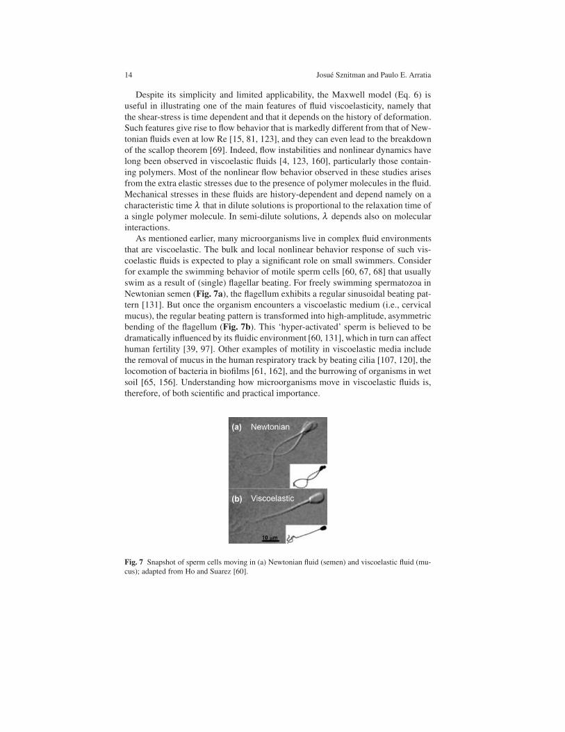
for example the swimming behavior of motile sperm cells [60, 67, 68] that usually
swim as a result of (single) flagellar beating. For freely swimming spermatozoa in
Newtonian semen (Fig. 7a), the flagellum exhibits a regular sinusoidal beating pat-
tern [131]. But once the organism encounters a viscoelastic medium (i.e., cervical
mucus), the regular beating pattern is transformed into high-amplitude, asymmetric
bending of the flagellum (Fig. 7b). This ‘hyper-activated’ sperm is believed to be
dramatically influenced by its fluidic environment [60, 131], which in turn can affect
human fertility [39, 97]. Other examples of motility in viscoelastic media include
the removal of mucus in the human respiratory track by beating cilia [107, 120], the
locomotion of bacteria in biofilms [61, 162], and the burrowing of organisms in wet
soil [65, 156]. Understanding how microorganisms move in viscoelastic fluids is,
therefore, of both scientific and practical importance.
Fig. 7 Snapshot of sperm cells moving in (a) Newtonian fluid (semen) and viscoelastic fluid (mu-
cus); adapted from Ho and Suarez [60].

Locomotion in Complex Fluids: Experiments 15
Despite many recent efforts to be discussed below, the effects of bulk fluid elas-
ticity on the motility behavior of live organisms at low Re are still not clear and well
understood. In order to provide the reader with some basic insight into this issue, we
turn to our favorite dimensionless parameters and their physical meaning. The first,
of course, is the Reynolds number defined earlier as Re=ρUL/µ ; Re is a measure
of the relative importance of the fluid inertia to viscous forces. Because the length
scale L of microorganisms is very small, magnitudes of Re are also small (Re≪ 1)
and viscous forces dominate over inertial forces. We saw earlier that for E. coli
swimming, Re is estimated to be approximately 10−4. The effects of fluid elasticity
are often measured using the Deborah number, defined as De= λ f where λ is the
fluid relaxation time and f is the organism’s beating frequency. Note that De= 0 for
Newtonian fluids and De→ ∞ for purely elastic solids. One could imagine that fluid
elasticity may begin to play a dominant role for De> 1. If one considers the beating
frequency f of sperm cells to range from 20 to 50 Hz and the relaxation time λ of
cervical mucus to range from 1 to 10 s (depending on factors like hydration, among
others), one can expect fluid elasticity to play a significant role on the motility of
spermatozoa. One can also compare the ratio of the (fluid) elastic time scale λ to
the (fluid) viscous time scale ρL2/µ . This is the so-called Elasticity number, defined
as El=λ µ/ρL2. Elastic effects are expected to dominate for El> 1. Because of the
nonlinear (squared) dependence of El on the (swimmer) length scale L, one antici-
pates the effects of fluid elasticity to become increasingly important for swimming
microorganisms.
4.1.1 Swimming in Viscoelastic Fluids: Expectations
In 1979, Chaudhury [24] attempted to incorporate the effects of fluid elasticity on
animal locomotion using a series of expansions similar to Taylor’s analysis and a
second-order fluid. It was then predicted that fluid elasticity could either increase
or decrease the propulsion speed of the waving sheet (Fig. 2), depending on the
value of Re. Later, inspired by experimental observations of spermatozoa swim-
ming in mucus [60, 131], the effects of elasticity on beating flagellar structures were
considered in Stokes flow using the Maxwell model [46]. It was shown that self-
propulsion was not affected by viscoelasticity even at large Deborah numbers (De),
where De=λ f . Here, λ is the fluid relaxation time and f is the beating frequency.
However, the total work decreased with increasing De. It was then suggested that a
microorganism could swim faster in a viscoelastic fluid with the same expenditure
of energy compared with a Newtonian fluid.
More recently, Lauga [82] showed that, for a 2D waving sheet (Fig. 2), elas-
tic stresses could significantly alter the organism speed and the work required to
achieve net motion. Using nonlinear viscoelastic fluid models such as the Oldroyd-
B and the FENE-P models, it was shown that the sheet’s forward speed U in a purely
elastic fluids is given by [82]

16 Josue Sznitman and Paulo E. Arratia
U
UN
=1+De2(ηs/η)
1+De2, (7)
where UN is the swimming speed of the sheet in a viscous Newtonian fluid (i.e.,
Taylor’s original result) and ηs is the solvent viscosity; note that the solution vis-
cosity η is assumed to be the sum of the solvent viscosity and the polymer viscosity
such that η = ηs +ηp. Hence, for a given (i.e. prescribed) swimming gait UN ≥U ,
that is, elastic stresses reduce the overall speed of the waving sheet. A similar re-
sult was also obtained for a waving cylinder by Fu et al. [45]. These important re-
sults imply that fluid elasticity can reduce the swimming speed of microorganisms
when compared to simple, Newtonian fluids. An important caveat, of course, is that
organisms may compensate the reduction in velocity by increasing their beating
frequency and/or concurrently decreasing their body wavelength. In other words,
microorganisms can alter their swimming kinematics to adjust or adapt to varying
fluidic environments.
Fig. 8 (a,b) Polymer stress tensor fields for a finite length, two-dimensional undulating sheet. The
ellipses in the figures represent the directions and degree of distension of the polymer field. The
arrows represent the fluid velocity on the immersed filament. (c) Normalized average swimming
speed of the sheet as a function of Deborah number. Note the enhancement in propulsion at De
≈ 1. Figures adapted from [140].
Numerical simulations have also been used to address the role of fluid elas-
ticity on the swimming behavior of microorganisms. In particular, Teran and co-
workers [140] considered two-dimensional swimming ”free” sheets (i.e., with free
head and tail) of finite length L in viscoelastic fluids. The simulations were per-
formed by solving Stokes equation using the Oldroyd-B model as the constitutive
equation using an immersed boundary method. The simulations show that, for ac-
centuated tail motions, the sheet swims faster at De ≈ 1 than in a Newtonian fluid.
This regime corresponds to where ”swimmer” stroke frequency matches the fluid
relaxation time. This is unlike Eq. (7) which predicts that the swimmer speed in vis-
coelastic fluids is always slower than in Newtonian fluids. The simulations do show
that for De > 1, the swimming speed decreases as De increases.
As briefly discussed above, fluid elasticity can strongly affect both the swim-
ming dynamics and kinematics of small organisms even at low Re. Recent ana-

Locomotion in Complex Fluids: Experiments 17
lytical works predict that fluid elasticity hinders swimming speed while numerical
simulations show that it is possible to obtain an enhancement in self-propulsion in
a regime where the fluid relaxation time matches the swimmer stroke frequency,
that is De≈ 1. Despite such important advances, it is still not clear whether elastic
stresses enhance or hamper self-propulsion since theoretical and numerical results
are model dependent. So the question still stands: does fluid elasticity enhance or
hinder self-propulsion at low Re? Perhaps experiments will shed more light into this
important question.
5 Experiments in Viscoelastic Fluids
5.1 Scaled-up experiments
While some specific aspects of the role of elastic stresses on the swimming behavior
of microorganisms have been recently addressed both theoretically and numerically,
there is still a dearth of systematic experiments addressing the role of viscoelastic-
ity on swimming of microorganisms at low Re. Part of the problem is undoubtedly
the difficulty in identifying a model organism or swimmer that is both able to move
in different types of media and relatively easy to image and track. To circumvent
some of these difficulties, many investigators choose to build instead macroscopic-
scaled versions of the microorganisms’ propulsion mechanism [71, 73, 74, 91]. Such
scaled-up experiments can provide a wealth of information that would be other-
wise difficult to obtain with live microorganisms (e.g., detailed velocity fields). In
this section, we discuss an interesting experimental setup proposed by Liu and co-
workers [91] in which a scaled-up model of bacterial filaments is investigated.
The experimental setup is shown in Fig. 9 and consists of a large cylindrical tank
filled with either a viscous Newtonian fluid (silicon oil) or a viscoelastic fluid (poly-
meric solution). A rigid helix rotating at speed ω is slowly immersed or plunged at
a constant speed V into the the fluid-filled tank. Helices of varying pitch angles are
used to mimic the geometry of bacterial flagellar filaments (e.g. E. coli). The hydro-
dynamic force exerted on the helices by the fluid is measured by placing the tank
on top of a sensitive digital balance. In these experiments, zero-force swimming is
achieved by adjusting the translation speed until the measured axial force is zero.
Because the helix is inserted from above, a positive vertical force on the helix rep-
resents a drag, and a negative vertical force on the helix is a thrust. The force-free
swimming speed is measured as a function of helix rotation rate, helix geometry
(i.e., pitch angle), and fluid properties (i.e., Newtonian vs. viscoelastic).
Because the fluids are very viscous, the Reynolds number is well below 0.01
and inertial effects are negligible. In order to decouple the effects of fluid elasticity
from those of rate dependent viscosity (e.g., shear-thinning) common in polymeric
solutions, a nearly constant-viscosity, elastic fluid was prepared. Such fluids are of-
ten called “Boger fluids” in reference to David Boger who first proposed the use

18 Josue Sznitman and Paulo E. Arratia
Fig. 9 (a) Scale-up mechanical apparatus used for measuring the motility of a rotating helix. The
helical structure, shown in (b), is slowly immersed into a Newtonian or viscoelastic fluids and
rotates about the vertical direction. The net hydrodynamic force on the helix is determined by a
laboratory balance beneath the tank as shown in (a). (c) Normalized propulsion speed as a function
of Deborah number for two polymeric solutions and helices with different pitch angles. Figure
adapted from [91].
of such model fluids [16]. Boger fluids are constructed by using a small quantity
(usually in part per millions) of high molecular weight (MW) flexible polymer dis-
solved in a very viscous solvent. Because the polymer contribution to the overall
solution viscosity is small, the solution viscosity is overwhelmed by the viscosity
of the Newtonian solvent. The addition of high MW flexible polymers is able to
add elasticity to the fluids. As a result, Boger fluids have nearly-constant viscos-
ity while still possessing elasticity. In the work of Liu et. al [91], Boger fluids are
prepared by dissolving either 3000 ppm or 6000 ppm of poly-isobutylene (PIB) in
polybutene (solvent). The average relaxation time λ for both polymeric solutions is
approximately 0.6 s.
The main result of the scaled-up experiment is shown in Fig. 9 (rightmost panel).
The investigators find an enhancement of the measured swimming speed of a rotat-
ing helix in a viscoelastic fluid near De=1, where De=ωλ/2π . This result is similar
to the enhancement observed in numerical simulations of 2D flexible filaments in
Oldroyd-B fluids by Teran and co-workers [140] and of helical filaments [128], but
is in contrast with the decrease observed in analytical calculations [43, 82] and ex-
periments with live organisms [124]. As the rotating speed (and De) increases, the
helix propulsion speed decreases even below the purely viscous Newtonian speed.
An important take-away message from these scale up experiments is that it ap-
pears that the nature of the dependence of propulsion speed on fluid elasticity (or
De) depends strongly on the geometry of the waveform used for swimming. This is
made obvious by the sensitivity of the peak enhancement of swimming speed on the
pitch angle of the helix, as shown in Fig. 9 (rightmost panel). This is an important
point which we will further discuss later in this chapter.
Locomotion in Complex Fluids: Experiments 19
While scale-up or mechanical mock experiments can provide much useful infor-
mation, they cannot fully capture the complexity of live organisms. Therefore, it is
important to perform systematic studies using live organisms. In the next section,
we will discuss experiments using a well known biological model system, namely,
the nematode Caenorhabditis elegans.
5.2 Experiments with live organisms
As discussed above (and in other parts of this book), there has been much interest in
understanding the swimming behavior of microorganisms in complex fluids, in par-
ticular fluids possessing elasticity. Example of such fluids include materials made up
of organic components such as glucose, amino acids, and soluble proteins amongst
other. This is the case for internal body linings including gastrointestinal [27], respi-
ratory [111] and urogenital mucus [39]. Many microorganisms live in such complex
fluidic environments including sperm cells as well as many types of bacteria and al-
gae. Clearly, understanding the effects of fluid elasticity on the swimming behavior
of microorganisms is of both scientific and practical interest.
Both numerical simulations and theoretical analyses have been used to gain in-
sight into the problem of swimming in viscoelastic fluids. Results seem to vary
depending on the type of constitutive model and on the swimmer waveform. For ex-
ample, it was shown that elasticity augments propulsion speed for an infinite wav-
ing sheet immersed in a second-order fluid [24]. But recent analyses using both
Oldroyd-B and FENE models showed that for the case of an infinite undulating
sheet [82] and cylinder [43], viscoelasticity decreases swimming speed compared
to Stokesian Newtonian cases. By contrast, a two-dimensional numerical simulation
for a finite undulating sheet using the Oldroyd-B model showed that fluid elasticity
could in fact augment swimming speed when the Deborah number (De) is approxi-
mately 1 [140]. A recent experiment using a mechanical scale up model of a helical
flagellum, showed that there is indeed an enhancement in propulsion speed at De≈1 [128].
Despite these important recent developments, experiments with live organisms
have been lacking and the effects of fluid elasticity on swimming behavior of mi-
croorganisms are still not clear [78]. In this section, we will focus on swimming
experiments with a biological model system, namely the nematode Caenorhabdi-
tis elegans. Model organisms are non-human species which are extensively studied
to understand particular biological phenomena. Examples include the zebra fish, E.
coli, fruit fly (Drosophila melanogaster), and mice, among many others. The idea is
that discoveries made in model organisms will provide insight into the workings of
other non-model organisms. In the case of swimming, the hope is that by choosing
a model organisms, discoveries made with the nematode C. elegant can be used to
understand other organisms.

20 Josue Sznitman and Paulo E. Arratia
Fig. 10 (a) Snapshot of the nematode C. elegans moving in an water-like buffer solution. The red
line along the nematode correspond to the body centerline or ”skeleton”, obtained using image
analysis. The centroid (dashed) and the trajectory of the tail (blue) are tracked at a sampling rate
of 125 frames per second. (b) Body-shape lines as a function of time color coded by time over one
beating period.
5.2.1 C. elegans: an attractive model organism for swimming studies
An interesting model system that has received much attention in the biological
community is the the nematode Caenorhabditis (C.) elegans, which is a small,
multi-cellular, free-living roundworm found in soil environments. Much is known
about the nematode’s genetics and physiology; its genome has been completely se-
quenced [19] and a complete cell lineage has been established [20]. These nema-
todes are equipped with 95 muscle cells that are highly similar in both anatomy and
molecular makeup to vertebrate skeletal muscle [154]. Their neuromuscular system
controls their body undulations which allows C. elegans to swim, dig, and crawl
through diverse environments. The wealth of biological knowledge accumulated to
date makes C. elegans ideal candidates for investigations that combine aspects of
biology, biomechanics, and the fluid mechanics of propulsion.
Figure 10 shows an image of an adult, wild-type C. elegans swimming in a water-
like buffer (M9) solution. The nematode is characterized by a relatively long and
quasi-cylindrical body shape (Fig. 10). Its length can vary from 50 µm (infants) to
1 mm (adults) while its radius is approximately 80 µm. The nematode length-scale
is an important feature, since imaging the motion of micro-organisms and the flow
induced by them is not a trivial task.

Locomotion in Complex Fluids: Experiments 21
5.2.2 Swimming experiments with C. elegans: Dilute polymeric solutions
We now discuss swimming experiments using the nematode C. elegans in some de-
tail. Experiments with two main type of fluids, namely Newtonian and viscoelastic,
are discussed. Experiments are performed in small channels that are made of acrylic
and are 1.5 mm wide and 500 µm deep; they are sealed with a thin (0.13 mm) cover
glass. In order to minimize three-dimensional motion, the channels are relatively
shallow, yet the nematode is able to freely move. The swimming motion of C. ele-
gans are imaged using standard bright-field microscopy using a fast CMOS camera.
The image acquisition rate are kept constant at 125 frames per second to guarantee
small linear displacements along the nematodes body between consecutive frames.
All data presented here pertain to nematodes swimming at the center plane of the
fluidic channel. Out-of-plane recordings are discarded. An average of 15 nematodes
is recorded for each experiment.
An important consideration in swimming experiments with live organisms is the
fluid medium. Fluids must be ”constructed” or developed such that they possess
the desirable rheological property (elasticity, shear-thinning, etc) but without being
toxic to the organism. In the experiments to be discussed here, Newtonian fluids of
different shear viscosities µ are prepared by mixing two low molecular weight oils
(Halocarbon oil, Sigma-Aldrich). Viscoelastic fluids are prepared by adding small
amounts of carboxymethyl cellulose (CMC, 7 × 10⁵ MW) into de-ionized water.
CMC is a long, flexible polymer with an overlap concentration (c∗) of approxi-
mately 104 ppm. In order to rule out the effects of shear-rate dependent viscosity, an
aqueous solution of the stiff polymer Xanthan Gum (XG) that is shear-thinning but
possesses negligible elasticity is also used in experiments.
Fig. 11 (Left) Fluid shear viscosity curves for the flexible carboxy-methyl cellulose (CMC) and
semi-rigid xanthan gum (XG) solutions. The concentrations of CMC solution ranges from 1000
ppm to 8000 ppm by weight, from bottom to top in the plot. Solid circles represent the 3000 ppm
XG aqueous solution. The values of the power law index n are 0.65 and 0.35 for the 8000 ppm
CMC and the 3000 ppm XG solutions, respectively. Table: The power law indexes of the CMC
aqueous solutions

22 Josue Sznitman and Paulo E. Arratia
Fluid Rheology - Viscosity Data: An important step in these experiments is
fluid rheological characterization. How viscous or elastic are the fluids? A strain-
controlled rheometer RSF III (TA Instruments) with a cone-and-plate geometry is
used to characterize the rheological properties of the CMC and XG solutions. As
shown in Fig. 11, the viscosities of these polymeric solutions share similar mono-
tonic decrease as the shear rate increases, that is they are slightly shear-thinning. The
viscosity curve of polymeric solutions are fitted with the power-law fluid model of
the type µ = µ0|γ|n−1, where µ0 is the viscosity factor and n is the power law index.
The power law index n is fitted to all CMC and XG solutions (see table in Fig. 11).
The CMC solutions show relatively weak shear-thinning behavior, particularly in
the shear rate range of 1 s−1 to 20 s−1. This is the range of shear-rates produced by
the swimming C. elegans in fluids. As the CMC concentration in solution increases,
shear-thinning effects also increases. In the most concentrated CMC solution, i.e. at
8000 ppm, the power law index is approximately 0.65. As a comparison, the xan-
than gum solution at 3000 ppm shows much stronger shear-thinning behavior, and
the power law index for this xanthan gum solution is 0.35. Note that the mixture
of low molecular weight Halocarbon oils show constant shear viscosity and are not
shown.
Fluid Rheology - Relaxation Times: Shear-viscosity or flow curves are not suffi-
cient to describe the material properties of viscoelastic fluids which are inherently
time-dependent (see Eq. 6). An important quantity used to characterize viscoelastic
fluids is the fluid relaxation time λ . Measuring λ is not a trivial task for several rea-
sons including the fact that most real viscoelastic fluids have not one but an spectrum
of relaxation times; λ can also be shear-rate dependent. What is usually reported in
the literature (and used in many models) is the longest, most dominant value of
λ . There are several ways to obtain λ including (i) measurements of first normal
stress difference N1 from steady rheology combined with an appropriate constitu-
tive model, (ii) oscillatory or frequency dependent measurements in which both the
viscous G” and elastic G′ moduli are measured for small strains, and (iii) stress re-
laxation experiments. For more detail information on rheological measurements and
applications, please see [124].
For the experiments discussed in this section, the values of λ for all the viscoelas-
tic CMC solutions are obtained using a stress relaxation technique after a sudden
applied strain or shearing displacement. This technique is sometimes referred to as
step-strain stress relaxation. In the experiment, the time decay of the shear stress is
described by a relaxation modulus G(t) (Fig. 12), which is fitted with the general-
ized linear viscoelastic model of a single relaxation time of the type G(t)=Goe−t/λ .
By varying the polymer concentration in solution, the values of λ can range by as
much as an order of magnitude from 0.4 s for the most dilute concentration (1500
ppm) to about 5.6 s for the most concentrated solution (8000 ppm). The value of λfor all CMC solutions are shown in the Table in Fig. 12,
Swimming Kinematics: Now that the fluids have been characterized, we can begin
to discuss the swimming experiments using C. elegans. Because it is important to
establish a baseline, results obtained with the viscoelastic fluids (CMC solutions) are
compared to swimming in Newtonian fluids (halocarbon oils). An important quan-

Locomotion in Complex Fluids: Experiments 23
Fig. 12 (Left) Stress relaxation data and (Table) fluid relaxation time λ for all polymeric (CMC)
solutions.
tity that is used to characterize the swimming behavior of undulatory swimmers such
as the nematode C. elegans is the bending curvature, defined as κ(s, t) = dφ/ds=.
Here, φ is the angle made by the tangent to the x-axis in the laboratory frame at each
point along the body centerline, and s is the arc length coordinate spanning the head
of the nematode (s = 0) to its tail (s = L). Figure 13a shows the spatio-temporal
evolution of the nematode’s body curvature κ(s, t) for 3T, or 3 swimming cycles.
The contour plots show the existence of periodic, well-defined diagonally oriented
lines characteristic of bending waves, which propagate in time along the nematode
body length. By taking the Fourier transform of the contour plots (along the axis of
time), a single peak a 2 Hz is found indicating that the nematode beating is periodic
in time (Fig. 13b). Other kinematic metrics such as wavelength (1 mm) and wave
speed (5 mm/s) can also be extracted from the contour plots.
Propulsion Speed: Newtonian vs Viscoelastic: Now, it is possible to address the
question of whether fluid elasticity hinders or enhances the propulsion speed of live
organisms using C. elegans. The average nematode forward velocity U is calculated
by differentiating the nematode’s centroid position with respect to time (Fig. 10).
For nematodes swimming in a Newtonian fluid of shear-viscosity µ of 5 mPa·s(or 5 × the viscosity of water), the value of U is approximately 0.4 mm/s and the
Reynolds number (Re=ρUL/µ) is approximately 0.05. Hence, the model organism
C. elegans can be considered a low Re swimmer.
The nematode’s swimming speed as a function of fluid viscosity for both New-
tonian and viscoelastic (CMC) solutions is shown in Fig. 14a. For relatively low
viscosity values, the swimming speed is independent of fluid viscosity µ and the
values of U are nearly identical for both cases. For µ > 30 mPa·s, the swimming
speed decreases with increasing µ even for Newtonian fluids. This decrease in U
is most likely due to the nematode’s finite power. Note that, for a nematode swim-
ming with constant power at low Re, P ∼ µU2 where P is power. Results show that,

24 Josue Sznitman and Paulo E. Arratia
Fig. 13 The kinematics of swimming C. elegans at low Re number in viscous Newtonian fluids
(Re≈ 0.1). (a) Contour plot of the measured curvature (κ) along the nematodes “skeleton” or body
centerlines as a function of time. The y-axis corresponds to the dimensionless position s/L along
the nematode’s body where s = 0 is the head and s = L the tail. (b) Frequency spectra of κ at
different selected positions s/L. The nematodes beating frequency peaks at a single value ( 2.0
Hz), irrespective of the location s/L.
over the admittedly limited range of µ , the nematode’s propulsion speed shows a
decay that is slower than µ−1/2 , which strongly suggests that the nematode does
not swim with constant power. The maximum power generated by the organism is
approximately 200 pW, calculated for µ = 30 mPa·s.
Importantly, the values of U for viscoelastic fluids are found to be 35 % lower
than the Newtonian fluid of same shear viscosity (Fig. 14a). For example, the ne-
matode’s swimming speeds for the viscoelastic and Newtonian cases are 0.18 mm/s
and 0.25 mm/s, respectively, even though the shear viscosity for both fluids is 300
mPa·s (Fig. 14a). The decrease in swimming speed in CMC (polymeric) solutions
does not seem to be due to shear-thinning effects since nematode swimming in the
non-viscoelastic, shear-thinning fluid (XG) showed no apparent decrease in propul-
sion speed (Fig. 14a, triangle symbol) compared to the Newtonian case.
So far, experiments using an undulatory, low Re swimmer (i.e. C. elegans)
show that for similar viscosities fluid elastic stresses seem to hinder the organisms’
propulsion speed. An important question is whether the organism is responding or
adapting to the extra elastic stresses present in the fluid or are the polymer molecules
toxic to the organism? In other words, is the observed decrease in swimming speed
due to hydrodynamics or biology? This is a difficult question to answer with cer-
tainty, but one can address it at least in part by comparing the swimming phenotypic
behavior (i.e. kinematics) between Newtonian and viscoelastic fluids. The wave-
speed c produced by the nematode is of particular interest since it has been shown
that the values of c seem to change significantly once the nematode adopts a differ-
ent swimming gait, i.e. swimming versus crawling [125]. The wave-speed can be
easily measured from the curvature contour plots. The inset of In Fig. 14a shows the
nematode’s bending wave speed c as a function of fluid viscosity. Results indicate

Locomotion in Complex Fluids: Experiments 25
Fig. 14 (a) Nematode’s swimming speed U as a function of shear viscosity µ for Newtonian (red
circle) and viscoelastic (blue square) fluids. Triangle symbol represents the non-elastic xanthan
gum solution. The data shows that fluid elasticity decreases the nematode’s swimming speed when
compared to a Newtonian fluid of same viscosity. For µ > 30 mPa·s , the nematode’s swimming
speed decreases indicating a limit in power for this type of organism. Inset in (a) shows the nema-
tode’s wavespeed as a function of viscosity for all fluids. There is not apparent difference between
the different fluids at a given viscosity value. (b) Normalized swimming speed as a function of
Deborah number. Experiemental data is plot together with numerical and theoretical predictions.
Note that swimming speed decreases as De is increased.
that viscoelasticity has negligible effect on the nematode’s swimming kinematics.
That is, the changes in kinematics including the decrease in beating frequency and
wave speed are due to viscous effects only. In addition, there is no evidence of
change in motility gait (e.g. swimming to crawling) as µ increases since the beating
amplitudes remain constant (A= 0.26 mm) even for the most viscous fluid (µ = 400
mPa·s).
The effects of fluid elasticity on the nematode’s swimming behavior are best il-
lustrated by plotting the normalized swimming speed U/UN as a function of the
Deborah number (De = λ f ), where UN is the Newtonian speed. Figure 14(b) shows
that the normalized swimming speed decreases monotonically with De, and reaches
an asymptotic value of 0.4 as De is further increased. In other words, as the elas-
tic stresses increase in magnitude in the fluid, it introduces a larger resistance to
propulsion, therefore decreasing the nematode’s swimming speed.
Comparing Experiments to Calculations: We can now compared the experimen-
tal results to the numerical and theoretical prediction discussed above. Of course,
such comparisons is not quite fair because there are significant differences be-
tween the experiments and the calculations. For example, most calculations are two-
dimensional (2D) while the nematode is allowed to swim in 3D. Most importantly,
while the calculations assume a prescribe kinematics or waveform, the nematode is
free to choose its own. Nevertheless, qualitative assessments can be made.

26 Josue Sznitman and Paulo E. Arratia
We begin by noting that for all the experiments presented in this section, the ratio
of the solvent viscosity to the total solution viscosity is below 0.05, which is similar
to the calculations [82, 43]. As mentioned before, for the case of an infinitely long,
2D waving sheet and cylinder with prescribed beating pattern, it is predicted that U
decreases with increasing De. While the experimental data supports the predicted
trend, at least qualitatively, there are quantitative discrepancies between the exper-
imental and theoretical results as shown in Figure 14(b). Of course, quantitative
agreement is not expected. Some of the possible reasons for the observed discrep-
ancies may be the finite length of the swimmer and the assumption of small beating
amplitude in the theoretical works. That is, only small deflections are considered for
both the waving sheet and cylinder while the nematode shows significant bending.
Nevertheless, the theoretical models are able capture the main trends in the experi-
mental data and perform surprisingly well.
How do the experimental results compare to the 2D numerical simulations [140,
128]? Remember that the simulations predict an interesting enhancement of the
sheet swimming speed at De= 1. The experimental results do not reveal such swim-
ming speed enhancement, although a scale up mechanical experiment did find such
enhancement [91]. Nevertheless, for De > 1, the simulation predicts a gradual de-
crease in U . The discrepancies between the experiment and the simulations are most
likely due to the difference in the swimming beating patterns. While simulations
used a left-moving traveling wave with an amplitude that increased from head to
tail, the experiments with C. elegans reveal a traveling wave with an exponential
decay from head to tail.
A Possible Mechanism - The Role of Extensional Viscosity: So what could explain
the decrease in swimming speed for nematodes moving in viscoelastic fluids? One
possible explanation may be related to the extensional viscosity of polymeric fluids.
The reader may be very familiar with the concept of shear viscosity µ , which is the
fluid resistance to a shear deformation. The concept of extensional viscosity may be
less familiar because it is not usually taught in standard fluid dynamics textbooks.
Simply put, extensional viscosity ηe is the fluid resistance to an extensional defor-
mation that is devoid of shear; such flows are sometimes called shear-free. Hence,
the material property of interest In shear-free flows such as biaxial stretching and
elongation flows is the fluid extensional viscosity ηe.
For Newtonian fluids, the extensional viscosity is equal to three times the shear
viscosity such that ηe = 3µ . This result was first reported by Trouton over a century
ago in 1906 [144], and the quantity ηe/µ is often refereed as the Trouton ratio; for
Newtonian liquids, the Trouton ratio is constant (Tr = ηe/µ = 3). Viscoelastic flu-
ids such as solutions containing flexible polymers, however, behave quite differently.
Many experiments have shown that the extensional viscosity of liquids containing
flexible polymers can be orders of magnitude larger than the extensional viscosity
of Newtonian fluids [5, 96]. This is true even for viscoelastic and Newtonian fluids
that have similar values of µ . In addition, while Newtonian fluids exhibit ηe values
that are independent of strain, viscoelastic fluids show strain hardening behavior. It
should be evident that viscoelastic and Newtonian fluids will behave quite differ-
ently in flows with a strong extensional component.

Locomotion in Complex Fluids: Experiments 27
But, how can extensional viscosity explain the reduced swimming speed of C.
elegans in viscoelastic fluids? A clue maybe in the velocity fields produced by the
swimming nematodes. Figure 15 show typical streamlines computed from experi-
mentally measured velocity fields using particle tracking methods for both the (a)
Newtonian and (b) the viscoelastic cases. Overall, the streamlines display large re-
circulation flow structures, or vortices, that are attached to the nematode’s body.
While the large scale patterns are similar for both cases, detail inspections shows
the appearance of a distinct hyperbolic point near the nematode for the viscoelastic
case. It is important to note that flow near such hyperbolic points is purely exten-
sional. The hypothesis that the decreased in swimming speed (in the nematode case,
at least) is mostly likely due to the sudden increase of elastic stresses near regions of
high velocity gradients such as hyperbolic points. Near such regions, the extensional
viscosity of a solution of flexible polymers can be orders of magnitude larger than
a Newtonian fluid. Polymer molecules can be easily aligned and stretched, which
results in an increase in hydrodynamic drag along the molecules and poses an addi-
tional resistance to fluid transport and swimming.
Fig. 15 (a) Streamlines computed from instantaneous velocity fields of Newtonian (Re < 10−3)
and (b) polymeric (Re < 10−3;De = 3.0) fluids. Arrows in (a,b) indicate flow direction and the box
in (b) shows a hyperbolic point in the flow.
In summary, experiments with the nematode C. elegans shows that fluid elasticity
can hinder its swimming speed. Further, it appears that the nematode’s swimming
speed decreases with increasing fluid elasticity, that is, U decreases as the Deborah
number is increased. This trend is predicted by both numerical simulations [refs] and
theory [refs], but the agreement is only qualitative. Hence, there is plenty of room for
refining the experiments, theory, and simulations. It is clear that knowledge of the
velocity fields is important in determining how fluid elasticity affects the organism’s
swimming kinematics and dynamics. In the case of dilute polymeric solutions, one is
interested in the interactions between the polymer molecules (fluid microstructure)
and the velocity field produced by the swimmer, as discussed above. In the next
section, we will discuss the undulatory swimming of C. elegans in non-dilute (i.e.

28 Josue Sznitman and Paulo E. Arratia
semi-dilute and concentrated) solutions, where polymer networks rather than single
molecules are of interest.
5.2.3 Swimming experiments with C. elegans: Beyond the dilute regime
In this section, we will briefly discuss the swimming behavior of C. elegans in semi-
dilute and concentrated polymeric solutions. Such solutions are characterized by
the formation of polymer networks, and the interplay between the fluid’s internal
structure (e.g. polymer networks) and self-propulsion is critical to many biological
processes such as reproduction [59], bacterial infection [63], and bio-degradation in
soil [2]. Early experimental observations have revealed that polymer networks can
enhance the swimming speed of flagellated bacteria moving in solutions containing
long-chain polymer molecules [13, 121]. For these small organisms (L < 10 µm),
it has been argued that the main mechanism for this propulsion enhancement is
due to the benefits of pushing against a quasi-rigid polymer network [13, 95]. It
is worth noting that the exact mechanism responsible for the observed propulsion
enhancement is still not clear.
The flow behavior of non-dilute, concentrated polymeric solutions are strong
function of the mechanical properties of the network. The role of the mechanical
properties of fluid internal networks on an organism’s swimming behavior has re-
cently been investigated in numerical [42, 37, 95, 103] and theoretical [90] studies.
Numerical studies of swimming in structured fluids have postulated that the shapes
and dynamics of internal networks are accounted for by two effective anisotropic
viscosities [95, 103], which qualitatively explain some of the observed propulsion
enhancement in microorganisms [13, 121]. Such anisotropic viscosities, however,
are difficult to measure and apply to quantitative analysis. In heterogeneous, gel-
like environments, modeled by embedding stationary objects in an incompressible
viscous fluid, the swimming speed of a microorganism can be enhanced by the un-
derlying structures in the fluid [90]. For internal networks made of small molecules,
such as a binary blend of two intermixed fluids, a two-fluid model predicts an en-
hancement in swimming speed for stiff and compressible networks [42], and a re-
duction in swimming speed when local distributions of volume fractions of the two
phases scale differently for thrust and drag [37]. Overall, the observed propulsion
speed variations in these studies underscore the important role of the fluid internal
structures on the swimming behavior of microorganisms.
But let’s see how the fluid internal structures, in this case polymer networks,
affect the swimming behavior of C. elegans. In this subsection we will discuss a re-
cent experimental investigation similar to the one just discussed above, except that
the polymeric solutions are non-dilute and possess networks; more details can be
found elsewhere [48]. Polymer networks are formed by controlling the concentra-
tion of the bio-compatible rod-like polymer xanthan gum (XG) in water. Polymer
concentration ranges from 300 ppm to 5000 ppm by weight. These XG solutions
transition from the semi-dilute to the concentrated regime at a concentration of ap-
proximately 3000 ppm [48]. This is made clear by plotting the solutions zero-shear

Locomotion in Complex Fluids: Experiments 29
viscosity µ0 as a function of polymer concentration, as shown in 16(a). Note that the
values of µ0 increase as the polymer concentration c is increased, as expected. But
we find a change in slope as the solution transitions from the semi-dilute to the con-
centrated regime at a concentration of approximately 2800 ppm. This transition is
commonly interpreted as a structural transition [115, 32]. In concentrated solution,
the shape and dynamic properties of polymer networks dominate flow behaviors;
in semi-dilute solution, the hydrodynamic interactions among individual polymers
dominate flow behaviors [32].
Fig. 16 (Left) Xanthan gum (XG) zero-shear viscosity µ0 as a function of polymer concentration
c. The values of µ0 increases with polymer concentration, as expected. The change in slope at
c ≈ 2800 ppm is often associated with a structural transition. (Right) Nematode swimming speed
U as a function of concentration. Swimming speed exhibits a rapid increase as the solution enters
the concentrated regime at approximately 2800 ppm.
Effects of polymer networks on swimming speed: The swimming speed of C. el-
egans in XG solutions is investigated as a function of polymer concentration, as
shown in Figure 16(b). Results show that U remains relatively constant for poly-
mer concentrations below 3000 ppm. Surprisingly, however, the data shows sudden
increase in U for concentrations above 3000 ppm. The values of U are maintained
around 0.15 mm/s in semi-dilute solutions (c < 2800 ppm) but they quickly rise
by 65% to about 0.25 mm/s in concentrated solutions (c > 2800 ppm) despite a
significant increase in solution viscosity. As expected, the swimming speed ulti-
mately decreases as the concentration is further increased due to the nematode’s
finite power output [124]. An increase in U with viscosity has been previously re-
ported for microorganisms moving in structured gel-like media, but the mechanisms
are still not well understood [13, 121]. A recent theoretical work suggests that such
increase may be due to the presence of polymer networks in the media and that
microorganisms may be able to push against such quasi-static networks and move
more efficiently [95]. However, because of the large difference in length scales be-
tween the nematode (≈ 1 mm) and the polymer networks (≈ 10 µm), this notion
does not adequately explain the observed propulsion enhancement.

30 Josue Sznitman and Paulo E. Arratia
By comparing the velocity fields produced by the nematodes in semi-dilute and
concentrated regimes (not shown here), a recent experimental investigation [48]
showed that the observed enhancement in U for C. elegans swimming in concen-
trated polymer solutions is most likely related to shear-induced fluid anisotropy.
That is, the increase in U observed in Fig. 16(b) is probably due to the anisotropic
response of the fluid microstructure to applied stress due to the nematodes swim-
ming motion. In short, the undulatory swimming motion of C. elegans induces a
structural anisotropy which leads to an increase in the effective drag coefficient ra-
tio Cn/Ct (see RFT equation in Section 1) and consequently an enhancement in U .
The above results show that the nematode C. elegans can swim faster in concen-
trated solutions than in semi-dilute solutions. This is an unexpected result since the
fluid viscosity increases as polymeric solution transitions from the semi-dilute to
the concentrated regime. This sudden increase in U in fluids with polymer networks
is thought to be connected to the anisotropic response of the fluid microstructure to
applied shear stresses due the nematode’s motion [48]. While intriguing, the pro-
posed mechanism is speculative due to the difficulty to measure or visualize the
polymer microstructure during swimming. In this cases, numerical simulations and
theoretical calculations can provide much needed clarity and understanding to the
problem.
5.2.4 Swimming experiments with C. elegans: Final Remarks
In this section, we discussed the swimming behavior of the nematode C. elegans in
Newtonian and viscoelastic (i.e. polymeric) fluids. For dilute polymeric solutions, a
recent experiment show that fluid elasticity hinders the swimming speed of nema-
todes by 40 % compared to Newtonian fluids. The swimming speed is also shown to
decreases as elasticity (i.e. Deborah number) increases. On the other hand, for con-
centrated polymeric solutions, the presence of polymer networks seem to enhance
swimming speed by as much as 65 % when compared to semi-dilute and dilute poly-
meric solutions. These results underscore the importance of the fluid microstructure
and its interactions with the applied stresses generated by the swimmer. Perhaps
the main message so far is that it is difficult to quantitatively describe a priori the
motility of microorganisms in complex fluids without knowledge of the interactions
between the fluid microstructure and the applied stresses. It becomes clear that these
non-trivial interactions need to be accounted for in theoretical calculations and nu-
merical simulations.
5.3 Fluid-assisted locomotion in complex fluids: Artificial
swimmers
As shown above, fluid elasticity can significantly affect the swimming behavior
of live organisms. In the case of a slender undulatory swimmer (e.g. C. elegans),

Locomotion in Complex Fluids: Experiments 31
fluid elasticity is shown to hinder the organism propulsion speed. In this section, we
will explore a different, and perhaps simpler, question: Can fluid elasticity enable
propulsion?
To answer the above question, one needs to think back to the “scallop theorem”,
which tell us that only non-reciprocal deformations of the swimmer can break time-
reversal symmetry and result in net motion [112]. The main assumptions of the the-
orem is that the swimmer is moving at low Reynolds numbers (Re < 0.1) and that
the fluid is purely viscous or Newtonian. So, in order to break the “scallop theorem”
or kinematic reversibility one needs to increase the amount of inertia in the system
(i.e. increase Re) and/or to use fluids that are not Newtonian and possess nonlin-
ear rheological behavior. In this section, we will review recent experiments [69] in
which artificial particles with reciprocal swimming strategies are able to break the
scallop theorem once immersed in complex fluids. Net motion happens even in the
absence of inertia (low Re). The experiments focus mainly on the role of viscoelas-
ticity, and two main types of fluids will be used: (i) dilute polymeric solutions [69]
and (ii) wormlike micellar solutions [47].
Before we begin, it is worth noting that the possibility that fluid elasticity can en-
able rather than modify propulsion circumventing the scallop theorem is still largely
unexplored. Propulsion enabled by fluid elasticity has been predicted for three spe-
cial cases of reciprocal motion: a flapping surface extending from a plane [106, 108];
a sphere which generates small-amplitude sinusoidal motion of fluid along its sur-
face [85]; and a “wriggling” cylinder with reciprocal forward and backward strokes
at different rates [45]. However, there remains little experimental demonstration,
and such propulsion of free, finite-amplitude swimmers have been seldom studied.
5.3.1 Experiments with reciprocal swimmers: Can fluid elasticity enable
propulsion?
Here, we briefly discuss recent experiments [69, 47] in which a single rigid object,
in this case a dumbbell particle or dimer, is actuated in a reciprocal manner in very
viscous fluids. In the experiments, the dimer such as the one shown in Fig. 17 is
immersed in a fluid and repeatedly reoriented by a magnetic field. The effects of
inertia are absent due to the high fluid viscosity (∼ 10 Pa·s), resulting in Re ≪0.1 comparable to that of a swimming microorganism. By applying only magnetic
torques, the apparatus reciprocally actuates just one degree of freedom in the system,
the dimer’s orientation a. For a purely viscous Newtonian fluid at low Re, the authors
found no net motion because a(t) is cyclic; this is as expected. Yet when a small
amount of polymer or surfactants are added to the fluid, making it viscoelastic, the
same cyclic stroke results in propulsion, in a direction set by the dimer’s shape and
boundary conditions.
Experimental Setup: Before diving into the discussion, let us briefly describe the
experimental setup. More details can be found elsewhere [69, 47]. The artificial
swimmer is a polar (asymmetric) dimer (Fig. 17a); symmetric dimers are also used
for control but no net motion is expected. The polar dimer consists of a piece of

32 Josue Sznitman and Paulo E. Arratia
Fig. 17 (a) Snapshot of polar dimer. An epoxy bead is attached to a steel wire to form polar
(asymmetric) dimers. (b) Top view of experiment. Two aligning electromagnets at constant current
are orthogonal to two driving magnets, controlled by a computer. (c) The dimer with magnetization
m experiences torque τmag to align with the magnetic field. Dimer orientation a oscillates around
〈a〉, which is parallel to Balign.
carbon steel wire of length 2Rdimer = 2.5–3 mm and diameter 230 µm, with an
epoxy bead of diameter 2Rdimer ∼ 500 µm at one end. The dimer is then immersed in
a fluid bath that is surrounded by four electromagnets; a schematic of the apparatus
is shown in Fig. 17(b). The dimer has orientation a and is magnetized with moment
m = am, so that a uniform magnetic field B reorients it with torque τmag = m×B,
as depicted in Fig. 17(c). In short, four electromagnets reorient the dimer in a fluid
cell. A reciprocal “wiggling” motion is achieved with two diametrically opposed
electromagnets generating a constant field Balign, and a pair of orthogonal magnets
generating the AC field component Bdrive. The driving magnet current is controlled
by a computer via a power amplifier. The magnitude of Balign and amplitude of
Bdrive are O(103 G). The dimer has orientation a and is magnetized with moment
m = am, so that a uniform magnetic field B reorients it with torque τmag = m×B,
as depicted in Fig. 17(c).
Working Fluids: The dimer is immersed in a container (50 mm tall, 30 mm in
diameter) of either Newtonian or viscoelastic fluid (Fig. 17b). The Newtonian fluid
is a 96%-corn syrup aqueous solution (by mass) with a kinematic viscosity µ/ρ of
approximately 4× 10⁴ cSt. Two viscoelastic solutions are prepared: a dilute poly-
meric solution and a wormlike micellar (WLM) solutions. The polymeric solution is
made by adding 0.17% (by mass) of high-molecular-weight polyacrylamide (PAA,
MW = 1×10⁶) to a viscous Newtonian solvent (93%-corn syrup aqueous solution).
The solution has nearly constant shear viscosity of approximately 50 Pa·s and a
relaxation time λ of approximately 2 s.
The WLM solution is prepared by slowly adding 130 mM hexadecyltrimethy-
lammonium bromide (CTAB) to an aqueous solution of 130 mM sodium salicylate
(NaSal). This type of WLM solutions is known to form long wormlike micelles
that continuously break and reform due to thermal fluctuations leading to viscoelas-
tic stress relaxation. The relaxation time λ of the WLM solution is approximately
1.5 s [47]. Because the main rheological property of interest is viscoelasticity and
the fluid relaxation times λ of both solutions are known, it is possible to define a
Deborah number (De). For this oscillating (time-dependent) system, it is customary

Locomotion in Complex Fluids: Experiments 33
to define De as the product of the longest relaxation time of the fluid λ and the
driving frequency fdrive.
Swimming with reciprocal motion: Now that the methods are in place, we can
ask an important question: Can the nonlinear rheological properties of a given fluid
enable propulsion at low Re? Before we discuss the results, it is worth making sure
that artifacts that could lead to net motion are not being introduced in the experi-
ment. To that end, the investigators [47] calibrated their results against a polar and
symmetrical dimer immersed in a viscous Newtonian fluid with similar shear vis-
cosity as the polymeric and WLM solutions. As shown in Fig. 18(a), they found
negligible net displacement, comparable to the effects of magnetic drift and sed-
imentation when driving is turned off altogether (Fig. 18a). Furthermore, the di-
rection of displacement in a Newtonian fluid is not controlled by particle shape or
boundary conditions, confirming that it is not hydrodynamic in origin. Experiments
with a symmetric dimer in a viscoelastic medium far from any boundaries also yield
negligible net displacement.
Fig. 18 Centroid trajectories from ◦ to + reciprocally actuated polar dimer (leftmost panel) in
Newtonian, dilute polymeric solution, and wormlike micellar (WLM) solutions at low at high
Deborah numbers. No appreciable net motion is found in the Newtonian case. Appreciable net
motion is found once small amounts of polymers or surfactants (WLM) are added to a Newtonian
solvent.
Evidence of purely elastic propulsion is shown in Fig. 18(b)-(d) for the polar
dimer immersed in dilute polymeric (b) and WLM solutions (c,d). Overall, the
data show a striking contrast between performing reciprocal motion in Newtonian
and in complex, viscoelastic fluids. For example, Figure 18(b) shows that in di-
lute polymeric solutions, far from any boundaries, the polar dimer at at De = 5.7( fdrive = 2.8 Hz; Re = 1.2× 10⁻⁴) is able to achieve net motion at constant speed
even under reciprocal forcing. The same is true for the WLM solution (Figure 18b,d)
. It is interesting to note that, unlike the polymeric solution case, the swimming di-
rection in the WLM solutions seems to depend on De. For low elasticity values
(De < 1), the particle moves towards the bead while the particle moves preferen-
tially towards the rod for cases in which actuating frequency is approximately simi-
lar to the fluid relaxation time (De ∼ 1). The dimer directional dependence on De in
WLM solutions is still not understood.
The net motion shown in Fig. 18 is quantified by plotting the dimer net displace-
ment as a function of time. Figures 19(a) and (b) show the net displacement for

34 Josue Sznitman and Paulo E. Arratia
the dilute polymeric and WLM solutions, respectively. Both cases show dimer dis-
placements well above the noise or drift level (shaded area). Clearly, a much larger
displacement can be achieved with the WLM solutions, although the mechanisms
are still unknown. The data also shows that the dimer can move either towards the
bead (positive velocity) and towards the rod (negative velocity) depending on the
De as discussed above.
Fig. 19 Dimer net displacement as a function of time. The reciprocally actuate dimer is immersed
in (a) polymeric and (b) wormlike micellar solutions. Shaded area correspond to the the dimmer
drift due to small but finite magnetic gradients.
The displacement data show that fluid elasticity can indeed break the scallop
theorem and produce net motion even in reciprocally actuated particles. This is fas-
cinating because it opens the door for a new way to achieve locomotion, one that
relies on the fluid itself. In order to gain further understanding on the effects of fluid
elasticity on the dimer motion, it is best to plot the displacement data as a function
of De. Figure 20(a,b) show the dimer velocity as a function of De for the dilute
polymeric and WLM solutions, respectively. Recall that the positive velocity means
that the polar dimer is moving toward its bead and negative velocity means that the
dimer is moving toward its rod. For the polymeric solution case, the velocity in-
creases monotonically as a function of De. In fact, the velocity seems obey a De2
scaling.
The effects of elasticity are less obvious for the WLM solutions. While it is clear
that the polar dimer speed increases as De increases, the velocity trend is more
complex. It seems that for low elasticity values (0 < De < 1, the trend and dimer
swimming direction resemble the polymeric solution data. For for De≥ 1, the dimer
dimming direction reverses, and for De >> 1 the dimer comes to a complete stop.
While the exact mechanisms leading to such behaviors in WLM solutions are not
well understood, the data underscore the rich nonlinear behavior encountered in
complex fluids.
In summary, we find that polar particles (dimers) undergoing reciprocal motion
can indeed achieve locomotion at low Re in complex, viscoelastic fluids such as di-
lute polymeric and wormlike micellar (WLM) solutions. It is important to note that
this type of actuation yields no net motion in a purely Newtonian fluid. But a small
amount of polymer or surfactants in the fluid adds an elastic response to the driving,
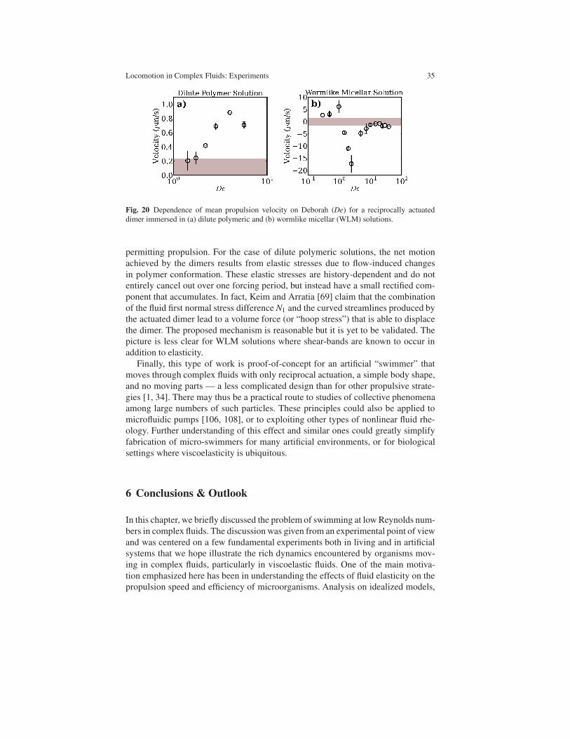
Locomotion in Complex Fluids: Experiments 35
Fig. 20 Dependence of mean propulsion velocity on Deborah (De) for a reciprocally actuated
dimer immersed in (a) dilute polymeric and (b) wormlike micellar (WLM) solutions.
permitting propulsion. For the case of dilute polymeric solutions, the net motion
achieved by the dimers results from elastic stresses due to flow-induced changes
in polymer conformation. These elastic stresses are history-dependent and do not
entirely cancel out over one forcing period, but instead have a small rectified com-
ponent that accumulates. In fact, Keim and Arratia [69] claim that the combination
of the fluid first normal stress difference N1 and the curved streamlines produced by
the actuated dimer lead to a volume force (or “hoop stress”) that is able to displace
the dimer. The proposed mechanism is reasonable but it is yet to be validated. The
picture is less clear for WLM solutions where shear-bands are known to occur in
addition to elasticity.
Finally, this type of work is proof-of-concept for an artificial “swimmer” that
moves through complex fluids with only reciprocal actuation, a simple body shape,
and no moving parts — a less complicated design than for other propulsive strate-
gies [1, 34]. There may thus be a practical route to studies of collective phenomena
among large numbers of such particles. These principles could also be applied to
microfluidic pumps [106, 108], or to exploiting other types of nonlinear fluid rhe-
ology. Further understanding of this effect and similar ones could greatly simplify
fabrication of micro-swimmers for many artificial environments, or for biological
settings where viscoelasticity is ubiquitous.
6 Conclusions & Outlook
In this chapter, we briefly discussed the problem of swimming at low Reynolds num-
bers in complex fluids. The discussion was given from an experimental point of view
and was centered on a few fundamental experiments both in living and in artificial
systems that we hope illustrate the rich dynamics encountered by organisms mov-
ing in complex fluids, particularly in viscoelastic fluids. One of the main motiva-
tion emphasized here has been in understanding the effects of fluid elasticity on the
propulsion speed and efficiency of microorganisms. Analysis on idealized models,

36 Josue Sznitman and Paulo E. Arratia
following the early work of G. I. Taylor in simple Newtonian fluids, showed that in
general fluid elasticity hinders propulsion. Numerical simulations, however, found
that fluid elasticity could in fact enhance propulsion under certain conditions (De
∼ 1). Experiments seem to confirm such predictions, at least qualitatively. For ex-
ample, the swimming speed and efficiency of C. elegans effectively decreases once
the organism is immersed in dilute viscoelastic fluids, while propulsion speed in-
creased for De ∼ 1 in scale up mechanical experiments. In more concentrated poly-
meric solutions, the nematode C. elegans showed a remarkable increase in speed for
solutions possessing polymer networks. Overall, we find that it is very hard to an-
swer question on whether fluid elasticity enhances or hinders the propulsion speed
of microorganisms. In fact, it may be an unfair question altogether as the answer
depends on the type of swimming kinematics (i.e., waveform) and the interactions
of the swimmer with the fluid microstructure (i.e., polymer or particles). That is, the
local details may matter quite bit. Nevertheless, it is clear from both experiments
and simulations that fluid elasticity affects, if not dramatically alters, the motility
behavior of microorganisms.
Parallel to these considerations, we also discussed an exciting avenue of new de-
velopments, that is the possibility of fluid-assisted propulsion. This type of propul-
sion is driven by the nonlinear rheological properties of the fluid such as viscoelas-
ticity and formation of shear-bands. We showed that even for reciprocally-actuated
polar particles, the extra elastic stresses of polymeric fluids can lead to net motion
at low Reynolds numbers. It is important to note that under the same conditions,
the same particle in a Newtonian fluid achieves no net motion due to kinematic re-
versibility. The experiments show that the particle net motion and speed increases
as the elasticity increases. These findings are exciting because such discovery opens
a new mechanism for the study of “active” particles and collective behavior in com-
plex fluids.
So where do we go from here? Of course there is still much to be done and under-
stood. The question of how microorganisms move in complex fluids is still largely
unanswered. We still do not know how microorganisms move in rheologically com-
plex material that possess rate-dependent viscosity, yield stress, and thixotropy. But
a picture is starting to emerge, and it seems that the interaction of the swimmer
with the fluid microstructure is very important. That is in a way not surprising since
microorganisms with different swimming kinematics will likely produce different
velocity fields in a given fluid, and the bulk response will be determined by the way
the fluid microstructure interacts with the velocity field. For example, it is well-
known that extensional flows are more efficient in stretching and aligning polymer
molecules than shear flows, and swimmers that produce flows with a large exten-
sional component are dominated by the fluid extensional viscosity. On the other
hand, swimmers that produce large amounts of curvature may experience a vis-
coelastic instability due to hoop stresses. So knowledge of the velocity field is quite
important, but equally important is how the molecules or particles respond to the
imposed velocity field. In a way, the swimmer may be thought of as a local probe
for fluid rheology. An exciting and important direction that would allow us to gain
a more complete understanding of swimming in complex fluids is to determine how

Locomotion in Complex Fluids: Experiments 37
single polymer molecules, particles, and networks interact with microorganisms.
This direction will ultimately take us from the continuum to a statistical mechanics
description of the problem. The challenge lies in connecting the statistical represen-
tation with the continuum description.
Finally, one can also ask the question of how multiple swimmers interact in New-
tonian fluids and in complex fluids. One would anticipate that the fluid microstruc-
ture will significantly alter the way multiple microorganisms interact. The type of
work is related to the field of active matter or fluid in which active or live particles
are present in the fluid medium. Active fluids differ from their passive counterpart
in that the active particles have the ability to absorb and dissipate energy and to
generate motion and mechanical stresses in the fluid medium. Importantly, these
active particles can drive the system out of equilibrium even in the absence of ex-
ternal forcing. Active fluids exhibit novel properties not seen in conventional (pas-
sive) complex fluids such as large-scale flows and collective motion on length scales
much greater than the particle dimensions [25], anomalous shear-viscosity [130], gi-
ant density fluctuations [105], and enhanced fluid mixing [77]. While much recent
progress has been made, the dynamics and flow behavior (rheology) of such active
complex fluids are still poorly understood. In this case, one can take advantage of a
vast body of knowledge developed to understand passive complex fluids.
Acknowledgements The authors would like to thank David Gagnon, Nathan Keim, Alexander
Leshansky, and Xiaoning Shen for help with the text and in drafting illustrations. This work was
supported by the US-Israel Binational Science Foundation (BSF grant nr. 2011323) and J. Sznit-
man was supported in part by the European Commission (FP7 Program) through a Career Integra-
tion Grant (PCIG09-GA-2011-293604). P.E. Arratia was supported in part by the Army Research
Office through award W911NF-11-1-0488.
References
1. Abbott, J.J., Peyer, K.E., Lagomarsino, M.C., Zhang, L., Dong, L., Kaliakatsos, I.K., Nelson,
B.J.: How should microrobots swim? Int. J. Robot Res. 28 1434 (2009)
2. Alexander, M.: Introduction to soil microbiology. Malabar, FL: R.E. Krieger (1991)
3. Aranson, I.: The aquatic dance of bacteria. Physics 6, 61 (2013)
4. Arratia, P.E., Thomas, C.C., Dioro, J., Gollub, J.P.: Elastic instabilities of polymer solutions
in cross-channel flow. Phys. Rev. Lett. 96, 144502 (2006)
5. Anna, S.L., McKinley, G.H.: Elasto-capillary thinning and breakup of model elastic liquids.
J. Rheol. 45, 115-138 (2001)
6. Berg, H.C.: How to track bacteria. Rev. Sci. Instrum. 42, 868 (1971)
7. Berg, H.C.: The tracking microscope. Adv. Opt. Elect. Microsc. 7, 1–15 (1978)
8. Berg, H.C.: Motile behavior of bacteria. Phys. Today 53, 24–29 (2000)
9. Berg, H.C.: The rotary motor of bacterial flagella. Annu. Rev. Biochem. 79, 19–54 (2003)
10. Berg, H.C.: E. coli in Motion. New York: Springer (2004)
11. Berg, H.C.: Bacterial flagellar motor. Curr. Biol. 18, R689–R691 (2009)
12. Berg, H.C., Brown, D.A.: Chemotaxis in Escherichia coli analysed by three-dimensional
tracking. Nature 239, 500–504 (1972)
13. Berg, H.C., Turner, L.: Movement of microorganisms in viscous environments. Nature. 278,
349-351 (1979)

38 Josue Sznitman and Paulo E. Arratia
14. Berke, A.P., Turner, L., Berg, H.C., Lauga, E.: Hydrodynamic attraction of swimming mi-
croorganisms by surfaces. Phys. Rev. Lett. 101, 038102 (2008)
15. Bird, R.B, Curtiss, C.F., Armstrong, R.C., Hassager, O.: Dynamics of Polymeric Liquids:
Fluid Mechanics. John Wiley & Sons, New York (1987)
16. Boger, D.V.: Highly elastic constant-viscosity fluid. J. Non-Newt. Fluid Mech. 3, 87-91
(1977)
17. Bray, D.: Cell Movements. New York: Garland (2000)
18. Brennen, C., Winet, H.: Fluid mechanics of propulsion by cilia and flagella. Annu. Rev. Fluid
Mech. 9, 339–398 (1977)
19. Brenner, S. The genetics of Caenorhabditis elegans, Genetics 77, 71-94 (1974)
20. Byerly, L., Cassada, R., Russell, R.: The life cycle of the nematode Caenorhabditis elegans.
I. Wild-type growth and reproduction. Dev. Biol. 51, 23-33 1976
21. Chen, C.-Y., Chen, C.-Y., Lin, C.-Y., Hu, Y.-T.: Magnetically actuated artificial cilia for opti-
mum mixing performance in microfluidics. Lab Chip (2013) doi: 10.1039/C3LC50407G
22. Chattopadhyay, S., Moldovan, R., Yeung, C., Wu, X.-L.: Swimming efficiency of bacterium
Escherichia coli. Proc. Natl. Acad. Sci. USA 103, 13712–13717 (2006)
23. Chattopadhyay, S., Wu, X.-L.: The effect of long-range hydrodynamic interaction on the
swimming of a single bacterium. Biophys. J. 96, 2023–2028 (2009)
24. Chaudhury, T.K.: On swimming in visco-elastic liquids. J. Fluid. Mech. 95, 189-197 (1979)
25. Cisneros, L.H., Cortez, R., Dombrowski, C., Goldstein, R.E., Kessler, J.O.: Fluid dynamics
of self-propelled micro-organisms, from individuals to concentrated populations. Exp. Fluids
43, 737–753 (2007)
26. Cohen, N., Boyle, J.H.: Swimming at low Reynolds number: a beginner’s guide to undulatory
locomotion. Contemp. Phys. 51, 103–123 (2010)
27. Corazziari, E.S.: Intestinal mucus barrier in normal and inflamed colon. J. Pediatr. Gastroen-
terol. Nutr. 48, S54–S55 (2009)
28. Cosson, J., Huitorel, P., Gagnon C.: How spermatozoa come to be confined to surfaces. Cell
Motil. Cytoskeleton 54, 56–63 (2003)
29. Croll, N.A.: The behaviour of nematodes: Their Activity, Senses, and Responses. Edward
Arnold, London (1970)
30. Denissenko, P., Kantsler, V., Smith, D.J., Kirkman-Brown, J.: Human spermatozoa migration
in microchannels reveals boundary-following navigation. Proc. Natl. Acad. Sci. USA 109,
8007-8010 (2012)
31. Di Leonardo, R., Dell’ Arciprete, L., Angelani, L., Iebba, V.: Swimming with an image. Phys.
Rev. Lett. 106, 038101 (2011)
32. Doi, M., Edwards, S.F.: The theory of polymer dynamics. New York: Oxford University Press
(1988)
33. Dombrowski, C., Cisneros, L., Chatkaew, S., Goldstein, R.E., Kessler, J.O.: Self-
concentration and large-scale coherence in bacterial dynamics. Phys. Rev. Lett. 93, 098103
(2004)
34. Dreyfus, R., Baudry, J., Roper, M.L., Fermigier, M., Stone, H.A., Bibette, J.: Microscopic
artificial swimmers. Nature 437, 862–865 (2005)
35. Drescher, K., Goldstein, R.E., Michel, N., Polin, M., Tuval, I.: Direct measurement of the
flow field around swimming microorganisms. Phys. Rev. Lett. 105, 168101 (2010)
36. Drescher, K., Leptos, K.C., Goldstein, R.E.: Rev. Sci. Instrum. 80, 014301 (2009)
37. Du, J., Keener, J.P., Guy, R.D., Fogelson, A.L. Low-Reynolds-number swimming in viscous
two-phase fluids. Phys. Rev. E. 85 036304 (2012)
38. Dunkel, J., Heidenreich, S., Drescher, K., Wensink, H.H., Bar, M. Goldstein, R.E.: Fluid
dynamics of bacterial turbulence. Phys. Rev. Lett. 110, 228102 (2013)
39. Fauci, L.J., Dillon, R.: Biofluidmechanics of reproduction. Annu. Rev. Fluid Mech. 38, 371–
394 (2006)
40. Francis, R.J.B., Chatterjee, B., Loges, N.K., Zentgraf, H., Omran, H., Lo, C.W.: Initiation and
maturation of cilia-generated flow in newborn and postnatal mouse airway. Am. J. Physiol.
Lung Cell. Mol. Physiol. 296, L1067–L1075 (2009)

Locomotion in Complex Fluids: Experiments 39
41. Fung, Y.C.: Biomechanics: Mechanical Properties of Living Tissues. Springer Verlag, Second
Edition (2004)
42. Fu, H.C., Shenoy, V.B., Powers, T.R.: Low-Reynolds-number swimming in gels. Europhys.
Lett. 91 24002 (2010)
43. Fu, H.C., Powers, T.R., Wolgemuth, C.: Theory of swimming filaments in viscoelastic media.
Phys. Rev. Lett., 99 258101 (2007)
44. Fu, H.C., Wogelmuth, C.W., Powers, T.R.: Beating patterns of filaments in viscoelastic fluids.
Phys. Rev. E 78, 041913 (2008)
45. Fu, H.C., Wogelmuth, C.W., Powers, T.R.: Swimming speeds of filaments in nonlinearly vis-
coelastic fluids. Phys. Fluids 21, 033102 (2009)
46. Fulford, G.R, Katz, D.F, Powell., R.: Swimming of spermatozoa in a linear viscoelastic fluid.
Biorheology 35, 295309 (1998)
47. Gagnon, D.A., Keim, N.C., Shen, X.N., Arratia, P.A.: Fluid-Induced Propulsion in Wormlike
Micellar Solutions. Submitted for Review (2014)
48. Gagnon, D.A., Shen, X.N., Arratia, P.A.: Undulatory swimming in fluids with polymer net-
works. Europhys. Lett. 104 14004 (2013)
49. Gibbons, I.R.: Cilia and flagella of eukaryotes. J. Cell. Biol. 91, 107s–124s (1981)
50. Ghosh, A., Fischer, P.: Controlled propulsion of artificial magnetic nanostructured propellers.
Nano Lett. 9, 2243–2245 (2009)
51. Ghosh, R., Sznitman, J.: Visualization of nematode C. elegans swimming in a drop, J. Vis.
15, 277-279 (2012).
52. Gray, J.: The movement of sea-urchin spermatozoa. J. Exp. Biol. 32, 775–801 (1955)
53. Gray, J., Hancock, G.J.: The propulsion of sea-urchin spermatozoa. J. Exp. Biol. 32, 802–814
(1955)
54. Gray, J., Lissmann, H.W.: The locomotion of nematodes. J. Exp. Biol. 41, 135–154 (1964)
55. Groisman, A., Steinberg, V.: Elastic turbulence in a polymer solution flow. Nature 405, 53–55
(2000)
56. Groisman, A., Steinberg, V.: Efficient mixing at low Reynolds numbers using polymer addi-
tives. Nature 410, 905–908 (2001)
57. Guasto, J.S., Johnson, K.A., Gollub, J.P.: Oscillatory flows induced by microorganisms swim-
ming in two dimensions. Phys. Rev. Lett. 105, 168102 (2010)
58. Guasto, J.S., Rusconi, R., Stocker, R.: Fluid mechanics of planktonic microorganisms. Annu.
Rev. Fluid Mech. 44, 373–400 (2012)
59. Guzick, D.S., Overstreet, J.W., Factor-Litvak, P., Brazil, C.K., Nakajima, S.T., Coutifaris, C.,
Carson, S.A., Cisneros, P., Steinkampf, M.P., Hill, J.A., Xu, D. Vogel, D.L.: Sperm morphol-
ogy, motility, and concentration in fertile and infertile men. New Engl. J. Med. 345 1388–1393
(2001)
60. Ho, H.C., Suarez, S.S.: Hyperactivation of mammalian spermatozoa: function and regulation.
Reprod. 122, 519–5126 (2001)
61. Houry, A., Gohar, M., Deschamps, J., Tishenki, E., Aymerich, S., Gruss, A., Briandet, R.:
Bacterial swimmers that infiltrate and take over the biofilm matrix. Proc. Natl. Acad. Sci.
USA 109, 13088–13093 (2012)
62. Jana, S., Ho Um, S., Jung, S.: Paramecium swimming in capillary tube. Phys. Fluids 24,
041901 (2012)
63. Josenhans, C., Suerbaum, S.: The role of motility as a virulence factor in bacteria. Int. J. Med.
Microbiol. 291 605–614 (2002)
64. Juarez, G., Lu, K., Sznitman, J., Arratia, P.E.: Motility of small nematodes in wet granular
media. Europhys. Lett. 92, 44002 (2010)
65. Jung, S., Winter, A.G., Hosoi, A.E.: Dynamics of digging in wet soil. Int. J. Nonl. Mech. 46,
602 (2011)
66. Katz, D.F., Blake, J.R., Paverifontana, S.L.: On the movement of slender bodies near plane
boundaries at low Reynolds-number. J. Fluid Mech. 72, 529–549 (1975)
67. Katz, D.F., Bloom, T.D., Bondurant, R.H.: Movement of bull spermatozoa in cervical mucus.
Biol. Reprod. 25, 931–937 (1981)

40 Josue Sznitman and Paulo E. Arratia
68. Katz, D.F., Millis, R.N., Pritchett, T.R.: Movement of human spermatozoa in cervical mucus.
J. Reprod. Fertil. Suppl. 53, 259–265 (1978)
69. Keim, N.C., Garcia, M., Arratia, P.E.: Fluid elasticity can enable propulsion at low Reynolds
number. Phys. Fluids. 24 081703 (2012)
70. Kim, M.J., Bird, J.C., Van Parys, A.J., Breuer, K.S., and Powers, T.R.: A macroscopic scale
model of bacterial flagellar bundling. Proc. Natl. Acad. Sci. USA 100, 15481–15485 (2003)
71. Kim, M.J., Kim, M.J., Bird, J.C., Powers, T.R., Breuer, K.S.: Particle image velocimetry
experiments on a macro-scale model for bacterial flagellar bundling. Exp. Fluids 37, 782–788
(2004)
72. Kim, M.J., Breuer, K.S.: Controlled mixing in microfluidic systems using bacterial chemo-
taxis. Anal. Chem. 43, 955–959 (2007)
73. Kim, M.J., Breuer, K.S.: Microfluidic pump powered by self-organizing bacteria. Small 43,
111–118 (2008)
74. Koehler, S.A., Powers, T.R.: Twirling elastica: kinks, viscous drag, and torsional stress. Phys.
Rev. Lett. 85, 4827–4830 (2000)
75. Konig, K., Svaasand, L., Liu, Y.G., Sonek, G., Patrizio, P., Tadir, Y., Berns, M.W., Tromberg,
B.J.: Determination of motility forces of human spermatozoa using an 800 nm optical trap.
Cell. Mol. Biol. 42, 501–509 (1996)
76. Korngreen, A., Priel, Z.: Simultaneous measurement of ciliary beathing and intracellular cal-
cium. Biophys. J. 67, 377–380 (1994)
77. Kurtuldu,H., Guasto, J.S., Johnson, K.A., Gollub, J.P.: Enhancement of biomixing by swim-
ming algal cells in two-dimensional films. Proc. Nat. Acad. Sci. 108, 10391-10395 (2011)
78. Lai, S.K., Wang, Y.Y., Wirtz, D., Hanes, J.: Micro- and macrorheology of mucus. Adv. Druf
Deliv. Rev. 61, 86–100 (2009)
79. Larson, R.G.: Instabilities in viscoelastic flows. Rheol. Acta 31, 213–236 (1992)
80. Larson, R.G., Shaqfeh, E.S.G., Muller, S.J.: Purely elastic instability in Taylor-Couette flow.
J. Fluid Mech. 218, 573–600 (1990)
81. Larson, R.G.: The Structure and Rheology of Complex Fluids. Oxford University Press
(1999)
82. Lauga, E.: Propulsion in a viscoelastic fluid. Phys. Fluids 19, 083104 (2007)
83. Lauga, E.: Life at high Deborah number. Europhys. Lett. 86, 64001 (2009)
84. Lauga, E., DiLuzio, W.R., Whitesides, G.M., Stone, H.A.: Swimming in circles: motion of
bacteria near solid boundaries. Biophys. J. 90, 400–412 (2006)
85. Lauga, E., Powers, T.R.: The hydrodynamics of swimming microorganism. Rep. Prog. Phys.
72, 096601 (2009)
86. Lebois, P., Sauvage, F., Py, C., Cardoso, O., Ladoux, B., Hersen, P., Di Meglio, J.M.: Loco-
motion control of Caenorhabditis elegans through confinement. Biophys. J. 102, 27912798
(2012).
87. Leoni, M., Kotar, J., Bassetti, B., Cicuta, P., Lagomarsino, M.C.: A basic swimmer at low
Reynolds number. Soft Matter 5, 472–476 (2009)
88. Lenaghan, S.C., Davis, C.A., Henson, W.R., Zhang, Z., Zhang, M.: High-speed microscopic
imaging of flagella motility and swimming in Giardia lamblia trophozoites. Proc. Natl. Acad.
Sci. USA 108, E550–E558 (2011)
89. Lighthill, J.: Flagellar hydrodynamics. Soc. Ind. Appl. Math. Rev. 18, 161–230 (1976)
90. Leshansky, A.M.: Enhanced low-Reynolds-number propulsion in heterogeneous viscous en-
vironments. Phys. Rev. E. 80 051911 (2009)
91. Liu, B., Powers, T.R., Breuer, K.S.: Force-free swimming of a model helical flagellum in
viscoelastic fluids. PNAS 108 1951619520 (2011)
92. Litt, M., Khan, M.A., Wolf, D.P.: Mucus rheology: a relation to structure and function.
Biorheology 13, 37–48 (1976)
93. Lyons, R.A., Djahanbakhch, O., Mahmood, T., Saridogan, E., Sattar, S., Sheaff, M.T., Naf-
talin, A.A., Chenoy R.: Fallopian tube ciliary beat frequency in relation to the stage of men-
strual cycle and anatomical site. Human Rep. 17, 584–588 (2002)
94. Lyons, R.A., Saridogan, E., Djahanbakhch, O.: The reproductive significance of human Fal-
lopian tube cilia. Hum. Reprod. Update 12, 363–372 (2006)

Locomotion in Complex Fluids: Experiments 41
95. Magariyama, Y., Kudo, S.: A mathematical explanation of an increase in bacterial swimming
speed with viscosity in linear-polymer solutions. Biophys. J. 83, 733–739 (2002)
96. McKinley, G.H., Sridhar, T.: Filament-stretching rheometry of complex fluids. Ann. Rev.
Fluid Mech. 34, 375-415 (2002)
97. McPartlin, L.A., Suarez S.S., Czaya, C.A., Hinrichs, K., Bedford-Guaus, S.J.: Hyperactiva-
tion of stallion sperm is required for successful in vitro fertilization of equine oocytes. Biol.
Reprod 81, 199–206 (2009)
98. Mitchell, D.R.: Chlamydomonas flagella. J. Phycol. 36, 261–273 (2000)
99. Montenegro-Johnson, T.D., Smith, D.J., Loghin, D.: Physics of rheologically enhanced
propulsion: Different strokes in generalized Stokes 25, 081903 (2013)
100. Moore, H., Dvorakova, K., Jenkins, N., Breed, W.: Exceptional sperm cooperation in the
wood mouse. Nature 418, 174–177 (2002)
101. Murphy, D.W., Webster, D.R., Yen, J.: A high-speed tomographic PIV system for measuring
zooplanktonic flow. Limnol. Oceanogr. Methods 10, 1096–1112 (2012)
102. Niebur, E., Erdos, P.: Theory of the locomotion of nematodes. Biophys. J. 60, 1132–1146
(1991)
103. Nakamura, S., Adachi, Y., Goto, T., Magariyama, Y.: Improvement in motion efficiency of
the sprirochete Brachyspira pilosicoli in viscous environments. Biophys. J. 90 3019-3026
(2006)
104. Nakahari, T., Nishimura, A., Shimamoto, C., Sakai, A., Kuwabara, H., Nakano, T., Tanaka,
S., Kohda, Y., Matsumura, H., Mori, H.: The regulation of ciliary beat frequency by ovarian
steroids in the guinea pig Fallopian tube: interactions between oestradiol and progesterone.
Biomed. Res. 32, 321–328 (2011)
105. Narayan, V., Ramaswamy, S., Menon, N.: Long-lived giant number fluctuations in a swarm-
ing granular nematic. Science 317, 105-108 (2007)
106. Normand, T., Lauga, E.: Flapping motion and force generation in a viscoelastic fluid. Phys.
Rev. E. 78 061907 (2008)
107. Oldenburg, A.L., Chhetri, R.K., Hill, D.B., Button, B.: Monitoring airway mucus flow and
ciliary activity with optical coherence tomography. Biomed. Opt. Exp. 3, 1970–1992 (2012)
108. Pak, O.S., Normand, T., Lauga, E.: Pumping by flapping in a viscoelastic fluid. Phys. Rev. E.
81 036312 (2010)
109. Pawashe, C., Floyd, S., Sitti, M.: Multiple magnetic microrobot control using electrostatic
anchoring. Appl. Phys. Lett. 94, 164108 (2009)
110. Polizu, S., Savadogo, O., Poulin, P., Yahia, L.: Applications of carbon nanotubes-based bio-
materials in biomedical nanotechnology. J. Nanosci. Nanotech. 6, 1883–1904 (2006)
111. Powell, R.L., Aharonson, E.F., Schwarz, W.H., Proctor, D.F., Adams, J.K., Reasor, M.: Rheo-
logical behavior of normal tracheobronchial mucus of canines. J. Appl. Physiol. 37, 447–451
(1974)
112. Purcell, E.M.: Life at low Reynolds number. Am. J. Phys. 45, 3–11 (1977)
113. Qian, B., Powers, T.R., Breuer, K.S.: Shape transition and propulsive force of an elastic rod
rotating in a viscous fluid. Phys. Rev. Lett 100, 078101 (2008)
114. Riedel, I.H., Kruse, K., Howard, J.: A self-organized vortex array of hydrodynamically en-
trained sperm cells. Science 309, 300–303 (2005)
115. Rodd, A.B., Dunstan, D.E., Boger, D.V.: Characterisation of xanthan gum solutions using
dynamic light scattering and rheology. Carbohyd. Polym. 45 159–174 (2000)
116. Rodenborn, B., Chen, C.-H., Swimmey, H.L., Liu, B., Zhang, H.P.: Propulsion of microor-
ganisms by a helical flagellum. Proc. Natl. Acad. Sci. USA 110, E338–E347 (2013)
117. Rotschild, L.: Non-random distribution of bull spermatozoa in a drop of sperm suspension.
Nature 198, 1221–1222 (2005)
118. Sakar, M.S., Lee, C., Arratia, P.E.: Flagellar dynamics in viscous fluids. Phys. Fluids 21,
091107 (2009)
119. Saintillan, D.: A quantiative look into microorganism hydrodynamics. Physics 3, 84 (2010)
120. Satir, P., Sleigh, M.A.: The physiology of cilia and mucociliary interactions. Annu. Rev.
Physiol. 52, 137–155 (1990)

42 Josue Sznitman and Paulo E. Arratia
121. Schneider, W.R., Doetsch, R.N.: Effect of Viscosity on Bacterial Motility. J. Bacteriol. 117
696-701 (1974)
122. Setter, E., SBucher, I., Haber S: Low-Reynolds-number swimmer utilizing surface traveling
waves: analytical and experimental study. Phys. Rev. E 85, 066304 (2012)
123. Shaqfeh, E.S.G.: Purely elastic instabilities in viscometric flows. Annu. Rev. Fluid Mech. 28,
129–185 (1996)
124. Shen, X.N., Arratia, P.E.: Undulatory Swimming in Viscoelastic Fluids. Phys. Rev. Lett. 106,
208101 (2011)
125. Shen, X.N., Sznitman, J., Krajacic, P, Lamitina, T., Arratia, P.E.: Undulatory locomotion of
Caenorhabditis elegans on wet surfaces. Biophys. J. 102, 2772–2781 (2012)
126. Shields, A.R., Fiser, B.L., Evans, B.A., Falvo, M.R., Washburn, S., Superfine, R.: Biomimetic
cilia arrays generate simultaneous pumping and mixing regimes. Proc. Natl. Acad. Sci. USA
36, 15670–15675 (2010)
127. Sitti, M.: Miniature devices: voyage of the microrobots. Nature 458, 1121–1122 (2009)
128. Spagnolie, S.E., Liu, B., Powers, T.R.: Locomotion of helical bodies in viscoelastic fluids:
enhanced swimming at large helical amplitudes. Phys. Rev. Lett. 111, 068101 (2013)
129. Sokolov, A., Aranson, I.S., Kessler, J.O., Goldstein, R.E.: Concentration dependence of the
collective dynamics of swimming bacteria. Phys. Rev. Lett. 98, 158102 (2007)
130. Sokolov, A., Aranson, I.S.: Reduction of Viscosity in Suspension of Swimming Bacteria.
Phys. Rev. Lett. 103 24, 148101 (2009)
131. Suarez, S.S., Ho, H.C.: Hyperactivated motility in sperm. Reprod. Domest. Anim. 38, 119–
124 (2005)
132. Suarez, S.S., Pacey, A.A.: Sperm transport inthe female reproductive tract. Human Reprod.
Update 12, 23–37 (2006)
133. Sznitman, J., Shen, X., Sznitman, R., Arratia, P.E.: Propulsive force measurements and flow
behavior of undulatory swimmers at low Reynolds number. Phys. Fluids 22 121901 (2010)
134. Tadir, Y., Wright, W.H., Vafa, O., Ord, T., Asch, R.H., Berns, M.W.: Force generated by
human sperm correlated to velocity and determined using a laser generated optical trap. Fertil.
Steril. 53, 944–947 (1990)
135. Tam, P.Y., Katz, D.F., Berger, S.A.: Non-linear properties of cervical mucus. Biorheology
17, 465–478 (1980)
136. Tao, W., Zhang, M.: A genetic algorithm-based area coverage approach for controlled drug
delivery using microrobots. Nanomed. 1, 91–100 (2005)
137. Taylor, G.I.: Analysis of the swimming of microscopic organisms. Proc. R. Soc. A. 209,
447–461 (1951)
138. Taylor, G.I.: The action of waving cylindrical tails in propelling microscopic organisms. Proc.
R. Soc. A. 211, 225–239 (1951)
139. Teff, Z., Priel, Z., Gheber, L.A.: Forces applied by cilia measured on explants from mucocil-
iary tissue. Biophys. J. 92, 1813–1823 (2007)
140. Teran, J., Fauci, L., Shelley, M.: Viscoelastic Fluid Response Can Increase the Speed and
Efficiency of a Free Swimmer. Phys. Rev. Lett. 104, 038101 (2010).
141. Tierno, P., Golestanian, R., Pagonabarraga, I., Sagues, F.: Controlled swimming in confined
fluids of magnetically actuated colloidal rotors. Phys. Rev. Lett. 101, 218304 (2008)
142. den Toonder, J.M., Onck, P.R.: Microfluidic manipulation with artificial/bioinspired cilia.
Trends Biotech. 31, 85–91 (2012)
143. Trouilloud, R., Yu, T.S., Hosoi, A.E., Lauga, E.: Soft swimming: exploiting deformable in-
terfaces for low Reynolds number locomotion. Phys. Rev. Lett. 101, 048102 (2008)
144. Trouton, F.T.: On the coefficient of viscous traction and its relation to that of viscosity. Proc.
R. Soc. Lond. A. 77, 426440 (1906)
145. Trueman, E.R.: The locomotion of Soft-Bodied Animals. J.W. Arrowsmith Ltd., Bristol, UK
(1975)
146. Turner, L., Ryu, W.S.: Real-time imaging of fluorescent flagellar filaments. J. Bacteriol. 182,
2793–2801 (2000)
147. Velez-Cordero, J.R., Lauga, E.: Waving transport and propulsion in a generalized Newtonian
fluid. J. Non-Newt. Fluid Mech. 199, 37-50 (2013)
Locomotion in Complex Fluids: Experiments 43
148. Vicsek, T., Zafeiris, A.: Collective motion. Phys. Reports 517, 71–140 (2012)
149. Von Stetina, S.E., Treinin, M., Miller, D.M.: The motor circuit. Int. Rev. Neurobiol. 69, 125–
167 (2006)
150. Wallace, H.R.: The dynamics of nematode movement. Annu. Rev. Phytopathol. 6, 91–114
(1968)
151. Weiss, P.: Artificial animalcules: In the microscopic realm, machines learn to swim. Science
News 169, 107–108 (2006)
152. Wereley, S.T., Meinhart, C.D.: Recent advances in micro-particle image velocimetry. Annu.
Rev. Fluid Mech. 42, 557–576 (2010)
153. Werner, M., Simmons, L.W.: Insect sperm motility. Biol. Rev. 83, 191–208 (2008)
154. White, J.G., Southgate, E., Thomson J.N., Brenner, S.: The structure of the nervous system
of the nematode Caenorhabditis elegans. Philo. T. Roy. Soc. B 314, 1–340 (1986)
155. Wiggins, C.H., Riveline, D., Ott, A., Goldstein, R.E.: Trapping and wiggling: Elastohydro-
dynamics of driven microfilaments. Biophys. J. 74, 1043–1060 (1998)
156. Winter, A.G., Deits, R.L.H., Hosoi, A.E.: Localized fluidization burrowing mechanics of
Ensis directus. J. Exp. Biol. 215, 2072–2080 (2012)
157. Wood, W.: The Nematode Caenorhabditis elegans. Cold Spring Harbor Laboratory (1988)
158. Wooley, D.M.: Motility of spermatozoa at surfaces. Reprod. 126, 259–270 (2003)
159. Wu, X.L., Libchaber, A.: Particle diffusion in a quasi-two-dimensional bacterial bath. Phys.
Rev. Lett. 84, 3017–3020 (2000)
160. Xi, L., Graham, M.D.: A mechanism for oscillatory instability in viscoelastic cross-slot flow.
J. Fluid. Mech. 622, 145–165 (2000)
161. Yang, J., Wolgemuth, C.W., Huber, G.: Kinematics of the swimming of spiroplasma. Phys.
Rev. Lett. 102, 218102 (2009)
162. Yazdi, S., Ardekani, A.M.: Bacterial aggregation and biofilm formation in a vortical flow.
Biomicrofluidics 6, 044114 (2012)
163. Yu, T.S., Lauga, E., Hosoi, A.E.: Experimental investigations of elastic tail propulsion at low
Reynolds number. Phys. Fluids 18, 091701 (2006)
164. Zhang, L., Abbott, J.J., Dong, L., Peyer, K.E., Kratochvil, E., Zhang, H., Bergeles, C., Nel-
son, B.J.: Characterizing the swimming properties of artificial baterial flagella. Nano Lett. 9,
3663–3667 (2009)
165. Zhong, S., Moored, K.W., Pinedo, V., Garcia-Gonzalez, J., Smits, A.J.: The flow field and
axial thrust generated by a rotating rigid helix at low Reynolds numbers. Exp. Therm. Fluid
Sci. 46, 1–7 (2013)
![Locomotion [2015]](https://static.fdocuments.in/doc/165x107/55d39c9ebb61ebfd268b46a2/locomotion-2015.jpg)