Vertebroplasty as a treatment for osteoporotic compression fractures

DISSERTATION ON
LOCKING COMPRESSION PLATING FOR OSTEOPOROTIC AND PERI-ARTICULAR
FRACTURES – A SHORT TERM OUTCOME ANALYSIS
Submitted forM.S.Degree examination
Branch II – Orthopaedic Surgery
INSTITUTE OF ORTHOPAEDIC AND TRAUMATOLOGYMADRAS MEDICAL COLLEGE &
GOVERNMENT GENERAL HOSPITAL,THE TAMILNADU DR.M.G.R. MEDICAL UNIVERSITY
CHENNAI
MARCH 2010

CERTIFICATE
This is to certify that this dissertation entitled “Prospective study on the Locking
compression plating for osteoporotic and peri-articular fractures.” submitted by
Dr.Navin Balasubramanian appearing for Part II, M.S. Branch II - Orthopaedics
degree examination in March 2010 is a bonafide record of work done by him under my
direct guidance and supervision in partial fulfilment of regulations of The Tamil Nadu
Dr. M.G.R. Medical University, Chennai.
I forward this to The Tamil Nadu Dr. M.G.R. Medical University, Chennai, Tamil
Nadu, India.
Prof. MAYIL VAHANAN NATARAJAN M.S.Ortho., M.Ch. Ortho (Liverpool) .Ph.D(Ortho Onco)., D.Sc.,FAMS.,FRCS(Eng)Director ,Institute of Orthopaedics and TraumatologyMadras Medical College & Government General HospitalChennai- 600 003
DEAN,Madras Medical College,Govt. General Hospital,Chennai - 600 003.
ACKNOWLEDGEMENT
Any prospective study in a place as big as this Institution requires the support and guidance of
a lot of people. It would only be appropriate if all the hours they have put in are properly remembered
and acknowledged. My sincere thanks to Dr.MOHANASUNDARAM M.D., Dean, Madras Medical
College, for permitting the utilization of the resources and clinical materials in this hospital.
I will forever be indebted to my guide and our Head of the Department Padma
Laureate PROF. MAYIL VAHANAN NATARAJAN, M.S.Ortho., M.Ch., Ortho
(Liverpool) ., D.Sc.,Ph.D., ( Orthopaedic Oncology)FAMS.,FRCS(Eng). He has always been a
constant source of inspiration with his advice and guiding me through the finer aspects
of this study. Without him this study would not have been possible.
My sincere thanks and gratitude to PROF. R.H.GOVARDHAN M.S.Ortho.,
D.Ortho., for his support, guidance and encouragement during the study.
My sincere thanks and gratitude to PROF .V.THULASIRAMAN M.S.Ortho.,
D.Ortho., for his constant inspiration and guidance during this study.
My sincere thanks and gratitude to PROF. S. SUBBIAH., M.S.Ortho., D.Ortho., for
his valuable guidance.
I am grateful to Dr.R.SUBBIAH. M.S.Ortho. D.Ortho, Reader in Spine Surgery, for
his support in this study.
I am profoundly thankful to Dr.NALLI R UVARAJ,M.S. Ortho., D.Ortho., DNB
Ortho., Reader in Spine Surgery, for all his valuable inputs to this study.
My special thanks to Dr. ANTONY VIMALRAJ for his constant encouragement
and valuable guidance throughout the study.
I sincerely thank Dr.R.SELVARAJ, Dr. V. MAZHAVAN, Dr.B.PASUPATHI,
Dr.T.R.RAMESH PANDIAN, Dr.N.B. THANMARAN, Dr. A.
SHANMUGASUNDARAM, Dr.VELMURUGAN, Dr. S. KARUNAKARAN, Dr. NALLI.
R.GOPINATH, Dr. K.P.MANIMARAN, Dr. PRABHAKARAN Dr N. MUTHALAGAN,
Dr. SENTHIL SAILESH, Dr. SASIDHARAN who have each been great mentors in their
own way.
I must thank all my fellow post graduates, staff members of the Department of
Orthopaedic Surgery, theatre staff and Anesthetists who have all chipped in with their
support during the study.
Last but not the least I am immeasurably indebted to all our patients who
consented and cooperated with this study and without whom this study would not have
been possible
INDEX
S.No. TOPIC Page No.
1. INTRODUCTION 1
2. ANATOMICAL CONSIDERATIONS 4
3. OSTEOPOROSIS- Overview 20
4. SURGICAL CONSIDERATIONS IN OSTEOPOROSIS 22
5. ETIOLOGY 26
6. TREATMENT OPTIONS 27
7. HISTORY OF PLATING 31
8. AIM 41
9. MATERIALS & METHODS 42
10. ILLUSTRATIONS 45
11. OBSERVATION & RESULTS 56
12. DISCUSSION 62
13. CONCLUSION 65
14. REFERENCES 67
15. PROFORMA 71
16. MASTER CHART 75

INTRODUCTION
Peri-articular and osteoporotic fractures of long bones are
becoming more common and are very challenging injuries to treat even
for a veteran orthopaedician. Peri-articular fractures occur in two
different age groups -due to different types of injuries. In young patients
peri-articular fractures occur due to high velocity injury such as road
traffic accidents, fire arm injuries and sport’s injuries while in elderly
patients with osteoporosis it occurs usually due to low velocity injury like
fall during walking. Also these conditions do result from fractures in the
young treated by conservative methods and which in the long term end up
in non-unions and further more these conditions are compounded by
disuse osteoporosis.
Because of the proximity of peri-articular fractures to the
corresponding joints, regaining full motion and function may be difficult.
Also achieving full union rates are increasingly difficult because of the
lack of availability of good bone stock which is very common in peri-
articular fractures because of the cancellous nature of the metaphyseal
fragment. The incidences of malunion, nonunion, and infection are
relatively high in many reported series. In older patients, treatment may
be complicated by coexisting osteoporosis.
6
There are multiple options for the treatment of these fractures with
their associated merits and demerits. Anatomical restoration of the
articular surface in cases of peri-articular fractures and good fracture
alignment and adequate compression in osteoporotic fractures along with
secure fixation of both proximal and distal fragments are the key to
achieve good functional outcome in these fractures to prevent early
secondary osteoarthritis.
Treatment of these fractures have been a controversial subject over
the past two decades. There have been a changing philosophy towards
surgical treatment of these complicated fractures. Close management of
these fractures was the treatment of choice until 1970. This was due to
non availability of appropriate implants and lack of proper techniques.
Apart from the usual problems of confining elderly patient to bed,
conservative methods at any age may be complicated by joint stiffness,
malunion and nonunion.
Early surgical stabilization can facilitate care of the soft tissue,
permit early mobility and reduces the complexity of nursing care. Open
reduction and internal fixation has been advocated, using implants,
including the conventional dynamic compression plates,angle blade plate,
fickle devices, Rush roads, Ender nails, dynamic condylar screw,
7
condylar buttress plate and interlocking nails.
The use of fixed angle devices require certain amount of bone
stock present, which limits their use in some fracture types. The
conventional plates are also associated with their own demerits such as
screw pullout, implant failure and unstable fixation needing postoperative
immobilisation. Delay in postoperative mobilization results in stiffness of
the joint which is an indicator of poor outcome.
A locking plate decreases the screw-plate toggle and motion at the
bone-screw interface and provides more rigid fixation. Rigid fixation is
felt to be one key to the successful treatment of these fractures. But
fixation in osteoporotic and comminuted fractures is difficult to obtain
anatomical reduction and adequate purchase.
So now with the evolution of locking compression plating for
osteoporotic and peri-articular fractures especially for the comminuted
intra – articular fractures many of the older demerits could be addressed
which includes the increased stability due to locking compression plating
principle, multiple screw options in the distal fragment providing option
for fixing the multiple fragments restoring the anatomical congruity and
providing stable fixation of the distal fragment with the proximal
fragment with resulting increased stability allowing for early
8
mobilization.
Anatomical considerations:
The most commonly encountered regions where peri-articular
fractures pose a problem are in order of occurrence distal radius, distal
femur, proximal humerus and distal tibia. Hence their relevant anatomy
will be discussed in particular with associated radiological assessment of
such fractures.
DISTAL RADIUS :
Distal Radius Fracture Anatomy
• Distal radius carries 80% of axial load
• ROM-80°dorsiflexion, 85°palmarflexion, 90°pro\sup,25°radial
deviation,35°ulnar deviation
• Distal radius 3 column anatomy: Radial column (strong cortical
bone), Intermediate column (contains lunate facet and sigmoid
notch); Distal ulna column (contains TFCC)
• Radial inclination=23°,radial length=12mm, volar tilt=11°, ulnar
variance -0.6mm, scapholunate angle = 60° +/- 15°
9

•
• Sensory branch of radial nerve becomes subcutaneous 5-10cm
proximal to radial styloid in interval between brachioradialis and
ECRL. It bifurcates before wrist. Dorsal branch 1-3cm radial to
Listers. Supplies 1st and 2nd web spaces. Palmar branch passes
within 2cm of 1st dorsal compartment provides sensation to
dorsolateral thumb after passing directly over EPL.
• Palmar cutaneous branch of the Median nerve arises from the
Median nerve @4-6cm proximal to the volar wrist crease and
travels between the FCR and median nerve. Supplies sensation to
10
the thenar area.
• Dorsal cutaneous branch of ulnar n arises deep to FCU, becomes
SQ 5cm from pisiform-has multiple branches
Distal Radius Fracture Xray
• PA , Lateral wrist films. Normal radiographic parameters: Radial
inclination=23°,radial length=12mm, volar tilt=11°, scapholunate
angle = 60° +/-15°. Assess ulnar variance, carpal alignment and
sigmoid notch conguence
• Signs of DRUJ injury: fracture at the base of the ulnar styloid,
widening of the DRUJ space seen on the P/A xray, >20° of dorsal
radial angulation, and >5 mm of proximal displacement of the
distal part of the radius.
• 1mm-2mm sagital CT best to view articular depression fx
• MRI if TFCC or scapholunate ligment tears suspected
Distal Radius Acceptable Reduction
• <2mm articular stepoff
• <5mm shortening
• <10° dorsal tilt
11

Distal Radius FractureFx Classification/Treatment
12

• AO Classification / Treatment : AO Classification
• AO Type A=extra-articular
• AO Type B=partial articular; fx of radial styloid, medial corner,
die-punch fracture of central articular surface
• AO Type C=complex articular; high-energy, none of the articular
surface remains in continuity with the metaphysic. Involvement of
>50% of the diameter of the metaphysic as seen on any radiograph,
comminution of at least 2 corticies of the metaphysic, or >2.0mm
of shortening of the radius.
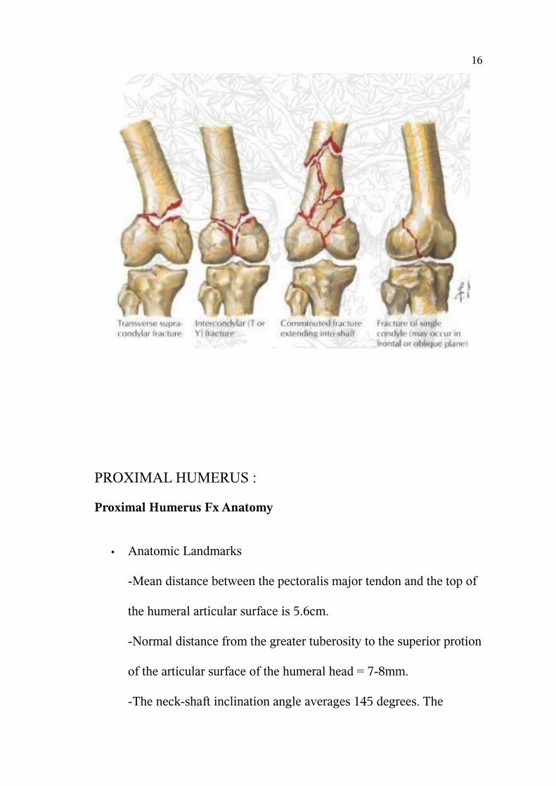
DISTAL FEMUR :
Distal Femur Fx Anatomy
• Hoffa Fragment = coronal (frontal) plane fragment associated with
comminution in the intercondylar notch. Present in @1/3 of Type C
fractures.
Distal Femur Fx Clinical Evaluation
• ATLS resuscitation. These can be high enegery injuries, assessment
should begin with the A,B,C's.
• Obvious deformity of knee/thigh often with limb shortening
13
• Document neurovascular exam before and after any treatment.
Distal Femur Fx Xray
• A/P and lateral views of the knee.
• CT: Ct scan is nearly always indicated for pre-operative planning
as there is a high association with coronal plane fractures which are
difficult to see on plane films.
Distal Femur Fx Classification/Treatment
• AO Classification
• Type A=extraarticular
• Type B=unicondylar fractures
• Type C=intrarticular fractures
14
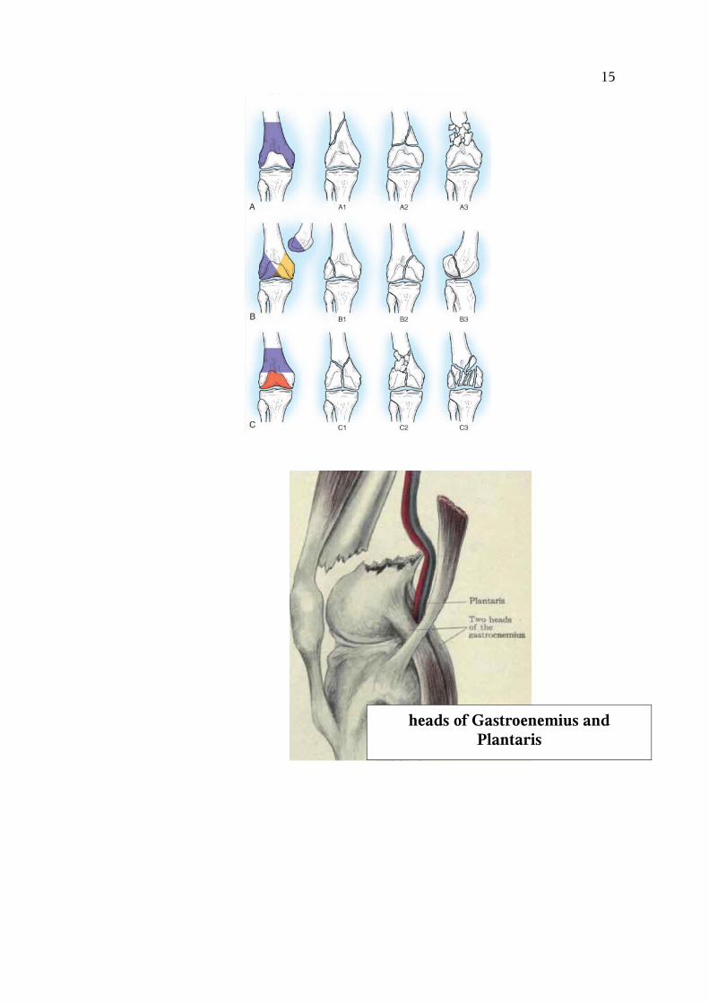
heads of Gastroenemius and Plantaris
15
PROXIMAL HUMERUS :
Proximal Humerus Fx Anatomy
• Anatomic Landmarks
-Mean distance between the pectoralis major tendon and the top of
the humeral articular surface is 5.6cm.
-Normal distance from the greater tuberosity to the superior protion
of the articular surface of the humeral head = 7-8mm.
-The neck-shaft inclination angle averages 145 degrees. The
16
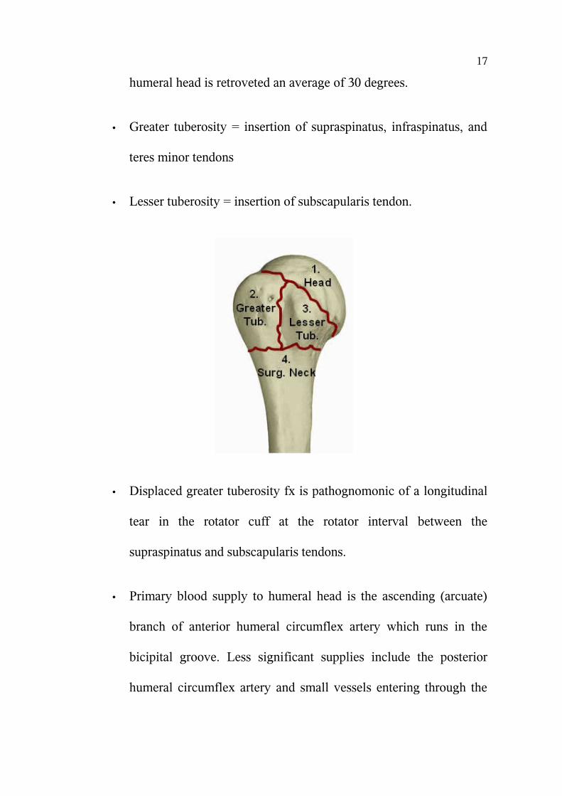
humeral head is retroveted an average of 30 degrees.
• Greater tuberosity = insertion of supraspinatus, infraspinatus, and
teres minor tendons
• Lesser tuberosity = insertion of subscapularis tendon.
• Displaced greater tuberosity fx is pathognomonic of a longitudinal
tear in the rotator cuff at the rotator interval between the
supraspinatus and subscapularis tendons.
• Primary blood supply to humeral head is the ascending (arcuate)
branch of anterior humeral circumflex artery which runs in the
bicipital groove. Less significant supplies include the posterior
humeral circumflex artery and small vessels entering through the
17

rotator cuff insertions.
• Humeral head vascularity after fracture can be estimated by the
amount of metaphyseal head extension, <8mm is associated with
ischemia; Medial hinge disruption >2mm is associated with
ischemia. If both indicate ischmia the positive predictive value of
ischemia for an anatomic neck fx is 97%.
• Most common site of injury to the axillary artery is in the third
part(named in relation to the pec minor) of the artery at the origin
of the anterior and posterior humeral circumflex arteries.
• Deforming forces: Pectoralis major pulls the shaft medially,
anteriorly and internally rotates. Supraspinatus abducts the head
fragment in two part fractures. If greater tuberosity is fractured it is
pulled superiorly and posteriorly by the supraspinatus and
infraspinatus. Lesser tuberosity fractures are pulled medially.
18

Proximal Humerus Fx Clinical Evaluation
• Generally complain of shoulder pain after a fall
• Swelling and ecchymosis in shoulder which can expend into chest
wall and down arm.
• Document NV exam, especially axillary nerve.
• Assess for head injury, LOC, cardiac/neurologic reasons for fall.
Proximal Humerus Fx Xray / Diagnostic Tests
19
• AP , scapular lateral and axillary views. Ensure humeral head is not
dislocated.
• AP in external rotation best demonstrates greater tuberosity
fractures. AP in internal rotation best demonstrates lesser
tuberosity fractures.
• CT may be useful in determining fracture type (head splitting) and
displacement, especially in greater tuberosity fractures. Helpful for
pre-op planning.
• MRI generally not useful.
• Consider EMG/NCV if neurologic injury is suspected, occurs in
67% of proximal humerus fractures.
• Angiogram: consider for diminished radial/brachial pulse,
expanding hematoma, changing neurologic status.
20
Proximal Humerus Fx Classification / Treatment
• Neer classification based on parts(shaft, head, GT, LT);
displacement = >10mm or >45 degree angulation.. Poor
interobserver reliability. CT improves interobserver reliablity.
• Minimally displaced = sling, PT within 2 wks. Functional
outcome, ROM and pain are significantly better when PT is started
within first two weeks..
• Proximal Humerus Surgical Indications: fx 1cm displaced or have
>45 degrees angulation or >10mm tuberosity displacement, open
fx, unable to reduce by closed means,
• Greater tuberosity 2-part fracture
• Surgical neck 2-part fracture
• Lesser tuberosity 2-part fracture
• Valgus impacted 4-part fx with good bone: ORIF. Non-op
outcomes = 80% adjusted Constant score
• 4-Part Fracture: insufficient evidence is available to determine best
treatment option. Increased pain with non-op treatment, equivalent
21
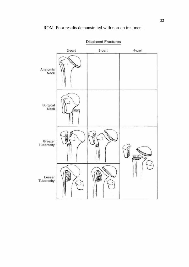
ROM. Poor results demonstrated with non-op treatment .
22
DISTAL TIBIA
Pilon Fracture Etiology / Epidemiology / Natural History
• Pilon fractures are fractures involving the articular weight bearing
surface of the distal tibia.
• Usually high energy axial load (MVC, fall from height),
occasionally low-energy rotation/torsion
• Foot postion determines fracture pattern: if plantar flexed =
posterior tibial fragment, neutral = entire articular surface,
dorsiflexed=anterior fragments
• 7-10% of tibial fractures
Pilon Fracture Anatomy
• Distal tibia fractures within 5cm of the ankle
Pilon Fracture Clinical Evaluation
• Assess vascularity by evaluating dorsalis pedis and posterior tibial
pulses as well as distal capillary refill
• Evaluate soft tissues.
23
Pilon Fracture Xray
• A/P, lateral and mortise views of the ankle; A/P, lateral tibia films
• CT scan indicated for pre-op planning, CT scans should be done in
traction with 3D reconstructions.
Pilon Fracture Classification/Treatment
• Soft tissues injuries are classified according to Tscherne and
Gotzen.
• Open fracture classified per Gustilo and Anderson.
24

• Principles of Treatment: restoration of fibular length, anatomic
reduction of tibial articular surface, bone grafting of metaphyseal
defects, medial buttress plating to prevent varus
• Treatment: initial fixation of the fibula with temporary spanning
external fixation with delayed conversion to internal fixation when
soft tissues permit, generally 14-21 days.
• closed fractures should be placed in calcaneal traction and a
Bohler-Braun frame.
• open fractures/compartment syndromes should be taken to OR for
2-pin traveling traction. (one 6mm centrally threaded calcaneal pin
and one proximal tibia pin at level of the fibular head with
quadrilateral frame.
• AO comprehensive Classification of Fractures of long Bones
• Ruedi Allgower Classification.
• Type A=extra-articular=@92% good/excellent results,
• Type B=partial articular=@85% good/excellent results,
• Type C=complete articular=@60% good/excellent results
25
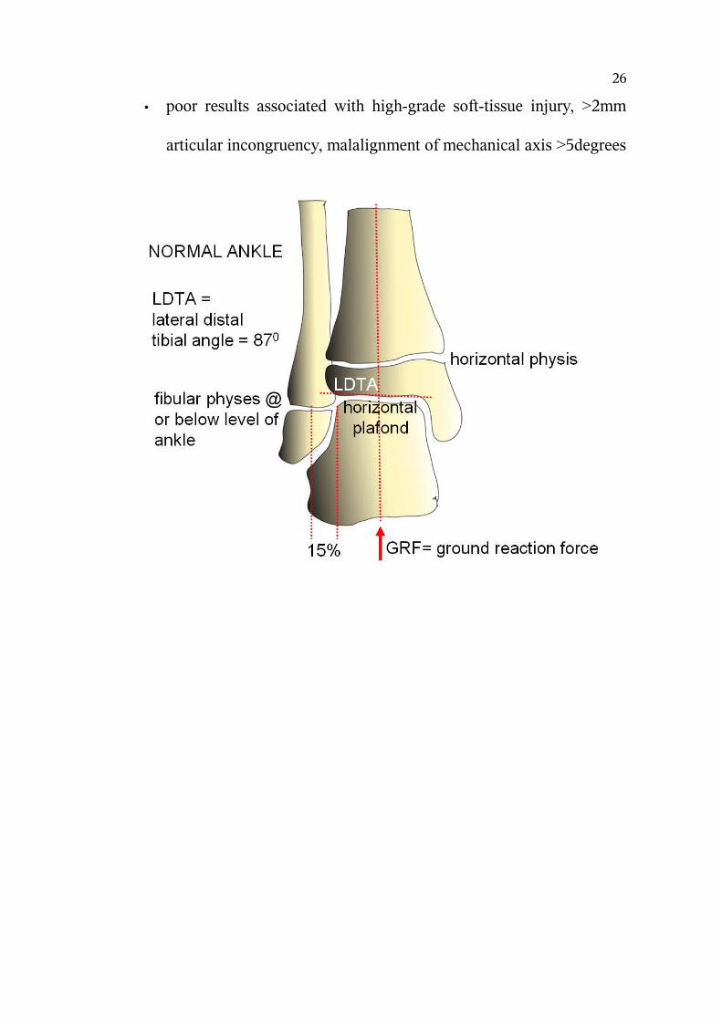
• poor results associated with high-grade soft-tissue injury, >2mm
articular incongruency, malalignment of mechanical axis >5degrees
26
OSTEOPOROSIS
Osteoporosis is a condition marked by reduced bone strength,
which can lead to an increased risk of fractured, or broken, bones.
Osteoporosis was first observed in Egypt in 990 BC and has therefore
been known about for many centuries. The strength of a person’s bones
is affected by their bone mass (amount of bone) and bone quality.
Osteoporosis is the major underlying cause of fractures in
postmenopausal and older women. Fractures occur most often in bones of
the hip, spine and wrist, but any bone can be affected. Some fractures can
be permanently disabling, especially when they occur in the hip. One of
the commonest risk factors associated with fracture is a fall .
Approximately one third of community dwellers aged 65 years or
more and 50% to 60% of residents of nursing and old people's homes fall
each year with women falling more than men . Fractures, dislocations, or
serious soft tissue injuries result from about 10% to 15% of the falls in
patients living in the community and from about 15% to 20% of falls in
institutionalized patients (14,15,16). Fractures occur in 3% to 12% of
falls in the elderly being more common in women than men (10).
Fragility fractures also impose an enormous cost on society. Hip fracture
is a major cause of hospital admission in the elderly.
27
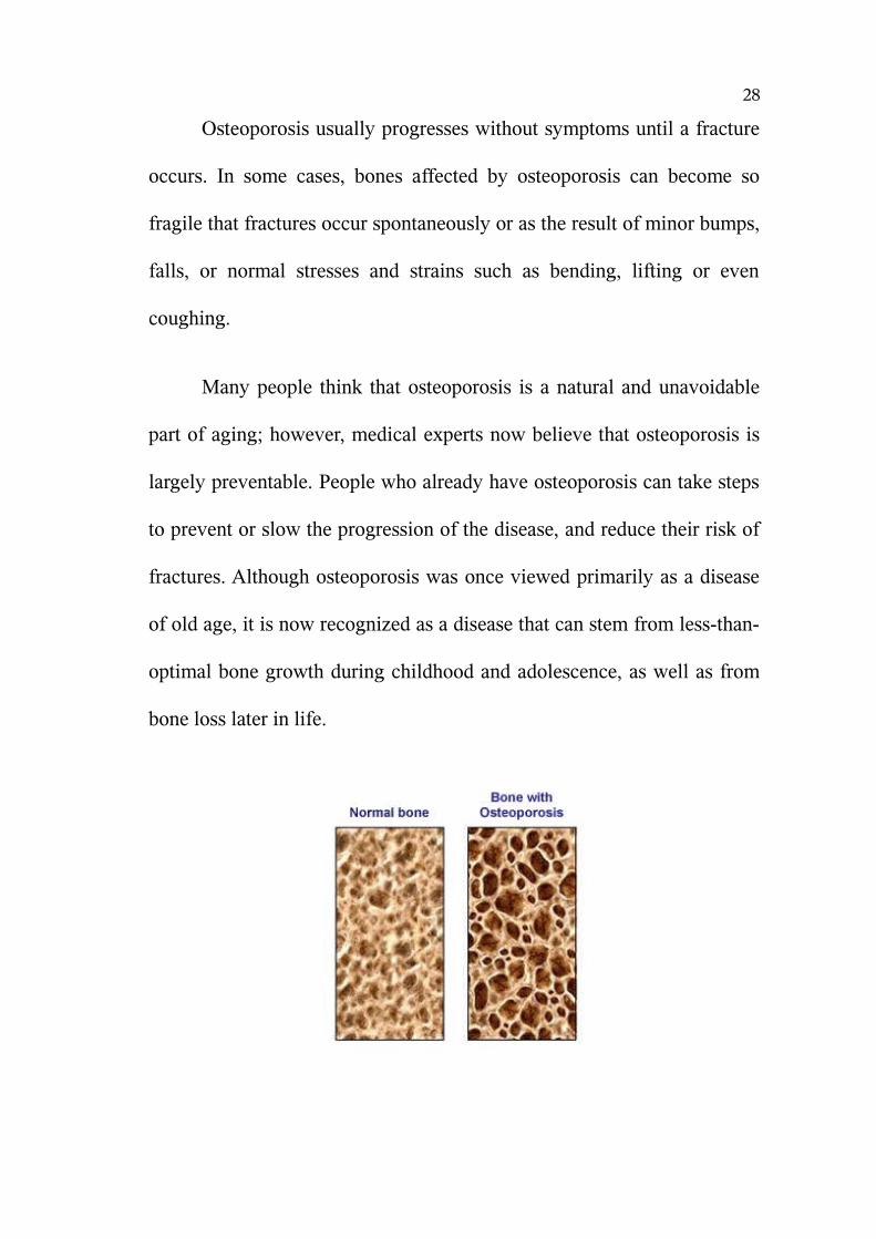
Osteoporosis usually progresses without symptoms until a fracture
occurs. In some cases, bones affected by osteoporosis can become so
fragile that fractures occur spontaneously or as the result of minor bumps,
falls, or normal stresses and strains such as bending, lifting or even
coughing.
Many people think that osteoporosis is a natural and unavoidable
part of aging; however, medical experts now believe that osteoporosis is
largely preventable. People who already have osteoporosis can take steps
to prevent or slow the progression of the disease, and reduce their risk of
fractures. Although osteoporosis was once viewed primarily as a disease
of old age, it is now recognized as a disease that can stem from less-than-
optimal bone growth during childhood and adolescence, as well as from
bone loss later in life.
28
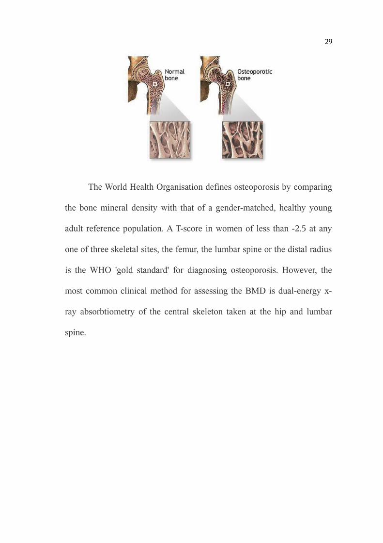
The World Health Organisation defines osteoporosis by comparing
the bone mineral density with that of a gender-matched, healthy young
adult reference population. A T-score in women of less than -2.5 at any
one of three skeletal sites, the femur, the lumbar spine or the distal radius
is the WHO 'gold standard' for diagnosing osteoporosis. However, the
most common clinical method for assessing the BMD is dual-energy x-
ray absorbtiometry of the central skeleton taken at the hip and lumbar
spine.
29
SPECIFIC SURGICAL CONSIDERATIONS FOR TREATING
FRACTURES IN AN OSTEOPOROTIC BONE
If an older patient with osteoporosis sustains a fracture there are
several important age-related factors to consider when planning
treatment. The functional demands in the elderly are different from young
healthy people and long-term immobilization in bed must be avoided.
Delaying fracture treatment by more than one day has been reported to
increase mortality in the elderly . Thus, it is probably even more
important in the elderly to achieve a stable fracture fixation that will
reduce pain and facilitate mobilization.
Reduced bone mass, increased bone brittleness, and structural
changes such as medullary expansion must be taken into account in the
osteoporotic patient when deciding the type of surgical method to be
used. It must also be understood that the osteoporotic patient usually has
low physical demands and a reduced life expectancy when making a
decision regarding treatment. For example long-term complications
following arthroplasty will not occur in the majority of elderly patients.
Thus, joint replacement surgery is a good option after displaced femoral
neck fractures as the stability provided by the implant permits immediate
weightbearing and mobilization . The major problem in osteoporotic
30

fracture treatment is fixation of the device to the bone as bone failure is
much more common than implant breakage. Internal fixation devices
such as sliding nail plates, intramedullary nails, and tension band
constructs that permit skeletal loading minimize stress at the implant
bone interface.
Some osteoporotic fractures are also associated with bone loss. If
this occurs it is important to achieve bone contact between the two main
fragments even if this results in shortening of the extremity. Good bone
contact will improve the chance of healing, reduce the healing period, and
also reduce the strains on the fixation device. If plates are used these
should be used as tension bands which require cortical contact opposite
the plates. In addition long plates, where the spacing of the screws are
more important than the number of screws, should be used as they will
distribute the forces over a larger area reducing the risk of bone failure .
Several types of fragility fractures such as fractures of the humerus, distal
radius, and closed fractures of the tibial diaphysis can be mobilized in a
sling, cast, or brace . Immobilization in casts has the disadvantage of
immobilizing the joints adjacent to the fracture often leading to joint
stiffness. Furthermore, a cast does not control fracture shortening which
is often seen in osteoporotic bone; and if the subcutaneous tissue is very
31
mobile, as it often is in the elderly, cast fixation will not provide adequate
fracture fixation. External fixators can be used but the main problem with
external fixation in osteoporotic bone is the same as for screw fixation,
namely loss of fixation. Loosening of the device is often followed by pin
infection and local bone resorption sometimes leading to a secondary
fracture at the pin site . The introduction of hydroxyapatite coated pins
has reduced the complication as fixation is improved compared to using
titanium-coated and standard pins .
Another method of improving fixation and avoiding bone
resorption is to anchor the screws with polymethylmethacrylate bone
cement. This can be inserted into the bone and allowed to harden before
drilling or it can be inserted into the screw holes just before the screws
are inserted. The screws can then be tightened after the cement hardens .
If this method is used it is important that the cement does not penetrate
the fracture so as to interfere with fracture healing.
Metaphyseal fractures in osteoporotic bone are associated with
specific fixation problems as the metaphyseal fragment is often very
small. To improve fixation and resist bending forces a screw and plate
construct with a locked angle between the plate and metaphyseal screw is
often used. Recently locked plates have been introduced threaded screw
32
holes in the plates, which create angular stability between the screws and
the plates.
The LISS system (less invasive stabilization system) and the LCP
(locking compression plates) are examples of such plates. The LCP
provides 3 times greater stability than a standard lateral condylar buttress
plate and about 2.5 times greater stability than a 95-degree condylar plate
in axial loading . Biomechanically this is explained by the fact that the
LCP also uses multiple screws for metaphyseal fixation. A particular
problem that often rules out the use of screws and plates in osteoporotic
bone is the periprosthetic fracture. These can be treated with plates using
wires for fixation around the femoral shaft
33
ETIOLOGY
Cancellous metaphyseal regions of bones in many ways behave
like osteoporotic bones in that they are the weak link between dense
cortical bone and adjoining joint surface. Hence both in the elderly and
young alike periarticular fractures and osteoporotic fractures are
addressed in similar ways. However the nature of violence in both age
groups is different. In the young age groups they mostly occur following
high energy violence like Road traffic accidents, fall from heights, fall of
heavy objects on them. In the elderly population though these similar
fractures do occur following trivial trauma like accidental fall.
34

TREATMENT
All fractures both in shaft and in particular the metaphyseal
cancellous regions need accurate alignment of joint surface and
immobilisation. This is of paramount importance as maintaining joint
motion is imperative. Also in osteoporotic bones the healing potential of
these bones is also altered. These potential hazards must be addressed if
we are to provide a normal mobile stable joint to the patients.
Non operative Treatment:
Earlier till 1970’s most of the fractures were treated with closed
reduction methods and immobilized in plaster cast . They were followed
primarily because of the lack of appropriate implants and adequate
surgical constraints. For non displaced and stable fractures, bracing can
provide enough stability to control pain and allow healing; however,
bracing cannot control alignment or length because immobilizing the
joint above and below is impossible. Hence the fallacies of plaster casting
techniques were instrumental in the new age of operative fixation
techniques.
35
Surgical Treatment:
Surgical treatment requires reduction followed by fixation to
maintain alignment. Options include external fixation or internal fixation.
Internal fixation is with intramedullary devices (eg, flexible rods, more
rigid retrograde or antegrade rods) or extramedullary plates and screws.
Distal Radius fractures treated with External Fixator and
ligamentotaxsis :
This device allows distraction of the fracture fragment and prevents
collapse of the cancellous bone. The drawbacks of this are poor patient
compliance in that this requires atleast 6 weeks of application, cannot
correct three dimensional deformities and leads to wrist stiffness.
Proximal humeral fractures treated with tension band principle and
intramedullary devices :
These devices are used in simple Neer’s two part fractures but
comminuted fractures and fractures involving head of the humerus cannot
be satisfactorily managed.
36

Supracondylar femur fracture treated with a dynamic condylar
screw plate:
This device allows fixed-angle stabilization of the fracture, which
usually prevents late loss of reduction, but it is technically limited
because it cannot be used to fix multiple fragments.
Supracondylar femur fracture treated with a blade plate:
This device allows fixed-angle stabilization of the fracture, which
usually prevents late loss of reduction, but it is technically limited
because it cannot be used to fix multiple fragments.
Supracondylar femur fracture treated with a supracondylar buttress
plate:
This device provides multiple holes for screw fixation of multiple
fragments, but it is not a fixed-angle implant so it may cause late
deformity.
37
Supracondylar femur fracture treated by retrograde intramedullary
nail:
Intramedullary devices are mechanically stronger than plates but
have limited ability to control multiple fragments and require exposure
through the knee joint.
Supracondylar femur fracture treated with Zickel flexible
intramedullary rods:
These devices act as an internal splint and can be placed rapidly
with minimal blood loss and surgical exposure but do not control length
and alignment.
Supracondylar femur fracture treated with external fixation and
minimal internal fixation:
This technique allows immediate restoration of length and
alignment with minimal surgical exposure, but it often cannot hold the
alignment in the long term and has associated problems with pin care.
38
Supracondylar femur fracture treated with a tibial buttress plate:
This type of plate is rarely used for these fractures but can allow
low-profile fixation of stable fracture patterns.
Distal tibial fractures treated with interlocking nails, K wire fixation,
External fixator :
These implants do not provide adequate stable fixation also they do
not conform to the anatomic shape of the tibial plafond. Hence their use
in distal tibial fractures is limited.
39
HISTORY OF PLATING
The date that a bone plate was first used on bone is reported to be
1565 (300 years before general anesthesia). That plate was used to repair
a cleft palate and was made out of molded gold. The late 1880's brought
the next major change in bone plating; surgeons began burying the bone
screws below the skin. There were many designs and ideas that developed
over the next 70 years. Unfortunately, malunions, nonunions and bone
infections were issues due to lack of sterile techniques, and bone plates
that were biomechanically unable to provide rigid fixation. Robert Danis
(1880-1962) developed the ideas of compression plating and
experimented with many different designs during his lifetime.
Modern bone plating started in the 1950's when a group of 15
surgeons lead by Maurice Muller formed AO/ASIF (Albeitgemeinshaft
fur osteosynthenfragen/ Association for the study of internal fixation) to
improve the principles of bone plating. AO remains purely a medical
organization to advance the study of fracture treatment while Synthes is
the commercial arm of the AO.
The original plates had round holes. If compression was needed for
the fracture, a separate device was needed to accomplish this. The
Dynamic Compression Plate (DCP was introduced in 1969 and was the
40
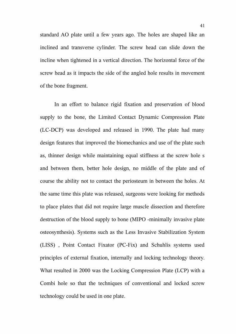
standard AO plate until a few years ago. The holes are shaped like an
inclined and transverse cylinder. The screw head can slide down the
incline when tightened in a vertical direction. The horizontal force of the
screw head as it impacts the side of the angled hole results in movement
of the bone fragment.
In an effort to balance rigid fixation and preservation of blood
supply to the bone, the Limited Contact Dynamic Compression Plate
(LC-DCP) was developed and released in 1990. The plate had many
design features that improved the biomechanics and use of the plate such
as, thinner design while maintaining equal stiffness at the screw hole s
and between them, better hole design, no middle of the plate and of
course the ability not to contact the periosteum in between the holes. At
the same time this plate was released, surgeons were looking for methods
to place plates that did not require large muscle dissection and therefore
destruction of the blood supply to bone (MIPO -minimally invasive plate
osteosynthesis). Systems such as the Less Invasive Stabilization System
(LISS) , Point Contact Fixator (PC-Fix) and Schuhlis systems used
principles of external fixation, internally and locking technology theory.
What resulted in 2000 was the Locking Compression Plate (LCP) with a
Combi hole so that the techniques of conventional and locked screw
technology could be used in one plate.
41

The original AO principles were:
Anatomic fracture reduction & fixation (as we know not always
possible).
Rigid fracture stability (not always possible).
Preservation of blood supply through careful soft tissue approaches
and fracture reduction techniques (sometimes the blood supply is
damaged from the injury).
Early return to function of the plated limb (difficult in veterinary
patients to control the amount of use).
With the understanding that not all fractures can be reconstructed,
the "rules" have been somewhat modified to:
Long bong bones must have axial re-alignment but not necessarily
anatomic perfection. Anatomic reduction is still necessary for
joints.
Appropriate construct stability to ensure fracture healing via direct
or indirect healing.
Atraumatic approaches and fracture reduction or minimally
invasive approaches.
Early return to mobility.
42
Fractures can and will heal under both conditions but that is if the
appropriate condition is chosen for the appropriate fracture
situation!
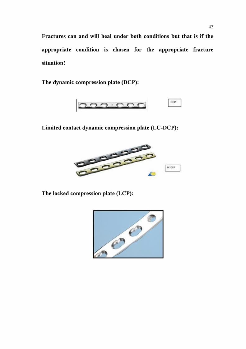
The dynamic compression plate (DCP):
Limited contact dynamic compression plate (LC-DCP):
The locked compression plate (LCP):
43

A cortical screw, a locked screw and the StarDrive head on the locked
screw:
4.5mm Distal Femur Locking compression Plate:
The plate system has many similarities to traditional plate fixation
44
methods with few improvements such as
Locking screws provides fixed angle construct and improved
fixation in osteoporotic bones
1. The screws do not rely on plate bone compression
2. Multiple screw fixation in distal fragment allows
improved fixation
3. Anatomically shaped plates is contoured to match the
contour of the bone and hence intra-operative contouring
is not required.
4. Combi - holes have additional dynamic compression
holes providing options for axial compression in addition
to locking mechanism.
45
CONVENTIONAL BONE PLATING VERSUS LOCKED COMPRESSION
PLATING
Conventional bone plates depend on direct plate to bone and screw
to bone friction to maintain fracture fixation. Therefore the plates must be
perfectly contoured prior to application to the bone. Fracture reduction
can be lost from axial loads causing excessive shear forces on the
construct that are greater than the frictional loads between the bone-plate-
screw construct. The cortical screws can toggle which leads to screw
loosening and loss of plate-bone fixation. Each screw works
independently; the construct depends on a single screw's stiffness or
pullout strength.
The biomechanical goals of the LCPs are to increase the stiffness
of the construct in a biological environment. The LCP is a fixed angle
construct that does not rely on screw purchase in bone. Once the screw is
locked into the plate, the fixed-angle converts shear stress into
compressive stress at the screw-bone interface. The load is now
perpendicular to the screw axis. In order for the construct to fail under an
axial load, the bone must collapse in compression. Therefore, the strength
in the LCP is the sum of all the screw and plate interfaces.
Locking screws are designed with smaller threads because they are
46
not used to generate compression between the plate and the bone. They
have a larger core diameter that ensures greater bending and shear
strength and dissipate the load over a larger area of bone. They have the
new Star Drive head that allows 65% greater insertion torque than
conventional hexagonal drivers. The Star Drive is self- retaining (stays on
the screw driver without a holding device). The locked screw has a
conical, double-lead thread design that facilitates alignment with the
threaded plate hole.
To date, there are no randomized clinical trials in human or animals
comparing the LCP plate to conventional plates (DCP and LC-DCP) in
patients with similar fractures. The plates are studied and compared in
vitro (human and animal) and in case series' and are where the
information on LCP principles and indications come from. The purported
indications for LCPs include:
1. Patients with poor quality bone (osteoporosis, osteomyelitis)
2. Complex periarticular fracture (especially when contouring may be
difficult in the metaphyseal area)
3. Inability to get minimal number of conventional screw cortices,
4. Periprosthetic fractures
5. Nonunions from failed fixations (cortex or cancellous screw
stripping or screw back-out)
47
6. Polytrauma cases (especially when the fractures cannot be
anatomically reconstructed).
In vitro studies in bone models do show that locked screw constructs
fail at higher loads than cortex screws and their advantage is magnified in
osteoporotic bone.
Technical and biological LCP aspects that are not known when
used in veterinary patients are: the ideal number of locked screws on
either side of the fracture, the number of unicortical versus bicortical
screws necessary for success, indications for some plate contouring
(although not exact contouring), the effects of combining conventional
screws and locked screws in the same construct, indications for double
plating or adding additional implants (such as plate rod constructs), if
there are additive biological effects on fracture healing when LCPs are
placed minimally invasively. It is technically possible to place locking
plates and screws minimally invasively with proper fluoroscopic
equipment. In human studies there is little mechanical advantage in
placing more than 2 locked screws on either side of the fracture. This may
be quite different in animal patients that cannot be strictly confined or
have multiple limbs fractured. Fracture fixation failures with LCPs do
occur; the clinical case application will address some of the reasons for
this.
48
AIM OF THE STUDY
The aim of the study is to analyze the short term results in terms of
union and functional outcome for osteoporotic and periarticular fractures
treated with locking compression plating.
49

MATERIALS & METHODS
This is a study conducted in the Department of Orthopaedics,
Madras Medical College, Government General Hospital, Chennai.
This study is a prospective study Conducted in the Department of
Orthopaedics from September 2007 to September 2009 with a sample
size of 21 cases.
Patients:
Patients were randomly selected from among the admissions to the
Orthopaedic ward in the Department of Orthopaedics, Government
General Hospital, Chennai and recruited into the study prospectively
based on the following criteria:
Inclusion criteria:
1. Age more than 16 years.
2. Osteoporotic bones either disuse or pathological bones.
3. Fractures occurring at or near joints namely distal femur,
proximal humerus, distal radius, distal tibia, proximal tibia.
4. Osteoporotic non-unions
50
5. Patients who consents to be included in the study.
Exclusion criteria:
1. Exclusion criteria were skeletal immaturity
2. Patients with tumourous conditions.
3. Severe articular comminution not possible to be reconstructed
with internal fixation.
4. Undisplaced fracture patterns needing only conservative
management.
5. Patients not willing for internal fixation.
Study protocol:
A total of 21 patients with osteoporotic and periarticular fractures
were included in the study as per the criteria outlined previously.
On admission detailed examination of the patients was carried out
after hemodynamic stabilization. Patients were then immobilized on a
plaster of Paris
Then standard Antero – Posterior and Lateral view X – Rays are
taken and the fracture configuration noted. Computerized Tomography is
51

also taken when needed to assess the exact alignment of the fragments.
The fracture is classified using the various classification systems earlier
described.
Then after the assessment for anesthetic fitness open reduction and
internal fixation of the fracture is done using locking compression plate.
POST OPERATIVE ASSESSMENT USING HHS and DASH
Scoring systems :
HSS (hospital for special surgery) score
Pain
Walking (none to severe): points 15–0
At rest (none to severe): points 15–0
Function
Walking (unlimited to unable): points 12–0
Stairs (normal to with support): points 5–2
Transfer (normal to with support): points 5–2
RoM (80°–120°): points 10–15
Muscle strength (grade 5–0): points 15–0
52
Flexion deformity (none to >20°):points 10–0
Instability (none to >15°): points 5–0
Subtractions
One cane: 1 point
One crutch: 2 points
Two crutches: 3 points
Extension lag (5°–15°): 2–5 points
Deformity (every 5°): 1 point
Excellent = 85 points or more, good = 70–84 points, fair = 60–69
points, poor = less than 60 points.
53
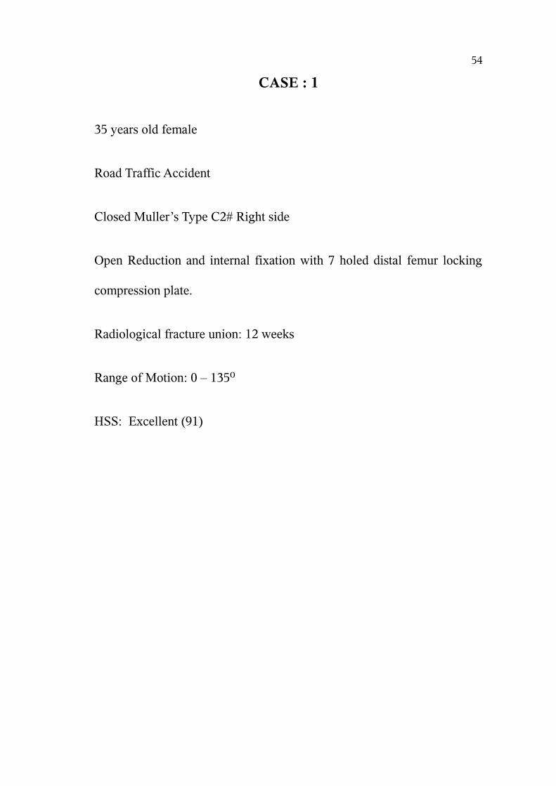
CASE : 1
35 years old female
Road Traffic Accident
Closed Muller’s Type C2# Right side
Open Reduction and internal fixation with 7 holed distal femur locking
compression plate.
Radiological fracture union: 12 weeks
Range of Motion: 0 – 135⁰
HSS: Excellent (91)
54
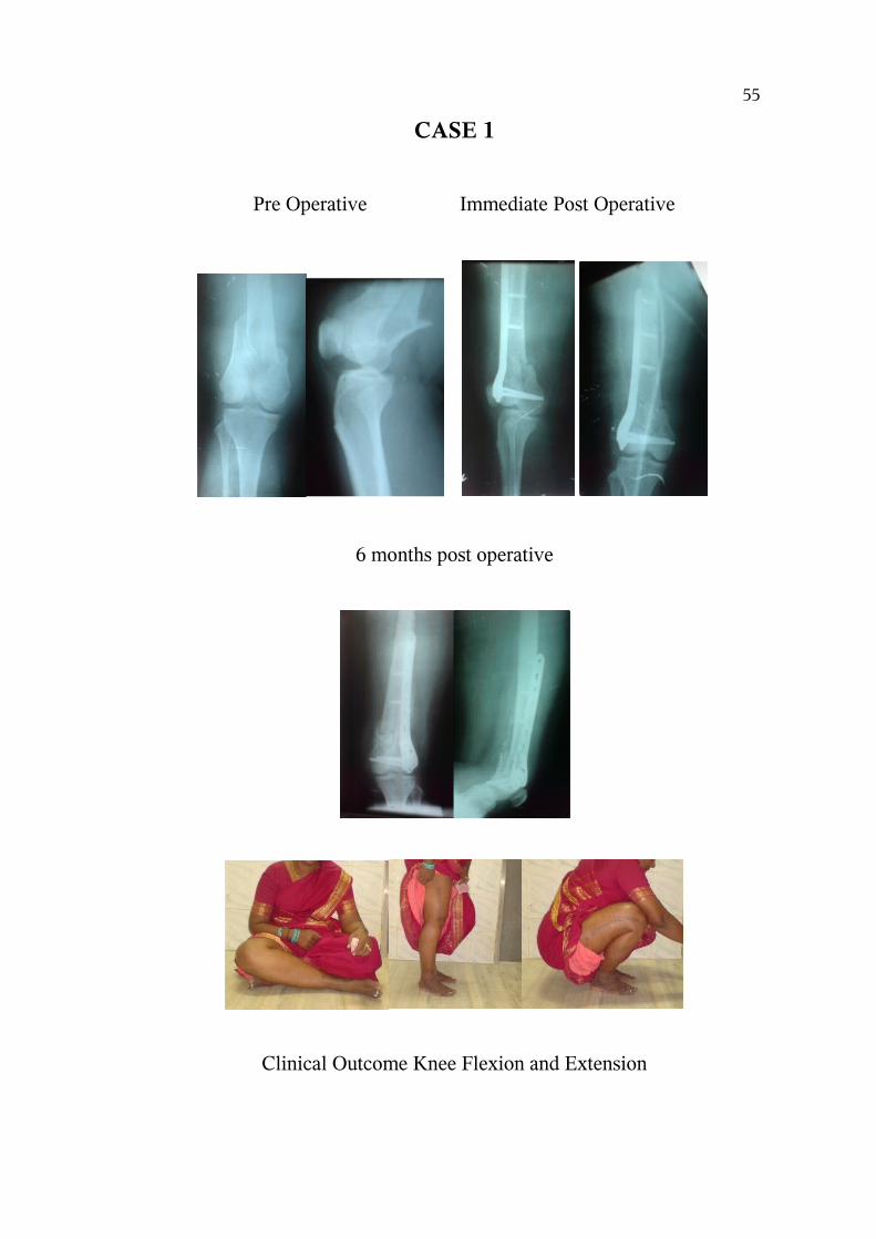
CASE 1
Pre Operative Immediate Post Operative
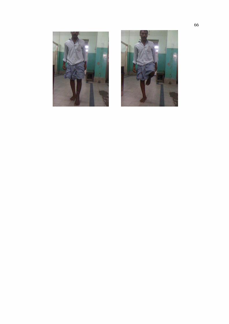
6 months post operative
Clinical Outcome Knee Flexion and Extension
55

CASE 2
22 years old male
Sustained RTA and fall
Comminuted fracture Distal Radius – Right
Open Reduction and Internal Fixation using Distal Radius Locking
Compression Plate
Radiological union : 12 weeks
Range of motion : Dorsiflexion 35 deg palmar flexion 50 deg
DASH score : 5.0 ( Good)
56
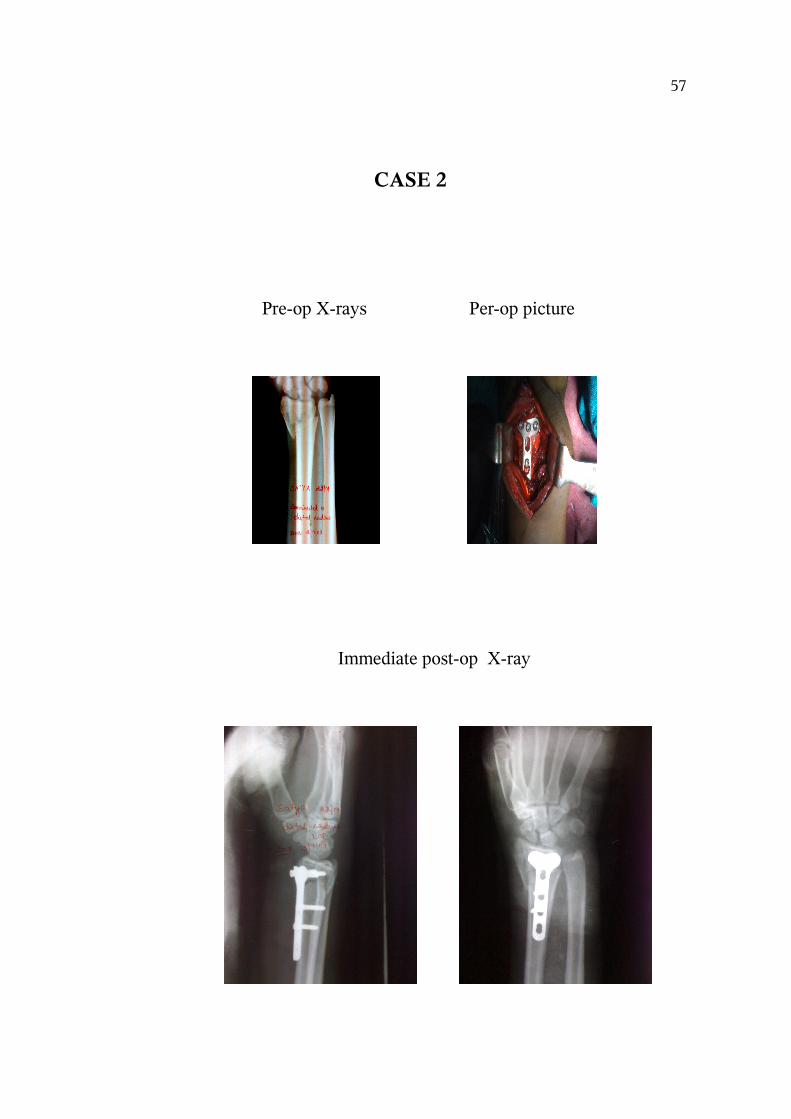
CASE 2
Pre-op X-rays Per-op picture
Immediate post-op X-ray
57
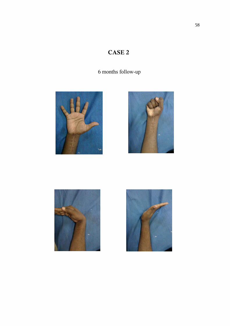
CASE 2
6 months follow-up
58
CASE 3
40 year old Female
Sustained accidental fall from two wheeler and sustained injury to
Right leg
Had a Pilon fracture Right
Treated by Open Reduction and Internal Fixation with Distal Tibial
Locking plate and Fibular plating
Radiological union : 12 weeks
Functional outcome : Excellent with full weight bearing walking
with no secondary osteoarthrosis of the ankle
59
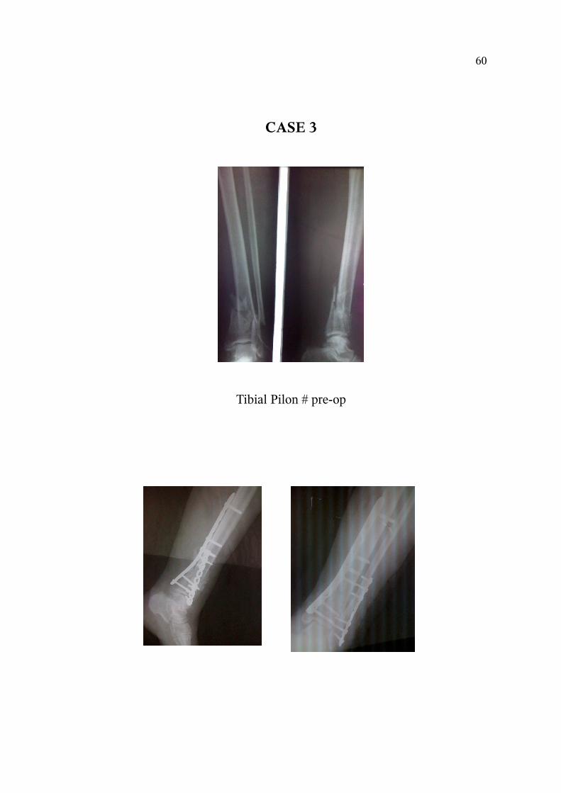
CASE 3
Tibial Pilon # pre-op
60
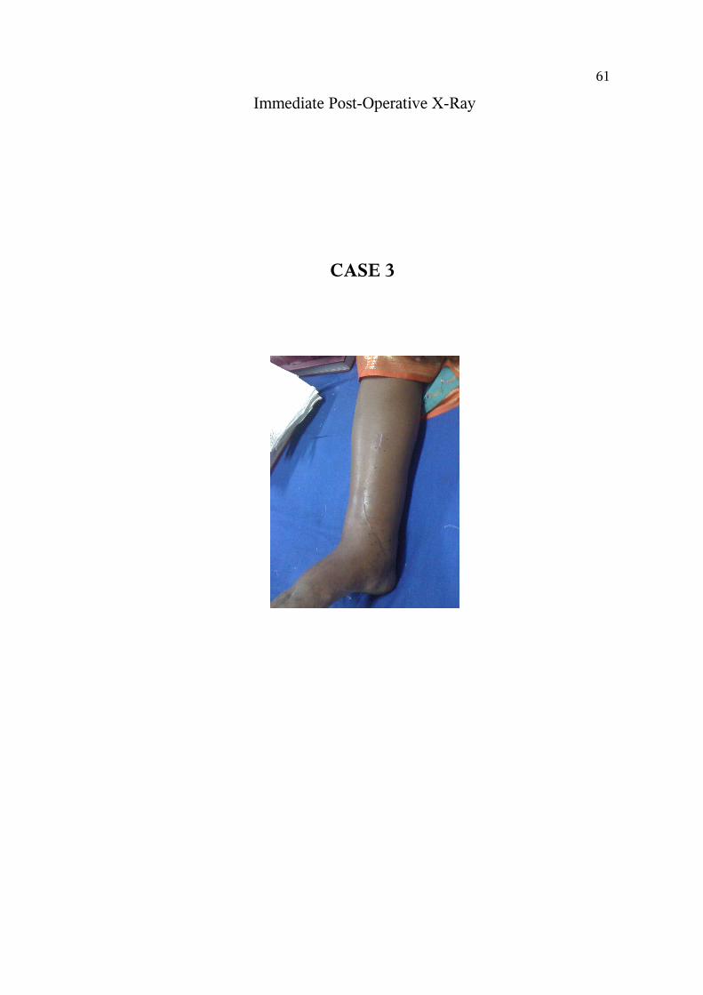
Immediate Post-Operative X-Ray
CASE 3
61
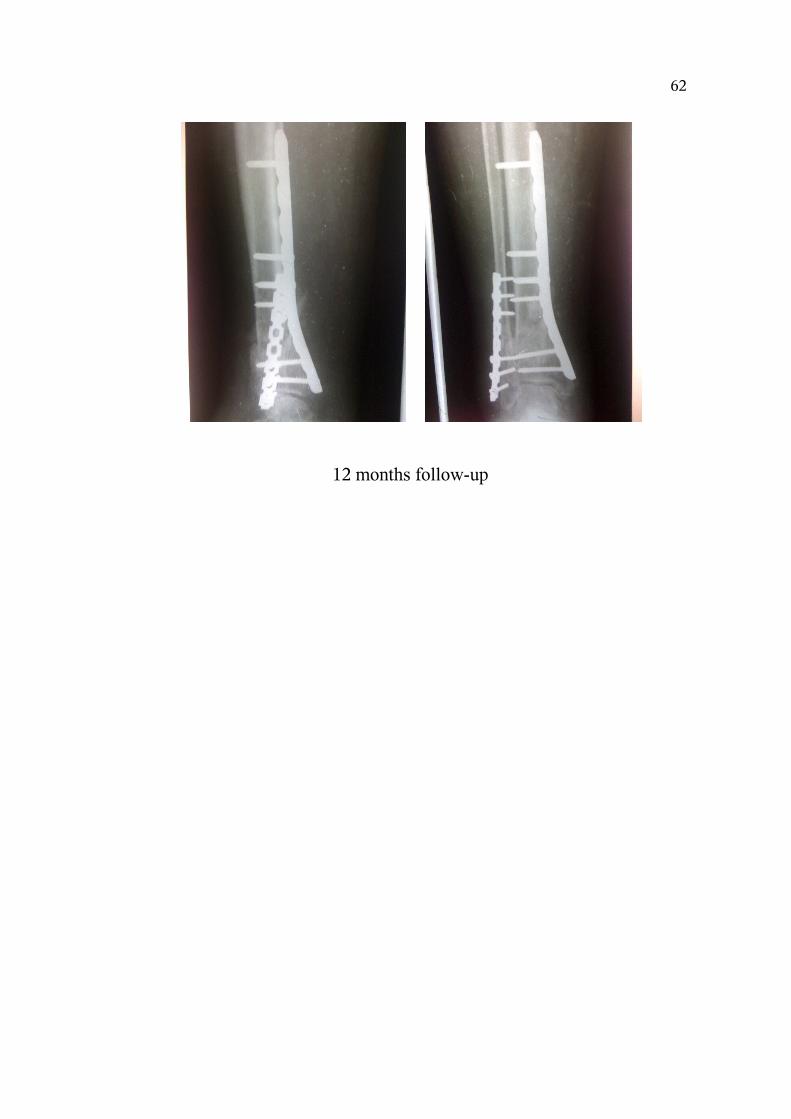
12 months follow-up
62
CASE 4
32 year old Male
Sustained RTA and had a Grade II compound fracture of Both
Bone Leg Left side
Initially treated with Wound Debridement and External Fixator
followed by flap cover
At 5 months he developed non-union at fracture site
Treated with Open Reduction and Narrow Dynamic Locking
Compression Plate with Bone Grafting
Radiological union : 14 weeks
Functional Outcome : Excellent outcome. Full weight bearing
walking with full range of motion
63
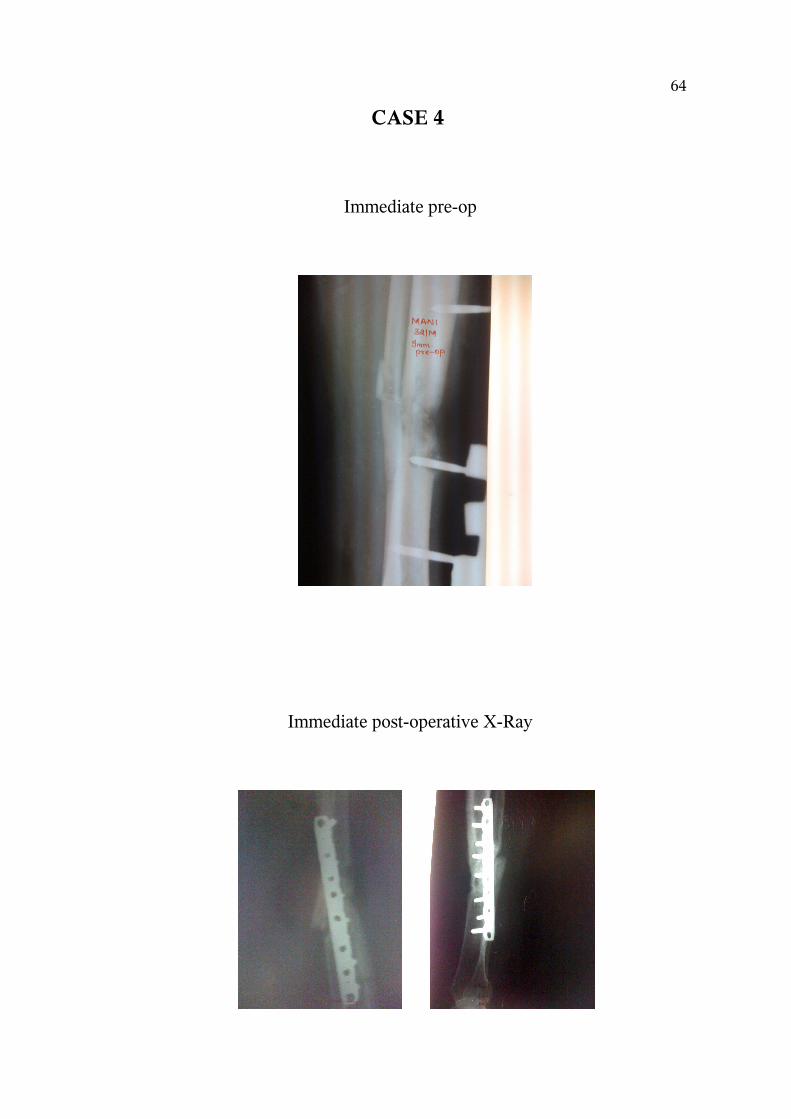
CASE 4
Immediate pre-op
Immediate post-operative X-Ray
64
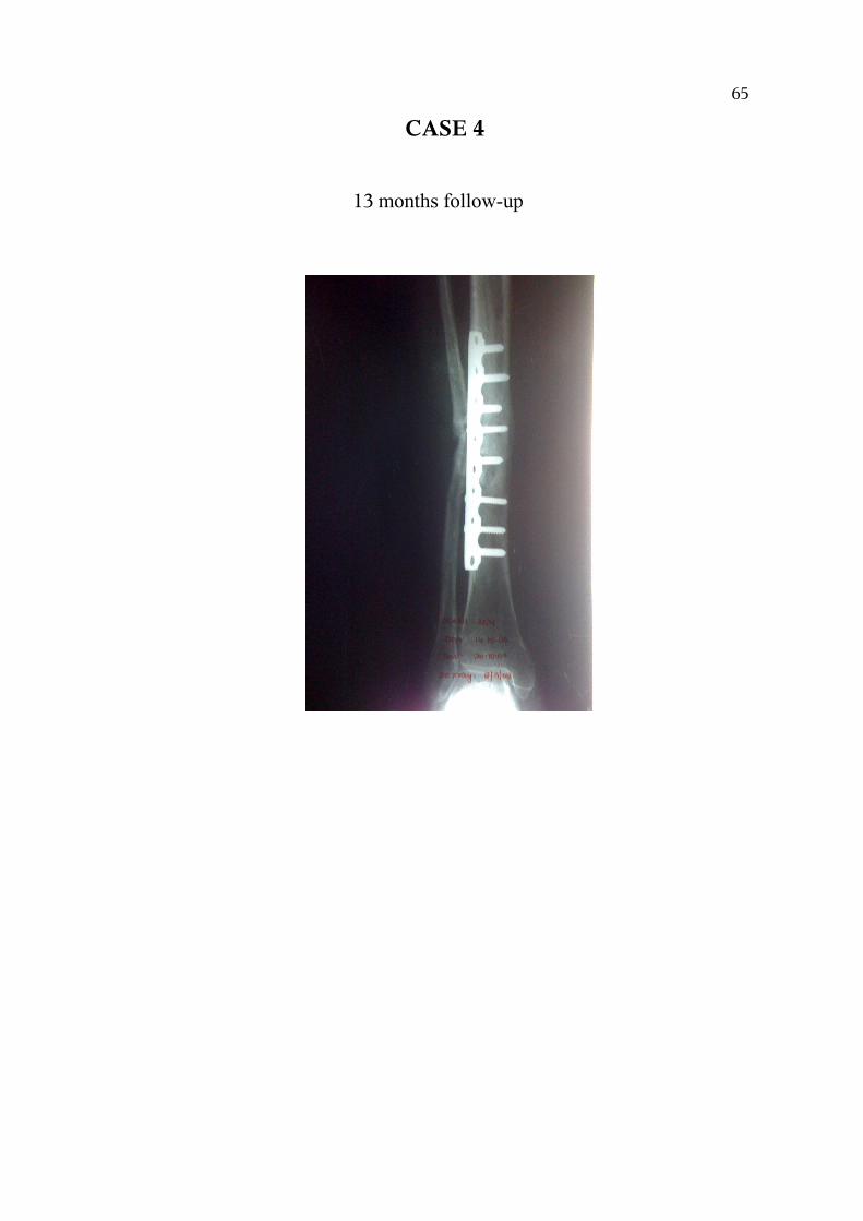
CASE 4
13 months follow-up
65
66
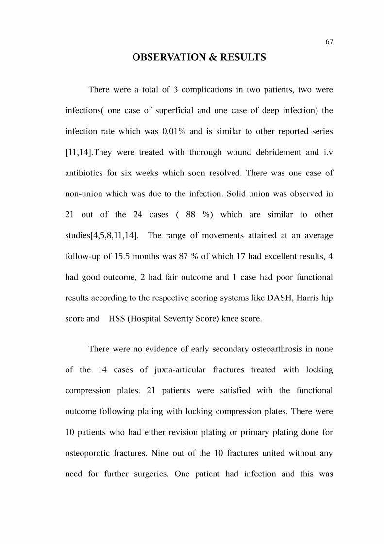
OBSERVATION & RESULTS
There were a total of 3 complications in two patients, two were
infections( one case of superficial and one case of deep infection) the
infection rate which was 0.01% and is similar to other reported series
[11,14].They were treated with thorough wound debridement and i.v
antibiotics for six weeks which soon resolved. There was one case of
non-union which was due to the infection. Solid union was observed in
21 out of the 24 cases ( 88 %) which are similar to other
studies[4,5,8,11,14]. The range of movements attained at an average
follow-up of 15.5 months was 87 % of which 17 had excellent results, 4
had good outcome, 2 had fair outcome and 1 case had poor functional
results according to the respective scoring systems like DASH, Harris hip
score and HSS (Hospital Severity Score) knee score.
There were no evidence of early secondary osteoarthrosis in none
of the 14 cases of juxta-articular fractures treated with locking
compression plates. 21 patients were satisfied with the functional
outcome following plating with locking compression plates. There were
10 patients who had either revision plating or primary plating done for
osteoporotic fractures. Nine out of the 10 fractures united without any
need for further surgeries. One patient had infection and this was
67
attributed to the poor skin condition and soft tissue condition due to
multiple failed procedures in him.
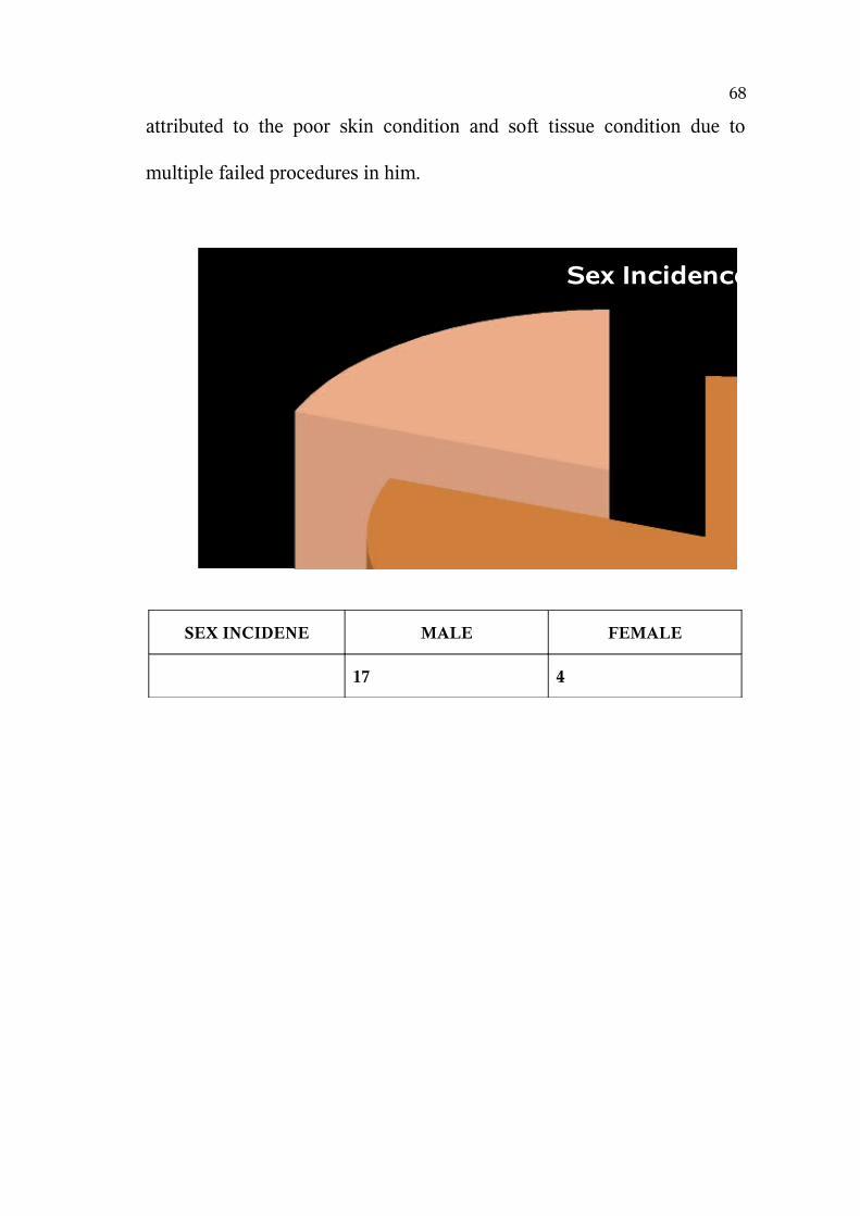
Sex Incidence
MalesFemales
SEX INCIDENE MALE FEMALE
17 4
68
Dista
l Fem
ur
Proxim
al H
umer
us
Dista
l Rad
ius
Diaph
ysial
Dista
l Tibial
0
1
2
3
4
5
6
7
8
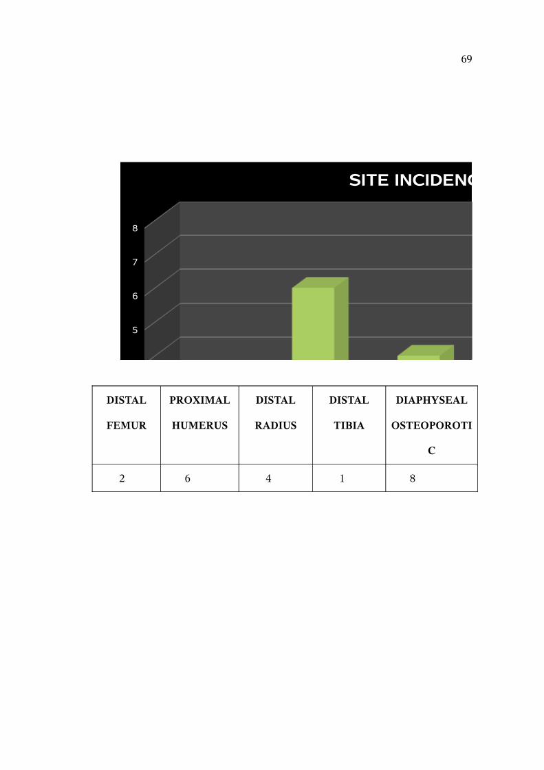
SITE INCIDENCE
SITE INCIDENCEDISTAL
FEMUR
PROXIMAL
HUMERUS
DISTAL
RADIUS
DISTAL
TIBIA
DIAPHYSEAL
OSTEOPOROTI
C
2 6 4 1 8
69

UNION RATE
UnionNon-Union
UNION 19
NON-UNION 2
70

COMPLICATIONS
InfectionsNon Union
INFECTION 1
NON-UNION 2
71

11 to 20
21 to 30
31 to 40
41 to 50
51 to 60
0
1
2
3
4
5
6
7
AGE INCIDENCE
AGE INCIDENCE 11-20 years 3
21-30 years 6
31-40 years 6
41-50 years 4
51-60 years 1
72
DISCUSSION
The recent evolution in reduction and internal fixation of
fractures is based on an improved understanding of the biology of bone,
of the biomechanics of fracture fixation and fracture healing and on the
analysis of previous failures. Improvements in implant designs play an
important role in avoiding possible complications and in achieving the
primary goals of operative fracture treatment.
The evolution of locking compression plates in the fixation of
specific fracture characteristics has revolutionized the treatment of
complicated and failed previous internal fixation procedures. Our study
was done to analyse the usefulness of such locking plates in osteoporotic
and periarticular fractures and results were computed and compared with
similar studies done by other surgeons.
Gardner et al. in 2002 reported his series of 36 cases of proximal
humeral fractures treated with proximal humeral LCP and reported two
cases of humeral necrosis which was not seen in our study. Breakage of
implant was seen in one patient which was also not encountered in our
study. The DASH score reported was 18.0 which was similar to the
DASH score of 19.0 in our study.
73
Bjorkenheim et al (2004) reported a series of 72 patients with
proximal humeral fractures treated with PHILOS plate and reported a
union rate of 94 %. There were three cases of non-union (0.04 %). Our
series also had similar results.
Sommer et al. (2004) reported four cases of implant failure with
locking plates and attributed this to poor technical application and also
poor choice of appropriate implant rather than to the features of locking
plate itself. His experience highlights the importance of detailed
understanding of the biomechanical principles of plate fixation as well as
meticulous pre-operative planning.
Ring et al. (2004) reported his series of 24 cases of osteoporotic
non-unions of diaphyseal fractures treated with locking compression
plates and reported a union rate of 97 % with two cases requiring
additional bone grafting to achieve union.
Kassab et al. (1998) in his series of 44 patients with diaphyseal
osteoporotic non-unions and achieved solid union in 40 cases ( 90 %). In
our series it was 88 % which was comparable. There were three cases of
persistent non-unions which required secondary bone grafting and
revision internal fixation.
74
CONCLUSION
This study highlights the role of locking compression plating in
complex osteoporotic and peri-articular fractures in which conventional
dynamic compression plates and reconstruction plates would fail
prematurely. The correct application of locking compression plates
requires a long learning curve and spurious use will negate the
advantages of the locking plates.
The results of our study have confirmed earlier reports that locking
compression plates provide better fixation in osteoporotic fractures. The
chances of implant failure are less as the screws are firmly position inside
the bone. Also since these plates are limited contact plates there is less
contact between plate and the bone and hence there is minimal disruption
of sub-periosteal blood supply to the fracture ends and this aids in
fracture union. The locked nuts prevent further tightening of the screws
and hence reduction is maintained and secondary angular deformities are
prevented.
In periarticular fractures when the required cortical purchases are
not possible on either side of the fracture site, these specially designed
plates allow adequate cortical purchases. Also metaphyseal fractures
behave like osteoporotic fractures since they are primarily cancellous
75

bone stable rigid fixation is not possible in all cases with conventional
plates.
We have used locking compression plates in both osteooporotic and
juxta-articular fractures and have found to be implant of choice in these
fractures. The union rates achieved by us is 88 % which is comparable to
other studies. Also the low infection in our study and the non-union rate
are also comparable to similar studies done by other groups.
Hence locking compression plates are special implants which have
been specifically designed for clinical application in osteoporotic and
juxta-articular fractures.
76
REFERENCES
1. Gardner MJ, Brophy RH, Campbell D, Mahajan A, Wright TM.
The mechanical behaviour of locking compression pplates
compared with dynamic compression plates in a cadaver radius
model. J Orthop Trauma. 2005 Oct;19(9):597-603.
2. Plecko M, Kraus A. Internal fixation of proximal humerus fractures
using the locking compression proximal humeral plate. Oper
Orthop Traumatol.2005 Feb;17(1):25-50.
3. Bjorkeenheim JM, Pajarinen J, Savolainen V. Internal fixation of
proximal huneral fractures with a locking compression plate : a
retrospective evaluation of 72 patients followed for a minimum of
1 year. Acta Orthop Scand. 2004 Dec;75(6):741-5.
4. Frankhauser F, Boldin C, Schippinger G, Haunschmid C,
Szyskowitz R. A new locking plate for unstable fractures of the
proximal humerus. Clin Orthop Relat Res. 2005 Jan;(430):176-81.
5. Sommer C, Babst R, Mullerr M, Hanson B. Locking compression
plate loosening and plate breakage : a report of four cases. J Orthop
Trauma. 2004 Sep;18(8):571-7
77
6. Egol KA, Kubiak EN, Fulkerson E, Kummer Fj, Koval J.
Biomechanics of locked compression plate and screws. J Orthop
Trauma. 2004 Sep;18(8): 488-93.
7. Kaab MJ, Frenk A, Schmeling A, Schaser K, Schutz M, Haas NP.
Locked internal fixator : sensitivity of screw/ plate stability to the
correct insertion angle of the screw. J Orthop Trauma.2004
sep;18(8):483-7.
8. Ring D, Kloen P. Kadzielski J, Helfet D, Jupiter JB. Locking
compression plates for osteoporotic nonunions of the diaphyseal
humerus. Clin Orthop Relat Res. 2004 Aug;(425):50-4.
9. Korner J, Diederichs G, Arzdorf M, Lill H, Josten C, Schneider E,
Linke B. A biomechanical evaluation of methods of distal humerus
fracture fixation using locking compression plates versus
conventional reconstruction plates. J Orthop Trauma.2004 May-
Jun;18(5):286-93.
10.Gautier , Sommer C. Guidelines for the clinical application of the
LCP. Injury 2003.Nov;34 Suppl 2:B63-76.
11.Sommer C, Gautier E, Muller M, Helfet DL, Wagner M. First
clinical results of the locking compression plate. Injury 2003.
78
Nov;34 Suppl 2:B43-54.
12.Korner J, Lill H, Muller LP, Rommens PM, Schneider E, Linke B.
The LCP-concept in the operative treatment of distal humerus
fractures-biological, biomechanical and surgical aspects. Injury
2003. Nov;34 Suppl 2:B20-30.
13.Cole PA, Zlowodski M, Kregor PJ. Less Invasive Stabilization
System (LISS) for fractures of the proximal tibia: indications,
surgical technique and preliminary results of the UMC Clinical
Trial. Injury 2003 Aug;34 Suppl 1:A16-29.
14.Kassab SS, Mast JW, Mayo KA. Patients treated for nonunions
with plate and screw fixation and adjunctive locking nuts. Clin
Orthop Relat Res. 1998 Feb;(347):86-92.
15. The Evolution of AO/ASIF Bone Plating Equipment: Are They
Better or Just Different? Amy S. Kapatkin, DVM, MS, DACVS
Department of Surgical & Radiological Sciences, University of
California, Davis
16. AO Manual of Fracture Management. Internal Fixators: Concepts
and Cases Using LCP and LISS. Wagner M, Frigg R (eds.),
Thieme, Stuttgart, 2006
79

17. Egol KA, Kubiask EN, Fulkerson E, et al: Biomechanics of locked
plates and screws. J Orthop Trauma 18(8): 488-493, 2004
18. Perren SM: Evolution of the internal fixation of long bone
fractures. The scientific basis of biologic internal fixation:
choosing a new balance between stability and biology. J Bone Jt
Surg 84B:1092-1110, 2002
80
PROFORMA
TOPIC : Locking Compression Plate in Osteoporotic and Juxta-articular fractures
Dr. Navin Balasubramanian P.G M.S (Orth)
CASE :
NAME : AGE / SEX :
ADDRESS : D.O.A :
D.O.S :
D.O.D :
Presenting complaints :
If female whether Pre- / Post-menopausal
Examination :
X-Ray :
Diagnosis :
Treatment :
Procedure done :
Post-op protocol :
Advice on discharge :
81

FOLLOW-UP NOTES
Time since surgery
X-Ray Findings
Range of Movements
FunctionalAssessment
Any complications
Special Points :
82

83