LOCALPHOTODYNAMICTHERAPY(PDT)OFRATC6GLIOMAXENOGRAFTS ...
7
LOCAL PHOTODYNAMIC THERAPY (PDT) OF RAT C6 GLIOMA XENOGRAFTS WITH Pd-BACTERIOPHEOPHORBIDE LEADS TO DECREASED METASTASES AND INCREASE OF ANIMAL CURE COMPARED WITH SURGERY Smadar SCHREIBER 1,2 , Shimon GROSS 1,2 , Alex BRANDIS 2 , Alon HARMELIN 3 , Varda ROSENBACH-BELKIN 2 , Avigdor SCHERZ 2 and Yoram SALOMON 1 * 1 Department of Biological Regulation, The Weizmann Institute of Science, Rehovot, Israel 2 Department of Plant Sciences, The Weizmann Institute of Science, Rehovot, Israel 3 Experimental Animal Center, The Weizmann Institute of Science, Rehovot, Israel Photodynamic therapy (PDT), locally applied to solid C6 rat glioma tumors in the foot of CD1 nude mice, eradicated the primary tumor and also decreased the rate of groin and lung metastases. Pd-Bacteriopheophorbide (Pd-Bpheid), a novel photosensitizer synthesized in our laboratory, was used in our study. The primary lesion in the hind leg was treated by an i.v. injection of 5 mg/kg of Pd-Bpheid and immediate illumination (650 – 800 nm, 360 J/cm 2 ). This protocol and the surgical amputation of the leg were compared for local and metastasis responses. Following PDT, hemorrhage, inflam- mation with tumor necrosis and flattening were observed and histologically verified in the photodynamically treated tumor. Whereas local tumor control rates were up to 64% following PDT, in surgically treated animals, local tumor control was absolute. The rates of metastases in the groin and the lungs were at least 12-fold lower in the photodynam- ically treated animals compared with untreated or surgery- treated groups. The overall cure rates after PDT or surgery were 36% and 6%, respectively, at 8 weeks. These findings suggest that local PDT with Pd-Bpheid, which acts primarily on the tumor vasculature, efficiently eradicates the solid C6 tumors. In addition, the local PDT of the primary lesion has beneficial therapeutic effects on remote C6 metastasis, which is not obtained with surgery. It is therefore suggested, that although surgery is highly efficient for the immediate removal of the primary tumor, it lacks such systemic, ther- apeutic effects on distant metastases. Pd-Bpheid-PDT may thus offer a potentially superior curative therapy for C6 gli- oma tumors in the limb by eradicating the target tumor and by reducing the rate of metastasis in the groin and lung, possibly due to innate immunity. © 2002 Wiley-Liss, Inc. Key words: photodynamic therapy; surgery; Pd-bacteriopheophor- bide; C6 glioma tumor; metastasis Photodynamic therapy (PDT) is based on the destruction of tumors by cytotoxic reactive oxygen species (ROS) produced upon local tumor illumination in patients administered with a photosen- sitizer. 1–3 Following health agency approval for photofrin- based PDT in many countries, this anti-cancer treatment modality en- tered clinical use for the local treatment of an increasing number of indications including skin, esophageal, lung, gastric, cervical and bladder cancers. 4 PDT is usually considered a local anti-tumor treatment modal- ity. However, reports from several laboratories suggest that PDT also induces beneficial systemic effects. Following in vitro hema- toporphyrin-based PDT, adhesiveness and metastatic potential de- cline in DHD-K12-cultured colon carcinoma cells. Moreover, in- travenous or s.c. injection of these PDT-treated cells to rats resulted in a reduced number of lung metastases compared with untreated cell injection. 5,6 Although this observation may be due to local photodynamic damage, the potential beneficial effect may be viewed as systemic. Other in vivo studies showed that local PDT with various photosensitizers mediates long-term tumor immunity and resistance to tumor rechallenge. It was shown that the activa- tion of natural killer (NK) cells and T lymphocytes by PDT, along with induction of specific anti-tumor antibodies, 7 play a major role in the curative response. 8,9 Moreover, adoptive transfer of lym- phocytes, which were tumor-sensitized by photofrin-based PDT in immunocompetent mice, improved the tumor response of SCID mice to PDT. 9 In addition, it was shown that neutrophils are indispensable for successful PDT. Moreover, PDT of neutropenic mice (treated with rabbit anti-rat neutrophil serum) did not retard tumor growth relative to controls. 10,11 Other studies investigated the effect of local in vivo PDT on distant metastasis. 12,13 One study reported a decrease and the other reported an increase in metastasis compared with surgery. We have recently developed a new line of highly efficient photosensitizers based on bacteriochlorophyll (A. Brandis et al., 2001; private communication). 14,15 The first in this series bacteri- ochlorophyll-Serine (Bchl-Ser) was found to be phototoxic in cultured melanoma cells with an LD 50 value of 100 nM. This value is about 100 times lower than that of the hematoporphyrin deriv- ative Photosan. 16,17 When tested on melanoma xenografts in mice 18 –20 and DS sarcoma in rats, 21 over 80% of the tumors were cured after a single PDT session. The anti-tumor effects of Bchl- Ser using a protocol with no injection-illumination time interval were ascribed mainly to the anti-vascular action of this treatment modality. 20 When tested under the same conditions in the same cells, 16 the new derivative used here, Pd-Bacteriopheophorbide (Pd-Bpheid) (Fig. 1) shows about 1,000-fold higher phototoxicity than Photosan and greater anti-tumor efficiency than Bchl-Ser. 15,22 The high efficiency of these new sensitizers as PDT agents prompted us to examine their local and systemic effects on distant metastases. In the present report, we investigated the local control of C6 glioma tumors and the therapeutic effects on distant metas- tases (groin and lung) following Pd-Bpheid based PDT. MATERIAL AND METHODS Materials DMEM/F12, Fetal calf serum and Glutamine were from Bio- logical Industries (Kibbutz Beit Haemek, Israel). Phosphate-buff- ered saline (PBS) was from Sigma Chemical Co. (Israel). Penicil- Abbreviations: Bchl-ser, Bacteriochlorophyll-serine; NK, natural killer cell; Pd-Bpheid, Palladium-bacteriopheophorbide; PDT, photodynamic therapy; ROS, reactive oxygen species. Grant sponsor: STEBA BIOTECH N.V., The Netherlands. *Correspondence to: Department of Biological Regulation, Weizmann Institute of Science, 76100 Rehovot, Israel. Fax: 972-8-934-4116. E-mail: [email protected] Received 24 August 2001; Revised 13 November 2001; Accepted 14 December 2001 DOI 10.1002/ijc.10299 Published online 7 March 2002 in Wiley InterScience (www.interscience. wiley.com). Int. J. Cancer: 99, 279 –285 (2002) © 2002 Wiley-Liss, Inc. Publication of the International Union Against Cancer
Transcript of LOCALPHOTODYNAMICTHERAPY(PDT)OFRATC6GLIOMAXENOGRAFTS ...
LOCAL PHOTODYNAMIC THERAPY (PDT) OF RAT C6 GLIOMA XENOGRAFTSWITH Pd-BACTERIOPHEOPHORBIDE LEADS TO DECREASED METASTASESAND INCREASE OF ANIMAL CURE COMPARED WITH SURGERYSmadar SCHREIBER
1,2, Shimon GROSS1,2, Alex BRANDIS
2, Alon HARMELIN3, Varda ROSENBACH-BELKIN
2, Avigdor SCHERZ2 and
Yoram SALOMON1*
1Department of Biological Regulation, The Weizmann Institute of Science, Rehovot, Israel2Department of Plant Sciences, The Weizmann Institute of Science, Rehovot, Israel3Experimental Animal Center, The Weizmann Institute of Science, Rehovot, Israel
Photodynamic therapy (PDT), locally applied to solid C6rat glioma tumors in the foot of CD1 nude mice, eradicatedthe primary tumor and also decreased the rate of groin andlung metastases. Pd-Bacteriopheophorbide (Pd-Bpheid), anovel photosensitizer synthesized in our laboratory, was usedin our study. The primary lesion in the hind leg was treatedby an i.v. injection of 5 mg/kg of Pd-Bpheid and immediateillumination (650–800 nm, 360 J/cm2). This protocol and thesurgical amputation of the leg were compared for local andmetastasis responses. Following PDT, hemorrhage, inflam-mation with tumor necrosis and flattening were observedand histologically verified in the photodynamically treatedtumor. Whereas local tumor control rates were up to 64%following PDT, in surgically treated animals, local tumorcontrol was absolute. The rates of metastases in the groinand the lungs were at least 12-fold lower in the photodynam-ically treated animals compared with untreated or surgery-treated groups. The overall cure rates after PDT or surgerywere 36% and 6%, respectively, at 8 weeks. These findingssuggest that local PDT with Pd-Bpheid, which acts primarilyon the tumor vasculature, efficiently eradicates the solid C6tumors. In addition, the local PDT of the primary lesion hasbeneficial therapeutic effects on remote C6 metastasis,which is not obtained with surgery. It is therefore suggested,that although surgery is highly efficient for the immediateremoval of the primary tumor, it lacks such systemic, ther-apeutic effects on distant metastases. Pd-Bpheid-PDT maythus offer a potentially superior curative therapy for C6 gli-oma tumors in the limb by eradicating the target tumor andby reducing the rate of metastasis in the groin and lung,possibly due to innate immunity.© 2002 Wiley-Liss, Inc.
Key words: photodynamic therapy; surgery; Pd-bacteriopheophor-bide; C6 glioma tumor; metastasis
Photodynamic therapy (PDT) is based on the destruction oftumors by cytotoxic reactive oxygen species (ROS) produced uponlocal tumor illumination in patients administered with a photosen-sitizer.1–3 Following health agency approval for photofrin- basedPDT in many countries, this anti-cancer treatment modality en-tered clinical use for the local treatment of an increasing number ofindications including skin, esophageal, lung, gastric, cervical andbladder cancers.4
PDT is usually considered a local anti-tumor treatment modal-ity. However, reports from several laboratories suggest that PDTalso induces beneficial systemic effects. Following in vitro hema-toporphyrin-based PDT, adhesiveness and metastatic potential de-cline in DHD-K12-cultured colon carcinoma cells. Moreover, in-travenous or s.c. injection of these PDT-treated cells to ratsresulted in a reduced number of lung metastases compared withuntreated cell injection.5,6 Although this observation may be due tolocal photodynamic damage, the potential beneficial effect may beviewed as systemic. Other in vivo studies showed that local PDTwith various photosensitizers mediates long-term tumor immunityand resistance to tumor rechallenge. It was shown that the activa-tion of natural killer (NK) cells and T lymphocytes by PDT, alongwith induction of specific anti-tumor antibodies,7 play a major rolein the curative response.8,9 Moreover, adoptive transfer of lym-
phocytes, which were tumor-sensitized by photofrin-based PDT inimmunocompetent mice, improved the tumor response of SCIDmice to PDT.9 In addition, it was shown that neutrophils areindispensable for successful PDT. Moreover, PDT of neutropenicmice (treated with rabbit anti-rat neutrophil serum) did not retardtumor growth relative to controls.10,11 Other studies investigatedthe effect of local in vivo PDT on distant metastasis.12,13 One studyreported a decrease and the other reported an increase in metastasiscompared with surgery.
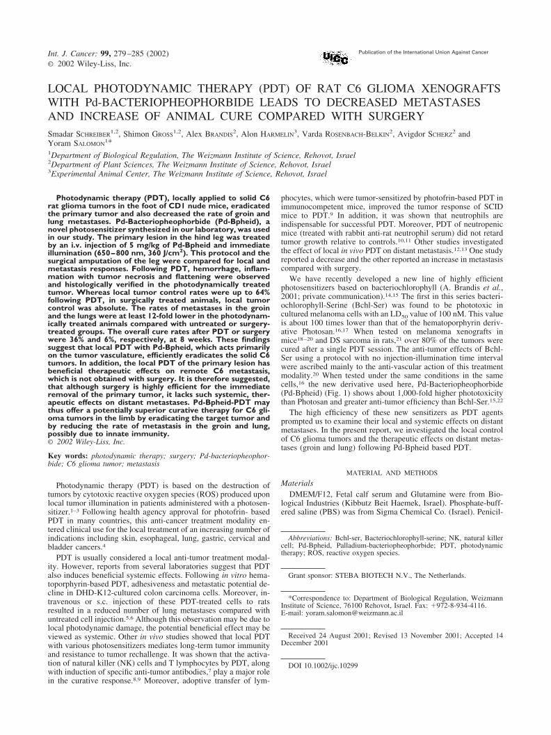
We have recently developed a new line of highly efficientphotosensitizers based on bacteriochlorophyll (A. Brandis et al.,2001; private communication).14,15 The first in this series bacteri-ochlorophyll-Serine (Bchl-Ser) was found to be phototoxic incultured melanoma cells with an LD50 value of 100 nM. This valueis about 100 times lower than that of the hematoporphyrin deriv-ative Photosan.16,17 When tested on melanoma xenografts inmice18–20 and DS sarcoma in rats,21 over 80% of the tumors werecured after a single PDT session. The anti-tumor effects of Bchl-Ser using a protocol with no injection-illumination time intervalwere ascribed mainly to the anti-vascular action of this treatmentmodality.20 When tested under the same conditions in the samecells,16 the new derivative used here, Pd-Bacteriopheophorbide(Pd-Bpheid) (Fig. 1) shows about 1,000-fold higher phototoxicitythan Photosan and greater anti-tumor efficiency than Bchl-Ser.15,22
The high efficiency of these new sensitizers as PDT agentsprompted us to examine their local and systemic effects on distantmetastases. In the present report, we investigated the local controlof C6 glioma tumors and the therapeutic effects on distant metas-tases (groin and lung) following Pd-Bpheid based PDT.
MATERIAL AND METHODS
MaterialsDMEM/F12, Fetal calf serum and Glutamine were from Bio-
logical Industries (Kibbutz Beit Haemek, Israel). Phosphate-buff-ered saline (PBS) was from Sigma Chemical Co. (Israel). Penicil-
Abbreviations: Bchl-ser, Bacteriochlorophyll-serine; NK, natural killercell; Pd-Bpheid, Palladium-bacteriopheophorbide; PDT, photodynamictherapy; ROS, reactive oxygen species.
Grant sponsor: STEBA BIOTECH N.V., The Netherlands.
*Correspondence to: Department of Biological Regulation, WeizmannInstitute of Science, 76100 Rehovot, Israel. Fax: �972-8-934-4116.E-mail: [email protected]
Received 24 August 2001; Revised 13 November 2001; Accepted 14December 2001
DOI 10.1002/ijc.10299Published online 7 March 2002 in Wiley InterScience (www.interscience.
wiley.com).
Int. J. Cancer: 99, 279–285 (2002)© 2002 Wiley-Liss, Inc.
Publication of the International Union Against Cancer

lin-streptomycin was from Bio-Lab laboratories (Jerusalem,Israel). Ketamine was from Rhone Merieux (Lyon, France). Xy-lazine was from Bayer (Leverkusen, Germany). Oxycodone wasfrom Rafa (Israel).
Cell cultureC6 rat glioma cells were cultured as monolayers in DMEM/F12
supplemented with glutamine, penicillin-streptomycin and 10%fetal calf serum. Cells were cultured at 37°C in an atmosphere of5% CO2 and passages were made every 3–4 days.
AnimalsCD1 male nude mice, 6 weeks old (28–32 g), were supplied
from the pathogen-free colony of the Experimental Animal Unit ofthe Weizmann Institute, Rehovot, Israel.
Tumor modelCultured C6 cells (1 � 106/0.02 ml) collected in PBS were
s.c.-injected in the distal dorsal foot of the hindleg of anesthetizedmice. When the needle was injected below the skin, the handle ofthe syringe was withdrawn to verify that it had not penetrated ablood vessel. In this way, systemic dissemination of tumor cellswas avoided. Tumors grew to a size of 6–8 mm (the treatmentsize) within 3 weeks. Tumors (� � 7 mm) located at least 3 mmdistal to the ankle were used in these experiments. Animals wereeuthanized upon tumor burden (size � � 15 mm, ulceration andbleeding) or large groin metastasis (increasing animal weight,beyond 110% the weight at treatment). All animal procedures wereapproved by the Institutional Subcommittee on Research for An-imal Care.
AnesthesiaMice were anesthetized by i.p. injection of Ketamine (5 mg/kg)
and Xylazine (1 mg/kg, 40 �l) 3 min before treatment. This dosewas sufficient to keep the animal anesthetized for about 40 min.
AnalgesiaOxycodone (12mg/liter) in 5% sucrose was provided as drinking
water immediately after treatment to the untreated control, PDTand surgery groups, for 1 week.
EuthanizationAnimals were euthanized by i.p. injection of Ketamine (15
mg/kg) and Xylazine (3 mg/kg, 120 �l).
SensitizerPd-Bpheid (A. Brandis et al., 2001; personal communication)15
(Fig. 1), which was synthesized in our laboratory, was dissolved in10 ml of a Cremophor based vehicle by sonication. The resultingsolution was centrifuged at 13,900g for 1 min in an Eppendorfcentrifuge and the supernatant retained. Sensitizer concentrationswere calculated assuming ε � 3.5 � 104 (� � 762 nm) usingwater-diluted samples. The sensitizer solution was stored in thedark at room temperature and used over several days with occa-sional spectroscopic examination for purity.
Light sourceA xenon LS3-PDT lamp (Biospec, Russia) with a 650–800 nm
spectral window and water filter was used. Light intensity deliv-ered via optical fibers was measured before every experimentusing a power meter (Ophir Optronics, Israel). A light powerdensity of 150 or 200 mW/cm2 was used. Aside from the tumortreatment site, the treated animal was protected from light withaluminum foil.
Photodynamic treatmentThe anesthetized mouse was placed in a home-built chamber
and the sensitizer (Pd-Bpheid, 5 mg/kg in �0.1 ml Cremophorvehicle) was i.v. injected (bolus). The tumor (� � 7 mm) wasimmediately illuminated (light spot diameter 1.1 cm) for 30 min asdescribed previously.20 The treatment dose rates were 150–200mW/cm2 equivalent to 270–360 J/cm2. During illumination theskin temperature at the illuminated site was elevated by no morethan 1°C as determined with an electronic thermometer (Newtron,TM-5005-Single I/P, Taiwan).
Parameters of response to PDTTumor necrosis and flattening were used to indicate local re-
sponse. The cure of the animals (no tumor and no metastasis) wasmonitored for 8 weeks. Tumors were photographed with a MX-2900 zoom Fujifilm digital camera, before and at different timesafter PDT.
SurgeryAfter anesthesia, the tumor-bearing leg of the mouse was tightly
ligated with 3-0 silk, 1–2 mm above the ankle joint. Amputationthrough the ankle, in a mid-articular plane, was then performed.Tumor cell dissemination during surgery was avoided, by ampu-tating only after blood vessel ligation. Subsequently, an analgesicwas given for 1 week.
Groin metastasesMetastases in the groin were inspected visually and by palpa-
tion. Metastases were photographed as above and histologicallyconfirmed as described below.
Lung metastasesAt different times following treatment, the animals were eutha-
nized and the lungs were removed. Lung metastases were macro-scopically detected as white colonies, projecting from the pinklung surface. Metastases measuring over 0.5 mm in diameter werecounted and considered significant. For fixation, the lungs were im-mersed in Bouin’s solution for 4 days. Lung surface metastases werephotographed and histologically confirmed as described below.
HistologyTumors and groin metastases were dissected. The samples in-
cluded the skin, subcutaneous tissue and the tumor mass. First, thesamples were fixed in 4% formaldehyde in phosphate buffer, pH7.4 and embedded in paraffin. Next, the paraffin blocks were cut(4–5 �m thick) through the skin in a midtangential plane. Finally,the specimens were stained with hematoxylin and eosin (H&E)
FIGURE 1 – Pd-Bpheid: Absorption spectrum, chemical formula andspectral window for PDT. The absorption spectrum (300–900 nm) inchloroform and chemical formula of Pd-Bpheid (insert) are shown.Also indicated are the major transition bands (By, Bx, Qx and Qy) andtheir respective maximal absorption. The spectral window used in thePDT protocol (650–800 nm) is indicated by dotted square.
280 SCHREIBER ET AL.

and micrographs were taken using an Olympus AX-70 microscope(Olympus) equipped with a Leaf MicroLumina camera (Scitex,Hertzelia, Israel).
Statistical analysisThe results were evaluated using unpaired 1-tailed t tests and
1-tailed tests of proportions. A p-value 0.05 was consideredstatistically significant.
RESULTS
Rat C6 glioma metastatic tumor modelAlthough rat C6 glioma variants are mostly nonmetastatic, it
was of interest that the specific cell line that we used shows anappreciable frequency of metastases when injected into the mousefoot. Therefore, we first characterized this C6 glioma tumor model,in particular the metastasis formation. Briefly, C6 cells were in-jected into the hindleg of the mice with a tumor-take of 100%(n�48). The primary tumors reached a diameter of 6–8 mmwithin 3 weeks (n�20), 10–12 mm by 5 weeks (n�12) and 13–15mm at 7 weeks (n�16). Macroscopic metastases to the groinappeared by 3 weeks as a protruding mass that reached a diameterof 5–10 mm at 7 weeks (Fig. 2a). Macroscopic metastases to thelungs appeared at 5 weeks as projecting white colonies on the pinklung surface. At 7 weeks, these colonies (1–10/lung) measured
0.5–5 mm in diameter (Fig. 2b). The frequency of metastases atthis time was 62% in the groin and 50% in the lung. All metastaseswere confirmed by histological examination of the groin (Fig. 2c)and lung (Fig. 2d).
Local response of C6 glioma xenograft to Pd-Bpheid-PDTWe next determined the local response of the C6 xenografts to
Pd-BP-PDT. Tumor-bearing mice were injected with 5 mg/kgPd-BP and illuminated at light doses of 270 (group 1, n�8) or 360J/cm2 (group 2, n�11). Tumor response was monitored for 5weeks (Fig. 3a). Inflammation and necrosis developed over 2–5days with ultimate tumor flattening by days 7–10 at the 2 lightdoses used. No macroscopic tumors were seen between 16–35days in the animal group 2, where most wounds healed and thelocal cure rate was 64%. In contrast, recurrences developed in allanimals in group 1 around day 18 (Fig. 3a). Tumors in this and theuntreated control (group 3, n�16) were allowed to grow to the sizelimit at which time the animals were euthanized. One example oflocal tumor cure followed for 142 days is illustrated in Figure 3b.An example of a tumor (13 mm ) in an untreated animal at 7weeks after C6 cell injection is shown in Figure 2a.
Histology of C6 tumors after Pd-Bpheid-PDTPd-Bpheid-PDT-induced damage to tumor and blood vessels
was examined by histology 3, 24 and 48 hr after treatment using 3
FIGURE 2 – C6 rat glioma xenograft: primary tumor, groin and lung metastases. The CD1 nude mouse is shown 7 weeks after C6 tumor cellinjection. (a) The primary tumor (13 mm ) and the groin metastasis (10 mm , encircled) are shown. (b) The mouse was sacrificed and thelungs removed and embedded in Bouin’s solution for 4 days and then photographed. The lung surface metastases are encircled in black. (c)Histology: lymph node metastasis (groin). (d) Histology: lung metastases. Meta, metastasis; lymp, lymphocytes.
281LOCAL PHOTODYNAMIC THERAPY OF RAT C6 GLIOMA XENOGRAFTS

animals per time point (Fig. 4). The histology of the C6 tumorbefore treatment is shown in Figure 4 (untreated). Three hoursafter treatment (Fig. 4, 3 hr), most tumor blood vessels werecongested but intact; however, a few were mildly damaged, result-ing in mild multifocal hemorrhage. Most of the tumor cells werenot histologically damaged. The small number of damaged tumorcells were superficial cells adjacent to the light source. Mild,subcutaneous infiltration, including perivascular cuffing, consistedmainly of neutrophils and some round cells were seen adjacent tothe tumor. Perivascular cuffing was also noticed within the tumormass. Twenty-four hours post PDT, numerous blood vessels weredamaged, causing severe hemorrhage and tumor necrosis. At this
stage, the inflammatory reaction was moderate (Fig. 4, 24 hr). Forty-eight hours after PDT, necrosis was widespread and included most ofthe tumor (Fig. 4, 48 hr). At this time individual blood vessels, tumorand inflammatory cells were hardly discernible. These results suggestthat the blood vessels are a primary target of Pd-Bpheid-PDT and thatPDT initiates the inflammatory response.
The response of distant metastasis to local Pd-Bpheid-PDTof the primary tumor
To evaluate whether PDT has a systemic effect on distant metas-tasis, we compared the frequency of groin and lung metastases in 4groups of animals. The experiment included 3 experimental groupsdescribed above: PDT with 5 mg/kg Pd-Bpheid using a light dose of270 (group 1, n�8), or 360 J/cm2 (group 2, n�11) or untreated (group3, n�16). An additional group was surgically treated (group 4, n�16)and the tumor-bearing foot was amputated.
Groin metastases. The appearance of groin metastases was firstobserved on day 4 in the untreated and surgically treated animalgroups. In both Pd-Bpheid-PDT groups, groin metastasis appearedby day 18 (Fig. 5). The incidence of metastasis in the untreated andsurgically treated animal groups increased with time to include themajority of the animals (62%) and (88%), respectively, by day 35.In contrast, the incidence of metastases remained unchanged (25–27%) in both PDT groups from 21–35 days. These results implythat local PDT leads to a delay in the appearance of and a decreasein the incidence of groin metastasis, compared with both surgeryand no treatment.
Lung metastases. The average number of lung metastases pertreated animal on day 35 is shown in Table I. The mean number oflung metastasis in untreated animals (2.12) was �2.5-fold higherthan in surgically treated animals (0.81) and �10-fold higher thanin animals that underwent Pd-Bpheid-PDT (0–0.18). The frequen-cies of lung metastases were the lowest following PDT (Table I).As with groin metastases, the lowest incidence in the number ofdistant lung metastasis was seen in both PDT groups, with eithera partial or complete local tumor response. However, in this casesurgical treatment was beneficial compared with untreated ani-mals.
Animal cure. Spontaneous cure (no tumor and no metastasis)was never seen in untreated animals. After surgical amputation ofthe tumor-bearing foot there was obviously complete control of theprimary tumor. However, most animals developed metastasis by 8weeks and only 1/16 (6%) was cured. PDT with 360 J/cm2 led tothe cure of 4/11 animals (36%), whereas use of a lower light dose(270 J/cm2) led to no cure since local tumor control did not lastbeyond day 18 (Fig. 3a). The results presented in Table I show thatby providing local and distant tumor control and cure, PDT ap-pears to be superior to surgical treatment.
DISCUSSION
Our study demonstrates that local Pd-Bpheid-based PDT isefficient for both local and systemic control of C6 glioma xeno-grafts in mice. Using Pd-Bpheid-PDT, the success rate of tumorflattening in the foot was 64% (Fig. 3). Pd-Bpheid-PDT of the C6glioma tumor model appears to be based on vascular destruction(Fig. 4), similar to Bchl-Ser-based PDT of mouse melanoma.20
PDT is generally considered a local treatment modality; how-ever, our results demonstrate that treatment of a C6 xenograft inthe foot of the mouse with Pd-Bpheid and light had a beneficialeffect on distant metastases. Pd-Bpheid-PDT reduced the rate ofgroin metastases by approximately 2-fold (from 62 to 25–27%)and lung metastases by at least 12-fold (from 2.1 to 0–0.18/mouse) and provided an overall cure of 36% compared withuntreated controls where no cure was observed. In spite of the highshort-term effectiveness of surgery, the results of this treatmentwere inferior to PDT, with only 6% overall cure. Groin metastasesafter surgery were observed in most of the mice (88%) at a slightlyhigher rate than that seen in the untreated group (62%). This
FIGURE 3 – Local response of rat C6 xenograft to PDT. (a) Micewith C6 glioma xenografts (� �7 mm) in the hindleg were treatedwith 5 mg/kg Pd-Bpheid and illuminated at 270 J/cm2 (group 1, n�8,open circles) or 360 J/cm2 (group 2, n�11, open diamonds) or un-treated (group 3, n�16, open squares). The incidence of flat tumors asa function of time after PDT is shown. ap0.05 by 1-tailed test forproportions, testing Pc� Pt vs. PcPt, where Pc � proportion foruntreated controls and Pt � proportion for treated subjects. All otherdetails were as described in Material and Methods. (b) The clinicalcourse of events in a tumor bearing animal (group 2) before and afterPDT is presented. Photographs at day of treatment (0 day) and 4, 7, 16,35 and 142 days after PDT are shown. By day 4, partial necrosis wasseen. By day 7, tumor flattening was observed with an eschar coveringthe wound. By day 35, the wound healed and the animal was cured asseen by day 142.
282 SCHREIBER ET AL.

apparent increase is however not statistically significant (p�0.05).In comparison to untreated control, the rate of lung metastases inthe surgery group was 2.5-fold lower (0.8/mouse), but this differ-ence was also not statistically significant (p�0.05). Interestingly,reducing the light intensity by 25% to 270 J/cm2, induced a similarresponse in the distant metastasis (Fig. 5, Table I), whereas thelocal response was only temporal with 100% regrowth (Fig. 3a).
Changes in tumor-host relationship, including the immune stateof the host, tumor angiogenesis and tumor cell adhesion, mayeither encourage or prevent the development of microscopic me-tastases to clinical manifestation.
The effect of local PDT on distant metastases was reported inonly a handful of cases with quite varying results. A decreased rateof lung metastases relative to surgery or no treatment, following
Photofrin II-based PDT of s.c. Lewis lung carcinoma in C57BL/6mice was reported by Gomer.12 Similarly, PDT with indocyaningreen of s.c. DMBA-4 mammary tumors in Wistar Furth ratsdecreased the volume and the number of cases of axillary and inguinalmetastasis, compared with no treatment.7 In contrast, Momma andHasan13 showed that BPD-based PDT of R3327-MatLyLu prostatecarcinoma in Dunning rats increased the number of lung metastasis(but not the frequency of animals with metastasis) compared withboth untreated and surgically treated groups. The differences betweenthe reported results and the results of our study may be due todifferences in tumor type, site of implantation, animal species, PDTprotocol and surgical techniques used.
Recent studies showed that PDT of sarcomas with 2-iodo-5-ethylamino-9-diethylaminobenzo(�)-phenothiazinium chloride8 or
FIGURE 4 – Histology of C6 tumors after Pd-Bpheid-PDT. C6 glioma xenografts (��7 mm)in the hindleg were treated with 5 mg/kg Pd-Bpheid and illuminated at 360 J/cm2. Tumorswere harvested before and 3, 24 and 48 hr aftertreatment, fixed and prepared for histological ex-amination. Micrographs at 2 magnifications ofeach time point are presented. bv-blood vessel,hem-hemorrhage, nec-necrosis, neu- neutrophils,tum-tumor. All other details were as described inMaterial and Methods.
283LOCAL PHOTODYNAMIC THERAPY OF RAT C6 GLIOMA XENOGRAFTS

photofrin9 induce long-term tumor immunity. Moreover, PDTcured the tumor only in immunocompetent but not in severelycombined immunodeficient mice. It was also shown that activationof natural killer cells (NK) and lymphocytes by PDT plays a majorrole in the curative response. Induction of long-term tumor resis-tance and the production of specific tumor antibodies in responseto PDT and imuno-adjuvant were also reported.7 A rational con-nection between PDT and tumor immunity can be supported byfact that in order to initiate an immune response, dendritic cells,the most potent antigen-presenting cells, must be activated byendogenous signals like those expressed by stressed necrotic cellsor damaged blood vessels. They then obtain the capacity to acti-vate an antigen-specific T-cell response. Such responses are notinduced by healthy or apoptotic cells. Thus, PDT of tumor tissue
may create just the right set of conditions needed for the inductionof tumor immunity.23
Although CD1 nude mice are deficient in T cells, NK activationagainst C6 tumors and the induction of tumor-specific antibodiesby Pd-Bpheid-PDT are possible explanations for the observeddecrease in metastasis development (Fig. 5, Table I).
Still another explanation to the lower metastasis incidenceafter PDT may be related to changes in tumor cell adhesion.Colon cancer cells treated in vitro by PDT loosely adhere toendothelial cells and induce a smaller number of metastasiswhen injected i.v.5 or s.c.,6 as compared with untreated cells.
The fact that C6 metastasis appeared above the tumor-bearingfoot, following amputation, suggests that spontaneous micro-scopic dissemination had occurred, before surgery. It has beenreported in the literature that surgery may cause immunosup-pression,24,25 consistent with an increase in the rate of metas-tases. However, following surgery in this model, the rates ofmetastases in the lung and groin were lower and higher, respec-tively, compared with untreated control but these differenceswere not statistically significant. We therefore conclude thatimmunosuppression seems not to play an important role in thismodel (Fig. 5, Table I). The methodological approach of simulta-neously recording metastatic rates in 2 anatomical locations thatrepresent different target tissues further strengthens this claim.
The development of tumors and metastases depends on an-giogenesis.26 –28 Angiostatic substances (like endostatin or an-giostatin) circulate in tumor-bearing animals and suppress me-tastasis.26,29 –31 Eradication of the primary tumor may thereforechange the balance between angiostatic and angiogenic sub-stances. Consequently, after surgical removal of the primarytumor, when the levels of such putative substances abruptly fall,metastasis can grow and become apparent. However, in this C6model, the arguments that stand against the significance ofimmunosuppression also put in question the importance of theremoval of putative angiostatic factors by surgery. The possibleinfluence of PDT on the balance of angiostatic and angiogenicsubstances has, to the best of our knowledge, not been reported.In this respect, it is interesting to note that local X-ray irradi-ation of FSA-II tumors is followed by an increase in endosta-tin32 and the suppression of angiogenesis at a distal site.33
Although systemic effects may be engraved in clinically usedPDT, the basis of these observations and their generality withrespect to sensitizer type, treatment protocol and the tumor typeare not clear. Future experiments with immunocompetent ani-mals will enable the elucidation of the mechanistic basis andpossible clinical relevance of the described observations.
ACKNOWLEDGEMENTS
Y.S. is the incumbent of the Charles and Tillie Lubin Chair ofBiochemical Endocrinology. This study serves as partial fulfill-ment of the Ph.D. thesis requirements for S.S. at the FeinbergGraduate School, The Weizmann Institute of Science. We wish tothank Prof. R.S. Kenett for his help in the statistical analysis andMs. R. Tsoref for her devoted secretarial help.
FIGURE 5 – Time course of the appearance of C6 groin metastasisafter local PDT or surgical amputation of the tumor bearing leg. Theappearance of groin metastases in the animal groups described inFigure 3a was monitored at different times after beginning of theexperiment. The experimental groups were either treated with Pd-Bpheid and illuminated at 270 J/cm2 (group 1, n�8 open circle), or360 J/cm2 (group 2, n�11, open diamond) or untreated controls (group3, n�16, open squares) or surgically treated (group 4, n�16, trian-gles). Groin metastases were visually inspected and then verified bypathological examination and the percentage of animals, out of thetotal treated animals, with metastasis in the groin was calculated.Control untreated tumors continued to grow and the animals had to besacrificed 5 weeks after beginning of the experiment according toanimal welfare regulations. bp0.05 by 1-tailed test for proportions,testing Pc � Pt vs. Pc � Pt, where Pc � proportion for untreatedcontrols and Pt � proportion for treated subjects. Using the same testfor comparing surgery to untreated resulted in p�0.05. All otherdetails were as described in Material and Methods.
TABLE I – LOCAL AND DISTANT TUMOR RESPONSE TO PDT AND SURGICAL AMPUTATION
Treatment Number ofmice, n
Local tumor control(day 35, %)
Frequency groinmetastasis
(day 35, %)
Frequency lungmetastasis
(day 35, %)
Mean lung metastasisper animal, n � SE
(day 35, %)
Cure(8 weeks, %)
None 16 0 62 50 2.12 � 0.72 0Amputation 16 1001 88 25 0.81 � 0.43 6Pd-Bpheid 270 J/cm2 8 0 252 02 03 0Pd-Bpheid 360 J/cm2 11 641 272 92 0.18 � 0.183 361
1p 0.05 by 1-tailed test for proportions, testing Pc � Pt vs. Pc Pt, where Pc � proportion for untreated controls and Pt � proportion fortreated subjects.–2p 0.05 by one tailed test for proportions, testing Pc � Pt vs. Pc � Pt, where Pc � proportion for untreated controls and Pt �proportion for treated subjects.–3p 0.05 by an unpaired one tailed t test for Mc � Mt vs. Mc � Mt, where Mc, Mt represent the expected numberof lung metastasis per animal in control and treated groups, respectively.
284 SCHREIBER ET AL.

REFERENCES
1. van Hillegersberg R, Kort WJ, Paul Wilson JH. Current status ofphotodynamic therapy in oncology. Drugs 1994;48:510–27.
2. Bonnet R. Photodynamic therapy in historical perspectives. Rev Con-temp Pharmacother 1999;10:1–17.
3. Sibata CH, Colussi VC, Oleinick NL, et al. Photodynamic therapy: anew concept in medical treatment. Braz J Med Biol Res 2000;33:869–80.
4. Dougherty TJ, Gomer CJ, Henderson BW, et al. Photodynamic ther-apy review. J Natl Cancer Inst 1998;90:889–905.
5. Foultier MT, Vonarx-Coinsmann V, Cordel S, et al. Modulation ofcolonic cancer cell adhesiveness by haematoporphyrin derivative pho-todynamic therapy. J Photochem Photobiol B 1994;23:9–17.
6. Rousset N, Vonarx V, Eleouet S, et al. Effects of photodynamictherapy on adhesion molecules and metastasis. J Photochem PhotobiolB 1999;52:65–73.
7. Chen WR, Zhu WG, Dynlacht JR, et al. Long-term tumor resistanceinduced by laser photo-immunotherapy. Int J Cancer 1999;81:808–12.
8. Hendrzak-Henion JA, Knisely TL, Cincotta L, et al. Role of theimmune system in mediating the antitumor effect of benzophenothia-zine photodynamic therapy. Photochem Photobiol 1999;69:575–81.
9. Korbelik M, Dougherty GJ. Photodynamic therapy-mediated immuneresponse against subcutaneous mouse tumors. Cancer Res 1999;59:1941–6.
10. de Vree WJ, Essers MC, de Bruijn HS, et al. Evidence for animportant role of neutrophils in the efficacy of photodynamic therapyin vivo. Cancer-Res 1996;56:2908–11.
11. de Vree WJ, Essers MC, Koster JF, et al. Role of interleukin 1 andgranulocyte colony-stimulating factor in photofrin-based photody-namic therapy of rat rhabdomyosarcoma tumors. Cancer Res 1997;57:2555–8.
12. Gomer CJ, Ferrario A, Murphree AL. The effect of localized porphy-rin photodynamic therapy on the induction of tumour metastasis. Br JCancer 1987;56:27–32.
13. Momma T, Hamblin MR, Wu HC, et al. Photodynamic therapy oforthotopic prostate cancer with benzoporphyrin derivative: local con-trol and distant metastasis. Cancer Res 1998;58:5425–31.
14. Scherz A, Salomon Y, Fiedor L. Catalytic condensation of chlorophylland bactheriochlorophyll derivatives. US patent #5955:585, 1998.
15. Scherz A, Salomon Y, Brandis A, et al. Palladium-substituted bacte-riochlorophyll derivatives and use thereof. International PCT PatentApplication: No. PCT/IL99/00673, 1999.
16. Rosenbach-Belkin V, Chen L, Fiedor L, et al. Serine conjugates ofChlorophyll and Bacteriochlorophyll photocytotoxicity in vitro andtissue distribution in mice bearing Melanoma tumors. PhotochemPhotobiol 1996;64:174–81.
17. Rosenbach-Belkin V, Chen L, Fiedor L, et al. Photodynamic tumortherapy, 2nd and 3rd generation photosensitizers. In: Moser J, ed.London: Harwood Academic Publishers, 1998. 117–26.
18. Zilberstein J, Scherz A, Bromberg A, et al. Mechanisms involved inchlorophyll based photoinduced cell damage: photodynamic therapy
of melanoma. Meeting of the Society of Magnetic Resonance. Nice,France, 1995;3:1681.
19. Zilberstein J, Scherz A, Bromberg A, et al. The effects of Bacterio-chlorophyll based PDT on tumor vasculature. Meeting of the Societyof Magnetic Resonance. New York, 1996.
20. Zilberstein J, Schreiber S, Bloemers MCWM, et al. Anti-vasculartreatment of solid melanoma tumors with bacteriochlorophyll-serinebased photodynamic therapy. Photochem Photobiol 2001;73:257–63.
21. Kelleher DK, Thews O, Rzeznik J, et al. Water-filtered infrared-Aradiation: a novel technique for localized hyperthermia in combina-tion with bacteriochlorophyll-based photodynamic therapy. Int J Hy-perthermia 1999;15:467–74.
22. Koudinova N, Pinthus JH, Brandis A, et al. Curative and palliativeeffects of a novel photodynamic agent (Pd bacteriopheophorbide) onprostate cancer: in vivo studies Using human prostate cancer xeno-grafts. CapCURE 8th Annual Scientific Retreat. Lake Tahoe Nevada,2001. www.capcure.org.
23. Gallucci S, Lolkema M, Matzinger P. Natural adjuvants: endogenousactivators of dendritic cells. Nat Med 1999;5:1249–55.
24. Ben-Eliyahu S, Page GG, Yirmiya R, et al. Evidence that stress andsurgical interventions promote tumor development by suppressingnatural killer cell activity. Int J Cancer 1999;80:880–8.
25. Lundy J, Lovett EJD, Hamilton S, et al. Halothane, surgery, immu-nosuppression and artificial pulmonary metastases. Cancer 1978;41:827–30.
26. O’Reilly MS, Wiederschain D, Stetler-Stevenson WG, et al. Regula-tion of angiostatin production by matrix metalloproteinase-2 in amodel of concomitant resistance. J Biol Chem 1999;274:29568–71.
27. Rivas MJ, Arii S, Furutani M, et al. Expression of human macrophagemetalloelastase gene in hepatocellular carcinoma: correlation withangiostatin generation and its clinical significance. Hepatology 1998;28:986–93.
28. Yoon SS, Eto H, Lin CM, et al. Mouse endostatin inhibits theformation of lung and liver metastases. Cancer Res 1999;59:6251–6.
29. O’Reilly MS, Pirie-Shepherd S, Lane WS, et al. Antiangiogenicactivity of the cleaved conformation of the serpin antithrombin. Sci-ence 1999;285:1926–8.
30. Hahnfeldt P, Panigrahy D, Folkman J, et al. Tumor developmentunder angiogenic signaling: a dynamical theory of tumor growth,treatment response and postvascular dormancy. Cancer Res 1999;59:4770–5.
31. Wen W, Moses MA, Wiederschain D, et al. The generation of en-dostatin is mediated by elastase. Cancer Res 1999;59:6052–6.
32. Ramsay J, Zietman A, Preffer F, et al. The growth and cell kinetics ofa secondary tumor after radiation or surgical treatment of the primarytumor. Int J Radiat Oncol Biol Phys 1989;17:809–13.
33. Hartford AC, Gohongi T, Fukumura D, et al. Irradiation of a primarytumor, unlike surgical removal, enhances angiogenesis suppression ata distal site: potential role of host-tumor interaction. Cancer Res2000;60:2128–31.
285LOCAL PHOTODYNAMIC THERAPY OF RAT C6 GLIOMA XENOGRAFTS