Localization of Trichinella spiralis in muscles of ... - Parasite · LOCALIZATION OF TRICHINELLA...
3
L OCALIZATION OF TRICHINELLA SPIRALIS IN MUSCLES OF COMMERCIAL AND PARASITOLOGIC INTEREST IN PORK RIBICICH M., MIGUEZ M., ARGIBAY T., BASSO N. & FRANCO A.* Summary: Trichinellosis is widespread around the world with different representatives of the genus Trichinella found in almost every continent. In Argentina the main source of transmission for the disease to humans is pig meat infected with Trichinella spiralis. The object of this work was to determine the distribution of Trichinella larvae in fresh meat cuts which are sold for human consumption and in the muscles traditionally used for the disease diagnosis at meat-packing plants. Cranial muscles to the last rib showed more Trichinella spiralis larvae than those with a caudal location (p < 0.01). No significant differences were found (p > 0.05) between bilateral left and right muscles. Significant larval concentrations were found in the neck muscles, even in carcasses with a low parasitic load; these muscles are used to prepare cold meats (boston butt). Commercial cuts of meat had a substantial larval burdens in animals experimentally infected with 500 to 5,000 Trichinella spiralis larvae, with parasite burdens similar to infection levels in muscles evaluated at the meat packing plant. KEY WORDS : Trichinella spiralis, pigs, meat inspection. I n Argentina the main source of transmission of tri- chinellosis to humans is pig meat infected with Tri- chinella spiralis. The muscles where Trichinella spiralis larvae esta- blish vary according to the host (Gould et al., 1970; Campbell et al., 1993; Reina et al, 1996; Kapel et al., 1994; Pozio et al., 1998). The muscles used for slaughter inspection for Trichinella infection (dia- phragm, tongue, masetters, intercostals) are those that contain the highest number of larvae per gram (Kotula et al., 1984; Zimmerman et al., 1970). The objective of this work was to determine the distribution of larvae of fresh meat cuts which are sold for human consump- tion and in the muscles traditionally used for inspec- tion in meat-packing plants. MATERIALS AND METHODS * Faculty of Veterinary Sciences. University of Buenos Aires, Chor- roarin 280 (1427) Argentina. Correspondence: Mabel Ribicich. Tel.: 54-11-4580-2820 - Fax: 54-11-4514-8962. E-mail: [email protected] PIG INFECTION T he work was carried out with 14 commercial hybrid pigs, Landrace and Large White cross- bred, originating from a cattle-breeding ranch in the province of Buenos Aires. They were 60 days old at the beginning of the experiment and weighed approximately 20 kg each. All animal work was per- formed in the swine breeding facility belonging to the Faculty of Veterinary Sciences at the University of Buenos Aires. The parasite Trichinella spiralis was maintained by serial passage in female CF-I mice obtained from the Department of Parasitology, ANLIS. Dr. Carlos G. Malbrán, Argentina. Muscle larvae were recovered from mice by acid- pepsin digestion of striated muscle collected at least 30 days after infection. Pigs were orally inoculated with infective larvae of Tri- chinella spiralis using a Coopers® Cattle pellet appli- cator bolus-flowing gun. The pigs were experimentally inoculated with doses of 100 (group 1), 500 (group 2), 5,000 (group 3) or 50,000 (group 4) larvae of Trichi- nella spiralis. TISSUE RECOVERY On day 100 post-inoculation, all 14 pigs were sedated and euthanized by exsanguination. Following eutha- nasia, the animals were eviscerated and tongue muscle, tongue base, masseters, diaphragm, intercostals and com- mercial cuts such as ham, shoulder, boston butt, fillet, spare ribs and belly were collected for artificial digestion. DIGESTION TESTING Distribution of T. spiralis larvae in each tissue sample was determined by digestion of 100 g of tissue (Gam- ble, 1996). STATISTICAL ANALYSIS A test for proportion was made with the Z statistic, using the software Statistic for Windows. Differences were considered significant at p < 0.05. S246 X th ICT. August 2000 Parasite, 2001, 8, S246-S248 Article available at http://www.parasite-journal.org or http://dx.doi.org/10.1051/parasite/200108s2246
Transcript of Localization of Trichinella spiralis in muscles of ... - Parasite · LOCALIZATION OF TRICHINELLA...

LOCALIZATION OF TRICHINELLA SPIRALIS IN MUSCLES OF COMMERCIAL AND PARASITOLOGIC INTEREST IN PORK
RIBICICH M., MIGUEZ M., ARGIBAY T., BASSO N. & FRANCO A.*
S u m m a r y :
Trichinellosis is widespread around the world with different representatives of the genus Trichinella found in almost every continent. In Argentina the main source of transmission for the disease to humans is pig meat infected with Trichinella spiralis. The object of this work was to determine the distribution of Trichinella larvae in fresh meat cuts which are sold for human consumption and in the muscles traditionally used for the disease diagnosis at meat-packing plants. Cranial muscles to the last rib showed more Trichinella spiralis larvae than those with a caudal location (p < 0.01). No significant differences were found (p > 0.05) between bilateral left and right muscles. Significant larval concentrations were found in the neck muscles, even in carcasses with a low parasitic load; these muscles are used to prepare cold meats (boston butt). Commercial cuts of meat had a substantial larval burdens in animals experimentally infected with 5 0 0 to 5 ,000 Trichinella spiralis larvae, with parasite burdens similar to infection levels in muscles evaluated at the meat packing plant.
KEY WORDS : Trichinella spiralis, pigs, meat inspection.
In Argentina the main source of transmission of trichinellosis to humans is pig meat infected with Trichinella spiralis.
The muscles where Trichinella spiralis larvae establish vary according to the host (Gould et al., 1970; Campbell et al., 1993; Reina et al, 1996; Kapel et al., 1994 ; P o z i o et al., 1 9 9 8 ) . T h e m u s c l e s used for slaughter inspection for Trichinella infection (diaphragm, tongue, masetters, intercostals) are those that contain the highest number of larvae per gram (Kotula et al., 1984; Zimmerman et al., 1970) . The objective of this work was to determine the distribution of larvae of fresh meat cuts which are sold for human consumption and in the muscles traditionally used for inspection in meat-packing plants.
MATERIALS AND METHODS
* Faculty of Veterinary Sciences. University of Buenos Aires, Chor-roarin 280 (1427) Argentina. Correspondence: Mabel Ribicich. Tel.: 54-11-4580-2820 - Fax: 54-11-4514-8962. E-mail: [email protected]
P I G INFECTION
The work was carried out with 14 commercial hybrid pigs, Landrace and Large White crossbred, originating from a cattle-breeding ranch in
the province of Buenos Aires. They were 60 days old at the beginning o f the exper iment and w e i g h e d approximately 20 kg each. All animal work was performed in the swine breeding facility belonging to the Faculty of Veterinary Sciences at the University of Buenos Aires. The parasite Trichinella spiralis was maintained by serial passage in female CF-I mice obtained from the Department of Parasitology, ANLIS. Dr. Carlos G. Malbrán, Argentina. Muscle larvae were recovered from mice by acid-pepsin digestion of striated muscle collected at least 30 days after infection.
Pigs were orally inoculated with infective larvae of Trichinella spiralis using a Coopers® Cattle pellet applicator bolus-flowing gun. The pigs were experimentally inoculated with doses of 100 (group 1), 500 (group 2) , 5,000 (group 3) or 50,000 (group 4) larvae of Trichinella spiralis.
TISSUE RECOVERY
On day 100 post-inoculation, all 14 pigs were sedated and euthanized by exsanguination. Following euthanasia, the animals were eviscerated and tongue muscle, tongue base, masseters, diaphragm, intercostals and commercial cuts such as ham, shoulder, boston butt, fillet, spare ribs and belly were collected for artificial digestion.
DIGESTION TESTING
Distribution of T. spiralis larvae in each tissue sample was determined by digestion of 100 g of tissue (Gamble, 1996) .
STATISTICAL ANALYSIS
A test for proportion was made with the Z statistic, using the software Statistic for Windows. Differences were considered significant at p < 0.05.
S246 Xth ICT. August 2000 Parasite, 2001, 8, S246-S248
Article available at http://www.parasite-journal.org or http://dx.doi.org/10.1051/parasite/200108s2246
CONTROL AND VETERINARY DIAGNOSIS
RESULTS
Both animals in group 0 were negative by artificial digestion. The predilection sites of muscle larvae of Trichinella spiralis in pigs infected
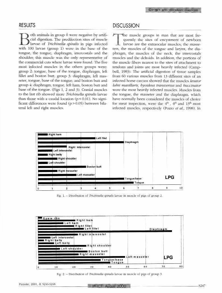
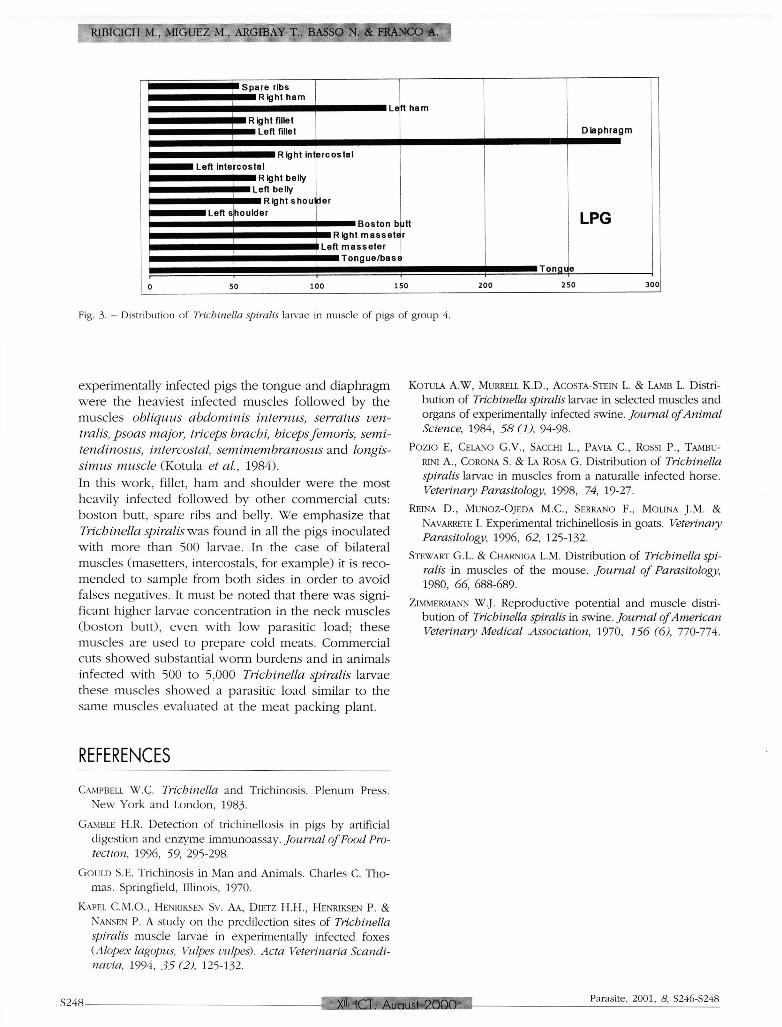
with 100 larvae (group 1) were in the base of the tongue, the tongue, diaphragm, intercostals and the shoulder; this muscle was the only representative of the commercial cuts where larvae were found. The five most infected muscles in the others groups were : group 2: tongue, base of the tongue, diaphragm, left fillet and boston butt. group 3: diaphragm, left mas-seter, tongue, base of the tongue, and boston butt and group 4: diaphragm, tongue, left ham, boston butt and base of the tongue. (Figs 1, 2 and 3) . Cranial muscles to the last rib showed more Trichinella spiralis larvae than those with a caudal location ( p < 0 . 0 1 ) . No significant differences were found (p > 0 .05) be tween bilateral left and right muscles.
DISCUSSION
T he muscle groups in man that are most frequently the sites of encystment of newborn larvae are the extraocular muscles, the masse-
ters, the muscles of the tongue and larynx, the diaphragm, the muscles of the neck, the intercostals muscles and the deltoids. In addition, the portions of the muscle fibers nearest to the sites of attachment to tendons and joints are most heavily infected (Campbell, 1983) . The artificial digestion of tissue samples from 60 various muscles from 13 different sites of an infected horse carcass showed that the muscles levator labii maxillaris, hyoideus transversus and buccinator were the most heavily infected muscles. Muscles from the tongue, the masseter and the diaphragm, which have normally been considered the muscles of choice for meat inspection, were the 4 t h , 6 t h and 1 3 t h most infected muscles, respectively (Pozio et al., 1998). In
Fig. 1. - Distribution of Trichinella spiralis larvae in muscle of pigs of group 2.
Fig. 2. - Distribution of Trichinella spiralis larvae in muscle of pigs of group 3.
Parasite, 2001, 8, S246-S248 Xth ICT. August 2000 S247
R I B I C I C H M., M I G U E Z M., A R G I B A Y T. , B A S S O N. & F R A N C O A.
Fig. 3. — Distribution of Trichinella spiralis larvae in muscle of pigs of group 4.
experimentally infected pigs the tongue and diaphragm were the heaviest infected muscles fol lowed by the muscles obliquus abdominis internus, serratus ven-tralis, psoas major, triceps brachi, biceps femoris, semi-tendinosus, intercostal, semimembranosus and longis-simus muscle (Kotula et al, 1984) . In this work, fillet, ham and shoulder were the most heavily infected followed by other commercial cuts: boston butt, spare ribs and belly. W e emphasize that Trichinella spiralis was found in all the pigs inoculated with more than 500 larvae. In the case o f bilateral muscles (masetters, intercostals, for example) it is reco-mended to sample from both sides in order to avoid falses negatives. It must b e noted that there was significant higher larvae concentration in the neck muscles (boston butt), even with low parasitic load; these muscles are used to prepare cold meats. Commercial cuts showed substantial worm burdens and in animals infected with 500 to 5,000 Trichinella spiralis larvae these muscles showed a parasitic load similar to the same muscles evaluated at the meat packing plant.
KOTULA A . W , MURRELL K.D., ACOSTA-STEIN L. & LAMB L. Distribution of Trichinella spiralis larvae in selected muscles and organs of experimentally infected swine. Journal of Animal Science, 1984, 58 ft), 94-98.
POZIO E, CELANO G . V . , SACCHI L., PAVIA C , ROSSI P., TAMBU-RINI A., CORONA S. & LA ROSA G . Distribution of Trichinella spiralis larvae in muscles from a naturalle infected horse. Veterinary Parasitology, 1998, 74, 19-27.
REINA D., MUNOZ-OJEDA M . C , SERRANO F. , MOLINA J . M . & NAVARRETE I. Experimental trichinellosis in goats. Veterinary Parasitology, 1996, 62, 125-132.
STEWART G . L . & CHARNIGA L.M. Distribution of Trichinella spiralis in muscles of the mouse. Journal of Parasitology, 1980, 66, 688-689.
ZIMMERMANN W . J . Reproductive potential and muscle distribution of Trichinella spiralis in swine. Journal of American Veterinary Medical. Association, 1970, 156(6), 770-774.
REFERENCES
CAMPBELL W.C. Trichinella and Trichinosis. Plenum Press. New York and London, 1983.
GAMBLE H.R . Detection of trichinellosis in pigs by artificial digestion and enzyme immunoassay. Journal of Food Protection, 1996, 59, 295-298.
GOULD S.E. Trichinosis in Man and Animals. Charles C. Thomas. Springfield, Illinois, 1970.
KAPEL C.M.O., HENRIKSEN Sv. AA, DIETZ H.H. , HENRIKSEN P. & NANSEN P. A study on the predilection sites of Trichinella spiralis muscle larvae in experimentally infected foxes (Alopex lagopus, Vulpes vulpes). Acta Veterinaria Scandinavia, 1994, 35 (2), 125-132.
S248 Xth ICT. August 2000 Parasite, 2001, 8, S246-S248