Mitochondrial dysfunction and endoplasmic reticulum stress ...
Upload
rakesh-tuliCategory
view
214download
0
Localization of Rabies Virus Glycoprotein into the EndoplasmicReticulum Produces Immunoprotective Antigen
Dinesh K. Yadav • Shadma Ashraf •
Pradhyumna K. Singh • Rakesh Tuli
Published online: 17 May 2012
� Springer Science+Business Media, LLC 2012
Abstract Rabies virus surface glycoprotein (rabies
G-protein) with (G?RS) and without (G-RS) endoplasmic
reticulum retrieval signal was expressed and characterized in
tobacco plants. Transgenically expressed rabies G-protein
was estimated at 0.015–0.38 % of total leaf protein. The
relative migration of the rabies G-protein on SDS-PAGE was
at the position, as anticipated for the viral coat protein
(*66 kDa). Immunolocalization by confocal microscopy
established that immunoprotective G?RS expressed in
tobacco was primarily confined to ER. G?RS showed
binding to Con A lectin and was susceptible to N-glycosidase
F activity similar to native rabies G-protein. However, the
G-RS transgenically expressed in tobacco leaves was gly-
cosylated differently and was resitant to N-glycosidase
F. Immunological studies and Rapid Fluorescent Foci Inhi-
bition Test (RFFIT) showed that G?RS was immunogenic
and immunoprotective, whereas G-RS was moderately
immunogenic but non-protective against live virus chal-
lenge. Hence, plants can express the antigenic component of
rabies virus with suitable glycosylation, which is important
to give protection against rabies virus infection.
Keywords Rabies G-protein � ER retrieval signal �Glycosylation � Rabies vaccine � Plant bioreactor
Abbreviations
BSA Bovine serum albumin
Con A Concanavalin A
ER Endoplasmic reticulum
G?RS Rabies G-protein with endoplasmic retrieval
signal
G-RS Rabies G-protein without endoplasmic retrieval
signal
PBS Phosphate buffer saline
RFFIT Rapid Fluorescent Foci Inhibition Test
VIR Authentic rabies virus
1 Introduction
Rabies is a major zoonotic disease of all mammals causing
about 55,000 annual deaths worldwide (http://www.who.
int/wer) [29]. Infection progresses to disease after the
rabies virus is transmitted through the bite of a rabid animal
to nervous system and is inevitably fatal. Rabies glyco-
protein, exposed on the surface of viral particle, is
responsible for binding to cellular receptor and entry into
host cells [3]. It is majorly responsible for the induction of
virus neutralizing antibodies and subsequent protection
after pre- and post-exposure vaccination [21]. Despite
many advances in rabies vaccine technology, the inci-
dences of rabies are high worldwide and need a safer,
cheaper and effective vaccine.
Plants have emerged as promising protein production
systems. Hepatitis B surface antigen was first human virus
antigen that was heterologously expressed in plant that
elicit immune response [14]. Majority of approved
D. K. Yadav � S. Ashraf � P. K. Singh � R. Tuli
National Botanical Research Institute, Rana Pratap Marg,
Lucknow 226001, India
D. K. Yadav (&)
Amity Education Valley, Amity University, Haryana,
Gurgaon 122413, Haryana, India
e-mail: [email protected]
R. Tuli
National Agri-Food Biotechnology Institute, C-127, Industrial
Area, Phase VIII, SAS Nagar, Mohali 160071, Punjab, India
123
Protein J (2012) 31:447–456
DOI 10.1007/s10930-012-9420-y
biopharmaceutical proteins are glycoproteins and glyco-
sylation is often essential for proper folding, stability,
solubility and desired biological activity [27]. Appropriate
cellular targeting of proteins in eukaryotic cells is impor-
tant for the glycosylation, correct folding, assembly, post-
translational modifications and stability of proteins in plant
cells. Typically, N-glycosylation initiates into the ER fol-
lowing co- or post-translational transfer of a preformed
dolichol phosphate-linked oligosaccharide onto the nascent
polypeptide through secretory pathway. ER provides an
oxidizing environment and abundance of molecular chap-
erones that influence folding and assembly of nascent
proteins. ER residency of a protein largely depends on the
presence of a unique tetra-peptide H/KDEL at C-terminus
[16]. The H/KDEL-dependent retrieval mechanism has
been observed in plants similar to yeast and mammalian
cells [5–7]. Plants can produce proteins with complex-type
N-linked glycans with a tri-mannosyl core substitution in
the ER, similar to that of mammalian glycosylation system.
Post ER processing of N-glycan chains in golgi apparatus is
different in plants as compared to mammalian cells [12].
The tri-mannosyl core in plants is often substituted by a b1, 2- xylose (Xyl) residue and/or a 1, 3-linked fucose (Fuc)
residue at the inner core of N-acetyl glucosamine (GlcNAc)
residue, instead of a 1, 6-linked Fuc residue, as in mam-
mals. In the present study we have shown the successful
expression of immunoprotective rabies glycoprotein in
plant expression system with suitable glycosylation that
can protect against rabies infection by retrieving and
retaining the protein into the ER.
2 Materials and Methods
2.1 Designing, Synthesis and Vector Construction
of Rabies Virus Surface Glycoprotein Gene
The strategy followed for designing, synthesis and vector
construction for gene expressing rabies G-protein with ER
retrieval signal (pSA5) has been described elsewhere
(Fig. 1a) [1]. The rabies G-protein with ER retrieval signal
was placed downstream of CaMV35S duplicated enhancer
promoter. The pathogen related signal peptide was used to
translocate the protein into the ER. Factor Xa peptidase site
(IEGR) was included before the rabies G-protein for
enzymatic removal of the Hisx6 affinity tag. The expression
cassette of rabies G-protein without ER retrieval signal was
constructed by deleting the SEKDEL from the gene. To
achieve this, chimeric rgp gene, containing plasmid
with SEKDEL pSA5 was used as template for amplifica-
tion using 50-CCAAGCTTTCTAGATAAACAATGAACT
TCCTCAAGTCATTC-30 (P1) as forward primer and
A
PR-S rgpCaMV 35S (His)6 IEGR SEKDEL TnosnptII TnosPnos
RBLB
B
PR-S rgp(His)6 IEGR
HindIII SacI
Pcec
HindIII HindIII
PR-S rgpCaMV 35S (His)6 IEGR SEKDEL TnosnptII TnosPnos
RBLB P1
P2
PR-S rgpPcec His6 IEGR TnosnptII TnosPnos
LB RBHindIIIHindIII SacI
p17+SX
pSA5
Fig. 1 Schematic diagram of
coding region of G?RS (a) and
G-RS (b) used for
Agrobacterium mediated
transformation to develop
transgenic tobacco plants.
Cloning strategy adopted for
G?RS was as described by
Ashraf et al. [1]. ER retrieval
signal, SEKDEL, was deleted in
G-RS (b) using primers (P1
and P2 shown by arrows).
Chimeric gene (G-RS) and
promoter Pcec were ligated into
pBI101 binary vector at HindIII
and SacI restriction sites to
obtain p17 ? SX
448 D. K. Yadav et al.
123
50-CGAGCTCTCATCACAAACGCGTCTCGCCTCC-30
(P2) as reverse primer. This amplicon was ligated
downstream of Pcec (complete expression cassette) pro-
moter which is reported to be stronger than the CaMV35S
promoter [23] and resulting expression cassette was cloned
in pBI101 at HindIII and SacI restriction sites to obtain
binary vector construct p17 ? SX (Fig. 1b).
2.2 Genetic Transformation of Tobacco and E. coli
Agrobacterium tumefaciens LBA4404 (pAL4404) was
transformed with pSA5 (G?RS) and p17 ? SX (G-RS)
recombinant plasmids by electroporation and was used for
transformation of tobacco (Nicotiana tabaccum cv. Petit
Havana) using leaf disc method [11]. Kanamycin resistant
T0 plantlets expressing G?RS and G-RS were planted in
soil for growth to mature vegetative stage in the green
house. The plasmid pSA33 [1] was used to transform
E. coli (BL21 DE3) to express rabies virus surface
G-protein using standard methods [22].
2.3 Screening and Molecular Analyses of Transgenic
Plants
The transgenic tobacco lines (T1) were confirmed by PCR
with gene specific end primers (forward primer 50-CCAA
GCTTTCTAGATAAACAATGAACTTCCTCAAGTCAT
T-30 and the reverse primer 50-GGATATAATCTTTCCGG
ACTGTGGAGTAACGGAGACCTCCCTACCGGT-30 for
G?RS and P2 as reverse primer for G-RS) using genomic
DNA and cDNA as template. More than 50 individual
transgenic plant lines expressing G?RS and G-RS were
screened for the high level expression of rabies G-protein
by sandwich enzyme-linked immunosorbent assay of total
soluble protein extracted in protein extraction buffer and
was probed with polyclonal horse anti-rabies antibodies
(1:10,000) [1] and only the promising transgenic lines were
included in subsequent experiments.
2.4 Northern Blotting
Total RNA was isolated from 100 mg fresh leaf of dif-
ferent transgenic lines using TRIZOL LS reagent (Invit-
rogen, USA) following the manufacturer’s instructions.
Forty lg denatured RNA was electrophoresed on 1.2 %
(w/v) denaturing agarose gel containing formaldehyde.
After electrophoresis, RNA was transferred onto Hybond
N? nylon membrane following standard protocol [22]. The
blot was hybridized with a[P32] dCTP labeled rgp gene
specific probe at 42 �C for 24 h in hybridization solution.
Finally, the blot was washed with 0.1 % SDS in 0.19SSC
and was exposed to phosphorescent screen and imaged on
Molecular Imager FX (Bio-Rad, USA).
2.5 Immunohistochemistry and Confocal Microscopy
Protoplasts were prepared from tobacco leaves according
to [10] with minor modifications. The protoplast isolation
buffer (1 % Cellulase R10; 0.25 % Macerozyme Onozuka
R10; 0.4 M D-Mannitol and 10 mM 2-(N-morpho-
lino)ethanesulfonic acid buffer pH 5.7) was preheated at
55 �C for 10 min and cooled to room temperature before
adding 0.1 % BSA, 5 mM b-Mercaptoethanol and 30 mM
CaCl2. Five mm leaf pieces were submerged into protoplast
isolation buffer for overnight at 28 �C. Isolated protoplasts
were filtered through 70lm nylon mesh and centrifuged for
5 min at 1009g. The intact protoplasts were gently re-
suspended in protoplast wash buffer (4 mM 2-(N-mor-
pholino) ethanesulfonic acid buffer; 0.5 M Mannitol and
2 mM KCl). Isolated protoplasts were adhered on poly
L-Lysine (100 lg/ml in 10 mM Tris pH 8.0) coated cover-
slips by incubating for 3 h at room temperature. Adhered
protoplasts were washed twice with PBS, air-dried and
fixed with 4 % formaldehyde for 1 h. Protoplasts were
permeabilized with 0.5 % Nonidet P-40 in PBS for 10 min.
Chlorophyll was leached out from the fixed protoplasts by
methanol for 2 9 10 min. Air-dried protoplasts were
rehydrated in PBS for 30 min. After blocking with 5 %
BSA in PBS, protoplasts were incubated over-night at 4 �C
with primary antibodies (monoclonal mouse anti rabies
G-protein, US Biologicals, 1:10 and polyclonal rabbit anti
calnexin IgG, 1:1,000 prepared in PBS-1 % BSA). Pro-
toplasts were incubated with Alexa Fluor� 488 conjugated
anti-mouse IgG (1:15) and Alexa Fluor� 594 conjugated
anti-rabbit IgG (1:1,000) at 4 �C for 6 h. Protoplasts were
washed with PBS for four times in between any two
incubation steps. Finally, protoplasts were air dried and
mounted on microscopic slides in Prolong� Gold Antifade
reagent (Molecular Probes) and allowed to cure in dark
at 4 �C for over-night. Flurographs were taken under
Confocal Laser Scanning Microscope (Bio-Rad, USA).
2.6 Purification of Plant Expressed Rabies G-protein
Microsomes were prepared and rabies G-proteins were
purified as described elsewhere [1].
2.7 Con A Affinity Chromatography of rabies
G-proteins
The total protein extracted from E. coli (induced by 1 mM
IPTG to express rabies G-protein) was solubilised in 1 M
urea and dialyzed against loading buffer consisting of
20 mM Tris–Cl pH 7.5 and 0.5 M NaCl. The G?RS and
Localization of Rabies Virus Glycoprotein 449
123
G-RS were prepared from transgenic tobacco leaves of
respective transgenic plants and were partially purified on
anion exchange chromatography [11]. G?RS and G-RS
enriched fractions were applied on to Concanavalin A
column (GE healthcare, USA) as per manufacturer’s
instructions. Similarly, whole cell extract of E. coli cells
was applied on to Concanavalin A column as negative
control. After washing the column, the bound glycoproteins
were desorbed by 0.1 M borate buffer pH 6.5. Unbound
and eluted fractions were analyzed by immunobloting to
establish glycosylation state of the recombinant rabies
G-proteins expressed in tobacco and E. coli.
2.8 N-glycosidase Sensitivity
Enzymatic deglycosylation of G?RS and G-RS along
with authentic rabies G-protein as positive control and
E. coli expressed rabies G-protein as negative control, was
performed with N-glycosidase A (Roche) and N-glycosi-
dase F (NEB, England). Rabies G-proteins from different
sources were treated with N-glycosidase A for 12 h at
37 �C following manufacturer’s instructions. After the
incubation, immunoblotting was performed using equine
anti-rabies antibodies. Similarly, rabies G-proteins from
different sources were denatured with 19 glycoprotein
denaturing buffer at 100 �C for 10 min. After addition of
NP-40 and G7 reaction buffer, diluted N-glycosidase F was
added and the reaction mixture was incubated overnight at
37 �C. Separation of reaction products was visualized by
immunoblotting.
2.9 Immunization of BALB/c Mice
BALB/c mice were primed by injecting 25lg of purified
G?RS and G-RS. Commercially available killed rabies
virus vaccine containing 25lg of equivalent G protein
(Rabipur, Aventis Pharma Ltd.) was used as positive control
while PBS served as a negative control. Priming was per-
formed with equal volume of Freund’s complete adjuvant.
Three booster doses were given on the 7th, 14th and 28th day.
First two boosters were given with Freund’s incomplete
adjuvant while last booster was given without any adjuvant.
Serum was collected from retro-orbital sinus on 35th day for
anti-rabies antibody titration. Immunized mice were chal-
lenged with live challenge virus standard rabies virus. The
challenge dose (109LD50) was injected intra-cerebrally and
mice were observed for 15 days. Experiments had three
biological repetitions and values are shown as mean.
2.10 Titration of Response in Immunized Mice Blood
The presence of rabies G-protein specific antibodies in
mouse sera was estimated by double sandwich enzyme-
linked immunosorbent assay. The 96 well microtiter plates
were coated with 100 ll/well of human anti-rabies immu-
noglobulin at 1:2,000 dilutions in bicarbonate buffer pH
9.6 and incubated overnight at 4 �C. Non specific sites
were blocked with 1 % BSA prepared in PBS-T and
incubated at 37 �C for 2 h. The wells were filled with
inactivated rabies virus, Rabipur, at 1:50 dilution in PBS-T
containing 0.25 % BSA and incubated overnight at 4 �C.
The wells were filled with 100 ll immunized mice serum
(1:500) in PBS-T containing 0.25 % BSA and incubated
overnight at 4 �C. The plates were incubated with 100 ll/
well of 1:40,000 dilution of alkaline phosphatase-conju-
gated anti-mouse anti-IgG (Sigma, St. Louis), horseradish
peroxidase conjugated anti-mouse anti-IgG1 (1:1,000) and
anti-mouse anti-IgG2a (1:1,000) in PBS-T containing
0.25 % BSA at 37 �C for 2 h. Plates were washed thrice
with PBS-T between each incubation step. The chromo-
genic reaction was allowed to take place by adding 100 ll/
well of p-nitro phenyl phosphate for alkaline phosphatase
and tetra-methylbenzidine for horseradish peroxidase con-
jugates and reaction was allowed for 30 min at 37 �C. The
enzymatic reaction was stopped by adding 3 N NaOH or
1 N sulfuric acid (50 ll/well). Absorbance was measured
at 405 nm and 450 nm, respectively.
2.11 RFFIT Test
Serum was collected on 35-day post immunization, labeled
and stored at 4 �C for further analysis. The RFFIT test was
performed at The Pasteur Institute of India (WHO Regional
Reference Laboratory, Coonoor, India), following the
procedure as described by Pandit et al. [20], including a
WHO reference serum. Results are expressed in IU/ml.
3 Results
3.1 Expression of G?RS and G-RS
Transgenic tobacco plants expressing G?RS and G-RS
proteins were screened for the presence of rgp gene.
Genomic DNA isolated from the promising transgenic
tobacco plants was used as template in PCR, using rgp
gene-specific end primers. DNA fragment of the expected
size of *1.6 kb was amplified (Fig. 2a); confirming the
insertion of rgp into the tobacco plant genome. RT-PCR
using cDNA template of representative transgenic lines,
amplified a fragment of *1.6 kb (Fig. 2b) establishing
full-length transcription of rgp gene. The full-length tran-
script was also noticed by northern blotting of total RNA of
representative transgenic lines (Fig. 2c). Western blot
analysis of the promising transgenic lines expressing
G?RS and G-RS protein showed a band of *66 kDa
450 D. K. Yadav et al.
123
when the blot was probed with anti-rabies glycoprotein
antiserum (Fig. 2e). Thus, the transgenic tobacco lines
expressed full-length, glycosylated G?RS and G-RS
proteins.
3.2 Immunolocalization and Confocal Microscopy
Immunolocalization was performed with isolated protop-
lasts from two types of transgenic and non-transgenic
tobacco leaves to establish the topographic locations of
rabies G-protein in cell. Calnexin, a chaperone intrinsic to
endoplasmic reticulum membrane, was used as internal
marker of ER. The red fluorescence emitting from anti-
calnexin fluorophor indicates the topographic location of
ER, whereas, the green fluorescence emitting from anti-
rabies G-protein tagged fluorophor shows its localization.
The protoplasts from non-transgenic tobacco emitted only
red fluorescence, showing the location of calnexin into ER
(Fig. 3 Top panel). The absence of green fluorescence
establishes the absence of rabies G-protein in non-trans-
genic protoplasts. However, the protoplasts from transgenic
tobacco leaf expressing rabies G-protein showed both, the
green and red fluorescence from the same location (Fig. 3
Middle and lower panel); indicating that calnexin and
rabies G-protein were co-localized in ER membrane.
However, in the transgenic tobacco leaf expressing G-RS,
the intensity of green fluorescence was remarkably low as
compared to G?RS (Fig. 3 Lower panel), representing the
ER localization of G-RS, similar to G?RS in ER
membrane.
3.3 Glycosylation Detection in Plant Derived Rabies
G-proteins
The rabies G-protein expressed in transgenic tobacco
leaves and E. coli were analyzed for glycosylation state
by observing their Con A lectin affinity. The G?RS and
G-RS proteins extracted from the transgenic tobacco were
1.6 kb
564
831980
15841330
2097/1904
4217/35505148/4973
2122
M NT 13B 5D
B C
NT 13B 5D
1.6 kb
1.6 kb
G–RSG+RS
831
2097
5148/4973
564
980
15841330
1904
35304217
2122
A
M NT 13B 18A 32B 5D 6B 9A 9B 12D
D
25
37
50
75
100
150
250
M VIR NT 13B 5D
25
37
50
75
100
150
250
E
66kDa
M VIR NT 13B 5D
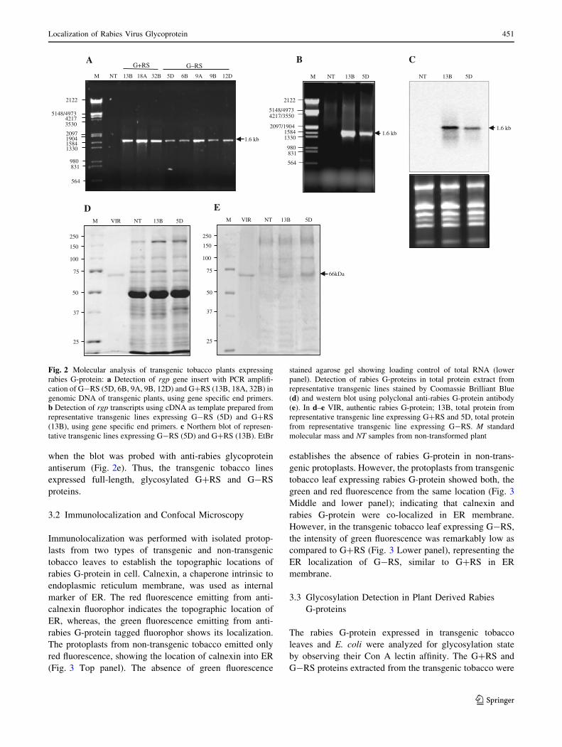
Fig. 2 Molecular analysis of transgenic tobacco plants expressing
rabies G-protein: a Detection of rgp gene insert with PCR amplifi-
cation of G-RS (5D, 6B, 9A, 9B, 12D) and G?RS (13B, 18A, 32B) in
genomic DNA of transgenic plants, using gene specific end primers.
b Detection of rgp transcripts using cDNA as template prepared from
representative transgenic lines expressing G-RS (5D) and G?RS
(13B), using gene specific end primers. c Northern blot of represen-
tative transgenic lines expressing G-RS (5D) and G?RS (13B). EtBr
stained agarose gel showing loading control of total RNA (lower
panel). Detection of rabies G-proteins in total protein extract from
representative transgenic lines stained by Coomassie Brilliant Blue
(d) and western blot using polyclonal anti-rabies G-protein antibody
(e). In d–e VIR, authentic rabies G-protein; 13B, total protein from
representative transgenic line expressing G?RS and 5D, total protein
from representative transgenic line expressing G-RS. M standard
molecular mass and NT samples from non-transformed plant
Localization of Rabies Virus Glycoprotein 451
123
purified on Q-Sepharose fastflow matrix and immunoaf-
finity column. Molecular mass of the purified rabies
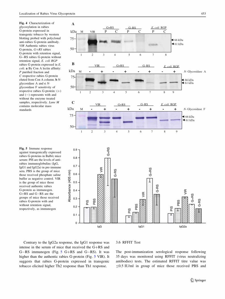
G-proteins (Fig. 4a lane 3 G?RS and lane 5 G-RS) was
similar to authentic rabies G-protein (Fig. 4a lane 2 VIR).
When rabies G-protein enriched fractions obtained from
the Q-Sepharose matrix were passed through Con A col-
umn and eluted with 0.1 M borate buffer, only one protein
from G?RS extract (Fig. 4a lane 4) co-migrated with the
authentic rabies G-protein and showed immunoreactivity
with anti-rabies G-protein antibodies. While the partially
purified G-RS, whose molecular mass was apparently
equal to the authentic rabies G-protein was not retained on
to the Con A column (Fig. 4a lane 6). The non-glycosyl-
ated E. coli expressed rabies G-protein whose molecular
mass was *61 kDa (Fig. 4a lane 7), was not retained on
the Con A column (Fig. 4a lane 8) as well.
3.4 Enzymatic Deglycosylation of Rabies G-proteins
with N-glycosidase A and N-glycosidase F
The rabies G-proteins from authentic rabies virus, G?RS,
G-RS and E. coli, were digested with N-glycosidase A
(Fig. 4b lane 3, 5, 7 and 9). The molecular mass of non-
glycosylated E. coli expressed rabies G-protein remained
unchanged (Fig. 4b lane 9). However, a reduction in
molecular mass was noticed in the other three cases. When
the authentic rabies G-protein, G?RS, G-RS and E. coli
expressed proteins were digested with N-glycosidase F,
only G?RS yielded a band of increased migration (Fig. 4c
lane 5) similar to authentic rabies G-protein (Fig. 4c lane
3). However, molecular mass of G-RS and non-glycos-
ylated E. coli expressed rabies G-proteins remained
unchanged (Fig. 4c lane 7 and 9).
3.5 Immune Response of Rabies G-proteins in Mouse
Antiserum
The serological analysis of immunized BALB/c mice
showed IgG response of G?RS higher than that of com-
mercial rabies virus vaccine. However, the IgG response
against G-RS was comparably lower (Fig. 5). The sub-
isotype immunoglobulin estimation in the serum of
immunized mice with G?RS and G-RS proteins showed
different response. The IgG2a response was highest in the
mice immunized with authentic rabies G-protein (Fig. 5
VIR); whereas it was slightly lower in the group of mice
immunized with G ?RS (Fig. 5 G?RS). The IgG2a
response in the group of mice that received G-RS protein
as immunogen was similar to the negative control (Fig. 5
G-RS).
Anti- Glycoprotein antiserum Anti-calnexin antiserum Superimposed
Non Transformed tobacco protoplast
G+RS tobacco protoplast
G-RS tobacco protoplast
Fig. 3 Immunolocalization of rabies G-protein expressed in trans-
genic tobacco leaf protoplast: Top panel represents immunofluores-
cence from non-transformed protoplast; middle panel represents
immunofluorescence from G?RS and lower panel represents
immunofluorescence from G-RS. Green fluorescence indicates
anti-rabies G-protein antibody labeled with Alexaflor-488 and red
fluorescence indicates anti-calnexin antibody labeled with Alexaflor-
520. Images were captured at 10 lm magnification
452 D. K. Yadav et al.
123
Contrary to the IgG2a response, the IgG1 response was
intense in the serum of mice that received the G?RS and
G-RS immunogen (Fig. 5 G?RS and G-RS). It was
higher than the authentic rabies G-protein (Fig. 5 VIR). It
suggests that rabies G-protein expressed in transgenic
tobacco elicited higher Th2 response than Th1 response.
3.6 RFFIT Test
The post-immunization serological response following
35 days was monitored using RFFIT (virus neutralizing
antibodies) tests. The estimated RFFIT titre value was
B0.5 IU/ml in group of mice those received PBS and
75
50
kDa P C P C P C
66 kDa61 kDa
AG+RS G–RS E. coli RGP
VIRM
C
61 kDa
50
75
kDa
66 kDa
G+RS G–RS E. coli RGPVIR
- + - + - + - + N- Glycosidase FM
B
50
75
kDa
66 kDa61 kDa
G+RS G–RS E. coli RGPVIR
- + - + - + - + N- Glycosidase AM
1 2 3 4 5 6 7 8
1 2 3 4 5 6 7 8 9
1 2 3 4 5 6 7 8 9
Fig. 4 Characterization of
glycosylation in rabies
G-protein expressed in
transgenic tobacco by western
blotting probed with polyclonal
anti-rabies G-protein antibody.
VIR Authentic rabies virus
G-protein, G?RS rabies
G-protein with retention signal,
G-RS rabies G-protein without
retention signal, E. coli RGPrabies G-protein expressed in E.coli. a By Con A lectin affinity:
P purified fraction and
C respective rabies G-protein
eluted from Con A column. b N-
glycosidase A and c N-
glycosidase F sensitivity of
respective rabies G-protein: (?)
and (-) represents with and
without the enzyme treated
samples, respectively. Lane Mcontains molecular mass
standards
PIS
PIS P
IS
PB
S
PB
S PB
S
VIR
VIR V
IR
G+
RS
G+
RS
G+
RS
G-R
S
G-R
S
G-R
S
0
0.1
0.2
0.3
0.4
0.5
0.6
0.7
0.8
0.9
IgG IgG1 IgG2a
Abs
orba
nce
(405
nm
)
Fig. 5 Immune response
against transgenically expressed
rabies G-proteins in Balb/c mice
serum: PIS are the levels of anti-
rabies immunoglobulins (IgG,
IgG1 and IgG2a) in pre-immune
sera. PBS is the group of mice
those received phosphate saline
buffer as negative control. VIR
is the group of mice those
received authentic rabies
G-protein as immunogen.
G?RS and G-RS are the
groups of mice those received
rabies G-protein with and
without retention signal,
respectively, as immunogen
Localization of Rabies Virus Glycoprotein 453
123
G-RS along with pre-immune sera. However, the RFFIT
titres value was C64.0 IU/ml and C12 IU/ml, in group of
mice those received Rabipur and G?RS, respectively
(Table 1).
3.7 Immunoprotection of Mice Challenged with Live
Rabies Virus
After the third booster, experimental mice were challenged
with live challenge virus standard rabies virus. The chal-
lenge dose of 109LD50 was administered intracerebrally to
all groups mice and observed for 14 days for the appear-
ance of rabies symptoms. Paralytic symptoms started
appearing after 5 days of challenge, in group of mice those
administered with PBS and G-RS. However, the mice
those received the authentic rabies vaccine and G?RS
remained healthy during the observation period. The
G?RS gave complete protection, similar to the commercial
authentic rabies vaccine (Fig. 6). Mice immunized with
PBS and G-RS died after 14 days of challenge.
4 Discussion
Plants have emerged as a highly versatile expression sys-
tem for the production of cost-effective and contamination-
safe recombinant biopharmaceutical proteins. Therefore,
strategic designing of antigen coding genes for high-level
expression and proper glycosylation is an important con-
sideration. The conserved KDEL/HDEL C-terminal motif
present on mature secretory proteins is recognized in golgi
apparatus and is responsible for the retrieval of proteins by
the transmembrane receptor, ERD2 [13]. In the present
study, two variants of strategically designed rgp were used
to study the possible role of ER retention signal, SEKDEL,
on biological activity of rabies antigen due to the possible
differences in glycosylation pattern. Therefore, SEKDEL
was placed on the C-terminus of rabies G-protein (G?RS)
for its retrieval back to ER, while it’s variant was devoid of
the SEKDEL motif (G-RS).
Estimation of rabies G-protein expression in transgenic
tobacco lines showed that G?RS was expressed at
0.015–0.38 %, while, G-RS ranged from 0.006–0.009 %
of total leaf protein. Stable expression of rabies G-protein
in transgenic plants has been reported at low levels and
detectable only through immunoprecipitation along with
reduced molecular mass of 60–62 kDa [15]. The lowered
molecular mass was attributed to differences in glycosyl-
ation, though not studied. Since the accumulation level of
protein is irrespective of mRNA level [25], the increased
accumulation of G?RS, unlike to that of G-RS (Fig. 3
lower panel), can be attributed to efficient retrieval of the
protein into ER (Fig. 3 middle panel). Thus, addition of
SEKDEL motif suggests enhanced accumulation of rabies
G-protein [28]. In absence of proper targeting signals,
proteins accumulated in the secretory system are secreted
to apoplast where stability of protein is much lower than in
the lumen of ER [4, 24]. Hence, due to the absence of
SEKDEL motif or any other specific cellular localization
signal in G-RS, it might have secreted into the apoplast
and its subsequent degradation leading to low expression
level as compared to G?RS. Thus, higher retrieval of
G?RS in ER and presence of fewer proteases could lead its
increased accumulation. The results from enzyme-linked
immunosorbent assay (data not shown) performed with
microsomal and cytosolic fractions prepared at high-speed
centrifugation also suggested that G?RS mainly was
confined to microsomal fraction while major part of G-RS
was detected in cytosolic fraction.
The secretory systems in animals, yeast and plants share
many similarities. The N-glycan structures of natural resi-
dent proteins of ER are devoid of golgi modifications and
serve as marker of a putative recycling function [9, 17].
The differential affinity of the two plant-expressed rabies
G-proteins for Con A showed the difference in their glycan
Table 1 Mean RFFIT serum titres (IU/ml, ±SD) at 35 days post-
immunization
Group Mean RFFIT titers
(IU/ml, ±SD)
PIS – (±nd)
PBS – (±nd)
VIR C64.0 (±2.0)
G?RS C12.0 (±4.0)
G-RS – (±nd)
– average RFFIT titer value B0.5 IU/ml, nd not detected
0
50
100
PBS
VIR
G+
RS
G-R
S
% S
urvi
val o
f im
mun
ized
mic
e
Fig. 6 Immunoprotective ability of different immunogens after intra-
cerebral challenge of immunized mice groups (of Fig. 5) with
109LD50 doses of challenge virus standard and observed up to
15 days
454 D. K. Yadav et al.
123
content. Con A lectin has affinity to bind with molecules
containing high a-D mannopyranosyl, a-D glucopyranosyl
and sterically related residues. The binding sugar requires
the presence of C-3, C-4 and C-5 hydroxyl groups which is
the feature of high mannose sugars. In the present study,
G?RS was retained on the Con A matrix, suggesting the
presence of biantennary high mannose type glycan moie-
ties on it (Fig. 4a lane 4), whereas the G-RS did not show
affinity and was not retained on to Con A column (Fig. 4a
lane 6). It suggests that G-RS contained modified and
complex type of glycans. This was supported by deglyco-
sylation assays (Fig. 4b and c). When rabies G-proteins
were treated with N-glycosidase A (that cleaves the N-
glycosidic linkage irrespective of any modification of
glycan side chain), the rabies G-proteins were reduced to a
molecular mass similar to non-glycosylated rabies G-pro-
tein expressed in E. coli (Fig. 4b lane 3, 5 and 7). When
rabies G-proteins were deglycosylated with N-glycosidase
F (that cleaves between the innermost GlcNAc and aspar-
agine residue only when the side chains of GlcNAc are not
modified), G?RS was cleaved (Fig. 4c lane 5) while
G-RS was resistant to N-glycosidase F activity (Fig. 4c
lane 7). It suggests that due to lack of ER retrieval signal,
SEKDEL, G-RS might travel deep up to median and
trans-golgi apparatus, where its terminal glycans and side
chains were diversely modified. G-RS might have glyco-
epitopes like a(1, 3)-fucose and b(1, 2)-xylose; as a(1, 3)-
fucosyltransferase and b(1, 2) xylosyltransferase are found
in medial cisternae of golgi apparatus. This study also
established that SEKDEL motif prevented plant-specific
modification of terminal glycans and side chains of Glc-
NAc residues of G?RS, which seems containing high
mannose type oligosaccharide structure [26]. Structural
analyses of plant ER resident proteins have shown that
natural reticuloplasmins bear a high mannose type oligo-
saccharide structure, which is common to plants and
mammals [4, 18, 19].
When the purified rabies G-proteins were injected into
the mice to test their immunogenicity; G?RS was found
more immunogenic than G-RS (Fig. 5). Titration of sub-
isotypic antibodies in the serum of immunized mice
showed that major immune response against plant derived
rabies G-protein was IgG1 type (Th2) and was two fold
higher for G?RS while, slightly higher in G-RS as
compared to the commercial vaccine. The IgG2a response
(Th1) was 1.4 fold higher in mice group immunized with
authentic rabies G-protein than in G?RS group. However,
there was no IgG2a response in G-RS group. Increased
IgG2a response in G?RS suggest a potent induction of
anti-viral effector functions through Th1 by TC cell acti-
vation, a feature that was absent in G-RS, which proved
more effective against rabies virus infection It is quite
evident from RFFIT assay which shows the presence of
protective level of virus neutralizing antibody in G?RS
(Table 1). It explains the immunoprotective efficiency of
G?RS similar to commercial vaccine against the live virus
challenge (Fig. 6). However, G-RS was unable to impart
comparable protection probably due to the presence of b1,
2 xylose- and/or a1, 3 fucose containing glycoepitopes,
which might induce rapid immune opsonization of dis-
tinctly glycosylated rabies antigen from the blood stream,
thus strongly compromising its efficacy [8]. Protection
against viral infection co-relates well with development of
the Th1 response [2].
Thus, the strategy to retrieve the recombinant rabies
G-protein within the ER was able to produce immuno-
protective rabies antigen besides enhancing its accumula-
tion. The results also established the importance of
appropriate strategies aiming plants as bioreactors for
production of designer vaccines.
Acknowledgments The work was carried out at National Botanical
Research Institute (NBRI), Lucknow. Council of Scientific and
Industrial Research, India, is gratefully acknowledged for financial
support to DKY as Senior Research Fellow at NBRI. We thank
Central Drug Research Institute, Lucknow for providing confocal
microscopy facility and Indian Veterinary Research Institute, Iza-
tnagar for conducting challenge virus standard challenge of immu-
nized mice. We thank Dr Rebecca Boston, University of North
Carolina, USA, for the generous gift of anti-calnexin antibody.
References
1. Ashraf S, Singh PK, Yadav DK, Shahnawaz M, Mishra S, Sawant
SV, Tuli R (2005) J Biotechnol 119:1–14
2. Cenna J, Tan GS, Papaneri AB, Dietzschold B, Schnell MJ,
McGettigan JP (2008) Vaccine 26:6405–6414
3. Coll SM (1995) Arch Virol 140:827–851
4. Fischer R, Stoger E, Schillberg S, Christou P, Twyman RM
(2004) Curr Opin Plant Biol 7:152–158
5. Frigerio L, Pastres A, Prada A, Vitale A (2001) Plant Cell
13:1109–1126
6. Gomord V, Denmat LA, Fitchette-Laine AC, Satiat-Jeunemaitre
B, Hawes C, Faye L (1997) Plant J 11:313–325
7. Gomord V, Faye L (1996) Plant Physiol Biochem 34:165–181
8. Gomord V, Fitchette AC, Menu-Bouaouiche L, Saint-Jore-Dupas
C, Plasson C, Michaud D, Faye L (2010) Plant Biotechnol J
8:564–587
9. Gomord V, Wee E, Faye L (1999) Biochimie 81:607–618
10. He ZH, Fujiki M, Kohorn BD (1996) J Biol Chem 271:
19789–19793
11. Horsch RB, Fry JE, Hoffmann NL, Eicholtz D, Rogers SG, Fraley
RT (1985) Science 227:1229–1231
12. Lerouge P, Cabanes-Macheteau M, Rayon C (1998) Fischette-
Laine0 AC, Gomord V, Faye L. Plant Mol Biol 38:31–48
13. Lewis MJ, Pelham HR (1992) Cell 68:353–364
14. Mason HS, Lam DM, Arntzen CJ (1992) Proc Nat Acad Sci U S
A 89:11745–11749
15. McGarvey PB, Hammond J, Dienelt MM, Hooper DC, Fu ZF,
Dietzschold B, Koprowski H, Michaels FH (1995) Biotechnology
13:1484–1487
16. Munro S, Pelham HRB (1987) Cell 48:899–907
Localization of Rabies Virus Glycoprotein 455
123
17. Navazio L, Baldan B, Mariani P, Gerwig GJ, Vliegenthart JF
(1996) Glycoconj J 13:977–983
18. Navazio L, Sponga L, Dainese P, Fitchette-Laine AC, Faye L,
Baldan B, Mariani P (1998) Plant Sci 131:35–42
19. Pagny S, Cabanes-Macheteau M, Gillikin JW, Leborgne-Castel
N, Lerouge P, Boston RS, Faye L, Gomord V (2000) Plant Cell
12:739–756
20. Pandit V, Joseph LN, Veerabhadran JS, Palaniappan C (1991)
Indian J Med Res 93:67–70
21. Perrin P, Thibodeau L, Sureau P (1985) Vaccine 3:325–332
22. Sambrook J, Fritsch EF, Maniatis T (1989) Molecular cloning: a
laboratory manual. Cold Spring Harbor Laboratory Press, Cold
Spring Harbor, New York
23. Sawant SV, Singh PK, Madnala R, Tuli R (2001) Theor Appl
Genet 102:635–644
24. Schouten A, Roosien J, Van Engelen FA, De Jong GA, Borst-
Vrenssen AW, Zilverentant JF, Bosch D, Stiekema WJ, Gommers
FJ, Schots A, Bakker J (1996) Plant Mol Biol 30:781–793
25. Stoger E, Vaquero C, Torres E, Sack M, Nicholson L, Drossard J,
Williams S, Keen D, Perrin Y, Christou P, Fischer R (2000) Plant
Mol Biol 42:583–590
26. Triguero A, Cabrera G, Cremata JA, Yuen CT, Wheeler J, Ra-
mirez NI (2005) Plant Biotechnol J 3:449–457
27. Walsh G, Jefferis R (2006) Nat Biotechnol 24:1241–1252
28. Wandelt CI, Khan MRI, Craig S, Schroeder HE, Spencer D,
Higgins TJV (1992) Plant J 2:181–192
29. World Health Organization (2010) Rabies vaccines: WHO posi-
tion paper. Weekly epidemiological record. World Health Orga-
nization, Geneva, Switzerland, 32, 309–320
456 D. K. Yadav et al.
123