Local opioid inhibition and morphine dependence of supraoptic ...
8
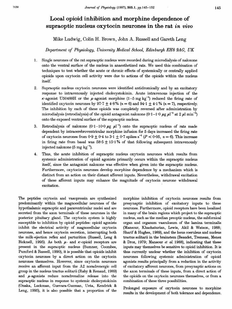
Journal of Physiology (1997), 505.1, pp.145-152 Local opioid inhibition and morphine dependence of supraoptic nucleus oxytocin neurones in the rat in vivo Mike Ludwig, Colin H. Brown, John A. Russell and Gareth Leng Department of Physiology, University Medical School, Edinburgh EH8 9AG, UK 1. Single neurones of the rat supraoptic nucleus were recorded during microdialysis of naloxone onto the ventral surface of the nucleus in anaesthetized rats. We used this combination of techniques to test whether the acute or chronic effects of systemically or centrally applied opioids upon oxytocin cell activity were due to actions of the opioids within the nucleus itself. 2. Supraoptic nucleus oxytocin neurones were identified antidromically and by an excitatory response to intravenously injected cholecystokinin. Acute intravenous injection of the K-agonist U50488H or the iu-agonist morphine (1-5 mg kg-) reduced the firing rate of identified oxytocin neurones by 97-7 + 4-8% (n = 6) and 94-1 + 4-1 % (n = 7), respectively. The inhibition by each of these opioids was completely reversed after administration by microdialysis (retrodialysis) of the opioid antagonist naloxone (0'1-1 0 jug 4t1-1 at 2 jul min-') onto the exposed ventral surface of the supraoptic nucleus. 3. Retrodialysis of naloxone (0 1-100 0ug jul-) onto the supraoptic nucleus of rats made dependent by intracerebroventricular morphine infusion for 5 days increased the firing rate of oxytocin neurones from 09 + 04 to 3 1 + 0 7 spikes s-' (P < 0'05, n = 6). This increase in firing rate from basal was 58-5 + 15.1 % of that following subsequent intravenously injected naloxone (5 mg kg-). 4. Thus, the acute inhibition of supraoptic nucleus oxytocin neurones which results from systemic administration of opioid agonists primarily occurs within the supraoptic nucleus itself, since the antagonist naloxone was effective when given into the supraoptic nucleus. Furthermore, oxytocin neurones develop morphine dependence by a mechanism which is distinct from an action on their distant afferent inputs. Nevertheless, withdrawal excitation of these afferent inputs may enhance the excitation. The peptides oxytocin and vasopressin are synthesized predominantly within the magnocellular neurones of the hypothalamic supraoptic and paraventricular nuclei and are secreted from the axon terminals of these neurones in the posterior pituitary gland. The oxytocin system is highly susceptible to inhibition by opioid peptides: opioid agonists inhibit the electrical activity of magnocellular oxytocin neurones, and hence oxytocin secretion, interrupting both the milk-ejection reflex and parturition (Russell, Leng & Bicknell, 1995). As both iu- and K-opioid receptors are present in the supraoptic nucleus (Sumner, Coombes, Pumford & Russell, 1990), it is possible that opioids inhibit oxytocin neurones by a direct action on the oxytocin neurones themselves. However, since oxytocin neurones receive an afferent input from the A2 noradrenergic cell group in the nucleus tractus solitarii (Raby & Renaud, 1989) and ,u-agonists reduce noradrenaline release into the supraoptic nucleus in response to systemic cholecystokinin (Onaka, Luckman, Guevara-Guzman, Ueta, Kendrick & Leng, 1995), it is also possible that a proportion of the magnitude of oxytocin neurone withdrawal morphine inhibition of oxytocin neurones results from presynaptic inhibition of excitatory inputs to these neurones. Furthermore, opioid receptors have been localized in many of the brain regions which project to the supraoptic nucleus, such as the median preoptic nucleus, the subfornical organ and organum vasculosum of the lamina terminalis (Mansour, Khachaturian, Lewis, Akil & Watson, 1988; Sharif & Hughes, 1989), and the locus coeruleus and nucleus tractus solitarii in the brainstem (Beaudet, Tremeau, Menez & Droz, 1979; Mansour et al. 1988), indicating that these inputs may themselves be sensitive to opioid inhibition. It is thus currently unclear whether the inhibition of oxytocin neurones following systemic administration of opioid agonists results principally from a reduction in the activity of excitatory afferent neurones, from presynaptic actions on the axon terminals of these inputs, from a direct action of the opioids on the oxytocin neurones themselves, or from a combination of these three possibilities. Prolonged exposure of oxytocin neurones to morphine results in the development of both tolerance and dependence. 7139 145
Transcript of Local opioid inhibition and morphine dependence of supraoptic ...
Journal of Physiology (1997), 505.1, pp.145-152
Local opioid inhibition and morphine dependence ofsupraoptic nucleus oxytocin neurones in the rat in vivo
Mike Ludwig, Colin H. Brown, John A. Russell and Gareth Leng
Department of Physiology, University Medical School, Edinburgh EH8 9AG, UK
1. Single neurones of the rat supraoptic nucleus were recorded during microdialysis of naloxoneonto the ventral surface of the nucleus in anaesthetized rats. We used this combination oftechniques to test whether the acute or chronic effects of systemically or centrally appliedopioids upon oxytocin cell activity were due to actions of the opioids within the nucleusitself.
2. Supraoptic nucleus oxytocin neurones were identified antidromically and by an excitatoryresponse to intravenously injected cholecystokinin. Acute intravenous injection of theK-agonist U50488H or the iu-agonist morphine (1-5 mg kg-) reduced the firing rate ofidentified oxytocin neurones by 97-7 + 4-8% (n = 6) and 94-1 + 4-1 % (n = 7), respectively.The inhibition by each of these opioids was completely reversed after administration bymicrodialysis (retrodialysis) of the opioid antagonist naloxone (0'1-1 0 jug 4t1-1 at 2 jul min-')onto the exposed ventral surface of the supraoptic nucleus.
3. Retrodialysis of naloxone (0 1-1000ug jul-) onto the supraoptic nucleus of rats madedependent by intracerebroventricular morphine infusion for 5 days increased the firing rateof oxytocin neurones from 09 + 04 to 3 1 + 0 7 spikes s-' (P < 0'05, n = 6). This increasein firing rate from basal was 58-5 + 15.1 % of that following subsequent intravenouslyinjected naloxone (5 mg kg-).
4. Thus, the acute inhibition of supraoptic nucleus oxytocin neurones which results fromsystemic administration of opioid agonists primarily occurs within the supraoptic nucleusitself, since the antagonist naloxone was effective when given into the supraoptic nucleus.Furthermore, oxytocin neurones develop morphine dependence by a mechanism which isdistinct from an action on their distant afferent inputs. Nevertheless, withdrawal excitationof these afferent inputs may enhance theexcitation.
The peptides oxytocin and vasopressin are synthesizedpredominantly within the magnocellular neurones of thehypothalamic supraoptic and paraventricular nuclei and are
secreted from the axon terminals of these neurones in theposterior pituitary gland. The oxytocin system is highlysusceptible to inhibition by opioid peptides: opioid agonistsinhibit the electrical activity of magnocellular oxytocinneurones, and hence oxytocin secretion, interrupting boththe milk-ejection reflex and parturition (Russell, Leng &Bicknell, 1995). As both iu- and K-opioid receptors are
present in the supraoptic nucleus (Sumner, Coombes,Pumford & Russell, 1990), it is possible that opioids inhibitoxytocin neurones by a direct action on the oxytocinneurones themselves. However, since oxytocin neurones
receive an afferent input from the A2 noradrenergic cellgroup in the nucleus tractus solitarii (Raby & Renaud, 1989)and ,u-agonists reduce noradrenaline release into thesupraoptic nucleus in response to systemic cholecystokinin(Onaka, Luckman, Guevara-Guzman, Ueta, Kendrick &Leng, 1995), it is also possible that a proportion of the
magnitude of oxytocin neurone withdrawal
morphine inhibition of oxytocin neurones results frompresynaptic inhibition of excitatory inputs to theseneurones. Furthermore, opioid receptors have been localizedin many of the brain regions which project to the supraopticnucleus, such as the median preoptic nucleus, the subfornicalorgan and organum vasculosum of the lamina terminalis(Mansour, Khachaturian, Lewis, Akil & Watson, 1988;Sharif & Hughes, 1989), and the locus coeruleus and nucleustractus solitarii in the brainstem (Beaudet, Tremeau, Menez& Droz, 1979; Mansour et al. 1988), indicating that theseinputs may themselves be sensitive to opioid inhibition. It isthus currently unclear whether the inhibition of oxytocinneurones following systemic administration of opioidagonists results principally from a reduction in the activityof excitatory afferent neurones, from presynaptic actions onthe axon terminals of these inputs, from a direct action ofthe opioids on the oxytocin neurones themselves, or from acombination of these three possibilities.
Prolonged exposure of oxytocin neurones to morphineresults in the development of both tolerance and dependence.
7139 145

M. Ludwig, C. H. Brown, J A. Russell and C. Leng
Tolerance is seen as a reduction in the magnitude of theinhibition which results from a given dose of morphine.Dependence is revealed by acute antagonism of the opioidagonist by naloxone which results in an immediate andlong-lasting morphine-withdrawal hyperexcitation of theoxytocin system; this is seen as marked increases in thefiring rate of oxytocin neurones in vivo, oxytocin hetero-nuclear RNA and immediate early gene expression in thesupraoptic and paraventricular nuclei, intra-supraopticnucleus release of oxytocin from dendrites and secretion ofoxytocin into the systemic circulation (Russell et al. 1995).
The A2 cell group which projects to the supraoptic nucleus(Raby & Renaud, 1989) also develops morphine dependenceand undergoes withdrawal excitation (Stornetta, Norton &Guyenet, 1993). Thus, morphine withdrawal excitation ofoxytocin neurones may result from excitation of afferentinputs such as that from the A2 cell group at this time.
To study whether ,u- and K-opioid agonists act within thesupraoptic nucleus to inhibit the activity of oxytocinneurones and whether morphine dependence of the oxytocinsystem develops at the level of the supraoptic nucleus, wecombined microdialysis, for local drug administration(retrodialysis) to the ventral surface of the nucleus, withextracellular recording of identified oxytocin neurones.
METHODSInduction of morphine tolerance/dependenceVirgin female Sprague-Dawley rats (220-350 g) were anaesthetizedby inhalation of 5% halothane in a mixture of 02 and N20 (bothflow rates at ca. 500 ml min-), and a 28 gauge stainless-steelcannula was stereotaxically implanted into the right lateral cerebralventricle (3 0 mm caudal and 2-0 mm lateral to bregma and 4'5 mmbelow the upper surface of the skull, Paxinos & Watson, 1986). Thecannula was attached via polythene tubing to a subcutaneousosmotic minipump (Alzet 2001; Charles River Ltd, Margate, Kent,UK). The pump and tubing contained morphine dissolved in sterilepyrogen-free water to deliver increasing doses over 5 days (10 and20 jug h-1 for 40 h each and 50 jug h-V for the remaining 40 h at1 jul h-'; Rayner, Robinson & Russell, 1988). Following surgeryanimals were housed individually with free access to food andwater.
Electrophysiology and retrodialysisThe pituitary stalk and right supraoptic nucleus were exposed bythe transpharyngeal approach under urethane anaesthesia (ethylcarbamate, 1-25 g kg-l i.P.), and a dialysis probe was used for localdrug administration (Ludwig & Leng, 1997). An in-house designedU-shaped microdialysis probe (total membrane length, 2-0 mm;Spectra/Por RC Hollow Fibers', Spectrum Med. Inc., Houston,TX, USA) was bent to position the loop of the membrane flat ontothe exposed ventral surface of the brain on the ventral glial laminaof the supraoptic nucleus after removal of the meninges. Thesupraoptic nucleus was dialysed with artificial cerebrospinal fluid(ACSF; pH 7-2, composition (mM): NaCl, 138; KCl, 3-36; NaHCO3,9-52; Na2HPO4, 0 49; urea, 2-16; CaCl2, 1-26; MgCl2, 1-18) at aflow rate of 2 jl# min-. The glass recording microelectrode (filledwith 0 9% saline, 20-40 MQ resistance) was placed through thecentre of the loop of the dialysis membrane. A stimulating electrode
(SNEX-200X, Clarke Electromedical) was placed on the neuralstalk of the pituitary gland and set to deliver single matchedbiphasic pulses (1 ms duration, <1 mA peak to peak) for anti-dromic identification of supraoptic nucleus neurones. A femoral veincatheter was inserted for systemic drug administration. Oxytocinneurones were distinguished from vasopressin neurones by theirfiring pattern (oxytocin neurones being non-phasic) and by theirdiffering responses to intravenously injected cholecystokinin(i.v. CCK, 20 jug kg-'), i.e. transient excitation of oxytocin neurones(at least 1 spike s-' increase in firing rate over 1 min within 4 minof injection) and no effect or short-term inhibition for vasopressinneurones (Renaud, Tang, McCann, Stricker & Verbalis, 1987).
Drug administrationU50488H (a selective K-agonist) or morphine (a selective ju-agonist)was injected i.v. (both 1-0-5-0 mg kg-' in an injection volume of0 5 ml kg-') until the firing rate of the cell being recorded wasinhibited to less than 0'2 spikes s-. Naloxone was retrodialysedonto the supraoptic nucleus (0-7 jgul-F after U50488H and0'1-100 jug ju-F in 10-fold increments after morphine, at 2 julmin-) for periods of 10 or 30 min. Since we were looking forinhibitory actions, cells which were silent or showed a very lowfiring rate were not studied in this experiment. At the end of eachexperiment the rats were killed by overdose of pentobarbitoneanaesthetic (60 mg kg-, i.v.).
Firing rate analysisThe firing rates of identified oxytocin cells were downloaded onto apersonal computer using the Spike2 software package (CambridgeElectronic Design, Cambridge, UK). The mean firing rate of eachcell was calculated for the 5 min period immediately before eachtreatment and for successive 5 min periods after treatment.Statistical analyses were completed using the SigmaStat® softwarepackage (Jandel Scientific GmbH, Erkrath, Germany). All responsesto drug administration were analysed by one-way repeatedmeasures (RM) analysis of variance (ANOVA). Where the Fratiowas significant this was followed by post hoc analyses using theStudent-Newman-Keuls test. All values are expressed as means +S.E.M. and differences were considered statistically significant ifP< 0'05. n, indicates the number of neurones.
DrugsMorphine sulphate was supplied by The Royal Infirmary ofEdinburgh (Edinburgh, UK); U50488H (trans-(±)-3,4-dichloro-N-methyl-N-(2-[1-pyrrolidinyl]cyclohexyl)benzeneacetamide) andnaloxone hydrochloride were purchased from Sigma; and cholecysto-kinin-(26-33)-sulphated (CCK) was from Bachem (Bachem Ltd,Saffron Walden, Essex, UK).
RESULTSEffects of retrodialysis of naloxone onto thesupraoptic nucleus on electrical activity of oxytocinneurones after acute U50488H or morphine inhibitionA single identified magnocellular oxytocin neurone wasrecorded from the supraoptic nucleus of thirteen rats. Themean spontaneous firing rate of the thirteen supraopticnucleus neurones was 3-4 + 0 5 spikes s-1; these cells werecharacterized as oxytocin neurones by their transientincrease in firing rate following i.v. CCK (1I4 + 0 3 spikes s-1increase averaged over 5 min). This firing rate and responseto systemic CCK are similar to those previously reported for
146 J Physiol.505.1

Opioid effects within the supraoptic nucleus
oxytocin neurones in both morphine-naive and -dependentrats (Brown, Munro, Murphy, Leng & Russell, 1996).
Six rats from which supraoptic nucleus neurones wererecorded were injected i.v. with U50488H in doses of1-5 mg kg-' until cessation of neuronal activity, or until nofurther inhibition of the neurones resulted (97 7 + 4-8%maximum inhibition, P< 0 05, n = 6, Figs IA and 2A).One neurone was recorded for a further 55 min afterU50488H injection during which time no recovery from the
A
n 30-0
Co
.? 20m
i 10
CO 0
L 0x0
CM
6 yLO 0
I3 l0
0
U50488H-induced inhibition was evident, confirming thelong duration of inhibition following these doses ofU50488H, as seen in previous studies (Pumford, Russell &Leng, 1993). In the other five rats, retrodialysis of 0 7 ,sg 1l-F(2 mM) naloxone onto the supraoptic nucleus for 10-30 mincompletely reversed U50488H-induced inhibition of oxytocinneurones. Subsequent i.v. injection of 5 mg kg-' U50488Hwithin 15 min of the termination of retrodialysis of naloxonedid not alter the firing rate of these neurones.
N0
NLX15 min
15 30 45Time (min)
l
Ml90
_- - N
o 00~60
-2 (.
1
0 25
LO (~~~~D
50 50
1 1cli Clf co)Cx CR00 02 0
75Time (min)
100 125
rl
0
I,
150
Figure 1. Retrodialysis of naloxone onto the supraoptic nucleus reverses opioid inhibition ofoxytocin neurones
The panels show the spontaneous firing rates (in 10 s bins) of two supraoptic nucleus oxytocin neurones
identified by their transient increase in firing rate following i.v. injection of 20 ,ug kg' CCK (CCK-1).A, i.v. injection of 1 mg kg-' U50488H (U50-1) eliminated the spontaneous activity of this neurone andreduced the magnitude of its response to a second injection of 20 jug kg-' CCK (CCK-2). Retrodialysis ofnaloxone (NLX, 07 jug uF-') onto the supraoptic nucleus over 15 min restored the spontaneous activity ofthe neurone and prevented further inhibition by a second injection of 5 mg kg-' U50488H (U50-2). B, i.v.
injection of 1 mg kg-' morphine (MOR-1) after 20 jug kg' i.v. CCK (CCK-1) markedly reduced thespontaneous activity and the CCK-responsiveness (CCK-2, 20 jug kg-) of this neurone. Retrodialysis ofnaloxone (NLX, 0 7 ug #1-') onto the supraoptic nucleus over 20 min restored the spontaneous activity ofthis neurone. This recording further indicates the temporal features of antagonism of systemically appliedmorphine by retrodialysed naloxone. Retrodialysis of naloxone reduced the inhibitory effect of morphinegiven during retrodialysis (MOR-2) and abolished the effect of morphine given 15 min after retrodialysis(MOR-3). Following termination of naloxone retrodialysis, a third injection of 20 jug kg' CCK (CCK-3)again produced a transient increase in spontaneous activity of this neurone. Subsequent repeated injectionof 1 mg kg-' morphine resulted in progressively greater inhibition of the spontaneous firing rate(MOR-4-7), presumably resulting from elimination of naloxone from the local environment.
B
-60
0en
Co2 0
r-
S 20
c)
114 1 1I
J Physiol.505.1 147
0
1

148 M. Ludwig, C. H. Brown,
In seven rats, morphine was injected iV. at doses of1-5 mg kg-', resulting in a 9441 + 44 % inhibition of thefiring rate of the neurones recorded (P < 0 05, n = 7,Figs 1B and 2B). In five of these seven neurones, naloxoneretrodialysed at concentrations of 04-IO0 pg 4ul-' onto thesupraoptic nucleus completely reversed the morphine-inducedinhibition, and subsequent IV. injection of 1 or 5 mg kg-'morphine within 15 min of the termination of naloxoneretrodialysis did not reduce the firing rate of these neurones.
Recordings from two of the cells were lost approximately 35and 50 min after injection of the 5 mg kg-' dose ofmorphine which silenced the cells; these cells were stillmaximally inhibited at the time that the recordings werelost.
Effects of retrodialysis of naloxone onto thesupraoptic nucleus on electrical activity of oxytocinneurones in morphine-dependent ratsAs in untreated rats, oxytocin neurones in morphine-dependent/tolerant rats are excited by systemic injection ofCCK (Brown et al. 1996). Here, oxytocin neurones inmorphine-dependent rats were characterized by theirtransient increase in firing rate following iV. CCK (14 +0 5 spikes s-' increase averaged over 5 min, n= 6). Naloxonewas retrodialysed onto the supraoptic nucleus in 10-foldincrements over the following ranges: 0-1-100 jug ul-' for10 min at each dose (from 10 ng ul-' in one case; n = 4) and1 0-100 fig ul-' for 30 min at each dose (n = 2). Naloxone(0x1-10*0 ug ul-) dose dependently increased the firing rate
A60-
Co0-4U0
0- 20-CD.=c._
B40-
3
co
0. 20-0
10-U-
I
o 0co LOcc 0
I
x-Jz
6
I
0uL
ITT
L A. Russell and C. Leng J Physiol. 505.1
of the six neurones recorded from 0 9 + 0 4 to341 + 0 7 spikes s-' (P < 0 05). The overall increase in firingrate over basal after retrodialysis of the 10 0 fug fl-' dose ofnaloxone was 58X5 + 151 % of that following subsequenti.v. administration of 5 mg kg-' naloxone, at which time thefiring rate of the neurones was 3x9 + 0 3 spikes s&' (Fig. 3).
DISCUSSIONThe present results demonstrate that inhibition ofsupraoptic nucleus oxytocin neurones following systemicadministration of fi- and K-opioid agonists is predominantlyattributable to opioid actions within the supraoptic nucleus,since the inhibition was fully reversed by administration ofthe opioid antagonist naloxone directly onto the nucleus.The present results cannot differentiate between post-synaptic actions on the oxytocin neurones themselves andpresynaptic inhibition at axon terminals impinging on theoxytocin neurones. The A2 cell group projection to thesupraoptic nucleus is subject to presynaptic inhibition bymorphine (Onaka et al. 1995), while the effects of opioids atthe axon terminals of other projections to the supraopticnucleus are unknown. U50488H hyperpolarizes bothoxytocin and vasopressin neurones in vitro, and theK-agonist dynorphin reduces the magnitude of excitatorypostsynaptic potentials evoked by focal stimulation insupraoptic nucleus neurones (Inenaga, Nagatomo, Nakao,Yanaihara & Yamashita, 1994). Morphine also inhibits bothoxytocin and vasopressin neurones in hypothalamic slices
Figure 2. Summary of the effects of retrodialysis ofnaloxone onto the supraoptic nucleus after inhibition ofoxytocin neurones by U50488H or morphineThe firing rates of identified supraoptic nucleus oxytocinneurones, means + S.E.M. (indicated by columns and bars)averaged over 5 min before (Basal) and after inhibition by1-5 mg kg-' iv. U50488H (U50-1, A) and 1-5 mg kg-' iv.morphine (MOR-1, B). After retrodialysis of 07 pg ul-'naloxone (NLX) the spontaneous activity was restored in bothA and B and subsequent injection of 1-5 mg kg' i.v.U50488H (U50-2, A) or 1-5 mg kg' iv. morphine (MOR-2,B) within 15 min of the termination of naloxone retrodialysisdid not then inhibit the activity of the neurones. *P< 0-001(one-way RM ANOVA); n = 5 in both A and B.
*
,,~m
zC
.-am 0

Opioid effects within the supraoptic nucleus
(Pittman, Hatton & Bloom, 1980; Wakerley, Noble & Clarke,1983). Both ,u- and K-agonists inhibit the spontaneousactivity of oxytocin neurones in vivo (Pumford et al. 1993),and also the responses of these neurones to all stimuliagainst which these opioids have been tested, includingparturition, suckling and increased osmolality (Russell et al.1995). While it is possible that all of the afferent inputs tooxytocin neurones possess both ju- and K-receptors, thesimplest explanation is that the predominant inhibitoryactions of opioids are those exerted directly upon theoxytocin neurones themselves.
Retrodialysis of naloxone onto the supraoptic nucleus ofmorphine-dependent rats elicited a dose-dependent excitation
of oxytocin neurones. Thus, morphine withdrawal excitationof oxytocin neurones occurs at the level of the supraopticnucleus. Again it is possible that this is manifested at thelevel of the oxytocin neurones themselves or presynapticallyon their afferent inputs or some combination of the two.
In the present study, following retrodialysis of naloxoneonto the supraoptic nucleus, systemic administration ofnaloxone further increased the firing rate of oxytocinneurones in the morphine-dependent rats. As administrationof naloxone onto the supraoptic nucleus completely reversedthe acute inhibition of oxytocin neurones by morphine orU50488H, it appears unlikely that naloxone at similar dosesfailed to saturate supraoptic nucleus ,u-receptors in the
5 mg kg-1i.v. NLX
1r
A
.-
co
C)
c,0)
1 0 ng pi'-NLX
10 min
0O1 ug il-1 l 0/g/II-1 10 0 /tg ul-NLX NLX NLX
I I I
LAjLJ50
f
0
B 60-
50-
° 40-
a)Q- 30-U)
20-0)
10
U-
10-
.......TI 1 , I ~~~~~~..,,,..,,,.._
Basal 01 lt9 I-, 1 0 jg 1I1-NLX NLX
luu ig BeforeNLX i.v. NLX
Figure 3. Local application of naloxone induces withdrawal excitation of supraoptic oxytocinneurones in morphine-dependent ratsA, the spontaneous firing rate (in 10 s bins) of a supraoptic nucleus oxytocin neurone in a morphine-dependent rat identified by its transient increase in firing rate following 20 ,g kg-l iv. CCK (CCK).Retrodialysis of increasing concentrations (10 ng /tl-F to 10 jug F1-') of naloxone (NLX) onto the supraopticnucleus over 10 min each induced a dose-dependent increase in the firing rate of this neurone. Subsequenti.v. injection of 5 mg kg-' naloxone (i.v. NLX) induced a further small rise in the firing rate of the neurone.
B, the firing rates (averaged over 5 min; means + s.E.M.) of identified supraoptic nucleus oxytocin neurones
recorded from morphine-dependent rats (n = 6) before (Basal) and after retrodialysis of increasingconcentrations (0- 1 #sg 1ul-' to 10 ,ag ul-F) of naloxone (NLX) and before and after iv. injection of 5 mg kg-lnaloxone (NLX). *P < 0 05 (one-way RM ANOVA).
Time (min)100 150
T
Afteri.v. NLX
J Physiol.505.1 149

M. Ludwig, C. H. Brown, J A. Russell and C. Leng
morphine-dependent rats. Thus, a more likely explanationfor the further increased activity following systemic naloxoneis that some excitatory inputs to the supraoptic nucleus alsoundergo morphine-withdrawal excitation.
Although is-receptors are widely distributed in the CNS(Mansour, Fox, Burke, Akil & Watson, 1995), few of theareas where 24-receptors are located display dependenceafter chronic morphine treatment (Nye & Nestler, 1996).Following naloxone-precipitated morphine withdrawal,there is intense induction of Fos protein expression in thesupraoptic and paraventricular nuclei reflecting thewithdrawal activation of oxytocin neurones at these sites,but strikingly there is little or no similar induction of Foselsewhere in the hypothalamus, and in particular none inregions adjacent to the supraoptic nucleus (Russell et al.1995; Jhamandas, Harris, Petrov & Jhamandas, 1996).However, neurones of the A2 and A6 noradrenergic cellgroups also exhibit morphine dependence (Aghajanian,1978; Stornetta et al. 1993) and these areas project to thesupraoptic nucleus (Cunningham & Sawchenko, 1991). Otherafferent neurones which have been studied, notably those inthe lamina terminalis, do not appear to be activated duringmorphine withdrawal (Murphy, Onaka, Brown & Leng,1997), but it is possible that withdrawal excitation ofoxytocin neurones in part reflects withdrawal excitation ofnoradrenergic afferents.
During morphine withdrawal noradrenaline is released invarious brain regions, such as the hippocampus (Done,Silverstone & Sharp, 1992), and this release is prevented byprior administration of the a2-adrenergic agonist clonidine(Silverstone, Done & Sharp, 1992). Clonidine also reducesthe behavioural signs of opiate withdrawal in humans (Gold,Redmond & Kleber, 1978) and in rats (Taylor, Elsworth,Garcia, Grant, Roth & Redmond, 1988). Thus, it has beensuggested that noradrenaline may play a pivotal role inmorphine withdrawal excitation in behavioural systems.However, the noradrenaline release into the supraopticnucleus following morphine withdrawal is rather modest(Murphy et al. 1997), being less than that which results fromsystemic administration of cholecystokinin (Onaka et al.1995), a stimulus which increases the activity of oxytocinneurones to a much lesser extent than morphine withdrawal(Brown et al. 1996).
Perhaps the most extensively studied region in whichmorphine dependence develops is the locus coeruleus, wheremorphine treatment increases G-protein subunit, adenylatecyclase, cyclic AMP-dependent protein kinase and tyrosinehydroxylase levels (Nestler, Alreja & Aghajanian, 1994).However, an increase in glutamate release into the locuscoeruleus during morphine withdrawal also contributes tothe withdrawal-induced activation (Aghajanian, Kogan &Moghaddam, 1994). Thus, in the locus coeruleus it appearsthat withdrawal excitation is generated by an interplaybetween intracellular mechanisms and extrinsic inputs.
Withdrawal excitation of oxytocin neurones is unchangedby central catecholaminergic lesion (Murphy, Brown, Leng &Russell, 1995) and is reduced, but not eliminated, by lesionof the region anterior and ventral to the third ventricle(AV3V) (Russell, Pumford & Bicknell, 1992). The projectionfrom the AV3V region to the supraoptic nucleus contains aglutamatergic component (Yang, Senatorov & Renaud, 1994)but few of the neurones that project from the AV3V regionto the supraoptic nucleus are activated by morphinewithdrawal (Murphy et al. 1997). Nevertheless, as in thelocus coeruleus, it seems that withdrawal excitation ofsupraoptic nucleus oxytocin neurones results from acombination of intrinsic and extrinsic factors, includinginputs from glutamatergic and/or noradrenergic neurones.
In any experiment involving focal administration of a drug,the concentration at its site of action is subject touncertainty. Inhibition of oxytocin cells by 1 mg kg-' i.v.U50488H is fully antagonized only by an i.v. dose ofnaloxone of 5 mg kg-' or higher (Pumford et al. 1993).Assuming conservatively that this dose of naloxone isdistributed in a volume of 10 ml per 100 g body weightgives an estimated effective concentration of 50 ,ug ml-' atits site of action. This is lower by a factor of about 15 thanthe concentration of naloxone in the dialysate itself.However, the tissue concentrations reached during dialysisare certainly much lower than the concentration in thedialysate. When dialysis is used conventionally to measurethe concentrations of substances in the extracellular fluid,typical recoveries are of the order of 2-10% of theextracellular concentration (Benveniste & Hiittemeier,1990), but the more appropriate calculation is based onestimates of efflux from dialysis probes. Most of the effluxfrom the probes in situ will not enter the supraoptic nucleus(since the probe is apposed to the ventral surface withoutpenetrating, only part of the membrane is in contact withbrain tissue), and most of what enters will be cleared by thevasculature, which is fully intact in this preparation (bloodflow through the supraoptic nucleus is amongst the highestof any brain region, and the vasculature occupies a highproportion of the volume of the supraoptic nucleus; Gross,Sposito, Pettersen & Fenstermacher, 1986). When Fluorogoldwas administered through dialysis probes apposed to theventral surface of the supraoptic nucleus, subsequenthistological analysis indicated little penetration of tissuebeyond about 200 ,um of the surface (Ludwig & Leng, 1997)- little more in fact than the ventral glial lamina of thesupraoptic nucleus, which contains only the dendrites ofsupraoptic neurones and not their cell bodies (Armstrong,Scholer & McNeill, 1982). The inference that penetration islimited is strongly supported by results obtained usingtetrodotoxin. Tetrodotoxin blocks action potentials inoxytocin neurones at a concentration of 10-6 M and below,but dialysis of 10-4 M tetrodotoxin, though it consistentlyblocks spontaneous activity in supraoptic neurones, does notgenerally block antidromically evoked action potentials,
J Physiol.505.1150

Opioid effects within the supraoptic nucleus
implying that the concentration achieved at the somata(rather than the dendrites) of supraoptic neurones followingeven 30 min of dialysis remains at least two orders ofmagnitude below the dialysate concentration (Ludwig &Leng, 1997). Thus, while naloxone is more lipophilic thanFluorogold and tetrodotoxin, it is likely that the diffusion ofnaloxone away from the dialysis probe is also highlyrestricted. Furthermore, there is a high density of both #a-and K-opioid receptors throughout the entire supraopticnucleus but not in the immediate vicinity surrounding thesupraoptic nucleus (Sumner et al. 1990). Therefore, it isprobable that the effects of naloxone observed in the presentstudy result from actions within the supraoptic nucleusitself.
Given the potency of directly applied naloxone inantagonizing the actions of systemically applied U50488H,it was surprising that higher concentrations were needed toevoke morphine withdrawal, since naloxone is approximately50-fold more potent at ,u-receptors than at the K-receptorsthrough which U50488H acts. However, although very lowdoses of systemically administered naloxone will excitesupraoptic oxytocin neurones in morphine-dependent rats(Leng, Russell & Grossmann, 1989), full withdrawal againrequires a systemic dose of 5 mg kg-', and an inferredmaximal effective tissue concentration of 50 jug ml-'. Thehighest dialysate concentration used in the present studywas 10 mg ml-', over two orders of magnitude greater thanthe maximum effective systemic dose; this dialysisconcentration appears to be appropriate for delivering aneffective concentration of naloxone to the supraoptic nucleus,perhaps with a requirement to penetrate beyond thedendritic layer to reach the cell bodies of the neurones toelicit morphine withdrawal excitation.
In conclusion, the acute inhibition of oxytocin neurones bycu- and K-opioid agonists are mediated within the supraopticnucleus, probably directly upon the oxytocin neuronesthemselves but possibly also by presynaptic inhibition.Furthermore, oxytocin neurones express morphine with-drawal excitation, and presumably morphine dependence,separately from their distant afferent inputs.
AGHAJANIAN, G. K. (1978). Tolerance of locus coeruleus neurones tomorphine and suppression of withdrawal response by clonidine.Nature 276, 186-188.
AGHAJANIAN, G. K., KOGAN, J. H. & MOGHADDAM, B. (1994). Opiatewithdrawal increases glutamate and aspartate efflux in the locuscoeruleus: an in vivo microdialysis study. Brain Research 636,126-130.
ARMSTRONG, W. E., SCHOLER, J. & MCNEILL, T. H. (1982).Immunocytochemical, Golgi and electron microscopiccharacterization of putative dendrites in the ventral glial lamina ofthe rat supraoptic nucleus. Neuroscience 7, 679-694.
BEAUDET, A., TREMEAU, O., MENEZ, A. & DROZ, B. (1979).Visualisation des recepteurs aux opiaces dans le locus coeruleus durat: etude autoradiographique a haute resolution apresadministration d'un analogue tritie de la met-enkephaline. ComptesRendus des Seances de l'Academie des Sciences - Serie D, SciencesNaturelles 289, 591-594.
BENVENISTE, H. & HtTTEMEIER, P. C. (1990). Microdialysis - theoryand application. Progress in Neurobiology 35, 195-215.
BROWN, C. H., MUNRO, G., MURPHY, N. P., LENG, G. & RUSSELL, J. A.(1996). Activation of oxytocin neurones by systemic cholecystokininis unchanged by morphine dependence or withdrawal excitation inthe rat. Journal of Physiology 496, 787-794.
CUNNINGHAM, E. T. JR & SAWCHENKO, P. E. (1991). Reflex control ofmagnocellular vasopressin and oxytocin secretion. Trends inNeurosciences 14, 406-411.
DONE, C., SILVERSTONE, P. & SHARP, T. (1992). Effect of naloxone-precipitated morphine withdrawal on noradrenaline release in rathippocampus in vivo. European Journal of Pharmacology 215,333-336.
GOLD, M. S., REDMOND, D. E. JR & KLEBER, H. D. (1978). Clonidineblocks acute opiate-withdrawal symptoms. Lancet ii, 599-602.
GROSS, P. M., SPOSITO, N. M., PETTERSEN, S. E. & FENSTERMACHER,J. D. (1986). Differences in function and structure of the capillaryendothelium in the supraoptic nucleus and pituitary neural lobe ofrats; evidence for the supraoptic nucleus as an osmoreceptor.Neuroendocrinology 44, 401-407.
INENAGA, K., NAGATOMO, T., NAKAO, K., YANAIHARA, N. &YAMASHITA, H. (1994). Kappa-selective agonists decreasepostsynaptic potentials and calcium components of action potentialsin the supraoptic nucleus of rat hypothalamus in vitro. Neuroscience58, 331-340.
JHAMANDAS, J. H., HARRIS, K. H., PETROV, T. & JHAMANDAS, K. H.(1996). Activation of nitric oxide-synthesizing neurones duringprecipitated morphine withdrawal. NeuroReport 7, 2843-2846.
LENG, G., RUSSELL, J. A. & GROSSMANN, R. (1989). Sensitivity ofmagnocellular oxytocin neurones to opioid antagonists in ratstreated chronically with intracerebroventricular (i.c.v.) morphine.Brain Research 484, 290-296.
LUDWIG, M. & LENG, G. (1997). Autoinhibition of supraoptic nucleusvasopressin neurones in vivo: a combined retrodialysis/electro-physiology study in rats. European Journal of Neuroscience (in thePress),
MANSOUR, A., Fox, C. A., BURKE, S., AKIL, H. & WATSON, S. J.(1995). Immunohistochemical localization of the cloned mu opioidreceptor in the rat CNS. Journal of Chemical Neuroanatomy 8,283-305.
MANSOUR, A., KHACHATURIAN, H., LEWIS, M. E., AKIL, H. &WATSON, S. J. (1988). Anatomy of CNS opioid receptors. Trends inNeurosciences 11, 308-314.
MURPHY, N. P., BROWN, C. H., LENG, G. & RUSSELL, J. A. (1995).Morphine-withdrawal excitation of oxytocin neurones in theabsence of their noradrenergic input. Analgesia 1, 594-597.
MURPHY, N. P., ONAKA, T., BROWN, C. H. & LENG, G. (1997). The roleof afferent inputs to supraoptic nucleus oxytocin neurones duringnaloxone-precipitated morphine withdrawal in the rat. Neuroscience80, 567-577.
NESTLER, E. J., ALREJA, M. & AGHAJANIAN, G. K. (1994). Molecularand cellular mechanisms of opiate action: studies in the rat locuscoeruleus. Brain Research Bulletin 35, 521-528.
NYE, H. E. & NESTLER, E. J. (1996). Induction of chronic Fos-relatedantigens in rat brain by chronic morphine administration. MolecularPharmacology 49, 636-645.
J Physiol 505.1 151

M. Ludwig, C. H. Brown, J A. Russell and 0. Leng
ONAKA, T., LUCKMAN, S. M., GUEVARA-GUZMAN, R., UETA, Y.,KENDRICK, K. M. & LENG, G. (1995). Presynaptic actions ofmorphine: blockade of cholecystokinin-induced noradrenalinerelease in the rat supraoptic nucleus. Journal of Physiology 482,69-79.
PAXINOS, G. & WATSON, C. (1986). The Rat Brain in StereotaxicCoordinates. Academic Press, Sydney.
PITTMAN, Q. J., HATTON, J. D. & BLOOM, F. E. (1980). Morphine andopioid peptides reduce paraventricular neuronal activity: studies onthe rat hypothalamic slice preparation. Proceedings of the NationalAcademy of Sciences of the USA 77, 5527-5531.
PUMFORD, K. M., RUSSELL, J. A. & LENG, G. (1993). Effects of theselective kappa-opioid agonist U50,488 upon the electrical activityof supraoptic neurones in morphine-tolerant and morphine-naiverats. Experimental Brain Research 94, 237-246.
RABY, W. N. & RENAUD, L. P. (1989). Dorsomedial medullastimulation activates rat supraoptic oxytocin and vasopressinneurones through different pathways. Journal of Physiology 417,279-294.
RAYNER, V. C., RoBINsON, I. C. & RUSSELL, J. A. (1988). Chronicintracerebroventricular morphine and lactation in rats: dependenceand tolerance in relation to oxytocin neurones. Journal ofPhysiology 396, 319-347.
RENAUD, L. P., TANG, M., MCCANN, M. J., STRICKER, E. M. &VERBALIS, J. G. (1987). Cholecystokinin and gastric distensionactivate oxytocinergic cells in rat hypothalamus. American Journalof Physiology 253, R661-665.
RUSSELL, J. A., LENG, G. & BICKNELL, R. J. (1995). Opioid toleranceand dependence in the magnocellular oxytocin system: aphysiological mechanism? Experimental Physiology 80, 307-340.
RUSSELL, J. A., PUMFORD, K. M. & BICKNELL, R. J. (1992).Contribution of the region anterior and ventral to the third ventricleto opiate withdrawal excitation of oxytocin secretion.Neuroendocrinology 55, 183-192.
SHARIF, N. A. & HUGHES, J. (1989). Discrete mapping of brain muand delta opioid receptors using selective peptides: quantitativeautoradiography, species differences and comparison with kappareceptors. Peptides 10, 499-522.
SILVERSTONE, P. H., DONE, C. & SHARP, T. (1992). Clonidine but notnifedipine prevents the release of noradrenaline during naloxone-precipitated opiate withdrawal: an in vivo microdialysis study inthe rat. Psychopharmacology 109, 235-238.
STORNETTA, R. L., NORTON, F. E. & GUYENET, P. G. (1993).Autonomic areas of rat brain exhibit increased Fos-like immuno-reactivity during opiate withdrawal in rats. Brain Research 624,19-28.
SUMNER, B. E., CooMBEs, J. E., PUMFORD, K. M. & RUSSELL, J. A.(1990). Opioid receptor subtypes in the supraoptic nucleus andposterior pituitary gland of morphine-tolerant rats. Neuroscience37, 635-645.
TAYLOR, J. R., ELSWORTH, J. D., GARCIA, E. J., GRANT, S. J., ROTH,R. H. & REDMOND, D. E. JR (1988). Clonidine infusions into thelocus coeruleus attenuate behavioral and neurochemical changesassociated with naloxone-precipitated withdrawal.Psychopharmacology 96, 121-134.
WAKERLEY, J. B., NOBLE, R. & CLARKE, G. (1983). Effects ofmorphine and D-Ala, D-Leu enkephalin on the electrical activity ofsupraoptic neurosecretory cells in vitro. Neuroscience 10, 73-81.
YANG, C. R., SENATOROV, V. V. & RENAUD, L. P. (1994). Organumvasculosum lamina terminalis-evoked postsynaptic responses in ratsupraoptic neurones in vitro. Journal of Physiology 477, 59-74.
AcknowledgementsM.L. is supported by a German Research Fellowship (DeutscheForschungsgemeinschaft) and C.H.B. by a Medical ResearchCouncil Realizing Our Potential Award.
Author's email addressM. Ludwig: [email protected]
Received 3 July 1997; accepted 4 August 1997.
152 J: Phy8iol.505.1