Local Anesthesia for Cataract...
10
In recent years, advances in cataract surgery have lead to greater levels of refractive precision, faster visual rehabilitation, and improved comfort and safety. Refinements in phacoemulsification techniques and intraocular lens (IOL) technology deserve much of the credit for these advances, but innovations in anesthe- sia, especially topical anesthesia, have also played an important role in improving outcomes and hastening visual recovery. While topical anesthesia is favored by many sur- geons for the majority of their cases today, proper patient screening and careful preoperative planning are necessary in order to choose the best anesthesia for an individual patient. Mastery of all of the avail- able techniques—intracameral, topical, parabulbar (sub-Tenon’s), peribulbar, and retrobulbar anesthe- sia—along with an understanding of their advantages and disadvantages, is necessary in order to provide the highest level of care for all patients. The goal of this chapter is to define and describe the indications and techniques for each of these approaches. APPLIED ANATOMY A basic knowledge of orbital anatomy is essential to understand the effects and potential complications of orbital anesthesia. Intraocular pressure may be elevated after the injection of even modest amounts of anesthetic into the orbit. The orbit has an average volume of 30 cc. A sudden increase in orbital volume associated with the injection of anesthetic results in the transmission of force anteriorly, causing compression of the globe. The floor of the orbit is the shortest of the orbital walls and extends only 35 to 40 mm from the orbital rim. The 38-mm needle used in retrobulbar anesthesia, therefore, has the potential to damage the optic nerve in a significant percentage of the population. 1 The abducens, oculomotor, and nasociliary nerves pass through the annulus of Zinn. The trochlear nerve enters outside of the annulus to supply the superior oblique. Placement of anesthetic within the intramus- cular cone, whose apex is the annulus of Zinn, typi- cally results in the paralysis of the oculomotor and the abducens but not the trochlear. The superior oblique is often spared, and cyclotorsion may still occur even with a well-placed retrobulbar injection. Sensory innervation to the cornea and superonasal conjunctivae is provided by the nasociliary nerve that is within the muscle cone. The remaining conjunctival sensation is provided by the remaining branches of ophthalmic nerve (frontal and lacrimal) and two divi- sions of the maxillary nerve, which supply the lower Chapter Local Anesthesia for Cataract Surgery Rom Kandavel, MD 1
Transcript of Local Anesthesia for Cataract...

�
In recent years, advances in cataract surgery have lead to greater levels of refractive precision, faster visual rehabilitation, and improved comfort and safety. Refinements in phacoemulsification techniques and intraocular lens (IOL) technology deserve much of the credit for these advances, but innovations in anesthe-sia, especially topical anesthesia, have also played an important role in improving outcomes and hastening visual recovery.
While topical anesthesia is favored by many sur-geons for the majority of their cases today, proper patient screening and careful preoperative planning are necessary in order to choose the best anesthesia for an individual patient. Mastery of all of the avail-able techniques—intracameral, topical, parabulbar (sub-Tenon’s), peribulbar, and retrobulbar anesthe-sia—along with an understanding of their advantages and disadvantages, is necessary in order to provide the highest level of care for all patients. The goal of this chapter is to define and describe the indications and techniques for each of these approaches.
Applied AnAtomyA basic knowledge of orbital anatomy is essential
to understand the effects and potential complications of orbital anesthesia.
Intraocular pressure may be elevated after the injection of even modest amounts of anesthetic into the orbit. The orbit has an average volume of 30 cc. A sudden increase in orbital volume associated with the injection of anesthetic results in the transmission of force anteriorly, causing compression of the globe.
The floor of the orbit is the shortest of the orbital walls and extends only 35 to 40 mm from the orbital rim. The 38-mm needle used in retrobulbar anesthesia, therefore, has the potential to damage the optic nerve in a significant percentage of the population.1
The abducens, oculomotor, and nasociliary nerves pass through the annulus of Zinn. The trochlear nerve enters outside of the annulus to supply the superior oblique. Placement of anesthetic within the intramus-cular cone, whose apex is the annulus of Zinn, typi-cally results in the paralysis of the oculomotor and the abducens but not the trochlear. The superior oblique is often spared, and cyclotorsion may still occur even with a well-placed retrobulbar injection.
Sensory innervation to the cornea and superonasal conjunctivae is provided by the nasociliary nerve that is within the muscle cone. The remaining conjunctival sensation is provided by the remaining branches of ophthalmic nerve (frontal and lacrimal) and two divi-sions of the maxillary nerve, which supply the lower
Chapter
Local Anesthesia forCataract Surgery
Rom Kandavel, MD
1

� Chapter 1
lid and conjunctiva (enters via the inferior orbital fora-men). All of these additional somatosensory nerves lie outside of the muscular cone. For this reason, a retro-bulbar block can still leave areas of the conjunctiva sensitive to pain and touch.
The dura surrounding the optic nerve is continu-ous with the dura of the brain. Inadvertent injection of anesthetic into the subdural space within the nerve, therefore, can result in brainstem anesthesia.
preoperAtive evAluAtionCareful patient screening is essential in order to
determine which form of anesthesia is best suited for an individual. A surgeon should develop a checklist to avoid missing data that can influence the choice of anesthesia. A history and physical examination, with review of medications, is an excellent starting point for evaluation. Particular attention should be given to the patient’s ability to communicate, lie flat and still, and follow directions. A history of congestive heart failure, chronic obstructive pulmonary disease, chronic bronchitis, claustrophobia, anticoagulation status, and use of alpha-blockers (tamsulosin) should be addressed with each patient.
Retrobulbar and peribulbar anesthesia generally provide excellent intraoperative pain control with the added benefit of complete or partial akinesia and visual block. General anesthesia may be utilized when generalized muscle paralysis is an additional factor to ensure surgical success. Topical anesthesia should be reserved for communicative and calm patients who
have no relevant comorbidities. The surgeon should be experienced and expecting a shorter surgery with-out anticipated complications or added procedures. Longer procedures that may require iris manipulation or scleral suturing may benefit from retrobulbar or peribulbar anesthesia for improved iris and ciliary body anesthesia. While most patients can lie still, some may not be able to follow directions and are not well suited for topical anesthesia. Patients who have psychiatric disease or other comorbidities that prevent them from lying still may be candidates for general anesthesia.
The information contained in Tables 1-1 and 1-2 can serve as general guidelines for anesthesia selec-tion. In some instances, reviewing the procedure and different anesthesia approaches with the patient is useful. This allows the patient to self-assess his or her preferences. The discussion also allows the patient to ask questions and develop greater comfort with the surgeon and surgery.
retrobulbAr AnesthesiAMultiple protocols have been published with a
common goal of improving the efficacy and safety of retrobulbar anesthesia. Complications arising from ret-robulbar anesthesia include retrobulbar hemorrhage, globe/nerve perforation, extraocular muscle injury, and brainstem anesthesia/death.2 Other disadvantages include the need for increased sedation, a postopera-tive eye patch, longer visual recovery, ptosis, chemosis, subconjunctival hemorrhage, and increased posterior pressure during surgery. The most feared complication
Table 1-1Contraindications to Local Anesthesia
RelativeTremorAnxietyClaustrophobiaChildrenPoor communication/language barrier/deafnessLong operative time
AbsoluteInability to cooperate (eg, schizophrenia, dementia)Uncontrolled coughing/movement disorder
Table 1-2Contraindications to Topical Anesthesia
RelativePhotophobiaAnxietyDeafnessLong operative time
AbsolutePoor communication/language barrier/deafnessCannot follow directionsInsufficient pain control (as in prior eye surgery)

Local Anesthesia for Cataract Surgery �
of retrobulbar injection, perforation of the globe, is more common with eyes of higher axial length and/or staphyloma.2
A well-placed retrobulbar block usually results in excellent akinesia and sensory block with some visual block also. As previously noted, motor nerves within the muscle cone, the abducens, oculomotor, as well as the sensory nasociliary nerve, are affected, but because the trochlear nerve passes outside the cone, superior oblique muscle innervation is usually spared and cyclo-torsion may still occur. Most surgeons supplement retrobulbar blocks with topical anesthesia to complete anterior segment anesthesia because portions of the trigeminal, which supply the conjunctiva and lid, also pass outside the muscle cone.
The goal of retrobulbar anesthesia is the place-ment of anesthetic into the intramuscular cone located behind the globe and anterior to the orbital apex. Structures traversed by the retrobulbar needle include the skin, orbital septum, periocular tissue/fat, and the intramuscular connective tissue. Structures to be avoided include blood vessels, extraocular muscles, the globe, and the optic nerve. The technique detailed below is designed to avoid these structures and give reliable and reproducible anesthesia. Each surgeon will develop personal amendments, but the basic tenets apply.
Injectable mixtures should include a total volume of 10 cc or less composed of 2% lidocaine without epinephrine mixed 50:50 with 0.75%. Note that this 50:50 mixture dilutes each component to half the original concentration. Some surgeons may prefer 4% lidocaine, if available, to yield a final effective concen-tration of 2% lidocaine. The addition of bupivacaine increases the duration of action. If hyaluronidase is available, it can also be added to the mixture to speed diffusion of the medication and improve akinesia and sensory block. Hyaluronidase can also decrease poste-rior pressure by causing the volume to distribute more quickly. Fifteen to 20 units of hyaluronidase per mL of solution can be used.
A 38-mm (1.5-inch) 23-gauge needle with a round-ed point (Atkinson) is preferred. A standard sharp point needle has the advantage of passing through tis-sues more easily with less discomfort, but the reduced sensory feedback during injection and higher potential for injury to ocular structures favors the Atkinson or blunt-tipped needle.3 A 10-cc syringe is also preferred over a 5-cc for better tactile control of injection pres-sure and enough volume to change needles and contin-ue with facial nerve blocks after retrobulbar injection using the same syringe.
Retrobulbar anesthesia is performed prior to sterile prep. The patient is positioned flat on the operative bed. At the level of the forehead, 1-inch silk, plastic, or paper tape can be used to secure the head to the table if an assistant is not available. Intravenous pro-pofol or Versed (Hospira, Lake Forest, IL) should be administered in conjunction with an analgesic, such as fentanyl, to help prepare the patient for injection. If propofol is used, time for the medication to take effect should be allowed. Testing the lack of orbicularis contraction by gently brushing the eyelashes can help verify adequate sedation.
Following surgery, the eye should be patched. This is because the retrobulbar block reduces sensation of the eye (which results in a reduced blink reflex), provides akinesia (which causes a transient diplo-pia), and reduces vision (which is frightening to the patient). The patch may be removed after 4 to 6 hours in patients who have received only lidocaine. When bupivacaine is used, the patch should remain for not less than 8 hours.
pArAbulbAr (sub-tenon’s) AnesthesiA
Some surgeons have adopted the technique of using a blunt-tipped cannula intraoperatively to inject the same anesthetic mixture. This is known as a para-bulbar block. Parabulbar blocks can be placed as a planned anesthesia or can be utilized intraoperatively if the patient is uncooperative or has inadequate pain control with topical/peribulbar anesthesia.
This technique avoids the hazards of a sharp needle placement into this space and is a safer alterna-tive to retrobulbar anesthesia, but it can also result in increased chemosis, subconjuctival hemorrhage, and incomplete anesthesia if the cannula is not advanced in the sub-Tenon’s space.4 Damage to the vortex veins has also been reported.5 Onset is rapid, but the added dis-section can add to operative time. The disadvantages such as the need for patching with delayed visual reha-bilitation apply, as with retrobulbar anesthesia.
peribulbAr AnesthesiAThe injection of anesthesia within the orbit with-
out directing the needle inside the muscle cone reduces the risk of damage to vital structures. The soft tissue, intramuscular septae are incomplete and allow for the diffusion of medication into the cone, resulting in akinesia and visual block, as well as sensory dein-nervation to the nasociliary and extraconal divisions

� Chapter 1
of first and second divisions of the trigeminal nerve. This technique relies on larger volumes (7 to 10 cc) and works best if supplemented by 500 units of hyal-uronidase. Sedation with propofol, as with retrobulbar anesthesia, is preferred.
Sensory block and akinesia are dependent on dif-fusion, therefore this technique requires reassessment of akinesia (if desired) after 5 to 7 minutes. If adequate medial rectus akinesia is not obtained, the peribulbar injection can be repeated using the same technique targeting the medial fat compartment. Up to 24% of patients will require this supplemental 3- to 5-cc block.6 The entrance site for the supplemental block is just nasal to the medial rectus, adjacent to the carun-cle, and parallel to the medial orbital wall in the same fashion as described above. Higher volumes overall are used, therefore orbital pressure is increased and ecchymosis and chemosis are more likely than with the retrobulbar block.7 Reports of retrobulbar hemorrhage and globe perforation have also been published but are less common. This technique has reported anesthetic pain control similar to retrobulbar placement, but has an improved safety profile.8 Overall, the advantages of peribulbar anesthesia should be weighed against the frequent need for supplemental anesthesia, incomplete akinesia, the larger volume of anesthesia, and longer time required for complete diffusion.
topicAl And intrAcAmerAl AnesthesiA
As phacoemulsification techniques have advanced, incision size has decreased, the need for iris manipula-tion has diminished, and operative time has lessened. These changes have resulted in a decrease in the need for complete akinesia, long duration of ocular anesthe-sia, and intensity of iris and ciliary body sensory block. Topical and intracameral anesthesia alone can provide adequate anterior segment anesthesia for noncomplex phacoemulsification with proper patient selection.9 Use in trabeculectomy, secondary sutured IOLs, and pterygium excision has also become more common.
Topical anesthesia avoids the systemic and ocular risks of the previously described modalities. In addi-tion, it allows for quick visual recovery. Monitored anesthesia care can be used, but surgery can also be performed without intravenous agents (discussed below). It should be noted that many surgeons who use retrobulbar or peribulbar block use topical and/or intracameral anesthetic in addition to help complete
the anterior segment sensory block. Communicative, calm, cooperative patients are candidates for topical anesthesia. Careful patient selection is important.
Multiple agents are available for topical anesthesia and include tetracaine 0.5% drops, Tetravisc 0.5% gel (Ocusoft, Richmond, TX), lidocaine 2% jelly, Xylocaine 4% (AstraZeneca, Wilmington, DE), and bupivacaine 0.75%. Topical agents are placed at least 5 to 10 minutes prior to surgery. They provide excellent intraoperative pain control and also allow the patient to have less discomfort from the Betadine prep prior to draping.
Drop preparations are generally administered in two to three repeated doses separated by 5 to 10 minutes. Gel preparations have the benefit of coating the eye without requiring repeated doses. If used prior to dilating agents, gels can interfere with absorption. Therefore, many surgeons place a liquid preparation such as proparacaine 0.5% or tetracaine drops first and then complete the dilation protocols. After the pupil is dilated and 5 to 10 minutes prior to entering the operating room, Tetravisc or lidocaine gel can be placed into the eye. Lidocaine gel can be more viscous and at times more difficult to place under the lids to anesthetize the superior and inferior conjunctiva and fornices.10 Tetravisc has an intermediate viscosity and therefore spreads like a liquid drop but also coats like a gel. Each surgeon should develop a simple, repro-ducible protocol for topical anesthesia that can be performed efficiently by the surgical staff. One other variant on this form of anesthesia includes soaking a sponge with both dilating and/or anesthetic drops (perilimbal anesthesia) and placing it in the inferior fornix for 10 to 15 minutes. Anecdotal reports suggest that soaked pledgets can deliver higher concentrations of both anesthetic and mydriatic medications, but the actual procedure of sponge placement can be more intrusive than drops alone.
Topical anesthesia alone may not provide adequate iris and ciliary body anesthesia. Therefore, many surgeons will supplement with intracameral 1% non-preserved lidocaine. After the initial paracentesis is created, approximately 0.5-cc nonpreserved lidocaine is instilled into the anterior chamber. Uncomplicated cataract surgery can be performed with topical anes-thesia alone, but prospective trials suggest an addition-al anesthetic benefit to intracameral lidocaine.11 This additional agent represents a very quick, extra step in cataract surgery. At 1% concentration, endothelial cell toxicity has not been demonstrated in humans.

Local Anesthesia for Cataract Surgery �
The additional anesthetic effect makes any iris touch or manipulation more comfortable. If a scleral sutured posterior chamber lens, pupil expansion device, or iris stretching is necessary, intracameral anesthesia can be a useful adjunct. Other agents such as epinephrine or phenylephrine can also be added to this intracameral solution. These and other techniques are discussed in other portions of this text.
FAciAl nerve blocksOccasionally, a patient may have difficulty with
relaxing his or her orbicularis oculi muscle. Many times this is anxiety related, other times it may be an idiosyncratic reflex specific to that individual. Psychiatric disease can be a risk factor. If intravenous agents fail to reduce squeezing, facial nerve blocks in combination with any of the anesthetic modalities above can allow the surgeon to have improved control. Generally, patients who require facial nerve blocks are good candidates for retrobulbar/peribulbar anesthesia because of associated Bell’s phenomenon. Facial nerve blocks can be performed at any portion of the extra-cranial course after it exits the stylomastoid foramen. The nerve gives off multiple branches as it courses from behind the ear over the angle of the mandible, penetrating the parotid gland and dividing into its ter-minal branches, including the temporal and zygomatic, which supply the orbicularis. Types of facial nerve blocks are differentiated by their location, and each has inherent advantages and disadvantages.
Careful placement of additional anesthetic in the inferior fornix and anterior lateral orbit as the needle is withdrawn during retrobulbar and peribulbar anes-thesia can also result in seventh nerve block in up to 88% of cases by continued diffusion.12 Although less reliable, this can obviate the need for a separate facial nerve block.
The Nadbath block is directed at the exit of the nerve at the stylomastoid foramen. Respiratory and vocal chord paralysis have been reported with inadver-tent injection into the jugular foramen.13-15 Prolonged facial nerve block has also been reported.15 This tech-nique avoids ecchymosis of the face and is less painful, but also can temporarily paralyze multiple divisions of the facial nerve.
The O’Brien is placed more distally just below the zygomatic arch, anterior to the tragus. This site can be more painful and can also cause paralysis of the lips and lower face in addition to the intended superior divisions.
The modified Van Lint targets the terminal branch-es at the lateral canthus and lid. This technique avoids the paralysis of the other divisions of the seventh nerve but can cause lid ecchymosis and edema.
Facial nerve blocks are best done with conscious sedation usually directly after retrobulbar or peribul-bar block while the propofol is still at maximal effect. The same 10-cc syringe can be used if appropriate by changing the needle to a conventional sharp point 1-inch 30-gauge or 27-gauge needle.
conscious sedAtion And GenerAl Anesthetic AGents
Cataract surgeons should possess a basic under-standing of common anesthetic agents. Many times feedback from the patient is only perceived and communicated to the surgeon intraoperatively. The surgeon may also better understand the needs of each patient, having treated him or her for many years, than the anesthesiologist present for the surgery. An understanding of the common medications and their relative analgesic, anxiolytic, and amnestic properties will allow the surgeon to help tailor preoperative plan-ning and intraoperative supplementation.
Monitored anesthesia care involves intravenous sedation and analgesia with noninvasive monitoring. This allows for less physical stress on the patient. The patient is able to respond to commands, facilitating surgery, and recovery is quicker. Conversion to gen-eral anesthesia is still possible. Commonly used single agents include opiates (fentanyl), benzodiazepines (midazolam), and propofol.
Propofol (Diprivan [AstraZeneca]) is a short-act-ing induction agent that provides temporary sedation without analgesia. Propofol can be used prior to ret-robulbar block placement. Although the block can be placed without propofol, this agent provides a short duration of deep sedation with amnesia. Testing lack of orbicularis contraction by gentle eyelash stimulation can be a helpful measure of adequate sedation prior to retrobulbar placement. Hypotension and temporary apnea are possible, therefore pulsoximetry and blood pressure monitoring are essential.
Fentanyl and midazolam (Versed) can be used alone or in conjunction. Fentanyl, a short-acting nar-cotic, provides analgesia with some mild anxiolysis. Midazolam is an excellent anxiolytic and can also have an amnestic effect. Midazolam is short acting, water soluble, and has no analgesic properties. Both have a quick onset of action and can be augmented dur-

� Chapter 1
ing surgery for added effect. Midazolam can have a disinhibiting effect that can result in a lack of patient cooperation. This disinhibition and confusion is more common in the elderly and quite rare in younger patients. In some circumstances, patients can attempt to sit up or remove their draping. Therefore, careful attention and communication with the patient and anesthesiologist during surgery should be maintained in order to continually assess patient comfort and mental status.
Cooperation with adequate pain and anxiety con-trol is the goal of every cataract surgery. The surgeon’s demeanor and communication can help supplement pharmacologic anesthesia. Some individuals may expe-rience pain but not alert the surgeon for fear of “inter-fering” with the surgery. It can be useful to briefly describe to the patient what to expect in the operating room and encourage him or her to verbally express discomfort so that added analgesia can be provided.
step-by-step ApproAch to retrobulbAr AnesthesiA
Step 1. Anesthetic Preparation. A 38-mm (1.5-inch) 23-gauge needle with a rounded point (Atkin-son) on a 10-cc syringe is preferred. Ten cc containing 2% lidocaine without epinephrine mixed 50:50 with 0.75% bupivacaine and 10 to 15 units hyaluronidase per cc (optional) can be used.
Step 2. Patient Position. The assistant should be pres-ent at the head of the bed, facing the feet, hold-ing the head securely with both hands. One finger can be used to lift the upper lid of the operative eye to allow the surgeon to visualize
the globe throughout the procedure (Figure 1-1). The surgeon should be on the same side of the bed as the operative eye. The lower eye lid skin should be cleaned with an alcohol swab.
Step 3. Needle Placement. The needle tip, bevel down, is advanced parallel to the orbital floor, entering at the lateral third of the inferior lid. The patient’s eye should be in primary position (Figures 1-2 and 1-3).
Step 4. Needle Advancement. The surgeon’s index finger can be used to palpate and displace the globe superiorly as the needle is positioned to create adequate space for the needle to pass inferior to the globe between the lateral and inferior rectus muscles. Resistance to the rounded needle can be noted when the orbital septum is reached. Once the needle has passed the equator of the globe (the halfway point of the needle should be at the level of the iris), the needle is then angled superior and slightly medial toward the muscular cone to a loca-tion posterior to the macula. A small amount of anesthesia can be injected as the needle is advanced.
Step 5. Entering the Muscle Cone and Injecting. Re-sistance and relief can be detected as the needle enters the muscle cone. The syringe plunger should be gently withdrawn to ensure a blood vessel has not been entered prior to injection. Depending on anticipated cone volume, 2.5 to 4.0 cc should be injected. An additional 1 to 2 cc can be injected as the needle is withdrawn.
Figure 1-1.Properpatientstabilizationandpositionforretrobulbar/peribulbaranesthesia.
Figure 1-2. The retrobulbar/peribulbar needleshould enter at the lateral one-third of the lowereyelid below the globe with the eye in primaryposition. Supplemental medial peribulbar blocksenterbetweenthecaruncleandmedialrectus.

Local Anesthesia for Cataract Surgery �
Step 6. Assessment. Gentle “on and off” digital pres-sure should be used for 2 to 4 minutes to help facilitate diffusion of the anesthesia. Checking for the amount of akinesia can help assess the success of the retrobulbar block within a few minutes of placement.
If progressive proptosis, hemorrhagic chemo-sis, or unexplained posterior pressure during surgery is detected, retrobulbar hemorrhage should be suspected. Immediate lateral can-thotomy and cantholysis is the treatment of choice. Some surgeons proceed with surgery immediately once retrobulbar pressure is re-duced. Most surgeons prefer to delay the pro-cedure and wait a sufficient time for recovery and reassessment.
step-by-step ApproAch to peribulbAr AnesthesiA
Step 1. Anesthetic Preparation. A 25-mm 23-gauge needle with a rounded point (Atkinson) on a 20-cc syringe is preferred. Ten cc contain-ing 2% lidocaine (without epinephrine) mixed 50:50 with 0.75% bupivacaine and 10 to 15 units hyaluronidase per cc is used.
Step 2. Patient Position. The assistant should be pres-ent at the head of the bed, facing the feet, holding the head securely with both hands. One finger can be used to lift the upper lid of the operative eye to allow the surgeon to visu-
alize the globe throughout the procedure. The surgeon should be on the same side of the bed as the operative eye. The lower eye lid skin should be cleaned with an alcohol swab.
Step 3. Needle Entry and Injection. The entry point is at the outer third of the lower eyelid where the floor meets the lateral wall. The needle is advanced, bevel down, parallel to the floor un-til the needle base is at the level of the iris. As-piration first and then 7 to 10 cc of anesthetic solution is injected.
Step 4. Supplemental Block. Supplemental block if incomplete anesthesia is placed in the same fashion medial to the medial rectus adjacent to the caruncle. The needle is advanced parallel to the medial wall and 3 to 5 cc of the same mixture is injected.
step-by-step ApproAch to pArAbulbAr (sub-tenon’s)
AnesthesiAStep 1. Conjunctival Incision. An incision is made
with a Wescott scissors between the superior rectus and lateral rectus 9 to 10 mm posterior to the limbus down to bare sclera. The scissors are used to bluntly dissect posteriorly to allow space to advance the cannula (Figure 1-4).
Step 2. Anesthetic Placement. A 5-cc syringe with a blunt-tipped cannula containing a 50:50 lido-caine 2% (without epinephrine) and bupiva-caine 0.75% mixture is advanced around the equator of the globe into the anterior intra-conal space. It is important to directly visual-ize the blunt cannula entering under Tenon’s capsule (Figure 1-5). The cannula should fol-low the curve of the globe posteriorly. Two to 3 cc should be injected.
step-by-step ApproAch to topicAl And intrAcAmerAl
AnesthesiA
Application of Topical Anesthetic Tetracaine or proparacaine is used in two to
three divided doses in each eye prior to sur-gery. The first dose is given just prior to di-lating agents and then repeated every 5 to 10 minutes with each application of dilating
Figure 1-3. The retrobulbar anesthesia needle isdirected toward a point posterior to the maculaafter being advanced past the equator of theglobe. (Medical illustration copyright © 2008Nucleus Medical Art. All rights reserved. www.nucleusinc.com.)
� Chapter 1
drops. One additional application just prior to surgery may be necessary. If Tetravisc is uti-lized, one dose 5 to 10 minutes prior to sur-gery is placed in each eye.
Intracameral Anesthesia Sterile, intracameral 1% nonpreserved lido-
caine in a 1-cc syringe with a blunt cannula is prepared. After the initial paracentesis is cre-ated, approximately 0.5-cc nonpreserved li-docaine is instilled into the anterior chamber. Viscoelastic should be instilled into the ante-rior chamber after at least 5 seconds to allow anesthetic effect.
step-by-step ApproAch to FAciAl nerve blocks
A 27-gauge or 30-gauge 1-inch needle on a 5-cc syringe is preferred. Two percent lidocaine with epinephrine is mixed 50:50 with 0.75% bupivacaine for facial nerve blocks (Figure 1-6).
NadbathStep 1. Palpate the Location of the Stylomastoid Fo-
ramen. Use an alcohol swab to clean the area. The needle is entered perpendicular to the skin 2 mm anterior to the anterior-superior margin of the mastoid process behind the ear.
Step 2. Anesthetic Application. Two to 3 cc of 2% li-docaine alone or mixed 50:50 with 0.75% bu-
pivacaine is injected. This technique avoids ec-chymosis of the face and is less painful but also can temporarily paralyze multiple divisions of the facial nerve.
O’BrienStep 1. Palpate the Zygomatic Process Anterior to
the Tragus. Use an alcohol swab to clean the area. This method involves blocking the nerve above the condyloid process anterior to the tragus just below the zygomatic process.
Step 2. Anesthetic Application. Inject a volume of 1 to 2 cc. This site can be more painful and can also cause paralysis of the lips and lower face in addition to the intended superior divisions.
Van Lint (Modified)Step 1. Primary Injection. Use an alcohol swab to
clean the lateral canthal area. At 1 cm lateral to the canthal angle advance the needle to the suborbicularis plane and then inject 1 to 2 cc. Be careful to avoid local, superficial blood vessels.
Step 2. Anesthetic Supplement. Via the same skin entrance, direct the needle cephalad and cau-dad into the lid. Inject 1 cc as the needle is withdrawn, in each direction. This technique avoids the paralysis of other divisions of the seventh nerve but can cause lid ecchymosis and edema.
Figure 1-4. Dissectiontobaresclerainthesupero-temporalquadrant. (PhotocourtesyofThomasA.Oetting,MS,MD.)
Figure 1-5. Thecannulaisadvancedinsub-Tenon’sspaceposteriorlyhuggingtheglobe. (Photocour-tesyofThomasA.Oetting,MS,MD.)
Local Anesthesia for Cataract Surgery �
reFerences1. Katsev DA, Drews RC, Rose BT. An anatomic study of ret-
robulbar needle path length. Ophthalmology. 1989;96:1221-1224.
2. Eke T, Thompson JR. Serious complications of local anaes-thesia for cataract surgery: a 1 year national survey in the United Kingdom. Br J Ophthalmol. 2007;91(4):470-475.
3. Waller SG, Toboada J, O’Connor P. Retrobulbar anesthesia risk. Do sharp needles really perforate the eye more easily than blunt needles? Ophthalmology. 1993;100(4):506-510.
4. Zafirakis P, Voudouri A, Rowe S, et al. Topical versus sub-Tenon’s anesthesia without sedation in cataract surgery. J Cataract Refract Surg. 2001;27(6):873-879.
5. Stevens JD. A new local anesthesia technique for cataract ex-traction by one quadrant sub-Tenon’s infiltration. Br J Ophthl-mol. 1992;76:670-674.
6. Hendrick SW, Rosenberg MK, Lebenbom-Mansour MH. Efficacy and safety of single injection peribulbar block per-formed by anesthesiologists prior to cataract surgery. J Clin Anesth. 1997;9(4):285-288.
7. Wang HS. Peribulbar anesthesia for ophthalmic procedures. J Cataract Refract Surg. 1998;14:441-443.
8. Davis DB 2nd, Mandel MR. Efficacy and complication rate of 16,224 consecutive peribulbar blocks. A prospective multi-center study. J Cataract Refract Surg. 1994;20(3):327-337.
9. Chuang LH, Yeung L, Ku WC, Yang KJ, Lai CC. Safety and efficacy of topical anesthesia combined with a lower con-centration of intracameral lidocaine in phacoemulsification: paired human eye study. J Cataract Refract Surg. 2007;33(2):293-296.
10. Amiel H, Koch PS. Tetracaine hydrochloride 0.5% versus lidocaine 2% jelly as a topical anesthetic agent in cataract surgery: comparative clinical trial. J Cataract Refract Surg. 2007;33(1):98-100.
11. Tseng SH, Chen FK. A randomized clinical trial of combined topical-intracameral anesthesia in cataract surgery. Ophthal-mology. 1998;105(11):2007-2011.
12. Martin SR, Baker SS, Muenzler WS. Retrobulbar anesthesia and orbicularis akinesia. Ophthalmic Surg. 1986;17:232-233.
13. Warner LO, Martino JD, Davidson PJ. Pulmonary edema af-ter Nadbath and retrobulbar blocks: a possible explanation. Anesth Analg. 1995;80(3):643.
14. Birt CM, Dixon WS, Dionne CL. Vocal cord paralysis with Nadbath facial block. Can J Ophthalmol. 1994;29(5):231-233.
15. Zahl K. Selection of techniques for regional blockade of the eye and adnexa. In: McGoldrick KE, ed. Anesthesia for Ophthal-mic and Otolaryngologic Surgery. Philadelphia, PA: WB Saunders Co; 1992:235-247.
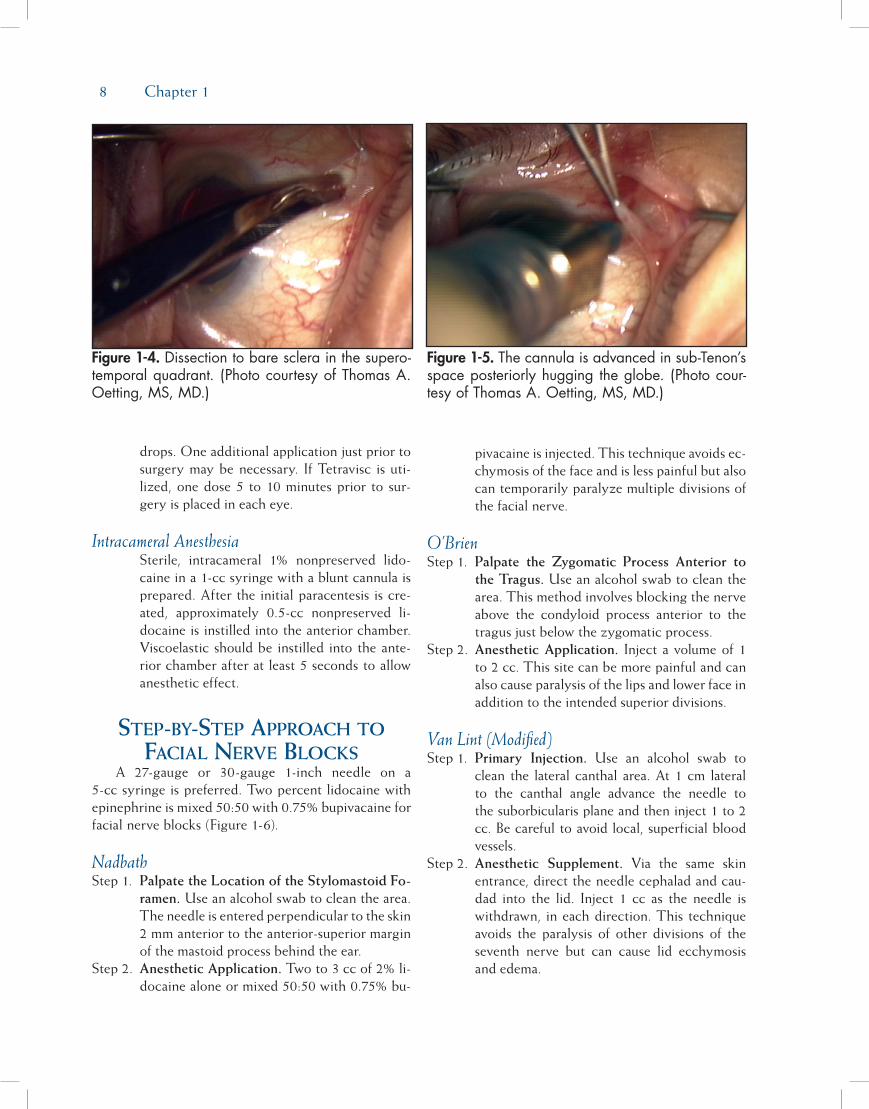
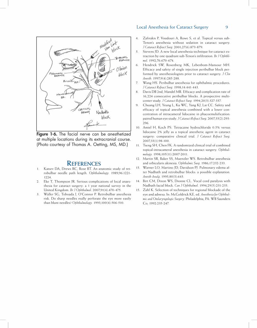
Figure 1-6. The facial nerve canbe anesthetizedatmultiplelocationsduringitsextracranialcourse.(PhotocourtesyofThomasA.Oetting,MS,MD.)