LJMU Research Onlineresearchonline.ljmu.ac.uk/id/eprint/9192/3/Acridone... · Ethnopharmacological...
32
Segun, P, Ismail, FMD, Ogbole, OO, Nahar, L, Evans, AR, Ajaiyeoba, EO and Sarker, SD Acridone alkaloids from the stem bark of Citrus aurantium display selective cytotoxicity against breast, liver, lung and prostate human carcinoma cells http://researchonline.ljmu.ac.uk/id/eprint/9192/ Article LJMU has developed LJMU Research Online for users to access the research output of the University more effectively. Copyright © and Moral Rights for the papers on this site are retained by the individual authors and/or other copyright owners. Users may download and/or print one copy of any article(s) in LJMU Research Online to facilitate their private study or for non-commercial research. You may not engage in further distribution of the material or use it for any profit-making activities or any commercial gain. The version presented here may differ from the published version or from the version of the record. Please see the repository URL above for details on accessing the published version and note that access may require a subscription. For more information please contact [email protected] http://researchonline.ljmu.ac.uk/ Citation (please note it is advisable to refer to the publisher’s version if you intend to cite from this work) Segun, P, Ismail, FMD, Ogbole, OO, Nahar, L, Evans, AR, Ajaiyeoba, EO and Sarker, SD (2018) Acridone alkaloids from the stem bark of Citrus aurantium display selective cytotoxicity against breast, liver, lung and prostate human carcinoma cells. Journal of Ethnopharmacology, 227. pp. 131-138. ISSN LJMU Research Online
Transcript of LJMU Research Onlineresearchonline.ljmu.ac.uk/id/eprint/9192/3/Acridone... · Ethnopharmacological...

Segun, P, Ismail, FMD, Ogbole, OO, Nahar, L, Evans, AR, Ajaiyeoba, EO and Sarker, SD
Acridone alkaloids from the stem bark of Citrus aurantium display selective cytotoxicity against breast, liver, lung and prostate human carcinoma cells
http://researchonline.ljmu.ac.uk/id/eprint/9192/
Article
LJMU has developed LJMU Research Online for users to access the research output of the University more effectively. Copyright © and Moral Rights for the papers on this site are retained by the individual authors and/or other copyright owners. Users may download and/or print one copy of any article(s) in LJMU Research Online to facilitate their private study or for non-commercial research. You may not engage in further distribution of the material or use it for any profit-making activities or any commercial gain.
The version presented here may differ from the published version or from the version of the record. Please see the repository URL above for details on accessing the published version and note that access may require a subscription.
For more information please contact [email protected]
http://researchonline.ljmu.ac.uk/
Citation (please note it is advisable to refer to the publisher’s version if you intend to cite from this work)
Segun, P, Ismail, FMD, Ogbole, OO, Nahar, L, Evans, AR, Ajaiyeoba, EO and Sarker, SD (2018) Acridone alkaloids from the stem bark of Citrus aurantium display selective cytotoxicity against breast, liver, lung and prostate human carcinoma cells. Journal of Ethnopharmacology, 227. pp. 131-138. ISSN
LJMU Research Online

1
Acridone alkaloids from the stem bark of Citrus aurantium L. (Rutaceae) display selective
cytotoxicity against breast, liver, lung and prostate human carcinoma cells
Peter A. Segun1,2,3, Fyaz M. D. Ismail2*, Omonike O. Ogbole1*, Lutfun Nahar2, Andrew R.
Evans2, Edith O. Ajaiyeoba1 and Satyajit D. Sarker2
1Department of Pharmacognosy, Faculty of Pharmacy, University of Ibadan, Ibadan, Nigeria
2Medicinal Chemistry and Natural Products Research Group, School of Pharmacy and
Biomolecular Sciences, Liverpool John Moores University, James Parsons Building, Byrom
Street, Liverpool L3 3AF, United Kingdom
3Department of Pharmacognosy, Faculty of Pharmacy, Olabisi Onabanjo University, Sagamu
Campus, Nigeria
Authors email address
Peter A. Segun: [email protected]
Fyaz M. D. Ismail: [email protected]
Omonike O. Ogbole: [email protected]
Lutfun Nahar: [email protected]
Andrew R. Evans: [email protected]
Edith O. Ajaiyeoba: [email protected]
Satyajit D. Sarker: [email protected]
*Authors for correspondence
E-mail: [email protected] , Phone no: +2348056434577
E-mail: [email protected], Phone no: +447734808544

2
Abstract
Ethnopharmacological relevance: Citrus aurantium L. (Rutaceae) is used, either singly or as
a part of a polyherbal preparation, in Nigerian traditional medicine for the management of
cancer and inflammatory diseases. Currently, there is a dearth of knowledge demonstrating its
anticancer potential.
Aim of the study: This study was carried out to determine the in vitro cytotoxicity of the crude
extract of the stem bark of C. aurantium, identify and isolate the bioactive constituents and to
establish the cytotoxicity of such constituents.
Material and Methods The powdered bark of C. aurantium was extracted with methanol at
room temperature (25-34 °C) and the crude extract was partitioned successively, with n-
hexane, dichloromethane (DCM) and methanol. Amongst the fractions, the DCM fraction was
the most active and compounds were isolated from this fraction using several chromatographic
techniques. The structures of the isolated compounds were elucidated by spectroscopic means
(mass spectrometry, one-dimensional and two-dimensional nuclear magnetic resonance). The
cytotoxicity of the extract, and the isolated compounds were evaluated by the MTT (3-(4,5-
dimethylthiazol-2-yl)-2,5-diphenyl tetrazolium) assay against four human cancer cell lines:
A549 (lung), HepG2 (liver), MCF7 (breast) and PC3 (prostate). The selectivity of the isolated
compounds was assessed using the normal human prostate epithelium cells (PNT2).
Results and Discussion: Of the three plant fractions, the DCM fraction showed significant
cytotoxicity, with its highest activity against A549 cells (IC50 = 3.88 µg/mL) and the least
activity on HepG2 cells (IC50 = 5.73 µg/mL). Six acridone alkaloids: citrusinine-I (1),
citracridone-I (2), 5-hydroxynoracronycine (3), natsucitrine-I (4), glycofolinine (5) and
citracridone-III (6), were isolated from the DCM fraction of C. aurantium. The isolated
compounds demonstrated potent to moderate cytotoxicity ((IC50 = 12.65 – 50.74 µM) against

3
the cancer cells under investigation. It is noteworthy that the compounds exerted cytotoxicity
at least four times more selective towards the carcinoma cells than the PNT2 cells.
Conclusion: The results obtained from this study have provided some evidence for the
ethnomedicinal use of C. aurantium against cancer and the acridone alkaloids present in its
stem bark, have appeared to be responsible for this effect. Further research to explore the
underlying molecular mechanism of the isolated acridone alkaloids is needful.
Keywords: Citrus aurantium; Rutaceae; acridone alkaloids; cytotoxicity, MTT assay
Abbreviations
A549, human lung adenocarcinoma; ANOVA, one-way analysis of variance; ATCC, American
type culture collection; CDCl3, chloroform; DMSO, dimethyl sulfoxide; ECACC, European
collection of authenticated cell cultures; HepG2, human liver adenocarcinoma; HPLC, high
performance liquid chromatography; HR-EI-MS, high resolution electrospray ionization mass
spectrometry; IC50; 50% inhibitory concentration; MCF7, human breast adenocarcinoma;
MTT, 3-(4,5-dimethylthiazol-2-yl)-2,5-diphenyl tetrazolium; NCI, national cancer institute;
NMR, nuclear magnetic resonance; OD, optical density; PBS, phosphate buffered-saline; PC3,
human prostate adenocarcinoma; PNT2, normal human immortalised prostate cell line; SI,
selectivity index; SS, stock solution; VLC, vacuum liquid chromatography; MeOH, methanol;
DCM, dichloromethane; n-hex, n-hexane; SEM, standard error of mean; COSY, correlated
spectroscopy; HMBC, heteronuclear multiple bond correlation; NOESY, nuclear Overhauser
effect spectroscopy; DEPTQ, distortionless enhancement by polarisation transfer; FBS, foetal
bovine serum; FHI, forest herbarium Ibadan; IR, infrared spectroscopy; m/z, mass to charge
ratio; WHO, world health organisation; Hz, hertz; CC, column chromatography; HSQC,
homonuclear single quantum correlation; UV, ultraviolet spectroscopy
4
1. Introduction
Citrus aurantium L. (Rutaceae), commonly known as bitter orange or sour orange (local name
in Yoruba, South West Nigeria, “Osan gaingain”), is used in traditional medicine to treat
cancer, diabetes, malaria, typhoid and worm infestations (Arias and Ramón-Laca, 2005). The
fruit extract of C. aurantium is used traditionally for the management anorexia and obesity. A
protoalkaloid, p-synephrine is the most important bioactive compound present in the fruits of
this plant (Stohs et al., 2011). This compound, due to its β-adrenergic activity, is responsible
for the reduction in appetite and weight gain associated with the folklore use of C. aurantium.
Hordenine, N-methyltyramine, octopamine and tyramine are other adrenergic amines present
in small amount, also contribute to the weight reduction activity of C. aurantium (Fugh-Berman
and Myers, 2004; Pellati and Benvenuti, 2007). There have been conflicting reports concerning
the safety of C. aurantium extracts and p-synephrine. While some studies associated the anti-
obesity effects of C. aurantium with reduction of locomotor activity and cardiovascular toxicity
in mice and rats (Calapai et al., 1999; Arbo et al., 2008; Arbo et al., 2009), several clinical
studies have shown that C. aurantium extract (at a dose of approximately 49 mg p-synephrine
daily) displayed no adverse effects on the blood pressure, blood chemistries,
electrocardiogram, heart rate, hepatic function and renal function (Kaats et al., 2013). Previous
ethnopharmacological studies have revealed that C. aurantium possesses several useful
biological activities which are summarised in Table 1.
Typical of the Citrus species, several flavonoids with antioxidant and anti-inflammatory
activities have been isolated from various parts of C. aurantium (Rio et al., 1992; Barreca et
al., 2011; Zhao et al., 2012). The seeds of C. aurantium are rich in limonoid glucosides; notably
isolimonic acid, ichangin, limonin glucoside, deacetylnomilin glucoside and obacunone
glucoside (Bennett et al., 1991). The triterpenoid, ichanexic acid, isolated from the ground

5
seeds of C. aurantium had significant chemopreventive activities on human colon cancer cells
(Jayaprakasha et al., 2008).
In our search for cytotoxic agents from natural sources, we have recently carried out an
ethnobotanical survey of medicinal plants used for cancer treatment amongst the Ijebu ethnic
group of southwestern Nigeria, and several traditional healers mentioned C. aurantium, as part
of herbal recipes for the treatment of cancer (Segun et al., 2018). Other studies within the same
region documented the anticancer ethnomedicinal use of C. aurantium (Soladoye et al., 2010;
Olujimi et al., 2014). Although C. aurantium is not indigenous to Nigeria, it has become a
widely used medicinal plant in the country.
This study was carried out to determine the in vitro cytotoxicity of the crude extract of the stem
bark of C. aurantium, identify and isolate the bioactive constituents and to establish the
cytotoxicity of such constituents. In the present study, the cytotoxicity of the stem bark of C.
aurantium on four human cancer cell lines: breast (MCF7), liver (HepG2), lung (A549) and
prostate (PC3) carcinoma cells in addition to the isolation and structural elucidation of six
known acridone cytotoxic alkaloids (1-6) are reported.
2. Material and methods
2.1 Instrumentation
Ultra Violet (UV) spectra were obtained on an Analytik Jena UV Visible spectrophotometer
(Germany) in methanol (MeOH). Infra-red (IR) spectra were recorded on a PerkinElmer FTIR
Spectrum BX spectrometer. Proton (1H) and 13C NMR spectra (1D and 2D experiments) were
recorded on the Bruker Avance III Spectrometer at 600 and 150 MHz, respectively. Chemical
shifts were reported in δ (ppm) using CDCl3 as the internal standard and the coupling constants
(J) are measured in Hertz (Hz). Mass spectrometric (HR-EI-MS) data were measured using the
Thermo Scientific LTQ Orbitrap XL mass spectrometer. Analytical TLC was performed using
pre-coated silica gel 60 F254 aluminium foil (Sigma-Aldrich, Germany) and spots were

6
visualised 254 nm after spraying with p-anisaldehyde-sulphuric acid reagent and heated at 70
°C. The HPLC analysis was performed on an Agilent 1260 infinity HPLC System coupled with
an Agilent 1260 diode array detector using an Ace-5 C18 column (150 × 21.2 mm, 5 µm
particle size, Hichrom Ltd) and HPLC grade solvents were purchased from Fischer Scientific
(Loughborough, UK). Absorbance measurements for the MTT assay were taken on a CLARIO
star microplate reader (BMG labtech, Germany).
2.2 Cell culture materials
Human adenocarcinoma cell lines: breast (MCF7), liver (HepG2), lung (A549) and prostate
(PC3) were purchased from the American Type Culture Collection (ATCC), (LGC Standards,
Middlesex, UK), and the normal human immortalised prostate cell line (PNT2) was obtained
from the European Collection of Authenticated cell cultures (ECACC), (Public Health
England, Salisbury, UK). Cell culture materials including Dulbecco’s Modified Eagle Medium
(DMEM), foetal bovine serum (FBS) and trypsin were obtained from Biosera (Nauaille,
France). Chemicals including 3-(4,5-dimethylthiazol-2-yl)-2,5-diphenyl tetrazolium (MTT),
vinblastine, trypan blue, phosphate buffered-saline (PBS) tablets and penicillin-streptomycin
antibiotics suspension were purchased from Sigma-Aldrich (Dorset, UK).
2.3 Plant material
Fresh stem bark of C. aurantium was purchased at the Bode herb market, Ibadan, Nigeria in
July 2016. The material was identified and authenticated at the Forest Herbarium Ibadan,
Nigeria, where a voucher specimen (FHI 111229) was deposited. The botanical name of the
plant was verified on the plant list database (http://www.theplantlist.org/; accessed
29/10/2016).
2.4 Extraction and isolation of active compounds
The air-dried and powdered stem bark of C. aurantium (1 kg) was extracted with MeOH (5 L
x 2) at room temperature (25-34 °C) with continuous stirring for 72 h. The combined filtrate

7
was concentrated under reduced pressure at 40 °C to give a dark brown crude extract (138 g).
A portion of this crude extract (80 g) was dissolved in MeOH (140 mL) and suspended in H2O
(60 mL) and partitioned successively with n-hexane (200 mL x 5), dichloromethane (DCM)
(200 mL x 5) and methanol (MeOH) (200 mL x 5). The DCM fraction (5 g) was subjected to
vacuum liquid chromatography (VLC) on silica gel (5 x 30 cm; < 55 in size) and eluted with
gradient mixtures of DCM/MeOH (10:1 to 5:5, each 250 mL). A total of 10 sub-fractions were
collected and pooled into four main fractions (Fractions A-D), based on analytical TLC profiles
of the fractions.
Fraction C (2 g) was chromatographed over a silica column gel (5 x 30 cm; 63–200 µm) and
successively eluted with n-hexane/EtOAc (3:7 to 1:10) and EtOAc/MeOH (1:10 to 5:5) by
gradient elution. A total of 180 sub fractions of (100 mL each), were collected and pooled into
16 sub-fractions (Sub-Fractions 1 - 16). Sub-fraction 3 (121 mg) was purified on reversed phase
preparative HPLC (mobile phase H2O/MeOH containing 0.1 trifluoroacetic acid (TFA); 0-5
min: 30-50% MeOH, 5-20 min: 50-80% MeOH, 20-30 min: 80-100% MeOH, 30-35 min:
100% MeOH, 35-40 min: 30% MeOH) to afford citrusinine-I (1, 7.7 mg; tR=12.3 min),
citracridone-I (2, 9.7 mg; tR=19.3 min) and 5-hydroxynoracronycine (3, 7.3 mg; tR=21.8 min).
Each time, UV detection were at 220, 254, and 320 nm and the flow rate was maintained at 10
mL/min. Sub-fraction 4 (81 mg) was purified on RP-Prep HPLC (mobile phase H2O/MeOH
containing 0.1 TFA; 0-30 min: 40-100% MeOH, 30-35 min: 100% MeOH, 35-40 min: 40%
MeOH) to afford natsucitrine-I (4, 6.4 mg; tR=15.1 min). Sub-Frac 5 (86 mg) was purified on
RP-Prep HPLC (mobile phase H2O/MeOH containing 0.1 TFA; 0-3 min: 30-75% MeOH, 3-
30 min: 75-100% MeOH, 30-33 min: 100% MeOH, 33-38 min: 30% MeOH) to afford
glycofolinine (5, 7.1 mg, tR=14.1 min). Sub-fraction 6 (91 mg) was purified on RP-PREP
HPLC (mobile phase H2O/ACN containing 0.1 TFA; 0-3 min: 30-50% ACN, 3-30 min: 50-

8
100% ACN, 30-33 min: 100% ACN, 33-38 min: 30% ACN) to afford citracridone-III (6, 9.9
mg, tR=13.2 min).
2.5 Cytotoxicity studies
2.5.1 Cell lines and cell culture
Human cancer cell lines: A549 (lung), HepG2 (liver), MCF7 (breast) and PC3 (prostate)
alongside the normal prostate epithelium cells (PNT2) were cultured in T75 flasks containing
DMEM supplemented with 10% FBS and 1% penicillin-streptomycin antibiotics suspension.
The cells were maintained in a humidified atmosphere, containing 5% CO2 at 37 °C and
passaged bi-weekly at a 1:3 dilution using 0.05% trypsin - 0.02% EDTA in PBS. Changes in
cell morphology, corresponding to the effects of test compounds, were monitored through an
inverted microscope (40x and 100x, Olympus CKX41, UK). Cells were counted using an
automated haemocytometer (C-Chip NanoEnTek, USA).
2.5.2 Preparation of extracts and compounds
Plants extracts and fractions were solubilised in 100% dimethyl sulfoxide (DMSO), to give a
stock solution (SS) of 100 mg/mL. Uniformity of the solution was ensured by stirring with
vortex agitation (1 min) prior to use. From the SS, serial dilutions were made in the cell culture
media to obtain working concentrations of 100, 20, 4, 0.8 and 0.16 µg/mL. For the pure
compounds, a SS of 10 M was prepared in 10% DMSO solution, thereafter, serial dilutions
were made to give working concentrations of 100, 50, 25, 12.5 and 6.25 µM for treatment with
MCF7, HepG2, A549 and PC3, and working concentrations of 800, 400, 200, 100 and 50 µM
for treatment with PNT2. For vinblastine (positive control), a SS of 10 µM was prepared in
10% DMSO solution, thereafter, serial dilutions were made to give working concentrations of
100, 50, 25, 12.5, 6.25 nM. Dilutions of the SS were made in cell culture media with the final
concentration of DMSO not exceeding 0.1%, which had previously been found not to be
cytotoxic (Basar et al., 2015).

9
2.5.3 Colorimetric MTT in vitro assay
Cytotoxicity testing was carried out using the MTT assay, which determines the number of
viable cells in culture (Gerlier and Thomasset, 1986). After repeated initial experiments, the
cell densities for optimal growth were found to be 2 x 10⁴, 4 x 10⁴, 1.2 x 10⁴, 2 x 10⁴ and 4 x
104 cells/well for MCF7, HepG2, A549, PC3 and PNT2, respectively. The exponential phases
of the cell lines were seeded into flat-bottomed, 24-well plate (Corning Costar® TC-treated,
Merck, UK) and incubated at 37°C/5% CO2 for 24 h to allow the cells to adhere to the surface
of the plate. After reaching about 90% confluence at 24 h, the old medium was removed by
vacuum suctioning, and the cells treated with varying concentrations (1 mL each) of the
extract(s), fraction(s) and compound(s), and incubated for 72 h. The first five wells of the 24-
well plate (A1-A5) contained the various concentrations of the test
extract/fractions/compounds, while the last well (A6) contained only 1 mL of the DMSO-
growth medium (negative control). The experiment was conducted in quadruplicate for each
test dilution, so that wells A1-D1, A2-D2, A3-D3, A4-D4 and A5-D5 contained different
concentrations of the extract/fractions/compounds. At the expiration of the incubation period,
the MTT assay was performed to measure the cell viability. The protocol described earlier in
the literature was adopted with slight modifications (Ogbole et al., 2017). Briefly, the old
medium was removed from each well by vacuum suctioning and replaced with 1 mL of 500
µg/mL MTT dye in PBS. The plates were incubated for 2 h at 37°C/5% CO2 to allow the
production of formazan crystals. After incubation, the medium was removed by vacuum
suctioning and 500 µL of propanol was added, under light protection, to dissolve the dark blue
formazan crystals. The plates were placed on a shaker for 10 min to enhance the dissolution of
preformed formazan crystals. The dilution in each 24-well plate was transferred into a 96 well
plate, so that the solution in A1 (24-well) was transferred to A1-A4 (96-well), B1 (24-well)
was transferred to B1-B4 (96-well) and so on. The absorbance of the solution was measured at

10
570 nm on a Clario Star microplate reader. The average optical density (OD) obtained from the
negative control wells was set arbitrarily at 100% and the average OD of treatment wells was
calculated relative to the negative control. In addition, the same process was carried out for
vinblastine (positive control) using the concentration range mentioned above. The cell viability
was calculated as follows:
% Inhibition =Average OD(control)−Average OD(treated)
Average OD(control) x 100
2.6 Statistical analysis
The results obtained were expressed as mean ± standard errors of means (SEM). The
experiments were conducted in quadruplicate on three different occasions. Statistical analysis
was done using GraphPad (version 5.01, GraphPad Prism Software Inc., San Diego, CA). One-
way analysis of variance (ANOVA) followed by Tukey’s test (each treatment compared to
control) were used to test for the statistical differences between the groups. Differences
between means were considered statistically significant when P<0.05. Inhibitory
concentrations at (IC50) were obtained from graphs plotted using non-linear regression equation
log (inhibitor) versus response - variable slope.
3. Results and discussion
3.1 Structure determination
As the DCM fraction of C. aurantium was the most cytotoxic, the activity-guided fractionation
of this fraction was performed using several chromatographic techniques, including vacuum
liquid chromatography (VLC), column chromatography and HPLC to afford compounds 1-6.
Spectroscopic analysis, mainly, 1H NMR, 13C NMR, 13C DEPTQ, 1H-13C HSQC, 1H-13C
HMBC and 1H-1H NOESY and mass spectrometry (MS) were used to unambiguously elucidate
the structures of the isolated compounds. All spectral data were compared with the published
literature data and the compounds were identified as citrusinine-I (1) (Wu and Furukawa,

11
1983), citracridone-I (2) (Wu et al., 1983), 5-hydroxynoracronycine (3) (Wu and Furukawa,
1983; Yamamoto et al., 1989), natsucitrine-I (4) (Juoichi et al., 1985), glycofolinine (5) (Ono
et al., 1995) and citracridone-III (6) (Yamamoto et al., 1989). This is the first report of the
isolation of these compounds from C. aurantium, though they have been reported from other
Citrus spp (C. sinensis (L.) Osbeck, Wu and Furukawa, 1983; C. reticulata Blanco, Wu et al.,
1983; C. natsudaidai (Yu. Tanaka) Hayata, Juoichi et al., 1985; C. sinensis (L.)
Osbeck,Yamamoto et al., 1989).
Figure 1: Structures of compounds 1-6
Citrusinine-I (1): pale yellow amorphous powder; UV (MeOH) λmax (log ε) 208 (0.13), 266
(015), 320 (0.07), 418 (0.02) nm; IR (KBr) νmax 3241, 2966, 1625, 1588, 1563, 1462, 1384,
1128, 1103 cm-1; positive HRESIMS m/z 302.1030 [M+H]+ (calcd for C16H16NO5, 302.1028);
1H-NMR and 13C-NMR (600/150 MHz, in CDCl3) shown on Tables 2 and 3, respectively.
Citracridone-I (2): orange amorphous powder; UV (MeOH) λmax (log ε) 202 (0.57), 268
(0.79), 328 (0.27) nm; IR (KBr) νmax 3199, 2967, 2643, 1681, 1625, 1587, 1566, 1174, 1129
cm-1; positive HRESIMS m/z 354.1338 [M+H]+ (calcd for C20H20NO5, 354.1336); 1H-NMR
and 13C-NMR (600/150 MHz, in CDCl3) shown on Tables 2 and 3, respectively.
12
5-hydroxynoracronycine (3): yellow amorphous powder; UV (MeOH) λmax (log ε) 208 (0.08),
266 (0.09), 284 (0.09), 322 (0.04), 418 (0.01) nm; IR (KBr) νmax 3250, 2960, 1625, 1588, 1563,
1359, 1128, 1103 cm-1; positive HRESIMS m/z 324.1233 [M+H]+ (calcd for C19H18NO4,
324.1230); 1H-NMR and 13C-NMR (600/150 MHz, in CDCl3) shown on Tables 2 and 3,
respectively.
Natsucitrine-I (4): yellow amorphous powder; UV (MeOH) λmax (log ε) 268 (1.28), 330 (0.33),
384 (0.17) nm; IR (KBr) νmax 3197, 2967, 2646, 1680, 1625, 1556, 1481, 1174, 1129 cm-1;
positive HRESIMS m/z 302.1025 [M+H]+ (calcd for C16H16NO5, 302.1023); 1H-NMR and 13C-
NMR (600/150 MHz, in CDCl3) shown on Tables 2 and 3, respectively.
Glycofolinine (5): yellow amorphous powder; UV (MeOH) λmax (log ε) 272 (2.36), 346 (0.65)
nm; IR (KBr) νmax 3194, 2967, 2646, 1680, 1625, 1566, 1480, 1174, 1129 cm-1; positive
HRESIMS m/z 332.1131 [M+H]+ (calcd for C17H18NO6, 332.1129); 1H-NMR and 13C-NMR
(600/150 MHz, in CDCl3) shown on Tables 2 and 3, respectively.
Citracridone-III (6): yellow amorphous powder; UV (MeOH) λmax (log ε) 204 (2.01), 272.0
(0.32) nm; IR (KBr) νmax 3241, 2966, 1625, 1588, 1564, 1462, 1384, 1128, 1103 cm-1; 1H-
NMR and 13C-NMR (600/150 MHz, in CDCl3) shown on Tables 2 and 3, respectively.
3.2 Cytotoxicity assay
The effects of the extract, fractions and isolated compounds of C. aurantium was tested on
MCF7, HepG2, A549 and PC3 cell lines. These cell lines were selected for this study, because
of high and increased in incidence rates of these cancers. The need to develop effective and
safe chemotherapeutic agents as novel treatments cannot be over emphasised. Breast, liver and
lung cancers account for 60% of the cancer cases among women globally, while the prostate
cancer is the most diagnosed cancer type in males (Torre et al., 2017). The DCM and MeOH
fractions of the stem bark of C. aurantium were found to possess activity below 100 µg/mL

13
against the cancer cells while the n-hexane fraction was inactive at 100 µg/mL against the
carcinoma cell lines (Table 4). However, only the DCM fraction had an activity that met the
specification of the National Cancer Institute (NCI), which requires a crude plant
extract/fraction to possess IC50 value < 30 µg/mL before it could be considered as active
(Rajkapoor et al., 2007).
Therefore, the DCM fraction was investigated further in this work. The DCM fraction showed
highest activity against A549 cells (IC50 = 3.88 µg/mL) and displayed the least activity on
HepG2 cells (IC50 = 5.73 µg/mL). The DCM fraction was subjected to further purification from
which six acridone alkaloids were isolated. The MTT cell viability assay was used to determine
the cytotoxicity of the fractions and the isolated compounds. This assay measures the ability of
the mitochondrial enzyme, succinate dehydrogenase, to reduce the soluble tetrazolium
containing yellow dye (MTT) into purple colour formazan crystals. Carcinoma cells rapidly
divide and so display high degree of metabolic activity and aid a high degree of MTT reduction.
The higher the number of viable cells in a culture, the higher the quantity of formazan that will
be produced, and this gives an indication of the degree of cytotoxicity of the test
extract/fraction/compound. Vinblastine, a reference compound used in several cytotoxicity
evaluations, was included as a positive control. All test compounds elicited dose-dependent
activity across the panel of human cancer cells (Figure 2), although with varying potencies as
revealed by their IC50 values (Table 5). In addition, all the test compounds exhibited time-
dependent activity as the extent of morphological damage to the cells observed in the
photomicrographs increased with prolonged exposure period. As the treatment time increased,
cells showed progressive loss of normal elongated shape, shrunk to smaller round cells with a
resultant loss of cell viability.

14
6.25 12.5 25 50 100 6.25 12.5 25 50 100 6.25 12.5 25 50 100 6.25 12.5 25 50 100
0
20
40
60
80
100
MCF7 A549 PC3 HepG2
***
******
***
***
******
***
***
***
*** ***
******
******
Compound 1
Via
bil
ity
(%
co
ntr
ol)
6.25 12.5 25 50 100 6.25 12.5 25 50 100 6.25 12.5 25 50 100 6.25 12.5 25 50 100
0
20
40
60
80
100
MCF7 A549 PC3 HepG2
**
***
***
***
***
***
***
***
******
***
**
***
***
Compound 2
Via
bil
ity
(%
co
ntr
ol)
6.25 12.5 25 50 100 6.25 12.5 25 50 100 6.25 12.5 25 50 100 6.25 12.5 25 50 100
0
20
40
60
80
100
MCF7 A549 PC3 HepG2
***
***
******
***
***
***
***
***
***
***
***
**
***
***
***
Compound 3
Via
bil
ity
(%
co
ntr
ol)
6.25 12.5 25 50 100 6.25 12.5 25 50 100 6.25 12.5 25 50 100 6.25 12.5 25 50 100
0
20
40
60
80
100
MCF7 A549 PC3 HepG2
**
***
***
***
**
***
***
***
***
***
******
***
******
Compound 4
Via
bil
ity
(%
co
ntr
ol)
6.25 12.5 25 50 100 6.25 12.5 25 50 100 6.25 12.5 25 50 100 6.25 12.5 25 50 100
0
20
40
60
80
100
MCF7 A549 PC3 HepG2
***
***
***
***
***
***
******
***
***
***
***
***
***
***
***
Compound 5
Via
bil
ity
(%
co
ntr
ol)
6.25 12.5 25 50 100 6.25 12.5 25 50 100 6.25 12.5 25 50 100 6.25 12.5 25 50 100
0
20
40
60
80
100
MCF7 A549 PC3 HepG2
***
***
***
***
***
***
******
******
******
***
******
Compound 6
***
Via
bil
ity
(%
co
ntr
ol)

15
Fig 2: Concentration-dependent compound-induced reduction in the viability of MCF-7, HepG2, A549 and PC3.
Cells were treated with a range of concentrations of the compounds for 72 h and the viability was assessed using
MTT assay. Vinblastine was used as a reference standard, with IC50 of approximately 20 – 40 nM across the panel
of carcinoma cells. According to the IC50 value, the rank order of increasing potencies of the compounds is
citracridone-I (2) > citracridone-III (6) > 5-hydroxynoracronycine (3) > glycofolinine (5) > natsucitrine-I (4) >
citrusinine-I (1). Values shown are means ± SEM (Standard error of the mean) of three different experiments.
**P< 0.01 and ***P< 0.001, compared to the negative control (DMSO only), evaluated using the Tukey multiple
comparison. X-axis: concentration in µM.
The acridones examined in this study could be classified into two groups: tricyclic alkaloids (1
(citrusinine-I), 4 (natsucitrine-I) and 5 (glycofolinine)) and tetracyclic alkaloids (2
(citracridone-I), 3 (5-hydroxynoracronycine) and 6 (citracridone-III). Generally, the tetracyclic
acridones displayed higher cytotoxicity towards the carcinoma cell lines than the tricyclic
acridones. A similar pattern was observed in a previous work (Kawaii et al., 1999). Comparing
the IC50 value for the cytotoxicity of the acridone alkaloids revealed important structural
requirements for the observed activity. Citracridone-I which possess a methoxyl group at the
C-5 position and a hydroxyl group at the C-6 position had the highest anti-proliferative activity
(IC50 value = 12.5 – 14.8 μM) amongst the alkaloids. Citracridone-III which has a hydroxyl
group attached to the C-5 position, unlike the methoxyl group of citracridone-I, had a reduced
antiproliferative activity (IC50 value = 18.2 – 20.9 μM). Further investigation of the tetracyclic
alkaloids revealed that 5-hydroxynoracronycine which lacked the hydroxyl group present in
citracridone-III, had the least cytotoxic activity on the panel of cell carcinoma with IC50 value
of 24.0 – 30.0 μM. Similar to the tetracyclic acridones, the presence of a methoxyl group and
hydroxyl group at C-5 and C-6, respectively increase the cytotoxicity of the tricyclic acridones
(glycofolinine > citrusinine-I; natsucitrine-I > citrusinine-I). In addition, methoxylation at the
C-4 position enhances cytotoxicity (glycofolinine > natsucitrine-I). All the acridone studied in
16
this report had a hydroxyl group at the C-1 position. A previous study that examined the
structure-activity relationships (SAR) of natural occurring acridones revealed that the
intramolecular hydrogen bonding between the hydroxyl group on C-1 position and the peri-
carbonyl function of the acridone nucleus is necessary for antiproliferative activity as the
replacement of the hydroxyl group with methoxyl group resulted in a marked decrease in
cytotoxicity (Michael, 2008). All acridones studied displayed low activity towards the HepG2
cells and high activity against MCF7 cells.
Previous antiproliferative studies on citrusinine-I reported varying cytotoxicity towards tumour
cells. An earlier report showed that citrusinine-I displayed a potent antiproliferative activity
towards HL-60 cell line with an IC50 value of 8.6 µM according to the alamar blue assay (Braga
et al., 2007), while another study reported an IC50 value less than 50 µM on HepG2 and KB
cell lines according to the MTT assay (Teng et al., 2005). However, citrusinine-I did not show
significant activity on COR-L23, C32, MCF7 and MRC-5 with IC50 value above 100 µM in
the sulforhodamine-B assay (Braga et al., 2007). These differences might be the result of
different cancer cell lines and cell proliferation methods used in the studies. Citracridone-I and
5-hydroxynoracronycine were reported to display moderate cytotoxicity towards AU565,
A431, SKBR-3 and T47D cell lines (Phetkul et al., 2014), while 5-hydroxynoracronycine
exhibited a moderate antiproliferative activity towards PC3 cell line (Happi et al., 2011). In an
in vitro assay of 12-O-tetradecanoylphorbol-13-acetate (TPA) induced Epstein–Barr virus
early antigen (EBV-EA) activation assay, citrusinine-I and glycofolinine displayed a better
anti-tumour promoting activity than β-carotene with IC50 value 300 mol ratio/TPA and 535 mol
ratio/TPA respectively (Itoigawa et al., 2003). The cytotoxicity of natsucitrine-I is reported in
this work for the first time.
Several studies have reported the antiviral potentials of the acridone alkaloids studied in this
paper. Glycofolinine displayed moderate activity against both HSV-1 and HSV-2 in

17
inactivation treatment and post-treatment with EC50 of 151 µM in both treatments
(Chansriniyom et al., 2009). Another study revealed that citrusinine displayed an EC50 of 0.56
µg/mL and 0.74 µg/mL against HSV-1 and HSV-2, respectively, while citracridone-I exhibited
an EC50 of 1.3 µg/mL and 4.8 µg/mL against HSV-1 and HSV-2, respectively (Yamamoto et
al., 1989). 5-Hydroxynoracronycine displayed a moderate anti-HIV-1 protease activity with an
IC50 value of 93.1 μM (Panthong et al., 2013) and displayed remarkable inhibitory effects on
Epstein-Barr virus activation (Takemura et al., 1995).
The goal of cancer chemotherapy is the ability of chemotherapeutic agents for cancer to
differentiate between cancer and normal human cells. Anticancer drugs are expected to exhibit
selective toxicity, as this is needful to overcome noxious side effects associated with their
usage. To measure the selectivity indices (SI) of the acridones used in this study, the normal
human prostate cells (PNT2) was included in the present study. Vinblastine, the positive
control, had a SI value ranging from 1.00 to 1.76 across the cancer cell lines, suggesting that
the drug cannot sufficiently differentiate between normal and cancer cells, a result in agreement
with a previous study (Ashidi et al., 2010). It is interesting to note that the cytotoxicity of these
compounds was significantly lower toward the normal human cell line compared to the
carcinoma cell lines (Table 6). The table revealed that the compounds exerted at least four
times higher cytotoxicity towards the carcinoma cells than the PNT2 cells. Citracridone-I (2)
had the highest SI value (approx. 7-13) across the panel of carcinoma cells, while citrusinine
(1) displayed the lowest SI value (approx. 4-5) against the tumour cells. These results suggest
that the acridone alkaloids have potentials in the development of low-toxicity chemotherapeutic
agents.
Citrus aurantium is widely used by traditional medical practitioners in southwestern Nigeria
for the treatment of breast cancer, prostate cancer, wounds and other inflammatory diseases
(Segun et. al., 2018). For the management of breast cancer, the stem bark of C. aurantium,

18
Entandrophragma utile (Dawe & Sprague) Sprague (Meliaceae) leaves, Annona muricata L.
(Annonaceae) leaves and the roots of Xylopia aethiopica (Dunal) A. Rich. (Annonaceae) are
boiled together in water for two hours. Three cups (approximately 20 mL per cup) of the
decoction is taken thrice daily for one month. In addition, the stem bark of C. aurantium,
Garcinia kola Heckel (Clusiaceae) fruits and Chrysophyllum albidum G.Don (Sapotaceae)
fruits are grounded in a mortar and mixed with palm oil. The paste obtained is applied on the
affected breast twice daily for up to two months. The result of the in vitro MTT assay has given
credence to the ethnomedicinal use of this plant. However, it is expedient to carry out further
in vivo toxicity studies to ascertain the safety of this medicinal plant, as a positive outcome
from such research might lead to the development of low-toxicity chemotherapeutic agents.
4. Conclusion
This study reported the antiproliferative potential of C. aurantium stem bark on four human
cancer cell lines. In addition, six acridone alkaloids have been reported from C. aurantium stem
bark. Additionally, the cytotoxicity of natsucitrine-I on any human cancer cell line is reported
for the first time. The six acridone alkaloids isolated from the active DCM fraction had varying
cytotoxicity, with citracridone-I displaying highest cytotoxicity across four human carcinoma
cell lines. The high selectivity displayed by the compounds suggest that the acridone alkaloids
have potential in the development of low-toxicity chemotherapeutic agents. The cytotoxicity
of the isolated compounds, may support the ethnomedicinal use of this plant, in the
management of cancer and other inflammatory diseases. Further study to explore the
underlying molecular mechanism of the isolated acridone alkaloids is needful.
Acknowledgement
19
The Commonwealth Scholarship Commission is hereby appreciated for a Split-Site PhD
scholarship award given to PAS (NGCN-2016-110) to carry out part of this work. This work
was taken in part from the PhD Thesis of PAS. The EPSRC National Mass Spectroscopy
Services, Swansea University, is gratefully acknowledged for providing the mass spectroscopic
data for purified compounds. Mr T.K Odewo (Forest Herbarium Unit, Forestry Research
Institute of Nigeria) is greatly appreciated for identification and authentication of the plant
material. The authors also thank Mrs Stephanie T. Guetchueng, Mrs Afaf Al-Groshi and Mrs
Shaymaa Al-Majmaie (Liverpool John Moores University) for help during this piece of work.
Authors contribution
PAS and OOO conceived and designed the experiments. PAS carried out the plant extraction
and the isolation of compounds. ARE provided materials and supervision for the biological
studies. FMDI, OOO and SDS supervised various part of the work. OOO, LN, EOA, ARE and
SDS assisted with the interpretation of various data. PAS, EOA and SDS contributed to the
writing of the manuscript. All authors read and approved the final version of the manuscript.
References
Ahangarpour, A., Oroojan, A.A., Amirzargar, A., Ghanavati, M., 2011. Antispasmodic effects
of Citrus aurantium flowers aqueous extract on uterus of non-pregnant rats. Iran. J. Reprod.
Med 9, 289.
Ammar, A.H., Bouajila, J., Lebrihi, A., Mathieu, F., Romdhane, M., Zagrouba, F., 2012.
Chemical composition and in vitro antimicrobial and antioxidant activities of Citrus aurantium
L. flowers essential oil (Neroli oil). Pak. J. Biol. Sci. 15, 1034-1040.

20
Arbo, M., Larentis, E., Linck, V., Aboy, A., Pimentel, A., Henriques, A., Dallegrave, E.,
Garcia, S., Leal, M., Limberger, R., 2008. Concentrations of p-synephrine in fruits and leaves
of Citrus species (Rutaceae) and the acute toxicity testing of Citrus aurantium extract and p-
synephrine. Food Chem. Toxicol. 46, 2770-2775.
Arbo, M.D., Schmitt, G.C., Limberger, M.F., Charão, M.F., Moro, Â.M., Ribeiro, G.L.,
Dallegrave, E., Garcia, S.C., Leal, M.B., Limberger, R.P., 2009. Subchronic toxicity of Citrus
aurantium L.(Rutaceae) extract and p-synephrine in mice. Regula. Toxicol. Pharmacol. 54,
114-117.
Arias, B.A., Ramón-Laca, L., 2005. Pharmacological properties of citrus and their ancient and
medieval uses in the Mediterranean region. J. Ethnopharmacol. 97, 89-95.
Ashidi, J.S., Houghton, P.J., Hylands, P.J., Efferth, T., 2010. Ethnobotanical survey and
cytotoxicity testing of plants of South-western Nigeria used to treat cancer, with isolation of
cytotoxic constituents from Cajanus cajan Millsp. leaves. J. Ethnopharmacol. 128, 501-512.
Barreca, D., Bellocco, E., Caristi, C., Leuzzi, U., Gattuso, G., 2011. Distribution of C-and O-
glycosyl flavonoids,(3-hydroxy-3-methylglutaryl) glycosyl flavanones and furocoumarins in
Citrus aurantium L. juice. Food Chem. 124, 576-582.
Basar, N., Oridupa, O.A., Ritchie, K.J., Nahar, L., Osman, N.M.M., Stafford, A., Kushiev, H.,
Kan, A., Sarker, S.D., 2015. Comparative cytotoxicity of Glycyrrhiza glabra roots from
different geographical origins against immortal human keratinocyte (HaCaT), lung
adenocarcinoma (A549) and liver carcinoma (HepG2) Cells. Phytother. Res. 29, 944-948.
Bennett, R.D., Miyake, M., Ozaki, Y., Hasegawa, S., 1991. Limonoid glucosides in Citrus
aurantium. Phytochemistry 30, 3803-3805.
Braga, P., Dos Santos, D., Da Silva, M., Vieira, P., Fernandes, J., Houghton, P., Fang, R., 2007.
In vitro cytotoxicity activity on several cancer cell lines of acridone alkaloids and N-

21
phenylethyl-benzamide derivatives from Swinglea glutinosa (Bl.) Merr. Nat. Prod. Res. 21, 47-
55.
Calapai, G., Firenzuoli, F., Saitta, A., Squadrito, F., Arlotta, M.R., Costantino, G., Inferrera,
G., 1999. Antiobesity and cardiovascular toxic effects of Citrus aurantium extracts in the rat:
a preliminary report. Fitoterapia 70, 586-592.
Chansriniyom, C., Ruangrungsi, N., Lipipun, V., Kumamoto, T., Ishikawa, T., 2009. Isolation
of acridone alkaloids and N-[(4-monoterpenyloxy) phenylethyl]-substituted sulfur-containing
propanamide derivatives from Glycosmis parva and their anti-herpes simplex virus activity.
Chem. Pharm. Bull. 57, 1246-1250.
Costa, C.A., Cury, T.C., Cassettari, B.O., Takahira, R.K., Flório, J.C., Costa, M., 2013. Citrus
aurantium L. essential oil exhibits anxiolytic-like activity mediated by 5-HT 1A-receptors and
reduces cholesterol after repeated oral treatment. BMC Comp. Alt. Med. 13, 42.
Fugh-Berman, A., Myers, A., 2004. Citrus aurantium, an ingredient of dietary supplements
marketed for weight loss: current status of clinical and basic research. Exp. Biol. Med. 229,
698-704.
Gerlier, D., Thomasset, N., 1986. Use of MTT colorimetric assay to measure cell activation. J.
Immunol. Methods 94, 57-63.
Happi, E.N., Waffo, A.F.K., Wansi, J.D., Ngadjui, B.T., Sewald, N., 2011. O-Prenylated
acridone alkaloids from the stems of Balsamocitrus paniculata (Rutaceae). Planta Med. 77,
934-938.
Itoigawa, M., Ito, C., Wu, T.-S., Enjo, F., Tokuda, H., Nishino, H., Furukawa, H., 2003. Cancer
chemopreventive activity of acridone alkaloids on Epstein–Barr virus activation and two-stage
mouse skin carcinogenesis. Cancer Lett. 193, 133-138.

22
Jayaprakasha, G., Mandadi, K., Poulose, S., Jadegoud, Y., Gowda, G.N., Patil, B.S., 2008.
Novel triterpenoid from Citrus aurantium L. possesses chemopreventive properties against
human colon cancer cells. Bioorg. Med. Chem. 16, 5939-5951.
Juoichi, M., Inoue, M., Fijitani, Y., Furukawa, H., 1985. Natsucitrine-I. II: New acridone
alkaloids from Citrus natsudaidai Hayata. Heterocycles 23, 1131-1134.
Kaats, G.R., Miller, H., Preuss, H.G., Stohs, S.J., 2013. A 60 day double-blind, placebo-
controlled safety study involving Citrus aurantium (bitter orange) extract. Food and Chem.
Toxicol. 55, 358-362.
Kang, P., Ryu, K.H., Lee, J.M., Kim, H.K., Seol, G.H., 2016. Endothelium- and smooth
muscle-dependent vasodilator effects of Citrus aurantium L. var. amara: Focus on Ca2+
modulation. Biomed. Pharmacother. 82, 467-471.
Kang, S.R., Park, K.I., Park, H.S., Lee, D.H., Kim, J.A., Nagappan, A., Kim, E.H., Lee, W.S.,
Shin, S.C., Park, M.K., 2011. Anti-inflammatory effect of flavonoids isolated from Korean
Citrus aurantium L. on lipopolysaccharide-induced mouse macrophage RAW 264.7 cells by
blocking of nuclear factor-kappa B (NF-κB) and mitogen-activated protein kinase (MAPK)
signalling pathways. Food Chem. 129, 1721-1728.
Kawaii, S., Tomono, Y., Katase, E., Ogawa, K., Yano, M., Takemura, Y., Ju-ichi, M., Ito, C.,
Furukawa, H., 1999. The antiproliferative effect of acridone alkaloids on several cancer cell
lines. J. Nat. Prod. 62, 587-589.
Michael, J.P., 2008. Quinoline, quinazoline and acridone alkaloids. Nat. Prod. Rep. 25(1), 166-
187.
Olujimi, O.O., Bamgbose, O., Arowolo, T., Steiner, O., Goessler, W., 2014. Elemental profiles
of herbal plants commonly used for cancer therapy in Ogun State, Nigeria. Part I. Microchem.
J. 117, 233-241.
23
Ogbole, O.O., Segun, P.A., Adeniji, A.J., 2017. In vitro cytotoxic activity of medicinal plants
from Nigeria ethnomedicine on Rhabdomyosarcoma cancer cell line and HPLC analysis of
active extracts. BMC Comp. Alt. Med. 17(1), 494.
Ono, T., Ito, C., Furukawa, H., Wu, T.S., Kuoh, C.S., Hsu, K.S., 1995. Two new acridone
alkaloids from Glycosmis species. J. Nat. Prod. 58, 1629-1631.
Panthong, K., Srisud, Y., Rukachaisirikul, V., Hutadilok-Towatana, N., Voravuthikunchai,
S.P., Tewtrakul, S., 2013. Benzene, coumarin and quinolinone derivatives from roots of Citrus
hystrix. Phytochemistry 88, 79-84.
Pellati, F., Benvenuti, S., 2007. Chromatographic and electrophoretic methods for the analysis
of phenetylamine alkaloids in Citrus aurantium. J. Chromatogr. A 1161, 71-88.
Phetkul, U., Phongpaichit, S., Watanapokasin, R., Mahabusarakam, W., 2014. New depside
from Citrus reticulata Blanco. Nat. Prod. Res. 28, 945-951.
Polo, C. M., Moraes, T. M., Pellizzon, C. H., Marques, M. O., Rocha, L. R. M., Hiruma-Lima,
C. A., 2012. Gastric ulcers in middle-aged rats: The healing effect of essential oil from Citrus
aurantium L.(Rutaceae). Evid. Based Complementary Altern. Med., 2012.
Rajkapoor, B., Sankari, M., Sumithra, M., Anbu, J., Harikrishnan, N., Gobinath, M., Suba, V.,
Balaji, R., 2007. Antitumor and cytotoxic effects of Phyllanthus polyphyllus on ehrlich ascites
carcinoma and human cancer cell lines. Biosci. Biotechnol. and Biochem. 71, 2177-2183.
Rio, J.A., Benavente, O., Castillo, J., Borrego, F., 1992. Neodiosmin, a flavone glycoside of
Citrus aurantium. Phytochemistry 31, 723-724.
Sanei-Dehkordi, A., Sedaghat, M.M., Vatandoost, H. and Abai, M.R., 2016. Chemical
compositions of the peel essential oil of Citrus aurantium and its natural larvicidal activity
against the malaria vector Anopheles stephensi (Diptera: Culicidae) in comparison with Citrus
paradisi. J. Arthropod Borne Dis. 10(4), 577

24
Sarrou, E., Chatzopoulou, P., Dimassi-Theriou, K., Therios, I., 2013. Volatile constituents and
antioxidant activity of peel, flowers and leaf oils of Citrus aurantium L. growing in Greece.
Molecules 18(9), 10639-10647.
Segun, P.A., Ogbole, O.O., Ajaiyeoba, E.O., 2018. Medicinal Plants Used in the Management
of Cancer among the Ijebus of Southwestern Nigeria. J. Herb. Med.
https://doi.org/10.1016/j.hermed.2018.04.002
Soladoye, M.O., Amusa, N., Raji-Esan, S., Chukwuma, E., Taiwo, A., 2010. Ethnobotanical
survey of anti-cancer plants in Ogun State, Nigeria. Ann. Biol. Res 1, 261-273.
Stohs, S.J., Preuss, H.G., Shara, M., 2011. The safety of Citrus aurantium (bitter orange) and
its primary protoalkaloid p-synephrine. Phytother. Res. 25, 1421-1428.
Takemura, Y., Ju-ichi, M., Ito, C., Furukawa, H., Tokuda, H., 1995. Studies on the Inhibitory
Effects of some Acridone Alkaloids on Epstein-Barr Virus Activation. Planta Med. 61, 366-
368.
Teng, W.Y., Huang, Y.L., Shen, C.C., Huang, R.L., Chung, R.S., Chen, C.C., 2005. Cytotoxic
acridone alkaloids from the stem bark of Citrus maxima. J. Chin. Chem. Soc. 52, 1253-1255.
Torre, L.A., Islami, F., Siegel, R.L., Ward, E.M., Jemal, A., 2017. Global cancer in women:
burden and trends. Cancer Epidemiol. 26, 444-457.
Wu, T., Furukawa, H., 1983. Acridone alkaloids. VII. Constituents of Citrus sinensis Osbeck
var. brasiliensis Tanaka. Isolation and characterization of three new acridone alkaloids, and a
new coumarin. Chem. Pharm. Bull. 31, 901-906.
Wu, T., Kuoh, C., Furukawa, H., 1983. Acridone Alkaloids. VI. The Contituents of Citrus
depressa. Isolation and Structure Elucidation of New Acridone Alkaloids from Citrus genus.
Chem. Pharm. Bull. 31, 895-900.

25
Yamamoto, N., Furukawa, H., Ito, Y., Yoshida, S., Maeno, K., Nishiyama, Y., 1989. Anti-
herpesvirus activity of citrusinine-I, a new acridone alkaloid, and related compounds. Antiviral
Res. 12, 21-36.
Zhao, H.Y., Yang, L., Wei, J., Huang, M., Jiang, J.G., 2012. Bioactivity evaluations of
ingredients extracted from the flowers of Citrus aurantium L. var. amara Engl. Food Chem.
135, 2175-2181.
26
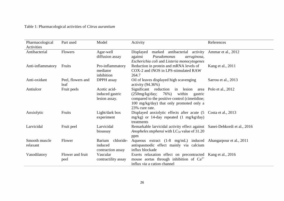
Table 1: Pharmacological activities of Citrus aurantium
Pharmacological
Activities
Part used Model Activity References
Antibacterial Flowers Agar-well
diffusion assay
Displayed marked antibacterial activity
against Pseudomonas aeruginosa,
Escherichia coli and Listeria monocytogenes
Ammar et al., 2012
Anti-inflammatory Fruits Pro-inflammatory
mediator
inhibition
Reduction in protein and mRNA levels of
COX-2 and iNOS in LPS-stimulated RAW
264.7
Kang et al., 2011
Anti-oxidant Peel, flowers and
leaf
DPPH assay Oil of leaves displayed high scavenging
activity (94.36%)
Sarrou et al., 2013
Antiulcer Fruit peels Acetic acid-
induced gastric
lesion assay.
Significant reduction in lesion area
(250mg/kg/day; 76%) within gastric
compared to the positive control (cimetidine;
100 mg/kg/day) that only promoted only a
23% cure rate.
Polo et al., 2012
Anxiolytic Fruits Light/dark box
experiment
Displayed anxiolytic effects after acute (5
mg/kg) or 14-day repeated (1 mg/kg/day)
treatments
Costa et al., 2013
Larvicidal Fruit peel Larvicidal
bioassay
Remarkable larvicidal activity effect against
Anopheles stephensi with LC50 value of 31.20
ppm
Sanei-Dehkordi et al., 2016
Smooth muscle
relaxant
Flower Barium chloride-
induced
contraction assay
Aqueous extract (1-8 mg/mL) induced
antispasmodic effect mainly via calcium
influx blockade
Ahangarpour et al., 2011
Vasodilatory Flower and fruit
peel
Vascular
contractility assay
Exerts relaxation effect on precontracted
mouse aortas through inhibition of Ca2+
influx via a cation channel
Kang et al., 2016

27
Table 2: 1H (600 MHz) NMR assignments for 1-6 in CDCl3.
Position 1 2 3 4 5 6 1H, J (Hz) 1H, J (Hz) 1H, J (Hz) 1H, J (Hz) 1H, J (Hz) 1H, J (Hz)
1 14.04, s 14.32, s 14.30, s 14.51, s 14.43, s 14.30, s
2 6.41, s 6.29, s 6.27, s 6.24, s 6.24, s 6.12, s
3 - - - - - -
4 - - - 6.24, s - -
4a - - - - - -
5 - - - - - -
6 7.14, dd (7.8, 1.4) - 7.33, d (7.9) - - -
7 7.18, t (7.8) 7.02, d (8.8) 7.19, t (7.9) 6.90, d (8.6) 6.92, d (8.8) 6.92, d (8.8)
8 7.96, dd (7.8, 1.4) 8.09, d (8.8) 7.94, dd (7.9, 1.2) 8.09, d (8.6) 8.11, d (8.8) 7.73, d (8.8)
8a - - - - - -
9 - - - - -
9a - - - - - -
10a - - - - - -
11 - 6.57, d (9.7) 6.70, d (9.7) - - 6.75, d (9.5)
12 - 5.62, d (9.7) 5.56, d (9.7) - - 5.65, d (9.5)
13 - - - - - -
14 - 1.54, s 1.53, s - - 1.54, s
15 - 1.54, s 1.53, s - - 1.54, s
NCH3 3.84, s 3.73, s 3.81, s 3.92, s 3.98, s 3.82, s
3-OCH3 3.82, s - - 3.84, s 3.96, s -
4-OCH3 3.99, s - - - 3.85, s -
5-OCH3 - 3.93, s - 3.70, s 3.70, s - Assignments were confirmed by DEPT, HSQC and HMBC experiments and by comparing with literature values
(Wu and Furukawa, 1983; Wu et al., 1983; Juoichi et al., 1985; Yamamoto et al., 1989; Ono et al., 1995)

28
Table 3: 13C (150 MHz) NMR and DEPTQ assignments for 1-6 in CDCl3.
Position 1 2 3 4 5 6
13C, DEPTQ 13C, DEPTQ 13C, DEPTQ 13C, DEPTQ 13C, DEPTQ 13C, DEPTQ
1 160.4 164.6 164.6 165.9 159.0 164.0
2 94.0 98.7 98.2 94.4 87.7 97.0
3 159.8 161.1 161.4 165.5 156.1 160.9
4 130.1 102.5 102.3 91.0 130.5 102.5
4a 142.2 147.2 147.8 147.1 143.1 148.0
5 146.2 135.8 137.2 134.2 134.4 134.6
6 120.6 154.4 120.4 154.8 154.7 150.9
7 123.1 112.0 112.0 111.2 111.7 111.7
8 118.9 123.4 123.2 124.0 123.9 123.7
8a 125.1 118.6 117.8 117.3 117.5 117.3
9 182.4 181.5 182.0 180.4 180.6 181.7
9a 106.7 106.8 107.3 105.0 105.7 105.9
10a 137.5 141.5 146.8 138.0 137.9 138.2
11 - 120.4 120.4 - - 120.7
12 - 124.7 124.0 - - 117.2
13 - 77.7 77.4 - - 76.3
14 - 27.2 27.2 - - 25.9
15 - 27.2 27.2 - - 25.9
NCH3 46.3 47.9 48.9 39.2 39.5 47.8
3-OCH3 60.3 - - 55.6 56.1 -
4-OCH3 56.8 - - - 60.8 -
5-OCH3 - 60.1 - 61.7 61.6 - Assignments were confirmed by DEPT, HSQC and HMBC experiments and by comparing with literature values
(Wu and Furukawa, 1983; Wu et al., 1983; Juoichi et al., 1985; Yamamoto et al., 1989; Ono et al., 1995)

29
Table 4: Antiproliferative Effects of the Fractions of the Stem Bark of Citrus aurantium
against Four Human Cancer Cell Linesa
Fraction
IC50 (µg/mL)b
MCF7 A549 PC3 HepG2
n-hexane > 100 > 100 > 100 > 100
DCM 5.12 ±0.54 3.88 ± 0.58 4.72 ± 0.23 5.73 ± 0.99
MeOH 90.6 ± 4.54 88.9 ± 1.23 78.2 ± 2.14 92.7 ± 4.11
aMCF7 - human breast cancer cells; A549- Human lung carcinoma cells; PC3 - human prostate
carcinoma cells; HepG2 - human hepatocellular carcinoma cells. bMTT method, with the cells
incubated with the fractions for 72 h (means ± SEM, n = 3). Vinblastine was used as the positive
control and reference compound.

30
Table 5: Antiproliferative Effects of Acridone Alkaloids from the Stem Bark of Citrus
aurantium against Four Human Cancer Cell Linesa
Compound
IC50 (µM)b
MCF7 A549 PC3 HepG2 PNT2
1 44.53 ± 1.45 35.76 ± 1.55 38.23 ± 1.11 50.74 ± 1.11 179 ± 4.11
2 12.65 ± 0.54 14.02 ± 0.11 14.88 ± 0.17 22.42 ± 0.17 151.5 ± 6.47
3 24.03 ± 1.05 25.94 ± 0.61 28.15 ± 0.68 30.09 ± 0.94 190.3 ± 3.81
4 31.11 ± 1.41 29.69 ± 0.44 27.44 ± 1.61 37.45 ± 0.79 205.1 ± 2.39
5 26.87 ± 0.57 25.02 ± 1.92 24.29 ± 0.49 26.38 ± 0.12 222.7 ±6.12
6 20.91 ± 0.17 20.36 ± 0.81 18.23 ± 0.53 32.62 ± 1.02 175.4 ± 2.11
Vinblastine 0.023 ± 0.01 0.037 ± 0.01 0.028 ± 0.01 0.021 ± 0.01 0.037 ± 0.01
aMCF7 - human breast cancer cells; A549 - human lung carcinoma cells; PC3 - human prostate
carcinoma cells; HepG2 - human hepatocellular carcinoma cells; PNT2 - normal human
prostate cells. bMTT method, with the cells incubated with the fractions for 72 h (means ±
SEM, n = 3). Vinblastine was used as the positive control and reference compound.

31
Table 6. The Selectivity Index (SI) of 1-6 and Vinblastine (VBN)
Compound MCF7 A549 PC3 HepG2
1 4.01 5.00 4.68 3.52
2 11.97 10.80 10.18 6.75
3 7.91 7.33 6.76 6.32
4 6.59 6.90 7.47 5.47
5 8.28 8.90 9.16 8.44
6 8.38 8.61 9.62 5.37
VBN 1.60 1.00 1.32 1.76
MCF7 - human breast cancer cells; A549 - Human lung carcinoma cells; PC3 - human prostate
carcinoma cells; HepG2 - human hepatocellular carcinoma cells. SI is the ratio of the IC50
values of the compound on PNT2 cells to those in the cancer cell lines.


![Synthesis of Acridone Base Phenolic Compounds for Antibacterial … · 2020. 3. 9. · acridone-N-acetic acid showed good antibacterial activity [5]. III. Singh P. et al.74 synthesized](https://static.fdocuments.in/doc/165x107/60b702fc3e696f369a20263a/synthesis-of-acridone-base-phenolic-compounds-for-antibacterial-2020-3-9-acridone-n-acetic.jpg)

