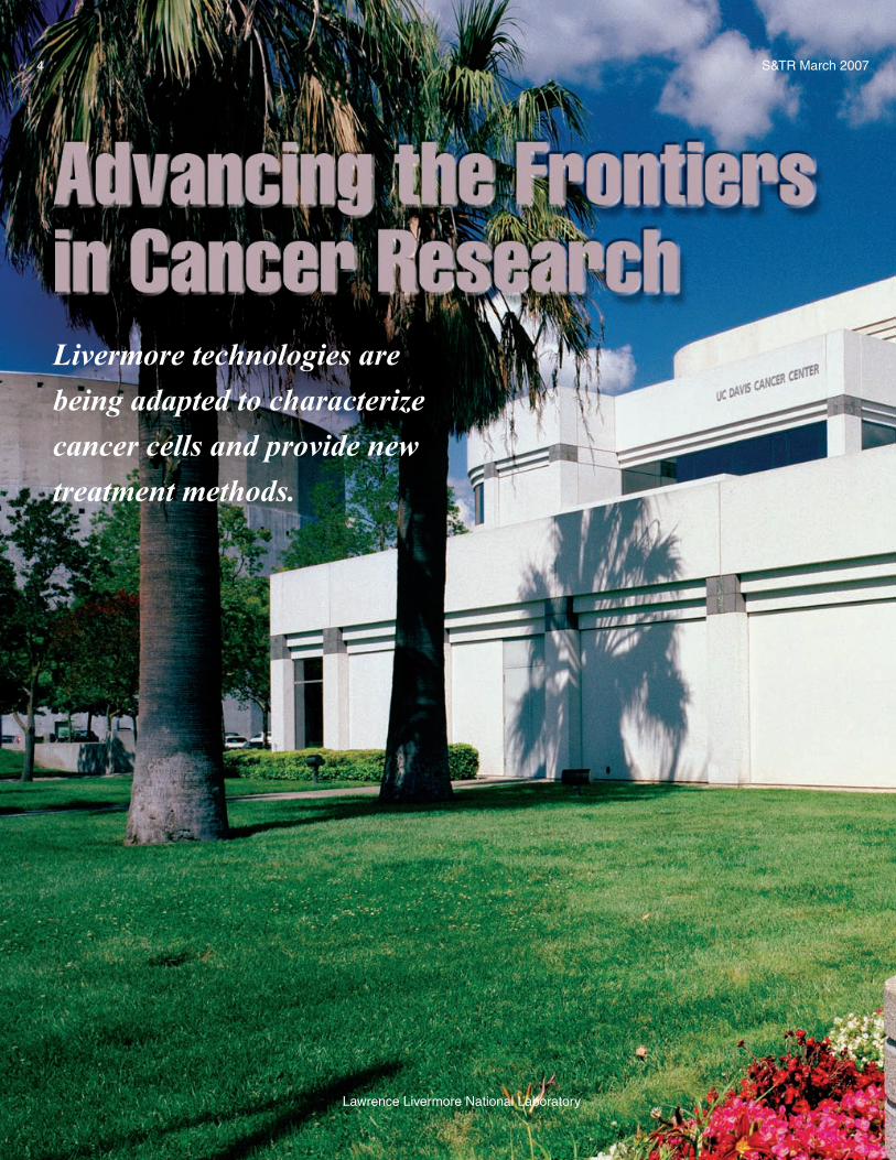
Livermore technologies are being adapted to characterize cancer cells and provide … ·...
8
Livermore technologies are being adapted to characterize cancer cells and provide new treatment methods. 4 Lawrence Livermore National Laboratory S&TR March 2007
-
Upload
trinhquynh -
Category
Documents
-
view
212 -
download
0
Transcript of Livermore technologies are being adapted to characterize cancer cells and provide … ·...
Livermore technologies are being adapted to characterize cancer cells and provide new treatment methods.
4
Lawrence Livermore National Laboratory
S&TR March 2007

MILLIONS of cell duplications occur in our bodies each day.
As an essential part of this process, the nucleotides that make up the DNA fingerprint—adenine, guanine, cytosine, and thymine—are reproduced with
perfect fidelity. Occasionally, an error occurs, and the
code is altered, or mutated, resulting in a coding sequence that may be
expressed as an abnormal cell. Typically, the body’s cellular machinery repairs these DNA mistakes, or its immune system disposes of the aberrant cells. With cancer, however, something in the genetic coding turns off the body’s immune mechanisms, allowing cells with cancerous mutations to avoid detection. Cancer cells can usually replicate more efficiently than normal cells, and they are difficult to eliminate. Blocking one metabolic pathway does not necessarily stop the cells from spreading because they often will adapt to another pathway.
Advances in Cancer Research 5
Lawrence Livermore National Laboratory
Cancer includes hundreds of diseases, each with a specific pattern of behavior and genetic makeup. No universal method can treat the many different types. Some cancers require surgery, while others are best treated with radiation, chemotherapy, or a combination of the two. To determine the best therapy for each patient, physicians need diagnostic tools that can detect the disease in its early stages and predict its course. But current technologies do not allow clinicians to precisely design individualized treatments. In addition, those who are treating cancer patients often are not aware of emerging technologies that could be useful.
S&TR March 2007
The University of California (UC) Davis
Cancer Center is a National Cancer Institute–
designated facility. The center cares for more
than 9,000 new cancer patients each year.
S&TR March 20076
Lawrence Livermore National Laboratory
Advances in Cancer Research
After many collaborations between Lawrence Livermore and the University of California (UC) Davis Cancer Center, researchers at both institutions began realizing that a formal partnership could help advance cancer research and make new treatment options available. The Laboratory’s advanced mass spectrometers, lasers, computational codes, and other instruments originally developed as national security technologies offered potential medical solutions.
Powerful Instruments Aid ResearchIn 2000, Lawrence Livermore and
the UC Davis Cancer Center formed a
strategic collaboration, combining UC Davis’s clinical expertise and basic science research resources with Livermore’s technologies. This collaboration represents the first time a major U.S. cancer center has teamed with a national laboratory, and the joint effort helped the center become a designated National Cancer Institute facility in July 2002. The partnership involves more than 200 physicians, molecular biologists, pharmacologists, physicists, chemists, and cell biologists. Physicians and laboratory clinicians work together to develop new cancer therapies, detection methods, and prevention strategies. (See S&TR, April 2001,
pp. 15–17.) Current efforts include adapting time-of-flight mass spectrometry to characterize individual cancer cells, using accelerator mass spectrometry (AMS) to tailor cancer therapy, and developing a proton accelerator to zap cancer cells.
Molecular biologist Jim Felton, a specialist in cancer causation and prevention, serves as an associate director for the Cancer Etiology, Prevention, and Control Program at the UC Davis Cancer Center. “In addition to its clinical expertise, UC Davis has a strong program in the molecular biology of cancer,” says Felton, who works in Livermore’s Chemistry, Materials, and Life Sciences (CMLS) Directorate. “We have instruments they can exploit.” For example, prior to the collaboration, few clinicians knew that AMS could be used to monitor a drug as it metabolizes, that lasers could provide a noninvasive approach to detect cancer, or that computational models could save time by predicting experimental outcomes.
The UC Davis Cancer Center is organized into six programs: molecular oncology; cancer biology in animals; developmental therapeutics; cancer etiology, prevention, and control; prostate cancer; and biomedical technology. Biologist Ken Turteltaub of CMLS serves as an associate director of shared resources, and Dennis Matthews, director of the Laboratory’s Center for Biotechnology, Biophysical Sciences, and Bioengineering, is an associate director of biomedical technology. Matthews and Felton help identify Livermore technologies that could be relevant to cancer research. Matthews says, “No other cancer center in the world has a dedicated biotechnologies program.”
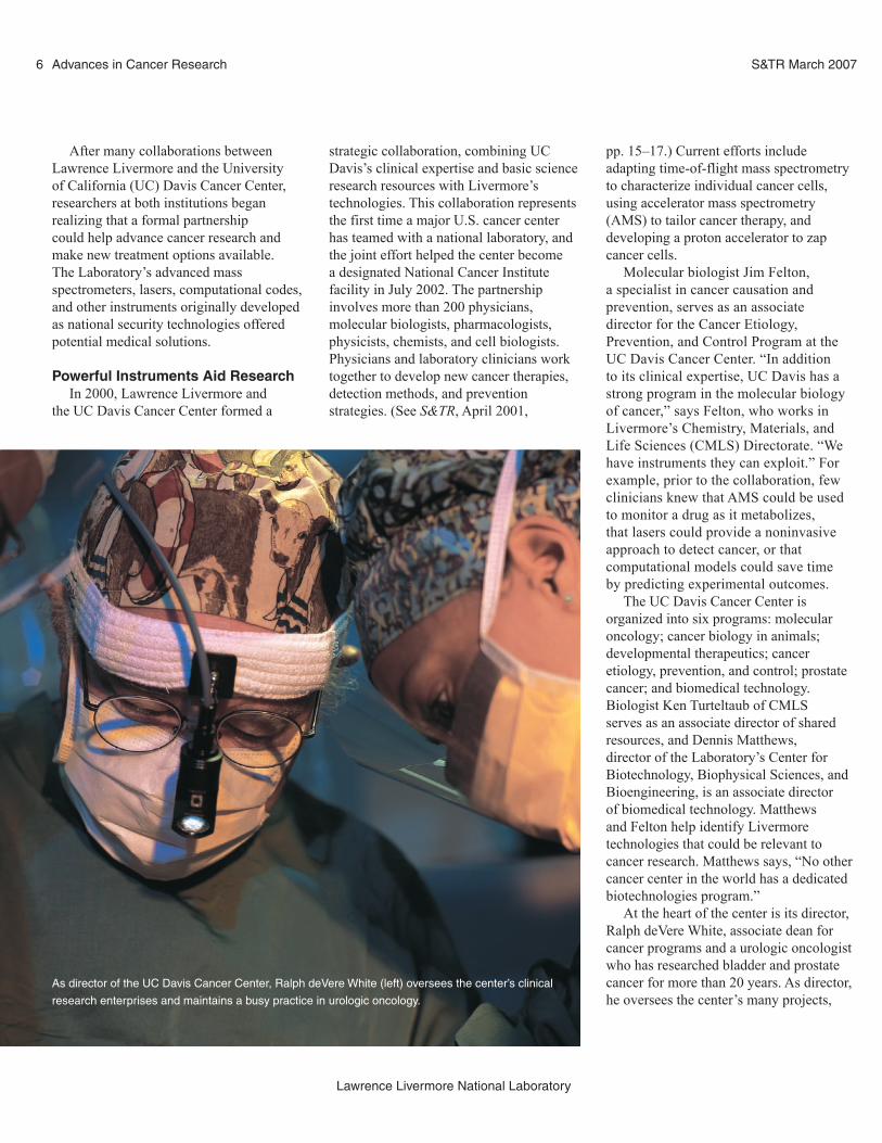
At the heart of the center is its director, Ralph deVere White, associate dean for cancer programs and a urologic oncologist who has researched bladder and prostate cancer for more than 20 years. As director, he oversees the center’s many projects,
As director of the UC Davis Cancer Center, Ralph deVere White (left) oversees the center’s clinical
research enterprises and maintains a busy practice in urologic oncology.
7
Lawrence Livermore National Laboratory
S&TR March 2007 Advances in Cancer Research
techniques that identify differences among the samples and classify the cells.
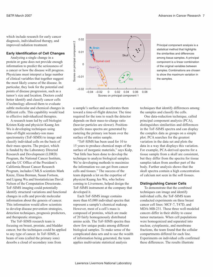
One data-reduction technique, called principal component analysis (PCA), distinguishes similarities and differences in the ToF-SIMS spectra and can display the complex data as groups on a simple plot. PCA searches for the greatest variation in the data set and plots the data in a way that displays this variation. For example, PCA-derived spectra for a tissue sample from a skull are consistent, but they differ from the spectra for tissue samples taken from another part of the body. Further analysis shows that the skull spectra contain a high concentration of calcium not seen in the soft tissues.
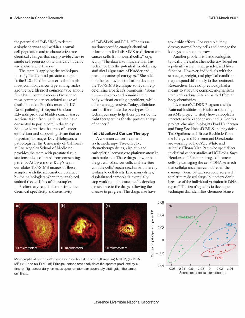
Distinguishing Cancer CellsTo demonstrate that the combined
techniques can image and identify individual cells, the ToF-SIMS team conducted experiments on three breast cancer cell lines: MCF-7, T47D, and MDA-MB-231. These three well-modeled cancers differ in their ability to cause tumor metastasis. When cell populations were homogenized and separated into nuclear, cytoplasmic, and membrane fractions, the team found that the cellular compartments differed for each line. Experiments on individual cells confirmed these differences. The results illustrate
which include research for early cancer diagnosis, individualized therapy, and improved radiation treatment.
Early Identification of Cell ChangesAnalyzing a single change in a
protein or gene does not provide enough information to predict the seriousness of a cancer or how the disease will progress. Physicians must interpret a large number of clinical variables that together suggest the most likely course of the disease. In particular, they look for the potential end points of disease progression, such as a tumor’s size and location. Doctors could better identify and classify cancer cells if technology allowed them to evaluate subtle molecular and chemical changes in different cells. This capability would lead to effective individualized therapies.
A research team led by cell biologist Kristen Kulp and physicist Kuang Jen Wu is developing techniques using time-of-flight secondary-ion mass spectrometry (ToF-SIMS) to image and classify individual cells on the basis of their mass spectra. The project, which is funded by the Laboratory Directed Research and Development (LDRD) Program, the National Cancer Institute, and the UC Office of the President’s California Breast Cancer Research Program, includes CMLS scientists Mark Knize, Elena Berman, Susan Fortson, and Ligang Wu and biostatistician David Nelson of the Computation Directorate. ToF-SIMS imaging could potentially identify structural variations and functional changes in cells and provide molecular information about the genesis of cancer. This information would allow scientists to develop disease prevention strategies, detection techniques, prognosis predictors, and therapeutic strategies.
The ToF-SIMS research team is focusing on breast, prostate, and bladder cancer, but the techniques could be applied to any type of cancer. In ToF-SIMS, a beam of ions (called the primary ions) desorbs a cloud of secondary ions from
a sample’s surface and accelerates them toward a time-of-flight detector. The time required for the ions to reach the detector depends on their mass-to-charge ratio (heavier particles are slower). Position-specific mass spectra are generated by rastering the primary ion beam over the surface of the entire sample.
“ToF-SIMS has been used for 10 to 15 years to produce chemical maps of the surface of inorganic materials,” says Kulp, “but little has been done to develop the technique to analyze biological samples. We’re developing methods to maximize the information we can get from cancer cells and tissues.” The success of the team depends a lot on the expertise of physicist Kuang Jen Wu, who before coming to Livermore, helped design the ToF-SIMS instrument at the company that developed it.
Each ToF-SIMS image contains more than 65,000 individual spectra that represent a sample’s chemical makeup. However, most of a cell’s mass is composed of proteins, which are made of 20 fairly homogenously distributed amino acids. The ToF-SIMS spectra thus show few unique peaks among different biological samples. To make sense of the complicated data sets and to use the wealth of information being generated, the team applies multivariate statistical analysis
0.02
0.01
0
–0.01
–0.02–0.04
Sco
res
on p
rinci
pal c
ompo
nent
2
–0.02 0 0.02Scores on principal component 1
Brain
Spinal cord
Heart
Liver
Skull
Rib
0.04 0.06 0.08
Principal component analysis is a
statistical method that highlights
the similarities and differences
among tissue samples. A principal
component is a linear combination
of the original variables between
samples. Combinations are chosen
to show the maximum variation in
the samples.
S&TR March 20078
Lawrence Livermore National Laboratory
Advances in Cancer Research
the potential of ToF-SIMS to detect a single aberrant cell within a normal cell population and to characterize rare chemical changes that may provide clues to single cell progression within carcinogenic and metastatic pathways.
The team is applying the techniques to study bladder and prostate cancers. In the U.S., bladder cancer is the fourth most common cancer type among males and the twelfth most common type among females. Prostate cancer is the second most common cancer-related cause of death in males. For this research, UC Davis pathologist Regina Gandour-Edwards provides bladder cancer tissue sections taken from patients who have consented to participate in the study. She also identifies the areas of cancer epithelium and supporting tissue that are important to image. David Seligson, a pathologist at the University of California at Los Angeles School of Medicine, provides the team with prostate tissue sections, also collected from consenting patients. At Livermore, Kulp’s team correlates ToF-SIMS images of these samples with the information obtained by the pathologists when they analyzed stained tissue slides of the samples.
Preliminary results demonstrate the chemical specificity and sensitivity
of ToF-SIMS and PCA. “The tissue sections provide enough chemical information for ToF-SIMS to differentiate cancer cells from normal cells,” says Kulp. “The data also indicate that this technique has the potential for defining statistical signatures of bladder and prostate cancer phenotypes.” She adds that the team wants to further develop the ToF-SIMS technique so it can help determine a patient’s prognosis. “Some tumors develop and remain in the body without causing a problem, while others are aggressive. Today, clinicians can’t differentiate the two types. Our techniques may help them prescribe the right therapeutics for the particular type of cancer.”
Individualized Cancer TherapyA common cancer treatment
is chemotherapy. Two effective chemotherapy drugs, cisplatin and carboplatin, contain one platinum atom in each molecule. These drugs slow or halt the growth of cancer cells and interfere with the cells’ repair mechanism, thereby leading to cell death. Like many drugs, cisplatin and carboplatin eventually stop working—the cancer cells develop a resistance to the drugs, allowing the disease to progress. The drugs also have
toxic side effects. For example, they destroy normal body cells and damage the kidneys and bone marrow.
Another problem is that oncologists typically prescribe chemotherapy based on a patient’s weight, age, gender, and liver function. However, individuals with the same age, weight, and physical condition may respond differently to the treatment. Researchers have not previously had a means to study the complex mechanisms involved as drugs interact with different body chemistries.
Livermore’s LDRD Program and the National Institutes of Health are funding an AMS project to study how carboplatin interacts with bladder cancer cells. For this project, chemical biologists Paul Henderson and Sang Soo Hah of CMLS and physicists Ted Ognibene and Bruce Buchholz from the Energy and Environment Directorate are working with deVere White and scientist Chong Xian Pan, who specializes in clinical cancer studies at UC Davis. Says Henderson, “Platinum drugs kill cancer cells by damaging the cells’ DNA so much that cellular enzymes cannot repair the damage. Some patients respond very well to platinum-based drugs, but others don’t because of the individual variation in DNA repair.” The team’s goal is to develop a technique that identifies chemoresistance
Micrographs show the differences in three breast cancer cell lines: (a) MCF-7, (b) MDA-
MB-231, and (c) T47D. (d) Principal component analysis of the spectra produced by a
time-of-flight secondary-ion mass spectrometer can accurately distinguish the same
cell lines.
–0.08 –0.06 –0.04 –0.02 0 0.02 0.04
MDA-MB-231
MCF-7
T47D
Scores on principal component 1
Sco
res
on p
rinci
pal c
ompo
nent
2
0.06
0.04
0.02
0
–0.02
–0.04
(a) (b) (c) (d)
10 micrometers 10 micrometers 10 micrometers
9
Lawrence Livermore National Laboratory
S&TR March 2007 Advances in Cancer Research
so oncologists can customize a patient’s treatment.
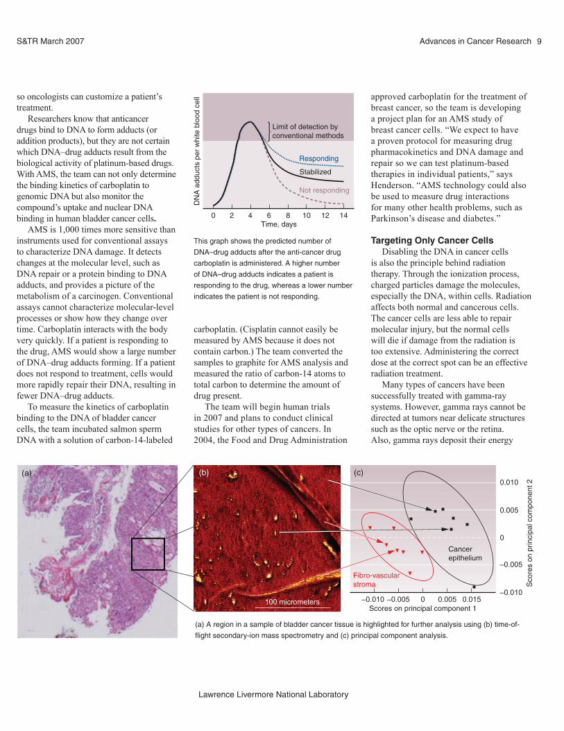
Researchers know that anticancer drugs bind to DNA to form adducts (or addition products), but they are not certain which DNA–drug adducts result from the biological activity of platinum-based drugs. With AMS, the team can not only determine the binding kinetics of carboplatin to genomic DNA but also monitor the compound’s uptake and nuclear DNA binding in human bladder cancer cells.
AMS is 1,000 times more sensitive than instruments used for conventional assays to characterize DNA damage. It detectschanges at the molecular level, such as DNA repair or a protein binding to DNA adducts, and provides a picture of the metabolism of a carcinogen. Conventional assays cannot characterize molecular-level processes or show how they change over time. Carboplatin interacts with the body very quickly. If a patient is responding to the drug, AMS would show a large number of DNA–drug adducts forming. If a patient does not respond to treatment, cells would more rapidly repair their DNA, resulting in fewer DNA–drug adducts.
To measure the kinetics of carboplatin binding to the DNA of bladder cancer cells, the team incubated salmon sperm DNA with a solution of carbon-14-labeled
carboplatin. (Cisplatin cannot easily be measured by AMS because it does not contain carbon.) The team converted the samples to graphite for AMS analysis and measured the ratio of carbon-14 atoms to total carbon to determine the amount of drug present.
The team will begin human trials in 2007 and plans to conduct clinical studies for other types of cancers. In 2004, the Food and Drug Administration
100 micrometers
Fibro-vascularstroma
Cancerepithelium
0.010
0.005
0
–0.005
–0.010–0.010 –0.005 0 0.005 0.015
Scores on principal component 1
Sco
res
on p
rinci
pal c
ompo
nent
2
(a) (b) (c)
(a) A region in a sample of bladder cancer tissue is highlighted for further analysis using (b) time-of-
flight secondary-ion mass spectrometry and (c) principal component analysis.
This graph shows the predicted number of
DNA–drug adducts after the anti-cancer drug
carboplatin is administered. A higher number
of DNA–drug adducts indicates a patient is
responding to the drug, whereas a lower number
indicates the patient is not responding.
Limit of detection byconventional methods
Responding
Stabilized
Not responding
0 2 4 6Time, days
DN
A a
dduc
ts p
er w
hite
blo
od c
ell
8 10 12 14
approved carboplatin for the treatment of breast cancer, so the team is developing a project plan for an AMS study of breast cancer cells. “We expect to have a proven protocol for measuring drug pharmacokinetics and DNA damage and repair so we can test platinum-based therapies in individual patients,” says Henderson. “AMS technology could also be used to measure drug interactions for many other health problems, such as Parkinson’s disease and diabetes.”
Targeting Only Cancer CellsDisabling the DNA in cancer cells
is also the principle behind radiation therapy. Through the ionization process, charged particles damage the molecules, especially the DNA, within cells. Radiation affects both normal and cancerous cells. The cancer cells are less able to repair molecular injury, but the normal cells will die if damage from the radiation is too extensive. Administering the correct dose at the correct spot can be an effective radiation treatment.
Many types of cancers have been successfully treated with gamma-ray systems. However, gamma rays cannot be directed at tumors near delicate structures such as the optic nerve or the retina. Also, gamma rays deposit their energy

S&TR March 200710
Lawrence Livermore National Laboratory
Advances in Cancer Research
uniformly, beginning near the body’s surface and continuing well beyond the cancer site to damage healthy tissues.
In 1946, physicist Robert Wilson, then at Lawrence Berkeley National Laboratory, proposed using protons for radiotherapy. Two years later, experiments at Lawrence Berkeley confirmed the benefit of this approach. Protons deliver most of their energy at a specific depth in the tissue, which is determined by the protons’ energy. Low-energy protons travel a short distance and deliver their energy, while higher-energy protons travel farther before releasing theirs. A proton beam can thus be calibrated to target specific areas of the body. Its precision can be so exact that healthy tissue a few millimeters from the target area receives almost no radiation.
Proton beam therapy has been available since 1990, but few organizations have the resources to build and operate the required facilities. In the past, large particle accelerators such as cyclotrons or synchrotrons were required to produce high-energy proton beams. These instruments occupy as much space as a basketball court, and construction alone costs up to $200 million. Concrete walls also must be built to protect people outside the treatment room from
the radiation generated. The U.S. has only a few of the 20 or so facilities available worldwide. The development of a compact proton accelerator could dramatically reduce the cost of building proton therapy centers.
Choosing the Best MaterialsIn 2003, the LDRD Program funded
a project to explore the concept of a compact proton accelerator called a dielectric wall accelerator. The project team, led by physicist George Caporaso of Livermore’s Physics and Advanced Technologies Directorate, originally focused on defense-related applications until Dennis Matthews proposed designing a version for cancer therapy. Caporaso says, “The principle behind the compact system is an accelerator tube composed of sections, or rings, each with transmission lines. A series of switches sequentially apply voltage to the transmission lines, and the high electric field propels the particles through the vacuum. The timing of the switches controls the accelerator wave so that its speed matches the particle’s speed.”
The accelerator uses an insulating pipe, or dielectric wall, to maintain a vacuum while preventing the accelerator from short-circuiting. A common problem with insulators is a phenomenon called
surface flashover. During high-voltage conditions, the electrodes that accelerate the charged particles produce unwanted electrons that gain energy from the strong electric field. These electrons then collide with the insulator’s surface, which liberates additional electrons and gas molecules adsorbed on the insulator. This process continues, eventually resulting in a gas buildup that sparks.
In the 1980s, researchers found they could design accelerators to withstand a higher electric field by imbedding thin metal layers within the insulator. They hypothesized that the metal layers interrupt the surface flashover process by deflecting or capturing the unwanted electrons before they can collide with the insulator surface. In Livermore’s dielectric insulator, conducting layers made of a metal such as stainless steel alternate with insulating layers of a plastic, such as polystyrene. The technology won an R&D 100 Award in 1997. (See S&TR, October 1997, pp. 16–17.)
John Harris, an engineer in Livermore’s Accelerator Design and Code Group, is conducting experiments to determine requirements for the high-gradient insulators to be used in a dielectric wall accelerator for proton therapy. In the Livermore design, insulator rings are
Plate with cells Cells grown in mediaspiked with carbon-14-labeled
carboplatin
Carbon-14-labeled drugbinds to DNA
Measuredby AMS
Responder
Nonresponder
Isolatedcells
H2N Pt O COOH
DNA–drug adduct
Carboplatin
ONH2
H2N
Pt
NH2
ExtractedDNA
A sample from bladder cancer cells is incubated
with a solution of carbon-14-labeled carboplatin to
measure the kinetics of carboplatin binding to the
DNA. Cells are then isolated from the solid sample
and an accelerator mass spectrometer (AMS)
measures the ratio of carbon-14 atoms to total
carbon to determine the amount of drug present.
COOH = carboxylic group compound, H2N and
NH2 = amino group compounds, O = oxygen, and
Pt = platinum.
11
Lawrence Livermore National Laboratory
S&TR March 2007 Advances in Cancer Research
stacked to form a pipe. Air is removed from inside the pipe, providing a vacuum through which the proton beam travels. Transmission lines stacked along the pipe’s outside surface can be activated by fast switches to produce an electric field in the pipe, which accelerates the proton beam. The electric field must be very strong for a compact accelerator to reach the energy levels needed in proton therapy. “The requirements for the vacuum insulation are thus quite challenging,” says Harris. “We’re starting to understand a lot more about how the insulators work, and how we can make them work even better.”
Caporaso’s team estimates that a compact system designed with a dielectric wall accelerator could achieve the required energies—from 70 megaelectronvolts for eye tumors to 250 megaelectronvolts for tumors deep in the body—with electric fields of up to 100 megavolts per meter. This design would eliminate such devices as bending magnets, which have large space requirements and generate unwanted radiation. Says Caporaso, “A dielectric wall accelerator only 2.5 meters long could generate the range of energy produced by traditional proton radiotherapy machines.”
The Laboratory Science and Technology Office and UC Davis Health System are funding a 20-centimeter-long prototype that should be ready next year. “Our goal is to build the machine as small as possible while ensuring that its wall is strong enough to hold gradient,” says Caporaso. “At the same time, we want the machine’s architecture to be such that it delivers the required energy at a fraction of the cost of traditional systems.”
A Strategic CollaborationThe technology could also be used
to generate radioisotopes for medical applications. As with proton radiotherapy, radiopharmaceuticals must be made at large facilities and flown to hospitals on request. However, radioisotopes begin decaying right away, and many of those used in medical applications decay rapidly. With compact, inexpensive accelerators, hospitals could produce these isotopes on site for immediate use. In addition to medical applications, the technology could also be applied to screen luggage.
The strategic collaboration between Livermore and UC strengthens basic science research and maximizes the
capabilities of both institutions. Says Kulp, “We’re making advances in our studies on cancer cells because we can pull together a multidisciplinary team that includes a chemist, a biologist, a pathologist, a physicist, and a statistician.” Together, researchers have pioneered many technologies to help in the fight against cancer.
“When we formed the integrated cancer program in 2000, we laid out an exciting vision of what the collaboration might bring,” says deVere White. “I doubt any of us could have imagined the success it already has become or the potential for good that it holds for the future.”
—Gabriele Rennie
KeyWords:accelerator mass spectrometry (AMS), cancer, carboplatin, cisplatin, dielectric wall accelerator, principal component analysis (PCA), proton accelerator, proton beam therapy, time-of-flight secondary-ion mass spectrometry (ToF-SIMS), University of California (UC) Davis Cancer Center.
For further information contact Jim Felton
(925) 422-5656 ([email protected]).
High-gradientinsulator
Transmission line
(a) (b) (c)
A compact dielectric wall accelerator is composed
of a stack of rings made from thin, alternating
layers of a metal and an insulator: (a) side view,
(b) top view, and (c) sample ring. Transmission
lines embedded in the rings produce the electric
fields that propel charged particles along the
tube. As the particles travel, a series of switches
open and close, controlling the voltage applied to
transmission lines in each section.