
litdoc28917653AA_20110831103810
8
GE Healthcare The Trap Platform. Small-scale protein sample preparation. Get it right from the start. Trap Product Profile
description
sd fff
Transcript of litdoc28917653AA_20110831103810
imagination at work
GE Healthcare
The Trap Platform.
Small-scale proteinsample preparation.
Get it right from the start.
Trap
Product Profile

The small-scale preparation of protein samplesOptimized for downstream applications
The small-scale preparation of protein samples is key to the success and quality of subsequent downstream experimental work and analyses. This critical aspect of protein science has hardly changed for decades and is increasingly regarded as a major limiting step in the study of proteins.
The continuous development of the Trap platform is designed to bring total solutions to the ever increasing demands of the protein researcher.
Building on the experience and knowledge of proteins gained over decades in our laboratories together with the heritage from the HiTrap™ platform of protein capture products, the Trap platform is being developed further with the introduction of new SpinTrap™, GraviTrap™, and MultiTrap™ formats for various steps in protein sample preparation.
The Trap platform is a range of application-based protein preparation products designed to enable researchers to achieve high reproducibility, yield, and purity of their proteins in line with the specific demands of downstream techniques or analytical methods, e.g. 1-D, 2-D electrophoresis, Western blotting, liquid chromatography, protein interaction, and mass spectrometry.
Optimization guides help you achieve maximum performance for your specific application and for the characteristics of your protein of interest. All Trap protocols are completely transparent. Full information about formats, media, buffers, and methodologies is provided to support and enable further optimization by the researcher.
The current Trap product range includes products for tagged protein preparation, desalting, and protein enrichment. Continuous development of the platform will extend this range to cover all protein preparation needs.
Find the right protein sample preparation product for your application in this Product Profile or in the Protein Sample Preparation Selection Guide at:
www.gehealthcare.com/trap
HiTrap
MultiTrap
SpinTrap
GraviTrap
Trap

Application-focusedproduct selectionThe Trap platform focuses on optimizing protein sample preparation for typical downstream applications. The selection guide at www.gehealthcare.com/trap makes choosing the right product easy – simply click on the arrow pointing towards your intended analysis method.
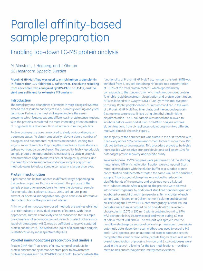
Parallel affinity-based sample preparationEnabling top-down LC-MS protein analysis
M. Almstedt, J. Hedberg, and J. ÖhmanGE Healthcare, Uppsala, Sweden
Protein G HP MultiTrap was used to enrich human α-transferrin (hTf) more than 100-fold from E. coli extract. The eluate resulting from enrichment was analyzed by SDS-PAGE or LC-MS, and the yield was sufficient for extensive MS analysis.
IntroductionThe complexity and abundance of proteins in most biological systems exceed the resolution capacity of every currently existing analytical technique. Perhaps the most striking example is the serum proteome, which features extreme differences in protein concentrations, with the proteins considered the most interesting often ten orders of magnitude less abundant than albumin or immunoglobulins.
Protein analyses are commonly used to study various disease or treatment states. To obtain statistically relevant data a number of biological and experimental replicates are needed, leading to a large number of samples. Preparing the samples for these studies is tedious work and a source of error. The demand for highly reproducible sample preparation approaches is increasing as protein analysis and proteomics begin to address actual biological questions, and the need for convenient and reproducible sample preparation methodologies to reduce sample complexity is larger than ever.
Protein fractionationA proteome can be fractionated in different ways depending on the protein properties that are of interest. The purpose of the sample preparation procedure is to make the biological sample, for example, blood, plasma, tissue, urine, cell culture, plant extract, or bacteria, manageable enough to enable an informative characterization of the protein(s) of interest.
Affinity- and immunocapture-based methods are well-established to enrich subsets or individual proteins of interest. With these approaches, sample complexity can be reduced so that a simple one-dimensional separation procedure such as electrophoresis or liquid chromatography (LC), may be sufficient to resolve captured protein constituents. The typical end-point of a proteomic analysis is identification by mass spectrometry (MS).
Parallel immunocapture preparation and analysisProtein G HP MultiTrap is one of a new range of products for protein enrichment by immunocapture prior to downstream protein analyses such as SDS-PAGE and LC-MS. To demonstrate the
functionality of Protein G HP MultiTrap, human transferrin (hTf) was enriched from E. coli cell containing hTf added to a concentration of 0.15% of the total protein content, which approximately corresponds to the concentration of a medium-abundant protein. To enable rapid downstream visualization and protein quantitation, hTf was labeled with CyDye™ DIGE Fluor Cy5™ minimal dye prior to mixing. Rabbit polyclonal anti-hTf was immobilized in the wells of a Protein G HP MultiTrap filter plate, and the antibody-protein G complexes were cross-linked using dimethyl pimelimidate dihydrochloride. The E. coli sample was added and allowed to incubate before wash and elution. SDS-PAGE analysis of three elution fractions from six replicates originating from two different multiwell plates is shown in Figure 2.
The majority of the enriched hTf was eluted in the first fraction with a recovery above 50% and an enrichment factor of more than 100 relative to the starting material. This procedure proved to be highly reproducible with relative standard deviations well below 10% for both target protein recovery and specific purity.
Reversed-phase LC-MS analyses were performed and the starting material and hTf-enriched elution fraction were compared. Start material was diluted with the elution buffer to a suitable protein concentration and thereafter treated the same way as the enriched sample. Tricarboxyethylphosphine was added to reduce the disulfide bonds of the proteins and cysteines were alkylated with iodoacetamide. After alkylation, the proteins were cleaved into smaller fragments by addition of stabilized porcine trypsin and incubated overnight at room temperature. Ten microliter of each sample was injected on a C18 enrichment column and desalted on-line using the Ettan™ MDLC chromatography system. Bound peptides were then separated on an analytical C18 reversed-phase column (0.075 × 150 mm) with a gradient from 0 to 67% (v/v) acetonitrile in 0.1% formic acid and water during 60 min at a flow rate of 200 nl/min. The effluent was sprayed into the nanoflow electrospray source of an ion trap mass spectrometer. An automatic data-dependent scan method was used to acquire MS and MS/MS spectra, and an automated protein database search completed the identification of the peptide fragments as well as the overall identification of proteins. Human and E. coli databases were used in the search, allowing for the two modifications — oxidized methionines and carboxyamido-methylated cysteines.

Proteins detectedAbout 50 different proteins were detected and identified with confidence (p<0.01) in the start material. These proteins were mainly high-abundant E. coli proteins, including proteins involved in the protein synthesis machinery, metabolic enzymes, and various heat-shock proteins (Fig 3). They were generally identified by only one or two peptide fragments. hTf was not detected in the start material. In contrast, the identification of proteins in the enriched sample showed hTf as the major protein hit with high confidence, represented by 48 unique hTf-derived peptide fragments covering 70% of the precursor sequence (Fig 3). Spectra were obtained with signal strength that permitted detailed information extraction. The remaining proteins identified were either ribosomal proteins or proteins closely associated with the ribosomal complex. Notably, the yield in this particular experiment was high enough to allow the
This article was first published in GEN,Genetic Engineering News, December 2006.
Protein G Sepharose HP
Antibody
Protein mix
Protein of interest
Imobilization
Capture
Wash
Elution
Sample collection
Enrichment
Digestion
LC-MS/MS analysis
Fig 1. Applied sample preparation work flow.
1 2 3 4 5 6 7 8 9 10 11 12 13 14 15 16 17 18 19 20 21 22 23
Start material
Second elution,replicates 1–6
First elution,replicates 1–6
Third elution,replicates 1–6
Fig 2. Enrichment of Cy5-labelled human serum transferrin (hTf) using cross-linked a-transferrin antibodies and Protein G Sepharose High Performance in a multiwell format. Start material was E.coli protein (5 mg/mL) containing hTf (7.5 µg/mL). Depicted is the analysis by SDS-PAGE of six replicates from three elution steps. The gel was stained by Deep Purple Total Protein Stain and scanned in the Ettan DIGE Imager using excitation and emission wavelengths specific for Cy5 (red) and Deep Purple (green), respectively.
Fig 3. Proteins and peptides identified by LCMS/ MS analysis with p values less than 0.01. Upper panel: The number of unique proteins identified in each sample, i.e., start material (green) and hTf enriched material (blue), respectively. The respective protein positions in the graph relate to expectation value (X-axis) and intensity (Y-axis). The sizes of the bubbles represent the number of different peptides identified per protein. Lower panel: Detailed information on the sequence coverage of hTf (red boxes) compared to the theoretical sequence (black line), along with an example of an MS/MS spectrum of the indicated peptide with b- and y-ion series annotated.
use of only 5% of the first eluted fraction, indicating that protein(s) with significantly lower abundance than reviewed in this study may well be analyzed using the same protocol.
ConclusionsAn immunoprecipitation sample preparation method based on Protein G HP MultiTrap proved to be convenient and highly reproducible, generating an enriched sample that could be analyzed with an SDS-PAGE or LC-MS approach. The results demonstrate the strength of reducing sample complexity prior to analysis. In addition, the yield of the enriched protein from the model system used in this study was sufficient to enable an extensive MS analysis.
Reference1. Data File: Protein G SpinTrap and Protein G MultiTrap, GE Healthcare, 28-9067-90,
Edition AA (2006).
10
-700 -500 -300
log expectation value
-100 100
100
80
60
40
20
0
9876
43210
5
Proteins identified in start material Proteins identified after enrichment
log
inte
nsity
10
-700 -500 -300
y“1
y“2
y“3
y“4
y“5
y“6
y“7
y“8
log expectation value
-100 100
9876
43210
5
log
inte
nsity
hTf
hTf spiked in E.coli Protein G Sepharose HP eluate
49 proteins 14 40 proteins
93 peptides 4 187 peptides
b3
b4
b5
b6
b7
b8
b9
200 400 600 800 1000
Protein G Sepharose HP eluate
Trap toolkit Protein G HP MultiTrapSample: 5 mg/ml E .coli protein containing 7.5 µg/ml hTfSample volume: 0.2 mlAntibody: Polyclonal rabbit anti-hTfBinding buffer: Tris buffered saline (TBS: 50 mM Tris, 150 mM NaCl, pH 7.5)Wash buffer: TBS, 2 M urea, pH 7.5Elution buffer: 0.1 M glycine-HCl, 2 M urea, pH 3.0

Reproducible protein enrichmentSpinTrap enriches proteins with very low variation between replicates. Typical recovery rates vary by less than 10% (Fig 1 and 2). Similarly, MultiTrap plates allow preparation of up to 96 samples in parallel with well-to-well variation below 10% (Fig 3). SpinTrap and MultiTrap formats are designed so that methods developed in one format can be simply transferred to the other. For projects requiring high throughput, MultiTrap gives significant cost savings.
7.5 3.7 1.88 0.94 0.47 1 2 3 4 5 6 7 8 9 10 11 12
Replicates 1–12 Standards (µg/ml)
la iret am gn it rat s fo
Reco
very
(%
)
Replicates 1–10
Replicates 1–12
Reco
very
of s
tart
ing
mat
eria
l (%
)
Fig 2. Recovery of total loaded material varied by 6% (relative standard
deviation), illustrated by the error bar on the column showing the average
of the 12 samples (red bar).
Fig 3. Enrichment of a known amount of HSA from an E. coli cell lysate (5 mg/ml
E. coli protein + 7.5 µg/ml HSA) using Protein G HP MultiTrap shows high well-to-well
reproducibility (relative standard deviation <10%). The protein recovery of the first
elution step from 10 wells is shown.
SpinTrap reproducibility SpinTrap reproducibility
MultiTrap reproducibility
Fig 1. Enrichment of human albumin (HSA) from E. coli lysates (5 mg/ml
E.coli protein + 7.5 µg/ml HSA) using Streptavidin HP SpinTrap. Twelve
columns were run in parallel. Recovery from the first elution is shown.
Known amounts of HSA were run as standards.
la iret am gn it rat s fo
Reco
very
(%
)
Replicates 1–10
Replicates 1–12
Reco
very
of s
tart
ing
mat
eria
l (%
)

Application- focused protocolsWe’ve added flexibility in the form of Optimization Guides to improve performance for both specific applications and the characteristics of your protein. Beyond simply cleaning up samples, you can, for example, prepare labeled and pooled protein samples upstream of DIGE analysis. You get consistent, label-independent protein enrichment – a key requirement for quantitative expression analysis.
2:1 1:3 1:3 1:2 1:2 1:1 1:1 2:1 1:3 2:1 1:3 1:2 1:2 1:1 1:1 2:1 1:3 2:1 1:3 1:2 1:2 1:1 1:1 2:1 2:1
E3 E2 E1
1:3 1:2 1:1
ref
0 1.0 2.0 3.0 4.0
1.0
2.0
3.0
4.0
Expected ratio
oitar derus aeM
Fig 5. The measured values in the first and the second elution steps in Figure 4
correspond well to expected values (R2 > 0.99).
Fig 4. SpinTrap and MultiTrap products ensure consistent preparation of differentially labeled proteins. Transferrin labeled with either Cy3 or Cy5 was added
in known Cy3:Cy5 ratios ranging from 1:3 to 2:1 to an E. coli lysate. Transferrin was then enriched using Protein A HP SpinTrap and analyzed for Cy3/Cy5 ratio
differences. Samples were collected in three elution steps (E1-E3), and separated by SDS-PAGE. Ref: 100% starting material (transferrin mixed and loaded directly
into the SpinTrap).
Expected vs. measured values for differential analysis (E1)
Preparing labeled protein samples before DIGE analysis

imagination at work
Product Format Code no.
Tissue homogenization
Sample Grinding Kit Microspin columns
80-6483-37
Enzyme regulation
Protease Inhibitor Mix Reagent 80-6501-23
Nuclease Mix Reagent 80-6501-42
Protein fractionation
2-D Fractionation Kit Reagents 80-6501-04
Protein depletion
Albumin and IgG Removal Kit Microspin columns
RPN6300
Protein enrichment
NHS HP SpinTrap SpinTrap 28-9031-28
Streptavidin HP SpinTrap SpinTrap 28-9031-30
Streptavidin HP MultiTrap MultiTrap 28-9031-31
Protein A HP SpinTrap SpinTrap 28-9031-32
Protein A HP MultiTrap MultiTrap 28-9031-33
Protein G HP SpinTrap SpinTrap 28-9031-34
Protein G HP MultiTrap MultiTrap 28-9031-35
Desalting/buffer exchange/clean-up
Disposable PD-10 Desalting Columns
Gravity column 17-0851-01
Mini Dialysis Kit, 1 kDa, 250 µl Dialysis tubes 80-6483-75
Mini Dialysis Kit, 1 kDa, 2 ml Dialysis tubes 80-6483-94
Mini Dialysis Kit, 8 kDa, 250 µl Dialysis tubes 80-6484-13
Mini Dialysis Kit, 8 kDa, 2 ml Dialysis tubes 80-6484-32
2-D Clean-Up Kit Reagents 80-6484-51
SDS-PAGE Clean-Up Kit Reagents 80-6484-70
Antibody purification
Ab SpinTrap SpinTrap 28-4083-47
Ab Buffer Kit Buffer kit 28-9030-59
Product Format Code no.
Histidine-tagged protein purification
His MultiTrap FF MultiTrap 28-4009-89His MultiTrap HP MultiTrap 28-4009-90His SpinTrap SpinTrap 28-4013-53His GraviTrap GraviTrap 11-0033-99His GraviTrap Kit GraviTrap 28-4013-51His Buffer Kit Buffer kit 11-0034-00HisTrap™ HP (1 ml) HiTrap 17-5247-01HisTrap HP (5 ml) HiTrap 17-5248-01HisTrap FF (1 ml) HiTrap 17-5319-01HisTrap FF (1 ml) HiTrap 17-5255-01HisTrap FF crude (1 ml) HiTrap 11-0004-28HisTrap FF crude (5 ml) HiTrap 17-5286-01Anti-His Antibody Reagent 27-4710-01
GST-tagged protein purification
GST MultiTrap FF MultiTrap 28-4055-00
GST MultiTrap HP MultiTrap 28-4055-01GST Purification Module GraviTrap 27-4570-02GST SpinTrap Purification Module SpinTrap 27-4570-03GST Detection Module Reagents 27-4590-01
GST 96-Well Detection Module 96-well plate 27-4592-01
pGEX-vectors Reagent see www.gehealthcare.com/
lifesciences for details
5’ pGEX Sequencing Primer Reagent 27-1410-01
3’ pGEX Sequencing Primer Reagent 27-1411-01GSTrap™ HP (1 ml) HiTrap 17-5281-01GSTrap HP (5 ml) HiTrap 17-5282-01GSTrap FF (1 ml) HiTrap 17-5130-02GSTrap FF (5 ml) HiTrap 17-5131-01
GSTrap 4B (1 ml) HiTrap 28-4017-45GSTrap 4B (5 ml) HiTrap 28-4017-48
Anti-GST Antibody Reagent 27-4577-01PreScission™ Protease Reagent 27-0843-01Thrombin protease Reagent 27-0846-01
Factor Xa thrombin protease Reagent 27-0849-01
Miscellaneous
MultiTrap collection plate MultiTrap 28-4039-43
Ordering information
GE, imagination at work and GE Monogram are trademarks of General Electric Company.
CyDye, Cy5, Drop design, Ettan, GraviTrap, GSTrap, HiTrap, HisTrap, MultiTrap, PreScission, and SpinTrap are trademarks of GE Healthcare companies.
CyDye: This product or portions thereof is manufactured under an exclusive license from Carnegie Mellon University under US patent number 5,268,486 and equivalent patents and patent applications in other countries.
Purification and preparation of fusion proteins and affinity peptides comprising at least two adjacent histidine residues may require a license under US patent numbers 5,284,933 and 5,310,663 and equivalent patents and patent applications in other countries (assignee: Hoffman La Roche, Inc).
© 2007 General Electric Company – All rights reserved.First published Jan. 2007
All goods and services are sold subject to the terms and conditions of sale of the company within GE Healthcare which supplies them. A copy of these terms and conditions is available on request. Contact your local GE Healthcare representative for the most current information.
GE Healthcare Europe GmbHMunzinger Strasse 5, D-79111 Freiburg, Germany
GE Healthcare UK LtdAmersham Place, Little Chalfont, Buckinghamshire, HP7 9NA, UK
GE Healthcare Bio-Sciences Corp800 Centennial Avenue, P.O. Box 1327, Piscataway, NJ 08855-1327, USA
GE Healthcare Bio-Sciences KKSanken Bldg. 3-25-1, Hyakunincho Shinjuku-ku, Tokyo 169-0073, Japan
28-9176-53 AA 01/2007
www.gehealthcare.com/trap
GE Healthcare Bio-Sciences ABBjörkgatan 30SE-751 84 UppsalaSweden


