


Liporegulation in diet-induced obesity: The antisteatotic role of ...
29
Liporegulation in diet-induced obesity: The antisteatotic role of hyperleptinemia Young Lee 1 *, May-Yun Wang 1 *, Tetsuya Kakuma 1 , Zhuo-Wei Wang 1 , Evelyn Babcock 2 , Kay McCorkle 3 , Moritake Higa 1 , Yan-Ting Zhou 1 and Roger H. Unger 1,3 1 Gifford Laboratories, Touchstone Center for Diabetes Research, Department of Internal Medicine, University of Texas Southwestern Medical Center, Dallas, Texas 75390-8854; 2 Department of Radiology, University of Texas Southwestern Medical Center, Dallas, Texas 75390-9085; 3 Veterans Affairs Medical Center, Dallas, Texas 75216. * Contributed equally as co-first authors Running Title: Antisteatotic role of the hyperleptinemia of obesity Address Correspondence to: Roger H. Unger, M.D. Center for Diabetes Research University of Texas Southwestern Medical Center 5323 Harry Hines Blvd. Dallas, TX 75390-8854 Phone: 214/648-3488 FAX: 214/648-9191 E-Mail: [email protected] JBC Papers in Press. Published on November 28, 2000 as Manuscript M008553200 by guest on February 1, 2018 http://www.jbc.org/ Downloaded from
Transcript of Liporegulation in diet-induced obesity: The antisteatotic role of ...
Liporegulation in diet-induced obesity:
The antisteatotic role of hyperleptinemia
Young Lee1*, May-Yun Wang1*, Tetsuya Kakuma1, Zhuo-Wei Wang1,
Evelyn Babcock2, Kay McCorkle3, Moritake Higa1,
Yan-Ting Zhou1 and Roger H. Unger1,3
1Gifford Laboratories, Touchstone Center for Diabetes Research, Department of Internal
Medicine, University of Texas Southwestern Medical Center, Dallas, Texas 75390-8854;
2Department of Radiology, University of Texas Southwestern Medical Center, Dallas,
Texas 75390-9085; 3Veterans Affairs Medical Center, Dallas, Texas 75216.
* Contributed equally as co-first authors
Running Title: Antisteatotic role of the hyperleptinemia of obesity
Address Correspondence to: Roger H. Unger, M.D. Center for Diabetes Research University of Texas Southwestern Medical Center 5323 Harry Hines Blvd. Dallas, TX 75390-8854 Phone: 214/648-3488 FAX: 214/648-9191 E-Mail: [email protected]
JBC Papers in Press. Published on November 28, 2000 as Manuscript M008553200 by guest on February 1, 2018
http://ww
w.jbc.org/
Dow
nloaded from

2 SUMMARY
To test the hypothesis that the physiologic liporegulatory role of hyperleptinemia
is to prevent steatosis during caloric excess, we induced obesity by feeding normal
Sprague Dawley rats a 60% fat diet. Hyperleptinemia began within 24 h and increased
progressively to 26 ng/ml after 10 weeks, correlating with a ~150-fold increase in body
fat (r=0.91; p<0.0001). During this time triacylglycerol (TG) content of nonadipose
tissues rose only 1-2.7-fold, implying antisteatotic activity. In rodents without leptin
action (fa/fa rats, ob/ob and db/db mice) receiving a 6% fat diet, nonadipose tissue TG
was 4-100 times normal. In normal rats on 60% fat peroxisome proliferator-activated
receptor-α (PPARα) protein and L-carnitine palmitoyl transferase-1 (L-CPT-1) mRNA
increased in liver. In their pancreatic islets, fatty acid (FA) oxidation increased 30%
without detectable increase in expression of PPARα or oxidative enzymes, while
lipogenesis from [14C]-glucose was slightly below that of the 4% fat-fed rats (p<0.05).
Tissue-specific overexpression of wild-type leptin receptors in the livers of fa/fa rats, in
which marked steatosis is uniformly present, reduced TG accumulation in liver but
nowhere else. We conclude that a physiologic role of the hyperleptinemia of caloric
excess is to protect nonadipocytes from steatosis and lipotoxicity, by preventing
upregulation of lipogenesis and by increasing FA oxidation.
KEY WORDS: obesity, leptin, hyperleptinemia, steatosis, PPARα, liporegulation,
lipotoxicity, leptin receptor (OB-R)
by guest on February 1, 2018http://w
ww
.jbc.org/D
ownloaded from

3 INTRODUCTION
Compelling theoretical considerations, coupled with corroborating experimental
evidence, argue against the conventional view that the physiologic role of leptin is to
prevent obesity. First, plasma leptin levels of rodents and humans are low in the lean and
high in the obese (1), hardly the credentials of an antiobesity hormone. Second, diet-
induced obesity is not prevented in hypoleptinemic mice by restoring their plasma leptin
levels to normal with recombinant leptin (2). Third, there is no evidence that
overnutrition and obesity have ever posed a serious survival threat in evolution; on the
contrary, the principal survival threat throughout evolution has been famine, against
which obesity provides a measure of protection, as the “thrifty gene” hypothesis
maintains (3). Finally, it seems implausible to suggest that hormones evolve for the
purpose of preventing the clinical consequences of their own deficiency; just as insulin
evolved to confer advantages in nutrient metabolism, rather than to prevent diabetic
ketoacidosis, so leptin must have evolved, not to prevent its deficiency syndrome,
obesity, (4), but rather to confer a metabolic advantage that has not as yet been identified.
We had previously suggested that the metabolic advantage conferred by the
hyperleptinemia of obesity might be prevention of overaccumulation of triacylglycerols
(TG) in nonadipose tissues (5). Clearly, leptin does have powerful antilipogenic activity
in some such tissues (6). In the absence of leptin action, lipogenesis is increased and
fatty acid (FA) oxidation is reduced (7), accounting for the steatosis and lipotoxicity that
occurs in such circumstances (7-9). For example, in Zucker Diabetic Fatty (ZDF) rats
with a loss-of-function mutation in the leptin receptors (10,11), tissue TG ranges from
10-50 times the normal content (8) and is associated with functional impairment of
pancreatic β-cells (12,13) and myocardium (9) and insulin resistance (14). Ultimately,
by guest on February 1, 2018http://w
ww
.jbc.org/D
ownloaded from
4 the progressive overaccumulation of lipids causes death of cells in pancreatic islets and
myocardium, resulting in diabetes and myocardial failure, the most serious complications
of obesity. It has been proposed that the lipid overaccumulation enlarges the intracellular
pool of fatty acyl-CoA beyond the oxidative requirements of the cell (15), thereby
providing substrate for potentially destructive nonoxidative pathways such as de novo
ceramide formation (16) and lipid peroxidation (17,18).
If the foregoing abnormalities develop in the absence of leptin action, it follows
that leptin must be able to prevent them. Certainly hyperleptinemia induced by
adenoviral transfer of the leptin gene has remarkable lipopenic and antilipogenic activity
in tissues of normal rats, downregulating the expression of genes involved in lipogenesis,
while upregulating those genes involved in β-oxidation and thermogenesis (19).
Although they are consistent with putative antisteatotic activity of hyperleptinemia, such
studies do not prove that the actual physiologic role of adipocyte-derived hyperleptinemia
in obesity is to prevent the ectopic accumulation of TG in nonadipose tissues. This study
was designed to test this premise.
METHODS
Animals without leptin action: Three groups of rodents were employed. Obese
homozygous (fa/fa) Zucker diabetic fatty (ZDF)-drt rats, which are unresponsive to leptin
because of a loss-of-function mutation in their leptin receptor (10,11) and lean wild-type
(+/+) ZDF controls, were bred in our laboratory from [ZDF/Drt-fa (F10)] rats purchased
from Dr. R. Peterson (University of Indiana School of Medicine, Indianapolis). Two
groups of mice, C57 BL/6J-ob/ob, C57BL/KS-J-db/db and their wild-type controls,
C57BL/6J-+/+ and C57BL/KS-J-+/+ mice were purchased from Jackson Laboratory (Bar
Harbor, ME).
by guest on February 1, 2018http://w
ww
.jbc.org/D
ownloaded from
5
Animals with leptin action: To induce diet-induced obesity in normal rats,
Sprague Dawley rats, purchased from Charles River Laboratories, Raleigh, NC, were
employed. They were housed in individual metabolic cages (Nalgene, Rochester, NY)
with a constant temperature and 12 h of light and 12 h of darkness. Body weight and
food intake were measured weekly. Initially all rats were fed standard chow (Teklad 4%
Mouse/Rat diet, Madison, WI) ad libitum and had free access to water. At 4 weeks of
age they were either continued on this diet, which contains 24.8% protein, 4% fat and
3.94 Kcal/g, or they were switched to a high fat diet (Purina test Diet, Purina Mills, Inc.,
Richmond, IN) containing 60% fat, 7.5% carbohydrate, 24.5% protein and 6.7 Kcal/g in
order to produce diet-induced obesity.
Adenovirus transfer of OB-Rb cDNA to liver of fa/fa ZDF rats: In in vivo
experiments containing a total of 1x10¹² plaque-forming units of recombinant adenovirus
containing the cDNA of the leptin receptor, OB-Rb (AdCMV-OB-Rb) or as a control β-
galactosidase (AdCMV-β-gal), prepared as described previously (20), were infused into
conscious animals over a 10-min period through polyethylene tubing (PE-50, Becton
Dickinson) previously anchored in the left jugular vein of 9-week-old ZDF fa/fa rats
under sodium pentobarbital anesthesia, as described (20).
Expression of wild-type and mutated OB-Rb in liver and hypothalamus of fa/fa
rats: To compare the expression of wild-type OB-Rb in fa/fa rats with mutated OB-Rb,
total RNA of rat liver and hypothalamus was extracted using TRIzol reagent. Reverse
transcription of total RNA was carried out after treating RNA samples with RNase free-
DNase I. The first strand cDNA was then used to PCR-amplify an OB-R cDNA fragment
with OB-R-specific primers encompassing the region with the fa/fa mutation as described
(11). The conditions of PCR were as follows: denaturation for 45s at 92° C, annealing for
45s at 55° C, and elongation for 1 min at 72° C. The amplified PCR products were
digested with MspI at 37° C for one hour, then run on a 1.2% agarose gel.
by guest on February 1, 2018http://w
ww
.jbc.org/D
ownloaded from
6
Northern blot analysis: Total RNA was extracted by the TRIzol isolation
method (Life Technologies, Gaithersburg, MD) and Northern blot analysis was carried
out as described previously (21). cDNA probes for the oxidative enzymes, acyl CoA
oxidase (ACO) and liver-carnitine palmitoyl CoA transferase-1 (L-CPT-1) were prepared
by RT-PCR using the following primers: ACO-sense (2891-2910), 5'-
GCCCTCAGCTATGGTATTAC-3' and ACO-antisense (3505-3524), 5'-
AGGAACTGCTCTCACAATGC-3' (Gene Bank J02752); L-CPT-1-sense (3094-3113),
5'-TATGTGAGGATGCTGCTTCC-3' and L-CPT-1-antisense (3703-3722), 5'-
CTCGGAGAGCTAAGCTTGTC-3' (Gene Bank L07736). The DNA fragment excised
after digesting pAC CMV-OB-Rb (13) with KpnI/HindIII restriction enzymes that
hybridizes only the intracellular domain of OB-Rb was also used as a probe of OB-Rb.
The hybridization signals were analyzed by Molecular Imager GS-363 (Bio-rad,
Hercules, CA). Values were normalized to the signal generated with an 18s ribosomal
RNA (rRNA) gene probe.
Multiplex reverse Transcriptase polymerase chain reaction (MPX-RT-PCR):
The procedure used was based on methods described by Jensen et al. (22) and O’Doherty
et al. (23). Total RNA (1 µg) was treated with RNase-free DNase (Promega), and first-
strand cDNA was generated with the oligo(dT) primer in the first-strand cDNA synthesis
kit (Clontech). MPX-RT-PCR was carried out in 25 µl reactions with 1.5 µl of the diluted
cDNA reaction as template mixed with 23.5 µl of PCR mix containing 1.25 units of Taq
polymerase and buffer (Roche Molecular Biochemicals); 25 µM of dATP, dTTP and
dGTP; 2.5 µCi of 2,500 Ci/mmol [α33P]dCTP (Amersham Pharmacia; 1 CI=37 GBq);
and 5 pmol each primer (Table 1). The standard thermal cycle profile was as follows for
lipogenic gene enzyme mRNA (FAS, ACC and GPAT) and β-oxidative enzyme gene
mRNA (L-CPT-1 and ACO): denaturation of 94° C for 1 min, annealing at 55° C for 1
min and extension at 72° C for 1 min for 24 cycles in liver and for 26 cycles in islets.
by guest on February 1, 2018http://w
ww
.jbc.org/D
ownloaded from

7
Reaction products were separated on 7 M urea, 1x TBE (0.1 M Tris base/83 mM
boric acid/1 mM EDTA) and 6% polyacrylamide gels, dried and PhosphorImager screens
were scanned by a Molecular Imager System (GS-363). TATA box binding protein
(TBP) mRNA was coamplified as an internal control, and data were expressed as ratios to
its signal.
To avoid biased results caused by potential interference between individual
amplicons, we analyzed the amplification kinetics of individual amplicons in reactions
where several products were coamplified. Representative experiments, in which mRNA
encoding lipogenic enzymes and TBP in pancreatic islets was simultaneously amplified,
show the noncompetitive amplification of individual products and their almost identical
rate of amplification, as indicated by the slopes within the exponential phase observed
from a linear regression analysis.
PPARαααα Immunoprecipitation: Fifty mg of liver from the rats was homogenized
in 2 ml of lysate buffer with proteinase inhibitors. A total of 100 µg of protein in 0.5 ml
of buffer was used for precipitation with 1:500 goat-anti-PPARα (C-20) (Santa Cruz
Biotechnology, Inc. Santa Cruz, CA). Protein-A beads from Pharmacia were used for
binding. Immunoblotting was carried out with rabbit-anti-PPARα from Calbiochem at
1:1500.
Magnetic nuclear resonance spectroscopy(MRS) and imaging (MRI): Using the
method of Stein et al. (24), proton magnetic resonance spectroscopy (MRS) and imaging
(MRI) data were obtained with a 4.7-T 40-cm-bore system (Omega chemical shift
imaging model, Bruker Instruments, Fremont CA) with a six-inch diameter birdcage coil.
Anesthetized rats were placed supine within the coil and positioned in the center of the
magnet. Proton spectra of the rat were resolved into water and fat resonances, the areas
of which were quantified using the nuclear magnetic resonance (NRM-1) software
program (Tripos Associates, St. Louis, MO), assuming equal line widths for both
resonances. Proton images were obtained from the abdominal region of each rat. Spin-
by guest on February 1, 2018http://w
ww
.jbc.org/D
ownloaded from
8
echo transaxial images were acquired with the following parameters: two transients,
recycle time (TR) = 500 msec, echo time (TE) = 16 msec, 2 mm slice thickness 2 mm
interslice gap, eight slices, a 140 mm FOV, and a 128 * 256 matrix. Images were
analyzed using NIH Image software (NIMH, Bethesda, MD).
TG content of tissues: Animals were sacrificed under sodium pentobarbital
anesthesia. Tissues were dissected and placed in liquid nitrogen. Total lipids were
extracted from about 100 mg of tissue by the method of Folch et al. (25), and dried under
N2 gas. TG was assayed by the method of Danno et al. (26).
Plasma measurements: Tail vein blood was collected in capillary tubes coated
with ethylenediaminetetraacetic acid. Plasma was stored at -20 oC. Plasma leptin was
assayed using the Linco leptin assay kit (Linco Research, St. Charles, MO). Plasma
glucose was measured by the glucose oxidase method using the glucose analyzer II
(Beckman, Brea, CA). Plasma free fatty acids (FFA) were determined using the
Boehringer Mannheim kit, (Indianapolis, IN). Plasma TG levels were measured by the
GPO-Trinder triglyceride kit (Sigma, St.Louis, MO).
[3H]-Palmitate Oxidation in Pancreatic Islets: Oxidation of [3H]-palmitate by
islets were determined as previously described (8). Groups of 100-200 islets were
incubated in duplicate with 1 mM 9,10-[3H]-palmitate for 3 days. Palmitate oxidation
was assessed by measuring tritiated water in the medium. Excess [3H]-palmitate was
removed by precipitating twice with an equal volume of 10% trichloroacetic acid with
2% BSA. Supernatants in a microcentrifuge tube were placed in a scintillation vial
containing unlabeled water and incubated at 50oC for 18 h. Tritiated water was measured
as described for use of [3H]-glucose (27).
U-[14C]glucose incorporation into lipids in islets: Incorporation of U-
[14C]glucose (14.6 mmol/l; New England Nuclear, Boston, MA) into lipids was measured
in islets as previously described in detail (28). About 200 islets were cultured for 3 days
in medium containing 8 mmol/l glucose. After 3 days in culture, lipids were extracted
by guest on February 1, 2018http://w
ww
.jbc.org/D
ownloaded from
9
from the islets according to the method of Bligh and Dyer (29), and counts incorporated
into total lipid were determined.
Statistical analyses: All values shown are expressed as mean ± SEM. Statistical
analysis was performed by two-tailed unpaired Student’s t-test by one-way analysis of
variance.
RESULTS
Response of leptin levels to caloric excess: If the function of leptin during caloric
excess is to minimize the accumulation of lipids in nonadipose tissues, hyperleptinemia
should begin promptly at the start of overnutrition and increase progressively as the
overnutrition continues. To test this, a group of 10 normal male Sprague-Dawley rats
was fed a diet in which 60% of the calories were derived from fat. Age-matched control
rats received a 4% fat diet. Both groups were observed for 70 days. Plasma leptin levels
in control rats were relatively unchanged, rising by only 0.04 ± 0.002 ng/ml per day to a
level of only 2.80 ± 0.77 ng/ml on the final day of the 70-day study. In rats on a 60% fat
diet, by contrast, plasma leptin rose to 4.3 ± 0.2 ng/ml (p<0.001) within 24 h and
increased progressively thereafter by 0.37 ± 0.07 ng/ml per day to a level of 26 ng/ml at
70 days (Figure 1A). In this group the rise in plasma leptin levels paralleled the
expansion in body fat mass, quantified by magnetic resonance spectrophotometry (MRS)
(Figure 1B); there was a highly significant correlation between body fat and the plasma
leptin level (r = 0.91; p<0.0001) (Figure 1C). Thus, leptin levels appear to respond
promptly to a caloric excess and they increase in proportion to enlargement of the adipose
mass, consistent with the postulated role.
TG deposition in nonadipose tissues in the presence of leptin action: If the
hyperleptinemia induced by high fat feeding does, in fact, protect nonadipose tissues of
normal rats from overaccumulation of lipids, their tissue TG content should remain low
during the development of obesity, despite the expansion of the adipose tissue mass and
by guest on February 1, 2018http://w
ww
.jbc.org/D
ownloaded from
10
the concomitant rise in plasma lipid levels. To test this, we measured tissue TG content
of nonadipose tissues 70 days after the start of the high fat diet, at which point total body
fat, measured by MRS, had increased ~150-fold above the pre-diet baseline (Figure 1B)
and plasma TG and FFA levels were significantly higher (Figure 2A). However, TG
content in nonadipose tissues increased only 1.0 to 2.7 fold above the baseline (Figure
2B). Thus, nonadipose tissues of leptin-responsive hyperleptinemic rats accumulated
only a small fraction of the total increase in body fat acquired over 70 days of excessive
fat intake, during which time the animals had became grossly obese (Figure 1B).
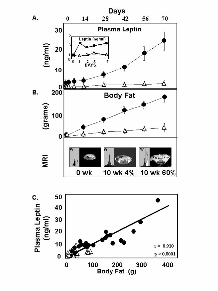
Mechanism of antisteatotic protection in liver: In the liver protection against
steatosis might involve, not only increased secretion of VLDL, but also enhanced FA
oxidation. In the latter case, an increase in the expression of PPARα and its target
enzymes, L-CPT-1 and ACO might be expected (30). To determine if the in vivo
protection against hepatic overaccumulation of TG in normal rats on a high fat diet is
mediated by this mechanism, we semiquantified PPARα protein and ACO and L-CPT-1
mRNAs in livers of normal rats receiving either a 60% or a 4% fat intake. PPARα protein
and L-CPT-1 mRNA were both significantly greater in the former group, but ACO
mRNA was not different (Figure 3A and B).
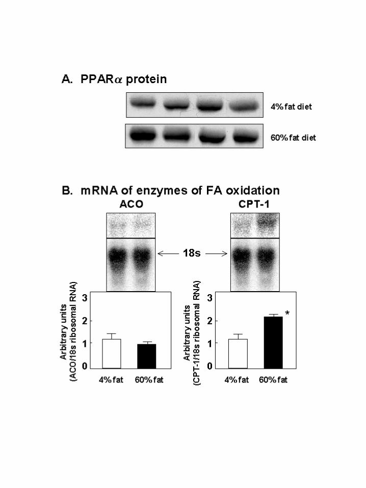
Mechanism of the antisteatotic protection in islets: Unlike liver, islets cannot
export excess FA, which may account for their vulnerability in obesity. To determine the
mechanism of the protection against lipid overaccumulation that prevails early in the
course of obesity, we measured the rate of oxidation of [3H]-palmitate in isolated islets of
Sprague-Dawley rats receiving either a 4% or 60% fat diet. Oxidation was 30% greater
in pancreatic islets of rats on the 60% fat diet than in controls on the 4% fat diet (Figure
4A). However, unlike in liver, no change in ACO or L-CPT-1 could be detected by
MPX-PCR (data not shown). These findings suggest that the preexisting oxidative
machinery of the islets was able to accommodate this increase in oxidation without an
increase in expression of genes encoding the enzymes.
by guest on February 1, 2018http://w
ww
.jbc.org/D
ownloaded from
11
We had previously reported that in the absence of leptin activity, as in fa/fa
ZDF rats, increased lipogenesis was the most important single factor in the ectopic
overaccumulation of lipids in islets (7,31). Accordingly, in normal rats the high fat diet
should not induce the increase in lipogenesis and lipogenic enzymes that had been
observed in fat-laden islets of the leptin-insensitive fa/fa rats. As shown in Figure 4B and
C, there was no increase in incorporation of 14C-glucose to lipids or in expression of
lipogenic enzymes; in fact, a small but significant decrease in lipogenesis and in FAS
mRNA was evident (Figure 4B and C). This was in sharp contrast to the ZDF fa/fa rats
in which lipogenesis was 2.5 times greater.
Ectopic TG deposition in the absence of leptin action: If the antilipogenic
protection observed in normal rats during caloric excess did, in fact, require the action of
the accompanying hyperleptinemia, rodent models with either a leptin deficiency (ob/ob
mice) or a loss-of-function mutation in their leptin receptors (db/db mice and ZDF fa/fa
rats) would be unprotected from lipid overaccumulation. We, therefore, measured the
plasma leptin levels (Figure 5A) and the TG content of islets, skeletal muscle, heart and
liver of these “unleptinized” rodents (Figure 5B). Although their diet contained only 6%
fat, the TG content of their nonadipose tissues ranged from ~4 to ~100-fold above normal
controls on the same diet. Thus, when leptin action is lacking, protection from lipid
overaccumulation in nonadipocytes is also lacking – even when the dietary fat intake is
normal.
Overexpression of wild-type OB-Rb in livers of ZDF fa/fa rats prevents
steatosis: If the marked hepatic steatosis and hypertriglyceridemia of obese ZDF fa/fa
rats is the result of lack of direct leptin action on the liver, transgenic overexpression of
the wild-type leptin receptor in the liver of these leptin-receptor-defective animals should
protect them. Therefore, we infused into 9-week-old ZDF fa/fa rats 1012 plaque-forming
units of recombinant adenovirus containing the cDNA of wild-type OB-Rb, the full-
length isoform of the leptin receptor (AdCMV-OB-Rb). AdCMV-β-galactosidase (β-gal)
by guest on February 1, 2018http://w
ww
.jbc.org/D
ownloaded from
12
was infused into age-matched ZDF fa/fa rats as a control. The wild-type OB-Rb
transgene introduced in vivo with an adenovirus vector was expressed exclusively in the
steatotic liver of the ZDF fa/fa rats (Figure 6A). None was detected in any other tissues,
including the hypothalamus.
One week after treatment with AdCMV-OB-Rb plasma TG levels of ZDF fa/fa
rats declined slightly to below pretreatment levels and remained significantly below the
controls for 3 weeks after AdCMV-OB-Rb treatment (Figure 5B). Liver TG content was
significantly below that of β-gal controls and untreated controls (Figure 5C), the result of
a delay in the increase in liver TG compared to the controls. TG content of heart and
skeletal muscle were unaffected (Figure 6B). Food intake in the two groups was identical
in the 2 adenovirus-treated groups (29.8 ± 1.4 g/d vs. 29.8 ± 1.5 g/d). Since the liver was
the only site of expression of the normal OB-Rb in these ZDF fa/fa rats and the only site
of antisteatotic action, we must assume that the elevated endogenous leptin levels, which
averaged 24 ± 2 ng/ml in AdCMV-OB-Rb-treated rats, and 28 ± 2 ng/ml in controls,
exerted a direct antisteatotic action on the liver. This strongly implies that the function of
hyperleptinemia of obesity is to prevent steatosis in tissues with functioning OB-Rb.
DISCUSSION
These findings support the concept that a physiologic role of leptin during
overnutrition is to confine storage of TG to adipocytes, cells specialized for this role, and
thus protect nonadipocytes from the adverse consequences of lipid overaccumulation.
This protection begins promptly at the start of overfeeding as the result of progressively
increasing hyperleptinemia that continues to rise for the duration of hypernutrition. This
appeared to minimize overaccumulation of lipids both by preventing the increase in
lipogenesis that occurs in the absence of leptin action (31), and through upregulation of
β-oxidative metabolism of the surplus fatty acids (7). Whereas in the liver there was an
increase in PPAR-α protein and CPT-1 mRNA, in pancreatic islets no such changes
by guest on February 1, 2018http://w
ww
.jbc.org/D
ownloaded from
13
could be detected, despite a 30% increase in the rate of 3H-palmitate oxidation. The
greater induction of FA β-oxidative enzymes in liver than in extrahepatic tissues confirms
a recent observation by Cook et al. (31).
In islets the antilipogenic action of hyperleptinemia appears to be at least as
important as the increase in FA oxidation in protecting islets from the lipid overload;
when leptin action is lacking, as in hyperphagic fa/fa ZDF rats, the fat-laden islets have
a high rate of 14C-glucose incorporation into lipids, in association with increased PPAR-γ,
ACC and FAS expression on a 6% fat intake (32). By contrast, in normal rats receiving
the 60% fat diet, these remained in the low normal range and the lipogenic rate declined.
When the antilipogenic effect of leptin is lacking, lipogenesis is excessive and cannot be
restrained by lipid excess (32).
The most compelling evidence in support of the antisteatotic role for leptin was
the in vivo demonstration in leptin-unresponsive fa/fa ZDF rats that transgenic
overexpression of the wild-type receptor in their livers prevented the severe hepatic
steatosis and hypertriglyceridemia that otherwise occurred. These findings are congruent
with earlier evidence of the antisteatotic action of recombinant leptin (33) and of
transplanted fat tissue in “fatless” mice with congenital lipodystrophy (34). Furthermore,
in our experiments the wild-type leptin receptors were expressed only in the liver and not
in the hypothalamus or anywhere else; it follows, therefore, that the endogenous
hyperleptinemia of those obese fa/fa rats must have acted directly via the transgenic OB-
Rb to prevent the lipid overaccumulation.
The prompt rise of plasma leptin levels on the very first day of the high fat diet,
and their high degree of correlation with the expanding body fat, are all consistent with
the response of an antilipogenic hormone with a physiologic liporegulatory mission,
namely to maintain FA homeostasis in nonadipocytes during overnutrition. This
protection may account for the fact that in hyperleptinemic rats and humans the lipotoxic
complications of diet-induced obesity do not appear until late in life, when leptin
by guest on February 1, 2018http://w
ww
.jbc.org/D
ownloaded from
14
effectiveness wanes (35,36). When leptin is absent, as in congenital generalized
lipodystrophy (33), or when leptin receptors are congenitally defective, as in ZDF rats,
these complications appear in severe form early in life.
It should be emphasized that we do not suggest that the direct antisteatotic activity
ascribed to the endogenous hyperleptinemia of obesity occurs in normal lean animals. It
appears to be a factor only during overnutrition when plasma leptin levels approach or
exceed the threshold for transport across the blood-brain barrier, which is probably in the
vicinity of 10 ng/ml (34). In the absence of overnutrition plasma levels are below 5
ng/ml and leptin action is presumed to be largely on the hypothalamic centers for control
of food intake and thermoregulation (35).
ACKNOWLEDGMENTS
The authors wish to thank Susan Kennedy for superb secretarial services. We
acknowledge the grant support of the Department of Veterans Affairs Institutional
Support (SMI 821-109), The National Institutes of Health (DK02700-37), The National
Institutes of Health/Juvenile Diabetes Foundation Diabetes Interdisciplinary Research
Program and Novo-Nordisk Corporation. We thank Cai Li, Ph.D., and Daniel Foster,
M.D. for critical review of this manuscript.
by guest on February 1, 2018http://w
ww
.jbc.org/D
ownloaded from
15 REFERENCES
1. Maffei, M., Halaas, J., Ravussin, E., Pratley, R.E., Lee, G.H., Zhang, Y., Fei, H.,
Kim, S., Lallone, R., Ranganathan, S., Kern, P.A., and Friedman, J.M. (1995)
Nature Med. 1, 1155-1161
2. Surwit, R.S., Edwards, C.L., Murthy, S., and Petro, A.E. (2000) Diabetes 49,
1203-1208
3. Neel, J.V. (1997) Nutr. Rev. 57, S2-S9
4. Halaas, J.L., Gajiwaia, K.S., Maffei, M., Cohen, S.L., Chalt, B.T., Rabinowitz,
D., Lallone, R.L., Burley, S.K., and Friedman, J.M. (1995) Science 269, 543-546
5. Unger, R.H., Zhou, Y.-T., and Orci, L. (1999) Proc. Natl. Acad. Sci. U.S.A. 96,
2327-2332
6. Shimabukuro, M., Koyama, K., Chen, G., Wang, M.-Y., Trieu, F., Lee, Y.,
Newgard, C.B., and Unger, R.H. (1997) Proc. Natl. Acad. Sci. U.S.A. 94, 4637-
4641
7. Lee, Y., Hirose, H., Zhou, Y.-T., Esser, V., McGarry, J.D., and Unger, R.H.
(1997) Diabetes 46, 408-413
8. Lee, Y., Hirose, H., Ohneda, M., Johnson, J.H., McGarry, J.D., and Unger, R.H.
(1994) Proc. Natl. Acad. Sci. U.S.A. 91, 10878-10882
9. Zhou, Y.-T., Grayburn, P., Karim, A., Shimabukuro, M., Higa, M., Baetens, D.,
Orci, L., and Unger, R.H. (2000) Proc. Natl. Acad. Sci. U.S.A. 97, 1784-1789
10. Iida, M., Murakami, T., Ishida, K., Mizuno, A., Kuwajima, M., and Shima, K.
(1996) Biochem. Biophys. Res. Comm. 224, 597-604
11. Phillips, M.S., Liu, O., Hammond, H., Dugan, V., Hey, P., Caskey, C.T., and
Hess, J.F. (1996) Nat. Genet. 13, 18-19
by guest on February 1, 2018http://w
ww
.jbc.org/D
ownloaded from
16 12. Hirose, H., Lee, Y.H., Inman, L.R., Nagasawa, Y., Johnson, J.H., and Unger,
R.H. (1996) J. Biol. Chem. 271, 5633-5637
13. Wang, M.-Y., Koyama, K., Shimabukuro, M., Newgard, C., and Unger, R.H.
(1998) Proc. Natl. Acad. Sci. U.S.A. 95, 714-718
14. McGarry, J.D. (1992) Science 258, 766-770
15. Unger, R.H. (1998) Trends in Endocrino. Metab, 7, 276-282
16. Shimabukuro, M., Zhou, Y.-T., Levi, M., and Unger, R.H. (1998) Proc. Natl.
Acad. Sci. U.S.A. 95, 2498-2502
17. Obeid, L.M., Linardic, C.M., Karolak, L.A., and Hannun, Y.A. (1993) Science
259, 1769-1771
18. Vincent, H.K., Powers, S.K., Stewart, D.J., Shanely, R.A., Demirel, H., and
Naito, H. (1999) Int. J. Obes. Relat. Metab. Disord. 23, 67-74
19. Zhou, Y.-T., Wang, Z.-W., Higa, M., Newgard, C.B., and Unger, R.H. (1999)
Proc. Natl. Acad. Sci. U.S.A. 96, 2391-2395
20. Chen, G.X., Koyama, K., Yuan, X., Lee, Y., Zhou, Y.-T., O’Doherty, R.,
Newgard, C.B., and Unger, R.H. (1996) Proc. Natl. Acad. Sci. U.S.A. 93, 14795-
14799
21. Kakuma, T., Lee, Y., Higa, M., Wang, Z.-W., Pan, W., and Unger, R.H. (2000)
Proc. Natl. Acad. Sci. U.S.A. 97, 8536-8541
22. Jensen, J., Serup, P., Karlsen, C., Nielsen, T.F., and Madsen, O.D. (1996) J. Biol.
Chem. 271, 18749-18758
23. O’Doherty, R.M., Jensen, P.B., Anderson, P., Jones, J.G., Berman, H.K.,
Kearney, D., and Newgard, C.B. (2000) J. Clin. Invest. 105, 479-488
24. Stein, D.T., Babcock, E.E., Malloy, C.R., and McGarry, J.D. (1995) Int. J.
by guest on February 1, 2018http://w
ww
.jbc.org/D
ownloaded from
17 Obesity 19, 804-810
25. Folch, J., Lees, M., and Stanley, G.H.S. (1957) J. Biol. Chem. 226, 497-509
26. Danno, H., Jicho, Y., Budiyanto, S., Furukawa, Y., and Kimura, S. (1992) J. Nutr.
Sci. Vitaminol. 38, 517-521
27. Milburn, J.L., Hirose, H., Lee, Y.H., Nagasawa, Y., Ogawa, A., Ohneda, M.,
BeltrandelRio, H., Newgard, C.B., Johnson, J.H., and Unger, R.H. (1995) J. Biol.
Chem. 270, 1295-1299
28. Chen, S., Ogawa, A., Ohneda, M., Unger, R.H., and McGarry, J.D. (1994)
Diabetes 43, 878-883
29. Bligh, E.G., and Dyer, W.J. (1959) Can. J. Biochem. Physiol. 37, 911-917
30. Zhou, Y.-T., Shimabukuro, M., Wang, M.-Y., Lee, Y., Higa, M., Milburn, J.L.,
Newgard, C.B., and Unger, R.H. (1998) Proc. Natl. Acad. Sci. U.S.A. 95, 8898-
8903
31. Cook, W.S., Yelandi, B.V., Ras, M.S., Hashimoto, T., and Reddy, K. (2000)
Biochem. Biophys. Res. Commun. 278, 250-257
32. Zhou, Y.-T., Shimabukuro, M., Lee, Y., Koyama, K., Higa, M., Ferguson, T., and
Unger, R.H. (1998) Diabetes 49, 1904-1908
33. Shimomura, I., Hamner, R.E., Ikemoto, S., Brown, M.S. and Goldstein, J.L.
(1999) Nature 401, 73-76
34. Gavrilova, O., Marcus-Samuels, B., Graham, D., Kim, J.K., Shulman, G.I.,
Castle, A.L., Venson, C., Eckhaus, M., and Reitman, M.L. (2000) J. Clin. Invest.
105, 271-278
35. Qian, H., Azain, M.J., Hartzell, D.L., and Baile, C.A. (1998) Proc. Soc. Exp. Biol.
Med. 219, 160-165
by guest on February 1, 2018http://w
ww
.jbc.org/D
ownloaded from
18 36. Scarpace, P., Matheny, M., Moore, R.L., and Tumer, N. (2000) Diabetes 49,
431-435
37. Wang, Z.-W., Zhou, Y.-T., Kakuma, T., Lee, Y., Higa, M., Kalra, S.P., Dube,
M.G., Kalra, P.S., Unger, R.H. (1999) Proc. Natl. Acad. Sci. U.S.A. 96, 10373-
10378
38. Friedman, J.M. (2000) Nature 404, 632-634
by guest on February 1, 2018http://w
ww
.jbc.org/D
ownloaded from

19 FIGURE LEGENDS:
Figure 1: Relationship of plasma leptin to total body fat in Sprague-Dawley rats
receiving diets with a fat content of 4% ! or 60% !. A) Plasma leptin
levels in the two groups; the inset provides a view of the daily leptin
profile for the first week of the study. B) Total body fat measured by
MRS. Representative spectral tracings and transrenal images are
displayed. C) The relationship between plasma leptin levels and body fat.
Figure 2: A) Comparison of the mean (± S.E.M.) plasma leptin, TG and FFA levels
in normal Sprague-Dawley rats fed a diet containing either 4% " (N=6)
or 60% (N=6) fat for 10 weeks. *p<0.001 B) The mean (± S.E.M.)
tissue TG content of 6 normal Sprague-Dawley rats on a 60% fat diet at 4
weeks of age before starting the high fat diet " and at 14 weeks of age
after 10 weeks on the 60% fat intake . TG content in islets is expressed
as ng/islet; in liver, heart and skeletal muscle, it is expressed as mg/g of
wet weight of tissue. Body fat, as determined by MRS, is expressed as
g/animal. *p<0.001
Figure 3: Mechanism of antisteatotic protection of liver during high fat feeding of
normal rats. A) PPARα protein measured by immunoprecipitation in liver
of 4 rats fed a diet containing 4% fat and 4 rats fed a 60% fat diet. B)
mRNA of enzymes of fatty acid β-oxidation, ACO and L-CPT-1,
quantified by northern hybridization. *p<0.01
Figure 4: Mechanism of antisteatotic protection of islets during high fat feeding of
normal rats. A) Comparison of rates of oxidation of [H3]-palmitate in rats
receiving a diet containing either 4% ! or 60% fat. *p<0.01. B)
by guest on February 1, 2018http://w
ww
.jbc.org/D
ownloaded from
20 Comparison of the rates of incorporation of [U-14C]-glucose into lipids
in normal Sprague-Dawley (SD) rats on a 4% ! or 60% fat intake.
These are significantly less than in islets of hyperphagic fa/fa ZDF rats on
a 6% fat intake . *p<0.01; §p<0.05. C) Comparison of expression of the
lipogenic enzymes acetyl CoA carboxylase (ACC), fatty acid synthase
(FAT) and glycerol-PO4 acyltransferase (GPAT) in islets of normal
Sprague-Dawley (SD) rats fed either 4% or 60% fat diet. As an internal
control for enzyme mRNA, TATA box-binding protein (TBP) was
employed. *p<0.005; ¶p<0.01; §p<0.05.
Figure 5: A) Comparison of mean (± SEM) plasma leptin, TG and FFA levels in
rodents that are either leptin-deficient (ob/ob mice) or unresponsive to
leptin because of loss-of-function mutation in its receptor gene (db/db
mice and fa/fa rats) and the corresponding wild-type (+/+) controls !.
*p<0.01; **p<0.001 B) Comparison of TG content in tissues of these
rodents. All animals received a diet containing 6% fat. *p<0.001
Figure 6: A) Effect of intravenous infusion of obese ZDF fa/fa rats with AdCMV-
OB-Rb or AdCMV-β-gal on the expression of wild-type receptor in the
liver and hypothalamus. B) Polymerase chain reaction-restriction
fragment length polymorphism (PCR-RFLP) of OB-Rb in liver and
hypothalamus of a lean wild-type (+/+) ZDF rat, an untreated obese (fa/fa)
ZDF rat and an obese fa/fa rat 4 weeks after treatment with either
AdCMV-βgal or AdCMV-leptin. Whereas in the untreated and AdCMV-
βgal-treated fa/fa rat only mutated OB-Rb is present, in the AdCMV-
leptin-treated fa/fa rat the OB-Rb matches that of the +/+ rat. C) Plasma
by guest on February 1, 2018http://w
ww
.jbc.org/D
ownloaded from
21 TG after AdCMV-OB-Rb " or AdCMV-βgal . **p<0.01. D)
Triacylglycerol (TG) content of liver in obese ZDF fa/fa rats after
treatment with AdCMV-OB-Rb # or AdCMV-βgal . *p<0.05. E)
Heart TG and skeletal muscle TG after AdCMV-OB-Rb # or AdCMV-
βgal . TG content in tissues of lean ZDF +/+ rats ! are shown for
comparison.
by guest on February 1, 2018http://w
ww
.jbc.org/D
ownloaded from
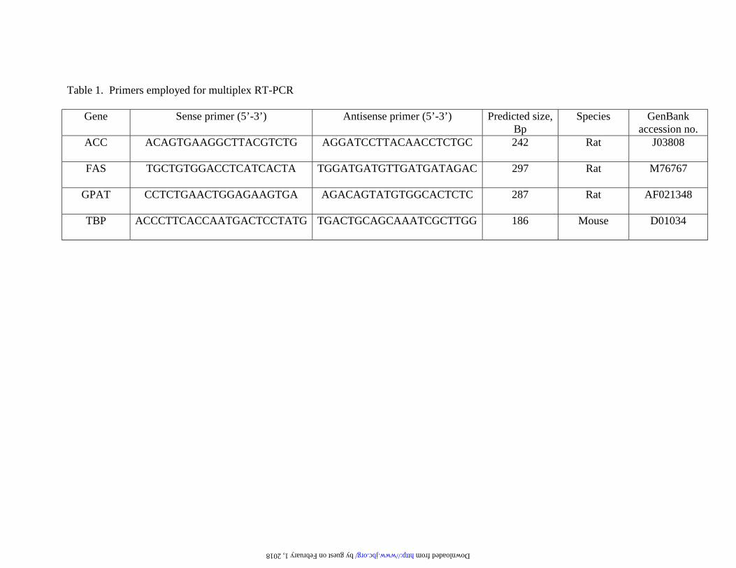
Table 1. Primers employed for multiplex RT-PCR
Gene Sense primer (5’-3’) Antisense primer (5’-3’) Predicted size, Bp
Species GenBank accession no.
ACC ACAGTGAAGGCTTACGTCTG AGGATCCTTACAACCTCTGC 242 Rat J03808
FAS TGCTGTGGACCTCATCACTA TGGATGATGTTGATGATAGAC 297 Rat M76767
GPAT CCTCTGAACTGGAGAAGTGA AGACAGTATGTGGCACTCTC 287 Rat AF021348
TBP ACCCTTCACCAATGACTCCTATG TGACTGCAGCAAATCGCTTGG 186 Mouse D01034
by guest on February 1, 2018 http://www.jbc.org/ Downloaded from
McCorkle, Moritake Higa, Yan-Ting Zhou and Roger H. UngerYoung Lee, May-Yun Wang, Tetsuya Kakuma, Zhuo-Wei Wang, Evelyn Babcock, Kay
Liporegulation in diet-induced obesity: The antisteatotic role of hyperleptinemia
published online November 28, 2000J. Biol. Chem.
10.1074/jbc.M008553200Access the most updated version of this article at doi:
Alerts:
When a correction for this article is posted•
When this article is cited•
to choose from all of JBC's e-mail alertsClick here
by guest on February 1, 2018http://w
ww
.jbc.org/D
ownloaded from