
Lipoprotein(a)-Associated Molecules Are Prominent...
12
CLINICAL RESEARCH Lipoprotein(a)-Associated Molecules Are Prominent Components in Plasma and Valve Leaflets in Calcific Aortic Valve Stenosis Michael Torzewski, MD, a Amir Ravandi, MD, PHD, b Calvin Yeang, MD, PHD, c Andrea Edel, PHD, b Rahul Bhindi, MD, b Stefan Kath, MD, a Laura Twardowski, MD, a Jens Schmid, PHD, c Xiaohong Yang, BS, d Ulrich F.W. Franke, MD, e Joseph L. Witztum, MD, f Sotirios Tsimikas, MD d VISUAL ABSTRACT Torzewski, M. et al. J Am Coll Cardiol Basic Trans Science. 2017;2(3):229–40. HIGHLIGHTS The LPA gene is the only monogenetic risk factor for CAVS, and OxPL and lysophosphatidic acid, generated by autotaxin from OxPL, are pro-inflammatory. Both autotaxin–apolipoprotein B and autotaxin–apo(a) were measureable in plasma. Immunohistochemistry revealed a strong presence of apo(a), OxPL, malondialdehyde-lysine, autotaxin, and macrophages, particularly in advanced lesions rich in cholesterol crystals and calcification. Six species of OxPL and lysophosphatidic acid, with aldehyde-containing phosphocholine-based OxPL most abundant, were identified and quantified after extraction from valve leaflets. We demonstrate the presence of a constellation of pathologically linked, Lp(a)-associated molecules in plasma and in aortic valve leaflets of patients with CAVS. These data are consistent with the hypothesis that Lp(a) is a key etiologic factor in patients with CAVS. From the a Department of Laboratory Medicine, Robert-Bosch-Hospital, Stuttgart, Germany; b Cardiac Sciences Program, University of Manitoba and Institute of Cardiovascular Sciences, St. Boniface Hospital, Winnipeg, Canada; c Dr. Margarete Fischer-Bosch Institute of Clinical Pharmacology and University of Tuebingen, Stuttgart, Germany; d Division of Cardiovascular JACC: BASIC TO TRANSLATIONAL SCIENCE VOL. 2, NO. 3, 2017 ª 2017 THE AUTHORS. PUBLISHED BY ELSEVIER ON BEHALF OF THE AMERICAN COLLEGE OF CARDIOLOGY FOUNDATION. THIS IS AN OPEN ACCESS ARTICLE UNDER THE CC BY-NC-ND LICENSE ( http://creativecommons.org/licenses/by-nc-nd/4.0/ ). ISSN 2452-302X http://dx.doi.org/10.1016/j.jacbts.2017.02.004
-
Upload
nguyenthuan -
Category
Documents
-
view
221 -
download
2
Transcript of Lipoprotein(a)-Associated Molecules Are Prominent...

J A C C : B A S I C T O T R A N S L A T I O N A L S C I E N C E V O L . 2 , N O . 3 , 2 0 1 7
ª 2 0 1 7 T H E A U T HO R S . P U B L I S H E D B Y E L S E V I E R O N B E H A L F O F T H E A M E R I C A N
C O L L E G E O F C A R D I O L O G Y F O U N D A T I O N . T H I S I S A N O P E N A C C E S S A R T I C L E U N D E R
T H E C C B Y - N C - N D L I C E N S E ( h t t p : / / c r e a t i v e c o mm o n s . o r g / l i c e n s e s / b y - n c - n d / 4 . 0 / ) .
I S S N 2 4 5 2 - 3 0 2 X
h t t p : / / d x . d o i . o r g / 1 0 . 1 0 1 6 / j . j a c b t s . 2 0 1 7 . 0 2 . 0 0 4
CLINICAL RESEARCH
Lipoprotein(a)-Associated MoleculesAre Prominent Components inPlasma and Valve Leaflets inCalcific Aortic Valve Stenosis
Michael Torzewski, MD,a Amir Ravandi, MD, PHD,b Calvin Yeang, MD, PHD,c Andrea Edel, PHD,b Rahul Bhindi, MD,bStefan Kath, MD,a Laura Twardowski, MD,a Jens Schmid, PHD,c Xiaohong Yang, BS,d Ulrich F.W. Franke, MD,e
Joseph L. Witztum, MD,f Sotirios Tsimikas, MDd
VISUAL ABSTRACT
F
U
F
Torzewski, M. et al. J Am Coll Cardiol Basic Trans Science. 2017;2(3):229–40.
rom the aDepartment of Laboratory Medicine, Robert-Bosch-Hospital, Stuttgart, Germ
niversity of Manitoba and Institute of Cardiovascular Sciences, St. Boniface Hospital, W
ischer-Bosch Institute of Clinical Pharmacology and University of Tuebingen, Stuttgart, Ger
HIGHLIGHTS
� The LPA gene is the only monogenetic
risk factor for CAVS, and OxPL and
lysophosphatidic acid, generated by
autotaxin from OxPL, are
pro-inflammatory.
� Both autotaxin–apolipoprotein B and
autotaxin–apo(a) were measureable in
plasma.
� Immunohistochemistry revealed a strong
presence of apo(a), OxPL,
malondialdehyde-lysine, autotaxin, and
macrophages, particularly in advanced
lesions rich in cholesterol crystals and
calcification.
� Six species of OxPL and lysophosphatidic
acid, with aldehyde-containing
phosphocholine-based OxPL most
abundant, were identified and quantified
after extraction from valve leaflets.
� We demonstrate the presence of a
constellation of pathologically linked,
Lp(a)-associated molecules in plasma and
in aortic valve leaflets of patients with
CAVS. These data are consistent with the
hypothesis that Lp(a) is a key etiologic
factor in patients with CAVS.
any; bCardiac Sciences Program,
innipeg, Canada; cDr. Margarete
many; dDivision of Cardiovascular

ABBR EV I A T I ON S
AND ACRONYMS
apo(a) = apolipoprotein(a)
apoB = apolipoprotein B
ATX = autotaxin
AVR = aortic valve replacement
CAVS = calcific aortic valve
stenosis
IgG = immunoglobulin G
Lp(a) = lipoprotein(a)
LysoPA = lysophosphatidic acid
LysoPC =
lysophosphatidylcholine
MDA = malondialdehyde
OxPL = oxidized phospholipid
OxPL-apo(a) = oxidized
phospholipid on apolipoprotein(a)
OxPL-apoB = oxidized
phospholipid on
apolipoprotein B-100
PC-OxPL = phosphocholine-
containingoxidizedphospholipids
RLU = relative light unit
Medicine, S
Surgery, Ro
San Diego,
Bosch Fou
HL119828,
co-invento
antibodies
and Ionis P
other auth
All authors
institution
informatio
Manuscript
Torzewski et al. J A C C : B A S I C T O T R A N S L A T I O N A L S C I E N C E V O L . 2 , N O . 3 , 2 0 1 7
Lp(a)-OxPL-ATX and CAVS J U N E 2 0 1 7 : 2 2 9 – 4 0
230
SUMMARY
ulp
be
La
nda
P0
rs o
use
har
ors
at
s a
n, v
re
The LPA gene is the only monogenetic risk factor for calcific aortic valve stenosis (CAVS). Oxidized phospho-
lipids (OxPL) and lysophosphatidic acid generated by autotaxin (ATX) from OxPL are pro-inflammatory. Aortic
valve leaflets categorized pathologically from both ATX–apolipoprotein B and ATX–apolipoprotein(a) were
measureable in plasma. Lipoprotein(a) (Lp[a]), ATX, OxPL, and malondialdehyde epitopes progressively
increased in immunostaining (p < 0.001 for all). Six species of OxPL and lysophosphatidic acid were identified
after extraction from valve leaflets. The presence of a constellation of pathologically linked, Lp(a)-associated
molecules in plasma and in aortic valve leaflets of patients with CAVS suggest that Lp(a) is a key etiologic factor
in CAVS. (J Am Coll Cardiol Basic Trans Science 2017;2:229–40) © 2017 The Authors. Published by Elsevier on
behalf of the American College of Cardiology Foundation. This is an open access article under the CC BY-NC-ND
license (http://creativecommons.org/licenses/by-nc-nd/4.0/).
W ith the extension of lifespan andaging of the population in the21st century, calcific aortic valve
disease (1), which encompasses early lesionsas well as stenotic disease (clinically seen ascalcific aortic valve stenosis [CAVS]), isbecoming an increasingly prevalent disorder.It is estimated that hemodynamically signifi-cant CAVS is present in >1 million patients in
the United States and that approximately 2.5 millioncases will be present worldwide by 2020 and 4.5 millionin 2030 (2). Aortic valve replacement (AVR) is the onlyoption for palliation of symptoms and prevention of com-plications. Unfortunately, by the time AVR is needed,patients are often octogenarians and a large percentageare not eligible for AVR due to advanced frailty and othersignificant comorbidities. For example, a recent reportcomparing surgical AVR versus transcatheter AVRrevealed 33% to 39% all-cause mortality, 12% to 19%rate of stroke, and 40% to 48% major adverse cardiacevent rate at 3-year follow-up in both groups (3).
SEE PAGE 241
Lipoprotein(a) (Lp[a]) is a genetically determined,likely causal, and independent risk factor for thepresence and progression of CAVS (4–8). Lp(a) is
izio Cardiovascular Center, University of California San Diego
rt-Bosch-Hospital, Stuttgart, Germany; and the fDivision of End
Jolla, California. This work was supported by the innovation
tion. Drs. Tsimikas and Witztum are supported by grants
1-HL088093, P01 HL055798, R01-HL106579, R01-HL078610, a
f and receive royalties from patents or patent applications o
d in biotheranostic applications. Dr. Tsimikas has a dual app
maceuticals, Inc. Dr. Witztum has received honoraria for con
have reported that they have no relationships relevant to th
test they are in compliance with human studies committe
nd Food and Drug Administration guidelines, including
isit the JACC: Basic to Translational Science author instruct
ceived December 21, 2016; revised manuscript received Febru
the major lipoprotein carrier of phosphocholine-containing oxidized phospholipids (PC-OxPLs) (9,10)that may contribute to inflammation and inducecalcification in valvular cells (11,12). For example, inthe ASTRONOMER (Aortic Stenosis ProgressionObservation: Measuring Effects of Rosuvastatin) trial(4), patients with elevated Lp(a) and OxPL on apoli-poprotein B-100 (OxPL-apoB) levels had the fastestprogression rate and higher need for AVR. Autotaxin(ATX), which breaks down lysophosphatidylcholine(LysoPC) derived from OxPL to lysophosphatidic acid(LysoPA), was also recently shown to be stronglyassociated with CAVS (13). In turn, because OxPL andoxidized low-density lipoprotein have been shown tobe present in CAVS (13–15), it is likely that a significantportion of the LysoPC is derived from hydrolysis of thetruncated oxidized sn2 fatty acids found in OxPL pre-sent in oxidized low-density lipoprotein, which aregenerated by such enzymes as lipoprotein-associatedphospholipase A2 and platelet-activating factor ace-tylhydrolase. In recent findings, ATX activity andeither Lp(a) or OxPL-apoB strongly interacted to pre-dict the presence of CAVS in patients undergoing AVRwith concomitant coronary artery disease (8).
There are no effective medical therapies to preventthe development or progression of CAVS, and statins
, La Jolla, California; eDepartment of Cardiovascular
ocrinology and Metabolism, University of California
fund of the Robert-Bosch-Hospital and the Robert
from the National Institutes of Health (R01 grants
nd R01-HL124174). Drs. Tsimikas and Witztum are
wned by the University of California San Diego on
ointment at the University of California San Diego
sulting for Ionis, CymaBay, and Prometheus Inc. All
e contents of this paper to disclose.
es and animal welfare regulations of the authors’
patient consent where appropriate. For more
ions page.
ary 1, 2017, accepted February 1, 2017.

FIGURE 1 ELISAs to Measure ATX-Lp(a) and ATX-apoB
Methodology of novel chemiluminescent enzyme-linked immunoadsorbent assay (ELISA)
to measure autotaxin (ATX)–apolipoprotein(a) (apo[a]) and ATX–apolipoprotein B (apoB)
complexes. ApoB-100 and lipoprotein(a) (Lp[a]) lipoproteins were captured from plasma
with specific antibodies bound to microtiter well plates. Conditions were established so
that the added plasma contains saturating amounts of lipoproteins to be captured. The
content of ATX mass on each lipoprotein was then detected with a goat anti-human
ATX antibody.
J A C C : B A S I C T O T R A N S L A T I O N A L S C I E N C E V O L . 2 , N O . 3 , 2 0 1 7 Torzewski et al.J U N E 2 0 1 7 : 2 2 9 – 4 0 Lp(a)-OxPL-ATX and CAVS
231
have failed to reduce progression of AVR in 4 ran-domized trials (16–20). Statins additionally maysignificantly raise plasma Lp(a) and OxPL-apoB (21),which may be counterproductive in preventing CAVS.For example, rosuvastatin raised Lp(a) levels 20% inthe ASTRONOMER trial (4). However, novel Lp(a)-lowering agents may be used in the near future totest the hypothesis that lowering Lp(a) reducesprogression of CAVS (22–25).
The objective of this study was 3-fold: 1) to developplasma measures of autotaxin carried by apoB andLp(a); 2) to define lysophophatidic and phosphocho-line-containing oxidized phospholipids within aorticvalve leaflets; and 3) to document the presence ofLp(a) and oxidation-specific epitopes in aortic valveleaflets obtained following aortic valve replacement.
METHODS
A variety of techniques were used to study patientswith mild to moderate CAVS and severe CAVSundergoing AVR. These techniques included novelenzyme-linked chemiluminescent assay of plasmacomponents, as well as immunohistochemistry andliquid chromatography-tandem mass spectroscopy ofextracts of aortic valve leaflets. Full details arepresented in the Supplemental Appendix.
ANTIBODIES TO Lp(a), ATX, AND OXIDATION-SPECIFIC
EPITOPES. LPA4 is a murine monoclonal immuno-globulin G (IgG) antibody to apo(a) that was generatedby immunizing mice with the apo(a) sequenceTRNYCRNPDAEIRP. E06 is a natural immunoglobulinM murine monoclonal antibody that binds to thephosphocholine head group of oxidized but notnative phospholipids. MDA2 is a murine IgG mono-clonal antibody that recognizes malondialdehyde-modified proteins and lipid adducts. An alkalinephosphatase–labeled goat anti-human ATXpolyclonal antibody was purchased from LifeTechnologies (Carlsbad, California).
NOVEL CHEMILUMINESCENT ENZYME-LINKED
IMMUNOADSORBENT ASSAY TO DETECT
LIPOPROTEIN-ASSOCIATED ATX. A sensitive andquantitative sandwich-based chemiluminescentenzyme-linked immunoadsorbent assay was used tomeasure ATX associated with plasma lipoproteinscontaining apolipoprotein B (apoB)-100, whichincludes apoB on very low density lipoprotein,intermediate-density lipoprotein, low-density lipo-protein, and Lp(a) (ATX-apoB) and also specificallyonly on Lp(a) (ATX-apo[a]) (Figure 1). Microtiter96-well plates were coated overnight at 4�C with an-tibodies MB47 to bind apoB-100 and LPA4 to bindLp(a) (all at 5 mg/ml antigen of 40 ml/well). Conditions
were established to ensure that the amount of plasmaadded was sufficient to provide a saturating and equalamount of each lipoprotein captured in each well.Excess material was washed off and the plates blockedwith 1% tris buffered saline/bovine serum albuminfor 45 min. After the plates were washed, ethyl-enediaminetetraacetic acid plasma was added at 1:50dilution (40 ml/well) for 75min to bind apoB-100, Lp(a),and high-density lipoprotein, respectively. After theplates were again washed, goat anti-human ATXantibody at 1 mg/ml (40 ml/well) was incubated with theplates for 60min. After washing excessmaterial off theplates, alkaline phosphatase–labeled goat anti-rabbitIgG (Sigma, St. Louis, Missouri) (40 ml/well) wasadded for 60min. After a final washing, Lumi-phos 530(Lumigen, Inc., Southfield, Michigan) (25 ml/well) wasadded for 75 min and luminescence read on a Dynexluminometer (Chantilly, Virginia). The results arereported as relative light units (RLUs) in 100 ms afterthe background (tris buffered saline/bovine serumalbumin) RLUs are subtracted. High and low valueswere added to each 96-well plate as internal controls.
PATIENTS WITH CAVS. The first group of patientscomprised 14 patients with mild to moderate CAVSwho had available ethylenediaminetetraacetic acidplasma samples stored frozen at –70�C. The second

TABLE 1 Baseline Clinical, Laboratory and Echocardiography
Variables of the CAVS Study Group
Age, yrs 76 (54–86)
BMI, kg/m2 27.3 (20.2–40.5)
Smoking
Never 41 (73.2)
Past 10 (17.9)
Present 5 (8.9)
Hypertension
No 5 (7.5)
Yes 62 (92.5)
Diabetes
No 49 (72.1)
Yes 19 (27.9)
Total cholesterol, mg/dl 183 (114–297)
LDL-C, mg/dl 103 (56–220)
Creatinine, mmol/l 79.6 (53.0–176.8)
hsCRP, mg/l 2.0 (0.0–29.0)
HbA1c, %Hb 5.8 (4.6–9.7)
NT-proBNP, pg/ml 547 (21–19,799)
Mean aortic valve gradient, mm Hg* 51.0 (14.9)
Mean aortic valve area, cm2† 0.71 (0.19)
Values are median (min–max) or n (%). *Only 26 patients had available echocar-diography data. †Only 36 patients had available echocardiography data.
CAVS ¼ calcific aortic valve stenosis; HbA1c ¼ glycosylated hemoglobin;hsCRP ¼ high-sensitivity C-reactive protein; LDL-C ¼ low-density lipoproteincholesterol; NT-proBNP ¼ N-terminal pro–B-type natriuretic peptide.
FIGURE 2 Correlation Between ATX-Apo(a) and ATX-apoB
Spearman correlation between plasma levels of ATX-apoB and
ATX-apo(a) complexes in patients with mild to moderate
calcific aortic valve stenosis. RLU ¼ relative light units; other
abbreviations as in Figure 1.
Torzewski et al. J A C C : B A S I C T O T R A N S L A T I O N A L S C I E N C E V O L . 2 , N O . 3 , 2 0 1 7
Lp(a)-OxPL-ATX and CAVS J U N E 2 0 1 7 : 2 2 9 – 4 0
232
group comprised 68 patients undergoing AVR forsymptomatic CAVS with prospective collection ofaortic valve leaflets and were recently described (26).Formalin-fixed nonrheumatic aortic valves withvarying degrees of macroscopic disease were analyzedfor the presence of apolipoprotein(a) (apo[a]), ATX,oxidation-specific epitopes, and macrophages. Thethird group comprised 4 patients who underwent AVRand liquid chromatography-tandem mass spectros-copy analysis of valve leaflets for the presence of spe-cific species of OxPL, and LysoPA was performed.
HISTOLOGICAL AND IMMUNOHISTOCHEMICAL ANALYSIS
OF VALVE LEAFLETS. Although all patients had a clin-ical diagnosis of CAVS, there was pathological vari-ability in the extent of involvement of the aortic valveleaflets. For this reason, valve leaflets were classifiedpathologically as grade 1 to 4, with a pathologicalclassification as previously described (26,27). In addi-tion, a semi-quantitative method was used to deter-mine the proportion of area stained by each antibody.The proportion of the areas stained for each epitoperelative to the area occupied by the various patholog-ical grades of CAVSwas estimated and assigned to 1 of 5scores: 0, <5%; 1, 6% to 25%; 2, 26% to 50%; 3, 51% to75%; or 4, 76% to 100%. A single aortic valve specimenusually comprises >1 pathological grade.
RESULTS
PRESENCE OF ATX IN CIRCULATING PLASMA IN
PATIENTS WITH MILD TO MODERATE CAVS. Lipo-protein-associated ATX, measured as ATX-apoB and
ATX-apo(a), were quantitated in 14 patients with mildto moderate CAVS. The Vpeak was 3.30 � 0.48 m/s,mean and peak gradients were 24.4 � 8.0 mm Hg and44.9 � 12.7 mm Hg, respectively, and aortic valve areawas 1.32 � 0.34 cm2. ATX-apoB and ATX-apo(a) couldbe directly measured on apoB and Lp(a), respectively,and they exhibited a modest correlation (r ¼ 0.58;p ¼ 0.028) (Figure 2). There were no significant cor-relations of ATX-apoB or ATX-apo(a) with Lp(a) andOxPL-apoB or oxidized phospholipid on apolipopro-tein(a) (OxPL-apo[a]) (not shown).
CLINICAL CHARACTERISTICS OF PATIENTS WITH
CAVS. Table 1 presents the clinical characteristics ofthe 68 patients with CAVS undergoing AVR. Baselinecharacteristics included 26.8% former or currentsmokers, 92.5% with hypertension, and 27.9% withdiabetes. Levels of low-density lipoprotein choles-terol tended to be in the normal range; Lp(a) levelswere not available because they are not generallymeasured in patients with CAVS. CAVS was severe,with a mean gradient of 51.0 � 14.87 mm Hg and anaortic valve area of 0.71 � 0.19 cm2.
PRESENCE OF apo(a), OXIDATION-SPECIFIC EPITOPES,
MACROPHAGES, AND CALCIFICATION IN AORTIC VALVE
LEAFLETS IN SEVERE CAVS. Aortic valve leaflet lesionsfrom 68 patients fulfilling the pathological criteria ofgrades 1 to 4 were examined. Figures 3 through 6

FIGURE 3 Presence of Apolipoprotein(a), Oxidation-Specific Epitopes, and Autotaxin in Grade 1 of Aortic Valve Disease
Sequential sections stained for calcification with alizarin red S and macrophages (CD68) (upper panel), apolipoprotein(a) (LPA4) and autotaxin
(ATX) (middle panel) as well as oxidized phospholipids (EO6) and malondialdehyde epitopes (MDA2) (lower panel). Note the lack of calcified
areas as well as the predominantly extracellular co-localization of the different antigens. In all panels, the aortic side of the valve is at the top.
J A C C : B A S I C T O T R A N S L A T I O N A L S C I E N C E V O L . 2 , N O . 3 , 2 0 1 7 Torzewski et al.J U N E 2 0 1 7 : 2 2 9 – 4 0 Lp(a)-OxPL-ATX and CAVS
233
display representative examples of grades 1 to 4 ofaortic valve calcification, as detected by alizarin redstaining and structural damage (26,27). With the useof the specific monoclonal antibody LPA4, apo(a) wasdetectable in every lesion examined.
Grade 1 lesions were characterized by a lack ofcalcified areas, and a predominant focal deposition ofapo(a) was present in the fibrosa (Figure 3). ATX, aswell as OxPL epitopes, stained by antibody E06, wereless evident but where present, they did co-localizewith apo(a). MDA-lysine epitopes, stained by anti-body MDA2, were diffusely present in the leaflet, butmacrophages stained by antibody to CD68 weresparse.
Grade 2 lesions were characterized by early devel-opment of calcified nodules and a more prominentdeposition of apo(a) in the fibrosa as well as in thespongiosa (Figure 4). ATX was present in areas of
apo(a) staining. OxPL epitopes as well as MDA-lysineepitopes were also more prominent than in grade1 but less evident than apo(a).
Grade 3 lesions were characterized by largecalcified nodules and/or cholesterol crystal deposits,and a more abundant deposition of apo(a) as well asATX compared with grade 1 and 2 lesions, mainlyaround cholesterol crystal deposits (Figure 5)and/or calcified areas (not shown). OxPL and MDA-lysine epitopes were much less prominent thanapo(a).
Grade 4 lesions were characterized by significantfibrosis and calcification grossly destroying thestructural integrity of the valvular cusp, as well asmore abundant localization of apo(a) as well asATX, throughout the lesions, including aroundcalcified areas (Figure 6). OxPL epitopes in grade 4lesions were highly co-localized with apo(a)
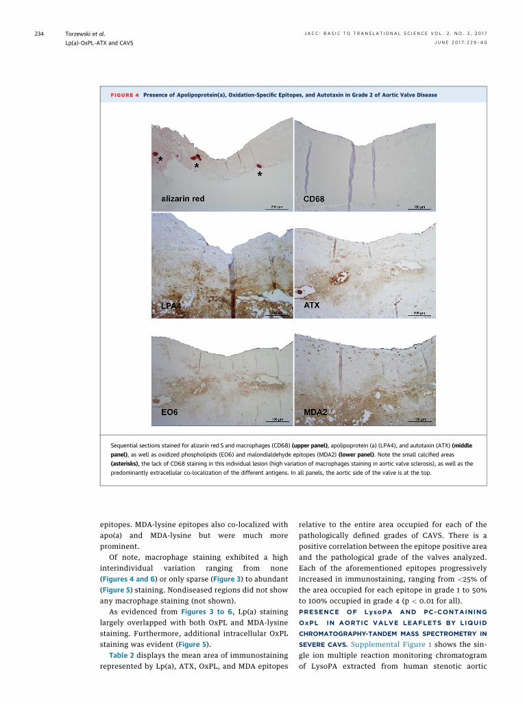
FIGURE 4 Presence of Apolipoprotein(a), Oxidation-Specific Epitopes, and Autotaxin in Grade 2 of Aortic Valve Disease
Sequential sections stained for alizarin red S and macrophages (CD68) (upper panel), apolipoprotein (a) (LPA4), and autotaxin (ATX) (middle
panel), as well as oxidized phospholipids (EO6) and malondialdehyde epitopes (MDA2) (lower panel). Note the small calcified areas
(asterisks), the lack of CD68 staining in this individual lesion (high variation of macrophages staining in aortic valve sclerosis), as well as the
predominantly extracellular co-localization of the different antigens. In all panels, the aortic side of the valve is at the top.
Torzewski et al. J A C C : B A S I C T O T R A N S L A T I O N A L S C I E N C E V O L . 2 , N O . 3 , 2 0 1 7
Lp(a)-OxPL-ATX and CAVS J U N E 2 0 1 7 : 2 2 9 – 4 0
234
epitopes. MDA-lysine epitopes also co-localized withapo(a) and MDA-lysine but were much moreprominent.
Of note, macrophage staining exhibited a highinterindividual variation ranging from none(Figures 4 and 6) or only sparse (Figure 3) to abundant(Figure 5) staining. Nondiseased regions did not showany macrophage staining (not shown).
As evidenced from Figures 3 to 6, Lp(a) staininglargely overlapped with both OxPL and MDA-lysinestaining. Furthermore, additional intracellular OxPLstaining was evident (Figure 5).
Table 2 displays the mean area of immunostainingrepresented by Lp(a), ATX, OxPL, and MDA epitopes
relative to the entire area occupied for each of thepathologically defined grades of CAVS. There is apositive correlation between the epitope positive areaand the pathological grade of the valves analyzed.Each of the aforementioned epitopes progressivelyincreased in immunostaining, ranging from <25% ofthe area occupied for each epitope in grade 1 to 50%to 100% occupied in grade 4 (p < 0.01 for all).PRESENCE OF LysoPA AND PC-CONTAINING
OxPL IN AORTIC VALVE LEAFLETS BY LIQUID
CHROMATOGRAPHY-TANDEM MASS SPECTROMETRY IN
SEVERE CAVS. Supplemental Figure 1 shows the sin-gle ion multiple reaction monitoring chromatogramof LysoPA extracted from human stenotic aortic

FIGURE 5 Presence of Apolipoprotein(a), Oxidation-Specific Epitopes, and Autotaxin in Grade 3 of Aortic Valve Disease
Sequential sections stained for alizarin red S and macrophages (CD68) (upper panel), apolipoprotein (a) (LPA4) and autotaxin (ATX) (middle
panel), as well as oxidized phospholipids (EO6) and malondialdehyde epitopes (MDA2) (lower panel). Note the predominance of cholesterol
crystals compared to calcified areas (*) in this individual lesion and co-localization of the different antigens around cholesterol crystal deposits
(#). Sometimes additional intracellular oxidized phospholipid staining is also evident (arrowheads). In all panels, the aortic side of the valve is
at the top.
J A C C : B A S I C T O T R A N S L A T I O N A L S C I E N C E V O L . 2 , N O . 3 , 2 0 1 7 Torzewski et al.J U N E 2 0 1 7 : 2 2 9 – 4 0 Lp(a)-OxPL-ATX and CAVS
235
valves, representing the most common LysoPA spe-cies seen in human plasma. In the 4 different stenoticaortic valves, 16:0 and 18:1 LysoPA species wereshown to be the most abundant species (Figure 7A).Variation in total amounts of LysoPA based onamount of valve tissue extracted was observeddespite using similar tissue wet weights (Figure 7B).Comparing LysoPA levels in each stenotic valveversus valvular gradients, there was a general trendof increased LysoPA levels and both mean and peakvalvular gradients (Figure 7C).
We also identified and quantitated the mostabundant fragmented PC-containing OxPL based on
known OxPL standards as described in the Methodssection. Supplemental Figure 2 shows the multiplereaction monitoring single ion plots for fragmentedPC-OxPL extracted from human stenotic aortic valveleaflets. The most abundant OxPL were PONPC andPAzPC, which were identified in all stenotic valvesstudied (Figure 8A). The aldehyde-containingPC-OxPL, which include POVPC and PONPC, werethe most abundant. The acid containing PC-OxPLconsisting of PAzPC, PGPC, and KODiaPC were thenext most prevalent fragmented species. Total PC-OxPL levels varied among the 4 valves examined(Figure 8B).

FIGURE 6 Presence of Apolipoprotein(a), Oxidation-Specific Epitopes, and Autotaxin in Grade 4 of Aortic Valve Disease
Sequential sections stained for alizarin red S and macrophages (CD68) (upper panel), apolipoprotein (a) (LPA4), and autotaxin (ATX) (middle
panel), as well as oxidized phospholipids (EO6) and malondialdehyde epitopes (MDA2) (lower panel). Note the co-localization of the
different antigens around and within heavily calcified areas (*) as well as the lack of CD68 staining in this individual lesion (high variation of
macrophages staining in aortic valve sclerosis). In all panels, the aortic side of the valve is at the top.
TABLE 2 Area of Involvement (Mean Positive Area) of Each Epitope
According to Pathological Grades of CAVS
Grade 1 Grade 2 Grade 3 Grade 4p Valuefor Trend
Apo(a) 1.3 (n ¼ 32) 1.9 (n ¼ 9) 1.6 (n ¼ 11) 2.3 (n ¼ 11) <0.001
Autotaxin 1.0 (n ¼ 2) 1.0 (n ¼ 3) 3.0 (n ¼ 4) 3.6 (n ¼ 5) <0.0001
OxPL 0.7 (n ¼ 32) 2.0 (n ¼ 8) 2.7 (n ¼ 11) 2.8 (n ¼ 11) <0.0001
MDA 1.6 (n ¼ 25) 2.4 (n ¼ 7) 3.1 (n ¼ 9) 4.0 (n ¼ 7) <0.0001
n represents the number of pathological grades analyzed within each category. Some of theleaflets harbored >1 different pathological grade, which is why the numbers analyzed aredifferent from those of leaflets/patients.
Apo(a) ¼ apolipoprotein(a); CAVS ¼ calcific aortic valve stenosis; MDA ¼ malondialdehyde;OxPL ¼ oxidized phospholipid.
Torzewski et al. J A C C : B A S I C T O T R A N S L A T I O N A L S C I E N C E V O L . 2 , N O . 3 , 2 0 1 7
Lp(a)-OxPL-ATX and CAVS J U N E 2 0 1 7 : 2 2 9 – 4 0
236
DISCUSSION
The present study showed that Lp(a)-associatedmolecules are present in plasma and aortic valveleaflets in patients with CAVS. Specifically, ATXcould be detected on apo(a) and apoB circulating inplasma, suggesting that it can be transported byLp(a) and delivered to aortic valve leaflets. Apo(a),the defining component of Lp(a), was also present inaortic valve leaflets from subjects with symptomaticCAVS who underwent AVR. In particular, apo(a),OxPL, MDA-lysine epitopes, and ATX that generatesLysoPA were particularly present in the most

FIGURE 7 Presence of LysoPA Species in Valve Leaflets and Relationship to
Valve Gradients
Prevalence of (A) individual lysophosphatidic acid (LysoPA) species and (B) total LysoPA
mass and (C) mean and peak valvular gradients in 4 different stenotic aortic valves.
Reverse-phase separation followed by tandem mass spectrometry detection of (A)
LysoPA species 16:0, 18:2, 18:1, 18:0, 20:4 and 22:6 in 4 aortic valves and (B) the total
LysoPA amounts per milligram of valve tissue extracted. (C) Represents the mean and
peak valvular gradients for each of the study valves.
J A C C : B A S I C T O T R A N S L A T I O N A L S C I E N C E V O L . 2 , N O . 3 , 2 0 1 7 Torzewski et al.J U N E 2 0 1 7 : 2 2 9 – 4 0 Lp(a)-OxPL-ATX and CAVS
237
advanced, pathologically defined grade 4 lesions.Importantly, apo(a), OxPL, MDA-lysine, and ATXwere located adjacent to prominent areas of extra-cellular aortic valve calcification. Finally, targetedanalysis of specific lipid species in stenotic valves byusing liquid chromatography-tandem mass spec-trometry directly confirmed the presence of themajor LysoPA species and a variety of PC-containingOxPL. Overall, these data suggest that after a latentperiod of prolonged plasma and leaflet exposure,these complexes may induce inflammation, calcifi-cation, and fibrosis and lend strong support to thehypothesis that the Lp(a)–ATX–OxPL axis is a keydeterminant of CAVS.
Lp(a) levels >30 to 50 mg/dl have been associatedwith CAVS in >10 epidemiological studies from NorthAmerican, European, and Asian populations(reviewed by Yeang et al. [11]). Moreover, the singlenucleotide polymorphism rs10455872 in the LPA genestudied in Mendelian randomization studies (5–7),and which is associated with elevated Lp(a) levels, isthe only monogenetic risk factor for CAVS (7), sug-gesting a causal role for Lp(a) in this disease. Aorticvalve calcification predicts progression of disease inhumans, and pathways driving aortic valve calcifica-tion are key risk factors for CAVS (28,29). The apo(a)moiety of Lp(a), via its lysine-binding domains, canbind to fibrin on denuded or injured endothelium(30,31), such as that on aortic valves subjected tomechanical stress in vivo, and accumulate in valveleaflets. Thereafter, the pro-inflammatory andpro-calcific cargo on Lp(a) such as OxPL, LysoPC, andLysoPA (implicated in ectopic calcification) maypromote CAVS.
Another important Lp(a)-associated molecule isATX, as shown in this study according to enzyme-linked immunoadsorbent assay and also byBouchareb et al. (13) in ultracentrifugally purifiedLp(a)-containing fractions. ATX is a secreted enzymethat is a member of the ecto-nucleotide pyrophos-phatase/phosphodiesterase family of ectoenzymes(ENPP) that hydrolyzes phosphodiester bonds ofvarious nucleotides. Unlike other ENPPs, ATX alsopossesses phospholipase D activity and catalyzes thehydrolysis of LysoPC into LysoPA. ATX seems to bethe predominant phospholipase D activity respon-sible for the generation of LysoPA levels in vivo, asheterozygous ATX knockout mice had one-half theLysoPA levels compared with their wild-type coun-terparts. Therefore, ATX is likely an importantcontributor of LysoPA found in aortic valve leafletseither via hydrolysis of LysoPC on Lp(a) ultimatelyretained in aortic valve leaflets or LysoPC from othersources.
Bone formation within the diseased aortic valveleaflets is driven by the differentiation of vascularcells into osteoblasts (32), via bone morphogenicprotein signaling and up-regulation of osteoblastictranscription factors, including RUNX2 and MSX2,which are up-regulated after exposure to oxidizedlow-density lipoprotein (reviewed by Yeang et al.

FIGURE 8 Presence of Individual PC-OxPL Species in Valve Leaflets
Liquid chromatography-tandem mass spectrometry analysis of the most abundant
fragmented phosphocholine-containing oxidized phospholipid (PC-OxPLs) compounds
extracted from human stenotic aortic valves and represented as (A) individual PC-OxPL
compounds or (B) as total PC-OxPL levels within each of the valves. Reverse phase
separation coupled with tandem mass spectrometry detection was used to detect
POVPC (1-palmitoyl-2-[5’-oxo-valeroyl]-sn-glycero-3-phosphocholine), PGPC (1-palmi-
toyl-2-glutaryl-sn-glycero-3-phosphocholine), PONPC (1-palmitoyl-2-[9’-oxononanoyl]-
sn-glycero-3-phosphocholine), KODiA-PC (1-[palmitoyl]-2-[5-keto-6-octene-dioyl]-sn-
glycero-3-phosphocholine), and PAzPC (1-palmitoyl-2-azelaoyl-sn-glycero-3-
phosphocholine).
Torzewski et al. J A C C : B A S I C T O T R A N S L A T I O N A L S C I E N C E V O L . 2 , N O . 3 , 2 0 1 7
Lp(a)-OxPL-ATX and CAVS J U N E 2 0 1 7 : 2 2 9 – 4 0
238
[11]). Both exogenously added OxPL (33,34) andLysoPA (35) were sufficient in differentiatingvascular cells or mesenchymal stem cells, respec-tively, into osteoblasts in culture. Interestingly,LysoPC also promoted osteoblast differentiationin vitro, but this outcome was completely dependenton ATX activity (13), implying that LysoPC is only anintermediary to LysoPA with respect to developmentof CAVS. Moreover, intraperitoneal LysoPA adminis-tration potentiated aortic valve calcification in amouse model of aortic stenosis (13), whereas the role
of OxPL in CAVS in animal models remains to bestudied (36). Our findings that OxPL was detectedimmunologically and by using mass spectrophotom-etry in calcified aortic valve leaflets, in conjunctionwith the observation by Bouchareb et al. (13) thatLysoPA was highly enriched in human aortic valveleaflets from subjects with CAVS, further supportsthe importance of these 2 Lp(a)-associated lipids asbiologically active mediators in the pathogenesisof CAVS.
Clinical data further exemplify the relevance ofLp(a), OxPL, and ATX toward the development andprogression of CAVS. A secondary analysis of theASTRONOMER trial showed that elevated Lp(a) andOxPL were predictive of a worse outcome in 220subjects with mild to moderate CAVS followed up for3.5 years. Those with the highest tertile of baselineLp(a) (>58.5 mg/dl) had faster progression rates(average peak velocity, 0.26 � 0.03 m/s/year vs.0.17 � 0.02 m/s/year) and had an approximately2-fold increased risk of a composite outcome of AVRand cardiac death, which increased to 5.5-fold if theywere younger than the median age of 57 years (36). Inconcert with the Lp(a) findings, those with thehighest tertile of OxPL-apoB (>5.50 nM) as well asOxPL-apo(a) (>33.5 nM) had an increased rate ofprogression and need for AVR consistent with thethesis that OxPL carried by Lp(a) participates in thepathogenesis of CAVS. In addition, in a case-controlanalysis of 150 subjects with CAD and CAVScompared with 150 individuals with CAD alone, anincreased risk of CAVS was associated with elevatedATX mass and ATX activity (37). Subjects withelevated ATX activity ($84 RFU/min) and eitherhigher Lp(a) ($50 mg/dl) or OxPL-apoB ($2.02 nM)had a dramatically higher risk of CAVS (3.46 [inter-quartile range: 1.40 to 8.58] nM; p ¼ 0.007) and (5.48[interquartile range: 2.45 to 12.27] nM; p < 0.0001),respectively (8). Overall, these findings suggest thatLp(a), OxPL, and ATX interact in mediating progres-sion of CAVS.
CLINICAL IMPLICATIONS. This study reinforces theroles of Lp(a), OxPL, and ATX as pathogenic riskfactors for CAVS. The hypothesis that Lp(a) loweringmay slow the progression of CAVS and need for AVRmay now be tested with the development of antisenseoligonucleotides that potently lower Lp(a) levels (23).It also implies that targeting OxPL or ATX may also beviable therapeutic approaches to inhibiting thedevelopment or progression of CAVS. The data fromthese 3 distinct group of subjects complement eachother, showing that: ATX can be transported by Lp(a)in plasma; is present in similar locations to apo(a),
PERSPECTIVES
COMPETENCY IN PATIENT CARE AND PROCEDURAL
SKILLS: Elevated Lp(a), OxPL-apoB, and ATX are abundant in
aortic valve leaflets obtained during AVR and may contribute to
the development and progression of CAVS.
TRANSLATIONAL OUTLOOK: Targeting elevated Lp(a),
OxPL-apoB, and ATX with therapeutic agents may be a viable
approach to prevent or reduce the rate of progression of CAVS.
J A C C : B A S I C T O T R A N S L A T I O N A L S C I E N C E V O L . 2 , N O . 3 , 2 0 1 7 Torzewski et al.J U N E 2 0 1 7 : 2 2 9 – 4 0 Lp(a)-OxPL-ATX and CAVS
239
OxPL, and areas of calcification in diseased aorticvalve leaflets; and that its product, LysoPA, can bedirectly detected in valve leaflets from subjects withCAVS.
STUDY LIMITATIONS. This study included tissueand/or blood samples from 3 distinct populations,and therefore direct comparison of plasma levels ofATX-apoB and ATX-apo(a) with histological assess-ment of aortic valve apo(a), ATX content, and spec-trophotometric quantitation of OxPL and LysoPAcontent was not feasible. Also, plasma levels of Lp(a)were not available in patients undergoing AVR due tolack of prior evidence of its etiologic importance tocorrelate the plasma levels with histological findings.Finally, the number of samples available for ATX as-says and for aortic valve leaflets available for liquidchromatography-tandem mass spectrometry analysisof OxPL and LysoPC was small, and these results willrequire validation in larger studies. Future studiesshould address whether elevated plasma levels ofATX-apo(a) and ATX-apo(a) correlate with clinicalseverity of CAVS and the presence of ATX, OxPL, andLysoPA valve content.
CONCLUSIONS
The observations in this study provide furtherevidence that an Lp(a)–OxPL-ATX–LysoPA axis may
be important for the pathogenesis of CAVS. Theseobservations provide a rationale for therapeuticattempts to reduce entry of Lp(a) into valve leaflets,or to inactivate its attendant OxPL (37), to reduce therisk of developing CAVS or to reduce its rate ofprogression and attendant complications and needfor AVR.
ACKNOWLEDGMENT The authors thank KerstinWinter for expert technical assistance.
ADDRESS FOR CORRESPONDENCE: Dr. SotiriosTsimikas, Vascular Medicine Program, Department ofMedicine, Sulpizio Cardiovascular Center, Universityof California San Diego, 9500 Gilman Drive, BSB 1080,La Jolla, California 92093-0682. E-mail: [email protected].
RE F E RENCE S
1. Rajamannan NM, Evans FJ, Aikawa E, et al.Calcific aortic valve disease: not simply a degen-erative process: a review and agenda for researchfrom the National Heart and Lung and BloodInstitute Aortic Stenosis Working Group. Executivesummary: calcific aortic valve disease-2011update. Circulation 2011;124:1783–91.
2. Yutzey KE, Demer LL, Body SC, et al. Calcificaortic valve disease: a consensus summary fromthe Alliance of Investigators on Calcific AorticValve Disease. Arterioscler Thromb Vasc Biol 2014;34:2387–93.
3. Deeb GM, Reardon MJ, Chetcuti S, et al. 3-Yearoutcomes in high-risk patients who underwentsurgical or transcatheter aortic valve replacement.J Am Coll Cardiol 2016;67:2565–74.
4. Capoulade R, Chan KL, Yeang C, et al. Oxidizedphospholipids, lipoprotein(a), and progression ofcalcific aortic valve stenosis. J Am Coll Cardiol2015;66:1236–46.
5. Kamstrup PR, Tybjaerg-Hansen A,Nordestgaard BG. Elevated lipoprotein(a) and riskof aortic valve stenosis in the general population.J Am Coll Cardiol 2014;63:470–7.
6. Arsenault BJ, Boekholdt SM, Mora S, et al.Impact of high-dose atorvastatin therapy andclinical risk factors on incident aortic valve
stenosis in patients with cardiovascular disease(from TNT, IDEAL, and SPARCL). Am J Cardiol2014;113:1378–82.
7. Thanassoulis G, Campbell CY, Owens DS, et al.Genetic associations with valvular calcification andaortic stenosis. N Engl J Med 2013;368:503–12.
8. Nsaibia MJ, Mahmut A, Boulanger MC, et al.Autotaxin interacts with lipoprotein(a) andoxidized phospholipids in predicting the risk ofcalcific aortic valve stenosis in patients with cor-onary artery disease. J Intern Med 2016;280:509–17.
9. Bergmark C, Dewan A, Orsoni A, et al. A novelfunction of lipoprotein [a] as a preferential carrierof oxidized phospholipids in human plasma. J LipidRes 2008;49:2230–9.
10. Leibundgut G, Scipione C, Yin H, et al.Determinants of binding of oxidized phospholipidson apolipoprotein(a) and lipoprotein(a). J LipidRes 2013;54:2815–30.
11. YeangC,WilkinsonMJ, TsimikasS.Lipoprotein(a)and oxidized phospholipids in calcific aortic valvestenosis. Curr Opin Cardiol 2016;31:440–50.
12. O’Brien KD, Reichenbach DD, Marcovina SM,Kuusisto J, Alpers CE, Otto CM. ApolipoproteinsB, (a), and E accumulate in the morphologicallyearly lesion of ’degenerative’ valvular aortic
stenosis. Arterioscler Thromb Vasc Biol 1996;16:523–32.
13. Bouchareb R, Mahmut A, Nsaibia MJ, et al.Autotaxin derived from lipoprotein(a) and valveinterstitial cells promotes inflammation andmineralization of the aortic valve. Circulation2015;132:677–90.
14. Cote N, Pibarot P, Pepin A, et al. Oxidized low-density lipoprotein, angiotensin II and increasedwaist circumference are associated with valveinflammation in prehypertensive patients withaortic stenosis. Int J Cardiol 2010;145:444–9.
15. Mahmut A, Boulanger MC, El Husseini D, et al.Elevated expression of lipoprotein-associatedphospholipase A2 in calcific aortic valve disease:implications for valve mineralization. J Am CollCardiol 2014;63:460–9.
16. Cowell SJ, Newby DE, Prescott RJ, et al.A randomized trial of intensive lipid-loweringtherapy in calcific aortic stenosis. N Engl J Med2005;352:2389–97.
17. Rossebo AB, Pedersen TR, Boman K, et al.Intensive lipid lowering with simvastatin andezetimibe in aortic stenosis. N Engl J Med 2008;359:1343–56.
18. Chan KL, Teo K, Dumesnil JG, Ni A, Tam J,ASTRONOMER Investigators. Effect of lipid
Torzewski et al. J A C C : B A S I C T O T R A N S L A T I O N A L S C I E N C E V O L . 2 , N O . 3 , 2 0 1 7
Lp(a)-OxPL-ATX and CAVS J U N E 2 0 1 7 : 2 2 9 – 4 0
240
lowering with rosuvastatin on progression ofaortic stenosis: results of the aortic stenosisprogression observation: measuring effects ofrosuvastatin (ASTRONOMER) trial. Circulation2010;121:306–14.
19. Dichtl W, Alber HF, Feuchtner GM, et al.Prognosis and risk factors in patients withasymptomatic aortic stenosis and their modulationby atorvastatin (20 mg). Am J Cardiol 2008;102:743–8.
20. Teo KK, Corsi DJ, Tam JW, Dumesnil JG,Chan KL. Lipid lowering on progression of mildto moderate aortic stenosis: meta-analysis ofthe randomized placebo-controlled clinical tri-als on 2344 patients. Can J Cardiol 2011;27:800–8.
21. Yeang C, Hung MY, Byun YS, et al. Effect oftherapeutic interventions on oxidized phospho-lipids on apolipoprotein B100 and lipoprotein(a).J Clin Lipidol 2016;10:594–603.
22. Graham MJ, Viney N, Crooke RM, Tsimikas S.Antisense inhibition of apolipoprotein (a) to lowerplasma lipoprotein (a) levels in humans. J LipidRes 2016;57:340–51.
23. Tsimikas S, Viney NJ, Hughes SG, et al. Anti-sense therapy targeting apolipoprotein(a): arandomised, double-blind, placebo-controlledphase 1 study. Lancet 2015;386:1472–83.
24. Raal FJ, Giugliano RP, Sabatine MS, et al.Reduction in lipoprotein(a) with PCSK9 mono-clonal antibody evolocumab (AMG 145): apooled analysis of more than 1,300 patients in 4phase II trials. J Am Coll Cardiol 2014;63:1278–88.
25. Gaudet D, Kereiakes DJ, McKenney JM, et al.Effect of alirocumab, a monoclonal proproteinconvertase subtilisin/kexin 9 antibody, on lip-oprotein(a) concentrations (a pooled analysis of150 mg every two weeks dosing from phase 2trials). Am J Cardiol 2014;114:711–5.
26. Twardowski L, Cheng F, Michaelsen J, et al.Enzymatically modified low-density lipoprotein ispresent in all stages of aortic valve sclerosis:implications for pathogenesis of the disease. J AmHeart Assoc 2015;4:e002156.
27. Warren BA, Yong JL. Calcification of the aorticvalve: its progression and grading. Pathology1997;29:360–8.
28. Jenkins WS, Vesey AT, Shah AS, et al. Valvular(18)F-Fluoride and (18)F-fluorodeoxyglucoseuptake predict disease progression and clinicaloutcome in patients with aortic stenosis. J Am CollCardiol 2015;66:1200–1.
29. Dweck MR, Pawade TA, Newby DE. Aorticstenosis begets aortic stenosis: between a rockand a hard place? Heart 2015;101:919–20.
30. Nielsen LB, Stender S, Kjeldsen K,Nordestgaard BG. Specific accumulation oflipoprotein(a) in balloon-injured rabbit aortain vivo. Circulation Res 1996;78:615–26.
31. Hughes SD, Lou XJ, Ighani S, et al. Lipoprotein(a)vascular accumulation inmice. In vivo analysis of therole of lysine binding sites using recombinantadenovirus. J Clin Invest 1997;100:1493–500.
32. Mohler ER 3rd, Gannon F, Reynolds C,Zimmerman R, Keane MG, Kaplan FS. Boneformation and inflammation in cardiac valves.Circulation 2001;103:1522–8.
33. Parhami F, Morrow AD, Balucan J, et al.Lipid oxidation products have opposite effectson calcifying vascular cell and bone cell dif-ferentiation. A possible explanation for theparadox of arterial calcification in osteoporoticpatients. Arterioscler Thromb Vasc Biol 1997;17:680–7.
34. Mody N, Parhami F, Sarafian TA, Demer LL.Oxidative stress modulates osteoblastic differen-tiation of vascular and bone cells. Free Rad BiolMed 2001;31:509–19.
35. Liu YB, Kharode Y, Bodine PV, Yaworsky PJ,Robinson JA, Billiard J. LPA induces osteoblastdifferentiation through interplay of two receptors:LPA1 and LPA4. J Cell Biochem 2010;109:794–800.
36. Yeang C, Cotter B, Tsimikas S. Experimentalanimal models evaluating the causal role of lip-oprotein(a) in atherosclerosis and aortic stenosis.Cardiovasc Drugs Ther 2016;30:75–85.
37. Leibundgut G, Witztum JL, Tsimikas S.Oxidation-specific epitopes and immunologicalresponses: translational biotheranostic implica-tions for atherosclerosis. Curr Opin Pharmacol2013;13:168–79.
KEY WORDS aortic valve stenosis,autotaxin, inflammation, Lp(a), oxidation-specific epitopes
APPENDIX For a supplemental methodssection as well as supplemental figures, pleasesee the online version of this article.


![Lipoprotein(a) and Oxidized Phospholipids Promote Valve ... · BACKGROUND Lipoprotein(a) [Lp(a)], a major carrier of oxidized phospholipids (OxPL), is associated with an increased](https://static.fdocuments.in/doc/165x107/5e5f5b29282d4a2232338b34/lipoproteina-and-oxidized-phospholipids-promote-valve-background-lipoproteina.jpg)


