Lipectomy associated to obesity produces greater fat ... · white adipose tissue of female compared...
13
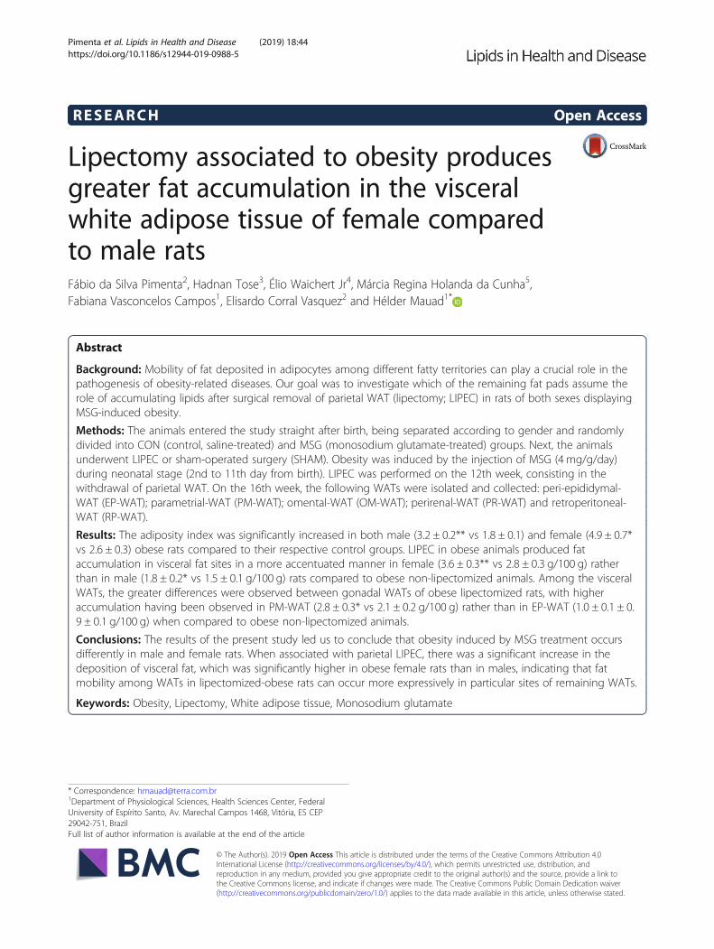
RESEARCH Open Access Lipectomy associated to obesity produces greater fat accumulation in the visceral white adipose tissue of female compared to male rats Fábio da Silva Pimenta 2 , Hadnan Tose 3 , Élio Waichert Jr 4 , Márcia Regina Holanda da Cunha 5 , Fabiana Vasconcelos Campos 1 , Elisardo Corral Vasquez 2 and Hélder Mauad 1* Abstract Background: Mobility of fat deposited in adipocytes among different fatty territories can play a crucial role in the pathogenesis of obesity-related diseases. Our goal was to investigate which of the remaining fat pads assume the role of accumulating lipids after surgical removal of parietal WAT (lipectomy; LIPEC) in rats of both sexes displaying MSG-induced obesity. Methods: The animals entered the study straight after birth, being separated according to gender and randomly divided into CON (control, saline-treated) and MSG (monosodium glutamate-treated) groups. Next, the animals underwent LIPEC or sham-operated surgery (SHAM). Obesity was induced by the injection of MSG (4 mg/g/day) during neonatal stage (2nd to 11th day from birth). LIPEC was performed on the 12th week, consisting in the withdrawal of parietal WAT. On the 16th week, the following WATs were isolated and collected: peri-epididymal- WAT (EP-WAT); parametrial-WAT (PM-WAT); omental-WAT (OM-WAT); perirenal-WAT (PR-WAT) and retroperitoneal- WAT (RP-WAT). Results: The adiposity index was significantly increased in both male (3.2 ± 0.2** vs 1.8 ± 0.1) and female (4.9 ± 0.7* vs 2.6 ± 0.3) obese rats compared to their respective control groups. LIPEC in obese animals produced fat accumulation in visceral fat sites in a more accentuated manner in female (3.6 ± 0.3** vs 2.8 ± 0.3 g/100 g) rather than in male (1.8 ± 0.2* vs 1.5 ± 0.1 g/100 g) rats compared to obese non-lipectomized animals. Among the visceral WATs, the greater differences were observed between gonadal WATs of obese lipectomized rats, with higher accumulation having been observed in PM-WAT (2.8 ± 0.3* vs 2.1 ± 0.2 g/100 g) rather than in EP-WAT (1.0 ± 0.1 ± 0. 9 ± 0.1 g/100 g) when compared to obese non-lipectomized animals. Conclusions: The results of the present study led us to conclude that obesity induced by MSG treatment occurs differently in male and female rats. When associated with parietal LIPEC, there was a significant increase in the deposition of visceral fat, which was significantly higher in obese female rats than in males, indicating that fat mobility among WATs in lipectomized-obese rats can occur more expressively in particular sites of remaining WATs. Keywords: Obesity, Lipectomy, White adipose tissue, Monosodium glutamate * Correspondence: [email protected] 1 Department of Physiological Sciences, Health Sciences Center, Federal University of Espírito Santo, Av. Marechal Campos 1468, Vitória, ES CEP 29042-751, Brazil Full list of author information is available at the end of the article © The Author(s). 2019 Open Access This article is distributed under the terms of the Creative Commons Attribution 4.0 International License (http://creativecommons.org/licenses/by/4.0/), which permits unrestricted use, distribution, and reproduction in any medium, provided you give appropriate credit to the original author(s) and the source, provide a link to the Creative Commons license, and indicate if changes were made. The Creative Commons Public Domain Dedication waiver (http://creativecommons.org/publicdomain/zero/1.0/) applies to the data made available in this article, unless otherwise stated. Pimenta et al. Lipids in Health and Disease (2019) 18:44 https://doi.org/10.1186/s12944-019-0988-5
Transcript of Lipectomy associated to obesity produces greater fat ... · white adipose tissue of female compared...
RESEARCH Open Access
Lipectomy associated to obesity producesgreater fat accumulation in the visceralwhite adipose tissue of female comparedto male ratsFábio da Silva Pimenta2, Hadnan Tose3, Élio Waichert Jr4, Márcia Regina Holanda da Cunha5,Fabiana Vasconcelos Campos1, Elisardo Corral Vasquez2 and Hélder Mauad1*
Abstract
Background: Mobility of fat deposited in adipocytes among different fatty territories can play a crucial role in thepathogenesis of obesity-related diseases. Our goal was to investigate which of the remaining fat pads assume therole of accumulating lipids after surgical removal of parietal WAT (lipectomy; LIPEC) in rats of both sexes displayingMSG-induced obesity.
Methods: The animals entered the study straight after birth, being separated according to gender and randomlydivided into CON (control, saline-treated) and MSG (monosodium glutamate-treated) groups. Next, the animalsunderwent LIPEC or sham-operated surgery (SHAM). Obesity was induced by the injection of MSG (4 mg/g/day)during neonatal stage (2nd to 11th day from birth). LIPEC was performed on the 12th week, consisting in thewithdrawal of parietal WAT. On the 16th week, the following WATs were isolated and collected: peri-epididymal-WAT (EP-WAT); parametrial-WAT (PM-WAT); omental-WAT (OM-WAT); perirenal-WAT (PR-WAT) and retroperitoneal-WAT (RP-WAT).
Results: The adiposity index was significantly increased in both male (3.2 ± 0.2** vs 1.8 ± 0.1) and female (4.9 ± 0.7*vs 2.6 ± 0.3) obese rats compared to their respective control groups. LIPEC in obese animals produced fataccumulation in visceral fat sites in a more accentuated manner in female (3.6 ± 0.3** vs 2.8 ± 0.3 g/100 g) ratherthan in male (1.8 ± 0.2* vs 1.5 ± 0.1 g/100 g) rats compared to obese non-lipectomized animals. Among the visceralWATs, the greater differences were observed between gonadal WATs of obese lipectomized rats, with higheraccumulation having been observed in PM-WAT (2.8 ± 0.3* vs 2.1 ± 0.2 g/100 g) rather than in EP-WAT (1.0 ± 0.1 ± 0.9 ± 0.1 g/100 g) when compared to obese non-lipectomized animals.
Conclusions: The results of the present study led us to conclude that obesity induced by MSG treatment occursdifferently in male and female rats. When associated with parietal LIPEC, there was a significant increase in thedeposition of visceral fat, which was significantly higher in obese female rats than in males, indicating that fatmobility among WATs in lipectomized-obese rats can occur more expressively in particular sites of remaining WATs.
Keywords: Obesity, Lipectomy, White adipose tissue, Monosodium glutamate
* Correspondence: [email protected] of Physiological Sciences, Health Sciences Center, FederalUniversity of Espírito Santo, Av. Marechal Campos 1468, Vitória, ES CEP29042-751, BrazilFull list of author information is available at the end of the article
© The Author(s). 2019 Open Access This article is distributed under the terms of the Creative Commons Attribution 4.0International License (http://creativecommons.org/licenses/by/4.0/), which permits unrestricted use, distribution, andreproduction in any medium, provided you give appropriate credit to the original author(s) and the source, provide a link tothe Creative Commons license, and indicate if changes were made. The Creative Commons Public Domain Dedication waiver(http://creativecommons.org/publicdomain/zero/1.0/) applies to the data made available in this article, unless otherwise stated.
Pimenta et al. Lipids in Health and Disease (2019) 18:44 https://doi.org/10.1186/s12944-019-0988-5
BackgroundObesity has become a major public health challenge [1],for the prevalence of overweight and obese people hasbeen increasing worldwide, being related to alarmingepidemiological risk factors for diabetes, cardiovasculardisease, cancer and premature death [2]. The genesis ofobesity and its physiological consequences have been ex-tensively studied. Our particular interest lies in under-standing the mobility of fat deposited in adipocytesamong different fatty territories, which could play a cru-cial role in the pathogenesis of obesity-related diseases.Specific conditions such as gender, blood circulation,
neurological factors and obesity itself could account forthe local differences observed among different fat depotsregarding cellular composition, size and function, mak-ing them act as “separate miniorgans” [3].It is well known that the fat stored in adipocytes is not
homogeneous. Its distribution among the different whiteadipose tissue (WAT) depots affects the production of in-flammatory cytokines and chemokines by pre-adipocytes,which is also influenced by age and body weight [3–5]. Thebulk of the body’s fat resides in subcutaneous WAT depots,which can be distinctive due to unique metabolic andphysiological characteristics. Also known as parietal WAT,these subcutaneous fat depots are located mainly in the in-ferior portion of the body, where they protect the musclesfrom fat infiltration by capturing the excess of lipids, con-tributing also to glucose homeostasis as metabolic dissipa-tors [6]. On the other hand, it is widely known thatupper-body adiposity constitutes an important risk factorfor many diseases, with visceral adiposity playing a majorrole in metabolic disorders, including insulin resistance,hyperinsulinemia, type-2 diabetes mellitus, among others[7–10]. Therefore, knowing the precise location andamount of the different fat depots in one’s body is criticalfor the comprehension of the actual role of regional adipos-ity as a risk factor in epidemiological studies [11].Due to culturally-defined beauty standards, surgical re-
moval of WAT through liposuction has been widelyemployed. It consists in the withdrawal of accumulationand/or bad distribution areas of parietal WAT, mainlyfrom the deep layer (lamellar). This procedure has alsobeen employed experimentally through parietal LIPEC,presenting controversial results concerning compensatoryincreases of WAT and the mobility of lipids among fat ter-ritories. This controversy is observed in both experimental[12, 13] and clinical studies [14–17]. Strong evidencepoints to changes taking place in the parietal WAT afterits resection, including fat deposition in non-lipectomizedareas. However, which are these areas, how do they behaveafter parietal LIPEC and are these issues influenced bygender are questions that remain to be fully answered.In order to assess these issues, we employed an experi-
mental model in which obesity was induced by treating
neonatal rats with monosodium glutamate (MSG). MSGprovokes damage in the central nervous system (CNS),predominantly in the hypothalamus arcuate nucleus(HAN), which is highly involved in the release of neuro-transmitters, peptides and hormones [18–21]. HAN le-sions occurring during the neonatal period lead toseveral neuro-endocrine changes in the regulation offood intake in rats, resulting in decreased naso-anallength, hyperlipidemia and, above all, obesity [22, 23]. Inaddition, it has been reported that adult rats treated withMSG displayed hyperinsulinemia, pointing to an impair-ment of insulin sensitivity [24].Despite the many results achieved so far on the connec-
tion between MSG treatment and obesity, the occurrenceof fat mobility among WATs in this experimental modelremains to be addressed, particularly following LIPEC inobese rats. Thus, the aim of this study was to investigatewhich of the remaining fat pads assume the key role of ac-cumulating lipids after surgical removal of parietal WATin rats of both sexes displaying MSG-induced obesity.
MethodsAnimal models and ethical issuesMale and female Wistar rats – included in the study straightafter birth – were maintained on a standard diet and tapwater, being exposed to a 12/12 h light-dark cycle at 23o C.First, the animals were separated according to gender andrandomly divided into the following groups: CON (control,saline-treated, male/female n = 11/10, respectively) andMSG (monosodium glutamate-treated male/female n = 15/16, respectively). Next, the animals underwent (LIPEC) orsham-operated surgery (SHAM), comprising the followinggroups: males CON-LIPEC (n = 4), CON-SHAM (n = 7),MSG-LIPEC (n = 10) and MSG-SHAM (n = 5); femalesCON-LIPEC (n = 5), CON-SHAM (n = 5), MSG-LIPEC(n = 11) and MSG-SHAM (n = 5).All experiments were conducted in accordance with the
U.S. National Institute of Health Guidelines for the Careand Use of Laboratory Animals, and study protocols werepreviously approved by our Institutional Ethics Commit-tee for the Use of Animals (Protocol # 034/2011).
Obesity-inducing treatmentObesity was induced by subcutaneous injection of MSG (4mg/g/day) during neonatal stage (2nd to 11th days frombirth). Control animals were submitted to the same sched-ule of injections, receiving NaCl hypertonic solution(1.45%) instead. At the age of 21 days, animals were weanedand placed in polypropylene boxes. All animals received thesame standard diet ad libitum throughout the study.
Surgical proceduresLIPEC was performed on the 12th week underanesthesia with chloral hydrate 10%, intraperitoneally.
Pimenta et al. Lipids in Health and Disease (2019) 18:44 Page 2 of 13
With the animals positioned in dorsal decubitus, theskin of the inguinal region and a small notch from themidpoint of the thigh to the knee joint of the animalswere pinched. Then, a divulsion was performed for thedetachment of WAT from the aponeurotic structuresand the skin, and also for the detachment of the adiposeprojection on the flanks until the reflection between theventral abdomen and the back-lobe and posterior with-drawal of parietal WAT. The skin was then sutured with4–0 cotton yarn, by continuous scrubbing. Removal ofthe parietal WAT was done bilaterally and weighed on aprecision scale. Sham surgery consisted in surgical inci-sion and cotton suture only.On the 16th week, the WATs were isolated and collected
from the following sites: parietal WAT (PT-WAT), only inanimals non-lipectomized at the 12th week;perigonadal-WATs: peri-epididymal-WAT (EP-WAT) andparametrial-WAT (PM-WAT); omental-WAT (OM-WAT),which corresponds to the greater omentum; perirenal-WAT(PR-WAT) and retroperitoneal-WAT (RP-WAT).Firstly, animals were decapitated and submitted to tricot-
omy in the abdominal anterior region and antero-medialrear paws. Next, animals were fixed in a surgical board insupine position. The individual procedures for the removalof each particular WATare briefly described below:
Parietal-WATThis procedure – performed only in sham groups(CON-SHAM and MSG-SHAM) – was conducted asdescribed above for the animals lipectomized at the 12thweek.
Perigonadal-WATsInitially, for male rats, a medium pubic xiphoid opening anda perigonadal adipose cushion were clamped. This tissuewas then pulled for testicular exposure and individualizationof peri-epididimal fat planes. In female rats, the uterus waspulled and sections of parametrial-WAT projections wereperformed.
Omental-WATsThis tissue was collected after traction of the spleen andthe stomach, exposing the greater curvature of the latterfor resection of the greater omentum.
Perirenal-WATIt was collected by bouncing the intestines laterally forvisualization of the kidney and its perirenal fat. By trac-tioning the kidney, the limits of this fat pad are exposedand excised. This procedure was performed bilaterally.
Retroperitoneal-WATAfter renal resection and subsequent removal of theperirenal-WAT and the adrenal gland, retroperitoneal fat
is easily visualized. It was resected from the pelvic regionup to below the diaphragm insertion, bilaterally.
Data analysisConsidering the variation in the total body weight of theanimals, all the WATs obtained (g) were normalized by100 g of body weight. The sum of all WATs – but theparietal-WAT that was removed in the 12th week – isreferred to as “total fats”. The Adiposity Index was cal-culated by the sum of total fats divided by corporalweight and multiplied by 100, representing the amountof fat from all WATs aforementioned by grams of ani-mal. The term “Visceral WAT” refers to the quantity offat accumulated in the viscera. It comprises the sum ofomental-, perirenal and epididymal−/parametrial-WATs.Extra-Visceral WAT represents the fat accumulated inthe abdomen, but not in the viscera. It was obtained bythe sum of retroperitoneal- and parietal-WATs, exceptin those cases where parietal fat was removed.
Statistical analysisThe results are expressed as the mean ± SEM. For theevaluation of the normality of the data distribution weused the D’Agostino and Pearson test. In addition, alldata were submitted to two-way analysis of variance(ANOVA) followed by Tukey test, and, for some of thedata, to the Student’s t-test. Statistical significance wasachieved when p < 0.05.
ResultsAnthropometric parameters of the different groups stud-ied are shown in Table 1. After 16 weeks, bothMSG-treated groups presented body weight valuessmaller than those of CON groups, which was true forboth sexes (although for sham females the change wasnot statistically significant). On the other hand, we ob-served that the LIPEC procedure did not induce changesin body weight in either CON or MSG-treated animals,which, again, was true for males and females. It isworthy of note that female animals weighted less thanmales to begin with. These results clearly show thatMSG treatment induced less body weight gain in bothmale and female animals, with LIPEC having no effectson this parameter.In order to evaluate actual fat accumulation in the ani-
mals from all groups, we determined the parameter totalfat (Table 1). We observed that, while LIPEC signifi-cantly increased the total fat in MSG-LIPEC male ratswhen compared to CON-LIPEC animals, no significantdifferences were observed regarding this parameter whenanimals belonging to MSG-LIPEC and MSG-SHAMgroups were compared between themselves, as well as be-tween CON-LIPEC and CON-SHAM groups. On theother hand, LIPEC induced opposite effects regarding
Pimenta et al. Lipids in Health and Disease (2019) 18:44 Page 3 of 13
total fat accumulation in CON and MSG-treated femalerats: while the procedure led to smaller total fat values inCON animals, significantly higher values were found inMSG-treated rats. In fact, total fat values in MSG- LIPECfemale rats were significantly higher than those of eitherMSG-SHAM and CON- LIPEC animals. These resultsclearly show that there is greater accumulation of total fatin obese female rats when compared to males, and thatLIPEC potentiated this accumulation.Considering that the procedures employed, i.e., LIPEC
and MSG treatment, produced different changes in thebody weight parameter, we have also analyzed their ef-fects on the adiposity index of the animals (Table 1),which permitted us to compare values of fat per gram ofcorporal weight among the groups. We observed thatMSG-treated male rats – LIPEC or not – presented sig-nificantly higher adiposity indexes when compared totheir respective controls, which was also true for femalerats. Taken together, these results indicate thatMSG-induced obesity led to a more pronounced fat ac-cumulation in females, with this accumulation beingeven higher when obesity was associated with LIPEC.We then asked ourselves how the distribution of fat in
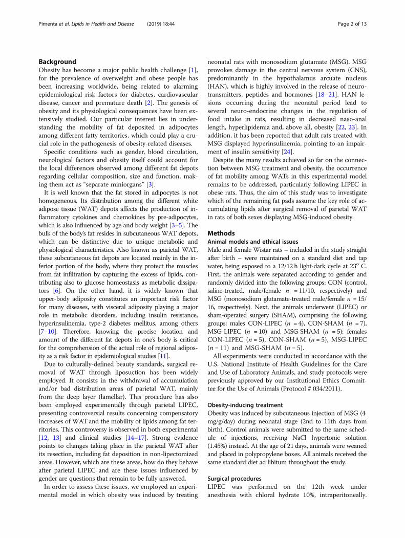
different corporal regions occurs after LIPEC, in bothcontrol and obese animals. To answer that, we separatedgroups of WATs based on their anatomical localizationand functional aspects. To begin with, we considered thegroup deemed visceral WAT (Fig. 1). We observed that,in males, while there was a significant increase in vis-ceral WAT in obese animals submitted to LIPEC whencompared to lipectomized control animals, obesity alonedid not have a significant effect (Fig. 1a). On the otherhand, in female animals, we observed that obesity aloneproduced a significant increase in visceral WAT whencompared to the sham control group. Moreover, obesityassociated to LIPEC induced significantly higher in-creases in visceral WAT not only compared to lipecto-mized control animals but also to no LIPEC obese ones(Fig. 1b). These data strongly indicate that the effects of
LIPEC on visceral WAT accumulation are much moresevere in obese female rats than in obese males.Next, we assessed the individual contribution of each one
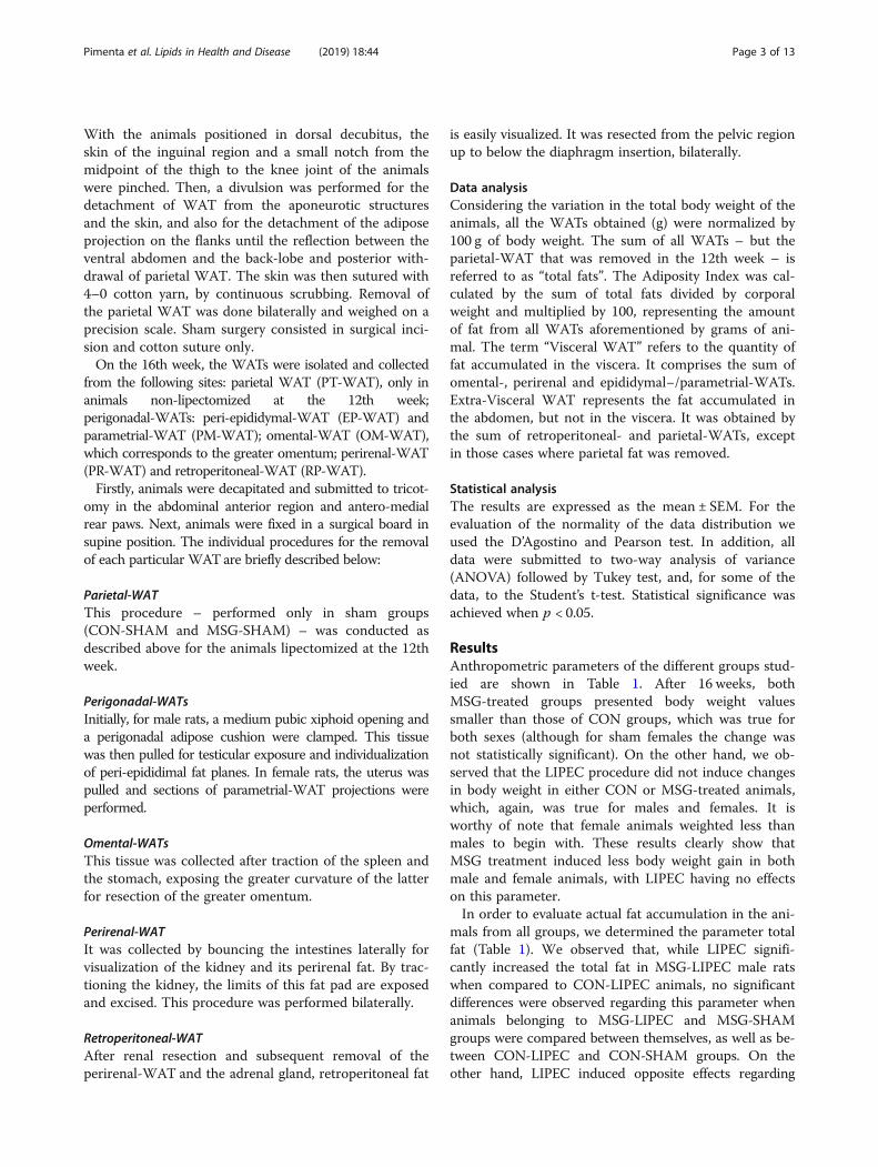
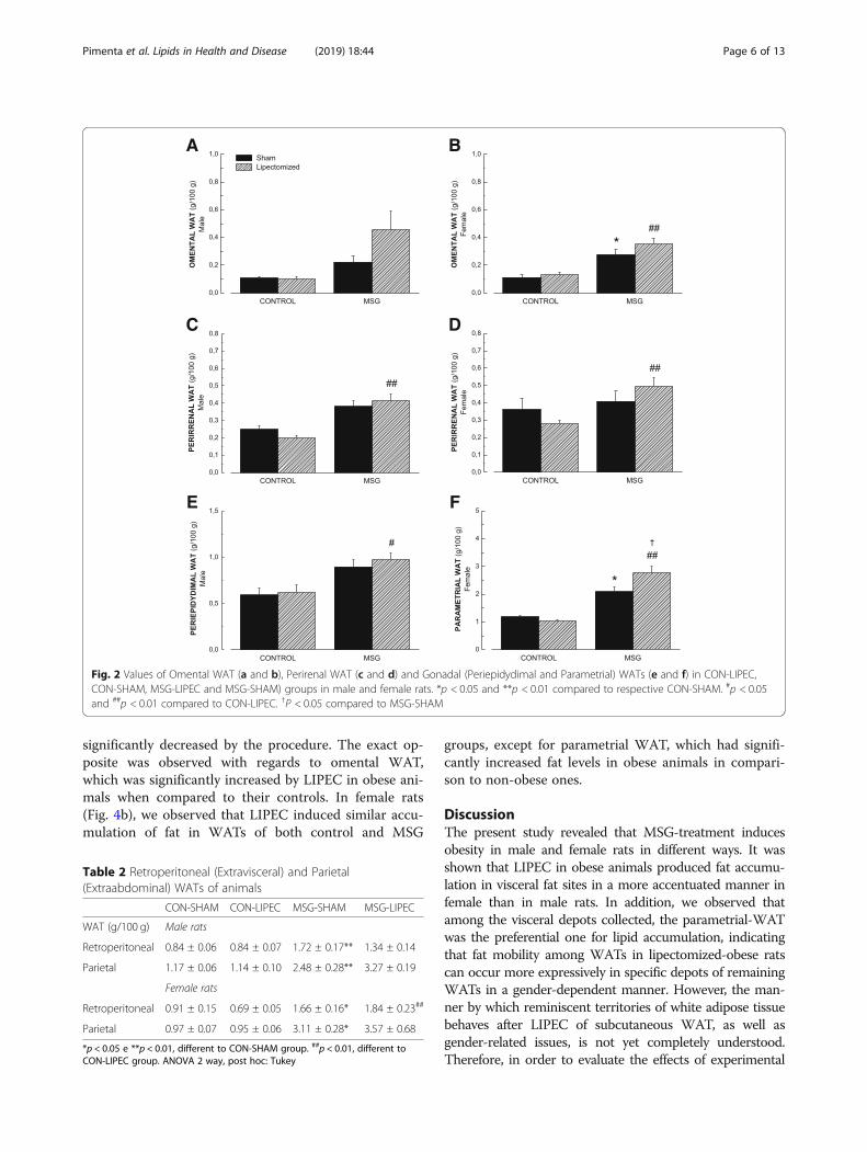
of the WATs that compose the visceral fat (Fig. 2). We didnot observe significant differences regarding the Omental-WAT in male rats (Fig. 2a). In females, we observed in-creased fat accumulation in both obesity-induced groupswhen compared to their respective controls (Fig. 2b).As to Perirenal-WAT, we observed, for both sexes,
that lipectomized obese animals presented higher fat ac-cumulation values when compared to lipectomized con-trol animals, with obesity by itself showing no significanteffects (Fig. 2).Finally, regarding Gonadal-WATs, we observed in-
creased values of Periepididymal WAT in lipectomizedobese animals when compared to their lipectomizedcontrols (Fig. 2e). Parametrial WAT values, on the otherhand, were found to be significantly higher in both MSGgroups when compared to their respective controls, aswell as when obese lipectomized animals were comparedto obese non-lipectomized animals (Fig. 2f ). Taken to-gether, these results suggest that the higher accumula-tion of visceral WATs observed in female rats is mainlydue to an increase in Parametrial WAT in obese ani-mals, particularly in those submitted to LIPEC.We have also dwelt upon the differences between extra-
visceral and extraabdominal fat accumulation, through theanalysis of retroperitoneal- and parietal-WATs (Table 2).We observed that, for both sexes, obesity itself inducedhigher retroperitoneal and parietal fat accumulation whencompared to control animals. In obese female rats, LIPECinduced a significantly higher accumulation of retroperi-toneal fat, clearly indicating that post-LIPEC fat mobilityto this pad is gender-dependent. It is important to pointout that, as in both lipectomized groups the parietal valuesare relative to the 12th week, while in sham-operatedgroups these values are relative to the 16th week, oneshould indeed expect no significant differences among an-imals undergoing the procedure with regards to their re-spective controls.The mobility of fat among WATs after MSG-induced
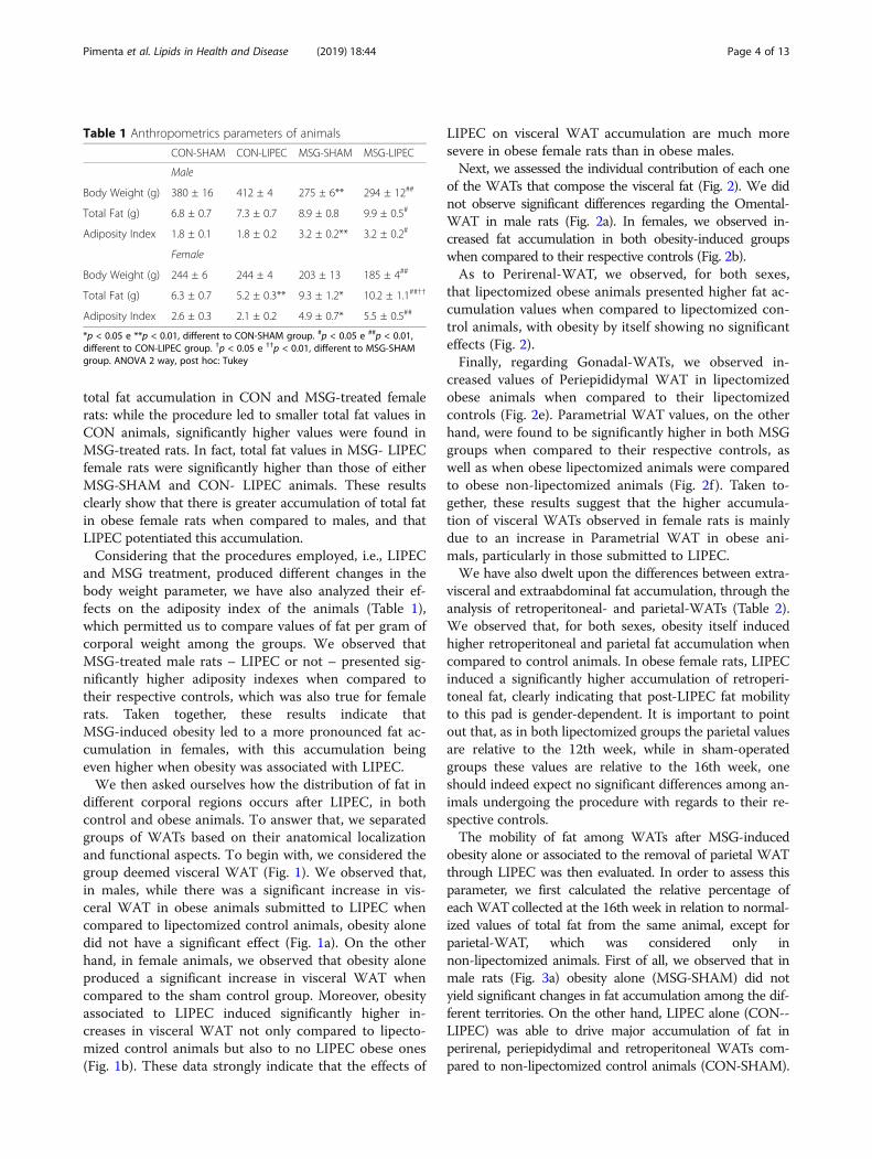
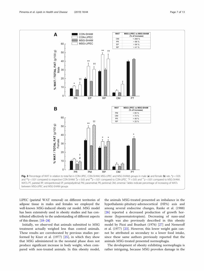
obesity alone or associated to the removal of parietal WATthrough LIPEC was then evaluated. In order to assess thisparameter, we first calculated the relative percentage ofeach WATcollected at the 16th week in relation to normal-ized values of total fat from the same animal, except forparietal-WAT, which was considered only innon-lipectomized animals. First of all, we observed that inmale rats (Fig. 3a) obesity alone (MSG-SHAM) did notyield significant changes in fat accumulation among the dif-ferent territories. On the other hand, LIPEC alone (CON--LIPEC) was able to drive major accumulation of fat inperirenal, periepidydimal and retroperitoneal WATs com-pared to non-lipectomized control animals (CON-SHAM).
Table 1 Anthropometrics parameters of animals
CON-SHAM CON-LIPEC MSG-SHAM MSG-LIPEC
Male
Body Weight (g) 380 ± 16 412 ± 4 275 ± 6** 294 ± 12##
Total Fat (g) 6.8 ± 0.7 7.3 ± 0.7 8.9 ± 0.8 9.9 ± 0.5#
Adiposity Index 1.8 ± 0.1 1.8 ± 0.2 3.2 ± 0.2** 3.2 ± 0.2#
Female
Body Weight (g) 244 ± 6 244 ± 4 203 ± 13 185 ± 4##
Total Fat (g) 6.3 ± 0.7 5.2 ± 0.3** 9.3 ± 1.2* 10.2 ± 1.1##††
Adiposity Index 2.6 ± 0.3 2.1 ± 0.2 4.9 ± 0.7* 5.5 ± 0.5##
*p < 0.05 e **p < 0.01, different to CON-SHAM group. #p < 0.05 e ##p < 0.01,different to CON-LIPEC group. †p < 0.05 e ††p < 0.01, different to MSG-SHAMgroup. ANOVA 2 way, post hoc: Tukey
Pimenta et al. Lipids in Health and Disease (2019) 18:44 Page 4 of 13
When associated to obesity (MSG-LIPEC vs MSG-SHAM),we observed that LIPEC promoted crescent accumulationof fat among the following WATs: omental (268%) > > peri-renal (86%) > periepidydimal (84%) > retroperitoneal (41%).These results indicate that while LIPEC, alone or associatedto obesity, promotes proportionally distinct levels of fat ac-cumulation in different WATs, obesity alone – in spite ofinducing higher adiposity – led to fat distribution in a moreuniform manner.In female rats (Fig. 3b), we found that obesity alone
led to proportionally higher levels of fat being distrib-uted to perirenal and parietal WATs. LIPEC alone pro-moted a major percentage of fat accumulation inparametrial and retroperitoneal WATs when comparedto non-lipectomized control animals. And, unlike in
male rats, the effect of LIPEC associated to obesityyielded the following crescent proportional accumula-tion: parametrial (85%) > omental (74%) > perirenal(48%) > retoperitoneal (25%), corroborating the results inwhich we observed major increased accumulation in vis-ceral fat.Finally, we analyzed the magnitude of the effects in-
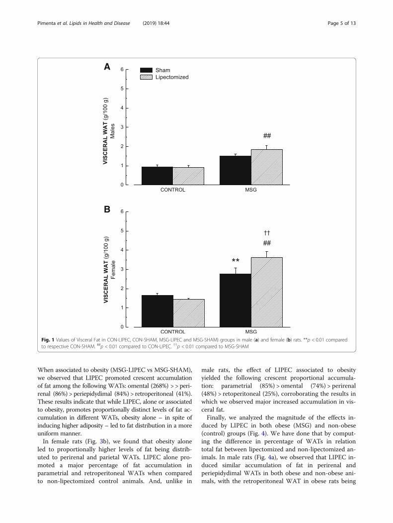
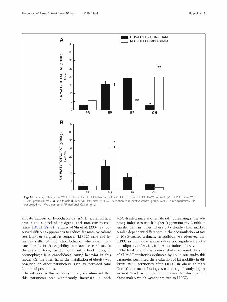
duced by LIPEC in both obese (MSG) and non-obese(control) groups (Fig. 4). We have done that by comput-ing the difference in percentage of WATs in relationtotal fat between lipectomized and non-lipectomized an-imals. In male rats (Fig. 4a), we observed that LIPEC in-duced similar accumulation of fat in perirenal andperiepidydimal WATs in both obese and non-obese ani-mals, with the retroperitoneal WAT in obese rats being
A
B
Fig. 1 Values of Visceral Fat in CON-LIPEC, CON-SHAM, MSG-LIPEC and MSG-SHAM) groups in male (a) and female (b) rats. **p < 0.01 comparedto respective CON-SHAM. ##p < 0.01 compared to CON-LIPEC. ††p < 0.01 compared to MSG-SHAM
Pimenta et al. Lipids in Health and Disease (2019) 18:44 Page 5 of 13
significantly decreased by the procedure. The exact op-posite was observed with regards to omental WAT,which was significantly increased by LIPEC in obese ani-mals when compared to their controls. In female rats(Fig. 4b), we observed that LIPEC induced similar accu-mulation of fat in WATs of both control and MSG
groups, except for parametrial WAT, which had signifi-cantly increased fat levels in obese animals in compari-son to non-obese ones.
DiscussionThe present study revealed that MSG-treatment inducesobesity in male and female rats in different ways. It wasshown that LIPEC in obese animals produced fat accumu-lation in visceral fat sites in a more accentuated manner infemale than in male rats. In addition, we observed thatamong the visceral depots collected, the parametrial-WATwas the preferential one for lipid accumulation, indicatingthat fat mobility among WATs in lipectomized-obese ratscan occur more expressively in specific depots of remainingWATs in a gender-dependent manner. However, the man-ner by which reminiscent territories of white adipose tissuebehaves after LIPEC of subcutaneous WAT, as well asgender-related issues, is not yet completely understood.Therefore, in order to evaluate the effects of experimental
A B
C D
E F
Fig. 2 Values of Omental WAT (a and b), Perirenal WAT (c and d) and Gonadal (Periepidydimal and Parametrial) WATs (e and f) in CON-LIPEC,CON-SHAM, MSG-LIPEC and MSG-SHAM) groups in male and female rats. *p < 0.05 and **p < 0.01 compared to respective CON-SHAM. #p < 0.05and ##p < 0.01 compared to CON-LIPEC. †P < 0.05 compared to MSG-SHAM
Table 2 Retroperitoneal (Extravisceral) and Parietal(Extraabdominal) WATs of animals
CON-SHAM CON-LIPEC MSG-SHAM MSG-LIPEC
WAT (g/100 g) Male rats
Retroperitoneal 0.84 ± 0.06 0.84 ± 0.07 1.72 ± 0.17** 1.34 ± 0.14
Parietal 1.17 ± 0.06 1.14 ± 0.10 2.48 ± 0.28** 3.27 ± 0.19
Female rats
Retroperitoneal 0.91 ± 0.15 0.69 ± 0.05 1.66 ± 0.16* 1.84 ± 0.23##
Parietal 0.97 ± 0.07 0.95 ± 0.06 3.11 ± 0.28* 3.57 ± 0.68
*p < 0.05 e **p < 0.01, different to CON-SHAM group. ##p < 0.01, different toCON-LIPEC group. ANOVA 2 way, post hoc: Tukey
Pimenta et al. Lipids in Health and Disease (2019) 18:44 Page 6 of 13
LIPEC (parietal WAT removal) on different territories ofadipose tissue in males and females we employed thewell-known MSG-induced obesity rat model. MSG modelhas been extensively used in obesity studies and has con-tributed effectively to the understanding of different aspectsof this disease. [18–24]Initially, we observed that animals submitted to MSG
treatment actually weighed less than control animals.These results are corroborated by previous studies per-formed by Kizer et al. (1977) [25], in which they showthat MSG administered in the neonatal phase does notproduce significant increase in body weight, when com-pared with non-treated animals. In this obesity model,
the animals MSG-treated presented an imbalance in thehypothalamic-pituitary-adrenocortical (HPA) axis andamong several endocrine changes, Ranke et al. (1988)[26] reported a decreased production of growth hor-mone (hyposomatotropism). Decreasing of naso-anallength was also previously described in this obesitymodel by Pizzi and Branhart (1976) [27] and Nemeroffet al. (1977) [23]. However, this lower weight gain can-not be attributed as secondary to a lower food intake,since these same authors previously reported that theanimals MSG-treated presented normophagia.The development of obesity exhibiting normophagia is
rather intriguing, because MSG provokes damage in the
A
B
Fig. 3 Percentage of WAT in relation to total fat in CON-LIPEC, CON-SHAM, MSG-LIPEC and MSG-SHAM) groups in male (a) and female (b) rats. *p < 0.05and **p < 0.01 compared to respective CON-SHAM. #p < 0.05 and ##p < 0.01 compared to CON-LIPEC. †P < 0.05 and ††p < 0.01 compared to MSG-SHAM.WATs: PT, parietal; RP, retroperitoneal; EP, periepidydimal; PM, parametrial; PR, perirenal; OM, omental. Tables indicate percentage of increasing of WATsbetween MSG-LIPEC and MSG-SHAM groups
Pimenta et al. Lipids in Health and Disease (2019) 18:44 Page 7 of 13
arcuate nucleus of hypothalamus (ANH), an importantarea in the control of orexigenic and anorectic mecha-nisms [18, 21, 28–34]. Studies of Shi et al. (2007, 35] ob-served different approaches to reduce fat mass by caloricrestriction or surgical fat removal (LIPEC) male and fe-male rats affected food intake behavior, which can impli-cate directly in the capability to restore visceral fat. Inthe present study, we did not quantify food intake, asnormophagia is a consolidated eating behavior in thismodel. On the other hand, the installation of obesity wasobserved on other parameters, such as increased totalfat and adipose index.In relation to the adiposity index, we observed that
this parameter was significantly increased in both
MSG-treated male and female rats. Surprisingly, the adi-posity index was much higher (approximately 2-fold) infemales than in males. These data clearly show markedgender-dependent differences in the accumulation of fatsin MSG-treated animals. In addition, we observed thatLIPEC in non-obese animals does not significantly alterthe adiposity index, i.e., it does not induce obesity.The total fats in the present study represent the sum
of all WAT territories evaluated by us. In our study, thisparameter permitted the evaluation of fat mobility in dif-ferent WAT territories after LIPEC in obese animals.One of our main findings was the significantly highervisceral WAT accumulation in obese females than inobese males, which were submitted to LIPEC.
A
B
Fig. 4 Percentage changes of WAT in relation to total fat between control (CON-LIPEC minus CON-SHAM) and MSG (MSG-LIPEC minus MSG-SHAM) groups in male (a) and female (b) rats. *p < 0.05 and **p < 0.01 in relation to respective control group. WATs: RP, retroperitoneal; EP,periepidydimal; PM, parametrial; PR, perirenal; OM, omental
Pimenta et al. Lipids in Health and Disease (2019) 18:44 Page 8 of 13
There are yet great controversy about the distributionand/or accumulation of lipids in the reminiscent WATdepots after LIPEC, as well as in relation the mecha-nisms involved, mainly when analyzed the influence ofgender. Different from our findings, previous studiesperformed by Shi et al. (2007) [35] observed that femalemice had a reduced capability to restore visceral fat(retroperitoneal depots) after fat loss by LIPEC. A pos-sible reason for this discrepant result could be explainedby the fact that we evaluate several visceral WAT depotsand tried to evaluate the association of obesity and par-ietal LIPEC in the rat model. However, it is important toemphasize that we choose the withdrawal of parietalsubcutaneous WAT depots because this has also been aroutine in humans.When focusing the metabolic role of different regions
of the body adipose tissue, cumulative data from the lastdecades of research consolidated the notion presented in1947 by Vague [36] that obesity is not a homogeneouscondition. Others have suggested that the data regionaldistribution of adipose tissue is important for the under-standing of obesity and its correlation with metabolicdisorders of glucose and lipids [37]. Several studies haveshown that the harmful influence of obesity on meta-bolic processes is mediated by intra-abdominal fat de-position. In this sense, visceral fat has been correlatedwith glucose intolerance and associated with hyperinsuli-nemia during the oral glucose tolerance test, suggestinga clinical picture of insulin resistance [38–41] as hasbeen evidenced by studies about the effects of neonatalMSG administration on insulin resistance. The increasein the adiposity of these animals was believed to be sec-ondary to the increase in sympathetic hypothalamic ac-tivity, which decreases the transport of GLUT-4 inbrown adipose tissue (BAT), consequently decreasingthermogenesis [42]. These effects could even be justifiedby the decrease of NPY levels in areas such as ANH andparaventricular nucleus (PVN) in the hypothalamus [43].However, a more plausible hypothesis is related to glu-cose metabolism, since the increased sympathetic activ-ity in visceral adipose depots seems too produce lipolysisand lipid mobilization by activating of β3-adrenoceptors(see Fig. 5). Previous studies in humans, showed that thelipolytic β3-adrenoreceptor sensitivity was 12 timeshigher in men, and the antilipolytic α2-adrenoreceptorsensitivity was 17 times lower in men, which lead us toconclude that in obesity, the catecholamine-induced rateof lipid mobilization from visceral fat is higher in menthan women [44].Nevertheless, the role of brown adipose tissue and its
effects to thermogenesis cannot be ruled out. Adipocytesfrom obese patients are characterized by altered endo-crine function, which leads to increased secretion of pro-inflammatory adipokines [45–47]. The activation of
inflammatory pathways leads to insulin resistance in per-ipheral tissues, such as skeletal muscle and adipose tis-sue itself [48]. Only in the last decade, that skeletalmuscle has been shown to behave an active endocrineorgan that releases myokine, which is described as beingable to communicate with cells in a local autocrine/para-crine manner within the muscles, or endocrine to distanttissues [49–51]. Myokines, peptides secreted by the mus-cles, are also produced by adipose tissue, activatingthermogenesis in the brown adipose tissue in vitro andin vivo and participating in some metabolic functionsand energy dissipation in these cells. Obese animalsshow higher expression of this peptide, suggesting somelevel of resistance to its effects [52, 53]. Perhaps, thiscould explain why, in the present study, LIPEC did notalter weight, total fat or adiposity index until the 16thweek (when the animals were sacrificed) in non-obeseanimals, contrasting with what was observed inMSG-treated obese animals.The novelty of the present study is the reporting of a
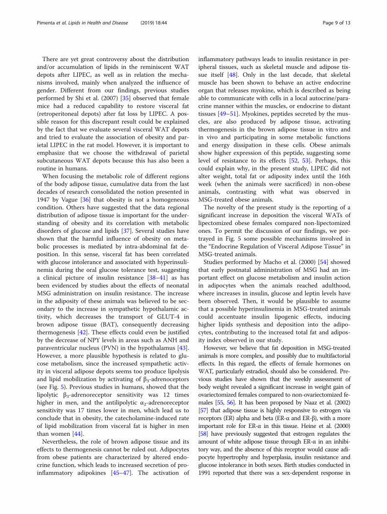
significant increase in deposition the visceral WATs oflipectomized obese females compared non-lipectomizedones. To permit the discussion of our findings, we por-trayed in Fig. 5 some possible mechanisms involved inthe “Endocrine Regulation of Visceral Adipose Tissue” inMSG-treated animals.Studies performed by Macho et al. (2000) [54] showed
that early postnatal administration of MSG had an im-portant effect on glucose metabolism and insulin actionin adipocytes when the animals reached adulthood,where increases in insulin, glucose and leptin levels havebeen observed. Then, it would be plausible to assumethat a possible hyperinsulinemia in MSG-treated animalscould accentuate insulin lipogenic effects, inducinghigher lipids synthesis and deposition into the adipo-cytes, contributing to the increased total fat and adipos-ity index observed in our study.However, we believe that fat deposition in MSG-treated
animals is more complex, and possibly due to multifactorialeffects. In this regard, the effects of female hormones onWAT, particularly estradiol, should also be considered. Pre-vious studies have shown that the weekly assessment ofbody weight revealed a significant increase in weight gain ofovariectomized females compared to non-ovariectomized fe-males [55, 56]. It has been proposed by Naaz et al. (2002)[57] that adipose tissue is highly responsive to estrogen viareceptors (ER) alpha and beta (ER-α and ER-β), with a moreimportant role for ER-α in this tissue. Heine et al. (2000)[58] have previously suggested that estrogen regulates theamount of white adipose tissue through ER-α in an inhibi-tory way, and the absence of this receptor would cause adi-pocyte hypertrophy and hyperplasia, insulin resistance andglucose intolerance in both sexes. Birth studies conducted in1991 reported that there was a sex-dependent response in
Pimenta et al. Lipids in Health and Disease (2019) 18:44 Page 9 of 13
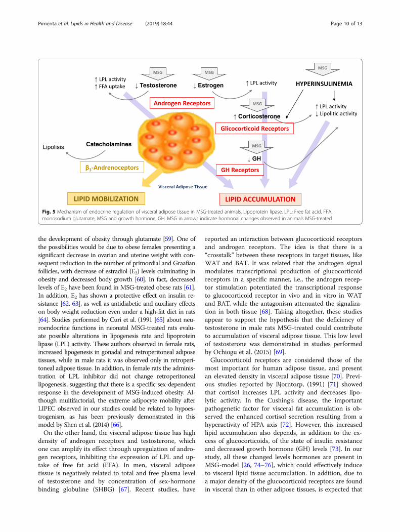
the development of obesity through glutamate [59]. One ofthe possibilities would be due to obese females presenting asignificant decrease in ovarian and uterine weight with con-sequent reduction in the number of primordial and Graafianfollicles, with decrease of estradiol (E2) levels culminating inobesity and decreased body growth [60]. In fact, decreasedlevels of E2 have been found in MSG-treated obese rats [61].In addition, E2 has shown a protective effect on insulin re-sistance [62, 63], as well as antidiabetic and auxiliary effectson body weight reduction even under a high-fat diet in rats[64]. Studies performed by Curi et al. (1991 [65] about neu-roendocrine functions in neonatal MSG-treated rats evalu-ate possible alterations in lipogenesis rate and lipoproteinlipase (LPL) activity. These authors observed in female rats,increased lipogenesis in gonadal and retroperitoneal adiposetissues, while in male rats it was observed only in retroperi-toneal adipose tissue. In addition, in female rats the adminis-tration of LPL inhibitor did not change retroperitoneallipogenesis, suggesting that there is a specific sex-dependentresponse in the development of MSG-induced obesity. Al-though multifactorial, the extreme adipocyte mobility afterLIPEC observed in our studies could be related to hypoes-trogenism, as has been previously demonstrated in thismodel by Shen et al. (2014) [66].On the other hand, the visceral adipose tissue has high
density of androgen receptors and testosterone, whichone can amplify its effect through upregulation of andro-gen receptors, inhibiting the expression of LPL and up-take of free fat acid (FFA). In men, visceral adiposetissue is negatively related to total and free plasma levelof testosterone and by concentration of sex-hormonebinding globuline (SHBG) [67]. Recent studies, have
reported an interaction between glucocorticoid receptorsand androgen receptors. The idea is that there is a“crosstalk” between these receptors in target tissues, likeWAT and BAT. It was related that the androgen signalmodulates transcriptional production of glucocorticoidreceptors in a specific manner, i.e., the androgen recep-tor stimulation potentiated the transcriptional responseto glucocorticoid receptor in vivo and in vitro in WATand BAT, while the antagonism attenuated the signaliza-tion in both tissue [68]. Taking altogether, these studiesappear to support the hypothesis that the deficiency oftestosterone in male rats MSG-treated could contributeto accumulation of visceral adipose tissue. This low levelof testosterone was demonstrated in studies performedby Ochiogu et al. (2015) [69].Glucocorticoid receptors are considered those of the
most important for human adipose tissue, and presentan elevated density in visceral adipose tissue [70]. Previ-ous studies reported by Bjorntorp, (1991) [71] showedthat cortisol increases LPL activity and decreases lipo-lytic activity. In the Cushing’s disease, the importantpathogenetic factor for visceral fat accumulation is ob-served the enhanced cortisol secretion resulting from ahyperactivity of HPA axis [72]. However, this increasedlipid accumulation also depends, in addition to the ex-cess of glucocorticoids, of the state of insulin resistanceand decreased growth hormone (GH) levels [73]. In ourstudy, all these changed levels hormones are present inMSG-model [26, 74–76], which could effectively induceto visceral lipid tissue accumulation. In addition, due toa major density of the glucocorticoid receptors are foundin visceral than in other adipose tissues, is expected that
Fig. 5 Mechanism of endocrine regulation of visceral adipose tissue in MSG-treated animals. Lipoprotein lipase, LPL; Free fat acid, FFA,monosodium glutamate, MSG and growth hormone, GH. MSG in arrows indicate hormonal changes observed in animals MSG-treated
Pimenta et al. Lipids in Health and Disease (2019) 18:44 Page 10 of 13
the lipid-accumulating effect of cortisol would be morepronounced in visceral than in other fat areas [71].The effects of GH on the regulation of visceral fat have
to be considered separately. A decrease in circulatingGH was previously demonstrated in human obesity andit was associated to an increase in visceral fat [71, 77].Otherwise, a reduction in visceral adipose tissue was ob-served in humans with acromegaly, a state of GH excess[78]. Thus, most action of GH on adipose tissue are toprevent lipid accumulation and to stimulate lipidmobilization. However, the regulation of adipose tissuemetabolism requires synergism with steroid hormones,and in relation to lipid accumulation, LPL is markedlyinhibited by GH in the presence of either testosterone orcortisol [71]. Considering that their receptors are par-ticularly dense in visceral adipose tissue, a more pro-nounced effect of GH would be expected in this tissue[71]. In this way, an important hallmark of MSG modelis the GH deficiency, which, in turn, would be consid-ered one of major factor to induce visceral fat accumula-tion, obviously in synergism to other changed hormonalfactors above described.Summarizing, low levels of estrogen/testosterone, defi-
ciency of GH and increased secretion of corticosterone,which has been observed in MSG-treated rats, could in-duce higher visceral fat accumulation. In female rats MSGsubjected to back of estrogen could justify whyparamentrial-WAT is more responsive to lipid accumula-tion, as has been observed in women in postmenopause,when abdominal fat cell present high LPL activity.The need to feel good about how we look is part of the
human condition. Overweight and obesity usually lead thepopulation to adopt miracle diets to lose weight or treatassociated illnesses such as dyslipidemias and diabetes. Onthe other hand, “nutraceuticals” and “functional foods”has been frequently used as an alternative biological inter-vention to pharmacological methods [79–82]. Chen et al.(2008) [83] that reported the beneficial Influences of redmold rice (RMR) on obesity and dyslipidemia. Some evi-dences have been showed that Dietary polyphenols (DPs)are effective and promote health via multiple signallingpathways (such as lipid anabolism/catabolism pathways,apoptotic pathways) [84]. A combined treatment withgenestein, quercetin, and resveratrol was shown higher in-hibition of adipogenesis in both primary human adipo-cytes and 3 T3-L1 murine adipocyte than individualmolecules [85]. In the near future, nutraceutical or func-tional foods, might be associated to pos-surgery proce-dures, as LIPEC, to prevent obesity, by the risk factor ofhealth that could be entailed.
ConclusionsThe results of the present study led us to conclude thatobesity induced by MSG treatment occurs differently in
male and female rats. When associated with parietalLIPEC, there is a significant increase in the deposition ofvisceral fat, which is significantly higher in obese femalethan in male rats. Finally, these results allow us to inferthat visceral WAT does not behave as a single tissue.
AbbreviationsANOVA: Analysis of variance; BAT: Brown adipose tissue; CNS: Central nervoussystem; CON: Control; EP: Epididymal; HAN: Hypothalamus arcuate nucleus;LIPEC: Lipectomy; MSG: Monosodium glutamate; OM: Omental;PM: Parametrial; PR: Perirenal; PVN: Paraventricular nucleus;RP: Retroperitoneal; SEM: Standart error mean; SHAM: Sham-operatedsurgery; WAT: White adipose tissue
AcknowledgementsNot applicable
FundingNo funding
Availability of data and materialsAll data generated or analyzed in this study are included in this manuscript.
Authors’ contributionsFSP, HT and HM conceived and designed the experiments; FSP, HT, EWJ andHM performed the experiments; FSP, EWJ, MRHC, FVC, ECV and HM analyzedthe data; FSP, FVC, ECV and HM wrote the paper. All authors read andapproved the final manuscript.
Ethics approval and consent to participateAll experiments were conducted in accordance with the U.S. NationalInstitute of Health Guidelines for the Care and Use of Laboratory Animals,and study protocols were previously approved by our Institutional EthicsCommittee for the Use of Animals (Protocol # 034/2011).
Consent for publicationNot applicable.
Competing interestsThe authors declare that they have no competing interests.
Publisher’s NoteSpringer Nature remains neutral with regard to jurisdictional claims inpublished maps and institutional affiliations.
Author details1Department of Physiological Sciences, Health Sciences Center, FederalUniversity of Espírito Santo, Av. Marechal Campos 1468, Vitória, ES CEP29042-751, Brazil. 2Laboratory of Translational Physiology and Pharmacology,Pharmaceutical Sciences Graduate Program, Vila Velha University (UVV),Avenida Comissário José Dantas de Melo, 21 Bairro Boa Vista II, Vila Velha, ESCEP 29102-920, Brazil. 3Departament of Medical Clinic, Escola Superior deCiências da Santa Casa de Misericórdia de Vitória (EMESCAM), Av. Nossa Sra.da Penha, 2190 - Bela Vista, Vitória, ES CEP 29027-502, Brazil. 4FaculdadeEstácio, Av. Dr. Herwan Modenese Wanderley, 1001. Bairro Jardim Camburi,Vitória, ES CEP 29092-095, Brazil. 5Sports Department, Center for PhysicalEducation and Sports, Federal University of Espírito Santo, Avenida FernandoFerrari, 514. Bairro Goiabeiras, Vitória, ES CEP 29075-910, Brazil.
Received: 16 November 2018 Accepted: 24 January 2019
References1. Kelly T, Yang W, Chen CS, Reynolds K, He J. Global burden of obesity in
2005 and projections to 2030. Int J Obes. 2008;32(9):1431–7.2. Haslam DW, James WP. Obesity Lancet. 2005;366(9492):1197–209.3. Tchkonia T, Thomou T, Zhu Y, Karagiannides I, Pothoulakis C, Jensen MD,
Kirkland JL. Mechanisms and metabolic implications of regional differencesamong fat depots. Cell Metab. 2013;17(5):644–56.
Pimenta et al. Lipids in Health and Disease (2019) 18:44 Page 11 of 13
4. Cartwright MJ, Schlauch K, Lenburg ME, Tchkonia T, Pirtskhalava T,Cartwright A, Thomou T, Kirkland JL. Aging, depot origin, and preadipocytegene expression. J Gerontol A Biol Sci Med Sci. 2010;65(3):242–51.
5. Tchkonia T, Morbeck DE, Von Zglinicki T, Van Deursen J, Lustgarten J,Scrable H, Khosla S, Jensen MD, Kirkland JL. Fat tissue, aging, and cellularsenescence. Aging Cell. 2010;9(5):667–84.
6. Cox-York K, Wei Y, Wang D, Pagliassotti MJ, Foster MT. Lower body adiposetissue removal decreases glucose tolerance and insulin sensitivity in micewith exposure to high fat diet. Adipocyte. 2014;4(1):32–43.
7. Björntorp P. Visceral obesity: a "civilization syndrome". Obes Res. 1993;1(3):206–22.8. Kissebah AH, Krakower GR. Regional adiposity and morbidity. Physiol Rev.
1994;74(4):761–811.9. Després JP, Lemieux I. Abdominal obesity and metabolic syndrome. Nature.
2006;444(7121):881–7.10. Van Gaal LF, Mertens IL, De Block CE. Mechanisms linking obesity with
cardiovascular disease. Nature. 2006;444(7121):875–80.11. Makrogiannis S, Caturegli G, Davatzikos C, Ferrucci L. Computer-aided
assessment of regional abdominal fat with food residue removal in CT.Acad Radiol. 2013;20(11):1413–21.
12. Michel C, Cabanac M. Lipectomy, body weight, and body weight set pointin rats. Physiol Behav. 1999;66(3):473–9.
13. Lacy EL, Bartness TJ. Effects of white adipose tissue grafts on total body fatand cellularity are dependent on graft type and location. Am J PhysiolRegul Integr Comp Physiol. 2005;289(2):R380–8.
14. Hernandez TL, Kittelson JM, Law CK, Ketch LL, Stob NR, Lindstrom RC,Scherzinger A, Stamm ER, Eckel RH. Fat redistribution following suctionlipectomy: defense of body fat and patterns of restoration. Obesity (SilverSpring). 2011;19(7):1388–95.
15. Benatti F, Solis M, Artioli G, Montag E, Painelli V, Saito F, Baptista L, Costa LA,Neves R, Seelaender M, Ferriolli E, Pfrimer K, Lima F, Roschel H, Gualano B,Lancha A Jr. Liposuction induces a compensatory increase of visceral fatwhich is effectively counteracted by physical activity: a randomized trial. JClin Endocrinol Metab. 2012;97(7):2388–95.
16. Swanson E. Photographic measurements in 301 cases of liposuction andabdominoplasty reveal fat reduction without redistribution. Plast ReconstrSurg. 2012;130(2):311e–22e.
17. Yazigi Solis M, Artioli GG, Montag E, de SallesPainelli V, Saito FL, Lima FR,Roschel H, Gualano B, Lancha Junior AH, Benatti FB. The liposuction-induced effects on adiponectin and selected cytokines are not affected byexercise training in women. Int J Endocrinol. 2014;2014:315382.
18. Olney JW. Brain lesions, obesity and other disturbances in mice treated withmonosodium glutamate. Science. 1969;164:719–21.
19. Olney JW. Glutamate-induced retinal degeneration in neo-natal mice.Electron microscopy of the acutely evolving lesion. J Neuropath Exp Neurol.1969;28:455–74.
20. Olney JW. Glutamate-induced retinal degeneration in neo-natal mice.Electron microscopy of the acutely evolving lesion. J Neuropath Exp Neurol.1969;28:455–74.
21. Lemkey-Johnston N, Reynolds WA. Nature and extent of brain lesions inmice related to ingestion of monosodium glutamate: A light and electronmicroscope study. J Neuropath exp Neurol. 1974;33:74–97.
22. Dolnikoff MS, Kater CE, Egami M, de Andrade IS, Marmo MR. Neonataltreatment with monosodium glutamate increases plasma corticosterone inthe rat. Neuroendocrinology. 1988;48(6):645–9.
23. Nemeroff CB, Grant LD, Bissette G, Ervin GN, Harrell LE, Prange AJ Jr.Growth, endocrinological and behavioral deficits after monosodium L-glutamate in the neonatal rat: possible involvement of arcuate dopamineneuron damage. Psychoneuroendocrinology. 1977;2(2):179–96.
24. Nenoff P, Remke H, Müller F, Arndt T, Mothes T. In vivo assessment ofinsulin binding in different organs of growing and adult glutamate-inducedobese rats. Exp Clin Endocrinol. 1993;101(4):215–21.
25. Kizer JS, Nemeroff CB, Youngblood WW. Neurotoxic amino acids andstructurally related analogs. Pharmacol Rev. 1977;29(4):301–18.
26. Remke H, Wilsdorf A, Müller F. Development of hypothalamic obesity ingrowing rats. Exp Pathol. 1988;33(4):223–32.
27. Pizzi WJ, Barnhart JE. Effects of monosodium glutamate on somaticdevelopment obesity in KK mice. Pharmacol Biochem Behav. 1976;5:551–7.
28. Olney JW, Ho OL. Brain damage in infant mice following oral intake ofglutamate, aspartate or cysteine. Nature (Lond). 1970;227:609–10.
29. Abraham R, Doughtery W, Goldberg L, Coulston F. The response of thehypothalamus to high doses of monosodium glutamate in mice and
monkeys. Cytochemistry and ultra- structural study of lysosomal changes.Expl Molec Path. 1971;15:43–60.
30. Burde RM, Acute BS, Kayes J. Monosodium glutamate: Acute effect of oraland subcutaneous administration on the arcuate nucleus of thehypothalamus in mice and rats. Nature, (Lond.). 1971;233:58–60.
31. Everly JL. Light microscopic examination of MSG-induced lesions in brain offetal and neonatal rats. Anat Rec. 1971;169:312.
32. Murakami U, Inouye M. Brain lesions in the mouse fetus ojed by maternaladministration of monosodium glutamate. Congenital Anomalies. 1971;11:171–7.
33. Olney JW, Sharpe LG, Feigin RD. Glutamate-induced brain damage in infantprimates. J Neuropath Exp Neurol. 1972;31:464–88.
34. Shimizu K, Mizutani A, Inouye M. Electron microscopic studies on thehypothalamic lesions in the mouse fetus caused by monosodiumglutamate. Teratology. 1973;8:105.
35. Shi H, Strader AD, Woods SC, Seeley RJ. Sexually dimorphic responses to fatloss after caloric restrictionor surgical lipectomy. Am J Physiol EndocrinolMetab. 2007;293:E316–26.
36. Vague J. La differe’nciation sexuelle, facteur determinant des formes del’obe’site. Presse Med. 1947;55:339–40.
37. Bouchard C, Despre’s J-P, Maurie’ge PO. Genetic and nongeneticdeterminants of regional fat distribution. Endocr Rev. 1993;14:72–93.
38. Fujioka S, Matsuzawa Y, Tokunaga K, Tarui S. Contribution of intra-abdominal fat accumulation to the impairment of glucoseand lipidmetabolism in human obesity. Metabolism. 1987;36:54–9.
39. Despre’s J-P, Nadeau A, Tremblay A, Ferland M, Moorjani S, Lupien PJ,The’riault G, Pinault S, Bouchard C. Role of deep abdominal fat in theassociation between regional adipose tissue distribution and glucosetolerance in obese women. Diabetes. 1989;38:304–9.
40. Pouliot M-C, Despre’s J-P, Nadeau A, Moorjani S, Prud’Homme D, Lupien PJ,Tremblay A, Bouchard C. Visceral obesity in men. Associations with glucosetolerance, plasma insulin and lipoprotein levels. Diabetes. 1992;41:826–34.
41. Jensen MD. Role of body fat distribution and the metabolic complicationsofobesity. J Clin Endocrinol Metab. 2008;93(11 Suppl 1):S57–63.
42. Morris MJ, Tortelli CF, Filippis A, Proietto J. Reduced BAT function as amechanism for obesity in the hypophagic, neuropeptide Y deficientmonosodium glutamate-treated rat. Regul Pept. 1998;75-76:441–7.
43. Tokuyama K, Himms-Hagen J. Brown adipose tissue thermogenesis, torpor,and obesity of glutamate-treated mice. Am J Phys. 1986;251(4 Pt 1):E407–15.
44. Lönnqvist F, Thorne A, Large V, Arner P. Sex differences in visceral fatlipolysis and metabolic complications of obesity. Arterioscler Thromb VascBiol. 1997;17:1472–80.
45. Trayhurn P, Beattie JH. Physiological role of adipose tissue: white adiposetissue as an endocrine and secretory organ. Proc Nutr Soc. 2001;60(3):329–39.
46. Sell H, Dietze-Schroeder D, Eckel J. The adipocyte-myocyte axis in insulinresistance. Trends Endocrinol Metab. 2006;17(10):416–22.
47. Sell H, Divoux A, Poitou C, Basdevant A, Bouillot JL, Bedossa P, Tordjman J,Eckel J, Clément K. Chemerin correlates with markers for fatty liver inmorbidly obese patients and strongly decreases after weight loss inducedby bariatric surgery. J Clin Endocrinol Metab. 2010;95(6):2892–6.
48. DeFronzo RA, Tripathy D. Skeletal muscle insulin resistance is the primarydefect in type 2 diabetes. Diabetes Care Suppl. 2009;2:S157–63.
49. Pedersen BK. The diseasome of physical inactivity--and the role of myokinesin muscle--fat cross talk. J Physiol. 2009;587(Pt 23):5559–68.
50. Pedersen BK. Exercise-induced myokines and their role in chronic diseases.Brain Behav Immun. 2011;25(5):811–6.
51. Pedersen BK, Febbraio MA. Muscles, exercise and obesity: skeletal muscle asa secretory organ. Nat Rev Endocrinol. 2012;8(8):457–65.
52. Raschke S, Eckel J. Adipo-myokines: two sides of the same coin-mediatorsof inflammation and mediators of exercise. Mediat Inflamm. 2013;2013:16.Article ID 320724.
53. Roca-Rivada A, Castelao C, Senin LL, Landrove MO, Baltar J, Crujeiras AB, et al.FNDC5/Irisin is not only a myokine but also an adipokine. PLoS One. 2013;8:e60563.
54. Macho L, Ficková M, Jezová, Zórad S. Late effects of postnataladministration of monosodium glutamate on insulin action in adult rats.Physiol Res. 2000;49(Suppl 1):S79–85.
55. Tofovic SP, Zhang X, Jacson EK, Dacic S, Petrusevska G. 2-Methoxyestradiolmediates the protective effects of estradiol in monocritaline-indicedpulmonary hypertension. Vasc Pharmacol. 2006;45:358–67.
56. Wegorzewska IN, Walters K, Weiser MJ, et al. Postovariectomy weight gainin female rats is reversed by estrogen receptor alpha agonist,propylpyrazoletriol. Am J Obstet Gynecol. 2008;199(1):67.
Pimenta et al. Lipids in Health and Disease (2019) 18:44 Page 12 of 13
57. Naaz A, Zak Causroczymski M, Heine P, et al. Effect of ovariectomy onadipose tissue of mice in the absence of estrogen receptor alpha (ER alpha):a potencial role for estrogen receptor beta (ER beta). Horm Metab Res, v.2003;34:758–63.
58. Heine PA, Taylor JA, Iwamoto GA, et al. Increased adipose tissue in maleand female estrogen receptor-alpha knockout mice. Proc Nati Acad Sci USA.2000;97(23):12729–34.
59. CCM N, Marmo MR, Egami M, Ribeiro EB, Andrade IS, Dolnikoff MS. Effect ofmonosodium glutamate treatment during neonatal development onlipogenesis rate and lipoprotein lipase activity in adult rats. Biochem Int.1991;24(5):927–35.
60. Miśkowiak B, Kesa B, Limanowski A, Partyka M, Filipiak B. Long-term effect ofneonatal monosodium glutamate (MSG) treatment on reproductive systemof the female rat. Folia Morphol (Warsz). 1999;58(2):105–13.
61. Castrogiovanni D, Gaillard RC, Giovambattista A, Spinedi E. Neuroendocrine,metabolic, and immune functions during the acute phase response ofinflammatorystress in monosodium L-glutamate-damaged, hyperadiposemale rat. Neuroendocrinology. 2008;88(3):227–34.
62. Kumagai S, Holmäng A, Björntorp P. The effects of estrogen andprogesterone on insulin sensitivity in female rats. Acta Physiol Scand. 1993;149(1):91–7.
63. Louet JF, LeMay C, Mauvais-Jarvis F. Antidiabetic actions of estrogen: insightfrom human and genetic mouse models. Curr Atheroscler Rep. 2004;6(3):180–5.
64. Bryzgalova G, Lundholm L, Portwood N, Gustafsson JA, Khan A, Efendic S,Dahlman-Wright K. Mechanisms of antidiabetogenic and body weight-lowering effects of estrogen in high-fat diet-fed mice. Am J PhysiolEndocrinol Metab. 2008;295(4):E904–12.
65. Curi CMN, Marmo MR, Egami M, Ribeiro EB, Andrade IS, Dolnikoff MS. Effectof monosodium glutamate treatment during neonatal development onlipogenesis rate and lipoprotein lipase activity in adult rats. Biochem Int.1991;24(5):927–35.
66. Shen M, Kumar SP, Shi H. Estradiol regulates insulin signalingandinflammation in adipose tissue. Horm Mol Biol Clin Investig. 2014;17(2):99–107.
67. Freedland ES. Role of a critical visceral adipose tissue threshold (CVATT) inmetabolic syndrome: implications for controlling dietary carbohydrates: areview. Nutr Metab (Lond). 2004;1(1):12.
68. Spaanderman DCE, Nixon M, Buurstede JC, Sips HC, Schilperoort M, KuipersEN, Backer EA, Kooijman S, Rensen PCN, Homer NZM, Walker BR, Meijer OC,Kroon J. Androgens modulate glucocorticoid receptor activity in adiposetissue and liver. J Endocrinol. 2018. Pii: JOE-18-0503.R1. https://doi.org/10.1530/JOE-18-0503.
69. Ochiogu IS, Ogwu D, Uchendu CN, Okoye ON, Ihedioha JI, Mbegbu EC.Effects of monosodium L-glutamate administration on serum levels ofreproductive hormones and cholesterol, epididymal sperm reserves andtesticular histomorphology of male albino rats. Acta Vet Hung. 2015;63(1):125–39.
70. Rebuffé-Scrive M, Lundholm K, Björntorp P. Glucocorticoid hormonebinding to human adipose tissue. Eur J Clin Investig. 1985;15:267–71.
71. Björntorp P. Endocrine abnormalities in obesity. Diabetes Rev. 1997;5:52–68.72. Wajchenberg BL, Bosco A, Marone MM, Levin S, Rocha M, Lerario AC, Nery
M, Goldman J, Liberman B. Estimation of body fat and lean tissuedistribution by dual energy x-ray absorptiometry and abdominal body fatevaluation by computed tomography in Cushing’s disease. J Clin EndocrinolMetab. 1995;80:2791–4.
73. Wajchenberg BL, Liberman B, Giannella Neto B, Morozimato MY, Semer M,Bracco LO, Salgado LR, Knoepfelmacher M, Borges MHS, Pinto ACAR, KaterC, Lengyel AMJ. Growth hormone axis in Cushing’s syndrome. Horm Res.1996;45:99–107.
74. Ribeiro EB, do Nascimento CM, Andrade IS, Hirata AE, Dolnikoff MS.Hormonal and metabolic adaptations to fasting in monosodium glutamate-obese rats. J Comp Physiol B. 1997;167(6):430–7.
75. Macho L, Jezova D, Zorad S, Fickova M. Postnatal monosodium glutamatetreatment results in attenuation of corticosterone metabolic rate in adultrats. Endocr Regul. 1999;33(2):61–7.
76. Guimarães ED, de Caires Júnior LC, Musso CM, Macedo de Almeida M,Gonçalves CF, Pettersen KG, Paes ST, González Garcia RM, de Freitas MathiasPC, Torrezan R, Mourao-Júnior CA, Andreazzi AE. Altered behavior of adultobese rats by monosodium L-glutamate neonatal treatment is related to
hypercorticosteronemia and activation of hypothalamic ERK1 and ERK2.Nutr Neurosci. 2017;20(3):153–60.
77. Glass AR. Endocrine aspects of obesity. Med Clin North Am. 1989;73:139–60.78. Brummer RJM, Lónn L, Kvist H, Grangård U, Bengtsson B-A, Sjöström L.
Adipose tissue and muscle volume determination by computedtomography in acromegaly before and 1 year after adenomectomy. Eur JClin Investig. 1993;23:199–205.
79. DeFelice SL. The nutraceutical revolution: its impact on food industry R&D.Trends Food Sci Technol. 1995;6:59–61.
80. Kalra EK. Nutraceutical–definition and introduction. AAPS PharmSciTech.2003;5:E25.
81. Scicchitanoa P, Camelib M, Maielloc M, Modestid PA, Muiesane ML, Novof S,Palmieroc P, Sabag PS, Pedrinellih R, Cicconea MM. Nutraceuticals anddyslipidaemia: beyond the common therapeutics. J Funct Foods. 2014;6:11–32.
82. Ton AMM, Arpini CM, Campagnaro BP, Pereira TMC, Vasquez EC. Alzheimer’sdisease: A brief update on the influence of gut microbiota and the impactof functional food. J Food Microbiol. 2018;2(1):11–5.
83. Chen WP, Ho BY, Lee CL, et al. Red mold rice prevents the development ofobesity, dyslipidemia and hyperinsulinemia induced by high-fat diet. Int JObes. 2008;32:1694–704.
84. Milenkovic D, Deval C, Gouranton E, Landrier JF, Scalbert A, Morand C,Mazur A. Modulation of miRNA expression by dietary polyphenols in apoEdeficient mice: A new mechanism of the action of polyphenols. PLoS One.2012;7:e29837.
85. Park HJ, Yang JY, Ambati S, Della-Fera MA, Hausman DB, Rayalam S, BaileCA. Combined effects ofgenistein, quercetin, and resveratrol in human and3T3-L1 adipocytes. J Med Food. 2008;11:773–83.
Pimenta et al. Lipids in Health and Disease (2019) 18:44 Page 13 of 13