Linda de Haas DPT, OCS, CHT - American Society of Hand ... · • Mnemonics • SALTR・ • S: ......
18
Hand Therapy Review Course UC Irvine Medical Center Orange, CA February 24‐26, 2017 Wrist, Hand and Thumb Fractures Linda de Haas PT, DPT, OCS, CHT 2 Copyright, 1996 © Dale Carnegie & Associates, Inc. Wrist & Hand Fractures Wrist & Hand Fracture Topics • Bony structure and anatomy • Bony injury and repair • Fracture types • Factors affecting healing and outcomes • Methods of fracture fixation • Common fractures Bony Structure & Anatomy Composition, Histology, Morphology, Anatomy, Classification Histology Bone Composition Organic Components 30% Dry Weight Type I Collagen Noncollagenous Proteins Growth Factors and Cytokines Inorganic Components 70% Dry Weight Calcium & Minerals Hydroxyapatite Water 6 Normal Osteoporotic
Transcript of Linda de Haas DPT, OCS, CHT - American Society of Hand ... · • Mnemonics • SALTR・ • S: ......

Hand Therapy Review CourseUC Irvine Medical Center
Orange, CAFebruary 24‐26, 2017
Wrist, Hand and Thumb Fractures
Linda de Haas PT, DPT, OCS, CHT
2
Copyright, 1996 © Dale Carnegie & Associates, Inc.
Wrist & Hand Fractures
Wrist & Hand Fracture Topics
•Bony structure and anatomy
•Bony injury and repair•Fracture types•Factors affecting healing and outcomes
•Methods of fracture fixation
•Common fractures
Bony Structure & Anatomy
Composition, Histology, Morphology, Anatomy, Classification
Histology
Bone CompositionOrganic Components 30% Dry Weight
Type I CollagenNoncollagenous ProteinsGrowth Factors and Cytokines
Inorganic Components 70% Dry Weight
Calcium & MineralsHydroxyapatiteWater
6
Normal Osteoporotic

Bone Cell Morphology
Osteoblasts : Bone forming cells,
Immature Osteocytes
Produce type I collagen
Line surface of bone
Osteocytes : Most numerous, Mature Osteocytes
Form latticework
Eventually become mineralized
Osteoclasts
Reabsorb bone
Produced from marrow cells
Bone Structure
Cortical bone – compact bone• Concentrated in diaphysis of long bone•Houses osteons (contain osteocytes)• 80% of skeletal mass• Slower to heal
Cancellous bone – trabecular/ spongy• 20% skeletal mass• Concentrated at epiphysis and metaphysis of long bones
•Higher metabolic turnover
"Structure of a Long Bone" by BruceBlaus ‐ Own work
Netter
Adult Bony Anatomy
Epiphysis : articular end of bone, which is coated with articular cartilage; has growth potential
Diaphysis : shaft of bone
Metaphysis : growing portion of the bone between the diaphysis and epiphysis
Periosteum : tough vascular covering of bone; thicker in children, thins with aging
Wikipedia
Growing Bony AnatomyEpiphysis
Physis : Growth Plate•Between metaphysis and epiphysis
•Fuses approximately age 20 for males and 18 for females
Metaphysis
Diaphysis•Periosteum•Endosteum
Bony Injury & RepairMethod of Injury, Fracture Repair
Indirect (Secondary) Fracture Repair
Results with non‐operative treatment of closed, well‐aligned, relatively stable fractures. Treated by immobilization with a cast or orthosis.
3 Phases1.Inflammatory Phase : first week after injury
2.Reparative Phase : 2‐3 weeks after injury3.Remodeling Phase : 3 weeks‐ 18 monthsafter injury

13
Healing Phases
Inflammatory: First week after injuryReparative: 2 - 3 weeks after injuryRemodeling: 3 weeks - 18 months after injury
Indirect Fracture Repair1. Inflammatory Phase
•Accumulation of a hematoma •between fracture ends •under elevated periosteum
•Bone necrosis (osteocytes lose nutrition)•Angiogenesis, restoration of blood supply•Proliferation of fibroblasts and osteoblasts• Invasion of leukocytes and macrophages
Indirect (Secondary)2. Fracture Reparative Phase
Repair Phase•Hematoma organizes – forms fibrin scaffold for repair cells
•External cartilaginous callus forms from periosteum and internal callus from endosteum
•New bone and osteoblasts bridge fracture site
Crucial period of immobilization during callus formation until clinical stability 4‐6
weeks after injury.
Indirect Fracture Repair3. Remodeling Phase
•Occurs over a prolonged period, 18‐24 months
•Continuous bone resorption/ bone formation
• Influenced by forces of stress•Bone is remodeled as osteoclasts reabsorb callous
Direct Fracture Repair
Occurs with open (surgical) treatment of fractures using pins, screws, plates, external fixation.
•Bone forms across approximated ends of bone
•Gap healing can occur for small defects
• Inflammatory phase skipped; goes straight to reparative phase
Fracture Complications
•Malunion / nonunion
• Joint stiffness•Tendon adhesion •Post‐traumatic arthritis
•Chronic Regional Pain Syndrome
•Open fractures: infection/including osteomyelitis

Fracture TypesImaging & Classification
Imaging Techniques
•X‐ray: standard/stress views, bone lesions
•Fluoroscopy : video of xray
•Arthrogram: dye injected into area –leakage shows tear
•MRI: soft tissue
•CT: complex fractures
Fracture Classification
•Location in Bone•Epiphysis•Diaphysis•Metaphysis
Salter‐Harris Fracture Classification
Pediatric fractures including physisSalter I ‐ through growth plate (physis) Salter II ‐through physis & part of metaphysis Salter III ‐ through physis; longitudinal fracture through epiphysisSalter IV ‐ longitudinal fracture
• extends into metaphysis, physis, and epiphysis;
• alignment necessary to restore articular surfaces
• physeal plate damage unknown
Salter V ‐ crush injury to germinal cells of epiphysis; premature closure of physeal plate
Salter Harris Fracture Classification
• Mnemonics
• SALTR・
• S:slipped (type I)・
• A: above (type II)・
• L: lower (type III)・
• T: through or transverse or together (type IV)・
• R: ruined or rammed (type V)
• SMACK・
• S: slipped (type I)・
• M: metaphyseal (type II)・
• A: articular-epiphyseal (type III・C: complete-metaphysis and epiphysis (type IV)・
• K: krushed! (type V)
Fracture Classification
AngleTransverseObliqueSpiralLongitudinal
ComplexitySimple (2 pieces)Comminuted (multiple pieces)
Closed versus OpenDegree of soft tissue damageOpen fractures carry risk of infection
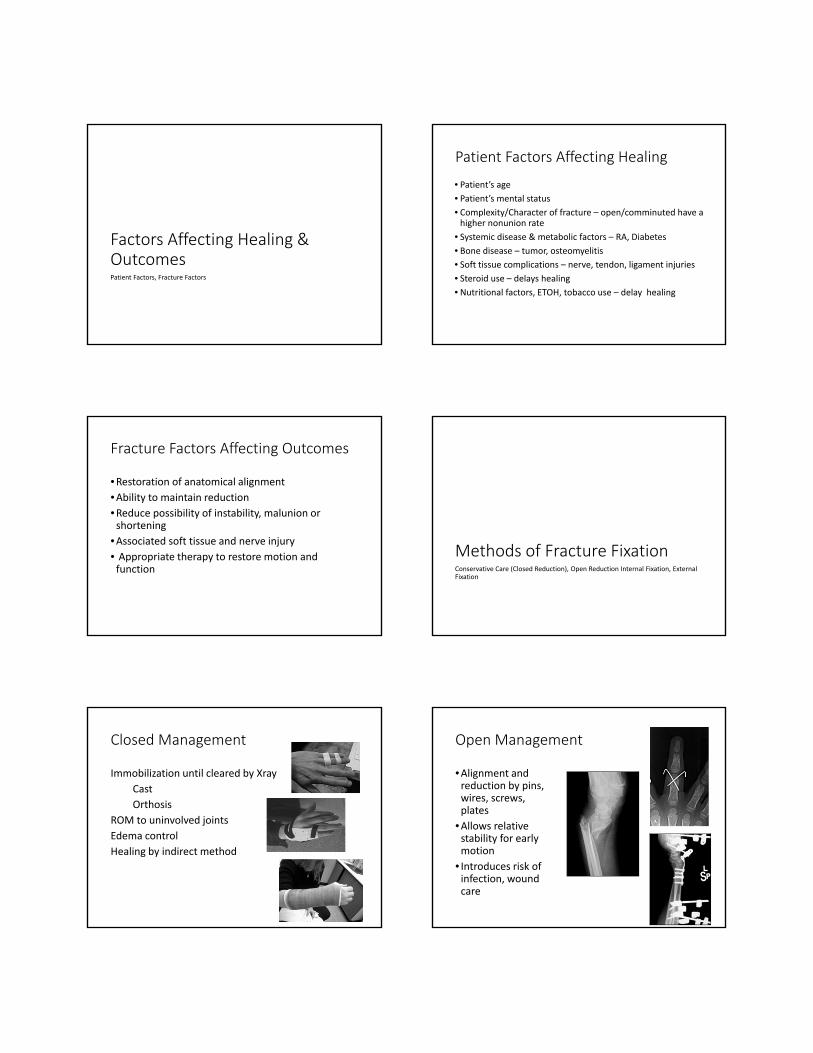
Factors Affecting Healing & OutcomesPatient Factors, Fracture Factors
Patient Factors Affecting Healing
• Patient’s age
• Patient’s mental status
• Complexity/Character of fracture – open/comminuted have a higher nonunion rate
• Systemic disease & metabolic factors – RA, Diabetes
• Bone disease – tumor, osteomyelitis
• Soft tissue complications – nerve, tendon, ligament injuries
• Steroid use – delays healing
• Nutritional factors, ETOH, tobacco use – delay healing
Fracture Factors Affecting Outcomes
•Restoration of anatomical alignment
•Ability to maintain reduction
•Reduce possibility of instability, malunion or shortening
•Associated soft tissue and nerve injury• Appropriate therapy to restore motion and function
Methods of Fracture FixationConservative Care (Closed Reduction), Open Reduction Internal Fixation, External Fixation
Closed Management
Immobilization until cleared by Xray
Cast
Orthosis
ROM to uninvolved joints
Edema control
Healing by indirect method
Open Management
•Alignment and reduction by pins, wires, screws, plates
•Allows relative stability for early motion
• Introduces risk of infection, wound care

Open Reduction and Internal Fixation (ORIF)
Indications•Unstable fractures•Fractures requiring early motion
•Fractures with high incidence of non‐union
•Methods: screws, plates, k‐wires, interosseous wires
External Fixation
External Fixation provides traction to prevent
fracture shortening or angulation.
Fracture ManagementTherapy Principles
• Immobilization
•Edema control
•AROM of uninvolved joints and surrounding soft tissue ASAP
•Protection of healing fracture while applying controlled stress to improve motion and strength.
Therapists should have an understanding of:
•Appropriate timing
•Fracture stability/alignment
•Operative procedures performed
•Potential dysfunction at uninvolved joints
Common FracturesDistal Radius Fractures, Carpal Fractures, Metacarpal Fractures, Phalanx Fractures
Distal Radius Fracture
Causes
•FOOSH•MVA
•Crush injuries• Low impact force in patients with osteoporosis
Distal Radius Alignment
•Radial Inclination = 21‐25°•Palmar Tilt = 11‐12°
•Radial Height = 10‐13mm
•All components are important for a stable & pain‐free wrist capable of accepting forces
36
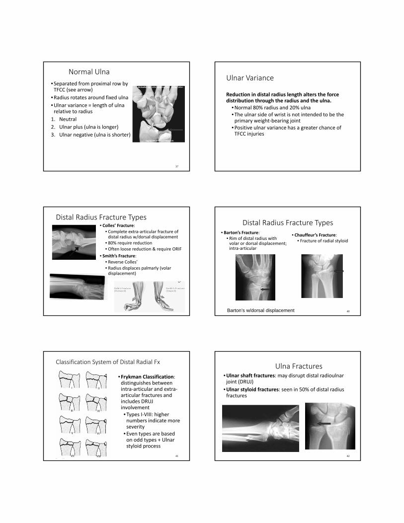
Normal Ulna
•Separated from proximal row by TFCC (see arrow)
•Radius rotates around fixed ulna•Ulnar variance = length of ulna relative to radius
1. Neutral
2. Ulnar plus (ulna is longer)
3. Ulnar negative (ulna is shorter)
37
Ulnar Variance
Reduction in distal radius length alters the force distribution through the radius and the ulna.
•Normal 80% radius and 20% ulna•The ulnar side of wrist is not intended to be the primary weight‐bearing joint
•Positive ulnar variance has a greater chance of TFCC injuries
Distal Radius Fracture Types• Colles’ Fracture:
• Complete extra‐articular fracture of distal radius w/dorsal displacement
• 80% require reduction• Often loose reduction & require ORIF
• Smith’s Fracture:• Reverse Colles’• Radius displaces palmarly (volar displacement)
39
Distal Radius Fracture Types• Barton’s Fracture:
• Rim of distal radius with volar or dorsal displacement; intra‐articular
• Chauffeur’s Fracture: • Fracture of radial styloid
40Barton’s w/dorsal displacement
Classification System of Distal Radial Fx
•Frykman Classification: distinguishes between intra‐articular and extra‐articular fractures and includes DRUJ involvement•Types I‐VIII: higher numbers indicate more severity
•Even types are based on odd types + Ulnar styloid process
41
Ulna Fractures•Ulnar shaft fractures: may disrupt distal radioulnar joint (DRUJ)
•Ulnar styloid fractures: seen in 50% of distal radius fractures
42

Distal Radius FracturesTherapeutic Intervention•Edema Control
• Compression• Elevation
•Digit Motion• Active• Passive
•ROM of Proximal Joints• Forearm • Elbow• Shoulder
Tendon Gliding Exercises
44
Hook fist
Tabletop
Flat fist
Composite fist
45
Blocked FDP
Blocked FDS
PROM into fist
Intrinsic stretch
Therapy: Non‐displaced Closed Reduction
• Initiate early treatment, if indicated, while wrist is immobilized
•4‐6 weeks: cast removed; fit with prefabricated or custom wrist splint; worn continuously or between exercise
•AROM to forearm, wrist 4‐6 weeks
•PROM, joint mobs 8 weeks
•Wean splint usage
•8‐12 weeks: strengthening
46
Therapy: Distal Radius ORIF
•Splint for protection •AROM can usually begin at 1‐2 weeks. Hardware provides stability
•Wean from splint and progress AROM/PROM weeks 5‐8
•Wrist and forearm strengthening at 8‐10 weeks
47
Therapy Distal Radius External Fixation
•Wound care‐light dressing if needed, care of other wounds per MD orders, pin site care
•Follow treatment guidelines for casted fracture
•Splinting as needed for ulnar support and/or web space
•Treat as normal fracture after removal

Wrist Fracture Complications
• Joint stiffness
• Malunion
• Complex Regional Pain Syndrome
• Median nerve compression
• Radiocarpal arthritis
• Weakness of grip
• Long‐term edema
• Carpal instability
• EPL rupture
• TFCC injury/tears
• Intrinsic contracture
• Persistent pain
• Need for further surgery
• Hardware failure
49 50
Wrist ROM Research and Therapy Goals
40º flexion 40º extension
40º combined RD/UD are
considered functional ROM at
the wrist
Patient can be fully functional with less than “normal” wrist ROM
Goal is functional, pain-free ROM
Distal Radius External Fixation Complications
•Pin tract infections•Non‐union•Median neuropathy• Irritation of dorsal sensory branch of radial nerve•RSD/CRPS•Damage to tendons or intrinsic hand muscles
.
Healed Distal Radius Fractures• Strengthening
• BTE• putty• wrist/FA curls
• FES – tendon excursion and retraining
• Composite dynamic splinting: extrinsic muscle tightness
• Refer for F.C.E. if unable to RTW
• Return to leisure activities modifications as needed
CARPAL FRACTURESFrequency
Types
Therapy Considerations
Carpal Fractures ‐Frequency
•Occur 1/10th as frequently as distal radius fractures•Scaphoid: account for 60‐70% of all carpal fractures•Triquetrum/Lunate: second most common‐ account for 20% of all fractures
•Others: combined account for only 7‐10% of carpal fractures

Carpal Fractures – Less Common
•Trapezium: often associated with fractures/dislocations involving thumb
•Pisiform: trauma over ulnar/volar wrist, proximal hypothenar eminence
•Capitate: rare because this carpal is in a centrally located and protected position
•Trapezoid: rare; crush or high energy impact
Scaphoid Fractures
• 90% occur from force applied with wrist in extension (FOOSH)
• Vulnerable to injury
• Difficult to diagnose ‐ often made based on clinical signs
• Delayed presentation on x‐ray
• Variable healing times according to fracture site
56
Healing Times: Scaphoid Fractures
•5‐20 weeks depending on location
•Blood supply enters distal to proximal
•Proximal Pole: poor vascular supply; longest healing time 16‐20 weeks
57
Non‐Operative Management and Therapy
• Immobilization 6‐12 weeks in cast/splint
•Forearm based thumb spica with IP free
•AROM to uninvolved joints including fingers, thumb IP
•Follow guidelines for healing/healed fractures
58
Therapy: ORIF with bone graft (Herbert screw, Russe Technique)
• Immobilize 4‐16 weeks in cast or orthosis
• Immobilized until bone union in a thumb spica with the IP free
•Orthosis can be worn post‐op or post immobilization for rigorous activity
Complications: Scaphoid Fractures
•Non‐union•Carpal instability•Carpal ligament injuries
•Delayed diagnosis•Normal anatomical alignment not restored
•AVN: Avascular Necrosis
60

Therapy: ORIF of Scaphoid
•AROM initiated when bone union is verified
•PROM 1 week later or per MD recommendations
•Strengthening 3‐4 weeks later or per MD
Triquetrum Fractures:
•2nd most common carpal fracture
•Caused by FOOSH with wrist extended and ulnarly deviated
•Requires 4‐6 weeks in short arm cast
Lunate Fractures:
•Often result of repeated compression or from FOOSH
•Potential to develop avascular necrosis (Kienbocks disease)
•Short arm cast x 6‐8 weeks
Kienbock’s•Avascular necrosis of the lunate
•Associated with negative ulnar variance
•Varying degrees of disability ‐ wrist pain, limited extension, weak grip
•Point tenderness over dorsal aspect of lunate
Carpal Fractures: Less Common 7‐10%
•Trapezium: associated with fracture/dislocation involving thumb
•Pisiform/Hamate: trauma over ulnar/volar wrist from fall or blow from handle of club, racquet or bat
•Capitate: rare because centrally located and protected
•Trapezoid: rare; crush or high energy impact
65
Hamate Fractures
•Easily missed, so look for tenderness over hook
•Body of hamate: usually stable, casted 4‐6 weeks
•Hook of hamate: can be associated with ulnar nerve involvement due to hemorrhage or compression in Guyon’s canal
•May require excision of hook
66

Therapy: Healed Carpal Fractures
•Follow treatment guidelines for distal radius fractures
•Focus on wrist and thumb ROM, composite flexion
•May require protective splint post cast removal and between exercise
•Evaluate/treat for intrinsic, extrinsic flexor/extensor tightness
•Use static progressive/dynamic splinting for wrist flex/extension
•Grip/wrist strengthening within pain free range
•Work hardening depending on RTW status
•Provide home exercise program
67
Carpal Fractures – Complications
•Re‐fracture‐ wrist absorbs stress during normal activities ‐ prolonged protective splinting helps to avoid
•Persistent pain‐ emphasize pain‐ free ROM over achieving maximum motion
• Non‐ union, malunion, and delayed union ‐ secondary to poor blood supply to many areas
METACARPAL FRACTURES
Metacarpal Fractures
•40‐50% all hand fractures•Quick healing•Commonly seen in 1st and 5th “border” digits
•Disruption of Interossei muscles
•Potential angulation with fisting from rotational deformity
•Extensor lag may result from metacarpal bone shortening
Metacarpal Fractures‐Orthosis
•Collateral ligaments at MP shorten in extension
•MP’s need to be immobilized in flexion, safe position
•Goal is to achieve alignment without rotation or shortening
Anatomical Considerations
• Shortening by 3‐5 mm can produce intrinsic/extrinsic imbalance
• Malrotation = finger overlap with fisting
• Angulation tolerated better in 4th/5th MCP
• Provide de‐rotational taping during early phases, if malrotation is noted
72

Non‐operative Management
• Non‐displaced/minimally displaced tend to be stable
• Splint or cast 3‐4 weeks • Safe position• PIP/DIPs optional per MD request
• Consider forearm‐ based versus hand‐ based splint depending on location of fracture
• Initiate AROM to uninvolved joints ASAP
• Initiate AROM for MP flex/ext as indicated by fracture location
• PROM to wrist, MPs 4‐6 weeks
• Strengthening 6‐8 weeks
73
Operative Indications
• Multiple fractures
• Open fractures
• Comminuted fractures
• Displaced fractures
• Multitrauma• I.e. head injury
• Spiral fractures• Tend to be unstable
• Oblique fractures• Tend to shorten
Therapy Timeline: Post‐Op
24 ‐ 48 Hours Post‐Op:
Fabricate protective splint
Edema management
AROM to MPs
A/PROM to uninvolved joints
Isolated ED glides2 Weeks Post‐Op:
Add scar massage 24 hours following suture removal if wounds are well healed.
3‐4 Weeks Post‐Op:
Splint may be discontinued and replaced with buddy taping
Continue taping until 6 weeks post‐op
6 Weeks Post‐Op:
Begin gradual strengthening program
75
Metacarpal Neck Fractures
•Most common site, is the weakest portion
•Caused by compressive force‐(blow with closed fist)
•Boxer’s Fracture in 4th or 5thmetacarpal
•Angulation (rotation deformity) can cause pain in palm, claw deformity, trapping of digits)
•Can protrude into palm
Unstable Metacarpal FractureTherapy Progression
•0‐4 wks: cast or clamshell splint
•4wks: D/C cast, AROM: resting splint between exercise; scar control
•2 wks after union: lifting up to 5 lbs., pinch•4 wks after union: resistive ex, lifting up to 25 lbs; D/C protective splint
•8 wks after union: return to sports; heavy work
Metacarpal FractureExternal Fixation
•Shortening of 3‐5 mm can produce intrinsic/extrinsic imbalance
•Malrotation= finger overlap
•Angulation tolerated better in 4/5th MC

Metacarpal Fracture, Stable ORIFTherapy Progression
•0‐3 days: protective splint; AROM all joints
•1‐3 wks: sutures removed, scar management; dynamic splinting prn
•2 wks after union: lifting up to 5 lbs•4 wks after union: resistive ex, lift up to 25 lbs; d/c splint
•8 wks after union: return to normal activities
Therapy Considerations: Metacarpal Fractures
•Manage edema (often excessive dorsal edema) to prevent tissue shortening in extended position
•Assess soft tissue over fracture site (extensor tendons)
•Use compressive dressings, elevation, Isotoner gloves, Tubigrip, Coban, etc.
80
Therapy to Increase Motion
• Hook Fist Full Fist • Isolates EDC glide
• Joint blocking
• Tendon gliding
• Static‐progressive splinting may be needed for MCP flexion
• Scar management against adhesions: elastomer, silicone gel sheets
81
Complications of MCP Fractures
•MP joint contractures common w/head and neck fractures•Extension contractures
•Decreased strength especially power grip in 4th/5th MCPs
•Decreased composite wrist/digit flexion due to extrinsic extensor tightness
82
Extensor Tendon Adherence• Little soft tissue between extensor tendons and metacarpals
• Results in MCP extension lags and extrinsic extensor tendon tightness
• Treatment: wrist flexion with composite fist, isolated EDC exercises, scar massage, scar molds, FES, combined wrist/finger flexion, continuous ultrasound, dynamic splinting
Specific Exercises
Extrinsic excursion
A: Synergistic wrist extension/finger flexion
B: Synergistic wrist flexion/finger extension
C: MCP blocking for extrinsic tendon extension
D. Composite extension and composite flexion
84D

PHALANGEAL FRACTURESProximal. Middle, and Distal
P1, P2, P3
Types of Phalangeal Fracture
•Oblique, transverse, spiral
•Spiral/oblique tend to be unstable
•Often cause soft tissue adherence
•Comminution = more soft tissue damage
•Stable fractures can be buddy taped
Management of P1, P2 FracturesSTABLE
• Simple nondisplaced/ fractures can be splinted and/or buddy taped
• Splint can be hand‐based or finger‐based depending on fracture location
• Consider hand‐based for children or at‐risk patients
UNSTABLE
• Displaced, mal‐aligned or intra‐articular fractures are surgical candidates
• 2‐3 days post‐op: remove bulky dressing and make dorsal/volar based splint to allow easy AROM (hand‐ or finger‐based)
87
P1, P2 Stable Management
• Weeks 0‐4: hand‐based, finger‐based or buddy taped
• AROM all joints
• 4 weeks (or when stable): PROM
• 2 weeks after union: dynamic splinting; d/c protective splint
• 4 weeks after union: PREs; lifting up to 25 lbs.
• 12 weeks (8 weeks after union): unrestricted activities
88
P1 Base Fractures
Hand‐based Dorsal Extension Block splint in safety position 2‐4 weeks for non‐articular fractures
• Design allows compression of fracture
• Allows protected early AROM
89
Therapy for Proximal and Middle Phalanx Fractures
•Edema control
•Protective splinting •Facilitate soft tissue glide over dorsal hood•Tendon glides
•Prevent PIP flexion contractures•Provide 3 point pressure with extension splinting
90
Digi Sleeve
3 Point Fixation for Correction of Flexion Deformity

91
• Facilitate soft tissue glide over dorsal hood
• A: FDP blocked flexion• B: FDS blocked flexion• C: Central slip/lateral bands gliding
• Blocking exercises
• Differential Tendon Gliding
• Composite wrist/digit flexion/extension
A
B
C
Therapy for P1/P2 Fractures
92
ORIF: P1/P2 Fractures
2‐5 Days Post‐op:
Replace post‐op dressing with protective splint
• Hand‐based for P1
• Hand‐based or finger‐based for P2 depending on patient compliance
Wound care as needed
Initiate AROM
Edema control
Follow same guidelines for non‐operative treatment
• Can initiate earlier PROM/strengthening due to rigid fixation
Complications of P1, P2 Fractures
•PIP flexion contracture ‐ splint in extension
• Limited active PIP extension ‐ use joint blocking
•Flexor tendon adherence at fracture site ‐ tendon gliding
93
Distal Phalanx Fractures
•Account for 50% of hand fractures
•Thumb/middle most commonly injured
•Crush injury is the most common mechanism
•Heal without excessive treatment
94
Therapy: Distal Phalanx Fracture
• DIP protective splinting; leave PIP free if possible
• Wound care
• Edema control
• Desensitization program
• Restore DIP ROM when appropriate
• Watch for extensor lag and modify exercises and splinting as needed
95
DIP protective splint
Tuft FracturesCrush/ comminuted fracture
Painful‐ hematoma
Soft tissue injury – debridment
Often involves the nail bed
Rehab:• Restore DIP motion without > 10⁰ extension lag
• Desensitization• Protective splinting to DIPleave PIP free

Mallet Fracture
• Inability to extend the distal phalanx at the DIP joint
• Two forms of mallet finger• Tendinous: extensor tendon rupture
• Bony: bony avulsion fracture of distal phalanx
• Causes a “flexion drop” deformity
97
Bony Mallet Therapy: Mallet Fractures
• DIP splint worn continuously• DIP in 0°• PIP not included in splint
• Splint usage 6‐8 weeks
• Different protocols for duration/weaning of splint
• Check for extensor lag, 5°considered within normal limits
98
Phalangeal Fracture Complications
•Numbness
•Cold sensitivity• Limited DIP ROM
•Nail abnormalities
•PIP flexion contracture‐splint in extension
• Limited active PIP extension‐ use joint blocking
•Flexor tendon adherence at fracture‐ reduced tendon gliding
Fractures of the Thumb
100
Treatment Guidelines
• Follow similar guidelines as for digits
• Special considerations: • Thumb joints lose motion faster than finger joints
• Motion at basal joint helps when MP joint motion is lost
• Prevent first web space contracture
101
Thermoplastic Web Space Splint
Thumb Fractures
Bennett’s Fracture
• Intra‐articular fracture + dislocation at first metacarpal base
• Intact oblique ulnar ligament stabilizes the small fracture fragment
• Metacarpal shaft is displaced proximally
• Closed vs. open reduction• Splint (short arm thumb spica with IP free) x 6 weeks
102

Post‐Op Treatment: Bennett’s Fracture
6 Weeks Post‐Op:
If the fracture is clinically healed, the short arm thumb spica cast is removed
AROM and gentle PROM to the wrist and thumb
Thumb spica splint worn between exercise sessions and at night
8 Weeks Post‐Op:
If the fracture is well healed• Discontinue splinting• Use taping or dynamic splinting as needed to increase ROM
Begin gentle strengthening10 Weeks Post‐Op:
• Return to normal activities
103
Thumb Fractures
Rolando’s Fracture
• Usually caused by axial loading of a partially flexed metacarpal, e.g. football
• Comminuted intra‐articular fracture at the first metacarpal base
• Surgery is almost always indicated and immobilized in thumb spica 4‐6 weeks
104
Ulnar Collateral Ligament Injury
Skiers thumb/Gamekeepers thumb
Closed reduction
• Hand based thumb spica splint x 6 weeks
Surgery if UCL rupture is complete
• Immobilized in thumb spica x 6 weeks
• Controlled AROM 3‐4 weeks post op
• Avoid lateral stress to thumb
References1. Chinchalkar SJ, Gan BS.Management of proximal interphalangeal joint
fractures and dislocations. J Hand Ther 2003; 16:117‐128.
2. Handoll HHG, Vaghela MV. Interventions for treating mallet finger injuries. Cochrane Database of Systematic Reviews 2004, Issue 3.
3. Kalainov DM, Hoepfner PE, Hartigan BJ, Carroll C, Genuario J. Nonsurgical treatment of closed mallet finger fractures. J Hand Surg 2005; 30A:580‐586.
4. Prosser R. Splinting in the management of proximal interphalangeal joint flexion contracture. J Hand Ther Oct‐Dec 1996; 9(4):378‐86.
5. Ruland R, Hogan C, Cannon D, Slade J. Use of dynamic distraction external fixation for unstable fracture‐dislocations of the Proximal InterphalangealJoint. J Hand Surg 2008;33:19‐25.
6. Prosser, R. Splinting in the management of proximal interphalangeal joint flexion contracture. J Hand Ther Oct‐Dec 1996; 9:378‐86.
7. Skirven TM, Osterman AL, Fedorczyk JM, Amadio PC, Eds. Rehabilitation of the Hand and Upper Extremity, Sixth Edition Philadelphia 2011 Elsevier Mosby