Limits of serological testing - Irish Blood Transfusion ... · PDF fileLimits of serological...
1
Limits of serological testing Sorcha Ni Loingsigh, Mark Lambert 1 , Barry Doyle, Stuart Adshead 1 Diagnostics Laboratory, Irish Blood Transfusion Service, National Blood Centre, James Street, Dublin 8, Ireland 2 Donor Grouping Laboratory, Irish Blood Transfusion Service, National Blood Centre, James Street, Dublin 8, Ireland Introduction Results Discussion & Conclusion The Rh system (ISBT 004) is currrently the most polymorphic blood group system, with over 60 blood groups described [International Society of Blood Transfusion, 2013]. RhD is the first blood group of the system and is still the most important clinically, indeed it is the most important protein blood group. Anti-D immunisation can readily occur in RhD- recipents. It is therefore essential that current typing strategies should ensure that blood is correctly typed for RhD. This is recommended in the ‘Guidelines for the Blood Transfusion Services in the UK’. Section 12.10.3 describes in a relatively unambiguous way the level of RhD typing that is acceptable for donors. The level of testing undertaken to confirm the true RhD type of donors varies throughout Europe and the world. A few National Blood Services in Europe confirm the RhD type of their RhD negative donors by testing for the presence of RHD. Of the five Blood Services in the geographical British Isles (IBTS, NIBTS, SNBTS, WBS, NHSBT) none go to this extent and only the NIBTS use an IAT-reactive anti-D for confirming the RhD type of their donors. In total 6 donor samples were investigated. Initially 2 RhD- C+ donor samples (in 2009 and 2010) were investigated due to extremely weak, but unrepeatable, reactions with one of the anti-D reagents used by the Donor Grouping Laboratory. Both samples were investigated and referred to the IBGRL. RHD sequencing identified both as weak D type 10 (RHD*01W.10), characterised by a 1177T>C nucleotide change, resulting in W393R amino acid change. Fig. 1. RHD Sequencing identifiying RHD*01W.10. In 2013, a pilot study was conducted which involved testing 315 RhD- C+/E+ donors with the RhD Kit. A further 4 RhD- C+ donors were identified. All four were negative with all the standard anti-D reagents used by both the Automated Donor and Red Cell Immunohaematology Laboratories. Fig. 2. Position of Tryptophan to Arginine change at amino acid residue 393 References Wagner FF, Frohmajer A, Ladewig B, Eicher NI, Lonicer CB, Müller TH, Siegel MH, Flegel WA. Weak D alleles express distinct phenotypes. Blood 2000;95:2699-2708. ISBT Working Party on Red Cell Immunogenetics and Blood Group Terminology webpage. Accessed 06/10/2013 - www.isbtweb.org/fileadmin/user_upload/WP_on_Red_Cell_Immunogenetics_an d/Updates/Table_of_blood_group_antigens_within_systems_v3_2_130331.pdf. RhesusBase Webpage. Accessed 06/10/2013. http://www.uni- ulm.de/~fwagner/RH/RB/. Flegel WA, von Zabern I, Wagner FF. Six years’ experience performing RHD genotyping to confirm D- red blood cell units in Germany for preventing anti-D immunizations. Transfusion 2009;49:465-471. Results Methods Mark Lambert Red Cell Referral Laboratory Irish Blood Transfusion Service National Blood Centre James’s Street Dublin 8 Ireland T.: +353-1-4322972 F.: +353-1-4322709 E.: [email protected] W.: www.giveblood.ie Donors in Ireland are tested, by the Automated Donor Grouping (ADG) Laboratory at the IBTS, for the presence of RhD using Diagast Totem and ImmunClone Anti-D duo (bromelised cells; Olympus PK73000). First time donors are tested with additional anti-D reagents (Bioscot RUM1 and Bioscot MS- 201). Donors with discrepant results are tested using BioRad DiaClon Product 50481. The Donor Grouping laboratory has begun evaluating testing of RhD- C+/E+ donors using the ALBAclone ® Advanced Partial RhD Typing Kit (RhD Kit), tested on the Zenyx QASAR IV. Discrepant samples are referred to the RCI Laboratory. Samples are tested by the AutoVue Innova [Products 707119 and 707135], and by tube agglutination using Bioscot reagents [anti-D BS226 and BS232]. Further testing was performed using the RhD Kit. The RhD Kit normally gives good results with most weak D and RhD variant samples. However, samples that have been referred for RhD investigation and give unexpected or negative results will also be tested by adsorption-elution with donor polyclonal anti-D (a pool of two donors with anti-D of 6 IU/ml). Methods Rh phenotyping (C/c, E/e) was performed by both the ADG and RCI laboratories. Samples that remained unresolved were referred to the International Blood Group Reference Laboratory for RHD gene sequencing. All 10 exons and flanking intronic regions were sequenced by Sanger sequencing. a) d) e) c) b) Fig 3. Samples tested with Biorad product 50481. DVI+ = detects DVI, DVI- does not detect DVI All 4 of the 2013 samples were weakly positive (0 – 1+) with the RhD Kit. Table 1. Anti-D reagents used by the ADG Laboratory Anti-D and Cell-Lines – Donor Laboratory Diagast Totem (P3x61) & (P3x21223B10) [IgM Blend] (P3x290) & (P3x35) [IgG Blend] ImmuClone Anti- D duo Th28 [IgG] MS26 [IgM] Bioscot RUM-1 RUM-1 [IgM] Bioscot MS-201 MS-201 [IgM] Biorad 50481 (ESD-1M) & (175-2) [DVI+] (LHM59/20) & (175- 2) [DVI-] Table 2. Anti-D reagents used by the RCI Laboratory Anti-D and Cell-Lines – RCI Laboratory Seraclone 226 MS-226 [IgM] Seraclone 232 MS-232 [IgM] Ortho Autovue 707119 ABDD D7B8 [IgM] RUM-1 [IgM] Ortho Autovue 707135 ABD D7B8 [IgM] ALBA RhD Kit 12 IgG Cell-Lines Polyclonal Anti-D Two donor plasmas (6.5 IU/ml) DNA from the four 2013 samples was referred to the IBGRL for RHD sequencing. All 4 were identified as weak D type 10 (RHD*01W.10). a) e) d) c) b) Fig. 3. Weak D types 1, 2 and 10, RhD+ and RhD- samples. Reactions observed with first 3 components of the RhD Kit. Weak D type 10 is a rare weak D type with an estimated frequency of 1/47,619 in SW Germany. In Ireland we estimate the frequency to be 1/97,500 of RhD- indivdulas (based on 6 donors and 1 patient (not handled by IBTS)). The frequency in RhD- C+/E+ is estimated at 1/79 donors (4 in 315 – detected in 2013). Although the reported RhD antigen density of 1,186 is not low compared to many other weak D types, the typing difficulties are likely explained by the reported heterogeneity seen in weak D type 10 individuals (only 50% staining observed by flow cytometry) [Wagner et al. 2000]. The weak D type 10 samples observed in the Irish donor pool are all associated with the DCe haplotype. This is different to the Weak D type 10 entry on RhesusBase (curated by Franz Wagner in Ulm University, Germany), which indictaes that the association is with the DcE haplotype. In a report on RHD genotyping of RhD- donors in Germany no weak D type 10 donors were detected, however this is most likely due to the fact that Germany tests RhD- donors with an IAT-reactive anti-D reagent [Flegel et al., 2009]. Weak D type 10 has similar RhD antigen density as weak D type 1, which been documented as causing anti-D immunisation in RhD- recipients. It could be expected that weak D type 10 might cause anti-D also. However, the clinical impact of not detecting weak D type 10 in the Irish RhD- donor pool is unclear. Units from two of the six donors were never transfused. While the remaining donors had varying numbers of transfused donations. Two had greater than 20 donations each. While tracing the recipients of units from these donors is ongoing, to date no anti-D allo- immunisation has been detected. Current policy for testing donor samples for RhD does not identify all weak D phenotypes. At a minimum all RhD- C+/E+ donors should have their RhD status confirmed by a more sensitive means such as an IAT-reactive anti- D, adsorption-elution with polyclonal anti-D or pooled testing of donor DNA for RHD-specific exons. This latter pooled DNA approach is becoming more widespread.
Transcript of Limits of serological testing - Irish Blood Transfusion ... · PDF fileLimits of serological...
Limits of serological testing
Sorcha Ni Loingsigh, Mark Lambert1, Barry Doyle, Stuart Adshead 1Diagnostics Laboratory, Irish Blood Transfusion Service, National Blood Centre, James Street, Dublin
8, Ireland 2 Donor Grouping Laboratory, Irish Blood Transfusion Service, National Blood Centre, James Street,
Dublin 8, Ireland
Introduction
Results
Discussion & Conclusion
The Rh system (ISBT 004) is currrently the
most polymorphic blood group system, with
over 60 blood groups described [International
Society of Blood Transfusion, 2013]. RhD is
the first blood group of the system and is still
the most important clinically, indeed it is the
most important protein blood group. Anti-D
immunisation can readily occur in RhD-
recipents.
It is therefore essential that current typing
strategies should ensure that blood is
correctly typed for RhD. This is recommended
in the ‘Guidelines for the Blood Transfusion
Services in the UK’. Section 12.10.3 describes
in a relatively unambiguous way the level of
RhD typing that is acceptable for donors.
The level of testing undertaken to confirm the
true RhD type of donors varies throughout
Europe and the world. A few National Blood
Services in Europe confirm the RhD type of
their RhD negative donors by testing for the
presence of RHD.
Of the five Blood Services in the geographical
British Isles (IBTS, NIBTS, SNBTS, WBS,
NHSBT) none go to this extent and only the
NIBTS use an IAT-reactive anti-D for
confirming the RhD type of their donors.
In total 6 donor samples were investigated.
Initially 2 RhD- C+ donor samples (in 2009 and
2010) were investigated due to extremely
weak, but unrepeatable, reactions with one of
the anti-D reagents used by the Donor
Grouping Laboratory.
Both samples were investigated and referred
to the IBGRL. RHD sequencing identified both
as weak D type 10 (RHD*01W.10),
characterised by a 1177T>C nucleotide
change, resulting in W393R amino acid
change.
Fig. 1. RHD Sequencing identifiying RHD*01W.10.
In 2013, a pilot study was conducted which
involved testing 315 RhD- C+/E+ donors with
the RhD Kit. A further 4 RhD- C+ donors were
identified. All four were negative with all the
standard anti-D reagents used by both the
Automated Donor and Red Cell
Immunohaematology Laboratories.
Fig. 2. Position of Tryptophan to Arginine change
at amino acid residue 393
References
Wagner FF, Frohmajer A, Ladewig B, Eicher NI, Lonicer CB, Müller TH, Siegel
MH, Flegel WA. Weak D alleles express distinct phenotypes. Blood
2000;95:2699-2708.
ISBT Working Party on Red Cell Immunogenetics and Blood Group Terminology
webpage. Accessed 06/10/2013 -
www.isbtweb.org/fileadmin/user_upload/WP_on_Red_Cell_Immunogenetics_an
d/Updates/Table_of_blood_group_antigens_within_systems_v3_2_130331.pdf.
RhesusBase Webpage. Accessed 06/10/2013. http://www.uni-
ulm.de/~fwagner/RH/RB/.
Flegel WA, von Zabern I, Wagner FF. Six years’ experience performing RHD
genotyping to confirm D- red blood cell units in Germany for preventing anti-D
immunizations. Transfusion 2009;49:465-471.
Results
Methods
Mark Lambert
Red Cell Referral Laboratory
Irish Blood Transfusion Service
National Blood Centre
James’s Street
Dublin 8
Ireland
T.: +353-1-4322972
F.: +353-1-4322709
W.: www.giveblood.ie
Donors in Ireland are tested, by the
Automated Donor Grouping (ADG) Laboratory
at the IBTS, for the presence of RhD using
Diagast Totem and ImmunClone Anti-D duo
(bromelised cells; Olympus PK73000). First
time donors are tested with additional anti-D
reagents (Bioscot RUM1 and Bioscot MS-
201).
Donors with discrepant results are tested
using BioRad DiaClon Product 50481. The
Donor Grouping laboratory has begun
evaluating testing of RhD- C+/E+ donors using
the ALBAclone® Advanced Partial RhD Typing
Kit (RhD Kit), tested on the Zenyx QASAR IV.
Discrepant samples are referred to the RCI
Laboratory. Samples are tested by the
AutoVue Innova [Products 707119 and
707135], and by tube agglutination using
Bioscot reagents [anti-D BS226 and BS232].
Further testing was performed using the RhD
Kit.
The RhD Kit normally gives good results with
most weak D and RhD variant samples.
However, samples that have been referred for
RhD investigation and give unexpected or
negative results will also be tested by
adsorption-elution with donor polyclonal anti-D
(a pool of two donors with anti-D of 6 IU/ml).
Methods
Rh phenotyping (C/c, E/e) was performed by
both the ADG and RCI laboratories.
Samples that remained unresolved were
referred to the International Blood Group
Reference Laboratory for RHD gene
sequencing. All 10 exons and flanking intronic
regions were sequenced by Sanger
sequencing.
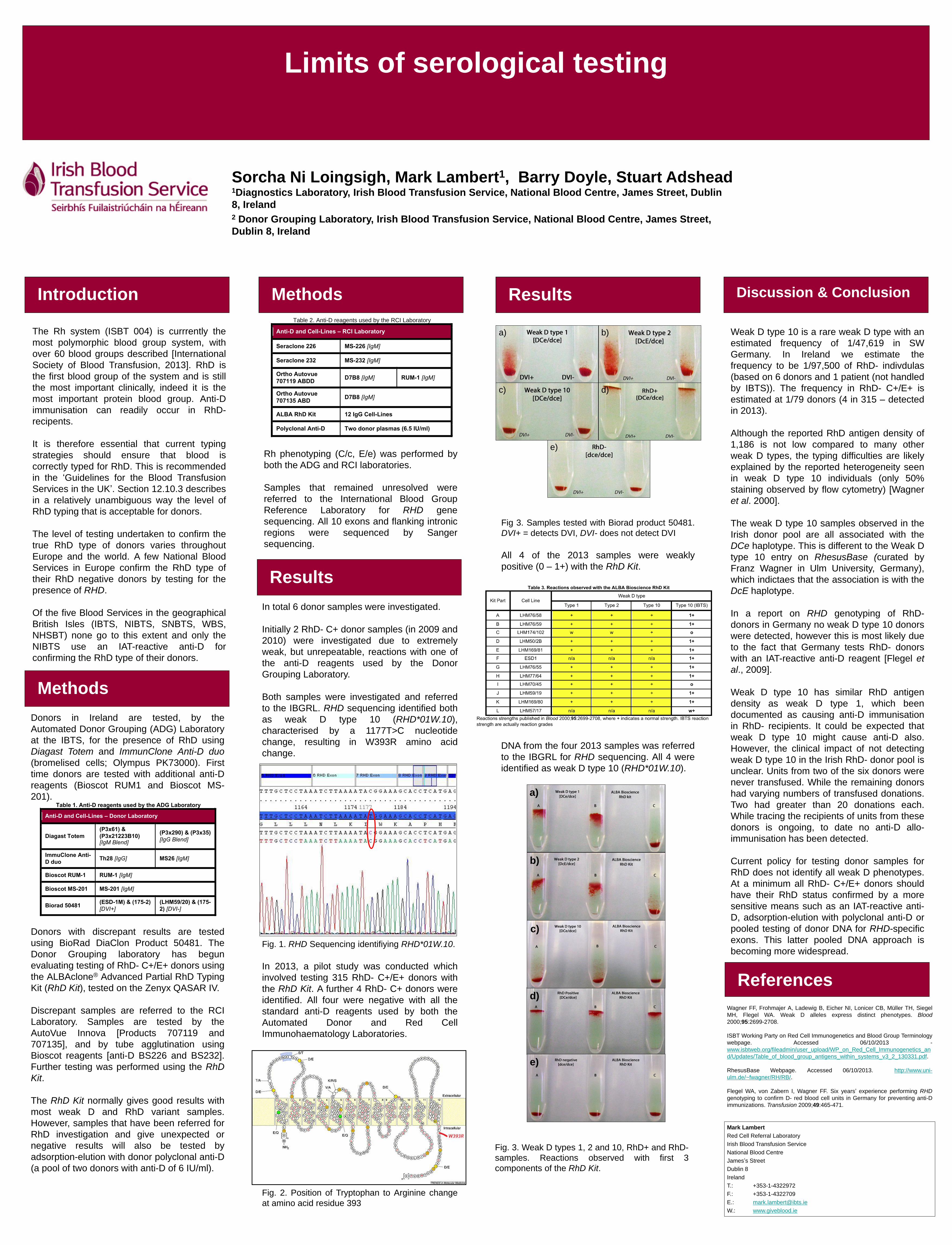
a)
d)
e)
c)
b)
Fig 3. Samples tested with Biorad product 50481.
DVI+ = detects DVI, DVI- does not detect DVI
All 4 of the 2013 samples were weakly
positive (0 – 1+) with the RhD Kit.
Table 1. Anti-D reagents used by the ADG Laboratory
Anti-D and Cell-Lines – Donor Laboratory
Diagast Totem
(P3x61) &
(P3x21223B10) [IgM Blend]
(P3x290) & (P3x35)
[IgG Blend]
ImmuClone Anti-
D duo Th28 [IgG] MS26 [IgM]
Bioscot RUM-1 RUM-1 [IgM]
Bioscot MS-201 MS-201 [IgM]
Biorad 50481 (ESD-1M) & (175-2)
[DVI+]
(LHM59/20) & (175-
2) [DVI-]
Table 2. Anti-D reagents used by the RCI Laboratory
Anti-D and Cell-Lines – RCI Laboratory
Seraclone 226 MS-226 [IgM]
Seraclone 232 MS-232 [IgM]
Ortho Autovue
707119 ABDD D7B8 [IgM] RUM-1 [IgM]
Ortho Autovue
707135 ABD D7B8 [IgM]
ALBA RhD Kit 12 IgG Cell-Lines
Polyclonal Anti-D Two donor plasmas (6.5 IU/ml)
DNA from the four 2013 samples was referred
to the IBGRL for RHD sequencing. All 4 were
identified as weak D type 10 (RHD*01W.10).
a)
e)
d)
c)
b)
Fig. 3. Weak D types 1, 2 and 10, RhD+ and RhD-
samples. Reactions observed with first 3
components of the RhD Kit.
Weak D type 10 is a rare weak D type with an
estimated frequency of 1/47,619 in SW
Germany. In Ireland we estimate the
frequency to be 1/97,500 of RhD- indivdulas
(based on 6 donors and 1 patient (not handled
by IBTS)). The frequency in RhD- C+/E+ is
estimated at 1/79 donors (4 in 315 – detected
in 2013).
Although the reported RhD antigen density of
1,186 is not low compared to many other
weak D types, the typing difficulties are likely
explained by the reported heterogeneity seen
in weak D type 10 individuals (only 50%
staining observed by flow cytometry) [Wagner
et al. 2000].
The weak D type 10 samples observed in the
Irish donor pool are all associated with the
DCe haplotype. This is different to the Weak D
type 10 entry on RhesusBase (curated by
Franz Wagner in Ulm University, Germany),
which indictaes that the association is with the
DcE haplotype.
In a report on RHD genotyping of RhD-
donors in Germany no weak D type 10 donors
were detected, however this is most likely due
to the fact that Germany tests RhD- donors
with an IAT-reactive anti-D reagent [Flegel et
al., 2009].
Weak D type 10 has similar RhD antigen
density as weak D type 1, which been
documented as causing anti-D immunisation
in RhD- recipients. It could be expected that
weak D type 10 might cause anti-D also.
However, the clinical impact of not detecting
weak D type 10 in the Irish RhD- donor pool is
unclear. Units from two of the six donors were
never transfused. While the remaining donors
had varying numbers of transfused donations.
Two had greater than 20 donations each.
While tracing the recipients of units from these
donors is ongoing, to date no anti-D allo-
immunisation has been detected.
Current policy for testing donor samples for
RhD does not identify all weak D phenotypes.
At a minimum all RhD- C+/E+ donors should
have their RhD status confirmed by a more
sensitive means such as an IAT-reactive anti-
D, adsorption-elution with polyclonal anti-D or
pooled testing of donor DNA for RHD-specific
exons. This latter pooled DNA approach is
becoming more widespread.