
Limit on the Role of Activity in Controlling the Release-Ready
13
Limit on the Role of Activity in Controlling the Release-Ready Supply of Synaptic Vesicles John F. Wesseling and Donald C. Lo Department of Neurobiology, Duke University Medical Center, Durham, North Carolina 27710 Typical fast chemical synapses in the brain weaken transiently during normal high-frequency use after expending their presyn- aptic supply of release-ready vesicles. Although it takes several seconds for the readily releasable pool (RRP) to refill during periods of rest, it has been suggested that the replenishment process may be orders of magnitude faster when synapses are active. Here, we measure this replenishment rate at active Schaffer collateral terminals by determining the maximum rate of release that can still be elicited when the RRP is almost completely exhausted. On average, we find that spent vesicles are replaced at a maximum unitary rate of 0.24/sec during periods of intense activity. Because the replenishment rate is similar during subsequent periods of rest, we conclude that no special mechanism accelerates the mobilization of neurotrans- mitter in active terminals beyond the previously reported, several-fold, residual calcium-driven modulation that persists for several seconds after bouts of intense synaptic activity. In the course of this analysis, we provide new evidence support- ing the hypothesis that a simple enzymatic step limits the rate at which reserve synaptic vesicles become ready to undergo exocytosis. Key words: synaptic physiology; presynaptic; synaptic vesi- cle; priming; short-term plasticity; readily releasable pool Recently there has been considerable interest in measuring the time required for reserve synaptic vesicles to become available to participate in neurotransmission at CNS synapses (Stevens and Tsujimoto, 1995; Rosenmund and Stevens, 1996; Dobrunz and Stevens, 1997; Stevens and Wesseling, 1998; Wu and Borst, 1999; Pyle et al., 2000). This work is important because a complete picture of the molecular basis of synaptic f unction requires a clear understanding of the kinetics of the various steps in the synaptic vesicle cycle, in addition to the identification of the enzymes involved. It is also important from a computational perspective, because the rate at which vesicles become ready to participate in synaptic transmission is a key element that determines the means by which information can be transferred between neurons via synapses. Typical presynaptic terminals contain hundreds of vesicles loaded with chemical transmitter, but at any given time, only a few of them are f unctionally available to be released quickly (Schikor- ski and Stevens, 1997, 2001). This release-ready subset of synaptic vesicles constitutes the readily releasable pool (RRP). When their RRPs empty during episodes of heavy use, synapses exhibit short-term depression, transiently becoming less reliable at trans- mitting information because they can no longer consistently pro- vide neurotransmitter for intercellular signaling (Elmqvist and Quastel, 1965; Rosenmund and Stevens, 1996). Previous studies have primarily measured the time it takes for the RRP to refill during periods of rest, most finding that recovery from depletion takes at least several seconds (Birks and MacIn- tosh, 1961; Elmqvist and Quastel, 1965; Stevens and Tsujimoto, 1995; Rosenmund and Stevens, 1996; Wu and Borst, 1999). The residual calcium that is cleared slowly from synaptic terminals after bouts of intense activity has been shown to accelerate the replenishment process several-fold (Dittman and Regehr, 1998; Stevens and Wesseling, 1998, 1999a; Wang and Kaczmarek, 1998). In those studies, however, the replenishment rate was monitored only during rest intervals that followed active epi- sodes. It has been suggested that there may be an additional, qualitatively different activity-dependent mechanism that can ac- celerate the rate at which neurotransmitter becomes available for release to a much greater extent during periods of intense pre- synaptic activity (Kusano and Landau, 1975), perhaps via a rapid refilling of the spent release-ready vesicles themselves (Pyle et al., 2000). In this study we examined the extent to which the RRP replen- ishment rate can be accelerated to counteract short-term depres- sion during heavy synaptic use. Using stimulation protocols that were sufficient to drive the RRPs of Schaffer collateral synapses into a near-empty steady state, we found that neurotransmitter becomes available for release only approximately two or three times as quickly when these synapses are active as it does during periods of rest, as predicted from the residual calcium-dependent type of replenishment-rate acceleration that has been reported previously (Stevens and Wesseling, 1998). We thus conclude that there is no special mechanism that accelerates the preparation of neurotransmitter for release in active terminals beyond this type of modulation that persists for several seconds after bouts of repetitive activity. MATERIALS AND METHODS All synaptic responses were measured from patch-clamped neurons held in whole-cell voltage-clamp mode. Preparation. Experiments were performed on transverse slices pre- pared from the hippocampi of 2- to 3-week-old mice. Mice were anes- thetized by inhalation of halothane and decapitated soon after the Received July 24, 2002; revised Sept. 6, 2002; accepted Sept. 9, 2002. Funding was provided by National Institutes of Health Grants NS10827 (J.F.W.) and NS32742 (D.C.L.) and the McKnight Endowment Fund (D.C.L.). We thank Dr. Dana Cohen, Dr. Isabel Pe ´rez-Otan ˜o, Dr. Iman Brivanlou, and Dr. Richard Aldrich for providing helpful suggestions. Correspondence should be addressed to John F. Wesseling, Duke University Medical Center 3209, Duke University Department of Neurobiology, Durham, NC 27710. E-mail: [email protected]. Copyright © 2002 Society for Neuroscience 0270-6474/02/229708-13$15.00/0 The Journal of Neuroscience, November 15, 2002, 22(22):9708–9720
Transcript of Limit on the Role of Activity in Controlling the Release-Ready
Limit on the Role of Activity in Controlling the Release-ReadySupply of Synaptic Vesicles
John F. Wesseling and Donald C. Lo
Department of Neurobiology, Duke University Medical Center, Durham, North Carolina 27710
Typical fast chemical synapses in the brain weaken transientlyduring normal high-frequency use after expending their presyn-aptic supply of release-ready vesicles. Although it takes severalseconds for the readily releasable pool (RRP) to refill duringperiods of rest, it has been suggested that the replenishmentprocess may be orders of magnitude faster when synapses areactive. Here, we measure this replenishment rate at activeSchaffer collateral terminals by determining the maximum rateof release that can still be elicited when the RRP is almostcompletely exhausted. On average, we find that spent vesiclesare replaced at a maximum unitary rate of 0.24/sec duringperiods of intense activity. Because the replenishment rate is
similar during subsequent periods of rest, we conclude that nospecial mechanism accelerates the mobilization of neurotrans-mitter in active terminals beyond the previously reported,several-fold, residual calcium-driven modulation that persistsfor several seconds after bouts of intense synaptic activity. Inthe course of this analysis, we provide new evidence support-ing the hypothesis that a simple enzymatic step limits the rateat which reserve synaptic vesicles become ready to undergoexocytosis.
Key words: synaptic physiology; presynaptic; synaptic vesi-cle; priming; short-term plasticity; readily releasable pool
Recently there has been considerable interest in measuring thetime required for reserve synaptic vesicles to become available toparticipate in neurotransmission at CNS synapses (Stevens andTsujimoto, 1995; Rosenmund and Stevens, 1996; Dobrunz andStevens, 1997; Stevens and Wesseling, 1998; Wu and Borst, 1999;Pyle et al., 2000). This work is important because a completepicture of the molecular basis of synaptic function requires a clearunderstanding of the kinetics of the various steps in the synapticvesicle cycle, in addition to the identification of the enzymesinvolved. It is also important from a computational perspective,because the rate at which vesicles become ready to participate insynaptic transmission is a key element that determines the meansby which information can be transferred between neurons viasynapses.
Typical presynaptic terminals contain hundreds of vesiclesloaded with chemical transmitter, but at any given time, only a fewof them are functionally available to be released quickly (Schikor-ski and Stevens, 1997, 2001). This release-ready subset of synapticvesicles constitutes the readily releasable pool (RRP). When theirRRPs empty during episodes of heavy use, synapses exhibitshort-term depression, transiently becoming less reliable at trans-mitting information because they can no longer consistently pro-vide neurotransmitter for intercellular signaling (Elmqvist andQuastel, 1965; Rosenmund and Stevens, 1996).
Previous studies have primarily measured the time it takes forthe RRP to refill during periods of rest, most finding that recoveryfrom depletion takes at least several seconds (Birks and MacIn-
tosh, 1961; Elmqvist and Quastel, 1965; Stevens and Tsujimoto,1995; Rosenmund and Stevens, 1996; Wu and Borst, 1999). Theresidual calcium that is cleared slowly from synaptic terminalsafter bouts of intense activity has been shown to accelerate thereplenishment process several-fold (Dittman and Regehr, 1998;Stevens and Wesseling, 1998, 1999a; Wang and Kaczmarek,1998). In those studies, however, the replenishment rate wasmonitored only during rest intervals that followed active epi-sodes. It has been suggested that there may be an additional,qualitatively different activity-dependent mechanism that can ac-celerate the rate at which neurotransmitter becomes available forrelease to a much greater extent during periods of intense pre-synaptic activity (Kusano and Landau, 1975), perhaps via a rapidrefilling of the spent release-ready vesicles themselves (Pyle et al.,2000).
In this study we examined the extent to which the RRP replen-ishment rate can be accelerated to counteract short-term depres-sion during heavy synaptic use. Using stimulation protocols thatwere sufficient to drive the RRPs of Schaffer collateral synapsesinto a near-empty steady state, we found that neurotransmitterbecomes available for release only approximately two or threetimes as quickly when these synapses are active as it does duringperiods of rest, as predicted from the residual calcium-dependenttype of replenishment-rate acceleration that has been reportedpreviously (Stevens and Wesseling, 1998). We thus conclude thatthere is no special mechanism that accelerates the preparation ofneurotransmitter for release in active terminals beyond this typeof modulation that persists for several seconds after bouts ofrepetitive activity.
MATERIALS AND METHODSAll synaptic responses were measured from patch-clamped neurons heldin whole-cell voltage-clamp mode.
Preparation. Experiments were performed on transverse slices pre-pared from the hippocampi of 2- to 3-week-old mice. Mice were anes-thetized by inhalation of halothane and decapitated soon after the
Received July 24, 2002; revised Sept. 6, 2002; accepted Sept. 9, 2002.Funding was provided by National Institutes of Health Grants NS10827 (J.F.W.)
and NS32742 (D.C.L.) and the McKnight Endowment Fund (D.C.L.). We thank Dr.Dana Cohen, Dr. Isabel Perez-Otano, Dr. Iman Brivanlou, and Dr. Richard Aldrichfor providing helpful suggestions.
Correspondence should be addressed to John F. Wesseling, Duke UniversityMedical Center 3209, Duke University Department of Neurobiology, Durham, NC27710. E-mail: [email protected] © 2002 Society for Neuroscience 0270-6474/02/229708-13$15.00/0
The Journal of Neuroscience, November 15, 2002, 22(22):9708–9720
disappearance of reflexive reactions to tail and foot pinches. Brains wereremoved rapidly and bathed in a chilled solution that had most of thesodium ions replaced with sucrose (in mM): 230 sucrose, 1.25 NaH2PO4,26 NaHCO3, 10 glucose, 3.5 KCl, 2.6 MgCl2, and 1.3 CaCl2. Thecerebellum and brain stem were dissected away, and 400-�m-thick coro-nal slices of the remaining brain were cut using a vibrating microtome.Hippocampal segments were dissected free, and area CA3 was removedfor all experiments except those in which recordings were made ofantidromic action potentials. The hippocampal slices were then gentlywashed three or four times in the physiological recording solution con-taining (in mM): 120 NaCl, 1.25 NaH2PO4, 26 NaHCO3, 10 glucose 3.5KCl, 2.6 CaCl2, 1.3 MgCl2, picrotoxin (50 �M), and D(�)APV (50 �M),except where indicated. Recording solutions were bubbled with a mixtureof 95% O2 and 5% CO2 for at least 20 min before addition of CaCl2 andMgCl2. The slices were maintained in an interface slice chamber andhumidified with the same gas mixture for between 2 and 10 hr beforebeing transferred to the submerged recording chamber.
Recording. Slices were submerged in the recording chamber with anylon mesh affixed to a platinum anchor. The solution in the 0.5 mlrecording chamber was exchanged 5–10 times per minute with continu-ously bubbled recording solution. Most recordings were performed using3–5 M� pipettes, filled with saline containing (in mM): 130 Cs-gluconate,5 CsCl, 5 NaCl, 2 MgCl2, 2 MgATP, 0.2 LiGTP, 1 EGTA, 0.2 CaCl2,and 10 HEPES. For experiments in which action potentials were firedantidromically, a solution containing (in mM) 140 K-gluconate, 9 NaCl, 1MgCl2, 2 MgATP, 0.2 LiGTP, 1 EGTA, 0.2 CaCl2, and 10 HEPES wasused instead. Intracellular solutions were adjusted to pH 7.2 and anosmolarity of 290 mOsm.
Stimulation. A monopolar silver/silver chloride electrode inserted intoa glass pipette (tip diameter between 20 and 40 �m) and filled withrecording solution was used for evoking action potentials in the Schaffercollaterals for all experiments presented here. EPSCs were evoked by upto 1 mA of current for 100 �sec. The more commonly used bipolarelectrodes made of tungsten transiently became substantially less effec-tive at eliciting action potentials over the course of single trials and weredeemed unsuitable for these experiments. The experiment summarizedin Figure 1 B provides a good test for whether a stimulating deviceintroduces unanticipated errors into the sorts of experiments detailedhere. Because small changes in stimulus intensity can lead to largechanges in the number of axons stimulated (Allen and Stevens, 1994), westress that it is not adequate to instead monitor only changes in the sizeof the electrical artifact associated with each stimulus.
Minimal stimulator settings (see Fig. 7) were determined during low-frequency stimulation as the strength needed to elicit successful synaptictransmission less than half of the time. To ensure that transmissionfailures did not result from nerve conduction failures arising from axonalthreshold fluctuations, minimal intensities were used only if it was pos-sible to both increase and decrease the stimulus intensity by severalpercentages without noticeably changing the probability of release.
Drugs. NBQX (3 �M) was added to block glutamatergic transmissionfor antidromic action potential recordings. Stock solutions of cyclothia-zide (CTZ) (20 mM) were made in DMSO and used at final concentra-tion of 100 �M where indicated. Kynurenic acid (KYN) was added inpowder form directly to the recording solution and then dissolved byvigorous stirring for several hours.
Experimental design. In general, it was often possible to repeat severaltrials of each experiment on individual preparations. To allow the syn-apses to recover completely between trials, at least 3 or 4 min was allowedfor rest before high-frequency stimulation was initiated (4 min for most;3 min for the minimal stimulation experiments). For the experimentsdocumented in Figures 1 A, 4, 5, 6, and 7, the experimental and controltrials were alternated. The order of the different types of trials summa-rized in Figure 3 was shuffled. Data were accepted only if the accessresistance was stationary throughout individual trials and also from trialto trial when the object was to compare synaptic response sizes betweentrials (see Figs. 1 A, 4, 6, and 7).
Analysis. Synaptic size was measured as the slope of the rising phase ofthe postsynaptic response estimated by fitting the rising segment between30 and 60% of the peak with a linear least squares fit. EPSCs recordedat �20 Hz were also measured as the current integral of the synapticresponse to confirm that the relative measurements of short-term depres-sion were accurate. Because typical synaptic responses took �25 msec todecay, measurements of synaptic potentials recorded at 40 Hz wereadjusted for the slope of the baseline response over the 5 msec thatpreceded the stimulus artifact (�15% of total measure). For 20 Hz
stimulation, a similar correction did not contribute substantially to themeasure. Because the rising phase of the smallest recordings made in1000 �M KYN were significantly contaminated by the stimulus artifact,the sizes of both the control and experimental recordings that involvedthis concentration of KYN were quantified as the current integral be-tween 15 and 24 msec after the stimulus artifact.
RESULTSWe studied the rate at which vesicular packets of transmitterbecome available for release at excitatory glutamatergic synapsesbetween hippocampal Schaffer collaterals and the CA1 pyramidalneurons in transverse slice preparations. When long-term changesare blocked pharmacologically with NMDA receptor antagonists,activity does not affect the sensitivity of the postsynaptic recep-tors at these synapses; the size of the postsynaptic responseelicited by the release of transmitter quanta stays constant duringperiods of intense use [Dobrunz and Stevens (1997); and see aseries of control experiments presented below]. This allowed usto monitor the rate of transmitter release during Schaffer collat-eral stimulation with standard electrophysiological recordings ofpostsynaptic CA1 neurons.
Sixty action potentials at 20 Hz exhaust the RRPTo measure the rate at which vesicles are prepared for exocytosisin active terminals, we developed a stimulation protocol that wassufficient to exhaust the RRP. Pilot experiments conducted onneurons grown in cell culture [similar to those described inRosenmund and Stevens (1996)] indicated that 40 or fewer actionpotentials in a high-frequency train only partially emptied theRRPs of those synapses, but that trains consisting of at least 60action potentials were sufficient to completely exhaust the pools(data not shown).
Because the technique used in the cell culture system to checkRRP fullness is not available in the slice preparation, an alternatetest was devised that involved switching the frequency of stimu-lation during a repetitive train. Because readily releasable vesiclesare functionally defined as the ones that are triggered for exocy-tosis directly by action potentials, increasing the stimulation ratewhen the RRP is still partially full should cause the rate ofexocytosis to increase. Conversely, when the pool is empty, thesynaptic strength is limited by the time it takes for fresh quanta oftransmitter to become available for release, and that rate appar-ently does not change with stimulus frequency (Eccles and Rall,1951; Curtis and Eccles, 1960; Elmqvist and Quastel, 1965; Ab-bott et al., 1997; Tsodyks and Markram, 1997; Stevens and Wes-seling, 1998). We thus reasoned that if the RRP was left empty bya train of 60 action potentials, subsequently doubling the stimu-lation frequency would not change the overall rate of exocytosis.
Figure 1 shows that 60 action potentials fired at 20 Hz nearlycompletely empty the RRPs of these synapses. Schaffer collateralswere stimulated 60 times at 20 Hz and then 21 more times eitherat 40 Hz or at 20 Hz as a control (Fig. 1A). During a brief settlingtime, the synaptic strength recorded at 40 Hz depressed quickly toone-half that recorded at 20 Hz (Fig. 1A, bottom versus topdashed lines). Because there were twice as many stimulations foreach unit of time during the faster stimulation, the overall steadyrate of release was the same at both frequencies. Thus, suddenlydoubling the stimulus frequency to 40 Hz did not substantiallyincrease the rate of transmitter release, suggesting that 60 actionpotentials at 20 Hz are sufficient to almost completely exhaustthe RRP.
This logic is valid only if the stimulating apparatus successfullyevoked action potentials in the same axons after each stimulus.
Wesseling and Lo • Limit on the Activity Dependence of Synaptic Vesicle Supply J. Neurosci., November 15, 2002, 22(22):9708–9720 9709

For example, it is possible that 20 Hz might represent an upperlimit on the rate of action potential firing, in which case, increas-ing the stimulation frequency to 40 Hz would not increase theoverall spike-firing rate in individual Schaffer collaterals. Tocontrol for this possibility, antidromic action potentials wererecorded in the cell bodies of CA3 pyramidal neurons during afrequency-switching stimulation protocol similar to the one usedabove. Using this approach, the stimulating apparatus was foundto have no problem evoking action potentials up to rates of atleast 40 Hz.
To test rigorously whether changing the stimulus frequency hadan effect on the excitability of Schaffer collaterals, we examinedthe firing behavior of axons when they were stimulated with theminimum strength that was required to evoke action potentials.By carefully adjusting the stimulation strength, we found a nar-row range of settings in which a fraction of the stimuli would failto trigger a regenerative current. This threshold window allowedthe quantification of very small changes in axonal excitability over
the course of the experiment [Allen and Stevens (1994) andreferences therein].
Figure 1B shows that the average probability of firing did notchange substantially during the experiment. Analysis was limitedto trials during which some, but not all, of the individual stimuliduring the first second of stimulation failed to elicit action poten-tials. No change was detected in the effective excitability of theSchaffer collaterals when the stimulation frequency was switchedfrom 20 to 40 Hz (Fig. 1B, compare bar 3 vs bar 4). Thisexperiment confirms that the stimulating apparatus was effectiveat evoking action potentials in the Schaffer collaterals at thefrequencies used for this study. Taken together, these resultsindicate that doubling the frequency of presynaptic action poten-tial firing did not substantially increase the rate of exocytosis inthese experiments, confirming that a 20 Hz train of 60 actionpotentials is sufficient to nearly completely exhaust the RRPs ofthese synapses.
Residual steady-state RRP fullnessHow close to empty is the pool after 3 sec of 20 Hz stimulation?The RRP is never expected to be truly empty for long periodsbecause the replenishment process continuously supplies freshquanta of transmitter to replace the spent ones. The average levelof the remaining steady-state fraction depends on both the poolreplenishment rate and the rate at which vesicles undergo exocy-tosis once they become available. Below, we introduce a formalmodel of pool replenishment that is used in the Appendix to showthat doubling the stimulation frequency should lower the standingsteady-state fullness of the RRP by a factor of �2. As detailed inthe Appendix, the steady-state levels of fullness at both stimula-tion frequencies can be estimated from the small, transient in-crease in the transmitter release rate that occurs as the sizes of theindividual responses settle to a new steady state after the stimulusfrequency is doubled. For the experiments documented in Figure1A, the brief release rate increase was quantified from the smalldifference in the sum of the responses elicited by 40 Hz stimula-tion compared with the corresponding sum of responses during 20Hz stimulation. The extra amount of release elicited by the 40 Hzstimulation was �2% of the aggregate response elicited by thefirst 60 stimuli. We show in the Appendix how this predicts thatthe RRPs of these Schaffer collateral synapses were �5% fullafter 60 stimulations at 20 Hz.
The RRP replenishment rate is slow at active synapsesThe preliminary studies confirmed that our experimental prepa-ration was appropriate for measuring the replenishment rate ofthe RRP at active synapses by showing that trains of at least 60stimuli (20 Hz) are sufficient to drive the RRP into a near-emptysteady state. Because synaptic vesicles can undergo exocytosisonly after having been readied for release, the rate at which freshtransmitter becomes available defines the sustained rate of releasewhen the RRP is in such a state. Likewise, the overall rate oftransmitter release must be equivalent to the rate of RRP replen-ishment so long as stimulation is rapid enough to keep the pool inthe near-empty steady state. An estimate of the rate constant forRRP replenishment can be determined by dividing this sustainedrate of release from exhausted terminals by the total capacity oftheir RRPs.
Lower bound for replenishment rateThe sum of all the responses evoked by the first 60 actionpotentials in a 20 Hz train yields an overestimate of the RRP
Figure 1. The RRP is emptied by 60 action potentials at 20 Hz. A,Schaffer collaterals were stimulated 60 times at 20 Hz and then 21 moretimes either at 40 or 20 Hz while postsynaptic responses were recorded bypatch clamping CA1 pyramidal neuron somata. Fluctuations caused bythe quantal nature of transmitter release, including frequent transmissionfailures, dominated the recordings during the 40 Hz stimulation, and sotraces were averaged across all experiments before being measured (eachpoint represents the average of 18 trials from 4 slices). The synapticresponse sizes were binned in groups of three, normalized by the size ofthe first response, and plotted versus the stimulus number. Diamondsrepresent responses of synapses stimulated at 20 Hz throughout; circlesare from trials where the frequency was doubled. The filled circles repre-sent 20 Hz responses; the open circles are 40 Hz responses. The value ofthe first bin is greater than 1 because of the short-term enhancementapparent during the first several responses. The top gray dashed linematches the steady-state response size for the last 21 responses whenrecorded at 20 Hz; the bottom line is drawn at exactly half that. Note thatduring the 40 Hz stimulation, the synaptic strength quickly settled to halfthat recorded at 20 Hz, matching the bottom dashed line and indicatingthat the RRP is left exhausted by the first 60 action potentials. Inset, Theaverages of the first 10 individual responses at 20 Hz, responses numbered51–60 at 20 Hz, and the first 10 responses at 40 Hz are all scaled by theirpeak size and overlaid. Because the individual responses evoked at 40 Hzdo not decay away completely in the 25 msec interstimulus interval, thecorresponding tail of the average 20 Hz response was first scaled to matchthe prestimulus artifact baseline and then subtracted from the average 40Hz response. Note that the shape of the EPSC did not change during theexperiment. B, The stimulus intensity was set within the narrow thresholdwindow as described in Results, and antidromic action potentials wererecorded in CA3 pyramidal neurons as Schaffer collaterals were stimu-lated repetitively for 3 sec at 20 Hz and then for 1 sec at 40 Hz. Theaverage probability of action potential firing given a stimulus (47 exper-iments, 2 neurons) was calculated for each second of stimulation andplotted versus time. Note that the firing probability did not change whenthe stimulus frequency was doubled (compare bars 3 and 4 ).
9710 J. Neurosci., November 15, 2002, 22(22):9708–9720 Wesseling and Lo • Limit on the Activity Dependence of Synaptic Vesicle Supply
capacity. Because such a stimulus train leaves the RRP nearlyempty, the sum of the responses must reflect release of the entirecontent of the pool, an amount that is equivalent to the RRPcapacity if stimulation is initiated when the RRP is full. Thereplenishment process is continuous, however, so some fractionof the cumulative response reflects release of transmitter thatbecame available during the 3 sec of stimulation.
The average response size during subsequent 20 Hz stimula-tion is directly proportional to the amount of pool replenishmentthat occurs during the brief interstimulus intervals, because eachaction potential leaves the RRP just as depleted as the previousone. Dividing the average response size during the fourth secondof 20 Hz stimulation by the sum of the response sizes elicited bythe first 60 action potentials yields a value of 0.75% for the dataplotted in Figure 2A (14 slices). Because there are 20 actionpotentials per second, this translates into a standard rate constantof 0.15/sec. This would be the value of the RRP replenishmentrate constant if no replenishment occurred during the first 3 secof stimulation, and the sum of the first 60 responses was therebydirectly proportional to the pool capacity. However, because thesum of the responses during the first 3 sec of stimulation is anoverestimate of the true RRP capacity, 0.15/sec represents a lowerbound for the RRP replenishment rate constant at active Schaffercollateral synapses.
Upper bound for replenishment rateAn upper bound can be calculated by assuming that the amountof RRP replenishment during each of the first 3 sec of stimulationis the same as it is during the fourth second when the refreshmentrate can be estimated as the rate of exocytosis. If so, the releaseof freshly prepared transmitter would account for �45% of thesum of responses elicited during the first 3 sec of stimulation.Excluding this fraction yields a rate constant for RRP replenish-ment of 0.28/sec. The release of replacement neurotransmitter isresponsible for �45% of the cumulative quantity released, how-ever, because the amount of replenishment during the first 2 secof stimulation when the RRP is still partially full must be less thanthe corresponding quantity when almost all the release sites arevacant. The actual rate constant describing RRP replenishmenttherefore must fall somewhere between the lower 0.15/sec esti-mate and 0.28/sec.
We note that this upper bound is based on the generallyaccepted classic concept that a functionally definable RRP doesindeed exist (Elmqvist and Quastel, 1965) and that the short-termdepression observed during the first several seconds of heavy useis not caused instead by a progressive reduction in the ability ofpresynaptic terminals to mobilize other internal transmitterstores. It is likely that this is a valid principle because most of thealternative possibilities can be ruled out on the basis of datapublished previously elsewhere (Rosenmund and Stevens, 1996;Dobrunz and Stevens, 1997; Stevens and Wesseling, 1999a). Be-low, we provide an additional set of experiments designed to testeven more thoroughly the integrity of the RRP itself.
A simple kinetic model for RRP replenishmentFirst, and to pinpoint the RRP replenishment rate constantwithin the range determined above, we present a quantitativemodel that allows us to gauge how much of the cumulativesynaptic response was caused by the release of transmitter thatbecame available during the experiments described above. Oursimple model is depicted schematically in Figure 2B and isrepresented formally by Equation 1:
dndt
� � � �N � n� � � � n, (1)
where the rate constant of pool filling, �, is the parameter that weevaluate, � is the unitary rate of exocytosis, n represents the
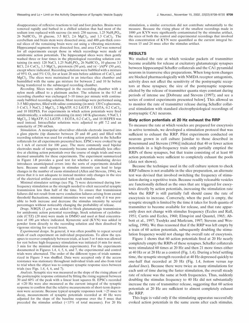
Figure 2. Readily releasable vesicles are replaced slowly during 20 Hzstimulation. A, Average relative synaptic strength is plotted as the Schaf-fer collaterals are stimulated at 20 Hz for 4 sec (each response wasnormalized by the size of the response to the first stimulus; experimentsare from 14 slices). The dashed gray line represents the fraction of thesynaptic response that is predicted to result from the exocytosis of vesiclesthat became available for release during the experiment (as derived in theAppendix). B, Working model of RRP replenishment. Left panel, In theresting synapse the spontaneous rate of exocytosis is low, and the RRPfills completely. The pool has a maximum capacity, and the high-energybarrier that keeps vesicles from fusing spontaneously is the rate-limitingelement that controls how quickly synaptic vesicles undergo exocytosis.Right panel, Episodes of high-frequency activity drive exocytosis quicklyenough to exhaust the RRP. With the pool empty, the rate at which newvesicles are made available limits the rate of transmitter release; theenergy barrier to fusion prevents vesicles from undergoing exocytosis inthe interval between action potentials, but as long as the high-frequencyspike train continues, the barrier is no longer the rate-limiting element inthe exocytic /endocytic cycle. This model accounts only for rate-limitingsteps in the exocytic /endocytic cycle during the first several seconds ofheavy use. Thereafter an additional element plays a role in controlling thedynamics of neurotransmitter mobilization. Endocytosis does not play arate-limiting role in this scheme and is not represented here. C, Thesimultaneous solutions for Equations 1A and 3A (Appendix) are plottedfor the data shown in A. The two independent equations relate the rateconstant of pool replenishment to the fraction of the full pool triggered torelease by isolated action potentials (fusion efficiency). When the corre-sponding values for the two parameters are plotted against each other, theresulting lines intersect where the refilling rate constant equals 0.24/secand the initial fusion efficiency is 0.044. Equation 1A depends only on thesteady-state response size after the pool has reached a near-empty steadystate and is represented by the gray diagonal line. Equation 3A depends onthe changing rate at which transmitter was released over the completecourse of the experiment and is represented by the black line. D, Theinitial fusion efficiency and the pool replenishment rate constant wereestimated separately by simultaneously solving Equations 1A and 3A foreach of the 14 experiments summarized in A and plotted versus eachother. Note that there is no evident correlation between these twoparameters.
Wesseling and Lo • Limit on the Activity Dependence of Synaptic Vesicle Supply J. Neurosci., November 15, 2002, 22(22):9708–9720 9711
number of vesicles available for release, and N is the capacity ofthe RRP (i.e., the total number of release sites in the RRP).
Equation 1 describes a realistic general model of RRP dynam-ics during the first several seconds of high-frequency use. Itassumes a pool of a fixed size (N) containing individual sites thatfill independently with first-order kinetics. The differential equa-tion is equivalent to the one used to characterize the kinetics ofpool replenishment in one of the earliest papers describing thetime course of RRP refilling at central synapses (Stevens andTsujimoto, 1995). Several nontrivial predictions of the modelhave since been tested, and it remains the simplest scheme that isconsistent with what is known about the kinetics of RRP replen-ishment. For example, the rate at which vesicles leave the RRPwithout undergoing exocytosis has been shown to be slow com-pared with the rate of pool filling (Murthy and Stevens, 1999).This, along with other observations (Stevens and Wesseling, 1998,1999b; Pyott and Rosenmund, 2002), implies that the pool has afixed size and is not instead in some steady-state equilibrium witha larger reserve pool (the reverse reaction would formally be anegligible component of �). An additional rate-limiting elementin the synaptic vesicle exocytic /endocytic cycle plays a role duringmore extensive use (Stevens and Wesseling, 1999b).
Equation 1 provides enough constraints to calculate the replen-ishment rate constant (�) from the data plotted in Figure 2A, asoutlined in the Appendix. The fraction of the total responsegenerated by transmitter that became available for release afterstimulation was initiated is plotted as a dashed line in Figure 2A.The simultaneous solution of two independent equations derivedfrom Equation 1 (Eqs. 1A and 3A) yields an average value for �of 0.24/sec for the experiments summarized in Figure 2A (n � 14slices). Although it is possible to devise alternative models thatgenerate slightly different estimates, the actual refreshment ratecould not be much faster, because this estimate is close to theupper bound (0.28/sec) derived without the constraints of a spe-cific model. It thus typically takes several seconds for fresh pack-ets of neurotransmitter to replace the expended readily releasablesupply during episodes of intense synaptic use.
Estimates of fusion efficiencyEquation 1 also provides information about the efficiency withwhich action potentials trigger exocytosis that can be used as aconsistency check for the logic presented so far. Although thepresent analysis does not provide meaningful absolute units for Nbecause neither the number of synapses activated simultaneouslynor the quantal response sizes are known, the fraction of the RRPthat was triggered to undergo exocytosis by the first action po-tential in each 20 Hz train can be calculated by dividing the sizeof the first response by N. This fraction is termed the initial fusionefficiency because it represents the efficiency with which isolatedaction potentials trigger the release of available vesicles at restingsynapses (Stevens and Wesseling, 1999a). The common solutionfor Equations 1A and 3A yields a value for this parameter of0.044, for the experiments summarized in Figure 2. (The individ-ual solutions for the two equations for the data plotted in Fig. 2Aare plotted in Fig. 2C.) This value fits within the published rangeof 0.03–0.07 for these synapses, giving us confidence that ourapproach measures the same kinetic elements that have beenstudied previously (Dobrunz and Stevens, 1997; Schikorski andStevens, 1997). Although we did observe a significant amount ofheterogeneity between preparations in both the rate of RRPreplenishment [range, 0.13–0.36; coefficient of variation (CV) �0.33; see also Stevens and Wesseling (1998)] and the initial fusion
efficiency (range 0.022–0.13; CV � 0.52), Figure 2D shows thatthe variation was not correlated between the two parameters (r �0.01).
Slowly dissipating, activity-dependent acceleration ofthe replenishment processIs neurotransmitter prepared for exocytosis much more quickly atactive nerve terminals than at quiescent ones? The average re-plenishment rate constant of 0.24/sec that we have determined forthe RRPs of active Schaffer collateral synapses is not much faster,if at all, than the resting refreshment rate measured at similartypes of synapses grown in cell culture [which range from 0.09/secto 0.4/sec (Stevens and Wesseling, 1998)]. To compare the replen-ishment rate during high-frequency stimulation with the rateduring rest intervals at the same Schaffer collateral synapses, wemeasured the complete time course over which the RRP refillsafter being depleted.
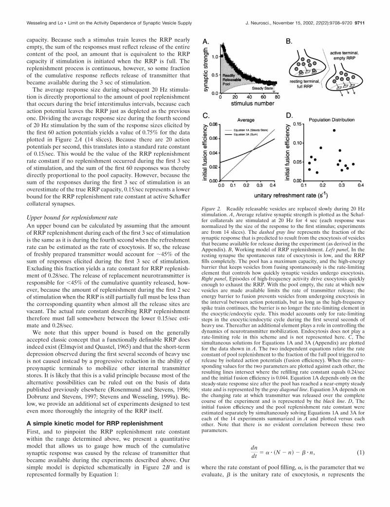
The experimental strategy used is conceptually similar to pre-viously published methods (Stevens and Tsujimoto, 1995; Stevensand Wesseling, 1999b). Experiments were conducted duringwhich the pool was emptied two times in succession, with anexperimentally varied rest interval as diagrammed at the top ofFigure 3. Trains of action potentials (80 at 20 Hz) were usedinstead of osmotic shocks to elicit exocytosis from the synapticterminals. An estimate of pool refilling during the rest intervalwas derived by dividing the sum of the first 60 responses duringthe second stimulus train by the corresponding sum of responsesevoked by the first one. To account for the steady-state amount ofrelease that persists after the RRP has already been exhausted,the sums were first adjusted by subtracting the equivalent measure
Figure 3. The RRP replenishment time course during periods of rest ispredicted by the refreshment rate when synapses are active. The RRP wasemptied with 80 action potentials (20 Hz), and subsequent recovery wasmonitored after an experimentally varied delay with an identical burst ofstimulation (diagrammed at top). The sums of the responses to the secondstimulus train were normalized as described in Results and plotted againstthe recovery interval. The curve is the predicted RRP refilling time coursecalculated with Equation 2 from the replenishment rate measured whenthe synapses were active (0.30/sec for these synapses; mean SEM; 9cells, at least 10 trials for each point). Inset, Average synaptic strengths ofthe responses elicited during the first and second stimulus trains for the 2sec recovery interval are plotted versus time during the trial ( filled circlesrepresent the responses that were used to estimate the RRP recovery after2 sec of rest, i.e., the first 60 responses of each stimulus train).
9712 J. Neurosci., November 15, 2002, 22(22):9708–9720 Wesseling and Lo • Limit on the Activity Dependence of Synaptic Vesicle Supply

derived from experiments when no time was allowed for recoverybetween the two stimulus trains.
As anticipated from Stevens and Wesseling (1998), the replen-ishment rate at Schaffer collateral synapses during long recoveryperiods is somewhat slower than the value for � estimated whenthe synapses are active. If the replenishment process remainedconstant after stimulation ceased, Equation 1 would predict asingle exponential time course of refilling with a time constant of3.3 sec (i.e., 1/�, � � 0.30/sec for this particular set of synapses).Instead, however, the recovery time course was more closelyapproximated by a somewhat slower, 5 sec exponential (recoverydata are plotted in Fig. 3; the single exponential is not shown).This difference was expected because the residual calcium thataccumulates in presynaptic terminals during high-frequency usehas been shown to accelerate the replenishment process several-fold (Stevens and Wesseling, 1998). During subsequent periods ofrest, the rate diminishes gradually as residual calcium is pumpedout of the terminals. Equation 1 can be modified to account forthe decaying effect of residual calcium during rest intervals,yielding:
n�t� � N � �1 � e���t�dt�, (2)
where �(t) is no longer a constant but decays to baseline along asingle exponential with a time constant of 10 sec during periods ofrest (Stevens and Wesseling, 1998). If �(t) starts at 0.30/sec at thebeginning of the recovery period and then slows down threefold,Equation 2 yields the dashed line in Figure 3, which provides agood approximation for the recovery time course.
The estimate of the RRP replenishment rate at active synapsesis thus consistent with the measured time course of pool refillingduring subsequent periods of rest. The slowly decaying, residualcalcium-dependent acceleration appears to be the only significantkinetic element that increases the rate at which neurotransmitterbecomes available for release during periods of heavy use.
Pool capacity does not depend on themeasurement methodologyAlthough the conclusion that it takes several seconds for freshvesicles laden with neurotransmitter to be prepared for exocytosisat active synapses does not depend on the particular formaltheory used to calculate the RRP refreshment rate, it is based onthe classic concept that a functionally definable subset of thesynaptic vesicles is available for action potential-triggered exocy-tosis. To test this generally accepted assumption more thor-oughly, we investigated the effects of changing either the effi-ciency with which individual action potentials trigger theexocytosis of readily releasable vesicles, or the rate of stimulation,on our measure of pool capacity.
CalciumRaising the extracellular concentration of calcium enhances thestrength of resting synapses by increasing the efficiency withwhich action potentials trigger exocytosis of release-ready vesi-cles, whereas magnesium has the opposite effect [Stevens andWesseling (1999a) and references therein]. Changing the ratio ofthe two divalent ions should not affect measurements of RRPcapacity if the RRP is a pool of fixed size.
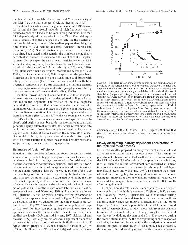
The capacity of the pool was measured from the responses to 80stimuli (20 Hz), with either 4.5 mM Ca/0.5 mM Mg or 2.5 Ca/2.5Mg in the bath; the normalized response sizes are plotted inFigure 4Ai. The total divalent ion concentration was kept con-stant because axonal excitability can be influenced by this param-
eter (Frakenhaeuser and Hodgkin, 1957). At the higher calciumconcentration, the average size of the first synaptic responseduring stimulation was approximately twice the size of the cor-responding one measured with the lower calcium concentration inthe bath. The synaptic strength depressed so much more quicklyin higher calcium, however, that the sums of all 80 responses werestatistically indistinguishable between the two conditions.
Neither the RRP capacity nor the replenishment rate calcu-lated as outlined in the Appendix differed significantly betweenconditions. The measured capacity was slightly larger under thehigh calcium condition (9%), and the replenishment rate wasslightly lower (0.26 vs 0.31/sec), both differences reflecting anonsignificantly lower (5%) average steady-state response to thefinal 20 stimuli under the high calcium condition. As expected, theincrease in the initial fusion efficiency calculated from the simul-
Figure 4. The capacity of the RRP does not depend on the measurementprotocol. A, Short-term depression was induced with 80 stimuli (20 Hz)with either 2.5 mM Ca/2.5 mM Mg (squares) in the bath or 4.5 Ca/0.5 Mg(circles) at the same synapses. i, Responses were normalized by theaverage size of the first response recorded at the lower calcium concen-tration and plotted versus stimulus number (3 slices, 7 trials for each). ii,The RRP replenishment rate constant and the initial fusion efficiencywere calculated by simultaneously solving Equations 1A and 3A, as inFigure 2C. The gray lines represent the analysis of responses evoked underthe high calcium condition; the black lines are for the lower calciumexperiments. The dashed lines represent the solutions for Equation 1A;the solid lines represent Equation 3A. Points of intersection represent thecommon solutions. Note that, as predicted, the fusion efficiency in highcalcium is greater than the fusion efficiency in low calcium by a factorsimilar to the amount of enhancement in synaptic strength observed afterswitching into the higher calcium-containing solution. B, Depression wasinduced with 81 stimuli at 20 and 40 Hz for the same synapses. i, Theresponse sizes were normalized by the average size of the first responsesin the stimulus trains and plotted versus stimulus number for both sets oftrials (3 slices, 11 trials for each). ii, Simultaneous solution of Equations1A (dashed lines) and 3A (solid lines). The gray lines represent the analysisof the 40 Hz responses; black lines represent the analysis of 20 Hzresponses.
Wesseling and Lo • Limit on the Activity Dependence of Synaptic Vesicle Supply J. Neurosci., November 15, 2002, 22(22):9708–9720 9713
taneous solutions of Equations 1A and 3A for both sets of data(Fig. 4Aii) closely matched the increase in synaptic strength thatoccurred when the calcium to magnesium ratio was increased.
Stimulation frequencyThe measurement of RRP capacity should also be independent ofthe particular stimulation protocol used to empty it. To test this,pool capacity was measured from 80 responses generated at either20 or 40 Hz (Fig. 4B). Although the sum of the responses evokedby the first 60 stimuli at 20 Hz was 28% greater than the corre-sponding sum of responses to 60 action potentials recorded at 40Hz, this difference was expected because there was twice as muchtime for replacement transmitter to become available for releaseduring the slower stimulus trains. When this factor is taken intoaccount as outlined in the Appendix, the resting capacity esti-mated from the responses evoked by the 20 Hz stimulation waswithin 5% of the value derived from the 40 Hz trials. Thesolutions of Equations 1A and 3A are plotted in Figure 4Bii. Asexpected, the RRP replenishment rate constants were similar forthe two data sets (0.29/sec for the 40 Hz stimulation protocol and0.26/sec for the 20 Hz protocol).
Together, these results indicate that the capacity of the RRPdoes not depend on the particular protocol used to measure it.They provide support for the general concept that the neurotrans-mitter used for synaptic signaling is drawn from a nonarbitraryRRP (Birks and MacIntosh, 1961; Elmqvist and Quastel, 1965)and for our particular model of RRP replenishment (Eq. 1).
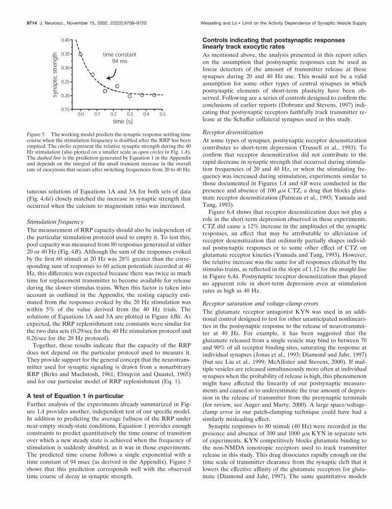
A test of Equation 1 in particularFurther analysis of the experiments already summarized in Fig-ure 1A provides another, independent test of our specific model.In addition to predicting the average fullness of the RRP undernear-empty steady-state conditions, Equation 1 provides enoughconstraints to predict quantitatively the time course of transitionover which a new steady state is achieved when the frequency ofstimulation is suddenly doubled, as it was in those experiments.The predicted time course follows a single exponential with atime constant of 94 msec (as derived in the Appendix). Figure 5shows that this prediction corresponds well with the observedtime course of decay in synaptic strength.
Controls indicating that postsynaptic responseslinearly track exocytic ratesAs mentioned above, the analysis presented in this report relieson the assumption that postsynaptic responses can be used aslinear detectors of the amount of transmitter release at thesesynapses during 20 and 40 Hz use. This would not be a validassumption for some other types of central synapses in whichpostsynaptic elements of short-term plasticity have been ob-served. Following are a series of controls designed to confirm theconclusions of earlier reports (Dobrunz and Stevens, 1997) indi-cating that postsynaptic receptors faithfully track transmitter re-lease at the Schaffer collateral synapses used in this study.
Receptor desensitizationAt some types of synapses, postsynaptic receptor desensitizationcontributes to short-term depression (Trussell et al., 1993). Toconfirm that receptor desensitization did not contribute to therapid decrease in synaptic strength that occurred during stimula-tion frequencies of 20 and 40 Hz, or when the stimulating fre-quency was increased during stimulation, experiments similar tothose documented in Figures 1A and 4B were conducted in thepresence and absence of 100 �M CTZ, a drug that blocks gluta-mate receptor desensitization (Patneau et al., 1993; Yamada andTang, 1993).
Figure 6A shows that receptor desensitization does not play arole in the short-term depression observed in these experiments.CTZ did cause a 12% increase in the amplitudes of the synapticresponses, an effect that may be attributable to alleviation ofreceptor desensitization that ordinarily partially shapes individ-ual postsynaptic responses or to some other effect of CTZ onglutamate receptor kinetics (Yamada and Tang, 1993). However,the relative increase was the same for all responses elicited by thestimulus trains, as reflected in the slope of 1.12 for the straight linein Figure 6Aii. Postsynaptic receptor desensitization thus playedno apparent role in short-term depression even at stimulationrates as high as 40 Hz.
Receptor saturation and voltage-clamp errorsThe glutamate receptor antagonist KYN was used in an addi-tional control designed to test for other unanticipated nonlineari-ties in the postsynaptic response to the release of neurotransmit-ter at 40 Hz. For example, it has been suggested that theglutamate released from a single vesicle may bind to between 70and 90% of all receptor binding sites, saturating the response atindividual synapses (Jonas et al., 1993; Diamond and Jahr, 1997)(but see Liu et al., 1999; McAllister and Stevens, 2000). If mul-tiple vesicles are released simultaneously more often at individualsynapses when the probability of release is high, this phenomenonmight have affected the linearity of our postsynaptic measure-ments and caused us to underestimate the true amount of depres-sion in the release of transmitter from the presynaptic terminals(for review, see Auger and Marty, 2000). A large space/voltage-clamp error in our patch-clamping technique could have had asimilarly misleading effect.
Synaptic responses to 80 stimuli (40 Hz) were recorded in thepresence and absence of 300 and 1000 �M KYN in separate setsof experiments. KYN competitively blocks glutamate binding tothe non-NMDA ionotropic receptors used to track transmitterrelease in this study. This drug dissociates rapidly enough on thetime scale of transmitter clearance from the synaptic cleft that itlowers the effective affinity of the glutamate receptors for gluta-mate (Diamond and Jahr, 1997). The same quantitative models
Figure 5. The working model predicts the synaptic response settling timecourse when the stimulation frequency is doubled after the RRP has beenemptied. The circles represent the relative synaptic strength during the 40Hz stimulation (also plotted on a smaller scale as open circles in Fig. 1A).The dashed line is the prediction generated by Equation 1 in the Appendixand depends on the integral of the small transient increase in the overallrate of exocytosis that occurs after switching frequencies from 20 to 40 Hz.
9714 J. Neurosci., November 15, 2002, 22(22):9708–9720 Wesseling and Lo • Limit on the Activity Dependence of Synaptic Vesicle Supply

that predict glutamate receptor saturation during normal synaptictransmission also predict that, in addition to partially blocking thesynaptic response, �300 �M KYN should shift the dose–responserelationship between transmitter release and postsynaptic re-sponse size well into the linear range (Diamond and Jahr, 1997).Figure 6B shows that neither treatment affected the accumulationof short-term depression, however, indicating that receptor satura-tion does not introduce a nonlinear component into our postsyn-aptic measure of transmitter release. Furthermore, KYN did re-duce the overall synaptic strength substantially (66% in 300 �M
KYN, 78% in 1000 �M KYN), making it unlikely that voltage-clamperrors contributed significant nonlinearities to our measurements.
Intersynaptic cross talkFinally, we provide a positive control for our assumption that thepostsynaptic responses to Schaffer collateral stimulation can beused as linear estimators of transmitter release. The CTZ andKYN experiments argue against receptor desensitization or sat-uration as significant contributions to short-term plasticity, but donot necessarily address other potential postsynaptic mechanisms.A more direct test has been provided by Dobrunz and Stevens(1997), who showed that the quantal sizes of responses to trans-mitter released from single synapses do not depress during repet-itive use, when stimulus strengths are so weak that only one
afferent synapse is activated at once. We typically evoked releasefrom tens of synapses simultaneously for our experiments, how-ever. Although there is no indication that transmitter released atthe same time from multiple synapses interacts with the AMPAtype of glutamate receptors in a nonlinear way at Schaffer collat-eral synapses (Asztely et al., 1997), cross talk between calyceal-type synapses has been reported to result in postsynaptic forms ofshort-term depression (Trussell et al., 1993; Neher and Sakaba,2001). Such a phenomenon, if operative at Schaffer collateralsynapses, might have led to an overestimate of the actual amountof presynaptic depression in our experiments.
An experiment designed to test this possibility instead verifiedthat the amount of depression in synaptic strength measured atmultiple, simultaneously active synapses was matched by de-creases in the probability of release at individual synapses. Short-term depression and RRP refilling were measured with pairs ofstimulus trains (80 stimuli at 20 Hz) separated by 2 sec restintervals as diagrammed at the top of Figure 7B. Weak stimuliwere interleaved with those of normal strength during experimen-tal trials. The weak stimulus intensities may have evoked actionpotentials in several axons, all with afferent synapses with lowrelease probabilities, or in single afferent synapses. Experimentaltrials were alternated with control trials during which only weak
Figure 6. CTZ and KYN do not affectshort-term depression. Ai, Forty Hertz stim-ulation. Schaffer collaterals were stimulated80 times at 40 Hz in the presence and ab-sence of 100 �M CTZ. Average responses tothe stimulus trains with ( gray traces) andwithout (black traces; scaled by 1.12) CTZare plotted (data are from 4 slices, 18 trialsfor each). The first six responses are plottedin the top panel. Note that CTZ amplifiedthe first response slightly more than the rest,possibly because of presynaptic side effectsof the drug. The entire electrophysiologicalrecordings (the trace gathered in the ab-sence of drug is scaled by 1.12), with thestimulus artifacts blanked (2 msec windows),
are plotted in the bottom panel. Aii, Twenty Hertz and frequency switching. Schaffer collaterals were stimulated 60 times at 20 Hz and then 21 more timesat 40 Hz as in Figure 1A in the presence and absence of 100 �M CTZ. The size of the synaptic response to each stimulus was normalized by the averagesize of the first control response (no drug). The size of each response recorded in CTZ is plotted against the size of the corresponding control responseto the same stimulus number (open circles represent responses at 40 Hz; filled circles are 20 Hz responses). The dashed gray line is straight, with a slopeof 1.12 (3 slices, 10 trials each). Inset, The average current traces representing the responses to the 20 stimuli preceding and after the frequency switchfor each condition are overlaid (the response recorded in the absence of drug was scaled by 1.12; the larger of the two sets of current deflections representsresponses recorded at 20 Hz; the baseline of the 40 Hz response average was calculated and subtracted as for the inset of Fig. 1 A). Note that the scaledresponses with and without drug are identical and that the average 40 Hz response size is approximately half that recorded at 20 Hz. B, Schaffercollaterals were stimulated at least 80 times at 40 Hz alternately in the presence and absence of KYN (300 �M: 3 slices, 22 trials each; 1000 �M: 3 slices,10 trials each). The normalized sizes of the control responses are plotted against the corresponding sizes of the responses gathered with KYN in thebath for each stimulus (i, 300 �M KYN; ii, 1000 �M KYN). The dashed lines are straight with slopes of 0.34 ( i) and 0.22 (ii).
Figure 7. Individual synapses behave likesimultaneously activated populations. Schaf-fer collaterals were stimulated with pairs of4-sec-long spike trains (20 Hz) separated by2 sec intervals. For half of the trials, only aminimal number of afferents were stimu-lated throughout. For the other half, stimuliof normal intensity were interleaved withthe weak stimuli. A, Typical, sequential rawdata example of a failure, a large response,and a minimal response during interleavedstimulation. B, C, The average sizes ( B) andprobability ( C) of successful transmissionsin response to the minimal stimuli were av-
eraged into 200 msec bins and plotted versus the time of the experiment. The squares represent the interleaved minimal responses; the circles summarizeexperiments in which only minimal stimuli were used throughout.
Wesseling and Lo • Limit on the Activity Dependence of Synaptic Vesicle Supply J. Neurosci., November 15, 2002, 22(22):9708–9720 9715
stimuli were used throughout. Cross talk between synapses wouldbe measurable as a change in the sizes of the successful responsesto weak stimulation caused by transmitter release elicited by thenormal intensity stimuli.
Figure 7 shows that neither the size of the successful minimalresponses (Fig. 7B) nor the probability of release (Fig. 7C) wasaffected by the transmitter release elicited by the large, inter-leaved stimuli. This result argues strongly against a contributionof intersynaptic cross talk to short-term depression.
The average amplitude of the successful minimal responsesduring both types of trials may have declined a small amountduring the first second of stimulation (Fig. 7B). The effect isattributable to the infrequent, simultaneous exocytosis of multi-ple vesicles, as expected of a stochastic release process when theoverall probability of release is as high as it was at the beginningof the weak stimuli trains (Zucker, 1973).
During 20 Hz use, the probability of release at the synapsesactivated by the weak stimuli depressed just as extensively as thesynaptic currents did in the other experiments reported here. TheRRP refreshment rate constant calculated by the method outlinedin the Appendix for the data plotted in Figure 7C is 0.23/sec. Thisvalue is indistinguishable from the rate constant calculated fromthe synaptic currents elicited by stronger stimulations. The RRPsrefilled �45% during the first 2 sec of rest, yielding an extrapo-lated replenishment rate constant of 0.30/sec, a value that is notsignificantly different from the rate constant calculated for thesynapses when active. These results provide assurance that thesynaptic currents measured for this study were linearly related tothe amount of release at multiple, simultaneously active synapses,and they confirm that it takes several seconds for neurotransmit-ter to replenish the RRP, even when synapses are active.
DISCUSSIONHere we show that a slowly dissipating, residual calcium-drivenacceleration mechanism is sufficient to account for the full rangeof activity-dependent modulation of the rate at which neurotrans-mitter is prepared for exocytosis at Schaffer collateral synapses.Fresh quanta of neurotransmitter were found to replace spentones in the RRP at an average rate of 0.24/sec when synapseswere active. During subsequent periods of rest, the RRP refilledwith an �5 sec exponential time course, as expected if thereplenishment process were to gradually decelerate during inac-tive periods as demonstrated previously (Stevens and Wesseling,1998). With no deceleration during rest intervals, the 0.24/sec rateconstant would predict an only slightly faster exponential refillingtime course with a 4.2 sec time constant. This leaves little kineticroom for a rapidly dissipating component of replenishment-rateenhancement that might have been missed by the earlier studyand indicates that the rate at which the RRP is replenished canbe accelerated only modestly, even during periods of heavysynaptic use.
A kinetically simple model (Eq. 1) of the rate-limiting mecha-nisms that control how long it takes for neurotransmitter to beprepared for release provides a good account of the dynamics ofshort-term plasticity analyzed here. In particular, Figure 5 showsthat when the RRP is in a near-empty steady state, our workingmodel predicts quantitatively the time course over which thesynaptic strength settles to a new steady state after perturbationsin the stimulus frequency, a behavior that is a hallmark offirst-order kinetic reactions.
Although the cell biological process of vesicular maturation iscomplex, involving endocytosis, vesicle genesis and loading, phys-
ical translocation and docking to the release sites, and biochem-ical priming (Sudhof, 1995), the observation that first-order ki-netics describe well the process by which vesicles are readied forexocytosis is consistent with the possibility that only a singlefirst-order enzymatic step in the exocytic /endocytic cycle is ratelimiting during the first several seconds of heavy synaptic use.The identity of the molecular machinery that limits how quicklyvesicles are prepared for exocytosis remains obscure, although itseems to be enzymatic in nature (Stevens and Wesseling, 1998,1999b; Pyott and Rosenmund, 2002) and may be related tobiochemical priming that takes place during or after physicaldocking to the active zone (Kawasaki et al., 1998). During ex-tended periods of synaptic activity, a second rate-limiting processalso plays a role in a longer-lasting form of synaptic depressionthat may be related to depletion of the reserve pool of vesicles orto some other rate-limiting element in the exocytic /endocyticcycle (Stevens and Wesseling, 1999b).
Does the readily releasable pool havekinetic subdivisions?Our working model makes no qualitative kinetic distinctionsamong readily releasable vesicles. The data presented here arealso consistent with models in which some of the release-readyvesicles are available for immediate exocytosis, and the rest areused to restock quickly this smaller pool when required, as long asthe rate of transfer between the two pools is fast on the time scaleof these experiments (Neher and Zucker, 1993; Voets et al., 1999;Voets, 2000). On the other hand, the phenomena modeled bythese more complicated schemes can often be explained equallywell if the efficiency with which action potentials trigger thefusion of individual release-ready vesicles is a heterogeneousproperty of the release sites themselves (Beutner et al., 2001;Sakaba and Neher, 2001). At present, we favor models in which allof the release-ready vesicles are prevented from undergoing exo-cytosis by a single type of energy barrier, because when therelease process is enhanced by residual calcium, the propensityfor exocytosis of the whole pool is potentiated in parallel (Stevensand Wesseling, 1999a).
Relation to earlier studiesOn the basis of electrophysiological data alone, the RRP refresh-ment rate that we have measured may pertain either to the timeit takes for reserve vesicles to be prepared for release or to someother mechanism of neurotransmitter mobilization such as directrefilling of the depleted readily releasable vesicles themselves.However, Pyle et al. (2000) recently measured the time it takes forfluorescently labeled reserve vesicles to replace spent readilyreleasable ones during periods of rest after episodes of high-frequency synaptic use at similar synapses in culture. They reporta single exponential recovery time course with a 7 sec timeconstant, derived from optical measurements, that is similar tothe 5 sec time constant for RRP refilling measured here, suggest-ing that the RRP is replenished primarily with waiting reservevesicles and not by some other means.
Several studies have reported that the transmitter mobilizationprocess is much faster when synapses are active than it is duringsubsequent rest intervals (Kusano and Landau, 1975; Pyle et al.,2000). In the earliest study to report such a phenomenon, Kusanoand Landau (1975) used a model in which action potentials alwaysrelease the same fraction of the RRP, and they concluded that theamount of short-term depression observed during high-frequencyuse is incompatible with a slow replenishment process at the
9716 J. Neurosci., November 15, 2002, 22(22):9708–9720 Wesseling and Lo • Limit on the Activity Dependence of Synaptic Vesicle Supply
squid giant synapse. It is now known, however, that the efficiencywith which action potentials trigger release is not stationary at themammalian central synapses that we study (Stevens and Wessel-ing, 1999a). Although squid synapses may behave differently fromSchaffer collateral synapses in this respect, it is also possible thatthe short-term depression in synaptic strength measured by Ku-sano and Landau (1975) did not correspond to the true extent ofRRP depletion, much as a similar analysis would lead to anunderestimate of pool depletion for the synapses in our prepara-tion. Such an underestimate may have led to an overestimate ofthe amount of activity-dependent acceleration in the replenish-ment process at the squid giant synapse.
Pyle et al. (2000) reported a faster time course for RRP refillingwhen measured electrophysiologically, from which they have con-cluded that individual readily releasable vesicles can be reusedrapidly several times before being replaced at a slower rate byvesicles from the reserve pool. The basis of our quantitativelydiffering electrophysiological results is uncertain. One possibilityis that the stimulation protocol used by Pyle et al. (2000) to elicitexocytosis in the electrophysiological portion of their study didnot completely empty the available pool. This might have resultedin a recovery time course that partially reflected short-term en-hancement in that a larger fraction of the available pool wouldhave been released by trains initiated after shorter rest intervalsthan by those initiated after longer periods of rest [for a moreextensive discussion of this issue, see Stevens and Wesseling(1999a)].
What is the role of the RRP?The release-ready supply of synaptic vesicles supports the abilityof synapses to communicate information encoded by the spike-firing rate. The hippocampal synapses that we have studied startwith a typical release probability of �30–40% (Hessler et al.,1993; Rosenmund et al., 1993; Allen and Stevens, 1994; Huangand Stevens, 1997). Although they fail to release transmitter inresponse to most individual action potentials, they can still reli-ably transmit information by using short bursts of action poten-tials that are sure to trigger some exocytosis when the RRP is atleast partially full. However, this option is no longer available tosynapses that have expended all of their release ready vesicles.After the RRP has been exhausted, the probability of releasebecomes inversely proportional to the stimulus frequency (Ecclesand Rall, 1951; Curtis and Eccles, 1960; Elmqvist and Quastel,1965; Abbott et al., 1997). Because transmitter release is stochas-tic and rare when synapses are depleted of their release-readyvesicles, increasing the frequency of presynaptic activity wouldhave minimal impact on the overall signal detected by postsyn-aptic neurons.
Because of the kinetic constraints imposed by a slow RRPreplenishment rate, information would likely be transmitted moreefficiently via these synapses by a neural code that relies onsporadic bursts of activity than on one that uses precise modula-tions in the frequency of continuous activity. Consistent with this,excitatory neurons in the hippocampus and other brain regions dotend to fire such bursts of spikes in awake and behaving mammals(Ranck, 1973; Scott et al., 1986; Newsome et al., 1989; Knierimand van Essen, 1992; Meister and Berry, 1999). This raises thefollowing question: is the slow replenishment rate an unavoidablebiochemical limitation that hampers the design of efficient neuralcircuits, or does the nervous system derive some computationalbenefit from such constraints on synaptic operation?
APPENDIXDerivation of the RRP refreshment rate fromEquation 1According to Equation 1, when the RRP starts off empty, andthere is no exocytosis (i.e., � � 0), the pool replenishes as afunction of time by:
n�t� � N � �1 � e���t�.
These conditions are approximated during the brief intervalsbetween stimuli when the RRP is in a near-empty steady state, asit is after the 60th action potential in a high-frequency train.While the pool is maintained in a steady state, the amount ofrefilling between stimuli must be equivalent to the amount ofexocytosis, r(�), elicited by each action potential. If � is thefrequency of stimulation (20 Hz or 40 Hz for this study):
r��� � N � �1 � e��
v �,
because 1/� is the time between stimuli. The solution for �depends on the pool capacity, N, which turns out to be anunwieldy parameter. A more convenient parameter is the initialfusion efficiency, fe, defined as the fraction of the RRP releasedby the first stimulus:
fe �r�1�
N,
where r(1) is the amount of transmitter released by the firststimulus. By rearranging, and substituting for N:
fe �r�1�
r�����1 � e
��
v �. (1A)
This equation relates two unknown variables, fe and �, anddepends on the steady-state rate of release elicited during con-tinued stimulation after the RRP has been emptied.
Another, independent equation relating the two unknown vari-ables can be derived from Equation 1 that, in contrast, dependson the rate of transmitter release during the first part of ahigh-frequency train. The capacity of the pool (and thence thefusion efficiency) could be estimated crudely by summing all ofthe quanta released by the first 60 action potentials, but thismethod overestimates the true capacity because fresh vesicles thatreplace those expended during stimulation are also released. Thetrue capacity is:
N � �i�1
S
r�i� � w�S�,
where r(i) is the amount of exocytosis elicited by the ith actionpotential, S is the number of stimuli in the pool depleting train(S � 80 in this case), and w(S) represents the number of vesiclesthat became newly available and subsequently underwent exocy-tosis during the stimulation. w(S) can be derived from Equation1 (see Lemma 1) as:
w�S� � �i�1
S
r�i� � �1 � e����S�i�
v �. (2A)
This formulation for w(S) was used to generate the dashed line inFigure 2A. By combining the previous two equations, we thusderive an equation for N:
Wesseling and Lo • Limit on the Activity Dependence of Synaptic Vesicle Supply J. Neurosci., November 15, 2002, 22(22):9708–9720 9717
N � �i�1
S
r�i� � e����S�i�
v .
Or, by substituting r(1)/fe for N:
fe �r�1�
�i�1
S
r�i� � e����S�i�
v
. (3A)
Equations 1A and 3A are independent because they depend ondifferent parts of the data. Equation 3A depends on the exocyticrate during the part of the stimulation that initially depletes theRRP, whereas Equation 1A depends only on the subsequentsteady-state rate of release. An analytic solution for � based onEquations 1A and 3A is messy, but when the two equations areevaluated numerically using the response sizes plotted in Figure2A as linear proxy for transmitter release, we calculate an averagevalue for � of 0.24/sec.
The simultaneous solution of Equations 1A and 3A, using thedata sets plotted in Figure 2A and Figure 4, Ai and Bi, arepresented graphically in Figure 2C and Figure 4, Aii and Bii. Foreach plot, the lines representing Equation 1A depend only on thesteady-state response size and pass through the origin. The linesrepresenting Equation 3A intersect the y-axis when the poolcapacity is equivalent to the cumulative amount of exocytosis,because when � � 0, no transmitter replenishes the pool (i.e.,w(S) � 0). The point of intersection is taken as the commonsolution for fe and �. A value for N can then be extracted directlyby dividing the size of the first response [r(1)] by fe.
We know from Stevens and Wesseling (1998) that � is notalways constant because the replenishment rate acceleratesseveral-fold during bursts of activity. During high-frequency stim-ulation, however, �(t) achieves its maximum value after onlyapproximately 10 action potentials (Stevens and Wesseling, 1998).This saturation happens so quickly during 20 Hz stimulation thatthe required theoretical corrections to Equations 1A and 3Achange the final result by a only few percentage points. By treating� as a constant during high-frequency stimulation, we calculate itsupper bound; the actual maximum replenishment rate is slightlyslower.
Estimates of steady-state RRP fullness predict thesynaptic response settling time course caused bystimulus frequency perturbationsWhen the RRP is in a near-empty steady state and the stimulusfrequency is suddenly doubled, as in the experiments documentedin Figure 1A, the synaptic strength quickly settles to half itsprevious value, as expected. During the settling time, the rate ofexocytosis is increased transiently, and so the 40 Hz stimulationelicits slightly more transmitter release than the corresponding 20Hz protocol. This extra amount comes to 2.4% of the total RRPcapacity calculated as above. Assuming that the pool is in a steadystate at this point (i.e., dn/dt � 0), from Equation 1 we get afractional fullness value (when n �� N) of:
nN
�n
N � n�
�
�.
If doubling the stimulus frequency also doubles �, the poolfullness during the 40 Hz stimulation should decline to half itssteady-state value at 20 Hz. The extra amount released when the
stimulus frequency is doubled would then be equal to that half,and so at 20 Hz the fractional fullness of the RRP at steady statemust be �4.8%. With this, we can solve for �:
� ��
0.048.
For these experiments, � � 0.25/sec, and so � � 5.2/sec duringthe fourth second of stimulation at 20 Hz (� � 10.4/sec at 40 Hz).From Equation 1 we get the general solution:
n �� � N � C � e�t������
� �,
where C is some constant that depends on the initial conditions.Perturbations in the pool fullness should thus decay back tosteady state with a time constant of 1/(� � �). In this case, thesteady-state value is changed abruptly, creating an analogoussituation. If our model is correct, the residual pool fullness, andthence the synaptic strength, is thus predicted to settle exponen-tially with a time constant of 94 msec after a sudden jump in thestimulus frequency from 20 to 40 Hz. This quantitative predictioncan be seen to match well the behavior that was observed exper-imentally (Fig. 5, dashed line).
We note that this prediction depends on the specific assump-tion that � only increases twofold when the stimulus frequency isdoubled. That would happen if each individual action potentialtriggered the exocytosis of waiting release-ready vesicles with thesame efficiency during the fourth second of 20 Hz stimulation asthey do when the stimulus frequency is switched instead to 40 Hz.We stress that during the first second or two of stimulation, theefficiency with which action potentials elicit exocytosis is knownto increase substantially within presynaptic terminals (Stevensand Wesseling, 1999a) in a manner that depends on the stimula-tion frequency (Zengel and Magleby, 1982; Zucker, 1989;Delaney and Tank, 1994). Because of this frequency facilitation, �might be expected to more than double when the stimulus fre-quency is doubled in these experiments, and our estimate of thesettling time course should thus be considered an upper boundestimate; that is, Equation 1 would also be consistent with a fastertime course of decay. We think that the assumption is accurate,however, because we have conducted a series of studies (data notshown) that indicate that the enhancing effect of rapid stimulationon the release machinery saturates during the first several secondsof 20 Hz use at these synapses, under these conditions (unpub-lished observations). This also explains why it takes a similarnumber of action potentials to empty the RRP when the entirestimulus train is delivered at 40 Hz (half as much time) as it doeswhen delivered at 20 Hz (Fig. 4B).
Lemma 1: derivation of Equation 2A from Equation 1Beginning with the assumption that Equation 1 describes thedynamics of RRP filling and emptying, consider the general casewhen the stimulation is initiated when the RRP is full. We canthen use mathematical induction to show that w(S), defined inEquation 2A, describes the amount of pool refilling that occursduring a stimulus train consisting of S stimuli.
Degenerate caseWe first show that Equation 2A holds for the shortest stimulustrains consisting of only two stimuli (S � 2). By definition, the firststimulus elicits r(1) amount of exocytosis from the RRP. Noadditional transmitter is released until the second stimulus elicits
9718 J. Neurosci., November 15, 2002, 22(22):9708–9720 Wesseling and Lo • Limit on the Activity Dependence of Synaptic Vesicle Supply

the additional exocytosis of r(2) transmitter (i.e., assume that � inEquation 1 is equal to zero during interstimulus intervals). Ac-cording to Equation 1, while � � 0, the empty space in the poolfills over time as:
w �t� � �N � n� � �1 � e���t�, (4A)
where w (t) is the amount of pool refilling over an interval duringwhich � � 0, t is time, and (N � n) is the empty space within theRRP when t � 0. Because, by assumption, the pool is full beforethe first stimulus, and r(1) is defined as the amount by which theRRP is diminished by the first stimulus, r(1) � (N � n) immedi-ately after the first stimulus, but before any time has passed forpool replenishment to occur. The duration of time between thetwo stimuli is just the reciprocal of the stimulation rate (1/�).Therefore, the amount of pool replenishment during the intervalbetween the first and second stimuli is:
w �1v� � r�1� � �1 � e��
1v� .
This quantity is the amount of pool replenishment during theentire two-stimuli train because, by definition, the stimulus trainends immediately after the last stimulus, and there is no moretime for additional replenishment to occur.
As defined in Equation 2A, w(2) is also equal to w (1/�):
w�2� � r�1� � �1 � e����2�1�
v � r�2� � �1 � e����2�2�
v �.
Because r�2� � �1 � e����2�2�
v � � 0,this algebraically simplifies to:
w�2� � r�1� � �1 � e����1�
v �.
Equation 2A is thus at least valid for the shortest stimulus trainsconsisting of only two stimuli.
Inductive caseIf we assume that Equation 2A is valid for the arbitrary case whenthe stimulus train consists of C � 1 stimuli, we can show that theequation is also valid when the stimulus train consists of C stimuli.Assume for induction:
w�C � 1� � �i�1
C�1
r�i� � �1 � e����C�1�i�
v � .
The amount of pool replenishment that occurs during a stimulustrain consisting of C stimuli is the sum of the amount occurringup to stimulus number C � 1, and the amount occurring betweenstimulus C � 1 and stimulus C. Because the time between stimuliis 1/�, from Equation 4A, the amount of pool replenishment
between the last two stimuli must be: E � �1 � e��
v � ,where E is thetotal amount of free space in the RRP immediately after stimulusnumber C � 1 (N � n in Eq. 4A). Because the amount of poolreplenishment during the first C � 1 stimuli of a train consistingof C stimuli must be the same as the amount of pool replenish-ment during the C � 1 stimuli of a train consisting of only C � 1stimuli, the total pool replenishment during a train of C stimuliwould be:
w�C� � �i�1
C�1
r�i� � �1 � e����C�1�i�
v � E � �1 � e��
v � .
E is just the difference between the transmitter release elicited bythe first C � 1 stimuli and the amount of pool replenishment thatoccurred during that time [i.e., w(C � 1)] and therefore can bedescribed by:
E � �i�1
C�1
r�i� � w�C � 1�.
Substituting for w(C � 1) and E, we have:
w�C� � �i�1
C�1
r�i� � �1 � e����C�1�i�
v �
��i�1
C�1
r�i� � �i�1
C�1
r�i� � �1 � e����C�1�i�
v �� � �1 � e��
v � .
This simplifies algebraically to:
w�C� � �i�1
C�1
r�i� � �1 � e����C�i�
v � .
And, because r�C� � �1 � e����C�C�
v � � 0, we conclude that Equa-tion 2A is valid for a stimulus train of C stimuli:
w�C� � �i�1
C
r�i� � �1 � e����C�i�
v � .
We have thus shown that if Equation 2A is valid for a stimulustrain of arbitrary length, then the equation must also be valid fora train that is longer by a single stimulus. Because we also showedthat Equation 2A is valid for the shortest trains consisting of onlytwo stimuli, it must also be true for trains of length 3, 4, and so onfor trains of any length by the principle of mathematical induc-tion. Taken together, this logic demonstrates that Equation 2Afollows from Equation 1 in all cases.
REFERENCESAbbott LF, Varela JA, Sen K, Nelson SB (1997) Synaptic depression
and cortical gain control. Science 275:220–224.Allen C, Stevens CF (1994) An evaluation of causes for unreliability of
synaptic transmission. Proc Natl Acad Sci USA 91:10380–10383.Asztely F, Erdemli G, Kullmann DM (1997) Extrasynaptic glutamate
spillover in the hippocampus: dependence on temperature and the roleof active glutamate uptake. Neuron 18:281–293.
Auger C, Marty A (2000) Quantal currents at single-site central syn-apses. J Physiol (Lond) 526:3–11.
Beutner D, Voets T, Neher E, Moser T (2001) Calcium dependence ofexocytosis and endocytosis at the cochlear inner hair cell afferentsynapse. Neuron 29:681–690.
Birks RI, MacIntosh FC (1961) Acetylcholine metabolism of a sympa-thetic ganglion. Can J Biochem Physiol 39:787–827.
Curtis DR, Eccles JC (1960) Synaptic action during and after repetitivestimulation. J Physiol (Lond) 150:374–398.
Delaney KR, Tank DW (1994) A quantitative measurement of the de-pendence of short-term synaptic enhancement on presynaptic residualcalcium. J Neurosci 14:5885–5902.
Diamond JS, Jahr CE (1997) Transporters buffer synaptically releasedglutamate on a submillisecond time scale. J Neurosci 17:4672–4687.
Dittman JS, Regehr WG (1998) Calcium dependence and recovery ki-netics of presynaptic depression at the climbing fiber to Purkinje cellsynapse. J Neurosci 18:6147–6162.
Dobrunz LE, Stevens CF (1997) Heterogeneity of release probability,facilitation, and depletion at central synapses. Neuron 18:995–1008.
Eccles JC, Rall W (1951) Repetitive monosynaptic activation of mo-toneurons. Proc R Soc Lond B Biol Sci 138:475–498.
Elmqvist D, Quastel DM (1965) A quantitative study of end-plate po-tentials in isolated human muscle. J Physiol (Lond) 178:505–529.
Wesseling and Lo • Limit on the Activity Dependence of Synaptic Vesicle Supply J. Neurosci., November 15, 2002, 22(22):9708–9720 9719
Frakenhaeuser B, Hodgkin AL (1957) The action of calcium on theelectrical properties of squid axons. J Physiol (Lond) 137:218–244.
Hessler NA, Shirke AM, Malinow R (1993) The probability of transmit-ter release at a mammalian central synapse. Nature 366:569–572.
Huang EP, Stevens CF (1997) Estimating the distribution of synapticreliabilities. J Neurophysiol 78:2870–2880.
Jonas P, Major G, Sakmann B (1993) Quantal components of unitaryEPSCs at the mossy fibre synapse on CA3 pyramidal cells of rathippocampus. J Physiol (Lond) 472:615–663.
Kawasaki F, Mattiuz AM, Ordway RW (1998) Synaptic physiology andultrastructure in comatose mutants define an in vivo role for NSF inneurotransmitter release. J Neurosci 18:10241–10249.
Knierim JJ, van Essen DC (1992) Neuronal responses to static texturepatterns in area V1 of the alert macaque monkey. J Neurophysiol67:961–980.
Kusano K, Landau EM (1975) Depression and recovery of transmissionat the squid giant synapse. J Physiol (Lond) 245:13–22.
Liu G, Choi S, Tsien RW (1999) Variability of neurotransmitter concen-tration and nonsaturation of postsynaptic AMPA receptors at synapsesin hippocampal cultures and slices. Neuron 22:395–409.
McAllister AK, Stevens CF (2000) Nonsaturation of AMPA and NMDAreceptors at hippocampal synapses. Proc Natl Acad Sci USA 97:6173–6178.
Meister M, Berry II MJ (1999) The neural code of the retina. Neuron22:435–450.
Murthy VN, Stevens CF (1999) Reversal of synaptic vesicle docking atcentral synapses. Nat Neurosci 2:503–507.
Neher E, Sakaba T (2001) Combining deconvolution and noise analysisfor the estimation of transmitter release rates at the calyx of held.J Neurosci 21:444–461.
Neher E, Zucker RS (1993) Multiple calcium-dependent processes re-lated to secretion in bovine chromaffin cells. Neuron 10:21–30.
Newsome WT, Britten KH, Movshon JA (1989) Neuronal correlates ofa perceptual decision. Nature 341:52–54.
Patneau DK, Vyklicky Jr L, Mayer ML (1993) Hippocampal neuronsexhibit cyclothiazide-sensitive rapidly desensitizing responses to kai-nate. J Neurosci 13:3496–3509.
Pyle JL, Kavalali ET, Piedras-Renteria ES, Tsien RW (2000) Rapidreuse of readily releasable pool vesicles at hippocampal synapses. Neu-ron 28:221–231.
Pyott SJ, Rosenmund C (2002) . The effects of temperature on vesicularsupply and release in autaptic cultures of rat and mouse hippocampalneurons. J Physiol (Lond) 539:523–535.
Ranck Jr JB (1973) Studies on single neurons in dorsal hippocampalformation and septum in unrestrained rats. I. Behavioral correlates andfiring repertoires. Exp Neurol 41:462–531.
Rosenmund C, Stevens CF (1996) Definition of the readily releasablepool of vesicles at hippocampal synapses. Neuron 16:1197–1207.
Rosenmund C, Clements JD, Westbrook GL (1993) Nonuniform prob-ability of glutamate release at a hippocampal synapse. Science262:754–757.
Sakaba T, Neher E (2001) Quantitative relationship between transmitterrelease and calcium current at the calyx of Held synapse. J Neurosci21:462–476.
Schikorski T, Stevens CF (1997) Quantitative ultrastructural analysis ofhippocampal excitatory synapses. J Neurosci 17:5858–5867.
Schikorski T, Stevens CF (2001) Morphological correlates of function-ally defined synaptic vesicle populations. Nat Neurosci 4:391–395.
Scott TR, Yaxley S, Sienkiewicz ZJ, Rolls ET (1986) Gustatory re-sponses in the nucleus tractus solitarius of the alert cynomolgus mon-key. J Neurophysiol 55:182–200.
Stevens CF, Tsujimoto T (1995) Estimates for the pool size of releasablequanta at a single central synapse and for the time required to refill thepool. Proc Natl Acad Sci USA 92:846–849.
Stevens CF, Wesseling JF (1998) Activity-dependent modulation of therate at which synaptic vesicles become available to undergo exocytosis.Neuron 21:415–424.
Stevens CF, Wesseling JF (1999a) Augmentation is a potentiation of theexocytotic process. Neuron 22:139–146.
Stevens CF, Wesseling JF (1999b) Identification of a novel process lim-iting the rate of synaptic vesicle cycling at hippocampal synapses.Neuron 24:1017–1028.
Sudhof TC (1995) The synaptic vesicle cycle: a cascade of protein-protein interactions. Nature 375:645–653.
Trussell LO, Zhang S, Raman IM (1993) Desensitization of AMPAreceptors on multiquantal neurotransmitter release. Neuron10:1185–1196.
Tsodyks MV, Markram H (1997) The neural code between neocorticalpyramidal neurons depends on neurotransmitter release probability.Proc Natl Acad Sci USA 94:719–723.
Voets T (2000) Dissection of three Ca 2�-dependent steps leading tosecretion in chromaffin cells from mouse adrenal slices. Neuron28:537–545.
Voets T, Neher E, Moser T (1999) Mechanisms underlying phasic andsustained secretion in chromaffin cells from mouse adrenal slices.Neuron 23:607–615.
Wang LY, Kaczmarek LK (1998) High-frequency firing helps replenishthe readily releasable pool of synaptic vesicles. Nature 394:384–388.
Wu LG, Borst JG (1999) The reduced release probability of releasablevesicles during recovery from short-term synaptic depression. Neuron23:821–832.
Yamada KA, Tang CM (1993) Benzothiadiazides inhibit rapid gluta-mate receptor desensitization and enhance glutamatergic synaptic cur-rents. J Neurosci 13:3904–3915.
Zengel JE, Magleby KL (1982) Augmentation and facilitation of trans-mitter release. A quantitative description at the frog neuromuscularjunction. J Gen Physiol 80:583–611.
Zucker RS (1973) Changes in the statistics of transmitter release duringfacilitation. J Physiol (Lond) 229:787–810.
Zucker RS (1989) Short-term synaptic plasticity. Annu Rev Neurosci12:13–31.
9720 J. Neurosci., November 15, 2002, 22(22):9708–9720 Wesseling and Lo • Limit on the Activity Dependence of Synaptic Vesicle Supply


![Sensor signal (CW limit, CCW limit, NORG "Origin proximity", ORG "Origin") [12V pull up photocoupler input] Servo pack signal (Servo ready, Positioning complete, Alarm) Emergency stop](https://static.fdocuments.in/doc/165x107/5e4e13665ce47400d9703c11/sensor-signal-cw-limit-ccw-limit-norg-origin-proximity-org-origin.jpg)