
Limbic system Neuroanatomy (Dr Muhammad Zeeshan Anwar)
23
Limbic System Dr Muhammad Zeeshan Anwar [email protected] Friday, January 30, 2015 Dr Muhammad Zeeshan Anwar [email protected] Clinical Neuroanatomy
-
Upload
muhammad-zeeshan-anwar -
Category
Education
-
view
555 -
download
7
Transcript of Limbic system Neuroanatomy (Dr Muhammad Zeeshan Anwar)

Limbic System
Dr Muhammad Zeeshan Anwar
Friday, January 30, 2015Dr Muhammad Zeeshan Anwar
Clinical Neuroanatomy
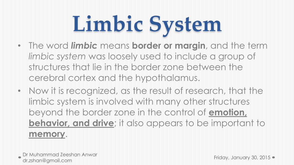
Limbic System• The word limbic means border or margin, and the term
limbic system was loosely used to include a group of
structures that lie in the border zone between the
cerebral cortex and the hypothalamus.
• Now it is recognized, as the result of research, that the
limbic system is involved with many other structures
beyond the border zone in the control of emotion,
behavior, and drive; it also appears to be important to
memory.
Friday, January 30, 2015Dr Muhammad Zeeshan Anwar
History• Paul Broca (1824-1880):
1878: “le grand lobe limbique”
Refers to a ring of gray matter on the medial aspect of the cerebral hemispheres.
• James Papez (1883-1958):
1930’s: defined a limbic system that might underlie the relationship between emotion and memory (Papez’ circuit).
Friday, January 30, 2015Dr Muhammad Zeeshan Anwar
Friday, January 30, 2015Dr Muhammad Zeeshan Anwar
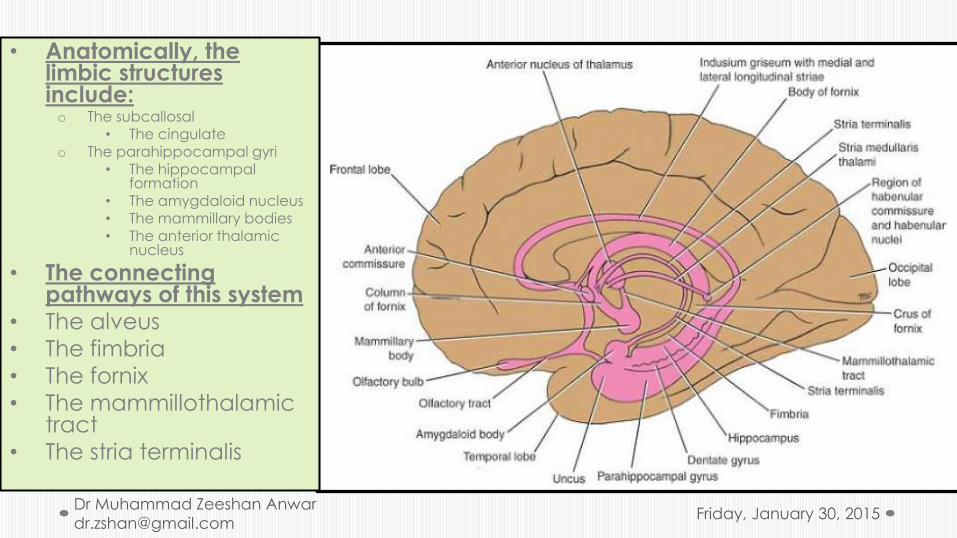
• Anatomically, the limbic structures include:o The subcallosal
• The cingulateo The parahippocampal gyri
• The hippocampal formation
• The amygdaloid nucleus• The mammillary bodies• The anterior thalamic
nucleus
• The connecting pathways of this system
• The alveus• The fimbria• The fornix• The mammillothalamic
tract• The stria terminalis
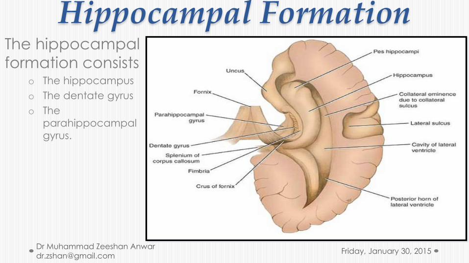
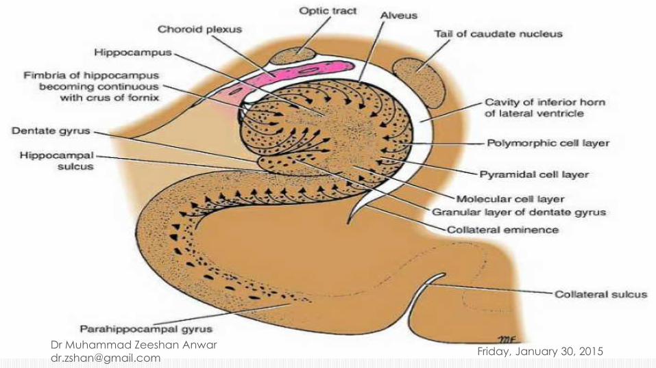
Hippocampal FormationThe hippocampal
formation consistso The hippocampus
o The dentate gyrus
o The
parahippocampal
gyrus.
Friday, January 30, 2015Dr Muhammad Zeeshan Anwar

The hippocampus• The hippocampus is a curved elevation of gray matter
that extends throughout the entire length of the floor of the inferior horn of the lateral ventricle.o Its anterior end is expanded to form the pes hippocampus. It is named
hippocampus because it resembles a sea horse in coronal section.
o The convex ventricular surface is covered with ependyma, beneath which lies a thin layer of white matter called the alveus.
o The alveus consists of nerve fibers that have originated in the hippocampus, and these converge medially to form a bundle called the fimbria.
o The fimbria, in turn, becomes continuous with the crus of the fornix.
• The hippocampus terminates posteriorly beneath the splenium of the corpus callosum
Friday, January 30, 2015Dr Muhammad Zeeshan Anwar
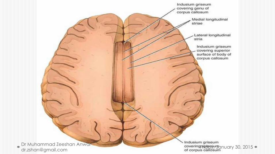
The dentate gyrus• The dentate gyrus is a narrow, notched band of gray matter
that lies between the fimbria of the hippocampus and the parahippocampal gyrus.
• Posteriorly, the gyrus accompanies the fimbria almost to the splenium of the corpus callosum and becomes continuous with the indusium griseum.o The indusium griseum is a thin, vestigial layer of gray matter that covers the superior
surface of the corpus callosum.
• Embedded in the superior surface of the indusium griseum are two slender bundles of white fibers on each side called the medial and lateral longitudinal striae.o The striae are the remains of the white matter of the vestigial indusium griseum.
• Anteriorly, the dentate gyrus is continued into the uncus.
Friday, January 30, 2015Dr Muhammad Zeeshan Anwar

Parahippocampal gyrus• The parahippocampal
gyrus lies between the
hippocampal fissure and
the collateral sulcus and
is continuous with the
hippocampus along the
medial edge of the
temporal lobe.
Friday, January 30, 2015Dr Muhammad Zeeshan Anwar

Amygdaloid Nucleus• The amygdaloid nucleus is so
named because it resembles an almond.
• It is situated partly anterior and partly superior to the tip of the inferior horn of the lateral ventricle .
• It is fused with the tip of the tail of the caudate nucleus, which has passed anteriorly in the roof of the inferior horn of the lateral ventricle.
• The stria terminalis emerges from its posterior aspect. The amygdaloidnucleus consists of a complex of nuclei that can be grouped into a larger basolateral group and smaller corticomedial group.
Friday, January 30, 2015Dr Muhammad Zeeshan Anwar
Connecting Pathways of the Limbic System
• The connecting pathways of the limbic system are:
• The alveus
• The fimbria
• The fornix
• The mammillothalamic tract
• The stria terminalis.
Friday, January 30, 2015Dr Muhammad Zeeshan Anwar
• The alveus:o consists of a thin layer of white matter that lies on the superior or ventricular surface of the
hippocampus, composed of nerve fibers that originate in the hippocampal cortex. The fibers converge on the medial border of the hippocampus to form a bundle called the fimbria.
• The fimbria:o Now leaves the posterior end of the hippocampus as the crus of the fornix. posteriorly and
superiorly beneath the splenium of the corpus callosum and around the posterior surface of the thalamus.
• The two crura now converge to form the body of the fornix, which is applied closely to the undersurface of the corpus callosum .As the two crura come together, they are connected by transverse fibers called the commissure of the fornix.
• These fibers decussate and join the hippocampi of the two sides.
o Anteriorly, the body of the fornix is connected to the undersurface of the corpus callosum by the septum pellucidum.
o Inferiorly, the body of the fornix is related to the tela choroidea and the ependymal roof of the third ventricle.
o The body of the fornix splits anteriorly into two anterior columns of the fornix, each of which curves anteriorly and inferiorly over the interventricular foramen (foramen of Monro).
• Then, each column disappears into the lateral wall of the third ventricle to reach the mammillary body.
Friday, January 30, 2015Dr Muhammad Zeeshan Anwar

• The mammillothalamic tract provides important connections between the mammillary body and the anterior nuclear group of the thalamus.
• The stria terminalis emerges from the posterior aspect of the amygdaloid nucleus and runs as a bundle of nerve fibers posteriorly in the roof of the inferior horn of the lateral ventricle on the medial side of the tail of the caudate nucleus. o It follows the curve of the caudate nucleus and comes to lie in the floor of the
body of the lateral ventricle.
Friday, January 30, 2015Dr Muhammad Zeeshan Anwar
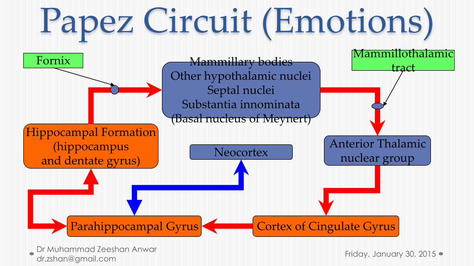
Papez Circuit (Emotions)Mammillary bodies
Other hypothalamic nucleiSeptal nuclei
Substantia innominata(Basal nucleus of Meynert)
Hippocampal Formation(hippocampus
and dentate gyrus)
Anterior Thalamicnuclear group
Cortex of Cingulate GyrusParahippocampal Gyrus
Neocortex
FornixMammillothalamic
tract
Friday, January 30, 2015Dr Muhammad Zeeshan Anwar

Structure of the Hippocampus and the Dentate Gyrus
• The cortical structure of the parahippocampal gyrus is six layered. • As the cortex is traced into the hippocampus, there is a gradual transition
from a six- to a three-layered arrangement.• These three layers are:
o The superficial molecular layer, consisting of nerve fibers and scattered small neuronso The pyramidal layer, consisting of many large pyramid-shaped neuronso The inner polymorphiclayer, which is similar in structure to the polymorphic layer of the cortex seen
elsewhere.
• The dentate gyrus also has three layers, but the pyramidal layer is replaced by the granular layer.
• The granular layer is composed of densely arranged rounded or oval neurons that give rise to axons that terminate on the dendrites of the pyramidal cells in the hippocampus. A few of the axons join the fimbria and enter the fornix.
Friday, January 30, 2015Dr Muhammad Zeeshan Anwar

Afferent Connections of the Hippocampus
• Afferent connections of the hippocampus may be divided into six groups:o 1. Fibers arising in the cingulate gyrus pass to the hippocampus.
o 2. Fibers arising from the septal nuclei (nuclei lying within the midline close to the anterior commissure) pass posterior in the fornix to the hippocampus.
o 3. Fibers arising from one hippocampus pass across the midline to the opposite hippocampus in the commissure of the fornix.
o 4. Fibers from the indusium griseum pass posteriorly in the longitudinal striae to the hippocampus.
o 5. Fibers from the entorhinal area or olfactory-associated cortex pass to the hippocampus.
o 6. Fibers arising from the dentate and parahippocampal gyri travel to the hippocampus.
Friday, January 30, 2015Dr Muhammad Zeeshan Anwar
Efferent Connections of the Hippocampus• Axons of the large pyramidal cells of the hippocampus
emerge to form the alveus and the fimbria. The fimbria continues as the crus of the fornix. The two cruraconverge to form the body of the fornix. The body of the fornix splits into the two columns of the fornix, which curve downward and forward in front of the interventricular foramina. The fibers within the fornix are distributed to the following regions:o Fibers pass posterior to the anterior commissure:
• 1. To enter the mammillary body, where they end in the medial nucleus.
• 2. To end in the anterior nuclei of the thalamus.
• 3. To enter the tegmentum of the midbrain.
o 4. Fibers pass anterior to the anterior commissure to end in the septal nuclei, the lateral preoptic area, and the anterior part of the hypothalamus.
o 5. Fibers join the stria medullaris thalami to reach the habenular nuclei.
Friday, January 30, 2015Dr Muhammad Zeeshan Anwar
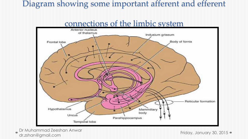
Diagram showing some important afferent and efferent
connections of the limbic system
Friday, January 30, 2015Dr Muhammad Zeeshan Anwar
Functions of the Limbic System• The limbic system, via the hypothalamus and its connections with the
outflow of the autonomic nervous system and its control of the endocrine system, is able to influence many aspects of emotional behavior. These include particularly:o the reactions of fear and anger and the emotions associated with sexual behavior.
• Converting recent memory to long-term memory.o A lesion of the hippocampus results in the individual being unable to store long term memory. o Memory of remote past events before the lesion developed is unaffected. This condition is called
anterograde amnesia.o It is interesting to note that injury to the amygdaloid nucleus and the hippocampus produces a greater
memory loss than injury to either one of these structures alone.
• There is no evidence that the limbic system has an olfactory function.• The various afferent and efferent connections of the limbic system provide
pathways for the integration and effective homeostatic responses to a wide variety of environmental stimuli.
Friday, January 30, 2015Dr Muhammad Zeeshan Anwar
Destruction of the Amygdaloid Complex
• Unilateral or bilateral destruction of the amygdaloid nucleus and the para-amygdaloid area in patients suffering from:
• aggressive behavior in many cases results in a decrease in aggressiveness,
• emotional instability
• Restlessness
• Increased interest in food
• Hypersexuality.
• There is no disturbance in memory.• Monkeys that have been subjected to bilateral removal of the temporal lobes
demonstrate what is known as the Klüver-Bucy syndrome.o They become docile and show no evidence of fear or anger and are unable to appreciate objects visually. They have
an increased appetite and increased sexual activity. Moreover, the animals indiscriminately seek partnerships with male and female animals.
• Precise stereotactic lesions in the amygdaloid complex in humans reduce emotional excitability and bring about normalization of behavior in patients with severe disturbances. No loss of memory occurs
Friday, January 30, 2015Dr Muhammad Zeeshan Anwar
Schizophrenia• The symptoms of schizophrenia include chronically disordered thinking, blunted
affect, and emotional withdrawal.
• Paranoid delusions and auditory hallucinations may also be present.
• Clinical research has shown that if the limbic receptors to dopamine are blocked by a pharmacologic agent, the worst symptoms of schizophrenia are lessened.
• Phenothiazine administration, for example, blocks the dopamine receptors in the limbic system.
• Unfortunately, this drug, as well as most other antipsychotic drugs, has major motor side effects on the dopaminergic receptors within the extrapyramidal system, producing abnormal involuntary movements. Research is now concentrating on finding a drug that will block the limbic dopamine receptors but without effect on the receptors of the extrapyramidal system (substantia nigra–corpus striatum).
• It is clear, however, that there is still no direct evidence that excessive production of dopamine by certain neurons actually contributes to schizophrenia.
Friday, January 30, 2015Dr Muhammad Zeeshan Anwar