L’ilot génomique pks chez Escherichia coli: structure ...
224
HAL Id: tel-00612631 https://tel.archives-ouvertes.fr/tel-00612631 Submitted on 29 Jul 2011 HAL is a multi-disciplinary open access archive for the deposit and dissemination of sci- entific research documents, whether they are pub- lished or not. The documents may come from teaching and research institutions in France or abroad, or from public or private research centers. L’archive ouverte pluridisciplinaire HAL, est destinée au dépôt et à la diffusion de documents scientifiques de niveau recherche, publiés ou non, émanant des établissements d’enseignement et de recherche français ou étrangers, des laboratoires publics ou privés. L’ilot génomique pks chez Escherichia coli : structure-fonction de la protéine ClbP et études épidémiologiques Damien Dubois To cite this version: Damien Dubois. L’ilot génomique pks chez Escherichia coli : structure-fonction de la protéine ClbP et études épidémiologiques. Médecine humaine et pathologie. Université d’Auvergne - Clermont-Ferrand I, 2011. Français. NNT : 2011CLF1MM04. tel-00612631
Transcript of L’ilot génomique pks chez Escherichia coli: structure ...

HAL Id: tel-00612631https://tel.archives-ouvertes.fr/tel-00612631
Submitted on 29 Jul 2011
HAL is a multi-disciplinary open accessarchive for the deposit and dissemination of sci-entific research documents, whether they are pub-lished or not. The documents may come fromteaching and research institutions in France orabroad, or from public or private research centers.
L’archive ouverte pluridisciplinaire HAL, estdestinée au dépôt et à la diffusion de documentsscientifiques de niveau recherche, publiés ou non,émanant des établissements d’enseignement et derecherche français ou étrangers, des laboratoirespublics ou privés.
L’ilot génomique pks chez Escherichia coli :structure-fonction de la protéine ClbP et études
épidémiologiquesDamien Dubois
To cite this version:Damien Dubois. L’ilot génomique pks chez Escherichia coli : structure-fonction de la protéine ClbP etétudes épidémiologiques. Médecine humaine et pathologie. Université d’Auvergne - Clermont-FerrandI, 2011. Français. �NNT : 2011CLF1MM04�. �tel-00612631�

UNIVERSITE BLAISE PASCAL UNIVERSITE D’AUVERGNE
Année 2011 N° d’ordre
ECOLE DOCTORALE
DES SCIENCES DE LA VIE ET DE LA SANTE
THESE
Présentée à l’Université d’Auvergne Pour l’obtention du grade de Docteur d’Université
Spécialité : Microbiologie
par
DUBOIS Damien
L’ilot génomique pks chez Escherichia coli : structure-fonction de la protéine ClbP et études épidémiologiques
Soutenue le 11 mars 2011
Président
F. DELBAC, Professeur, Université Blaise Pascal (Clermont-Ferrand)
Rapporteurs
C. GUILHOT, Directeur de recherche au CNRS, IPBS, Toulouse W. SOUGAKOFF, Maître de Conférences, Université Pierre et Marie Curie (Paris VI)
Membres
E. OSWALD, Professeur, Université Paul Sabatier (Toulouse III) R. BONNET, Professeur, Université d’Auvergne – Directeur de thèse
JE2526, usc-INRA 2018 Evolution des bactéries pathogènes et susceptibilité génétique de l’hôte
Clermont Université 28 place H. Dunant – 63001 Clermont-Ferrand Cedex

2

3
DUBOIS Damien
L’ilot génomique pks chez Escherichia coli : structure-fonction de la protéine ClbP et études épidémiologiques
RésuméL’ilot génomique pks de Escherichia coli et d’autres Enterobacteriaceae code des synthases de
polycétides et de peptides non ribosomaux qui permettent l’assemblage d’un composé hybride polycétide-peptide non ribosomal putative. Ce composé nommé colibactine induit des cassures double-brin de l’ADN des cellules eucaryotes.
La machinerie enzymatique codée par l’ilot pks comporte une protéine essentielle ClbP, atypique dans ce type d’ilot. Nous avons montré que ClbP possède une partie N-terminale catalytique et périplasmique, et une partie C-terminale associée à la membrane cytoplasmique. La structure cristalline de ClbP et des expériences de mutagenèse ont révélé un site actif à sérine et des caractéristiques structurales originales, qui sont compatibles avec une activité peptidase, confirmée par des analyses biochimiques. Dix homologues de ClbP ont été identifiés in silico dans des ilots génomiques de synthases de peptides non ribosomaux d’espèces bactériennes proches et éloignées. Tous les homologues testés ont présenté une promiscuité fonctionnelle avec ClbP. ClbP est donc le prototype d’une nouvelle sous-famille de peptidases, qui sont probablement impliquées dans la maturation de composés peptidiques non ribosomaux.
Par ailleurs, nous avons réalisé deux études épidémiologiques sur la prévalence de l’ilot pks dans l’espèce E. coli dans deux contextes physiopathologiques, l’urosepsis et les cancers coliques et rectaux. L’ilot pks était significativement associé aux souches issues d’urosepsis comparé à des souches commensales, et aux souches issues de biopsies de tumeurs coliques comparé à des souches commensales ou issues de biopsies de tumeurs rectales, de diverticuloses et de lésions iléales de maladie de Crohn.
Mots-clés : colibactine, peptidase, peptides non ribosomaux, urosepsis, cancer colorectal
SummaryThe pks genomic island of Escherichia coli and other Enterobacteriaceae encodes polyketide and
nonribosomal peptide synthases that build a putative hybrid PK-NRP compound. This compound designated Colibactin induces DNA double-strand breaks in eukaryotic cells.
The pks-encoded enzymatic machinery comprises an essential protein ClbP, atypical for this type of genomic islands. We report that ClbP harbors a catalytic and periplasmic N-terminal part, and a C-terminal part associated to the cytoplasmic membrane. ClbP crystal structure and mutagenesis experiments revealed a serine-active site and original structural features, which are compatible with peptidase activity confirmed by biochemical assays. Ten ClbP homologs were identified in silico in NRPS-encoding genomic islands of close and distant-related bacterial species. All tested ClbP homologs showed functional promiscuity with ClbP. ClbP is therefore a prototype of a new subfamily of peptidases, which are probably involved for the maturation of NRP compounds.
Furthermore, we undertook two epidemiological studies on the prevalence of pks island in E. coli in two pathophysiology contexts; urosepsis and colorectal cancers. The pks island was significantly associated with urosepsis strains compared to commensal strains, and strains isolated from biopsies of colon tumors compared with commensal strains or strains isolated from biopsies of rectal tumors, diverticulosis and ileal lesions of Crohn disease.
Key-words : colibactin, peptidase, nonribosomal peptides, urosepsis, colorectal cancer
Jury : Président : Mr DELBAC Frédéric
Rapporteurs : Mr GUILHOT Christophe Mr SOUGAKOFF Wladimir
Membres : Mr OSWALD Eric Mr BONNET Richard – Directeur de thèse
Date de soutenance : 11 mars 2011
Adresse de l’auteur : 4 allée de Layrac – 31700 Blagnac

4

5
A Frédérique,
A Anaé, puis-je te transmettre le sens de la curiosité
A Olivier, pour notre collaboration qui s’est arrêtée trop tôt
A tous ceux qui me sont chers

6

7
Remerciements
Je remercie sincèrement,
Messieurs les Docteurs Guilhot et Sougakoff, d’avoir aimablement accepté d’être les rapporteurs de cette thèse. Je suis fier de vous compter parmi mes juges et vous assure ma
reconnaissance et mon respect.
Monsieur le Professeur Delbac, d’avoir aimablement accepté de juger ce travail et de présider notre jury.
Monsieur le Professeur Oswald, pour votre collaboration, d’avoir aimablement accepté de juger cette thèse et de me permettre de poursuivre dans votre équipe.
Monsieur le Professeur Bonnet pour m’avoir accueilli comme Assistant et Doctorant dans son
laboratoire, pour sa disponibilité, son écoute, sa confiance, sa patience, son soutien et ses conseils amicaux. Cela a été un plaisir de travailler dans ton équipe et sous ta direction. Trouve ici toute ma gratitude et mon respect sincère.
Je tiens à remercier ceux sans qui ce travail n’aurait pas été possible,
Rolande, pour son dynamisme, son optimisme et son aide, Marlène, pour sa gentillesse, ses conseils et son aide,
cela a été un plaisir de travailler avec vous, au-delà de mes espérances en arrivant au fond du couloir,
Julien, pour les diverses collaborations et les nombreux échanges qui n’ont peut-être pas révolutionné la bactériologie mais qui ont fait avancer mes projets et mes idées, et « Viva San Diego !»,
Nathalie, pour sa rigueur scientifique, son dynamisme et son franc-parler, Olivier, qui nous a quitté prématurément, pour notre collaboration amicale,
Bernadette, pour sa collaboration et son enthousiasme pour la problématique, Pierre pour sa disponibilité, sa bonne humeur et nos échanges dans notre galère
« colorectale »,
Marie-Agnès pour ses conseils et son aide, Antony, pour nos échanges et sa bonne humeur constante ; je te souhaite de réussir avec ClbP
et ses semblables. Marie-Hélène et Claudine pour leur gentillesse et leur disponibilité.
Je remercie également,
Emilie et Caroline, mes successives charmantes collègues de bureau, Tous ceux de l’équipe « Evolution des bactéries pathogènes et susceptibilité de l’hôte », en
particulier Madame le Professeur Arlette Darfeuille-Michaud pour m’avoir accueilli dans son unité, Alexandra, pour son implication, et Sabah, pour son enthousiasme, Jean-Philippe pour ses conseils,
Tous ceux du laboratoire de bactériologie du CHU de Clermont-Ferrand pour leur accueil et leur bonne humeur,
Frédérique, mon épouse, pour sa compréhension et son soutien ces dernières années, pour avoir accepté les nombreuses absences,
Reçois ma profonde gratitude, Anaé, ma fille, pour ton sourire constant, avec toi, c’est facile d’être un papa qui « cherche ».

8

9
SOMMAIRE LISTE DES FIGURES........................................................................................................... 12
LISTE DES TABLEAUX ...................................................................................................... 14
LISTE DES ABREVIATIONS ............................................................................................. 15
INTRODUCTION.................................................................................................................. 17
CHAPITRE I – Revue bibliographique
I. LE CYCLE CELLULAIRE EUCARYOTE.............................................................. 23
A. STRATEGIE GENERALE DU CYCLE CELLULAIRE ............................................................ 23
B. LES PRINCIPAUX ACTEURS DE LA REGULATION DU CYCLE CELLULAIRE ..................... 241. LES PROTEINES KINASES DEPENDANTES DES CYCLINES (CDK) ...................................... 24 2. LES CYCLINES ................................................................................................................. 24 3. LA PHOSPHORYLATION DES CDK .................................................................................... 25 4. LES INHIBITEURS DE CDK ............................................................................................... 26 5. LA REGULATION DE LA LOCALISATION INTRACELLULAIRE DES CDK .............................. 26
C. LES PRINCIPAUX EVENEMENTS DE REGULATION DU CYCLE ......................................... 27
D. LES POINTS DE CONTROLE DE LA QUALITE DU CYCLE CELLULAIRE ............................ 281. LA SIGNALISATION DES ALTERATIONS DE L’ADN............................................................ 28 2. LES POINTS DE CONTROLE G1/S ET G2/M ....................................................................... 30 3. LES POINTS DE CONTROLE EN PHASE S............................................................................. 32 4. LE POINT DE CONTROLE DU FUSEAU MITOTIQUE .............................................................. 32
II. LES CYCLOMODULINES DE ESCHERICHIA COLI ........................................... 33
A. LE FACTEUR CYTOTOXIQUE NECROSANT CNF............................................................. 341. LES CNF VARIANTS......................................................................................................... 34 2. STRUCTURE...................................................................................................................... 34 3. SUPPORT GENETIQUE ....................................................................................................... 34 4. MODE D’ACTION .............................................................................................................. 35
a) Sécrétion ................................................................................................................. 35b) Translocation dans les cellules eucaryotes .......................................................... 36c) Modification de la cible moléculaire .................................................................... 36
5. EXPRESSION PHENOTYPIQUE ............................................................................................ 38 6. DISTRIBUTION.................................................................................................................. 39 7. ROLE DANS LE PROCESSUS INFECTIEUX............................................................................ 39 8. DES AGENTS CARCINOGENES POTENTIELS........................................................................ 40
B. LA TOXINE DE DISTENSION CYTOLETHALE CDT .......................................................... 431. LES VARIANTS CDT ET LEUR SUPPORT GENETIQUE ......................................................... 43 2. STRUCTURE DES CDT...................................................................................................... 43 3. MODE D’ACTION .............................................................................................................. 44
a) Sécrétion ................................................................................................................. 44

10
b) Translocation dans les cellules eucaryotes .......................................................... 44c) Altération de l’ADN et expression phénotypique ............................................... 46
4. DISTRIBUTION ET ROLE DANS LE PROCESSUS INFECTIEUX ................................................ 46 5. DES AGENTS CARCINOGENES POTENTIELS........................................................................ 47
C. LE FACTEUR D'INHIBITION DU CYCLE CIF..................................................................... 481. DISTRIBUTION ET SUPPORT GENETIQUE............................................................................ 48 2. STRUCTURE...................................................................................................................... 48 3. MODE D’ACTION ET EXPRESSION PHENOTYPIQUE ............................................................ 49 4. UN AGENT CARCINOGENE POTENTIEL .............................................................................. 49
D. LA COLIBACTINE ............................................................................................................ 511. SUPPORT GENETIQUE ....................................................................................................... 51 2. TRANSCRIPTION DE L’ILOT PKS ........................................................................................ 51 3. DISTRIBUTION DE L’ILOT PKS AU SEIN DES ENTEROBACTERIES ........................................ 53 4. ENVIRONNEMENT GENETIQUE DE L’ILOT PKS ................................................................... 53 5. EXPRESSION PHENOTYPIQUE ET MODE D’ACTION............................................................. 54 6. UN AGENT CARCINOGENE POTENTIEL .............................................................................. 56
III. LES POLYCETIDES ET LES PEPTIDES NON RIBOSOMAUX......................... 58
A. GENERALITES ................................................................................................................. 581. LES METABOLITES SECONDAIRES ..................................................................................... 58 2. INTERET DE L’ETUDE DES COMPOSES D’ORIGINE NATURELLE .......................................... 59
B. LA SYNTHESE DES COMPOSES PK ET NRP.................................................................... 611. LA LOGIQUE CHIMIQUE .................................................................................................... 62
a) L’activation des monomères................................................................................. 62b) L’élongation des oligomères ................................................................................. 64c) La modification des oligomères ............................................................................ 64
2. LES MACHINERIES ENZYMATIQUES .................................................................................. 65 a) Les chaines d’assemblage des composés PK ....................................................... 65b) Les chaines d’assemblage des composés NRP .................................................... 70c) Les chaines d’assemblages hybrides PKS-NRPS................................................ 75d) Les modifications enzymatiques après la chaine d’assemblage ........................ 75e) Les enzymes de réparation des chaines d’assemblage........................................ 75f) Les domaines manquants et autres violations de colinéarité ............................. 75g) Les fragments de chaine d’assemblage................................................................ 76
3. LA TAILLE DES CHAINES D’ASSEMBLAGE ......................................................................... 76 4. STRUCTURE DES CHAINES D’ASSEMBLAGE PKS ET NRPS............................................... 77
a) L’architecture modulaire...................................................................................... 77b) Reconnaissance entre les sous-unités protéiques................................................ 79c) Autres interactions................................................................................................. 80
C. FONCTIONS NATURELLES DES PK ET DES NRP ............................................................ 811. LA COLONISATION ........................................................................................................... 81
a) Armes de destruction massive ou molécules de signalisation ?......................... 81b) La protection contre les prédateurs eucaryotes ................................................. 82c) La mobilité, l’adhérence et la formation de biofilm ........................................... 82d) La chélation du fer et des autres ions métalliques.............................................. 83
2. LA SYMBIOSE ................................................................................................................... 85 3. LA VIRULENCE ................................................................................................................. 85

11
CHAPITRE II – Résultats expérimentaux
I. ARTICLE 1 – STRUCTURE-FONCTION DE LA PEPTIDASE CLBP ............... 89
II. ARTICLE 2 – LES CYCLOMODULINES DES E. COLI D’UROSEPSIS .......... 133
III. ARTICLE 3 - LES CYCLOMODULINES DES E. COLI DE TUMEURS .......... 145
CHAPITRE III – Discussion et Perspectives
I. FONCTION BIOCHIMIQUE DE CLBP ET DE SES ORTHOLOGUES ........... 169
A. CLBP, UNE PEPTIDASE DES COMPOSES NRP ............................................................... 169
B. RECHERCHE D’UN MOTIF CLIVE PAR CLBP ET SES ORTHOLOGUES ........................... 169
C. LOCALISATION DE CLBP, DE SES ORTHOLOGUES ET DE LA COLIBACTINE ................ 170
II. ROLE BIOLOGIQUE DE CLBP ET DE SES ORTHOLOGUES ........................ 173
A. CLBP ET LA SYNTHESE D’UN COMPOSE NRP BIOACTIF ............................................. 173
B. CLBP CLIVE UNE SEQUENCE D’ADRESSAGE ? ............................................................. 174
C. CLBP ET PROTECTION DE LA CELLULE PRODUCTRICE ............................................... 174
D. CLBP ET PROTECTION CONTRE DES NRP EXOGENES................................................. 175
III. DISTRIBUTION DES GENES CODANT LES PANRP........................................ 176
IV. FONCTIONS BIOLOGIQUES DE LA COLIBACTINE ...................................... 177
V. ROLE DE LA COLIBACTINE DANS LE PROCESSUS INFECTIEUX ............ 178
VI. ROLE DE LA COLIBACTINE DANS LE CANCER COLORECTAL................ 179
VII. AUTRES PERSPECTIVES RELATIVES A L’ETUDE DE L’ILOT PKS........... 185
VIII. CONCLUSION ........................................................................................................... 186
CHAPITRE IV – Autres publications
I. ARTICLE 4 – CTX-M ET VIRULENCE DE E. COLI K1 ...................................... 189
II. ARTICLE 5 – IDENTIFICATION DES STAPHYLOCOCCUS PAR SPECTROMETRIE DE MASSE ....................................................................................... 193
CHAPITRE V – Références bibliographiques

12
Liste des figures
Figure 1 : Contribution des éléments génétiques mobiles à l’évolution des E. coli
pathogènes 18
Figure 2 : Cladogramme montrant les phylogroupes A, B1, B2, D et E à partir de 20 génomes de E. coli et de Shigella 18
Figure 3 : Modèle classique du contrôle du cycle cellulaire chez les mammifères 23
Figure 4 : Régulation de CDK1 par les kinases CDK7 et Wee1/Myt1, la phosphatase Cdc25c et l’inhibiteur de CDK p21Waf1/Cip1 25
Figure 5 : Exemples de substrats des CDK 27
Figure 6 : Modèle des voies de signalisation des altérations de l’ADN 29
Figure 7 : Le point de contrôle G1/S 31
Figure 8 : Le point de contrôle G2/M 31
Figure 9 : Organisation génétique et modèle d’expression de la région hlyCABD-cnf1 du PAI-II de la souche UPEC J96 35
Figure 10 : Modèle d’action des CNF sur les cellules hôtes 37
Figure 11 : Organisation génétique et modèle d’action des CDT sur les cellules hôtes 45
Figure 12 : Localisation génétique et modèle d’action de Cif sur les cellules hôtes 50
Figure 13 : Carte schématique de l’ilot pks 52
Figure 14 : Unités transcriptionnelles de l’ilot pks 52
Figure 15 : Exposition de cellules HeLa à une infection transitoire à E. coli pks+ 55
Figure 16 : Modèle d'activation du point de contrôle G2/M dans les cellules HeLa exposées aux souches de E. coli pks+ 55
Figure 17 : Ponts anaphasiques et aberrations chromosomiques induits par une infection à E. coli pks+ 57
Figure 18 : Exemples de métabolites secondaires de type PK, NRP et hybrides utilisés en médecine 60
Figure 19 : Utilisation de monomères thioesters par les PKS et les NRPS 63

13
Figure 20 : Transfert et condensation des aminoacyl-thioesters lors de la formation d’un NRP 64
Figure 21 : Domaines essentiels constituant les modules des PKS et des NRPS 65
Figure 22 : Comparaison des PKS de type I et de type II 66
Figure 23 : Modification post-traductionnelle des domaines de Thiolation T par les PhosphoPantéthéinyl-Transférases 66
Figure 24 : Activités des domaines AT et KS des PKS 67
Figure 25 : Elongation de la chaine dans les systèmes PKS de type I 67
Figure 26 : Transformation du carbone β dans les PKS 69
Figure 27 : Organisation des PKS de type I ou modulaires synthétisant le 6-dEB 69
Figure 28 : Fonctionnement du domaine T et activités des domaines A et C des NRPS 71
Figure 29 : Les quatre processus de macrocyclisation des NRP 72
Figure 30 : Organisation des NRPS linéaires synthétisant la tyrocidine 74
Figure 31 : Modèles structuraux des modules de PKS de type I 78
Figure 32 : Structure cristalline du module de la dernière NRPS de la surfactine 79
Figure 33 : Domaines d’ancrage entre les sous-unités protéiques des systèmes de biosynthèse mixtes PKS-NRPS 80
Figure 34 : Acquisition du fer par E. coli chez les mammifères 84
Figure 35 : Exemples de composés NRPK impliqués dans la colonisation et la survie bactériennes 84
Figure 36 : Arbre phylogénétique des sous-types de domaines de condensation C de NRPS 171
Figure 37 : Modèle hypothétique d’initiation de la tumorigenèse colique par la colibactine 183

14
Liste des tableaux
Tableau 1 : Exemples de cyclomodulines bactériennes 33
Tableau 2 : Comparaison entre les effets cellulaires de CNF1 de E. coli et des facteurs de virulence majeurs (VacA et CagA) de H. pylori. 42
Tableau 3 : Caractéristiques des métabolismes primaires et secondaires 58

15
Liste des abréviations
6-dEB précurseur de l’érythromycine
A domaine d’Adénylation
ABC ATP Binding Cassette
ACP Acyl Carrier Protein
ADN Acide DésoxyriboNucléique
AIEC E. coli Adhérent et Invasif
AMP Adénosine MonoPhosphate
AmpC β-lactamase de classe C/de type AmpC
APC adenomatous polyposis coli
AT AcylTransférase
ATM Ataxia TelangiectasiaMutated protein
ATP Adénosine TriPhosphate
ATR ATM and Rad3-related protein
ATRIP ATR Interacting Protein
C domaine de Condensation
CagA Cytotoxin-associated protein A
CAK Cdk-Activating Kinase
COX2 Cyclo-OXygénase2
CCR Cancer ColoRectal
Cdc42 Rho GTPase
CDK Cyclin-Dependent Kinase
CDT Cytolethal Distending Toxin
Chk1 Checkpoint kinase 1
Chk2 Checkpoint kinase 2
Cif Cycle Inhibition Factor
CKI Cycline-dependent Kinase Inhibitor
CM Cyclomoduline
CNF Cytotoxic Necrotizing Factor
CoA Coenzyme A
Cy domaine de Cyclodéshydratation
DH Déshydratase
DNT Dermonecrotic Toxin
E. coli Escherichia coli
EHEC E. coli EntéroHémorragique
EPEC E. coli EntéroPathogène
ER Enoyl Réductase
ExPEC E. coli à l’origine de pathologies extra-intestinales
FAS Synthase d’acide gras
GFP Green Fluorescent Protein
KR Ketoacyl Réductase
KS Ketoacyl Synthase
LEE Locus d'effacement des entérocytes
MATE Multidrug And Toxic compound Extrusion
NF-kB Nuclear Factor-kappaB
NRP Peptide Non Ribosomal
NRPK Ensemble des PK, NRP et PK-NRP
NRPS Synthase de Peptides Non Ribosomaux
NTEC Necrotoxic E. coli
OX Domaine d’Oxidation
PAI Ilot de pathogénicité
PaNRP Peptidases associées aux NRP
PCP Peptide Carrier Protein
PDIM Phthiocérol dimycocérosates
PK Polycétide
PK-NRP Polycétide-Peptide non ribosomal
PKS Synthase de Polycétides
PKS-NRPS Synthase de PK-NRP
pRb Protéine du gène suppresseur de tumeur du rétinoblastome
PPTase PhosphoPantéthéinyl-Transférase
Rac Rho GTPase
Rho-GAP Rho-GTPase Activating Protein
RhoA GTPase
ROS Espèces réactives de l’oxygène
SST3 Système de Sécrétion de type III
T domaine de Thiolation
TE domaine ThioEstérase
UPEC Urinary Pathogenic E. coli
VacA Vacuolating cytotoxin
ZmA Zwittermicine A

16

17
Introduction
Découvert en 1885 par Théodore Escherich dans des selles de nourrissons,
Escherichia coli fait partie du microbiote intestinal, écosystème composé de plus de 5600
taxons différents [1]. Il colonise généralement le tractus gastro-intestinal des nourrissons dans
les heures qui suivent la naissance, puis établit une relation symbiotique qui perdure pendant
des décennies dans la couche muqueuse du côlon [2]. Compétiteur très performant sur ce site
surpeuplé, E. coli est l’anaérobie facultatif le plus abondant du microbiote intestinal humain,
qui assure des fonctions indispensables, comme la modulation du système immunitaire, la
dégradation des aliments, la synthèse de vitamines et la protection vis-à-vis de
microorganismes pathogènes. De plus, il participe au développement, à la différenciation, à
l’homéostasie des muqueuses et jouerait un rôle dans l’étiologie du cancer sporadique du
côlon [3-5].
E. coli peut devenir pathogène lors de l’affaiblissement des défenses de l’hôte et/ou
suite à l’acquisition d’attributs de virulence. Ces facteurs de virulence confèrent une capacité
à coloniser de nouvelles niches écologiques et sont codés sur des éléments génétiques mobiles
ou anciennement mobiles (figure 1) [2]. Ainsi, le génome de E. coli comprend le core-
génome constitué d’environ 2000 gènes essentiels aux fonctions cellulaires de base (par
exemple la traduction et le métabolisme) ainsi qu’un ensemble génique flexible. Ce dernier
est composé de 2068 à 3379 gènes par souche, issus d’un pool d’environ 15800 gènes codant
des fonctions optionnelles parfois impliquées dans la pathogénèse [6, 7]. Par ailleurs,
l’analyse du core-génome de E. coli a révélé cinq groupes phylogénétiques A, B1, B2, D et E
(figure 2) [8-15]. La distribution des facteurs de virulence varie selon les phylogroupes,
indiquant un rôle du « fond » génétique dans l’acquisition, le maintien et l’expression des
gènes de virulence [16-19].
18
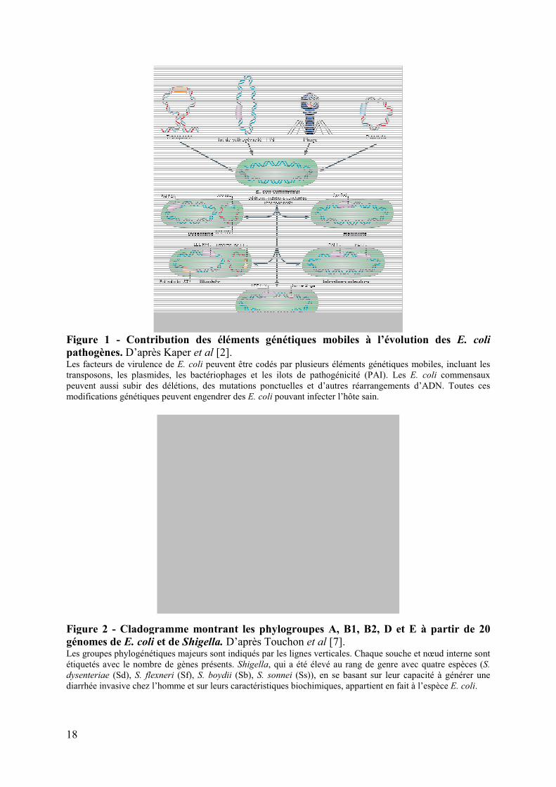
Figure 1 - Contribution des éléments génétiques mobiles à l’évolution des E. coli
pathogènes. D’après Kaper et al [2]. Les facteurs de virulence de E. coli peuvent être codés par plusieurs éléments génétiques mobiles, incluant les transposons, les plasmides, les bactériophages et les ilots de pathogénicité (PAI). Les E. coli commensaux peuvent aussi subir des délétions, des mutations ponctuelles et d’autres réarrangements d’ADN. Toutes ces modifications génétiques peuvent engendrer des E. coli pouvant infecter l’hôte sain.
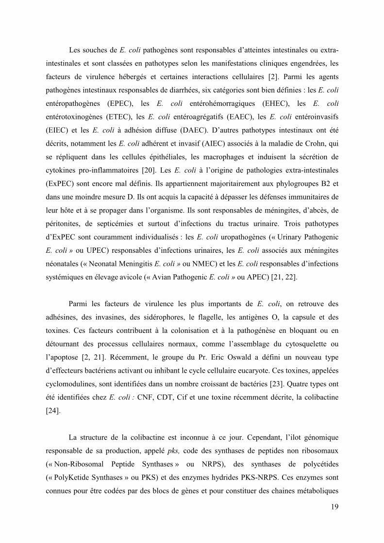
Figure 2 - Cladogramme montrant les phylogroupes A, B1, B2, D et E à partir de 20 génomes de E. coli et de Shigella. D’après Touchon et al [7]. Les groupes phylogénétiques majeurs sont indiqués par les lignes verticales. Chaque souche et nœud interne sont étiquetés avec le nombre de gènes présents. Shigella, qui a été élevé au rang de genre avec quatre espèces (S.
dysenteriae (Sd), S. flexneri (Sf), S. boydii (Sb), S. sonnei (Ss)), en se basant sur leur capacité à générer une diarrhée invasive chez l’homme et sur leurs caractéristiques biochimiques, appartient en fait à l’espèce E. coli.
19
Les souches de E. coli pathogènes sont responsables d’atteintes intestinales ou extra-
intestinales et sont classées en pathotypes selon les manifestations cliniques engendrées, les
facteurs de virulence hébergés et certaines interactions cellulaires [2]. Parmi les agents
pathogènes intestinaux responsables de diarrhées, six catégories sont bien définies : les E. coli
entéropathogènes (EPEC), les E. coli entérohémorragiques (EHEC), les E. coli
entérotoxinogènes (ETEC), les E. coli entéroagrégatifs (EAEC), les E. coli entéroinvasifs
(EIEC) et les E. coli à adhésion diffuse (DAEC). D’autres pathotypes intestinaux ont été
décrits, notamment les E. coli adhérent et invasif (AIEC) associés à la maladie de Crohn, qui
se répliquent dans les cellules épithéliales, les macrophages et induisent la sécrétion de
cytokines pro-inflammatoires [20]. Les E. coli à l’origine de pathologies extra-intestinales
(ExPEC) sont encore mal définis. Ils appartiennent majoritairement aux phylogroupes B2 et
dans une moindre mesure D. Ils ont acquis la capacité à dépasser les défenses immunitaires de
leur hôte et à se propager dans l’organisme. Ils sont responsables de méningites, d’abcès, de
péritonites, de septicémies et surtout d’infections du tractus urinaire. Trois pathotypes
d’ExPEC sont couramment individualisés : les E. coli uropathogènes (« Urinary Pathogenic
E. coli » ou UPEC) responsables d’infections urinaires, les E. coli associés aux méningites
néonatales (« Neonatal Meningitis E. coli » ou NMEC) et les E. coli responsables d’infections
systémiques en élevage avicole (« Avian Pathogenic E. coli » ou APEC) [21, 22].
Parmi les facteurs de virulence les plus importants de E. coli, on retrouve des
adhésines, des invasines, des sidérophores, le flagelle, les antigènes O, la capsule et des
toxines. Ces facteurs contribuent à la colonisation et à la pathogénèse en bloquant ou en
détournant des processus cellulaires normaux, comme l’assemblage du cytosquelette ou
l’apoptose [2, 21]. Récemment, le groupe du Pr. Eric Oswald a défini un nouveau type
d’effecteurs bactériens activant ou inhibant le cycle cellulaire eucaryote. Ces toxines, appelées
cyclomodulines, sont identifiées dans un nombre croissant de bactéries [23]. Quatre types ont
été identifiées chez E. coli : CNF, CDT, Cif et une toxine récemment décrite, la colibactine
[24].
La structure de la colibactine est inconnue à ce jour. Cependant, l’ilot génomique
responsable de sa production, appelé pks, code des synthases de peptides non ribosomaux
(« Non-Ribosomal Peptide Synthases » ou NRPS), des synthases de polycétides
(« PolyKetide Synthases » ou PKS) et des enzymes hydrides PKS-NRPS. Ces enzymes sont
connues pour être codées par des blocs de gènes et pour constituer des chaines métaboliques

20
synthétisant des composés chimiques de type polycétide-peptide non ribosomal
(« PolyKetide-Non-Ribosomal Peptide » ou PK-NRP), souvent toxiques et parfois utilisés en
thérapeutique [25]. Le rôle de la colibactine chez E. coli reste cependant à éclaircir. Par
ailleurs, l’ilot génomique pks produit une protéine essentielle appelée ClbP, qui est
inhabituelle dans ce type d’ilot et dont le rôle reste également à préciser.
L’objectif de ce travail était de caractériser sur le plan structural et fonctionnel la
protéine ClbP et d’explorer le rôle de l’ilot pks au travers d’une étude épidémiologique des
cyclomodulines de E. coli. Les résultats sont présentés sur articles, précédés par une revue
bibliographique portant sur le cycle cellulaire eucaryote, les cyclomodulines de E. coli, et les
composés PK-NRP. Les résultats sont ensuite discutés. Enfin, les publications réalisées en
marge de ce travail sont annexées à la fin de ce manuscrit.

21
Chapitre I
Revue
bibliographique

22

23
I. Le cycle cellulaire eucaryote
Le cycle cellulaire est un ensemble d'événements conservés et ordonnés, qui régissent
la croissance et la division cellulaire [26].
A. Stratégie générale du cycle cellulaire
Lorsqu’elles ne se divisent pas, les cellules sont dites quiescentes ou en phase G0. Elles
représentent une partie importante des cellules du corps humain. Le passage de ces cellules
dans une phase de prolifération nécessite des signaux mitogènes qui déclenchent le cycle
cellulaire. Ce dernier comprend une période de croissance cellulaire appelée interphase, suivie
de la mitose (figure 3) [26, 27]. L’interphase comprend trois phases : (i) la phase G1
(« Gap 1») qui prépare la synthèse de l'ADN et assure le passage d’un point de non-retour
rendant le processus de division indépendant des signaux mitogènes, (ii) la phase S («
Synthesis ») de réplication de l’ADN et (iii) la phase G2 (« Gap 2») de préparation à la mitose.
La phase M de mitose correspond à la ségrégation des chromosomes répliqués et à la
répartition des organites dans les cellules filles. Elle est classiquement subdivisée en 4 phases
appelées prophase, métaphase, anaphase et télophase. Ces processus sont étroitement régulés
par des mécanismes qui permettent ou limitent sa progression afin d’assurer l’intégrité des
cellules filles.
Figure 3 - Modèle classique du contrôle du cycle cellulaire chez les mammifères. Modifié d’après Malumbres et Barbacid, et Vermeulen et al [28, 29]. Les cyclines D se lient à CDK4 puis CDK6 pour former des complexes essentiels pour l'entrée en phase G1. Le complexe CDK2-cycline E déclenche la progression de la phase G1 en phase S. Le complexe CDK2-cycline A régule la phase S et son achèvement, relayé par le complexe CDK1-cycline A en fin de phase G2. Le complexe CDK1-cycline B déclenche ensuite la mitose. Le complexe CDK7-cycline H (complexe kinase activatrice) et les protéines Cdc 25 (phosphatases activatrices) activent les différentes CDKs.

24
B. Les principaux acteurs de la régulation du cycle cellulaire
La régulation du cycle cellulaire est assurée par les variations de l’activité des kinases
dépendantes des cyclines (« Cyclin-dependent Kinase » ou CDK). Elles-mêmes sont
modulées à quatre niveaux par : (i) l’association à des protéines activatrices, les cyclines, (ii)
des modifications de leur état de phosphorylation, (iii) la fixation de protéines inhibitrices, et
(iv) la régulation de leur localisation cellulaire.
1. Les protéines Kinases Dépendantes des Cyclines (CDK)
Les CDK forment une famille de sérine/thréonine kinases essentielles à la progression
du cycle cellulaire [30]. Globalement, l’activité CDK augmente à partir de la phase G1 pour
culminer et s’effondrer lors de la mitose [31]. Leur rôle est de phosphoryler, sur des sites
consensus, des protéines clés de la progression du cycle cellulaire. Leur niveau de production
dans la cellule est stable au cours du cycle cellulaire. Cependant, elles sont activées à des
points spécifiques du cycle cellulaire. Six CDK sont actives au cours du cycle cellulaire ;
CDK4, CDK6 et CDK2 interviennent pendant la phase G1, CDK2 en phase S, CDK1 en phase
G2 et M, et CDK7 à toutes les phases du cycle (figure 3) [28, 31].
2. Les Cyclines
Les CDK sont actives sous forme de complexe avec des protéines régulatrices
spécifiques, les cyclines [32, 33]. Vingt-cinq loci codent les cyclines chez l’homme [28]. Dix
cyclines appartenant à quatre classes différentes (A, B, D et E) sont nécessaires au cycle
cellulaire (figure 3). Leur concentration varie en fonction des phases du cycle cellulaire. Elles
portent des séquences d’ubiquitination permettant une dégradation efficacement par le
protéasome [34]. Les cyclines D et E sont principalement exprimées lors des phases G1 et sont
dégradées en phase S [31, 35, 36]. Les cyclines A et B sont majoritairement synthétisées lors
de la phase S et G2 respectivement et sont dégradées au début et en fin de mitose
respectivement [31, 34, 37].

25
3. La phosphorylation des CDK
L'activité CDK est aussi régulée par des phosphorylations activatrices ou inhibitrices
de résidus thréonine et tyrosine [29, 38, 39]. Elles seront actives ou non selon le profil des
phosphorylations portées. Le complexe CDK7-Cycline H, appelé aussi CAK (« Cdk-
Activating Kinase ») active les principaux complexes CDK-cycline par phosphorylation
(figure 3) [40]. L’activation passe également par la déphosphorylation d’autres sites par les
trois isoformes (a, b et c) des phosphatases Cdc25, qui jouent un rôle essentiel dans les
transitions G1/S et G2/M [41]. Par exemple, les kinases Wee1 et Myt1 agissent sur les résidus
14 et/ou 15 pour maintenir inactive CDK1 [42, 43]. Pour présenter une activité kinase, CDK1
doit d’une part, être phosphorylée sur le résidu 161 par CAK et d’autre part, être
déphosphorylée sur les résidus 14 et/ou15 par Cdc25c (figure 4).
Figure 4 - Régulation de CDK1 par les kinases CDK7 et Wee1/Myt1, la phosphatase Cdc25c et l’inhibiteur de CDK p21Waf1/Cip1
. La forme active de CDK1 est entourée en rouge.
D’après Vermeulen et al [29].

26
4. Les inhibiteurs de CDK
L'activité des CDK peut être bloquée par des protéines inhibitrices du cycle cellulaire,
appelées inhibiteurs de CDK (« Cycline-dependent Kinase Inhibitor » ou CKI). Elles se lient
aux CDK ou aux complexes CDK-cycline. Deux familles de CKI ont été décrites, la famille
INK4 et la famille Cip/Kip [44].
La famille INK4 (Inhibiteur de CDK4) comprend les protéines p15INK4b, p16INK4a,
p18INK4c et p19INK4d. Elles inactivent spécifiquement les CDK de la phase G1, CDK4 et CDK6
[45].
La deuxième famille d'inhibiteurs, la famille Cip/Kip comprend les protéines
p21Waf1/Cip1, p27Kip1 et p57Kip2 [46-48]. Ces inhibiteurs inactivent les complexes CDK-cycline
de la phase G1 et dans une moindre mesure, le complexe CDK1-cycline B de la phase M
(figure 4) [49]. La protéine p21Waf1/Cip1 induite par p53 inhibe également la synthèse de
l'ADN en se liant à l'antigène nucléaire de prolifération cellulaire PCNA (« Proliferation Cell
Nuclear Antigen ») [50].
5. La régulation de la localisation intracellulaire des CDK
La localisation intracellulaire des protéines effectrices du cycle cellulaire contribue
également à sa régulation. La cycline B contient un signal d'exclusion nucléaire, qui assure
une exportation active de la protéine jusqu'au début de la prophase [51]. Les kinases Wee1 et
Myt1 qui inactivent CDK1 sont situées respectivement dans le noyau et au niveau du
réticulum endoplasmique, et protègent la cellule d’une mitose prématurée [52, 53]. Le groupe
de protéines 14-3-3 régule le trafic intracellulaire de différentes protéines dont la phosphatase
Cdc25c. Elles maintiennent cette dernière dans le cytoplasme pendant l'interphase afin
d’éviter l’activation de CDK1 [54, 55].

27
C. Les principaux évènements de régulation du cycle
En phase G1 précoce, les complexes CDK4/6-cycline D phosphorylent différentes
protéines comme la protéine du gène suppresseur de tumeur du rétinoblastome (pRb), qui est
complexée à l’histone déacétylase et aux facteurs de transcription de la famille E2F [56, 57].
Cette phosphorylation libère les facteurs E2F qui régulent positivement la transcription des
gènes nécessaires pour la progression en phase S, tels que ceux de la cycline E et des Cdc25
(figure 5) [58].
Au cours de la transition de phase G1/S, le complexe CDK2-cycline E phosphoryle
pRb sur des sites additionnels afin d’assurer la libération complète des facteurs E2F, qui
induisent la transcription du gène codant la cycline A, nécessaire à la phase S [58-60]. Le
complexe CDK2-cycline E phosphoryle également l’histone H1 et ainsi participe à la
condensation des chromosomes nécessaire à la réplication de l'ADN (figure 5) [61, 62].
Figure 5 - Exemples de substrats des CDK. Modifié d’après Vermeulen et al [29].

28
En phase S, les CDK2 et CDK1 complexées à la cycline A régulent notamment
l’initiation de la réplication de l’ADN par la phosphorylation de la primase ADN polymérase
alpha (figure 5) [63].
Afin d’assurer la mitose, CDK1 complexée à la cycline A en phase G2 et à la cycline
B1 en fin de phase G2 et en début de mitose, phosphoryle de nombreuses protéines telles que
l’histone H1 et des protéines nécessaires au désassemblage de la membrane nucléaire et à
l’assemblage du fuseau mitotique (figure 5) [64].
D. Les points de contrôle de la qualité du cycle cellulaire
Pendant le cycle cellulaire, l’ADN doit être parfaitement conservé, dupliqué et réparti
entre les cellules filles. Pour assurer le bon déroulement de ces phénomènes, des points de
contrôle sont positionnés aux points critiques du cycle cellulaire : lors des transitions G1/S et
G2/M, et durant la phase S et la mitose.
1. La signalisation des altérations de l’ADN
Les points de contrôle sont activés par des voies de détection spécifiques des
dommages de l'ADN faisant intervenir quatre niveaux de signalisation (les protéines senseurs,
médiatrices, transductrices et effectrices) et arrêtent le cycle cellulaire afin de permettre la
réparation de l'ADN avant d’avancer dans le cycle ou d’activer la mort cellulaire par apoptose
en cas de dommages irréversibles (figure 6) [65-71].
La phosphorylation de l’histone H2AX par le senseur ATM (« Ataxia Telangiectasia
Mutated protein ») est un régulateur maître du recrutement des protéines de réparation aux
sites de coupure double-brin de l’ADN. Cette phosphorylation qui s’étend jusqu’à un
mégabase du site de coupure et permet la formation de foci de réparation aisément
visualisables en imagerie de fluorescence [72]. De plus, la dynamique des protéines de
signalisation et de réparation des altérations de l’ADN est assurée par un processus
d’ubiquitination influençant l’efficacité et la spécificité de la réparation de l’ADN [73].

29
Figure 6 - Modèle des voies de signalisation des altérations de l’ADN. Modifié d’après Sancar et al, et Zhou et Elledge [65, 66]. Dans les cellules de mammifères, la signalisation des points de contrôle est initiée notamment par les deux phospho-inositide kinases senseurs : ATM et ATR (« ATM and Rad3-related protein»), en complexe avec leurs partenaires de liaison respectifs MRN (Mre11-Rad50-Nbs1) et ATRIP (« ATR interacting protein »). Les cassures double-brin de l’ADN générées par les agents génotoxiques tels que les radiations ionisantes sont perçues par le complexe MRN, qui recrute alors ATM aux sites de dommage et favorise l’activation de cette kinase [70]. Lorsque les fourches de réplication arrivent aux cassures double-brin de l’ADN au cours des phases S et G2 du cycle cellulaire, ATR est activée, en aval d’ATM [67]. Inversement, lors d’arrêt des fourches de réplication ou d'exposition à une irradiation UV (cassures simple-brin) ATR est activée en amont d'ATM : les cassures simple-brin non réparées au cours des phases G1 et S génèrent lors du passage des fourches de réplication, des cassures double-brin qui activent ATM [71]. Les complexes senseurs d’ATM et d’ATR, avec l’aide de médiateurs, activent respectivement les kinases transductrices Chk1 et Chk2 [68]. Celles-ci inhibent ou activent en retour les effecteurs impliqués dans la réparation de l’ADN, la transcription, l’apoptose ou l’arrêt du cycle cellulaire. p53 et les Cdc25 participent directement à l’inhibition des transitions de phase G1/S et G2/M et la progression de la phase S. Selon le contexte, certaines molécules peuvent avoir des fonctions multiples dans cette voie de transduction du signal.

30
2. Les points de contrôle G1/S et G2/M
Selon la nature des lésions et la phase du cycle cellulaire, les kinases transductrices
Chk1 et Chk2 vont réguler les protéines effectrices dont p53 et Cdc25, qui participent
directement à l’inhibition des transitions G1/S (figures 7) et G2/M [65, 74-77].
Lors de dommages de l'ADN en phase G1, les transducteurs Chk1 et Chk2 activés
phosphorylent Cdc25a, favorisant sa liaison aux protéines 14-3-3 et son exclusion nucléaire
consécutive, ainsi que sa dégradation via la voie de l’ubiquitine [76]. CDK2 phosphorylée et
inactivée s’accumule et ne peut pas phosphoryler Cdc45 pour initier la réplication de l’ADN,
induisant un arrêt rapide du cycle cellulaire. Contrairement aux autres points de contrôle, le
maintien de l’arrêt du cycle cellulaire au point de contrôle G1/S est dépendant de la protéine
p53 [77]. p53 qui est phosphorylée par les senseurs ATM/ATR et par les transducteurs
Chk1/Chk2, induit la transcription de p21Waf-1/Cip1, qui se lie au complexe CDK4-cycline D
(ou Cdk4-Cyc D), l’empêchant de phosphoryler pRb [78]. pRb ne peut pas libérer le facteur
de transcription E2F nécessaire à la transcription des gènes de la phase S. p21Waf-1/Cip1 inactive
également le complexe CDK2-cycline E, assurant le maintien au point de contrôle G1/S.
Lors de dommages de l'ADN en phase G2, les cellules sont capables d’initier un arrêt
du cycle cellulaire en présence ou en absence de p53. Les transducteurs Chk1 et Chk2 activés
phosphorylent Cdc25a, qui ne peut plus activer le complexe CDK1-cycline B (Cdc2-cycline
B), bloquant l'entrée en mitose [75]. De même, Chk1 et Chk2 activés phosphorylent Wee1 qui
effectue une phosphorylation inhibitrice de CDK1 (figures 8). L’augmentation de p53
dépendante des dommages de l'ADN induit comme au cours du point de contrôle G1/S, la
transcription accrue de p21Waf-1/Cip1 et de protéines 14-3-3. p21Waf1/Cip1 se lie au complexe
CDK1-cycline B, empêchant son activité. L’augmentation de la liaison de la cycline B aux
protéines 14-3-3 l’exclut activement du noyau [74, 79].

31
Figure 7 - Le point de contrôle G1/S. D’après Sancar et al. [65].Les transducteurs Chk1 et Chk2 activés phosphorylent Cdc25a, favorisant sa liaison aux protéines 14-3-3 et son exclusion nucléaire consécutive, ainsi que sa dégradation via la voie de l’ubiquitine [76]. CDK2 phosphorylée et inactivée s’accumule et ne peut pas phosphoryler Cdc45 pour initier la réplication de l’ADN, induisant un arrêt rapide du cycle cellulaire (Cyc E/A : cyclines E ou A). Le maintien de l’arrêt du cycle cellulaire au point de contrôle G1/S est dépendant de la protéine p53 [77]. p53 qui est phosphorylée par les senseurs ATM/ATR et par les transducteursChk1/Chk2, induit la transcription de p21Waf-1/Cip1, qui se lie au complexe CDK4-cycline D (ou Cdk4-Cyc D), l’empêchant de phosphoryler pRb [78]. pRb ne peut alors pas libérer le facteur de transcription E2F nécessaire à la transcription des gènes de la phase S. p21Waf-1/Cip1 inactive également le complexe CDK2-cycline E, assurant le maintien au point de contrôle G1/S.
Figure 8 - Le point de contrôle G2/M. D’après Sancar et al. [65]. Les transducteurs Chk1 et Chk2 activés phosphorylent Cdc25a, favorisant sa liaison aux protéines 14-3-3 et son exclusion nucléaire consécutive. Cdc25a ne peut plus activer le complexe CDK1-cycline B (Cdc2-cycline B), bloquant l'entrée en mitose [75]. De même, Chk1 et Chk2 activés phosphorylent Wee1 qui effectue une phosphorylation inhibitrice de CDK1.

32
3. Les points de contrôle en phase S
La phase S du cycle cellulaire comprend trois points de contrôle qui ne requièrent pas
la protéine p53 : le point de contrôle de réplication, le point de contrôle intra-S et le point de
contrôle S/M [80]. Ces points de contrôle sont de découverte plus récente. Le point de
contrôle de réplication fait intervenir le complexe senseur ATR-ATRIP et inhibe l’initiation
de la réplication de l’ADN quand la progression des fourches de réplication est arrêtée en
réponse à différents stress. Le point de contrôle intra-S est activé via ATM par des cassures
double-brin de l’ADN. Les transducteurs Chk1 et Chk2 activés induisent alors la
phosphorylation et la protéolyse de la phosphatase Cdc25a, conduisant à l’inhibition des
complexes CDK2-cyclines E/A. Le point de contrôle S/M vérifie la duplication complète du
génome avant d’initier la division cellulaire. La cible clé de ce processus est le complexe
CDK1-cycline B.
4. Le point de contrôle du fuseau mitotique
Les protéines du point de contrôle du fuseau mitotique ou point de contrôle
métaphase-anaphase détectent un mauvais attachement des kinétochores des chromosomes
sur les microtubules des fuseaux mitotiques. Ce point de contrôle induit alors l’arrêt du cycle
cellulaire en métaphase afin d’éviter la mauvaise ségrégation des chromosomes et
l’aneuploïdie des cellules filles. L’arrêt du cycle cellulaire passe par un blocage de
l’ubiquitination et donc de la dégradation de la cycline B, évitant ainsi l’inactivation du
complexe CDK1-cycline B et donc la sortie de mitose [81].

33
II. Les cyclomodulines de Escherichia coli
Comme les virus qui perturbent le cycle cellulaire eucaryote pour leur réplication [82],
des bactéries pathogènes sont capables d'interférer activement avec le cycle cellulaire. Cette
activité repose sur la synthèse de toxines, appelées cyclomodulines [23] qui stimulent ou
inhibent la progression du cycle cellulaire (tableau 1). Quatre cyclomodulines ont été
identifiées chez E. coli : CNF, CDT, Cif et la colibactine.
Tableau 1 : Exemples de cyclomodulines bactériennes. D’après Nougayrède et al. [23].
Bactérie Effecteur * Mécanisme Inhibe la prolifération
Helicobacter pylori VacA Arrêt du cycle cellulaire des lymphocytes T en G1-S, inhibition de l’activation des lymphocytes T
Mycobacterium ulcerans Mycolactone Arrêt du cycle cellulaire en G0-G1
Fusobacterium nucleatum FIP Arrêt du cycle cellulaire des lymphocytes T en milieu de phase G1
Favorise la prolifération
Helicobacter pylori CagA Active la voie Ras-MEK-ERK, conduisant à une dispersion et une prolifération cellulaire
Bordetella spp. DNT Déamide la glutamine de Rho GTPases Forme des cellules géantes multinucléées Augmente la synthèse d’ADN
Pasteurella multocida PMT Active de multiples voies cellulaires de transduction du signal Puissant mitogène de divers types cellulaires
* effecteurs protéiques, sauf la mycolactone qui est un PK. FIP : « Fusobacterium immunosuppressive protein » ; DNT : « dermonecrotic toxin » ; PMT : « P. multocida
toxin ».

34
A. Le facteur cytotoxique nécrosant CNF
Le facteur cytotoxique nécrosant 1 ou CNF1 (pour « Cytotoxic Necrotizing Factor 1 »)
a été décrit pour la première fois en 1983 par Caprioli et al. chez E. coli. Il appartient au
groupe des cyclomodulines activant le cycle cellulaire. [83].
1. Les CNF variants
Trois variants de CNF1 ont été caractérisés : CNF2 et CNF3 chez E. coli, CNFY chez
Yersinia pseudotuberculosis [84-86]. CNF1 et CNF2 partagent 90% d’identité. CNF3 et
CNFY présentent respectivement 70% d’identité avec CNF1 et CNF2, et 65% d’identité avec
CNF1. Ces protéines s’apparentent par ailleurs à la toxine DNT de Bordedella par leur
domaine catalytique [87].
2. Structure
Les toxines de type CNF comprennent une chaine polypeptidique unique d’environ
115 kDa organisée en trois domaines fonctionnels [87]. La région N-terminale de la protéine
contient un domaine de liaison aux récepteurs cellulaires [88, 89]. La partie médiane formerait
un domaine central constitué par deux hélices hydrophobes pouvant initier la translocation du
domaine enzymatique dans le cytosol des cellules [90]. La partie carboxy-terminale contient
le domaine enzymatique qui porte la triade catalytique Val833, Cys866 et His881 [88, 91, 92].
3. Support génétique
Les gènes cnf1 et cnfy sont chromosomiques, alors que cnf2 est plasmidique (plasmide
Vir) [85, 93-95]. L’essentiel des études a porté sur le gène cnf1.
Le gène cnf1 est situé dans l'ilot de pathogénicité II (PAI-II) fréquemment observé
chez les E. coli du phylogroupe B2 [87]. Il se situe juste en aval de l'opéron hlyCABD codant
pour la production et la sécrétion de l’hémolysine � (figure 9) [87, 93, 94]. Une corégulation
transcriptionnelle de leurs gènes est proposée via 3 mécanismes : une zone promotrice
commune, l’action répressive de la protéine histone-like H-NS et une activité activatrice de
l’antiterminateur transcriptionnel RfaH. Cependant, un promoteur putatif indépendant a été

35
identifié dans les 60 nucléotides situés en amont du codon d’initiation de cnf1. Enfin, une
séquence anti-Shine-Dalgarno située à 150 paires de bases en aval du codon d’initiation de
cnf1 réprime la traduction de cnf1, probablement par la formation d'une structure en tige-
boucle qui séquestre le site de liaison au ribosome [89, 94, 96, 97].
Figure 9 - Organisation génétique et modèle d’expression de la région hlyCABD-cnf1 du PAI-II de la souche UPEC J96. D’après Lemonnier et al [87].L’antiterminateur transcriptionnel protéique RfaH interagit directement avec les complexes de transcription arrêtés sur les sites ops afin d’augmenter l’élongation de la transcription par un mécanisme qui empêche la pause et la fin de transcription. Dans le cas de l'opéron hlyCABD, les complexes de transcription amorcés par RfaH peuvent contourner l’arrêt de transcription au terminateur Thly situé entre les gènes hlyA et hlyB, permettant ainsi la transcription des gènes distaux hlyB, hlyD et cnf1. En plus de RfaH, la protéine histone-like H-NS, co-régule négativement les gènes de l’hémolysine et de cnf1.
4. Mode d’action
Le mode d’action des toxines CNF repose sur trois étapes : la sécrétion de la toxine, sa
translocation dans le cytosol des cellules cibles et la modification enzymatique des molécules
cibles.
a) Sécrétion
Les CNF sont sécrétées dans le milieu extérieur. Cependant, aucun peptide signal
détectable ou système de transporteur spécifique n’a été caractérisé. La sécrétion de CNF1 à
travers la membrane cytoplasmique implique la ferredoxine [98]. Elle est par ailleurs associée

36
aux vésicules de la membrane externe libérées par les bactéries [97, 99]. L’importance
physiologique de cette voie d’adressage reste toutefois à démontrer.
b) Translocation dans les cellules eucaryotes
CNF1 se lie à la protéine précurseur du récepteur de la laminine (37 kDa) à la surface
de la cellule hôte [100-102]. Deux régions en N-terminale (résidus 135 à 164) et C-terminale
(résidus 683 à 730) sont impliquées dans la fixation au récepteur [103, 104]. CNF3 et CNFy
se lieraient à des récepteurs cellulaires de surface différents [105, 106].
Après fixation à son récepteur, CNF1 entre dans les cellules hôtes via une endocytose
indépendante de la clathrine pour atteindre le compartiment endosomal tardif, permettant la
libération du domaine enzymatique et son transfert dans le cytosol grâce aux 2 hélices
hydrophobes du domaine médian [90, 102, 105, 107, 108].
c) Modification de la cible moléculaire
La cible cytoplasmique des CNF sont les GTPases Rho (figure 10A) [106, 108, 109],
des interrupteurs moléculaires de la superfamille Ras [110]. Le domaine enzymatique de
CNF1 assure la désamidation d'un résidu glutamine spécifique des protéines Rho ; le résidu
Gln63 de RhoA ou Gln61 de Rac et Cdc42 [111-114]. Cette modification compromet la
formation du complexe Rho-GAP (« GTPase Activating Protein ») nécessaire à l’inactivation
des protéines Rho et induit par conséquent une activation permanente des protéines Rho sous
forme de complexe avec le GTP. Ainsi activées en permanence, les protéines Rho sont
rapidement ubiquitinées et dégradées par le protéasome. Au total, CNF1 induit une activation
modérée et transitoire des protéines GTPases Rho [115-117]. Par ailleurs, les toxines CNF ont
un spectre d’action particulier ; CNF1 et CNF3 activent RhoA, Rac1 et Cdc42 [106] alors que
CNFY active sélectivement RhoA [109]. Les substrats préférentiels de CNF1 sont Rac et
Cdc42 et celui de CNF3, RhoA [106].

37
Figure 10 - Modèle d’action des CNF sur les cellules hôtes. D’après Nougayrède et al [23]. A. CNF se lie au récepteur cellulaire puis est internalisé par endocytose pour atteindre le compartiment
endosomal tardif acide, permettant le transfert de son domaine enzymatique dans le cytosol. Il déamide ensuite un résidu glutamine des GTPases Rho, bloquant celles-ci dans un état activé leur permettant d’activer de manière persistante leurs effecteurs. Ces effecteurs modifient le cytosquelette et favorisent la prolifération cellulaire.
B. Effet de CNF1 sur des cellules HeLa après 72 heures de contact. A gauche - Multinucléation des cellules révélée par marquage au DAPI. Au milieu - Fibres de stress révélée par la fluorescéine-phalloïdine. A droite - Analyse du cycle cellulaire montrant des cellules ayant un contenu en ADN élevé (4n, 8n,16n …).

38
5. Expression phénotypique
Les GTPases Rho contribuent à la régulation d'un large panel de processus cellulaires
en interagissant avec des effecteurs protéiques spécifiques [110]. Elles régulent l’organisation
et la dynamique des microtubules (ségrégation des chromosomes) et du cytosquelette d’actine
(polarité, migration et division cellulaires), et des évènements de la réponse immunitaire
(synapses immunologiques, phagocytose). Ces Rho GTPases contribuent à la progression du
cycle cellulaire en influençant le niveau de cycline D1 et d’inhibiteurs de CDK (p27Kip1 et
p21Waf1/Cip1) et prennent part à la transcription de nombreux gènes, tels que ceux intervenant
dans l’inflammation et l’apoptose. Les effets cytopathogènes induits par les CNF sont la
formation de fibres de stress d'actine 1 (figure 10B), de plaques d'adhérence focale 2 , de
lamellipodes, de filipodes 3 et de « rufflings » membranaires 4 [106, 108, 118, 119].
L’importante activité de « ruffling » permet aux cellules épithéliales de se comporter comme
des phagocytes. Elles développent une activité macropinocytique permettant la phagocytose
de différents types de particules, dont les bactéries [117, 120-122].
Les toxines CNF déclenchent la transition de phase G1/S et induisent la réplication de
l’ADN dans différentes cellules de mammifères. Probablement en raison d’ altérations du
fuseau mitotique, la ségrégation chromosomique et la cytocinèse 5 sont altérées. Des
aberrations nucléaires (multinucléation, segmentations nucléaires et mitoses multipolaires)
conduisant à des aneuploïdies sont associées à une augmentation de la teneur cellulaire en
ADN (4n, 8n, 16n) (figure 10B)[118, 119, 123-125].
CNF1, via les protéines Rho, active plusieurs voies de signalisation dont celles mettant
en jeu les kinases Jun, MAP p38 et NF-kB, des facteurs de transcription majeurs de la réponse
inflammation et de la différenciation cellulaire [114, 126].
1 Faisceaux filamentaires organisés dans lesquels des faisceaux de courts filaments d'actine de polarité alternée sont entremêlés avec des filaments bipolaires de myosine II, retrouvés généralement dans les cellules mobiles. 2 Structures d’adhérence caractéristiques des cellules en culture, correspondant aux sites dispersés et discontinus de contact très étroit avec la surface de la boite.3 Respectivement larges et fines extensions membranaires rigides faites de polymères d’actine utilisées par la cellule pour explorer son environnement à la recherche de matrice extra-cellulaire et à l'origine du mouvement amiboïde des cellules. 4 Surfaces cellulaires motiles contenant un maillage de filaments d’actine nouvellement polymérisés. 5 Phase finale de la division cellulaire qui correspond à la séparation physique des deux cellules filles.

39
6. Distribution
Les souches de E. coli cnf1+ (« Necrotoxic E. coli 1 » ou NTEC-1) sont issues de
l’homme et de nombreuses espèces de mammifères domestiques. Ces souches NTEC-1
appartiennent majoritairement au phylogroupe B2 [127-130]. Les souches cnf2+ (NTEC-2)
ont été isolées le plus souvent d’animaux [86, 131-134]. Récemment décrit, CNF3 n’a été
mise en évidence que dans des souches d’origine animale [84].
Les souches NTEC-1 sont fréquemment responsables de bactériémies [135] et
d'infections urinaires, avec une prévalence croissante allant de 21 à 78 % en fonction des
affections suivantes : cystites, pyélonéphrites et prostatites [127-129, 136-144]. Les souches
de E. coli responsables de méningites néonatales sont cnf1+ dans 10 à 13% des cas [130, 145,
146].
La participation des souches NTEC aux syndromes diarrhéiques de l’homme n’est pas
clairement établie, bien que les souches NTEC semblent exercer un effet pathogène sur
l'intestin, au moins chez les nouveau-nés et les jeunes enfants [133, 147-150]. L’implication
des souches NTEC-1 et -2 dans la survenue de syndromes dysentériques chez les animaux est
suggérée par quelques études épidémiologiques et un modèle expérimental [86, 134, 151,
152]. Cependant la pathogénie intestinale des NTEC repose probablement sur la production
combinée de plusieurs toxines (hémolysine, CNF, CDT, …).
7. Rôle dans le processus infectieux
La forte prévalence de CNF1 et CNF2 dans les souches ExPEC suggère un rôle dans
la pathogénie des infections extra-intestinales. CNF1 contribue à l’invasion du système
nerveux central par les souches de E. coli K1. Elle favorise l’internalisation de E. coli dans les
cellules endothéliales microvasculaires du cerveau humain in vitro et ainsi contribuerait à la
traversée de la barrière hémato-encéphalique [100, 153]. De même, CNF1 aiderait
l'internalisation des bactéries dans les cellules épithéliales, notamment de la vessie [117, 120-
122]. Ainsi, abritées des effecteurs du système immunitaire, les bactéries productrices peuvent
persister et former des réservoirs intracellulaires responsables des infections urinaires
récurrentes [154]. L'intoxication par CNF1 altère les jonctions intercellulaires et induit
l’apoptose de cellules épithéliales vésicales en culture, ce qui faciliterait l’invasion des tissus
sous-jacents par les bactéries [155].

40
CNF1 augmente la production de médiateurs de l’inflammation sur des cellules uro-
épithéliales en culture [156]. Des études menées à l’aide de modèles animaux d’infections
urinaires et de prostatites montrent que cette toxine contribue aussi aux dommages tissulaires
et à l’inflammation [156-159]. Des niveaux plus élevés des facteurs pro-inflammatoires MCP-
1 et IL-8 ont été mesurés dans l'urine de patients infectés par des UPEC cnf1-positives [140] ;
néanmoins, ceci n'exclut pas la participation d'autres facteurs bactériens, parmi lesquels
l’hémolysine α et le P-fimbriae, qui déclenchent également la production de médiateurs
inflammatoires par les cellules épithéliales intoxiquées [160, 161]. CNF1 pourrait donc
majorer les dommages de l'épithélium via l’inflammation.
CNF1 améliore aussi la survie bactérienne dans un modèle murin [158]. Plusieurs
études suggèrent que CNF1 retarde la transmigration des neutrophiles à travers l'épithélium et
réduit leurs fonctions antibactériennes (régulation négative de la phagocytose, altération de la
distribution des récepteurs CR3 du complément) [99, 162-164]. Bien qu’il favorise un état
inflammatoire, CNF1 faciliterait cependant la survie des bactéries face aux effecteurs du
système immunitaire inné en modulant leur fonction. CNF1 induirait une réponse
inflammatoire contrôlée ou en partie inopérante et permettrait ainsi une plus grande
persistance des bactéries [164].
En conclusion, les études in vitro et in vivo convergent vers un modèle dans lequel
CNF1 est un facteur de virulence favorisant l’internalisation des bactéries et modulant les
réponses inflammatoires. Il est donc concevable que les mécanismes d’action de CNF1
permettent d'augmenter la gravité et la chronicité des infections bactériennes.
8. Des agents carcinogènes potentiels
Plusieurs arguments soutiennent le rôle potentiel des CNF dans la transformation
cellulaire6.
La modification biochimique des protéines Rho induite par les CNF correspond à la
modification des protéines Ras d’origine génétique, observée dans de nombreuses tumeurs
[165]. Les toxines CNF dérégulent l’activité des GTPases Rho comme les effecteurs SopE et
SptP de Salmonella spp., bactéries dont le portage est associé au cancer de la vésicule biliaire
[166, 167].
6 Capacité acquise d’une cellule à proliférer de manière monoclonale et excessive, en échappant à l’homéostasie cellulaire.

41
Dans des cellules épithéliales, CNF1 induit une activation prolongée du facteur de
transcription NF-kB [126], un acteur reconnu de l’oncogenèse [168]. Cette activation induit
l’expression de COX2 [169], un gène majeur de la cascade des prostaglandines pro-
inflammatoires dont la surexpression est observée précocement dans de nombreux cancers
comme le cancer colorectal. NF-kB induit également l’expression et la libération de cytokines
pro-inflammatoires (IL-6, IL-8 et TNF-�) à partir des cellules épithéliales et endothéliales
[156, 164]. CNF1 en activant de façon prolongée la voie NF-kB favorise donc un état
inflammatoire chronique, qui est reconnu pour favoriser la carcinogenèse [170].
L’activité de CNF1 protège les cellules épithéliales de l'apoptose en augmentant
l’expression des facteurs anti-apoptotiques Bcl-2 et Bcl-XL, module l’autophagie, induit des
aneuploïdies et la formation de réseaux complexes de mitochondries ; des altérations pouvant
conduire à la transformation cellulaire [123, 171-173].
CNF1 promeut la rupture des jonctions cellulaires et la motilité cellulaire, par un
mécanisme dépendant de RhoA [117, 172, 174] ; un mécanisme proche de la transition
épithéliale-mésenchymateuse7 [175].
Enfin, l’activité de CNF1 s’apparente par de nombreux aspects à celle des protéines
CagA et VacA de H. pylori, un des meilleurs exemples de toxines carcinogènes (tableau 2).
7 Ensemble d’événements permettant aux cellules épithéliales de surmonter les contraintes physiques, telles que celles imposées par les jonctions intercellulaires et d’adopter le phénotype migratoire intervenant dans l’embryogénèse ou le processus métastatique des cellules cancéreuses.

42
Tableau 2 - Comparaison entre les effets cellulaires de CNF1 de E. coli et des facteurs de virulence majeurs (VacA et CagA) de H. pylori. D’après Travaglione et al [176].
CNF1 CagA VacA
Effets sur le cytosquelette et la morphologie cellulaire
- polymérisation de l’actine
- diffusion cellulaire
- multinucléation
- réarrangement de l’actine
- perte de polarité cellulaire
- dispersion/migration cellulaire
- perturbations de l’actine
- vacuolisation
Régulation de l’apoptose Effet anti-apoptotique Effet anti-apoptotique Effet pro-apoptotique
Production de médiateurs de l’inflammation
TNF-α
IL-6
IL-8
ROS
IL-8
ROS
TNF-α
IL-6
IL-1β
IL-8
Activation de facteurs de transcription
NF-kB NF-kB
NFAT
SRF
AP-1
NF-kB
NFAT
ATF-2
Effets sur le cycle cellulaireEndoréduplication
Arrêt en G2/M
Inhibition de la transition G1/S
Induction de la transition G1/S
Effets sur les mitochondries
Inhibition de la dépolarisation membranaire induite par les UVB
Réduction du potentiel de membrane, libération de cytochrome c
ROS : Espèces réactives de l’oxygène

43
B. La toxine de distension cytoléthale CDT
La toxine de distension cytoléthale (« Cytolethal Distending Toxin » ou CDT) a été
caractérisée en 1997 comme la première toxine bactérienne bloquant le cycle cellulaire en
transition de phase G2/M [177]. Ce type de toxine a été caractérisé chez de nombreuses
bactéries à Gram négatif pathogènes, comme E. coli, Shigella dysenteriae, Salmonella Typhi,
Haemophilus ducreyi, Helicobacter hepaticus, et Aggregatibacter (anciennement
Actinobacillus) actinomycetemcomitans [178]. CDT est constituée de trois sous-unités CdtA,
CdtB et CdtC.
1. Les variants CDT et leur support génétique
Cinq variants, CDT-I à CDT-V ont été caractérisés chez E. coli. Les sous-unités CdtB
sont plus conservées que les sous-unités CdtA et CdtC (>56% versus >36% et >33%
d’identité) [179-181].
Elles sont codées par trois gènes cdtA, cdtB et cdtC organisés en un opéron le plus
souvent chromosomique, parfois plasmidique (plasmide Vir codant CDT-III et CNF2). Leur
expression semble constitutive. Chez E. coli, ces gènes sont souvent flanqués de séquences
phagiques [95, 182-184].
2. Structure des CDT
Les toxines CDT appartiennent aux toxines holoprotéiques de type AB2. Les trois
sous-unités CdtA, CdtB et CdtC forment un complexe ternaire [185, 186]. CdtA et CdtC
partagent un motif lectine et sont responsables de la liaison de la toxine à des récepteurs
cellulaires membranaires [185, 187, 188]. CdtB, la sous-unité la plus conservée, présente une
homologie structurale et fonctionnelle avec les membres d'une superfamille de métallo-
enzymes incluant des nucléases et diverses phosphatases de seconds messagers lipidiques. Les
résidus His154, Asp229, His261 et Asp260 des sous-unités CdtB sont impliqués dans la
liaison de l’ion métallique Mg2+ et le processus enzymatique catalysé par la toxine [186, 189,
190].

44
3. Mode d’action
Le mode d’action des toxines CDT repose sur trois étapes : l’assemblage et la
sécrétion du complexe protéique ternaire, la translocation de la sous-unité CdtB dans le
cytosol des cellules cibles et l’altération de la cible, l’ADN (figure 11).
a) Sécrétion
Les trois protéines CdtA, CdtB et CdtC sont transloquées de façon indépendante dans
le périplasme par une séquence signal d’adressage [191, 192]. La sous-unité CdtA y subit un
greffage lipidique au niveau d’un domaine consensus appelé « lipobox », important pour une
production efficace de l’holotoxine [193]. Les trois sous-unités forment alors un complexe
périplasmique ternaire. Dans le surnageant de culture, la majorité des CDT reste étroitement
associée aux vésicules de la membrane externe [194].
b) Translocation dans les cellules eucaryotes
Deux types de récepteurs cellulaires ont été impliqués dans la liaison de CDT à la
surface cellulaire : des glycoprotéines contenant du fucose pour CDT-II [195] et des
glycosphingolipides GM3 pour la toxine CDT de A. actinomycetemcomitans [196]. Les
radeaux lipidiques riches en cholestérol de la membrane cytoplasmique sont également
impliqués dans l’adressage de CdtB dans la cellule cible [197]. Lors de la translocation de
CdtB dans la cellule cible, CdtA et CdtC demeureraient associées à la membrane (figure
11A). La sous-unité CdtB est internalisée par endocytose, transportée par voie rétrograde vers
l'appareil de Golgi et le réticulum endoplasmique, puis transloquée dans le noyau, son site
d'action [198-200]. L'entrée de CdtB dans le noyau repose sur un domaine de translocation
nucléaire [201].
La toxine CDT de Salmonella Typhi présente un comportement particulier.
L’expression de sa sous-unité CdtB est induite par l'invasion des cellules hôtes. Les gènes
cdtA et cdtC étant absents, CdtB s’associe alors avec les protéines PltA (toxine de type
pertussique A) et PltB (toxine de type pertussique B). Ce complexe désignée « toxine
typhoïde » (figure 11A) est empaqueté dans une structure membranaire appelée «puncta», et
transportée hors des cellules infectées. CdtB induit alors l’intoxication des cellules cibles par
son adressage aux cellules infectées par S. Typhi (mode autocrine) ou à des cellules non
infectées (mode paracrine) [202, 203].

45
Figure 11 - Organisation génétique et modèle d’action des CDT sur les cellules hôtes.D’après Ge et al et Nougayrède et al [23, 178].A. CDT est une toxine hétéro-trimérique dont les gènes codant les sous-unités sont organisés en opéron. CdtA et
CdtC sont nécessaires à la liaison de CDT à la surface de la cellule hôte. L’internalisation de CdtB est suffisante pour l’intoxication cellulaire. CdtB est internalisée par endocytose, transportée par voie rétrograde vers l'appareil de Golgi et le réticulum endoplasmique, puis transloquée dans le noyau où elle induit des altérations de l’ADN. La production et translocation de CdtB chez Salmonella Typhi fait intervenir les protéines PtlA et PtlB. Les dommages de l’ADN activent les voies de réponse ATM dépendante, entrainant un arrêt du cycle cellulaire en transitions de phase G1/S ou G2/M, voire une apoptose selon le modèle cellulaire. L’activation de RhoA par ATM peut conduire à la formation de fibres de stress et d’adhérences focales.
B. Effet d’une CDT purifiée de E. coli sur des cellules HeLa 72 heures après le début de l’exposition. A gauche - Elargissement des noyaux et absence de figures mitotiques révélés par marquage au DAPI. A droite - Analyse du cycle cellulaire montrant une majorité de cellules bloquées en phase G2.

46
c) Altération de l’ADN et expression phénotypique
Une fois dans le noyau, CdtB induit des dommages subtils de l’ADN détectables par la
phosphorylation des histones H2AX, un évènement de réponse caractéristique des cassures
double-brin de l’ADN [190, 204-208]. Cependant, l’action directe des CDT sur l’ADN reste à
démontrer. Deux études suggèrent des mécanismes par lesquels CdtB induirait des dommages
de l’ADN autres, notamment grâce à une activité phosphatidylinositol 3,4,5-triphosphate
phosphatase [209, 210].
Les dommages de l'ADN associés aux CDT de différentes espèces bactériennes
induisent l'activation de la kinase ATM, dans des cellules en prolifération ou non. Selon le
modèle cellulaire utilisé, un blocage du cycle cellulaire est observé en transitions de phase
G1/S ou G2/M (figure 11) [177, 178, 190, 205, 208, 211-214]. Le sort des cellules après
intoxication par CDT dépendrait aussi de la spécificité de liaison des différentes sous-unités
CdtA-CdtC aux récepteurs [215]. L’activation d’ATM par CDT active la protéine RhoA,
conduisant à la formation de fibres de stress et d’adhérences focales [207, 216].
4. Distribution et rôle dans le processus infectieux
Le rôle potentiel des CDT dans la pathogénicité de E. coli est uniquement étayé par
des études épidémiologiques. L’association préférentielle des gènes cdt au phylogroupe B2 de
E. coli a été rapportée dans une étude [217]. Cependant, la faible prévalence des gènes cdt
chez les UPEC (2 à 21%) et les souches responsables de bactériémies (10%) suggère que
CDT n’est pas un facteur de virulence majeur dans ces contextes [127, 128, 135, 144, 217].
La prévalence de cdtB chez les E. coli fécaux issus de patients présentant ou non des signes
digestifs est faible (1 et 5%), de même que chez les pathotypes intestinaux EPEC et EHEC
[128, 131, 179, 217]. Une étude cas-témoin portant sur les souches de E. coli cdt+ isolées
d’enfants n’a pas montré d’association entre ces souches et un épisode diarrhéique chez
l’enfant [218]. L’association cdt3/cnf2 a été observée essentiellement dans des souches de E.
coli isolées d’animaux, présentant fréquemment des troubles digestifs et des sepsis [132, 134].

47
Les toxines CDT joueraient un rôle important dans le pouvoir pathogène d’autres
espèces bactériennes. La toxine CDT de H. hepaticus (un pathogène murin entéro-hépatique)
est impliquée dans l’induction d’une inflammation et la persistance de l’infection au niveau
intestinal et hépatique [219-222]. Des rôles similaires ont été attribués aux toxines CDT de C.
jejuni [223-225]. Cependant les résultats diffèrent selon les modèles animaux utilisés [226].
La production de toxines CDT par H. ducreyi aggrave les chancres chez le lapin [227].
Les toxines CDT induisent la nécrose des cellules épithéliales et des fibroblastes,
cellules impliquées dans la réparation des lésions produites par les pathogènes [215]. Elles
ciblent aussi les cellules de la réponse immunitaire. Elles provoquent un arrêt du cycle
cellulaire suivi de l'apoptose des lymphocytes humains [213, 228, 229] et inhibent la
production d’oxyde nitrique par les macrophages murins d’une manière dose-dépendante
[230]. Aussi, il a été proposé que ces toxines agissent sur les cellules du système immunitaire
des muqueuses et donc altèrent la capacité de l'hôte à éliminer les bactéries productrices.
5. Des agents carcinogènes potentiels
La toxine CDT produite par la bactérie hépato-carcinogène H. hepaticus favorise le
développement de dysplasie hépatique dans un modèle murin [219]. L’activité génotoxique de
CDT favoriserait l’acquisition d’évènements mutationnels pouvant activer des gènes
oncogènes et/ou inactiver des gènes suppresseurs de tumeurs.

48
C. Le facteur d'inhibition du cycle Cif
Le facteur d'inhibition de cycle ou Cif (« Cycle Inhibition Factor ») est une
cyclomoduline protéique inhibant le cycle cellulaire. Elle est observée chez les pathotypes
intestinaux EPEC et EHEC [231-233]. Quatre protéines homologues de Cif ont été mises en
évidence chez Yersinia pseudotuberculosis, Photorhabdus luminescens, Photorhabdus
asymbiotica et Burkholderia pseudomallei [234, 235].
1. Distribution et support génétique
La prévalence du gène cif a été évaluée à moins de 2% chez des souches de E. coli
d’origine environnementale et animale (n=5 001) [233]. Ce gène est inactif dans 66% des
souches suite à un décalage du cadre de lecture ou à la présence d’une séquence d’insertion.
Un point commun des souches porteuses du gène est la présence du locus d’attachement et
d'effacement des entérocytes (locus LEE), qui est observé chez de nombreux EHEC et
caractérise le pathotype EPEC [233]. Ce locus code un système de sécrétion de type III
(SST3) permettant l’injection dans la cellule hôte d’effecteurs bactériens codés ou non par ce
locus. Bien que retrouvé principalement dans le groupe phylogénétique B1, le gène cif est
retrouvé dans les 4 principaux groupes phylogénétiques de E. coli. Il est cependant absent du
groupe E, incluant la plupart des souches d’EHEC de sérotype O157:H7 [233].
La toxine Cif n'est pas codée par LEE, mais par un prophage lambdoïde présent dans
la plupart des souches EHEC et EPEC (figure 12A) [232, 233]. Le prophage peut être induit
et former des particules phagiques infectieuses. La région d’insertion du prophage portant le
gène cif est très variable et porte de nombreuses traces de remaniements génétiques liés à
l’activité de séquences d’insertion [233]. Les gènes homologues de cif sont aussi présents
dans des régions génomiques fortement réarrangées et les bactéries les hébergeant possèdent
les gènes codant un SST3 fonctionnel [234].
2. Structure
Cif porte en C-terminal un domaine effecteur et en N-terminal, une séquence de
sécrétion et de translocation via le SST3 (figure 12A). Cette séquence d’adressage SST3 est
échangeable avec celles d’autres protéines effectrices utilisant le SST3 [236]. La structure
cristalline de Cif a révélé qu’elle est un membre divergent d’une superfamille d’enzymes

49
incluant des protéases à cystéine active, des acétyltransférases et des transglutaminases avec
qui elle partage 23 à 56% d’identité. Ces protéines présentent un repliement similaire et la
triade catalytique Cys109, His165 et Gln185 caractéristique de cette superfamille. Ce motif
est crucial pour l’activité biologique de Cif [235, 237, 238]. Cif est insensible à l’action des
inhibiteurs irréversibles des cystéine protéases mais sensibles à l’action des inhibiteurs
spécifiques des cystéine protéases de type papaïne [235, 238].
3. Mode d’action et expression phénotypique
Cif induit un effet cytopathique progressif caractérisé par le recrutement de
nombreuses plaques d'adhérences focales, l'assemblage de fibres de stress d'actine et
l'élargissement du noyau dû à l'inhibition de la mitose (figure 12B) [232, 239]. Le cycle
cellulaire s’arrête de façon irréversible en G2/M ou G1/S selon le stade du cycle lors de
l’intoxication [232, 240]. Les cellules synthétisent de l’ADN sans division cellulaire
(endoréduplication8). La teneur en ADN des cellules augmente ainsi et peut atteindre 16n
(figure 12B) [23, 232, 239]. Enfin, Cif peut provoquer une mort cellulaire retardée, 48 heures
après l'infection [241].
Le blocage du cycle cellulaire de l’hôte induit par Cif est lié à l'accumulation des CKI
p21Waf1/Cip1 et p27Kip1, suite à l'inhibition de leur dégradation par la voie ubiquitine/protéasome
(figure 12A) [240]. Cette accumulation entraine le maintien des CDK dans un état inactif
[231, 232, 239]. Récemment, Cui et al. ont montré que Cif et son homologue CHBP de B.
pseudomallei sont de puissants inhibiteurs de la voie d’ubiquitination eucaryote [242]. CHBP
déamide la glutamine 40 de l’ubiquitine et de la protéine ubiquitine-like Nedd8, alors que Cif
déamide spécifiquement Nedd8. Ces toxines altèrent ainsi le processus d’ubiquitination et
donc la dégradation de protéines comme p21Waf1/Cip1, p27Kip1 et RhoA. L’accumulation de ces
dernières rend compte de l’effet cytopathique progressif observé après intoxication.
4. Un agent carcinogène potentiel
L’activité de Cif sur l’ubiquitination suggère une activité pléïotropique, qui peut
affecter des protéines clés de la cancérogenèse comme p53 et les ubiquitine ligases [243]. Les
endoréduplications induites par Cif peuvent aussi engendrer des anomalies génétiques
susceptibles de favoriser le développement tumoral [244].
8 Duplication de l’ADN sans la séparation des chromosomes ni celle des cellules filles (cycle cellulaire sans phases G2 et M).

50
Figure 12 - Localisation génétique et modèle d’action de Cif sur les cellules hôtes.Modifié d’après Nougayrède et al [23]. A. Les souches de E. coli possédant un système de sécrétion de type III (SST3) codé par le locus d’attachement
et d'effacement des entérocytes (locus LEE), injectent directement dans la cellule hôte des effecteurs protéiques, dont Cif codé sur un prophage lambdoïde. Cif déamide la protéine ubiquitine-like Nedd8 sur la glutamine 40. Ces altérations affectent le processus d’ubiquitination par Nedd8 et donc la dégradation de protéines comme p21Waf1/Cip1, p27Kip1 et RhoA. L’accumulation de ces dernières rend compte de l’effet cytopathogène progressif.
B. Les cellules HeLa ont été infectées pendant 4 heures avec une souche de E. coli cif+ et analysées 72 heures après infection.
A gauche – Plaques d’adhérence focales révélées par la taline. Au milieu – Fibres de stress révélées par la rhodamine-phalloïdine. A droite – Analyse du cycle cellulaire montrant des cellules arrêtées en G2/M (majoritaires) et en G1, et des
cellules ayant poursuivi leur synthèse d’ADN sans mitose (endoréduplication).

51
D. La colibactine
La colibactine est une cyclomoduline récemment et fréquemment observée chez les
Entérobactéries dont E. coli [24]. Sa structure est inconnue à ce jour. Cependant, son support
génétique a été caractérisé ainsi que son activité biologique.
1. Support génétique
Le support génétique permettant la synthèse de la colibactine est un ilot génomique de
54 kb (figure 13) appelé pks [24]. Il contient 23 gènes putatifs dont 16 sont requis pour l'effet
toxique sur le cycle cellulaire.
L’analyse in silico des gènes essentiels a permis d’identifier leur fonction putative
[24] : trois gènes codent des PKS (clbC, clbI, clbO), trois gènes codent des NRPS (clbH, clbJ,
clbN) deux codent des PKS-NRPS (clbB et clbK) et six gènes codent des enzymes accessoires.
La coopération de ces enzymes codées par des blocs de gène est connue pour produire un
composé chimique hybride de type PK-NRP (cf chapitre III). La colibactine serait donc un
composé PK-NRP responsable de l’activité cyclomoduline. Parmi les protéines essentielles,
on dénombre aussi ClbL annoté « amidase » et ClbP annoté « protéine FmtA-like ». Ce type
de protéines n’est pas rapporté comme intervenant dans la biosynthèse de composés PK, NRP
ou PK-NRP.
L’ilot pks code sept protéines putatives non requises pour l’activité toxique [24] : la
protéine ClbR possédant un motif de liaison à l’ADN hélice-tour-hélice et pouvant intervenir
dans la régulation de cet ilot génomique, une pompe d’efflux ClbM, une petite protéine de
170 acides aminés (ORF2) et quatre protéines (une intégrase et trois transposases)
probablement impliquées dans l'histoire mobile de l’ilot.
2. Transcription de l’ilot pks
L’étude de la transcription de l’ilot pks par RT-PCR a révélé sept transcripts, dont
quatre sont polycistroniques. Ces polycistrons comprennent les gènes clbC à clbG (6,3 kb),
clbI à clbN (23,3 kb), clbO à clbP (3,9 kb) et clbR à clbA (0,9 kb) (figure 14) [245].
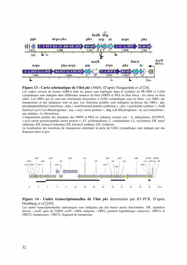
52
Figure 13 - Carte schématique de l’ilot pks (54kb). D’après Nougayrède et al [24]. Les cadres ouverts de lecture (ORFs) dont les gènes sont impliqués dans la synthèse de PK-NRP et l’effet cytopathique sont indiqués dans différentes nuances de bleu (NRPS et PKS en bleu foncé ; les autres en bleu clair). Les ORFs qui ne sont pas strictement nécessaires à l'effet cytopathique sont en blanc. Les ORFs des transposases et des intégrases sont en gris. Les fonctions prédites sont indiquées au-dessus des ORFs : ppt, phosphopantéthéinyl transférase ; nrps, « nonribosomal peptide synthetase » ; pks, « polyketide synthase » ; hcdh, hydroxyl acyl CoA déhydrogénase ; acp, « acyl carrier protein » ; dhg, α β déhydrogénase ; at, acyl-transférase ; am, amidase ; te, thioestérase. L'organisation prédite des domaines des NRPS et PKS est indiquée comme suit : A, adénylation; ACP/PCP, « acyl carrier protein/peptide carrier protein »; AT, acyltransférase; C, condensation; Cy, cyclization; ER, enoyl réductase; KR, ketoacyl réductase; KS, ketoacyl synthase; OX, oxidation. La localisation des insertions de transposons entraînant la perte de l'effet cytopathique sont indiqués par des drapeaux noirs et gris.
Figure 14 - Unités transcriptionnelles de l’ilot pks déterminées par RT-PCR. D’après Homburg et al [245]. Les unités transcriptionnelles opéroniques sont indiquées par des barres noires horizontales. DR, répétition directe ; asnW, gène de l'ARNt asnW ; intP4, intégrase ; ORF2, protéine hypothétique conservée ; ORF21 et ORF22, transposases ; ORF23, fragment de transposase.

53
Les niveaux de transcription en culture sous agitation sont supérieurs à ceux obtenus
en culture statique. La seconde partie de l’ilot pks (clbJ à clbQ) est transcrite à des niveaux
plus faibles quelles que soient les conditions de culture testées. La coculture des bactéries
avec des cellulaires eucaryotes n’a pas d’influence sur la transcription des gènes. Au cours de
la croissance bactérienne, une augmentation continue de l'expression des gènes est observée
en milieu minimum, LB ou DMEM avec un maximum en fin de phase exponentielle et en
début de phase stationnaire. Une influence de la source de carbone et d'énergie est observée
sur les niveaux de transcription en milieu minimum [245].
3. Distribution de l’ilot pks au sein des Entérobactéries
L’ilot pks fait partie du pool de gènes flexible de E. coli. Il est presque exclusivement
observé dans les isolats du groupe phylogénétique B2 ; seulement 3 isolats du groupe
phylogénétique B1 ont été rapportés. Sa prévalence est de 20 à 34% dans les isolats fécaux
(n=236) et 36 à 53% dans les ExPEC (n =215) [24, 246, 247]. L’ilot pks est statistiquement
associé aux souches de E. coli isolées lors de bactériémies et produisant de nombreux facteurs
de virulence, mais n’est pas le trait le plus prédictif d’une origine septique [247]. Il est
cependant absent des souches K12 de laboratoire et des souches de E. coli de pathotypes
intestinaux (n =737), de Shigella spp. (n=10) et de Salmonella enterica (n=114) [24, 246,
247].
L’ilot pks est observé dans d’autres genres de la famille des Enterobacteriaceae :
Klebsiella pneumoniae (3,5%, n=141), Enterobacter aerogenes (27,3%, n=11) et Citrobacter
koseri (n=1) [246]. La séquence de l’ilot pks est très conservée chez ces espèces
d’Enterobacteriaceae (>98% d’identité de séquences nucléotidiques). Des variations de
séquence ont été uniquement observées dans la région intergénique clbR-clbB. Cette région
contient un nombre variable (2 à 20) de répétitions en tandem d’octanucléotides. Aucune
corrélation entre la taille de la région, le nombre pair ou impair de répétitions, la parenté des
souches, le pathotype et l'activité cytopathogène de pks n’a pu être mise en évidence [246].
4. Environnement génétique de l’ilot pks
Chez les E. coli du groupe B2, l’ilot pks est inséré au niveau du locus de l’ARNt AsnW
[24], un « point chaud » d'intégration des éléments mobiles [248]. Fait intéressant, l’ilot
génomique pks chez K. pneumoniae, E. aerogenes et C. koseri, et les trois souches de E. coli
54
du groupe B1 est associé à des séquences mobiles apparentées aux éléments d'intégration et
de conjugaison (ICE), ICEEc1 et ICEKp1. Différents gènes asn-ARNt ont servi de sites
d'insertion chromosomique à ces éléments chez les souches de E. coli du groupe B1 et de K.
pneumoniae. Dans la souche C. koseri étudiée, l’ilot pks est inséré dans l’environnement
proche d’un asn-ARNt [246].
Chez E. coli, l’ilot pks est toujours retrouvé à proximité de l’ilot génomique HPI
(« High Pathogenicity Island »). La réciproque n’est pas vraie. L’ilot HPI est considéré
comme un facteur de virulence des ExPEC et permet la biosynthèse de la yersiniabactine, un
sidérophore de type NRP. La production de la colibactine est cependant indépendante de celle
de la yersiniabactine [24, 246].
5. Expression phénotypique et mode d’action
L'infection transitoire de lignées cellulaires de mammifères (cellules tumorales
humaines du col de l’utérus HeLa, cellules ovariennes de hamster CHO, cellules de
mélanomes humains A375 et cellules non tumorales des cryptes intestinales de rat IEC-6) par
des E. coli porteuses de l’ilot pks induit une mégalocytose caractérisée par un élargissement
progressif du corps cellulaire et du noyau, et l'absence de mitose (figure 15) [24, 249].
L'activité cytopathique nécessite un contact intime entre la cellule hôte et la bactérie, mais ne
requiert pas l'internalisation de la bactérie. Tuées par la chaleur, la sonication ou les
antibiotiques, les bactéries ne sont pas cytopathogènes [24].
L’analyse du cycle cellulaire de cellules tumorales infectées montre que le noyau des
cellules géantes a une teneur en ADN de 4n (figure 15). Cette observation, conjuguée à
l'absence de cellules en division indique que les cellules géantes sont bloquées à la transition
de phase G2/M (Fig. 3A).
Le blocage du cycle cellulaire est associé à une activation de la protéine ATM et une
phosphorylation dose-dépendante de l’histone H2AX (figure 15). Ces phénomènes
correspondent à l’activation du point de contrôle G2/M en réponse à des cassures double-brin
de l'ADN, qui ont été mises en évidence par le test de la comète (figure 15). La voie de
signalisation conduisant au blocage cellulaire en phase G2/M passe par l’activation de la voie
ATM-Chk2 (figure 16). De plus, la lignée CHO défective pour la protéine Ku80 (protéine
impliquée dans la voie de réparation de l’ADN par le système de « Non-Homologue End-
Joining ») et donc hypersensible aux cassures doubles brins de l’ADN meurt massivement
[250]. Cependant, l’action directe ou indirecte de la colibactine sur l’ADN reste à démontrer.

55
Figure 15 - Exposition de cellules HeLa à une infection transitoire à E. coli pks+. D’après Nougayrède et al [24].Les cellules HeLa ont été infectées pendant 4 heures avec une souche de E. coli K12 portant un BAC contenant l’ilot pks ou non (multiplicité d’infection de 100) ou un mélange de chaque souche (50:50). A. Mégalocytose des cellules révélée par coloration de Giemsa 72 h après infection. B. Analyse du cycle cellulaire 72 h après infection montrant un arrêt des cellules en G2/M. C. Phosphorylation des histones H2AX, témoin de cassures double–brin de l’ADN, analysée 4 heures après
infection. D. Test de la comète après infection ou traitement avec l’étoposide, un inhibiteur de la topoisomérase II humaine
qui induit des cassures double-brin de l’ADN.
Figure 16 - Modèle d'activation du point de contrôle G2/M dans les cellules HeLa exposées aux souches de E. coli pks+. D’après Nougayrède et
al [24].Le complexe enzymatique codé par l’ilot pks
synthétise un ou des composés de type PK-NRP, désigné « colibactin », qui induit, directement ou indirectement des cassures double brin de l'ADN dans les cellules hôtes eucaryotes. Ces dommages activent la voie de transduction du signal ATM-Chk2, conduisant à la séquestration cytoplasmique de Cdc25C et au défaut de déphosphorylation de Cdk1, induisant finalement un blocage du cycle cellulaire en phase G2. Les phophorylations sont représentées par des astérisques rouges.

56
6. Un agent carcinogène potentiel
Une seule et courte exposition de cellules épithéliales de mammifère en culture (lignée
CHO) à de faibles doses infectieuses de E.coli pks+ induit une réponse cellulaire aux
dommages de l'ADN, marquée par un blocage transitoire du cycle. Les cellules reprennent
leur division et présentent des signes de réparation incomplets de l'ADN, conduisant à des
ponts anaphasiques et des aberrations chromosomiques (figure 17) [250]. Dans des cultures
de différentes lignées cellulaires, des anomalies du nombre (aneuploïdie) ou de la structure
des chromosomes (micronoyaux, chromosomes en anneau et ponts anaphasiques) persistent
dans les cellules en division jusqu'à 21 jours après l'infection, indiquant l’apparition de cycles
de cassure-fusion-pont ayant un rôle majeur dans l’instabilité chromosomique [250].
L'infection d’entérocytes dans un modèle d’anse intestinale ligaturée de souris par une
souche de E. coli pks+ induit la formation de foyers d’histones H2AX phosphorylés
correspondant à une irradiation de 0,5 Grays [250]. Les cellules en culture montrent aussi une
augmentation significative de la fréquence des mutations et la formation de colonies
cellulaires indépendantes de l’ancrage [250]. Ces données suggèrent une activité mutagène et
de transformation cellulaire des E. coli pks+.

57
Figure 17 - Ponts anaphasiques et aberrations chromosomiques induits par une infection à E. coli pks+. D’après Cuevas-Ramos et al [250]. A. Pont anaphasique d’une cellule CHO 24 heures après infection. 72 heures après infection (i) anaphase
normale, (ii) pont anaphasique générant des ponts nucléoplasmiques (v), (iii) mitose multipolaire, et (iv) chromosome retardé générant des micronoyaux (vi). Barres : 10 µm.
B. Aberrations chromosomiques des cellules métaphasiques 24 heures après infection.

58
III.Les polycétides et les peptides non ribosomaux
A. Généralités
1. Les métabolites secondaires
Le métabolisme d’un organisme comprend le métabolisme primaire, essentiel à la
croissance et au développement, et le métabolisme secondaire, qui est souvent nécessaire à
l’interaction de l’organisme avec son environnement (Tableau 3) [251]. Chaque voie de
biosynthèse du métabolisme secondaire dérive d’un métabolisme primaire et implique des
réactions complexes et souvent hautement spécifiques qui aboutissent aux produits finis. Les
métabolites secondaires des micro-organismes, des plantes et des organismes marins forment
un immense réservoir de diversité chimique naturelle.
Les six voies principales de métabolisme secondaire synthétisent les terpénoïdes, les
alcaloïdes, les peptides, les oligosaccharides, les PK et les NRP [251, 252]. Des combinaisons
entre ces six voies principales de métabolisme secondaire peuvent se produire et permettent la
synthèse de nouveaux composés sans créer de nouvelles voies de biosynthèses comme par
exemples les molécules hybrides PK-NRP [253, 254].
L’analyse de l’ilot pks montre que la colibactine est un métabolite secondaire hybride
de type PK-NRP. La suite de ce chapitre se focalisera donc sur la biosynthèse et les fonctions
des composés PK, NRP et des hybrides PK-NRP.
Tableau 3 : Caractéristiques des métabolismes primaires et secondaires.D’après Hutter [251].

59
2. Intérêt de l’étude des composés d’origine naturelle
Les structures de plus de 200 000 métabolites secondaires ont été élucidées à ce jour,
nous fournissant une complexité et une diversité énorme de squelettes carbonés uniques. Ces
métabolites secondaires ont abouti à un arsenal de produits naturels aux propriétés spécifiques
qui sont à l’origine de médicaments inestimables [252].
En 1990, environ 80% des médicaments étaient soit des produits naturels, soit des
analogues hémi-synthétiques de ceux-ci [255]. L’essentiel de ces médicaments appartient aux
familles de composés PK, NRP et PK-NRP comme de nombreux antibiotiques (ex. : les
pénicillines, les céphalosporines, les tétracyclines, les macrolides comme l'érythromycine),
des antifongiques (ex : l’amphotéricine B et la nystatine), des antiparasitaires (ex. :
l'avermectine), des antipaludéens (ex. : l'artémisinine), des hypocholestérolémiants (ex. : la
lovastatine et ses nombreux analogues), des immunosuppresseurs (ex. : la cyclosporine, la
rapamycine et le tacrolimus) et des médicaments anticancéreux (ex. : la doxorubicine et la
bléomycine) (figure 18). Ces composés ont révolutionné la médecine et allongé l'espérance
de vie dans une grande partie du monde de près de 40 ans au début du 20ème siècle à plus de
77 ans aujourd'hui. Aussi, malgré l'expansion de la chimie médicinale synthétique dans les
années 1990, 50% des nouveaux médicaments restent encore aujourd’hui basés sur des
produits naturels [256]. Quatre raisons expliquent ce succès : [257] :
- la conception de produits actifs passe par l’élaboration de composés et de procédés ayant
la complexité retrouvée dans les composés naturels,
- des siècles d'évolution ont permis d'optimiser la structure des produits naturels, conduisant
souvent à des mécanismes d’adressage à la cible efficaces, une affinité sub-nanomolaire
pour les cibles et une grande spécificité,
- de très nombreux produits naturels restent à découvrir par l’exploration de nouvelles
niches écologiques et de nouveaux taxons microbiens,
- les progrès technologiques du séquençage, de la manipulation et de la reconstitution des
voies de biosynthèses offrent également de nouvelles perspectives.

60
Figure 18 - Exemples de métabolites secondaires de type PK, NRP et hybrides utilisés en médecine. Les structures, les micro-organismes producteurs et les activités pharmacologiques des composés sont indiqués.

61
B. La synthèse des composés PK et NRP
La biosynthèse des PK a été observée chez les bactéries, les champignons, les plantes
et les animaux, et celle des NRP principalement chez des micro-organismes tels que les
bactéries et les champignons [257, 258].
Les composés de type PK, NRP et PK-NRP (ensemble de composés désigné ci-après
NRPK) sont des oligomères synthétisés par des complexes multienzymatiques incluant des
mégasynthases (PK synthases ou PKS, NRP synthases ou NRPS et les PK-NRP synthases
hybrides ou PKS-NRPS) et des enzymes discrètes. Les gènes codant ces voies métaboliques
secondaires sont organisés en clusters, permettant une régulation coordonnée de la synthèse
des composés à la demande ou « à flux tendu » [259, 260]. Ces complexes multienzymatiques,
souvent appelés modules, assurent (i) l’activation de monomères destinés à l’assemblage, (ii)
l’assemblage des monomères en un oligomère linéaire par des étapes itératives de
condensation chimique, à l’image de la synthèse des protéines par le ribosome, et (iii) la
modification des monomères en cours ou après leur assemblage. Ces protéines agissent de
façon coordonnée selon une organisation évoquant une chaine d’assemblage d’automobiles, et
très fréquemment dans un ordre colinéaire à l’organisation génique [25, 261]. Les enzymes
discrètes modifient les monomères ou oligomères pendant ou après l’assemblage. Ainsi, la
structure unique des composés NRPK repose sur le nombre de modules enzymatiques mis en
jeu, l'ordre d’action des enzymes et la chimie catalysée par chaque enzyme dans et après la
chaine d’assemblage des monomères [25].

62
1. La logique chimique
L’assemblage des NRPK repose sur l’assemblage itératif de monomères d’acides et/ou
d’acides aminés en un oligomère, qui subit en sus des modifications enzymatiques complexes.
a) L’activation des monomères
Les monomères qui composent les PK et les NRP sont issus du métabolisme primaire.
D’autres monomères comme certains acides aminés non protéinogènes sont synthétisés à
partir des métabolites primaires par des voies métaboliques secondaires fréquemment codées
dans le cluster comprenant les gènes des mégasynthases. L’utilisation de ces monomères
repose sur leur activation sous forme de thioesters [261, 262].
Les composés PK sont constitués de monomères d’acyles portant un groupe carboxyle
activé permettant la condensation chimique avec l’oligomère en croissance. L’activation
repose sur une liaison au coenzyme A (CoA) par une liaison thioester (Figures 19A). Les
unités acyles sont des monoacyles comme l'acétyl-, le propionyl- et le benzoyl-CoA, ou des
variants, tels que le malonamyl-CoA ou le methoxymalonyl-CoA [263]. Le malonyl-CoA et
le méthylmalonyl-CoA sont les unités monomériques les plus fréquemment incorporées
pendant l’élongation de la chaine des PK.
Les NRP sont synthétisés à partir des 20 acides aminés protéinogènes, d’acides aminés
non protéinogènes et d’acides aryles (Figure 19B) [264, 265]. Au moins 350 monomères de
ce type sont dénombrés à ce jour. Ils sont à l’origine de l’essentiel de la diversité des
composés NRP. Leur activation repose sur la synthèse d’anhydrides couplés à l’AMP (Figure
19C).
Les monomères activés des PK et des NRP sont ensuite chargés sur les domaines de
thiolation T des synthases PKS et NRPS pour former respectivement des acyl- et aminoacyl-
thioesters conservant l’énergie d’activation (figure 19C) [266].

63
Figure 19 - Utilisation de monomères thioesters par les PKS et les NRPS.D’après Fischbach et Walsh [25]. A. Acyl-CoA thioesters couramment utilisés par les PKS pour l’initiation et l’élongation de la chaine.B. Monomères d’amino- et d’aryl-acides génériques et exemples d’acides aminés non protéinogènes (β-hydroxy-
tyrosine (β-OH-Tyr), 3,5-dihydroxyphénylglycine (3,5-Dpg), 4-hydroxyphénylglycine (4-Hpg)) utilisés par les NRPS pour l’initiation et l’élongation de la chaine.
C. Formation d’amino-acyl-thioester par les NRPS à partir d’amino-acyl-AMP.
!"#$%&'"()%%%% !"#$%&*+,$%
-&,.(+,/0. %%%%%%%%%%-)12+,'&,.(+,/0. %%%%% !)1+,/0. %
30. %4
%.5%3/!.$(6+'$%
A
B
C 78%%
!"#$%&'"()%%
'"(./&!+,/ -8%
β/9:/7+*%%%%%%%%%%%%%%%%%%%%;<=/>?@%%%%%%%%%%%%%%%%A/:?@%
'"(./&!+,/12".$B1$*%
>.'&"($%#$%12".,&1".(%

64
b) L’élongation des oligomères
L’élongation de l’oligomère repose la formation d’une liaison covalente avec un
monomère activé à chaque transfert de l’oligomère le long de la chaine enzymatique
d’assemblage [261, 262]. Ainsi, l’oligomère est transféré de la synthase n-1 sur l’acyl- ou
l’aminoacyl-thioesters de la synthase n, qui catalyse la condensation de l’oligomère avec
l’acyl- ou l’aminoacyl-thioesters (Figure 20). La condensation des acyl-thioesters forment les
liaisons C-C des oligomères PK. La condensation des aminoacyl-thioesters forment les
liaisons C-N (liaison peptidique) des oligomères NRP. Ce processus est répété tout au long de
la chaine d’assemblage selon un ordre guidé par l’agencement des synthases.
Figure 20 - Transfert et condensation des aminoacyl-thioesters lors de la formation d’un NRP. D’après Fischbach et Walsh [25]. L’aminoadipoyl-S-T1 est le donneur, et le cystéinyl-S-T2 l'accepteur pour la formation de la liaison peptidique, pour donner la protéine déacylée -S-T1et le dipeptidyl-S-T2. Domaines d’adénylation (A), de thiolation (T) et de condensation (C).
c) La modification des oligomères
L’oligomère peut être modifié sur la chaine d’assemblage multienzymatique au cours
des différentes phases d’élongation ou après celles-ci. Ces modifications sont assurées en
cours d’élongation par des domaines enzymatiques optionnels des mégasynthases, qui
peuvent assurer notamment l’épimérisation, la transméthylation des composés NRP ou la
réduction et la déshydratation des composés PK. Des enzymes discrètes, dites de « finition »,
et fréquemment codées dans le cluster comprenant les gènes des mégasynthases peuvent
également modifier l’oligomère au cours de sa synthèse ou après son assemblage. Ces
modifications nombreuses et complexes de l’oligomère sont à l’origine de la diversité des
composés, notamment de type PK, et sont essentielles à leur activité biologique [25].
! "#$
%&'(')&%*+ ! ",$
-*.)/&0*+ ! ",$
12&031%&'3*+ ! "#$

65
2. Les machineries enzymatiques
Les mégasynthases PKS, NRPS ou PK-NRPS assurent l’essentiel de la synthèse du
squelette carboné. Leur activité repose sur trois domaines de base [25, 261, 262] (Figure 21) :
1- le domaine Thiolation (T) de fixation des monomères,
2- le domaine KetoSynthase (KS) des PKS ou le domaine de Condensation (C) des NRPS,
3- le domaine AcylTransférase (AT) des PKS ou le domaine d’Adénylation (A) des NRPS.
L’association de ces trois types de domaine sur une ou plusieurs chaines peptidiques
définit un module enzymatique PKS ou NRPS.
A B
Figure 21 - Domaines essentiels constituant les modules des PKS (A) et des NRPS (B). D’après Fischbach et Walsh [25].
a) Les chaines d’assemblage des composés PK
(1) Organisation standard des PKS
Trois types de PKS ont été identifiés. Dans les PKS de type I (modulaires), les
domaines KS, AT et T sont fusionnés en cis pour former des modules, qui peuvent également
être fusionnés en une chaine peptidique pour former une mégasynthase multimodulaire.
L’ordre typique des domaines sur le module enzymatique est KS-AT-T. Dans les systèmes
PKS de type II (itératifs), les domaines existent sous forme de protéines distinctes
interagissant en trans pour former des complexes fonctionnels multimériques (Figure 22)
[25]. Enfin, les PKS de type III et apparentées se distinguent par l'absence du domaine T
[267].

66
!"#$%#&'(%#)############# !"#$%#&'(%#))#
A B
Figure 22 - Comparaison des PKS de type I et de type II. D’après Fischbach et Walsh [25]. A. Dans les systèmes de type I, les domaines protéiques sont connectés en cis.B. Dans les systèmes de type II, les domaines protéiques interagissent en trans.
Les domaines T des PKS de type I et de type II sont souvent désignés sous le nom de
protéines portant la chaine acyle ou ACP pour « Acyl Carrier Protein ». La chaine latérale
d'une sérine spécifique du domaine T est modifiée post-traductionnellement par la fixation
d'un bras flexible pantéthéinyle terminé par un groupe thiol libre (Figure 23). Cet amorçage
des domaines apo-T en domaines holo-T fonctionnels est effectué par des
PhosphoPantéthéinyl-Transférases (PPTases), souvent codées dans les clusters de gènes
codant les mégasynthases correspondantes [266, 268]. A partir d’un acyl-CoA activé, le site
holo-T peut alors fixer, grâce à la fonction thiol libre un monomère acyle pour former un acyl-
thioester prêt à être assemblé.
Figure 23 - Modification post-traductionnelle des domaines de Thiolation T par les PhosphoPantéthéinyl-Transférases (PPTases). D’après Fischbach et Walsh [25]. Les membres de cette classe d’enzymes catalysent le transfert d’un groupement PhosphoPantéthéinyl à partir d’un Coenzyme A vers une sérine conservée du domaine T. Cet « amorçage » des domaines T des chaines d’assemblage PKS et NRPS est identique.
*+,-&.+,#&/.+0#0.12%#

67
Le domaine AT sélectionne un acyl-CoA spécifique comme substrat. C’est le gardien
de la spécificité des monomères d’acyle incorporés. Il assure la fixation du groupement acyle
à l'extrémité thiolate du bras pantéthéinyle du domaine holo-T pour former un acyl-thioester
(acyl-S-T) activé et prêt à être assemblé (figure 24A). Le domaine KS est le catalyseur
permettant le transfert et la condensation de l’oligomère en croissance avec les monomères
selon un mécanisme en 3 phases, détaillé dans la figure 24B.
Figure 24 - Activités des domaines AT et KS des PKS. D’après Fischbach et Walsh [25]. A. Le domaine AT sélectionne le monomère acyl-CoA à incorporer et catalyse sa transthiolation au domaine T
en aval via un intermédiaire acyl-O-enzyme. B. Le domaine KS catalyse la formation d’une liaison C-C entre l’acyl-thioester en amont (à gauche),
transitoirement lié à KS et l’accepteur acyl carbanion en aval (à droite) résultant de la décarboxylation du acyl-S-T ajouté.
Au cours d’un assemblage utilisant un système PKS de type I, la chaine acyle en
croissance est transloquée du domaine Tn-1 vers le domaine Tn à chaque action d’un domaine
KS (Figure 25A). Le plus souvent, les domaines KS et T sont utilisés une seule fois. Les
systèmes PKS de type II itératif utilisent un seul domaine T, AT, et KS pour toutes les étapes
d’élongation. La chaine s’accroît à chaque cycle, mais reste attachée au même domaine T, et
les domaines AT et KS agissent de manière itérative (figure 25B). Dans ces systèmes itératifs,
le domaine KS est un hétérodimère d’une sous-unité active (KSα) et d’une sous-unité inactive
CLF (pour « Chain Length Factor »), qui forme un tunnel amphipathique dont la taille
contrôle le nombre d’itération réalisé [269].
Figure 25 - Elongation de la chaine dans les systèmes PKS de type I. D’après Fischbach et Walsh [25]. Chaque condensation résulte en une addition de deux ou trois carbones sur la chaine naissante, qui est simultanément transférée à partir d’un domaine T au suivant dans les PKS de type I.
*$������������
*%����������&�������##����A
B

68
(2) Les modules d’initiation et de terminaison
Les modules enzymatiques d’initiation et de terminaison fonctionnent différemment
des modules d’élongation décrits ci-dessus [25, 261, 262].
Un acyl-CoA est sélectionné par le domaine AT d'initiation et chargé sur le premier
domaine T. Le domaine KS est ainsi généralement absent du module d’initiation de la chaine
d’assemblage ou il y est non fonctionnel.
Le dernier module d'une chaine d'assemblage PKS entraîne la rupture de la liaison
thioester entre le domaine T final et l’oligomère terminé. Ce processus d’hydrolyse libérant le
composé est généralement catalysé par un domaine thioestérase (TE) de 35 kDa. Celle-ci
génère un acide libre à l’extrémité de l’oligomère ou une lactone cyclique par condensation
avec une fonction hydroxyle de la chaine PK [270].
(3) Les domaines optionnels et enzymes de finition
Les domaines facultatifs des PKS de type I et II sont les domaines catalytiques
kétoréductases (KR), déshydratases (DH) et enoylréductases (ER) [25, 271]. Ils opèrent
suivant la séquence KR > DH > ER ; le β-kétoacyl-S-T est d'abord réduit en β-hydroxyacyl-S-
T par l’action KR, puis ce dernier est déshydraté pour former un α,β-enoyl-S-T par l’action
DH, et enfin ce conjugué oléfine est saturé pour donner un acyl-S-T saturé suite à l’action ER
(Figure 26). Ces trois domaines enzymatiques catalysent au total la réduction du groupement
cétone β-C=O en méthyle β-CH2. Cependant, cette réduction est rarement complète et de
nombreux modules préservent les fractions intermédiaires β-hydroxyacyle ou α,β-enoyle en
raison de l'absence ou de la non-fonctionnalité de domaines ER, DH et/ou KR. Par ailleurs,
des modules C-méthyltransférases ainsi que des modules aminotransférases peuvent convertir
un β-kétoacyl-S-T en β-aminoacyl-S-T [272, 273].
Enfin des enzymes de finition peuvent agir en trans sur les intermédiaires acyl-S-T en
croissance, afin de catalyser des réactions de déshydratation, de décarboxylation, de réduction
ou d’addition de carbanions [25].
L’organisation standard des chaines d’assemblage de PK de type I peut être illustrée
par celle du 6-dEB (précurseur inactif de l’antibiotique érythromycine A), système modèle et
historique de PKS modulaires (figure 27) [274, 275].

69
Figure 26 - Transformation du carbone ββββ dans les PKS. D’après Fischbach et Walsh [25]. Le produit de condensation β-kétoacyl-S-T est réduit par un domaine kétoréductase (KR) pour former un β-hydroxyacyl-S-T, qui est déshydraté par un domaine déshydratase (DH) générant un α,β-enoyl-S-T (conjugué oléfine), qui est enfin réduit par un domaine enoylréductase (ER) pour donner un acyl-S-T saturé.
Figure 27 : Organisation des PKS de type I ou modulaires synthétisant le 6-dEB. D’après Meier et Burkart [261]. La première ligne indique les protéines multienzymatiques (DEBS : 6-dEB synthases) ; la deuxième ligne indique les modules constitués d’un nombre de domaines enzymatiques variable sur les différentes protéines (« Loading module » : module d’initiation ou de chargement ne possédant pas de domaine KS). Le domaine TE cyclise le PK en formant une lactone par condensation de la fonction acide libérée de la liaison thioester du domaine ACP final avec la dernière fonction hydroxyle de la chaine PK. Des enzymes de finition catalysent 2 hydroxylations et 2 glycosylations pour générer le composé final actif, l’érythromycine A.

70
b) Les chaines d’assemblage des composés NRP
(1) Organisation standard des NRPS
Trois types de NRPS sont décrits : les NRPS de type A (ou type linéaire) dont les
modules sont utilisés une seule fois, les NRPS de type B (ou type itératif), dont tous les
modules sont utilisés plusieurs fois et le type C (ou type non linéaire), dont certains domaines
sont absents et d’autres fonctionnent plus d’une fois [276].
Les NRPS suivent les préceptes généraux de la logique des PKS de type I. Le
mécanisme de biosynthèse est composé de chaines d'assemblage enzymatiques
multimodulaires qui contiennent un module pour chaque monomère d’acide aminé incorporé.
Par analogie avec les trois domaines d'un module PKS (KS-AT-T), il existe trois domaines de
base dans un module minimal de NRPS (Figure 28) [25, 261, 262] :
1- le domaine T (ou PCP pour « peptidyl carrier protein ») supporte la chaine peptidique en
croissance attachée à un groupement phosphopantéthéinyle ;
2- le domaine d’adénylation A est responsable de la sélection du monomère ;
3- le domaine de condensation C assure l'élongation de la chaine peptidique.
L’ordre typique des domaines sur le module multienzymatique NRPS est C-A-T.
(2) Les modules d’initiation et de terminaison
Le module d'initiation des lignes d'assemblage NRPS est généralement un module à
deux domaines « A-T1 » qui sélectionne le premier acide aminé et l'installe de façon covalente
sur T1 [25, 261, 262]. Le premier module peut présenter trois domaines C-A-T dans les
chaines d’assemblage synthétisant des NRP N-acylés ou lipopeptides, comme la daptomycine
(figure 18). Ce type de domaine C spécifique, appelé « Starter C » réalise le processus dit de
lipoinitiation : il fixe un acide carboxylique β-hydroxylé (typiquement un acide gras β-
hydroxylé) au premier acide aminé porté par le premier domaine T [277, 278].

71
Figure 28 - Fonctionnement du domaine T et activités des domaines A et C des NRPS. D’après Fischbach et Walsh [25]. Le fonctionnement des domaines T des chaines d’assemblage NRPS est équivalent à ceux des lignes d'assemblage des PKS. Chaque domaine apo-T doit être amorcé par un groupement phosphopantéthéine. Lors de l’assemblage du NRP, les résidus aminoacyl et les chaines peptidiques en croissance sont liés par un pont thioester au thiolate terminal du groupement prosthétique phosphopantéthéine. A. Le domaine A, par analogie avec le domaine AT des PKS sélectionne l'acide aminé à incorporer, active son
groupement carboxylate avec un ATP pour créer un aminoacyl-AMP (adénylation), puis fixe le résidu aminoacyl sur le thiolate du domaine T adjacent en aval (acylation).
B. Le domaine C, par analogie avec le domaine KS des PKS catalyse l’élongation de la chaine peptidique, formant une liaison N-C entre le peptidyl-S-Tn-1 amont (à gauche) à l’aminoacyl-S-Tn aval (à droite). Cet allongement de la chaine se produit de façon concomitante à la translocation de la chaine en croissance vers le Tn et préserve le radical aminé libre du résidu N-terminal.
Comme dans les PKS de type I, les modules de terminaison portent un domaine
thioesterase (TE) et ont généralement une organisation C-A-T-TE. Le domaine TE peut être
hydrolytique ou cyclisant comme observé dans les chaines d’assemblage PKS [279]. La
cyclisation est souvent un caractère fondamental car elle rigidifie la conformation du composé,
permet des interactions spécifiques avec les cibles biologiques et dans le cas des NRP,
confère une résistance aux protéases. La macrocyclisation des NRP peut se réaliser suivant
quatre processus différents (figure 29) [280]. La libération de l’oligomère sous forme linéaire
ou cyclique peut aussi être médiée par une réductase [281].
A
B

72
Figure 29 - Les quatre processus de macrocyclisation des NRP.D’après Sieber et Marahiel [280].

73
(3) Les domaines optionnels
Les composés NRP peuvent subir en cours de synthèse des modifications par des
domaines enzymatiques optionnels des enzymes NRPS.
Une caractéristique des NRP est l'abondance de résidus acides aminés de la série D.
Ces acides aminés sont générés par un système d’épimérisation des énantiomères usuels L en
énantiomères D [277]. Ce système est composé d’un domaine épimérase E associé aux
domaines standards dans l’ordre C-A-T-E qui modifie le dernier acide aminé incorporé [282,
283]. En aval du module C-A-T-E sur la chaine d’assemblage, un domaine de condensation C
spécifique, appelé DCL (donneur d’énantiomère D et accepteur d’énantiomère L) lie un acide
aminé L à la chaine peptidique en croissance terminée par l’acide aminé D modifié,
contrairement aux domaines de condensation usuels LCL, accepteurs et donneurs
d’énantiomères L. Certains domaines C couplent les deux fonctions de condensation et
d’épimérisation et sont appelés domaines « dual C/E » [284]. L’épimérisation des acides
aminés est parfois réalisée par une racémase discrète agissant en trans [285].
D'autres variants du domaine C appelés domaines Cy (pour cyclodéshydratation)
permettent l’ajout et la déshydratation de résidus Cys, Ser ou Thr pour former des
hétérocycles à cinq atomes (cycle thiazoline (Cys) ou oxazoline (Ser ou Thr)) [277, 286]. Ces
hétérocycles peuvent être ensuite modifiés par d’autres enzymes ou domaines. Le domaine
oxydase (Ox) est impliqué dans l'oxydation des cycles thiazolines facilement hydrolysables,
en hétérocycles thiazoles stables [287, 288]. De même, des réductases peuvent transformer les
cycles thiazolines en cycle tétrahydro-thiazolines [289].
De nombreux NRP contiennent des résidus d’acides aminés méthylés suite à l’action
de domaines N-méhyltransférases. Plus rarement des domaines C-méthyltransférases peuvent
agir sur des hétérocycles thiazolines[290, 291].
L’organisation des chaines d’assemblage de NRP peut être illustrée par celle de la
tyrocidine, un NRP cyclique (figure 30) .

74
Figure 30 - Organisation des NRPS linéaires synthétisant la tyrocidine. D’après Meier et Burkart [261]. La première ligne indique les protéines multienzymatiques (mégasynthases TycA, TycB et TycC) ; la deuxième ligne indique les modules constitués d’un nombre de domaines enzymatiques variable sur les différentes protéines : module 1 d’initiation ne possédant pas de domaine C ; modules 1 et 4 possédant un domaine E ; module 10 de terminaison possédant un domaine TE qui cyclise le NRP en formant un macrolactame par condensation de la fonction acide libérée de la liaison thioester du domaine PCP final avec la fonction amine N-terminale de la chaine NRP.

75
c) Les chaines d’assemblages hybrides PKS-NRPS
Les chaines d’assemblage hybrides contiennent des modules enzymatiques de type
PKS et NRPS. Ces modules PKS et NRPS peuvent aussi coexister sur une même protéine et
sont alors appelés PKS-NRPS [292-295]. Le fonctionnement de ces chaines d’assemblage
repose probablement sur les principes évoqués précédemment. Des investigations
complémentaires sont encore nécessaires, notamment pour expliquer le transfert de
l’oligomère et sa condensation lors des interfaces PKS/NRPS et NRPS/PKS.
d) Les modifications enzymatiques après la chaine d’assemblage
Les composés NRPK libérés du dernier module enzymatique de la chaine
d’assemblage subissent souvent des modifications supplémentaires jouant un rôle important
dans l'établissement de la bioactivité du composé. Ces modifications sont catalysées par des
enzymes de finition codées dans le cluster de gènes de biosynthèse du composé. Ces enzymes
catalysent typiquement des réactions d’oxydation, de O-/C-/N-glycosylation, de N-/O-
acylation, d'alkylation (méthylation généralement) et d’halogénation [265, 296-298].
e) Les enzymes de réparation des chaines d’assemblage
Les chaines de production des composés NRPK peuvent faire des erreurs, notamment
incorporer des monomères inadaptés sur les domaines T, bloquant la chaine d’assemblage.
Les clusters de gènes de biosynthèse des NRPK codent parfois des enzymes de réparation
discrètes appelées thioestérases TE de type II qui élimineraient les monomères inadaptés par
hydrolyse, régénérant ainsi les domaines T bloqués [299, 300].
f) Les domaines manquants et autres violations de colinéarité
Dans le petit échantillon des lignes d'assemblage étudiées à ce jour, des variantes ont
été détectées dans lesquelles des domaines normalement présents en cis sont manquants [301].
Par exemple, le complexe multienzymatique produisant la yersiniabactine contient trois
modules NRPS mais un seul domaine A. Il a été démontré que ce domaine active la L-
cystéine et la transmet en cis à un domaine T et en trans pour les deux autres domaines T
[302]. De nombreux gènes de PKS de type I ont été séquencés auxquels il manque un,
plusieurs ou l'ensemble des domaines AT [303-306]. Des domaines AT agissent alors en trans
pour fournir des unités malonyle ou méthylmalonyle aux domaines T dans les modules sans
modules AT.

76
Dans certaines voies de biosynthèse, des modules semblent être ignorés, probablement
en raison d’une non-fonctionnalité enzymatique ou architecturale de ces domaines : ce
phénomène est appelé le « domain skipping » [307, 308]. Par ailleurs, certaines chaines de PK
en croissance peuvent faire une pause et être allongées à deux reprises sur le même module, à
la manière d’un bégaiement, ce processus est dénommé le « domain stuttering » [309-311].
g) Les fragments de chaine d’assemblage
Des études bioinformatiques ont révélé des gènes codant pour des fragments de
chaines d’assemblage : des didomaines A-T ou des domaines A et T séparés (A/T) mais
adjacents se retrouvent en l'absence de tout autre composant de type NRPS. Ces domaines A
activent un L-acide aminé spécifique et l'installent sur le domaine T. Ce groupe aminoacyl lié
par un pont thioester au domaine T est alors le substrat pour une enzyme partenaire, qui induit
une transformation chimique au carbone β ou γ de l'aminoacyl-S-T. L’acide aminé modifié
peut alors être libéré par une thioestérase. Cette stratégie permet de piéger une fraction du
pool d'acides aminés protéinogènes et de les modifier en une forme non protéinogène
utilisable ensuite par différentes voies métaboliques secondaires [312]. Cette logique est aussi
connue dans des systèmes NRPS [313].
3. La taille des chaines d’assemblage
Les chaines d'assemblage des PKS de type I et des NRPS se composent parfois de
composants protéiques pouvant atteindre plusieurs mégadaltons. Ces chaines d’assemblage
sont comparables à un chapelet dans lequel chaque perle est un domaine replié de manière
autonome [25]. La chaine d'assemblage du 6-dEB comporte sept modules répartis sur trois
protéines d'environ 200 kDa chacune (figure 27) [275]. La synthétase de la cyclosporine
possède 41 domaines répartis sur 11 modules, regroupés dans une seule protéine de 15 000
résidus correspondant à une masse de 1,5 MDa [314], alors que le peptide cyclique qu’elle
synthétise n’est long que de 11 acides aminés.

77
De si grandes protéines sont vraisemblablement sensibles aux problèmes de
repliement, vulnérables à la protéolyse, et enclin à l'inactivation par mutation [25]. Le modèle
le plus commun d'organisation des modules sur plusieurs sous-unités protéiques peut aider à
éviter ces problèmes et pourrait également faciliter l'évolution des composants de ces chaines
d'assemblage par des additions, soustractions ou substitutions de domaines. Cependant, le
fractionnement d'une chaine d’assemblage en de multiples protéines crée d’autres
problèmes comme la reconnaissance inter-protéines lors du transfert des oligomères en
croissance entre les modules enzymatiques.
4. Structure des chaines d’assemblage PKS et NRPS
La biosynthèse catalysée par les chaines d’assemblage dépend de l'action successive
de plusieurs dizaines d’enzymes différentes. De nombreuses interactions protéine-protéine
doivent opérer de manière précise et coordonnée afin d’optimiser le processus d’assemblage.
Les détails moléculaires de ces interactions commencent à être déchiffrés.
a) L’architecture modulaire
La logique d'assemblage des NRPK est basée sur la liaison du substrat à un petit
domaine protéique T porteur. Cette microcompartimentalisation présente l’avantage de
séquestrer les substrats et les intermédiaires réactionnels loin de la concurrence d’autres
processus cellulaires, comme les autres lignes d'assemblage de NRPK [315]. Le bras
phosphopantéthéine des domaines T ou les domaines T eux-mêmes sont des éléments mobiles
oscillants, qui jouent un rôle essentiel dans l'optimisation des interactions avec les domaines
catalytiques des modules de PKS et de NRPS [316, 317]. Cependant, il n’existe pas
d’homologies significatives entre les domaines T des PKS et des NRPS et certains aspects de
leur mobilité reposeraient sur des mécanismes différents. Un ancrage temporaire du domaine
T des PKS sur les domaines catalytiques partenaires serait facilité par des interactions
électrostatiques, grâce à des motifs de reconnaissance du domaine T chargés négativement
[318]. Chez les NRPS, un changement de conformation du domaine T induit par le substrat
serait utilisé pour favoriser les interactions avec les domaines partenaires [316].
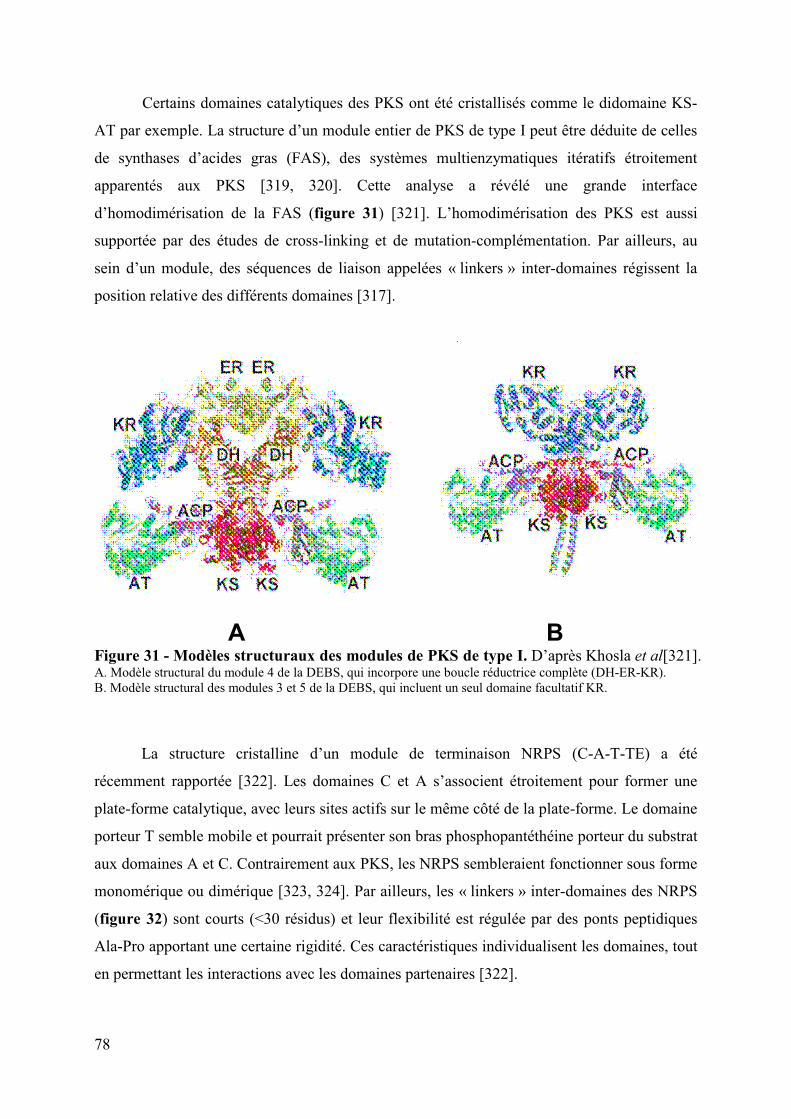
78
Certains domaines catalytiques des PKS ont été cristallisés comme le didomaine KS-
AT par exemple. La structure d’un module entier de PKS de type I peut être déduite de celles
de synthases d’acides gras (FAS), des systèmes multienzymatiques itératifs étroitement
apparentés aux PKS [319, 320]. Cette analyse a révélé une grande interface
d’homodimérisation de la FAS (figure 31) [321]. L’homodimérisation des PKS est aussi
supportée par des études de cross-linking et de mutation-complémentation. Par ailleurs, au
sein d’un module, des séquences de liaison appelées « linkers » inter-domaines régissent la
position relative des différents domaines [317].
A B Figure 31 - Modèles structuraux des modules de PKS de type I. D’après Khosla et al[321]. A. Modèle structural du module 4 de la DEBS, qui incorpore une boucle réductrice complète (DH-ER-KR). B. Modèle structural des modules 3 et 5 de la DEBS, qui incluent un seul domaine facultatif KR.
La structure cristalline d’un module de terminaison NRPS (C-A-T-TE) a été
récemment rapportée [322]. Les domaines C et A s’associent étroitement pour former une
plate-forme catalytique, avec leurs sites actifs sur le même côté de la plate-forme. Le domaine
porteur T semble mobile et pourrait présenter son bras phosphopantéthéine porteur du substrat
aux domaines A et C. Contrairement aux PKS, les NRPS sembleraient fonctionner sous forme
monomérique ou dimérique [323, 324]. Par ailleurs, les « linkers » inter-domaines des NRPS
(figure 32) sont courts (<30 résidus) et leur flexibilité est régulée par des ponts peptidiques
Ala-Pro apportant une certaine rigidité. Ces caractéristiques individualisent les domaines, tout
en permettant les interactions avec les domaines partenaires [322].

79
Figure 32 - Structure cristalline du module de la dernière NRPS de la surfactine.D’après Tanovic et al [322]. Domaines de condensation C, d’adénylation A, de thiolation PCP et thioestérase TE. Linker : séquences de liaison inter-domaines. Acore : sous-domaine principal N-terminal de A, Asub : sous-domaine C-terminal de A.
b) Reconnaissance entre les sous-unités protéiques
Les modules PKS et NRPS peuvent être répartis sur plusieurs protéines. Cette
organisation impose des transferts inter-protéiques entre les domaines T et KS des PKS, et les
domaines T et C des NRPS. Chaque protéine doit donc reconnaître spécifiquement la protéine
partenaire appropriée.
(1) Reconnaissance entre les sous-unités PKS
Dans les PKS, la région N-terminale réalise des interactions spécifiques stables avec la
région C-terminale de la sous-unité PKS partenaire. Ces régions, appelées domaines
d’ancrage, sont portables, c'est-à-dire qu’elles fonctionnent indépendamment de leur sous-
unité d’origine [317]. La stabilité des interactions entre ces domaines d’ancrage repose sur la
formation d’un complexe de 4 hélices α entrelacées, présentant des contacts hydrophobes et
des interactions électrostatiques critiques [325]. Ces domaines d’ancrage présentent deux
éléments de dimérisation (une interaction de 4 hélices α entrelacées et de 2 hélices α
enroulées à l’extrémité des domaines T et KS respectivement) qui interviennent aussi dans
l'association homodimérique des PKS [325]. Par ailleurs, des résidus de surface des domaines
T et KS sont aussi impliqués dans la discrimination des partenaires protéiques [326].
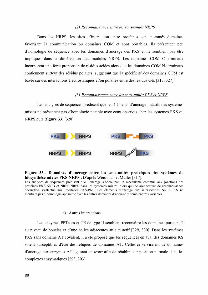
80
(2) Reconnaissance entre les sous-unités NRPS
Dans les NRPS, les sites d’interaction entre protéines sont nommés domaines
favorisant la communication ou domaines COM et sont portables. Ils présentent peu
d’homologie de séquence avec les domaines d’ancrage des PKS et ne semblent pas être
impliqués dans la dimérisation des modules NRPS. Les domaines COM C-terminaux
incorporent une forte proportion de résidus acides alors que les domaines COM N-terminaux
contiennent surtout des résidus polaires, suggérant que la spécificité des domaines COM est
basée sur des interactions électrostatiques et/ou polaires entre des résidus clés [317, 327].
(3) Reconnaissance entre les sous-unités PKS et NRPS
Les analyses de séquences prédisent que les éléments d’ancrage putatifs des systèmes
mixtes ne présentent pas d'homologie notable avec ceux observés chez les systèmes PKS ou
NRPS purs (figure 33) [328].
Figure 33 - Domaines d’ancrage entre les sous-unités protéiques des systèmes de biosynthèse mixtes PKS-NRPS . D’après Weissman et Muller [317].Les analyses de séquences prédisent que l’ancrage s’opère par un mécanisme commun aux jonctions des protéines PKS-NRPs et NRPS-NRPS dans les systèmes mixtes, alors qu’une architecture de reconnaissance alternative s’effectue aux interfaces PKS-PKS. Les éléments d’ancrage aux intersections NRPS-PKS ne montrent pas d’homologie apparente avec les autres domaines d’ancrage et semblent très variables.
c) Autres interactions
Les enzymes PPTases et TE de type II semblent reconnaître les domaines porteurs T
au niveau de boucles et d’une hélice adjacentes au site actif [329, 330]. Dans les systèmes
PKS sans domaine AT covalent, il a été proposé que les séquences en aval des domaines KS
soient susceptibles d'être des reliques de domaines AT. Celles-ci serviraient de domaines
d’ancrage aux enzymes AT agissant en trans afin de rétablir leur position normale dans les
complexes enzymatiques [293, 303].

81
C. Fonctions naturelles des PK et des NRP
Les organismes produisant des composés NRPK investissent une énergie parfois
énorme dans une machinerie enzymatique qui fabrique un produit unique. L'importance des
fonctions exercées par ces NRPK doit justifier le coût énergétique représenté par ces chaines
d’assemblage [25]. Si la diversité structurale de ces composés suggère des mécanismes
d’action différents, il semble que la finalité de ces composés est essentiellement de faciliter la
colonisation de certaines niches écologiques.
1. La colonisation
a) Armes de destruction massive ou molécules de signalisation ?
La grande majorité des antibiotiques utilisés pour traiter les infections sont des
composés de type NRPK d’origine environnementale. Leur rôle naturel a été généralement
déduit de leur activité thérapeutique. Dès 1940, Waksman et Woodruff ont proposé que ces
composés inhibent la multiplication ou tuent des organismes concurrents dans l’habitat
naturel de leur organisme producteur [331]. Toutefois, les molécules sélectionnées par
l'industrie pharmaceutique pour leurs propriétés antibiotiques à des concentrations
thérapeutiques auraient cependant des fonctions distinctes aux concentrations probablement
inférieures rencontrées dans la nature [332].
Les antibiotiques PK tels que l’érythromycine, la tétracycline et la rifampicine
induisent des modifications transcriptionnelles spécifiques chez les bactéries de façon dose-
dépendante et indépendante des voies de réponse bactérienne au stress [332, 333]. La
tétracycline induit par exemple l’expression du SST3 et la cytotoxicité de Pseudomonas
aeruginosa [334]. Par ailleurs, les antibiotiques déclenchent une réponse spécifique, qui peut
moduler les interactions au sein des communautés microbiennes [334, 335]. Ils peuvent donc
avoir un rôle de molécules de signalisation en plus de celui d’inhibiteurs. Cette hypothèse est
soutenue par la découverte d’une activité antibiotique pour des molécules bactériennes de
signalisation intra- et interspécifiques comme les homosérine lactones ou les
lantibiotiques qui sont impliqués dans le « quorum-sensing » [336-338]. Par exemple, une
homosérine lactone de P. aeruginosa inhibe les bactéries à Gram positif [337]. Le double rôle
de cette molécule du « quorum-sensing » pourrait rendre compte de la colonisation successive
de Staphylococcus aureus puis de P. aeruginosa chez les patients atteints de mucoviscidose
[335]. Les composés NRPK pourraient ainsi être des molécules de signalisation ayant un effet
synergique ou antagoniste sur les organismes présents dans la niche écologique de la bactérie
productrice.

82
b) La protection contre les prédateurs eucaryotes
La prédation représente un facteur de mortalité majeur des bactéries dans leur habitat
naturel. Pour se protéger les bactéries ont développé des stratégies de défense impliquant les
NRPK. La production de la sérawettine et de la surfactine par Serratia marcescens et certains
Bacillus spp. respectivement, induit un comportement d’évitement chez Caenorhabditis
elegans en agissant sur ses neurones chimiosensoriels [339]. La production du massétolide et
de la viscosine chez Pseudomonas fluorescens est induite par Naegleria americana et lysent
cette amibe prédatrice [340]. La production des toxines peut aussi être induite après ingestion
de la bactérie par le prédateur, comme pour le composé PK violacéine de Chromobacterium
violaceum qui cible des protozoaires non flagellés prédateurs [341].
c) La mobilité, l’adhérence et la formation de biofilm
Les lipopeptides (composés NRP portant un acide gras) biosurfactants peuvent
changer la viscosité des surfaces et influencer la mobilité et la différenciation multicellulaire
bactériennes [342, 343].
Sur les plantes, les lipopeptides peuvent agir comme des agents de mouillabilité de la
cuticule hydrophobe des feuilles, en réduisant la tension de surface, ce qui favorise la
solubilisation et la diffusion des substrats nécessaires à la croissance microbienne, et la
mobilité vers des niches plus riches en nutriments [344-346]. Il est proposé que les
lipopeptides soient impliqués dans la coordination de leur mouvement au cours du
« swarming »9 [347].
Les lipopeptides jouent un rôle important dans l’attachement aux surfaces et la
formation de biofilms chez Bacillus spp. et Pseudomonas spp. [342]. Selon leur structure, les
lipopeptides peuvent être orientés avec la partie hydrophile face à la surface cellulaire
bactérienne, et exposer leur partie hydrophobe vers l’extérieur, et donc faciliter l’attachement
aux surfaces hydrophobes ; quand l’orientation est inversée, ces composés favorisent
l’adhésion aux surfaces hydrophiles [348]. Les biofilms sont des regroupements structurés de
cellules bactériennes adhérant entre elles et incluses dans une matrice extracellulaire de
polymères qui adhère aux surfaces biologiques et non biologiques [349]. Certains lipopeptides
fonctionnent comme des molécules morphorégulatrices dans ce comportement multicellulaire
bactérien. La surfactine de Bacillus subtilis agit comme une molécule signal dans le
déclenchement de la formation de la matrice des biofilms [350, 351].
9 Déplacement bactérien en groupe sur une surface solide humide, précédant la formation de biofilm.

83
Les lipopeptides peuvent aussi influencer défavorablement l’attachement aux surfaces
et la formation de biofilm par d’autres micro-organismes. La surfactine réduit fortement
l’adhérence de pathogènes d’origine alimentaire tels que Listeria monocytogenes et
Enterobacter sakazakii [352], inhibe la formation de biofilm chez Salmonella Typhimurium
[353], altère la formation de structures aériennes chez Streptomyces coelicolor et affecte ainsi
sa capacité à coloniser une niche écologique [354].
d) La chélation du fer et des autres ions métalliques
Le fer est indispensable au développement des organismes. Les sidérophores,
molécules permettant la capture du fer, sont fréquemment des composés de type NRPK
sécrétés par les bactéries, les champignons et les plantes [355]. Ils sont produits et sécrétés
dans les environnements pauvres en fer libre. Chez les vertébrés, le fer est en grande partie lié
à des macromolécules. Lorsque les bactéries sont au contact de ces derniers, les sidérophores
sécrétés par les bactéries entrent en compétition avec les macromolécules eucaryotes pour la
chélation du fer et sont ensuite absorbés à l’aide de récepteurs spécifiques par les bactéries
[356]. De nombreux organismes sécrètent plus d'un sidérophore. Dans différentes conditions
environnementales et en fonction de l’état ionisé du fer, la sécrétion de l’un ou l'autre des
sidérophores semble prédominer. Certains sidérophores sont utilisés comme molécules de
stockage pour le fer, restituant lentement le fer une fois entrés dans la cellule.
Toutes les souches de E. coli produisent un sidérophore primaire de type NRP,
l’entérobactine (figures 34 et 35). Les mammifères ont développé une parade pour bloquer
l'acquisition du fer par ce sidérophore, dans le cadre de leur réponse immunitaire innée. La
lipocaline 2, ou sidérocaline, sécrétée par les macrophages et les hépatocytes lors d’une
infection séquestre l’entérobactine, empêchant E. coli d'acquérir le fer (figure 35) [357].
Certaines souches pathogènes de E. coli sécrètent des sidérophores secondaires, considérés
comme des facteurs de virulence comme la yersiniabactine et la salmochéline (figures 34 et
35). Cette dernière est une entérobactine glycosylée échappant à la séquestration par la
lipocaline [21, 358].
De nombreux lipopeptides bactériens ont un potentiel de chélation des ions
métalliques (Ca++, Na+, K+ et les métaux lourds). Certains protègeraient les bactéries des
effets toxiques d’ions métalliques en les chélatant. D’autres favorisent leur absorption et leur
utilisation par la bactérie à l’image des sidérophores et du fer [343].

84
Figure 34 - Acquisition du fer par E. coli chez les mammifères. D’après Wiles et al [21].L’hôte et le pathogène rivalisent pour l’acquisition du fer et ont développé des stratégies multiples pour surpasser l’autre. Le sidérophore entérobactine des souches de E. coli séquestre le fer avec une forte affinité, alors que la sidérocaline mammifère lie l’entérobactine et empêche son absorption par les bactéries. Des ExPEC portant le cluster de gènes iro peut modifier l’entérobactine par glycosylation, créant la salmochéline qui lie efficacement le fer mais n’est pas reconnue par la sidérocaline.
Figure 35 - Exemples de composés NRPK impliqués dans la colonisation et la survie bactériennes.
Entérobactine Sidérocaline mammifère
Salmochéline = Entérobactine glycosylée

85
2. La symbiose
De nombreuses souches de Bacillus spp. et de Pseudomonas spp. protègent les plantes
contre des infections bactériennes par la production d’antibiotiques. La souche de B. subtilis
6051 colonise les racines d’Arabidopsis thaliana et forme un biofilm stable et étendu. Ce
biofilm associé à la sécrétion de la surfactine protège la plante contre le pathogène
Pseudomonas syringae pv tomato [359]. La production du lipopeptide viscosinamide par
Pseudomonas fluorescens inhibe l’infection des racines de la betterave à sucre par
l’oomycète10Pythium ultimum, en inhibant la croissance de son mycélium et la motilité de ses
zoospores [360].
Certains lipopeptides ont aussi un effet bénéfique sur les plantes en stimulant leur
système immunitaire. De nombreux lipopeptides produits par des souches de Pseudomonas
spp. et Bacillus spp. non pathogènes déclenchent des réponses de défense chez les plantes
contre les champignons et les oomycètes [344, 361].
L'une des découvertes symbiotiques les plus surprenantes a été faite sur le champignon
Rhizopus microsporus, l'agent causal de la fonte des semis du riz. Un facteur de virulence du
champignon, la rhizoxine est un puissant PK cytotoxique qui inhibe l’assemblage des
microtubules. Les gènes produisant la toxine appartiennent à une bactérie symbiotique
intracellulaire du genre Bulkhoderia, localisée dans le cytosol fongique et indispensable à la
formation des sporanges et spores fongiques [362, 363]. En inhibant la mitose, la rhizoxine
détruit et tue les jeunes plants de riz, dont les tissus en décomposition profitent alors à l’hôte
fongique. Il s'agit d'un mutualisme intime ayant une fonction métabolique spécifique qui
influe sur le mode de vie du champignon. Deux organismes en symbiose peuvent ainsi être
bénéficiaires de la destruction d’un troisième.
3. La virulence
Deux excellents exemples d’implication des NRPK dans la virulence bactérienne ont
été mis en évidence dans le genre Mycobacterium.
M. ulcerans sécrète la mycolactone (figure 35), un PK de type macrolide, qui est un
facteur majeur de virulence. Cette bactérie est l'agent causal de l'ulcère de Buruli, une maladie
de peau invalidante sévissant en Australie et en Afrique de l'Ouest. Elle se caractérise par la
persistance de lésions nécrotiques de la peau et du tissu graisseux sous-jacent, l'absence de
réaction inflammatoire aiguë et de douleur. La mycolactone reproduit la plupart des signes de
10 Protistes filamenteux aquatiques, exclus phylogénétiquement du règne des champignons.

86
l’infection lorsqu'elle est injectée par voie intradermique à des cobayes. La mycolactone est
une cyclomoduline, qui induit un arrêt du cycle cellulaire en phase G0/G1, l'inhibition de la
synthèse des protéines et éventuellement la mort cellulaire [364]. Elle présente des propriétés
immunosuppressives en inhibant la voie NF-kB des monocytes et des lymphocytes T, la
production de cytokines, et la maturation et la migration des cellules dendritiques [365, 366].
Ces résultats suggèrent que la production de ce PK par M. ulcerans peut limiter à la fois
l’initiation des réponses immunitaires et le recrutement de cellules inflammatoires au site de
l’infection. En outre, l'anergie profonde et systémique des lymphocytes T aux antigènes
mycobactériens persiste de nombreuses années après un traitement curatif de l'ulcère de
Buruli [367]. Des isolats de M. ulcerans producteurs de mycolactone ont été retrouvés chez
plusieurs insectes aquatiques (punaises et araignées d’eau, moustiques) qui semblent être le
réservoir de ce pathogène [368, 369]. La mycolactone est essentielle à la colonisation initiale
et à long terme de la punaise d’eau par M. ulcerans [370] et est produite par certaines
mycobactéries aquatiques proches de M. ulcerans responsables de maladies chez les poissons
et les grenouilles [371].
Tous les membres du complexe M. tuberculosis et quelques autres espèces de
mycobactéries dont la plupart sont pathogènes pour l’homme et les animaux produisent un
groupe de lipides, les phthiocérol dimycocérosates (PDIM) (figure 35) [372]. Ces composés
sont produits par l’action combinée de PKS et de synthases d’acide gras (FAS), et se
localisent à la surface des bacilles où ils constituent une partie de la pseudo-membrane
externe de la bactérie [373]. Des modèles murins d'infection ont révélé la nécessité des PDIM
pour la multiplication de M. tuberculosis au cours de la phase aiguë de l'infection [374, 375].
Les PDIM participent à l’invasion des macrophages en favorisant la phagocytose et en
prévenant l’acidification phagosomale, par l’induction de changements dans l’organisation
des lipidiques de la membrane cytoplasmique des cellules hôtes[376]. Ils contribuent aussi à
la croissance initiale des bacilles tuberculeux en les protégeant du monoxyde d’azote. Enfin,
les PDIM jouent un rôle dans la modulation des réponses immunitaires précoces des
macrophages murins en altérant la production de cytokines pro-inflammatoires [377].
Les lipopeptides sont aussi impliqués dans le pouvoir pathogène de Pseudomonas chez
la plante. La production de syringomycine et de syringopeptine par P. syringae pv syringae
augmente substantiellement la sévérité de la maladie [378]. Le viscosine de P. fluorescens,
agent de la pourriture du brocoli, joue un rôle indirect dans la virulence en facilitant la
colonisation du tissu de la plante hôte [346].
87
Chapitre II
Résultats
expérimentaux
”Be naïve and try the experiment even if its chances of success are very small.”
Feodor F. K. LYNEN (1911-1979) Prix Nobel de physiologie/médecine en 1964

88
89
I. Article 1 – Structure-fonction de la peptidase ClbP
ClbP is the prototype of a periplasmic peptidase subgroup involved in the biosynthesis
of nonribosomal peptides.
Damien Dubois, Olivier Baron, Antony Cougnoux, Julien Delmas, Nathalie Pradel, Michèle
Boury, Bernadette Bouchon, Marie-Agnès Bringer, Jean-Philippe Nougayrède, Eric Oswald et
Richard Bonnet
Soumis à “Journal of Biological Chemistry”
Le gène clbP est l’un des 16 gènes de l’ilot pks nécessaires à son activité [24].
L’analyse in silico de la protéine correspondante suggère qu’il code une protéine
périplasmique fixée à la membrane interne et constituée de trois parties : (i) une séquence
signal clivable N-terminale dépendant de la voie de translocation Sec (ALA-QE 30-31), (ii)
un domaine principal périplasmique (positions 31 à 375), pouvant appartenir à la famille des
enzymes S12 de la classification MEROPS, et (iii) trois hélices transmembranaires en C-
terminal (résidus 390-412, 433-455, 465-485). Ce type de protéine n’avait pas été identifié
jusqu’alors comme intervenant dans la biosynthèse de PK ou de NRP.
Nous avons déterminé la structure cristallographique du domaine médian de ClbP par
diffraction aux rayons X avec une résolution de 2,4 Å. La structure de ce domaine, appelé
ClbPpep est similaire à celle des enzymes S12, notamment les β-lactamases de classe C et
surtout la peptidase Pab87 [379, 380]. La comparaison de la structure de ClbP avec ces
dernières a permis d’identifier la poche active. Elle forme une cavité ouverte sur le solvant,
profonde et exceptionnellement large, grâce au positionnement atypique d’une boucle (résidus
295 à 315) et de deux brins (b9 et b10, résidus 310 à 311 et 316 à 318 respectivement) à une
extrémité de la poche catalytique et à l’absence de deux hélices (h9 et h10) à l’autre extrémité
de la poche. Cette caractéristique de ClbP suggère une adaptation de la poche active de ClbP à
un substrat atypique.
Au fond de cette poche, nous avons observé des résidus correspondant aux motifs
critiques du site actif des enzymes de la famille S12, à savoir les motifs 95SxxK et 186Yxx, qui
sont connus pour catalyser le processus enzymatique. Le rôle de ces résidus a été exploré par
mutagénèse dirigée et analyse des effets cytopathogènes engendrés par une souche de E. coli
K12 contenant l’ilot pks défectif pour clbP et transcomplémentée par les mutants de ClbP.

90
Les résidus 95S, 98K et 186Y des motifs décrits ci-dessus se sont révélés essentiels, montrant
ainsi que ClbP agit selon un processus catalytique basé sur le mécanisme généralement
accepté pour des enzymes à sérine active S12. En revanche, les mutations des autres résidus
pointant vers le site actif n’affectaient pas sensiblement l’activité cytopathogène de l’ilot pks.
Enfin, l’analyse de la nature et du positionnement des résidus du site actif de ClbP
suggéraient une activité peptidasique. Nous avons confirmé l’activité peptidase de Clbpep à
l’aide de substrats chromogéniques des aminopeptidases, de même que sa capacité à lier un
composé de type NRP tel que l’imipénème. Par contre, ClbP ne présentait pas d’activité �-
lactamase notable et était incapable de conférer une résistance aux �-lactamines antibiotiques,
contrairement aux �-lactamases.
Par ailleurs, nous avons montré par fractionnement cellulaire que ClbP était associée à
la membrane cytoplasmique bactérienne. En utilisant une protéine de fusion peptide signal de
ClbP-GFP et des E. coli mutants isogéniques du système Sec, nous avons aussi confirmé que
la séquence N-terminale était une séquence d’adressage au périplasme dépendante de la voie
de translocation SRP/Sec/YidC. Ces données suggèrent une localisation périplasmique du
domaine catalytique ClbPpep, qui reste solidaire de la membrane via les 3 hélices
transmembranaires putatives C-terminales.
L’importance de ClbP pour l’activité de l’ilot pks suggérait que ClbP ait un rôle
critique, potentiellement conservé dans d’autres ilots génomiques de même nature. Nous
avons donc recherché des orthologues de ClbPpep dans les banques de données. Les
orthologues ont été analysés sur le plan phylogénétique, topologique et génomique. L’arbre
phylogénétique construit à partir des séquences peptidiques a révélé trois branches majeures.
La première portait ClbP et des homologues appelés ClbP-like qui présentaient une topologie
similaire. Ces protéines étaient dans 31% (n=19) des cas codées par des clusters de gènes
codant des NRPS ou PKS-NRPS. Elles étaient principalement (59/61) issues de bactéries
appartenant au phylum bactérien des Firmicutes (Bacillus et Clostridium). La deuxième
branche phylogénétique incluait les protéines « FmtA-like » du genre Staphylococcus. Ces
protéines de fonction inconnue présentent une topologie similaire à celle de ClbP, mais leurs
gènes n’étaient jamais observés à proximité de gènes codant des mégasynthases. La troisième
branche phylogénétique s’écartait distinctement des deux autres et regroupait des membres de
la famille S12 inclus dans l’analyse pour représenter les enzymes de cette famille dont les
activités biochimiques et/ou biologiques sont connues. Ces données suggéraient que ClbP et
les protéines ClbP-like d’une part, et les protéines FmtA-like d’autre part constituent des
sous-groupes distincts de la famille S12.

91
Afin de tester la proximité des protéines ClbP, ClbP-like et FmtA-like, nous avons
analysé la capacité de protéines ClbP-like représentatives (provenant de différents phyla, de
différents genres bactériens et associées génétiquement ou non à des ilots codant des
mégasynthases) et d’une FmtA-like à xéno-complémenter l’activité cytopathique d’une
souche de E. coli pks-positive défective pour ClbP. Une xéno-complémentation a été observée
pour toutes les protéines ClbP-like testées mais pas pour FmtA-like. Ces données soutiennent
la promiscuité fonctionnelle des protéines ClbP et ClbP-like. Elles révèlent aussi leur
tolérance au substrat car elles sont interchangeables alors qu’elles peuvent être issues d’ilots
génomiques codant des NRPS ou PKS-NRPS sans homologie significative.
Les protéines ClbP-like présentent peu de résidus à la fois conservés et spécifiques. La
plupart des résidus conservés chez ClbP et les protéines ClbP-like sont observés dans les
protéines FmtA-like et ne sont donc pas liés à leur spécificité fonctionnelle. Nous avons donc
investigué les propriétés physico-chimiques de surface de ClbP et des autres enzymes S12 de
structure connue afin d’identifier une caractéristique pouvant expliquer la spécificité des
ClbP-like. La poche catalytique de ClbP a présenté un potentiel de surface négatif, alors que
celui des autres enzymes S12 un potentiel de surface positif. Des résultats comparables ont été
observés pour les protéines ClbP-like (surface électronégative) et FmtA-like (surface
électropositive) à partir de structures modélisées par homologie. Le potentiel électrostatique
de surface de la poche catalytique des ClbP-like est ainsi corrélé à la capacité de restaurer la
bioactivité de l’ilot pks déficient pour ClbP, et semble donc être un déterminant majeur de
leur activité. Ce résultat était soutenu par le calcul du potentiel de surface des ligands connus
ou putatifs de ces enzymes, qui était complémentaire de celui des enzymes ; le potentiel
électrostatique de surface du composé PK-NRP Zwittermicine A (possédant une ClbP-like
dans son ilot de biosynthèse [381] était positif alors que celui des substrats des β-lactamases
ou des carboxypeptidases était négatif (β-lactamines et précurseurs du peptidoglycane).
Nous avons proposé finalement de désigner cette nouvelle sous-famille de peptidases à
sérine active, probablement impliquée dans la maturation finale de NRP bioactifs chargés
positivement, les peptidases associées aux NRP.

92

93
ClbP IS THE PROTOTYPE OF A PERIPLASMIC PEPTIDASE SUBGROUP INVOLVED IN THE BIOSYNTHESIS OF NONRIBOSOMAL PEPTIDES
Damien Dubois1,2, Olivier Baron3,4,5 =, Antony Cougnoux2, Julien Delmas1,2, Nathalie Pradel2, Michèle Boury3, Bernadette Bouchon6, Marie-Agnès Bringer2, Jean-Philippe
Nougayrède3,5, Eric Oswald3,4,5 and Richard Bonnet1,2
From CHU Clermont-Ferrand, Laboratoire de bactériologie clinique1, Clermont Université, JE 2526, USC-INRA 20182, Inserm U 9906, Clermont-Ferrand, France, and INRA, UMR12253,
CHU Toulouse, Laboratoire de Bactériologie-Hygiène4, Université de Toulouse, ENVT5,Toulouse, France
Running head: Structure and functionality of ClbP peptidase Corresponding authors: Richard Bonnet; [email protected]; Phone (+33) 4 73 75 49 20; Fax (+33) 4 73 75 49 22 or Eric Oswald; [email protected]; Phone (+33) 5 61 19 39 91; Fax (+33) 5 61 19 39 75 ✝ in memoriam
The pks genomic island of Escherichia coli encodes polyketide (PK) and nonribosomal peptide (NRP) synthases that allow assembly of a putative hybrid PK-NRP compound named colibactin that induces DNA double-strand breaks in eukaryotic cells. The pks-encoded machinery harbors an atypical essential protein, ClbP. Here we report that ClbP harbors a catalytic and periplasmic N-terminal part, and a C-terminal part associated to the cytoplasmic membrane. ClbP crystal structure and mutagenesis experiments revealed a serine-active site and original structural features compatible with peptidase activity confirmed by biochemical assays. Ten ClbP homologs were identified in silico in NRP genomic islands of close and distant-related bacterial species. All tested ClbP homologs were able to complement a clbP-deficient E. coli
mutant. ClbP is therefore a prototype of a new subfamily of periplasmic peptidases, which are involved for the maturation of NRP compounds. Such peptidases will be powerful tools for the manipulation of NRP biosynthetic pathways.
Polyketides (PK), nonribosomal peptide (NRP) and hybrid NRP-PK natural products have played a prominent role in and continue to inspire organic chemistry and medicines (1). Their biosynthesis occurs on multimodular enzymatic assembly lines, so-called megasynthases, and are typically released by thioesterases (2). The
conformations that establish biological activity are attained by diverse tailoring and editing enzymes such as acylases, alkylases, glycosylases, and oxidoreductases during chain growth and after thioesterase-mediated release. The dedicated tailoring enzymes are encoded by genes clustered with the assembly line genes for coordinated regulation.
Recently, Nougayrède et al. identified in Escherichia coli strains and other Enterobacteriaceae a large (54 kb) genomic island named pks, which encodes a typical cluster of NRP synthases (NRPS), PK synthases (PKS) and hybrid NRPS/PKS (3,4). The pks island was detected in E. coli strains involved in extraintestinal infections such as urinary tract infection and septicemia, but also in commensal strains from healthy people (3,5,6). The genetic and functional analyses of the pks island indicate that it encodes for synthesis of a PK-NRP hybrid compound, named colibactin. E. coli expressing this gene cluster causes DNA double-strand breaks in human eukaryotic cells upon infection (3). This DNA damage induces the phosphorylation of the H2AX histone ( H2AX) and activates the ATM-Chk2 signaling pathway that leads to transient G2/Mcell cycle-arrest and cell swelling (megalocytosis). Exposed cells exhibit signs of incomplete DNA repair leading to chromosomal instability, increase in gene mutation frequency and anchorage-independent colony formation, demonstrating the mutagenic and transforming potentials of colibactin (7).

94
In addition to eight NRP and PK megasynthases, the pks island encodes nine accessory, tailoring and editing enzymes (3). Eight of these proteins, including the protein ClbP, are required to induce the DNA damage in infected cells. No protein similar to ClbP has been reported so far as an accessory or tailoring enzyme. Here, we determined the crystal structure of ClbP main domain, its biochemical activity, its cellular localization, and we characterized distant ClbP homologous proteins that form a new subgroup of serine-reactive peptidases associated with NRP assembly lines.
Experimental procedures
Strains and plasmids. The bacterial strains and plasmids used in this study are listed in Suppl. Tables 3 and 4. DNA cloning. The DNA sequences of clbP were amplified from the genome of E. coli
strain IHE3034 (3). The DNA fragments encoding ClbP, the signal sequence of ClbP plus the first six amino acids of the mature part (ClbPss), and the peptidase domain of ClbP (designated ClbPpep) were cloned into pASK-IBA33plus, p8761, pET9a resulting in plasmids p33ClbP (ClbP + C-terminal in-frame RGS6His tag), p61ssClbP (ClbPss + C-terminal in-frame GFPmut2) and pClbPpep, respectively. In other experiments, ClbP-6His was subcloned from pMB702 into pBRSK vector to obtain pOB901. Plasmids were subsequently electroporated into competent E.
coli strains: p33ClbP into TG1 and DH10B/pBACpks∆clbP strains for cellular localization of ClbP and HeLa cells infection experiments, respectively; pOB901 into DH10B/pBACpks∆clbP strain for HeLa cellular challenges; p61ssClbP into wild-type and secY, yidC, SRP, mreB and ftsZ mutant E.
coli K-12 strains, for localization and topology analysis; and pClbPpep into BL21(DE3) strain for ClbPpep and derivative overexpression.
The DNA fragments encoding ClbP-like and FmtA-like proteins were cloned into pASK-IBA33plus (C-terminal in-frame RGS6His tag). Plasmids were subsequently electroporated into competent E. coli strain DH10B/pBACpks∆clbP. Primers used for
cloning are listed in Suppl. Table 5. For more details, see Suppl. Data. Site-directed mutagenesis experiments were performed using Quick-ChangeTM Site-Directed Mutagenesis kit (Stratagene, La Jolla, CA). For more details, see Suppl. Data. Translocation pathway and cellular fractionation. Cell induction assays of translocation pathway were performed as previously described (8). Only conditional secY and yidC mutants were available; the cold-sensitive secY-cs and yidC conditional mutants were used in this study. Cellular fractionation experiments were performed as previously described (8). Western-blots were performed with polyclonal rabbit anti-GFP (Invitrogen) at dilution of 1:10,000 and monoclonal anti-RGS6His according to the manufacturer’s instructions (Qiagen, Courtaboeuf, France). For more details, see Supp. Data. ClbPpep and ClbPpepY186E production and purification. The overexpression of ClbPpep and its mutant Y186E was performed in E.
coli BL21(DE3) from pClbPpep and pClbPpepY186E respectively. Purification was carried out by ion-exchange chromatography onto a HiTrapTM Q SepharoseTM High Performance column (Amersham Pharmacia Biotech) equilibrated with 20 mM Tris-HCl (pH 7.0) and eluted with a linear NaCl gradient (0 to 500 mM). The ClbPpep-containing elution peak was then purified by gel filtration chromatography onto a SuperoseTM 12 column (Amersham Pharmacia Biotech) in 100 mM NaCl, 5 mM phosphate buffer (pH 8.0). The ClbPpep-containing elution peak was concentrated by ultrafiltration to 10 mg/ml for crystallization. The enzyme was more than 95% homogeneous as judged by Coomassie blue staining after SDS-PAGE. For more details, see Suppl. Data. Crystallization and structure determination.ClbP crystals were grown in hanging drops over a well solution of 0.8 M monosodium dipotassium phosphate buffer (pH 7.0). Data were collected using a Q315r ADSC-CCD detector on ESRF beamline 14-4 at the European Synchrotron Radiation Facility (Grenoble, France). Reflexions were indexed,

95
integrated, and scaled using CCP4 package
(9). The initial model was obtained by
molecular replacement with the program
PHASER (10) and the X-ray structures 1E15,
1RGY and 1FR1 as a search model. The
structure was automatically and manually
refined with REFMAC5 and COOT programs,
respectively (11,12). Cross-validation was
used throughout and 5 % of the data were used
for the Rfree calculation. The stereochemical
quality of the models was monitored with the
PROCHECK program (13). Ramachandran
plots were calculated by RAMPAGE (14).
Processing and crystallographic refinement
statistics for ClbPpep crystal structure are
listed in Suppl. Table 1. The coordinates and
structural factors of ClbPpep have been
deposited in the RCSB Protein Data Bank,
entry 3O3V. For more details, see Suppl. Data.
Enzyme assays. D-aminopeptidase activity
was detected by monitoring the formation of
p-nitroaniline from Gly-, D-Ala, L-Ala, and L-
Ala-L-Ala-p-nitroanilide (Bachem, Weil am
Rhein, Germany) at 405 nm in a 100 mM Tris,
75 mM NaCl buffer (pH 7.5) at 25°C. β-
lactamase activity was tested by monitoring
the hydrolysis of nitrocefin (Calbiochem,
Darmstadt, Germany) at 482 nm in a 100 mM
Tris, 75 mM NaCl buffer (pH 7.5) at 25°C.
Sample preparation for mass spectrometry
analysis. ClbPpep (20 M) was incubated at
20°C for 24 h in a 100 mM Tris, 75 mM NaCl
buffer (pH 7.5) alone or with 20 mM
imipenem (Sigma-Aldrich), in a 1 mL final
volume. The reaction mixtures were desalted
by dialysis against a 5 mM ammonium
bicarbonate buffer before analysis by matrix-
assisted laser desorption/ionisation–time of
flight-mass spectrometry. For more details, see
Suppl. Data.
Cell culture, bacterial infections, H2AX staining and cell cycle analysis. Cell assays
were performed as previously described with
minor modifications (3). ~50% confluent
HeLa cell cultures were infected at a
multiplicity of infection (MOI) of 100. For
more details, see Suppl. Data.
Bioinformatics. The search for ClbP
homologous proteins and analysis of their
topology were performed using BLAST (15)
and InterProScan server
(http://www.ebi.ac.uk/Tools/InterProScan/).
Multiple sequence alignment of peptidase
domain and phylogenic tree were performed
using COBALT and TOPALi v2.5 softwares
(16,17). Analysis of the genetic context of
ClbP- and FmtA-like-encoding genes (more or
less 30 kb of both sides of the genes) was
performed manually from NCBI genome
database. The analysis of PK and NRP
megasynthase domains and their substrate
specificity was performed using the SEARCH
NRPS-PKS program
(http://www.nii.res.in/searchall.html).
Protonation, energy minimization, charge
assignation, computation of the electrostatic
surface potentials and visualization were
performed with VEGA-ZZ (18) using the
semi-empirical method AM1 with the
quantum chemistry program MOPAC2009
(19) (http://openmopac.net). For Molecular
modeling and Motif consensus elaboration, see
Suppl. Data.
RESULTS
In silico analysis of ClbP. The clbP
open reading frame was predicted to encode a
504 a.a. periplasmic inner membrane-anchored
protein comprising three domains: (i) a C-
terminal domain of three transmembrane
helices (residues 390-412, 433-455, 465-485),
(ii) an N-terminal domain supposed to be a
cleavable Sec-dependent signal sequence
(ALA-QE 30-31), and (iii) a large periplasmic
domain (positions 31 to 375). According to
primary structure comparisons, the last domain
exhibits the two conserved motifs (95
SMS98
K
versus SxxK and 186
YAS versus YxN) that
characterize the catalytic center of the
MEROPS S12 enzyme family (20), and is
therefore designated hereafter ClbPpep. The
S12 family is a heterogeneous group of active-
site serine enzymes, of which the substrates
are peptides or closely related compounds
such as β-lactams. The activity and biological
function of a few of these enzymes have been
described such as class C β-lactamases, R61
D-Ala-D-Ala carboxypeptidase B, DmpB
aminopeptidase, Pab87 peptidase, D-amino
acid amidase and EstB esterase (21-26).
However, the activity and the function of
96
numerous enzymes of the MEROPS S12 family remain unassigned, notably the FmtA-like proteins (27), which have been previously reported to be related to ClbP (3).
Structure determination of the ClbP peptidase domain. In an effort to further understand the function of ClbP, the structure of the ClbPpep peptidase domain was determined by X-ray diffraction to investigate its activity. The structure was refined against diffraction data extending to 2.4 Å resolution. The electron density map (Suppl. Fig. 1) showed well-defined density throughout most of the structure, with the exception of the six N-terminal residues. Diffuse density indicated disorder in the loop harboring residues 241 to 249. The stereochemical parameters of the model were satisfactory (Suppl. Table 1); a Ramachandran plot showed no residues in disallowed regions of ϕ/ψ space. The final model included three ClbPpep monomers and 277 water molecules.
Since the three molecules of ClbP were very similar, we thereafter confine our description of the structure to the monomer B, which yielded a slightly clearer density. The 3D structure of ClbPpep consists of two structural domains, all-� and �/� (Suppl. Fig. 2A). The �/� region is formed by residues 41–112 and 206–375 and the domain folds as a seven stranded anti-parallel �-sheet (b1, b2, b9, b10, b11, b12, b13) with six �-helices and three �-strands (b3, b4 and b7) packed on both faces of the sheet. The all-helical region (residues 113–205) contains four helices and loops. The conserved motifs 95SMSK and 186YAS, which correspond to S12 enzyme active site, are located in a large groove, between the two structural domains near the N-terminus of the first helix of the all-�domain.
The DALI program was used for a quantitative and exhaustive comparison of ClbPpep structure and those deposited in RCSB Protein Data Bank (28). As expected, the most pronounced homology was obtained with the structure of S12 family members (Suppl. Table 2), especially E. coli AmpC β-lactamase (PDB ID 2R9W, rmsd 2.1Å, Dali Z-score 37.6) and Pab87 peptidase (PDB ID 2QMI, rmsd 2.6Å, Dali Z-score 37.2). The
following analysis therefore focuses on the structural comparison of ClbPpep, AmpC β-lactamases and S12 MEROPS peptidases.
Although their overall fold was clearly similar, there are significant differences affecting the accessibility of the active site from the surrounding solvent. In comparison to AmpC β-lactamases, the upper part of the catalytic pocket was largely open, because of the absence of two helices (h9 and h10) (Suppl.Fig. 2A and 2E), as observed in Pab87 peptidase and R61 carboxypeptidase (23,29). However, the remaining structural element of ClbP in this area, the loop between residues 295 to 315, b9 and b10 strands (residues 310 to 311 and 316 to 318 respectively), moved away from the catalytic pocket, resulting in a widening of the catalytic groove, which was unusually larger than the known structures of S12 enzymes. In the bottom part of the catalytic pocket, ClbPpep and Pab87 peptidases harbored the additional helix h4’ (residues 166 to 170) unlike AmpC enzymes (Suppl.Fig. 2A and 2E). This helix slightly obstructed accessibility to the ClbPpep catalytic pocket. However, the entrance of the binding site was more open in ClbPpep than in S12 peptidases, which harbor one, two or several bulky helices in this area. Of note, between C337 and C367, ClbP harbored a disulfide bond, which links b12 strand (residues 335 to 341) with the C-terminal helix h11. To our best knowledge, this disulfite bond was not observed in other known structures of S12 enzymes. Overall, the ClbPpep structure was closely similar to that of S12 peptidases. However, ClbPpep had noticeable conformational differences in the active site, especially in the two extremities of the catalytic groove, which was unusually large (Suppl. Fig. 2B, 2C and 2D). This atypical shape suggests an adaptation of ClbP catalytic pocket to a specific substrate.
The catalytic residues and implications for catalysis. Striking structural similarities were observed between the active site of ClbPpep, class C �-lactamases and other S12 enzymes such as Pab87 peptidase(23,30,31). In particular, the location and geometry of catalytic residues such as S95, the nucleophile, Y186, the general base

97
and the main chain nitrogen atoms of positions 95 and 330 that form an oxyanion hole were similar (Fig. 1A and 1B). The positioning of the key residue Y186 and the presence of H327 (instead of the K residue of the KTG-box in β-lactamases) make, however, ClbPpep catalytic residues very similar to Pab87 peptidase. These results suggest that these enzymes and other S12 active-site serine enzymes share a catalytic mechanism.
Differences were observed in residues probably involved in substrate recognition. The motif YxN, which is observed in most S12 enzymes was replaced by the 186YAS motif in ClbP (Fig. 1B). S188 in ClbP was hydrogen bonded to the K98 residue of motif SxxK. In class C �-lactamases, the structural analog is N152. This residue shares similar interaction with the motif SxxK, but can also interact with ligands unlike S188 residue of ClbP (21). H257 of ClbP, which points towards the vicinity of S188, may replace N152 for substrate binding in this area of the active site.
Opposite to the 186YAS motif, the catalytic pocket of ClbP exhibits the HGG motif. The alignment of ClbP with class C �-lactamases revealed that HGG motif is a structural analog of KTG-box, which is a major wall of the catalytic cavity (Fig. 1B) (31). In class C �-lactamases, K can be replaced by H or R, and T may be replaced by S. A positively charged side chain followed by one bearing a hydroxyl group has therefore been suggested to be universally conserved. Of note, the last residue in ClbP motif is an atypical G328 residue instead of the canonical residue S or T, which are usually directly involved in substrate recognition via a hydrogen bond (Matagne et Al. 1998). However, at the vicinity (b11) (Fig. 1A andSuppl. Fig. 2E), positions 330 and 331 of ClbP harbor the unusual residues Q and N respectively, which points towards the catalytic pocket and may establish hydrogen bonds with substrates.
The residues E159 and F316 of ClbPpep are structural analogs of positions involved in the binding of the N-terminal and C-terminal parts of peptides in S12 amino and carboxy peptidases. These positions determine
the carboxypeptidase or aminopeptidase activity of S12 peptidases (32). According to previous works, the residues E159 and F316 of ClbP should favor aminopeptidase activity because the negative-charged residue E159 may accommodate the positive-charged N-terminal part of peptides, and the absence of positive-charged residue in position 316 may alter the accommodation of the negative-charged C-terminal part of peptides.
Overall, the analysis of residues located in ClbPpep catalytic pocket showed that the catalytic process mediated by this enzyme is based on the generally accepted mechanism for active-site serine peptidases. Despite the overall similarity of ClbP and AmpC β-lactamase structures, the structural elements surrounding the active serine more likely form a peptide-recognizing pocket with its own specificities, probably in relation with the biological role of ClbP.
Active site investigation by site-directed mutagenesis. The putative active site of ClbP was investigated by site-directed mutagenesis. Resulting mutants were assessed for their ability to restore the cytopathic activity of the pks-positive E. coli ∆clbP
isogenic mutant. Infected HeLa cells were analyzed for megalocytosis, G2/M cell cycle arrest, and phosphorylation of histone H2AX (3).
The Ser and Tyr residues of the two conserved motifs SxxK and YxN are involved in the hydrolytic mechanism mediated by S12 enzymes (33,34). To confirm the importance of these motifs and residues in ClbP,substitutions S95A, K98T and Y186G were introduced by site-directed mutagenesis residues into His-tagged ClbP. None of these mutants were able to restore the cytopathic activity of the pks-positive E. coli ∆clbPisogenic mutant unlike the wild-type ClbP protein (Fig. 1C and 1D). These resultsconfirmed the identification of the active site and the importance of ClbP main domain for the functionality of pks island.
The six positions 159, 188, 257, 316, 328 and 331 can participate in substrate binding in the ClbP catalytic pocket, because the site chain of these residues pointed

98
towards the catalytic pocket (Fig. 1A). The hydrogen bond donors S188 and H257 of ClbP-His were replaced by the residue A, which is not able to accept or give hydrogen bonds. The substitutions E159K and F316R were introduced into ClbP-His to modify the charges in the critical areas of the catalytic pocket. We also introduced the substitution G328S to check whether a small residue is required in the second position of the motif HGG to accommodate ClbP substrate. Substitution C337A was also introduced into ClbP-His to check the importance of the disulfide bond C337-C367 for stability or folding. No substitution altered the trans-complementing activity. ClbP activity may have been only weakly modified by the substitutions and the cellular assays are moderately sensitive. Unfortunately, efficient biochemical tests are not yet available to monitor ClbP activity. However, cellular tests are a good representation of the physiological context and the results are evidence of substrate tolerance of ClbP.
ClbP is a peptidase and a NRP-recognizing protein. As ClbP presents a S12 peptidase scaffold, peptidase activity was investigated using the reporters Gly-, D-Ala-, L-Ala-, and L-Ala-L-Ala-p-nitroanilides. The catalytic efficiency kcat/Km of ClbPpep against these substrates were 0.005, 0.018, 0.020, and 0.040 µM-1 min-1 respectively. This peptidase activity is weak in comparison to that of enzymes really adapted to these substrates such as S12 aminopeptidases (32). However, in a ClbPpep mutant harboring the substitution Y186E, there was no activity, which confirms the involvement of the ClbP binding site in the peptidase activity.
Imipenem is a NRP-type compound belonging to the chemical group of β-lactams, which efficiently bind members of the S12 enzyme family, such as carboxypeptidase and AmpC β-lactamases (35,36). ClbPpep incubated with imipenem was analyzed by mass spectrometry (Suppl. Fig. 3). Imipenem induced a mass shift consistent with the formation of imipenem acyl-enzyme complex of ClbPpep, as observed for β-lactam-binding proteins (37). Despite a high concentration of imipenem (ratio imipenem/ClbPpep, >1/1000),
part of ClbPpep exhibited a mass corresponding to the native protein. The results show that ClbP is able to bind NRP-type compounds, has a moderate β-lactam-recognizing activity, and was devoid of significant β-lactamase activity. The kcat value (< 0.001 µM-1 s-1) was around 1,000,000-fold lower than typical values of AmpC enzymes (38). Consequently ClbP in contrast to AmpC was not able to increase the resistance level of E. coli to β-lactam antibiotics.
ClbP is an SRP/Sec/YidC- and MreB-dependent inner membrane protein with an N-terminal periplasmic domain. The putative signal peptide and the translocation pathway of ClbP were investigated to determine its cellular location and topology. A ClbP-C-terminal His fusion protein was expressed in E. coli laboratory strain TG1 and western-blot analysis was performed on whole cell lysates and cellular fractions to localize ClbP (Suppl. Fig. 4A). We observed a reactive band with an apparent molecular mass compatible with ClbP-His mass since this band was missing from the whole cell lysate of E. coli carrying the empty plasmid. Suppl. Fig. 4A shows that ClbP-His was predominantly located in the inner membrane fraction, to a lesser extend in the spheroplast fraction but not in the periplasmic or cytoplasmic fractions. These results show that ClbP was associated to the inner membrane of E. coli and that its membrane insertion was independent of other pks island components.
The putative signal sequence of ClbP (ClbPss) plus the first six amino acids of the mature part was fused with a GFP reporter to give the fusion protein ClbPss-GFP. Western-blot analysis using a GFP-specific antibody was performed with E. coli TG1 cells expressing ClbPss-GFP and revealed the precursor (ClbPss-GFP) and mature (GFP) forms of the fusion protein (Suppl. Fig. 4B). This result confirmed the cleavage of the N-terminal part of the protein and the functionality of the signal sequence of ClbP.
SRP, SecY, and YidC are major components of the translocation/insertion pathways used by inner membrane proteins (39). MreB and FtsZ are proteins of the

99
bacterial cytoskeleton. MreB may be involved in cytoplasmic trafficking of proteins towards the Sec translocation system (40). To further analyze the membrane targeting of ClbPss, the translocation of the ClbPss-GFP fusion protein was analyzed by western blot analysis using SRP, secY, yidC, mreB and ftsZ mutants (Suppl. Fig. 4C). Processing was reduced in the secY and yidC conditional mutants and in the mreB mutant, but not in the wild-type strain and ftsZ mutant. No signal was observed with the SRP mutant, suggesting an absence of translocation and a degradation of the nascent peptide in the cytoplasm. Therefore, it is likely that the actin-like cytoskeleton MreB protein and the SRP/Sec/YidC pathway play an important role in the insertion of ClbP in the inner membrane and that the N-terminal cleavable signal sequence of ClbP is sufficient to address and translocate ClbP into E. coli inner membrane by the SRP/Sec/YidC insertion pathway.
GFP protein fused to signal sequences is a useful indicator to monitor the addressing of proteins to periplasm (40). GFP is incorrectly folded when it is translocated into the periplasm, and correctly folded when located in the cytoplasm. Only slight cytoplasmic fluorescence was observed in the E. coli TG1 strain after induction of ClbPss-GFP expression (Suppl. Fig. 5), suggesting that ClbPss address ClbP S12 large domain in the periplasm. Most of the mature GFP appeared to be in the periplasm (Suppl. Fig. 4B). This result confirmed that ClbP signal peptide is sufficient to address GFP to the inner membrane, after which the signal peptide is cleaved, resulting in the release of GFP into the periplasmic compartment. We can therefore proprose that the signal peptide is released from the mature moiety of ClbP for a productive function of the N-terminal periplasmic S12 domain of the protein, which remains associated with the inner membrane via transmembrane helices.
Identification of ClbP homologs across prokaryotic lineages. Since the functionality of the pks island is dependent on ClbP in E. coli (3), similar proteins may be observed in other bacteria producing bioactive PK-NRP compounds. We therefore screened
public protein and genomic databases to identify proteins homologous to ClbP peptidase domain. 77 homologs were obtained including the protein ZmaM, which may be involved in the synthesis of the PK-NRP antibiotic zwittermicin A (ZmA) (41). These proteins only shared 29.3% (ZP_04059476.1) to 42.7% (ZP_04215168.1) identity and all belonged to the MEROPS S12 enzyme family. The amino acid sequences and the predicted topology of both ClbP and the homologs were compared with those of representative members of the S12 family that have been assigned to a biological function and/or a biochemical activity. The resulting phylogenetic tree and the topologies revealed that the proteins clustered in three major branches supported by high bootstrap values (�64%) (Suppl. Fig. 6). One branch harbored the known representative members of S12 family, which were sub-clustered according to their biochemical activity (β-lactamases, peptidases, esterases and amidases). The second harbored the proteins FmtA-like (n=17) of Staphylococcus species (27). These proteins did not cluster with ClbP, but were phylogenetically closer to ClbP than the other members of the S12 family. In addition, their predicted topology was identical to that of ClbP unlike the other S12 family members. The third branch exhibited the longest phylogenic distance, which clearly defined a subgroup containing ClbP and most close-related proteins (n=60). These proteins, designated ClbP-like, exhibited a predicted topology identical to that of ClbP. Some (n=14), such as ZmaM, harbored in addition a putative ‘half-size’ ABC-type exporter domain in C-terminal. Except ClbP and its closest homolog (ClbP-like of Hahella chejuensis) produced by different γ-proteobacteria, all the other functional ClbP-like were identified in taxons of the Firmicute phylum (Bacillus and Clostridium geni), which is the main bacterial source of PK, NRP and/or hybrid PK-NRP compounds with the Actinobacteria phylum (42).
ClbP-like-encoding genes cluster with NRP megasynthases. PK, NRP and hybrid compounds are synthesized in bacteria by PK and NRP megasynthases, all or most of
100
which are encoded by genomic islands. The gene encoding ClbP and its homolog ZmaM are surrounded by such genomic islands (3,41). The genomic environment of 17 fmtA-like genes and 35 clbP-like genes were analyzed to find such genetic elements. No clues for the presence of megasynthase catalytic unit-encoding genes were revealed in the vicinity of any fmtA-like and 17 clbP-like genes. In contrast, putative NRPS- and PKS-encoding genes were observed in the vicinity of 18 clbP-like genes (Suppl. Fig. 7). Further investigations also revealed genes encoding for other putative proteins known to be involved in PK and NRP synthesis pathways such as PhosphoPantetheinyl Tranferases (PPTases), tailoring enzymes and transporters (2,43). They form 22 to 93 kb apparent gene clusters. The organization of these genetic elements did not share obvious similarity, except for the eight ZmaM-encoding gene clusters and the two observed in Bacillus pumilus. However, putative NRPS-encoding genes were present in all of these genomic islands unlike PKS-encoding genes, suggesting that the cognate products contain peptide moiety that may be related to the peptidase activity of ClbP-like proteins.
ClbP-like proteins display functional promiscuity. The previous results suggest that ClbP-like proteins are involved in a general mechanism of NRP compound processing. To test if these enzymes are functionally equivalent, we analyze the ability of six representative ClbP-like proteins (originating from different phyla, different geni, and encoded by genes inserted or not in NRP biosynthesis cluster) and one FmtA-like to xeno-complement the cytopathic activity of the pks-positive E. coli ∆clbP isogenic mutant. Megalocytosis, G2/M-cell cycle arrest and histone H2AX phosphorylation were restored by all ClbP-like proteins, whose gene is located in a NRPS-encoding genomic island (hereafter, ClbP-like proteins genetically linked to NRPS gene clusters from H.
chejuensis, Bacillus weihenstephanensis, Clostridium cellulolyticum and ZmaM from Bacillus cereus) or not (hereafter, ClbP-like proteins genetically unlinked to NRPS gene clusters from Bacillus mycoides and Bacillus
pseudomycoides) (Suppl. Fig. 8). In contrast, the FmtA-like protein did not restore cytopathic activity, despite its full expression and translocation to the inner membrane (Suppl. Fig. 8). This xeno-complementation thus supports the functional promiscuity of ClbP-like proteins, which constitute a family of peptidases designated NRP-associated peptidases. Moreover, these results are evidence of substrate tolerance of NRP-associated peptidases, and confirm data obtained in experiments that have investigated residues of the ClbP catalytic pocket potentially involved in substrate binding.
Sequence motifs and electrostatic surface properties of NRP-associated peptidases. NRP-associated peptidases display sequence similarities with penicillin-recognizing proteins, notably the conserved catalytic motifs SxxK, Yx(N/S/T/C) and (K/H)(S/T/G)G (44). Only a few other residues are 100% conserved, but most are also observed in FmtA-like proteins (Suppl. Fig. 9) and cannot be therefore linked to the functional specificity of NRP-associated peptidases. Nine matrix-based MEME motifs were identified in the sequences of NRP-associated peptidases (Suppl. Fig. 10A), whose five were located in the catalytic pocket. Five motifs were also observed in FmtA-like proteins. Of the four others, specific to NRP-associated peptidases, three were located in the ClbP catalytic pocket and probably involved in substrate accommodation (Suppl. Fig. 10B).
We investigated the electrostatic surface properties of NRP-associated peptidases in an attempt to identify structural properties that constitute their specificity. The electrostatic potential maps obtained from the crystal structure of ClbPpep and four other closely related S12 enzymes were compared with those obtained from FmtA-like and NRP-associated peptidase homology models (Suppl. Fig. 11A-M). The catalytic pockets of NRP-associated peptidases were located in a large groove with a negative potential surface. In contrast, the binding site of FmtA-like and the other S12 peptidases presented an overall positive potential surface. The electrostatic-potential maps of NRP-associated peptidases

101
and FmtA-like models revealed a correlation between the catalytic pocket electrostatic potential and the capacity to restore the bioactivity of pks island defective for ClbP production. The catalytic pocket electrostatic potential of NRP-associated peptidases may therefore be a major determinant of their activity by affecting the accommodation of substrates and their processing.
The distribution of charges on protein surface was compared to that of substrates or putative ligands. NAG-NAM-pentapeptide, benzylpenicillin and cephalothin, which are the substrate or inhibitors of carboxypeptidases and class C β-lactamases, exhibited an overall negative electrostatic potential (Suppl. Fig. 11P-S). This can favor their binding into the positive-charged catalytic pocket of carboxypeptidases and class C β-lactamases. Interestingly, the final bioactive PK-NRP compound ZmA and its putative preprocessing counterpart pre-zwittermicin, which is proposed by Kevany and coworkers to be cleaved by ZmaM (41), exhibited overall a positive potential surface (Suppl. Fig. 11N-O). This positive potential surface matches with the negative potential surface of the NRP-associated peptidase binding site. These results highlight the structural and functional differences between NRP-associated peptidases and other S12 enzymes and suggest that the former are a subgroup of the MEROPS S12 peptidase family, which is adapted to process positive-charged NRP or hybrid PK-NRP compounds.
DISCUSSION
The goal of this study was to identify the structure and function of the ClbP protein, which is essential to the genotoxic activity of the pks island. We extended investigations to ClbP-like proteins which are ClbP’s closest related proteins (30 to 42% identity), and are clustered on a distinct phylogenetic branch in the MEROPS S12 enzyme family.
The crystal structure of the ClbP enzymatic domain together with mutagenesis experiments and biochemical assays revealed the same α/β fold observed in the members of the MEROPS S12 family and a serine-reactive
active site conferring NRP-binding and peptidase activities necessary to colibactin bioactivity. The active site possessed original structural features. The shape of the ClbP active site forms an unusually wide, deep and negative-charged groove. Molecular modeling data suggest that these features are conserved in ClbP-like peptidases and differ from known structures of S12 members such as aminopeptidases, carboxypeptidases, Pab87 peptidases and AmpC β-lactamases (21,23,26,45). The catalytic cavity of these enzymes is smaller than that of ClbP and harbors positive charges, which match the negative charges of known substrates. In contrast, we observed that ClbP binding site is adapted to accommodate positive-charged and linear compounds such as certain PK-NRP compounds like ZmA. Bioinformatics and biochemical analyses of the ZmA biosynthesis enzymes performed by Kevany et al. suggest that the compound is initially biosynthesized as part of a large metabolite that is processed by a peptidase, resulting in the formation of ZmA(41). The only candidate enzyme encoded by the ZmA biosynthesis gene cluster to catalyze such a peptidase activity is ZmaM, a ClbP-like protein fused to cyclic peptide ABC exporter. These results highlight the structural differences between ClbP and other S12 peptidases and suggest that ClbP-like proteins can process positive-charged NRP or PK-NRP compounds.
ClbP and some ClbP-like-encoding genes belong to NRPS-encoding gene clusters. Of note, these clusters always contain NRPS-encoding genes. The ClbP-like proteins displayed functional promiscuity since they fully trans-complemented a pks+ E. coli
mutated for clbP. Likewise, ClbP-like peptidases shared a particular cellular localization and topology according to in silicoanalyses, namely a large N-terminal periplasmic peptidase domain anchored to the inner membrane by three C-terminal transmembrane helices. Hence, ClbP and the closest related ClbP-like proteins form a functional peptidase subgroup of S12 serine enzymes, which is probably involved in NRP or PK-NRP biosynthesis. We propose

102
therefore to designate this ClbP-like family the ‘NRP-associated peptidases’.
The major source of diversity in NRP compounds mainly derives from the building-block-inserting adenylation domain in each module of NRPSs (46). NRP-associated peptidases can contribute to the molecular diversity of NRP compounds. NRP cleavage by NRP-associated peptidases can be related to the action of tailoring enzymes in PK synthesis and confer another degree of structural scaffold diversity of NRP compounds. ClbP encoded by pks island and the other NRP-associated peptidases may process a pro-colibactin and other NRP compounds into biologically active molecules by cleaving a nonribosomal-borne peptide bond. The ClbP-like peptidase domain of ZmaM had been suggested to cleave a ‘pre-ZmA’ metabolite between the D-Asn and D-Ser, releasing ZmA and a fatty acyl-D-Asn (41). Interestingly, in silico analysis of NRPS substrate specificity showed that all gene clusters producing NRP-associated peptidases encode NRPS able to insert the close related residues Asn and/or Gln into putative NRP compounds, suggesting that these residues may be involved in substrate recognition by NRP-associated peptidases. Of note, ClbP-like proteins were the only proteins encoded by these islands that harbored a predicted translocation signal sequence and a predicted periplasmic catalytic domain, in contrast to other enzymes involved in biosynthesis of PK, NRP, and mixed compounds which are cytoplasmic proteins. Accordingly, NRP-associated peptidases may act outside the cytoplasm, after assembly line chains of NRP biosynthesis.
NRP-associated peptidases may delete a part of NRP compounds also involved in interactions with exporter and/or modification machinery. The antibiotic ribosomal peptides microcin V, bacteriocins and lantibiotics are produced as inactive pre-peptides including an N-terminal leader sequence, which presumably maintains the bacteriocins lato sensu in an inactive form within the producer cell and facilitates interactions with both modification machineries and exporters. This
leader sequence is usually cleaved during or after the export, respectively by cysteine peptidases fused to ABC transporters or by dedicated periplasmic serine peptidases (47,48).
Several NRP-associated peptidases harbor in C-term a typical ‘half-size’ ABC exporter domain, such as ZmaM (Suppl. Fig. 6). These ABC export systems are involved in the extrusion of noxious substances and the export of extracellular toxins (49). Five of the seven ABC exporters fused to NRP-associated peptidases belong to the family of cyclic peptide exporters (InterProScan accession number IPR005898). These ABC exporters were already proposed to transport NRP compounds (49). ClbP and other NRP-associated peptidases, did not harbor such an export domain, but their cognate gene clusters encode a MATE transporter (like ClbM in the pks island) or other efflux pumps. After the translocation of an inactive NRP compound from cytoplasm to periplasm via efflux pumps, the periplasmic location of NRP-associated peptidase domain may allow the maturation of the fully active toxin away from the potential cytoplasmic target of its producer cell. NRP-associated peptidases and efflux pumps can be therefore seen as a two-component resistance mechanism against the toxic activity generated by the cognate NRPS-encoding gene cluster (Suppl. Fig. 12).
NRP-associated peptidases genetically linked to NRPS gene clusters display a functional promiscuity with those genetically unlinked to NRPS gene clusters. These latter peptidases may also participate in the final step of the synthesis of a bioactive NRP compound whose most of genes encoding for its biosynthesis would be elsewhere in the genome. It had already been suggested that enzymes required for NRP and PK biosynthesis like PPTases are encoded in a distinct region of the main biosynthetic gene cluster in bacterial genomes (50). Thereby, we found one to three putative NRP biosynthetic clusters in 15 of 16 genomes possessing a NRP-associated peptidase genetically unlinked to a NRPS gene cluster.

103
In conclusion, ClbP is a prototype of a new subfamily of periplasmic serine-reactive peptidases probably acting as maturating enzymes for NRP compounds. We propose to designate these proteins NRP-associated peptidases. The location of ClbP and in particular its essential role in the production of the biological effect make this protein a key target for the control of the bioactivity of the PK-NRP-producing pks gene cluster, which
may affect commensalism, pathogenicity and constitute a predisposing factor for the development of colorectal cancer. Moreover, the engineering of NRP compounds should therefore take into account these new catalytic units, which can be interesting tools to modify the structures and the bioactivity of PK-NRP and NRP compounds, and can be a target for identifying bioactive molecules.
REFERENCES
1. Li, J. W., and Vederas, J. C. (2009) Science 325(5937), 161-165 2. Sattely, E. S., Fischbach, M. A., and Walsh, C. T. (2008) Nat Prod Rep 25(4),
757-793 3. Nougayrede, J. P., Homburg, S., Taieb, F., Boury, M., Brzuszkiewicz, E.,
Gottschalk, G., Buchrieser, C., Hacker, J., Dobrindt, U., and Oswald, E. (2006) Science 313(5788), 848-851
4. Putze, J., Hennequin, C., Nougayrede, J. P., Zhang, W., Homburg, S., Karch, H., Bringer, M. A., Fayolle, C., Carniel, E., Rabsch, W., Oelschlaeger, T. A., Oswald, E., Forestier, C., Hacker, J., and Dobrindt, U. (2009) Infect Immun 77(11), 4696-4703
5. Dubois, D., Delmas, J., Cady, A., Robin, F., Sivignon, A., Oswald, E., and Bonnet, R. (2010) J Clin Microbiol
6. Johnson, J. R., Johnston, B., Kuskowski, M. A., Nougayrede, J. P., and Oswald, E. (2008) J Clin Microbiol 46(12), 3906-3911
7. Cuevas-Ramos, G., Petit, C. R., Marcq, I., Boury, M., Oswald, E., and Nougayrede, J. P. (2010) Proc Natl Acad Sci U S A 107(25), 11537-11542
8. Pradel, N., Delmas, J., Wu, L. F., Santini, C. L., and Bonnet, R. (2009) Antimicrob Agents Chemother 53(1), 242-248
9. Potterton, E., McNicholas, S., Krissinel, E., Cowtan, K., and Noble, M. (2002) Acta Crystallogr D Biol Crystallogr 58(Pt 11), 1955-1957
10. McCoy, A. J., Grosse-Kunstleve, R. W., Adams, P. D., Winn, M. D., Storoni, L. C., and Read, R. J. (2007) J Appl Crystallogr 40(Pt 4), 658-674
11. Steiner, R. A., Lebedev, A. A., and Murshudov, G. N. (2003) Acta Crystallogr D
Biol Crystallogr 59(Pt 12), 2114-2124 12. Emsley, P., and Cowtan, K. (2004) Acta Crystallogr D Biol Crystallogr 60(Pt 12
Pt 1), 2126-2132 13. Laskowski, R. A., MacArthur, M. W., Moss, D. S., and Thornton, J. M. (1993) J
App Cryst 26, 283-291 14. Lovell, S. C., Davis, I. W., Arendall, W. B., 3rd, de Bakker, P. I., Word, J. M.,
Prisant, M. G., Richardson, J. S., and Richardson, D. C. (2003) Proteins 50(3), 437-450
15. Altschul, S. F., Madden, T. L., Schaffer, A. A., Zhang, J., Zhang, Z., Miller, W., and Lipman, D. J. (1997) Nucleic Acids Res 25(17), 3389-3402
16. Papadopoulos, J. S., and Agarwala, R. (2007) Bioinformatics 23(9), 1073-1079

104
17. Milne, I., Lindner, D., Bayer, M., Husmeier, D., McGuire, G., Marshall, D. F., and Wright, F. (2009) Bioinformatics 25(1), 126-127
18. Pedretti, A., Villa, L., and Vistoli, G. (2004) J Comput Aided Mol Des 18(3), 167-173
19. Stewart, J. J. P. (2008) MOPAC2009. In., Stewart Computational Chemistry, Colorado Springs, CO, USA
20. Rawlings, N. D., Morton, F. R., Kok, C. Y., Kong, J., and Barrett, A. J. (2008) Nucleic Acids Res 36(Database issue), D320-325
21. Beadle, B. M., Trehan, I., Focia, P. J., and Shoichet, B. K. (2002) Structure 10(3), 413-424
22. Pratt, R. F. (2008) Cell Mol Life Sci 65(14), 2138-2155 23. Delfosse, V., Girard, E., Birck, C., Delmarcelle, M., Delarue, M., Poch, O.,
Schultz, P., and Mayer, C. (2009) PLoS One 4(3), e4712 24. Okazaki, S., Suzuki, A., Komeda, H., Asano, Y., and Yamane, T. (2008) J
Synchrotron Radiat 15(Pt 3), 250-253 25. Wagner, U. G., Petersen, E. I., Schwab, H., and Kratky, C. (2002) Protein Sci
11(3), 467-478 26. Bompard-Gilles, C., Remaut, H., Villeret, V., Prange, T., Fanuel, L., Delmarcelle,
M., Joris, B., Frere, J., and Van Beeumen, J. (2000) Structure 8(9), 971-980 27. Komatsuzawa, H., Choi, G. H., Fujiwara, T., Huang, Y., Ohta, K., Sugai, M., and
Suginaka, H. (2000) FEMS Microbiol Lett 188(1), 35-39 28. Holm, L., and Sander, C. (1996) Science 273(5275), 595-603 29. Meroueh, S. O., Minasov, G., Lee, W., Shoichet, B. K., and Mobashery, S. (2003)
J Am Chem Soc 125(32), 9612-9618 30. Dodson, G., and Wlodawer, A. (1998) Trends Biochem Sci 23(9), 347-352 31. Matagne, A., Lamotte-Brasseur, J., and Frere, J. M. (1998) Biochem J 330 ( Pt 2),
581-598 32. Delmarcelle, M., Boursoit, M. C., Filee, P., Baurin, S. L., Frere, J. M., and Joris,
B. (2005) Protein Sci 14(9), 2296-2303 33. Chen, Y., Minasov, G., Roth, T. A., Prati, F., and Shoichet, B. K. (2006) J Am
Chem Soc 128(9), 2970-2976 34. Galleni, M., Lamotte-Brasseur, J., Raquet, X., Dubus, A., Monnaie, D., Knox, J.
R., and Frere, J. M. (1995) Biochem Pharmacol 49(9), 1171-1178 35. Matagne, A., Ghuysen, M. F., and Frere, J. M. (1993) Biochem J 295 ( Pt 3), 705-
711 36. Beadle, B. M., and Shoichet, B. K. (2002) Antimicrob Agents Chemother 46(12),
3978-3980 37. Drawz, S. M., Babic, M., Bethel, C. R., Taracila, M., Distler, A. M., Ori, C.,
Caselli, E., Prati, F., and Bonomo, R. A. (2010) Biochemistry 49(2), 329-340 38. Bauvois, C., Ibuka, A. S., Celso, A., Alba, J., Ishii, Y., Frere, J. M., and Galleni,
M. (2005) Antimicrob Agents Chemother 49(10), 4240-4246 39. Yuan, J., Zweers, J. C., van Dijl, J. M., and Dalbey, R. E. (2010) Cell Mol Life Sci
67(2), 179-199 40. Drew, D., Sjostrand, D., Nilsson, J., Urbig, T., Chin, C. N., de Gier, J. W., and
von Heijne, G. (2002) Proc Natl Acad Sci U S A 99(5), 2690-2695
105
41. Kevany, B. M., Rasko, D. A., and Thomas, M. G. (2009) Appl Environ Microbiol
75(4), 1144-1155 42. Donadio, S., Monciardini, P., and Sosio, M. (2007) Nat Prod Rep 24(5), 1073-
1109 43. Martin, J. F., Casqueiro, J., and Liras, P. (2005) Curr Opin Microbiol 8(3), 282-
293 44. Goffin, C., and Ghuysen, J. M. (2002) Microbiol Mol Biol Rev 66(4), 702-738,
table of contents 45. Lee, W., McDonough, M. A., Kotra, L., Li, Z. H., Silvaggi, N. R., Takeda, Y.,
Kelly, J. A., and Mobashery, S. (2001) Proc Natl Acad Sci U S A 98(4), 1427-1431
46. Fischbach, M. A., Walsh, C. T., and Clardy, J. (2008) Proc Natl Acad Sci U S A
105(12), 4601-4608 47. Cotter, P. D., Hill, C., and Ross, R. P. (2005) Nat Rev Microbiol 3(10), 777-788 48. Duquesne, S., Destoumieux-Garzon, D., Peduzzi, J., and Rebuffat, S. (2007) Nat
Prod Rep 24(4), 708-734 49. Saurin, W., Hofnung, M., and Dassa, E. (1999) J Mol Evol 48(1), 22-41 50. Lu, Y. W., San Roman, A. K., and Gehring, A. M. (2008) J Bacteriol 190(20),
6903-6908
FOOTNOTES
This paper is dedicated to the memory of our recently deceased friend and colleague Dr. Olivier Baron. We thank R. Perroux, M. Jan and C. Habouzit for technical assistance, Alexei Sorokin (INRA, Jouy en Josas, France), Michael G. Thomas (University of Wisconsin, Madison, USA), and Jihyun F. Kim (Korea Research Institute of Bioscience and Biotechnology, Daejeon, Republic of Korea) for providing B. weihenstephanensis KBAB4 strain, B. cereus UW85 strain and fosmid clone containing hch_03446 locus tag of H. chejuensis KCTC 2396T, respectively. We also thank C. Blavignac and C. Sczepaniak (CICS, Université d’Auvergne, France) for technical assistance in flux cytometry. This work was supported by le Ministère Français de l'Education Nationale, de la Recherche et de la Technologie, grant number JE2526 and l’Institut National de la Recherche Agronomique, grant number USC-2018. Molecular graphics images were produced using the UCSF Chimera package (NIH P41 RR-01081).

106
FIGURE LEGEND
Fig. 1. Key residues in ClbP active site. (A) Residues of ClbP located in close vicinity of the binding site. Hydrogen bonds are indicated by pink dash lines and the water molecules by the letter W. Carbon atoms of ClbP, Pab87 peptidase and AmpC β-lactamase are in green, yellow and grey respectively; oxygen atoms are in red and nitrogen atoms in blue. (B) Overlay of penicillin-recognizing motifs of ClbP, Pab87 peptidase and AmpC β-lactamase. The motifs SxxK, Yx(N/S/T) and (K/H)(S/T/G)G are numbered in red, blue and green respectively and residue numbering is according to ClbPpep sequence. (C, D) Phenotypic analysis of pks island activity in E. coli DH10B/pBACpks mutated or not for the clbP gene and trans-complemented with ClbP wild-type (WT) protein or the S95A, K98T or Y186G active site ClbP mutants. HeLa cells were infected 4 hours with 100 E. coli per cells, and then the cells were washed and incubated with gentamicin for 4-72 hours. Panel C: anti-His western-blot analysis of the expression of His-tagged WT and active site ClbP mutants in E. coli DH10B/pBACpks∆clbP after infection. Panels D: Host histone H2AX Ser139 phosphorylation (�H2AX) indicative of DNA double strand breaks was assayed by confocal immunofluorescence 4 hours after infection. DNA and �H2AX are pseudocolored in blue and red respectively (Bars = 20 �m). Cell swelling (megalocytosis) was observed following Giemsa staining 3 days after infection (Bars = 50 �m). G2-cell cycle arrest following DNA damage was assessed by flow cytometric DNA-content analysis, 48 hours after infection.
Figure 1.

107
SUPPLEMENTAL DATA
Supplemental experimental procedures
Strains, plasmids, and media. The strains and plasmids used in this study are listed in Suppl. Tables 3 and 4, respectively. E. coli strains were routinely grown at 37°C in Luria Bertani (LB) broth or on LB agar plates. Ampicillin (Amp) (100 �g/ml), kanamycin (50 µg/ml), chloramphenicol (25 µg/ml), glucose (0.2%), arabinose (0.2%), lactose (0.2%), IPTG (1 mM), or tetracyclin (200 ng/ml) were added as required to the medium. Precultures were grown from single colonies and used at 100-fold dilutions (50-fold for the YLS-3 mutant) for inoculation of the cultures. Non E. coli strains were grown for phenol-chloroform DNA extraction as recommended by the DSMZ or CIP collections, or by the givers.
DNA cloning for ClbP investigations. The DNA sequences of clbP were amplified from the genome of E. coli strain IHE30341. The DNA fragments encoding for the entire ClbP, the putative signal sequence of ClbP plus the first six amino acids of the mature part (ClbPss), and the peptidase domain of ClbP designated ClbPpep were amplified by PCR using clbPMK-F and clbPBN-R, clbPMK-F and clbPssBN-R, clbP-NdeI2-F and clbP-NotI2-R as primers (Suppl. Table 5), respectively. The PCR reactions for cloning experiments were performed using the High Fidelity Platinum® Taq polymerase or the Expand High Fidelity PCR System according to the manufacturer's instructions (Invitrogen, Cergy-Pontoise, France). The amplified fragments were purified, double digested by MfeI/BamHI for cloning at the EcoRI/BamHI sites of pASK-IBA33plus to obtain ClbP-RGS6His (ClbP + C-terminal in-frame RGS6His tag), double digested by MfeI/NheI for cloning at the EcoRI/NheI sites of p8761 to obtain p61ssClbP (ClbP signal sequence + C-terminal in-frame GFPmut2), or double digested by and cloned at the NdeI/NotI sites of pET9am to obtain pClbPpep (ClbP peptidase domain). In other experiments, ClbP-6His was subcloned from pMB702 (1) into pBRSK vector (2) to obtain pOB901. The constructions were checked by double-stranded DNA sequencing (Genome Express, Grenoble, France). Plasmids were subsequently electroporated into competent E. coli strains: p33ClbP into TG1 and DH10B/pBACpks∆clbP strains for cellular localization of ClbP and HeLa cellular challenges, respectively; pOB901 into DH10B/pBACpks∆clbP strain for HeLa cellular challenges; p61ssClbP into TG1, MC4100A, HPT406A, CU164A, MC1060, MYC-cs, YLS-3, and YLS-24 E. coli strains for ClbPss-GFP analysis; and pClbPpep into BL21(DE3) strain for ClbPpep and derivative overexpression (original strains are listed in Suppl. Table 3).
DNA cloning of ClbP homolog-encoding genes. The DNA fragments encoding for ClbP homologues of H. chejuensis KCTC 2396T (locus tag HCH_03446), B. cereus UW85 (locus tag zmaM), B.
weihenstephanensis KBAB4 (locus tag BcerKBAB4_1783), C. cellulolyticum H10 (locus tag Ccel_2389), B. mycoides DSM 2048 (locus tag bmyco0001_51990), B. pseudomycoides DSM 12442 (locus tag bpmyx0001_2150), and S. aureus Mu50 (locus tag SAV2441) were amplified by PCR using primers listed in Suppl. Table 5, purified, double digested by MfeI or EcoRI (according to primer name) and BamHI or XhoI for cloning at the EcoRI/BamHI or XhoI sites of pASK-IBA33plus (C-terminal in-frame with RGS6His tag) resulting in plasmids listed in Suppl. Table 4. The constructions were checked by double-stranded DNA sequencing (Genome Express, Grenoble, France). Plasmids were electroporated into competent E. coli DH5αF’ before isolation by miniprep. Plasmids were subsequently electroporated into competent final E. coli strain DH10B/pBACpks∆clbP.
Site-Directed Mutagenesis. The site-directed mutagenesis experiments were performed using Quick-ChangeTM Site-Directed Mutagenesis kit (Stratagene, La Jolla, CA). The substitutions E159K, Y176G, Y186E, S188A, H257A, G328S, G328S+F316R, N331A and C337A were individually introduced into ClbP cloned into p33ClbP plasmid and the substitution Y186E were introduced into ClbPpep cloned into pClbPpep plasmid. Reaction mixtures consisted of 20 ng template DNA, 125 ng each primer, 500 mM dNTPs, 1x buffer, and 2.5 U of PfuTurbo DNA polymerase in a final volume of 50 µl. The PCR program was as follows: initial denaturing at 95°C for 30 s, followed by 18 cycles at 95°C for 30 s, 55 to 60°C (according to the primer pairs) for 1 min, and 68°C for 14 min, and completed by

108
an additional 14 min at 68°C. Upon completion, 10 units of DpnI were added directly to the PCR mixture and digested at 37°C for 1 h. The mixture (1 µl) was directly transformed into DH5α and plated on LB supplemented with antibiotic according to the vector resistance. In other experiments, ClbP S95A, K98T and Y186G mutants were generated by ‘reverse PCR’ using pOB901 as template. Reverse PCR products were purified, ligated and transformed into DH5α. Successful mutations were confirmed by double DNA sequencing and the final constructs were used to transform E. coli
DH10B/pBACpks∆clbP for p33clbP and pOB901 derivative vectors, or E. coli BL21(DE3) for pClbPpep derivative vector. The mutagenic primers are listed in Suppl. Table 6.
Cell induction assays of translocation pathway, MreB and FtsZ mutants. Cell induction assays of translocation pathway were performed as previously described (3-5). We used the cold-sensitive secY-cs and yidC conditional mutants CU164A and MYC-cs, in which protein export is partially slowed down at 37°C and is severely affected upon exposure to 23°C. The following cold-sensitive and -insensitive strains harboring p61ClbPssGFP were all incubated in LB-ampicillin-glucose (0.2%) medium at 37°C (permissive temperature for cold-sensitive strains) with shaking until early exponential phase: MC4100A, MC1060 and MC1000 wild-type, cold-sensitive conditional translocation mutant CU164A (∆secY) and MYC-cs (∆yidC), and HPT406A (SRP mutant), YLS-3 (∆mreB) and YLS-24 (∆ftsZ) strains. The cultures were put on ice for 3 min and then kept at 23°C (nonpermissive temperature) for 1 h for conditional mutant induction. The cells were harvested by centrifugation, washed once with LB, and resuspended in LB-ampicillin-arabinose 0.2% medium and incubated a further 3 h at 23°C with shaking for induction of ClbPss-GFP expression.
Cellular fractionation experiments. Cellular fractionation experiments were performed as previously described (4,5). Crude extracts were prepared by centrifugation of 1 ml culture at OD600=0.5. The pellets were suspended in 0.1 ml Tris–HCl buffer (40 mM, pH 8) and 0.02 ml 1% protamine sulfate. Periplasm and spheroplasts were prepared by lysozyme/EDTA/cold osmoshock method and separated by centrifugation. Membranes and cytoplasmic soluble fractions were prepared from spheroplasts by breaking the cells by sonication and separated by ultracentrifugation at 120 000 x g for 90 min. Protease inhibitor Complete (Roche Diagnostics, Mannheim, Germany) was added at the maximum recommended concentration in all buffers used.
Western-blot experiments. Protein samples (20-25 µg for each sample) were resolved by SDS-PAGE separation electrophoresis on 12.5% polyacrylamide gels, immobilized onto polyvinylidene difluoride (PVDF) membranes and analyzed by immunoblot using the ECL blotting system according to the manufacturer’s instructions (Amersham Biosciences, Buckinghamshire, UK). Antisera were polyclonal rabbit anti-GFP (Invitrogen) at a dilution of 1:10,000 and monoclonal anti-RGS6His or anti-5His, which were used according to the manufacturer’s instructions (Qiagen, Courtaboeuf, France).
ClbPpep and ClbPpepY186E production and purification. pClbPpep plasmid encodes the peptidase domain of ClbP without the first 102 nucleotides and the last 390 nucleotides of the clbP gene. These sequences encode respectively for the signal peptide and three putative anchor hydrophobic transmembrane helices that were removed to facilitate cytoplasmic expression and protein purification, respectively. E. coli BL21(DE3) transformed with pClbPpep plasmid was cultured in Terrific Broth medium supplemented with 50 µg/ml kanamycin, 0.4 M D-sorbitol, and 2.5 mM β-betaine. The cell culture was grown at 37°C with shaking to an A600 nm ∼ 0.7. Overexpression of the clbPpep-encoding genes was induced with 0.2 mM isopropyl b-D-thiogalactopyranoside overnight at 25°C with shaking. Cells were harvested by centrifugation (10,000 g for ten minutes at 4°C), resuspended in 20 mM Tris-HCl buffer (pH 9.0) (4 mL/g of pellet). After addition of 5 µg lysozyme (2 hours at 4°C), cells were disrupted by ultrasonic treatment (four times for 30 seconds, each time at 20 W). The extract was clarified by centrifugation at 14,000g for 60 minutes at 4°C. After addition of 2 mg DNase I (15 min at room temperature; Roche), the supernatant (10,000 g for 60 minutes at 4°C) was dialyzed overnight against 20 mM Tris-HCl buffer (pH 9.0) and filtrated (0.22µm filter; Millex-GS, Millipore, Carrigtwohill, Ireland). Purification was carried out by ion-exchange chromatography onto a HiTrapTM

109
Q SepharoseTM High Performance column (10 ml; Amersham Pharmacia Biotech) equilibrated with 20
mM Tris-HCl (pH 7.0) and eluted with a linear NaCl gradient (0 to 500 mM). The ClbPpep-containing
elution peak was then purified by gel filtration chromatography onto a SuperoseTM 12 column (3.2 by
30 cm; Amersham Pharmacia Biotech.), which had been equilibrated and eluted with 100 mM NaCl 5
mM phosphate buffer (pH 8.0). ClbPpep-containing elution peak was extensively dialyzed against
50mM NaCl, 5 mM phosphate buffer (pH 8.0), concentrated by ultrafiltration to 10 mg/ml for
crystallization. The total protein concentration was estimated by the Bio-Rad protein assay (Bio-Rad,
Richmond, Calif.), with bovine serum albumin (Sigma Chemical Co.) used as a standard. The enzyme
was more than 95% homogeneous as determined by Coomassie blue staining after SDS-PAGE. The
N-terminal sequencing of the isolated protein was achieved by sequential Edman reaction carried out
at the (Plate-forme de Protéomique Analytique et Fonctionnelle) Institut National de Recherche
Agronomique (Nouzilly, France). Production and purification of ClbPpepY186E was performed in the
same way using the plasmid pClbPpepY186E.
Crystallization. ClbP crystals were grown in hanging drops over a well solution of 0.8 M monosodium
dipotassium phosphate buffer (pH 7.0). The drops comprised 2 l of protein solution (10 mg/ml in 50
mM NaCl 5 mM phosphate buffer pH 8.0) and 2 l of well solution. Crystals reached a size of 40 m
X 40 m X 70 m within four weeks. Before data collection, crystals were immersed for about 30 s in
a cryoprotectant solution (20% sucrose, 1.0 M monosodium dipotassium phosphate pH 7.0) and were
flashcooled in liquid nitrogen.
ΜΜΜΜass spectrometry. Sample preparation. ClbPpep (20 M) was incubated at 20°C for 24 h in a 100
mM Tris, 75 mM NaCl buffer (pH 7.5) alone, or with 20 mM imipenem (Sigma), in a 1 mL final
volume. The reaction mixtures were desalted by dialysis against a 5 mM ammonium bicarbonate
buffer, before analysis by matrix-assisted laser desorption/ionisation–time of flight (MALDI-TOF)-
mass spectrometry. For this, the sample was spotted onto the stainless steel target (Applied
Biosystems) and an equal volume of matrix solution (14mg/mL sinapinic acid in 50% acetonitrile in
water containing 0.1% trifluoroacetic acid) was deposited into the sample drop. Instrument settings. MALDI-TOF mass spectra were acquired on a Voyager DE-PRO mass spectrometer (Applied
Biosystems, Framingham, MA, USA) in linear positive mode, using an accelerating voltage of 25kV,
a grid voltage of 93%, a pulse delay extraction of 700ns, in the 15,000–50,000 m/z mass range.
Calibration was performed in a close external mode using a protein calibration mixture (LaserBio Labs,
Sophia-Antipolis, France).
Eukaryotic cell culture, bacterial infections, H2AX staining and cell cycle analysis. Cell assays
were performed as previously described, with minor modifications1. For bacterial infections, overnight
LB-amoxicillin-tetracycline cultures of bacteria were diluted in interaction medium (DMEM, 5 % fetal
calf serum (FCS), 25 mM HEPES, amoxicillin, tetracyclin). ~50% confluent HeLa cell cultures
(ATCC CCL2) were then infected with a multiplicity of infection (MOI) of 100. Cells were washed 4
h after inoculation and incubated in DMEM 10% FCS 200 g/ml gentamicin until analysis. Cell
morphology was examined by Giemsa staining at 72 hours after infection. For !H2AX staining, cells
were fixed at 4 or 20 hours after infection in 4% formaldehyde, and incubated with anti-
phospho(Ser139)-H2AX antibodies (JBW301, Upstate) followed by FITC- or rhodamine-conjugated
secondary antibodies. DNA was stained with DAPI or TO-PRO-3 (Invitrogen) and images were
acquired with a Zeiss LSM 510 Meta or Olympus IX70 laser scanning confocal microscope, the
confocal aperture being set to achieve a z optical thickness of ~0.5 m. For cell cycle analysis, cellular
suspensions were fixed and permeabilized with ethanol 70% at 48 hours after infection, then treated
with 50 g/ml propidium iodide and 250 g/ml RNAse. DNA-content data were acquired with a SLR
II or FACScalibur flow cytometer (Becton Dickinson) and analyzed with FlowJo software (Tree Star).
Motif consensus elaboration. The MEME (http://meme.sdsc.edu/meme/meme.html) software
program (version 4.1.0) was used for the identification of motifs from ClbP-like sequences exhibiting
trans-complementing activity (6,7). We then applied the MAST program (version 4.0)(8), using the
motif weight matrix from the MEME program, to search for the motif in all FmtA-like and ClbP-like
sequences as well as for a random sequence model based on the letter composition of the target

110
sequence. These scores are translated into position P values, which represent the probability of a randomly selected position in a randomly generated sequence having a match score at least as large as that of the given position. The best (i.e., lowest) position P values are then adjusted to take into account the length of the sequence. Only the motif hits with a position-specific goodness-of-fit P value of less than 10-4 were considered to identify the motif in the sequence.
Molecular modeling. The crystal structure of representative MEROPS S12 enzymes (1EI5, 2EFU, 1HVB, 1CI8, 2QMI, 3BLS, 1FR1, 1GCE) and ClbPpep were used as inputs for the program MODELLER (9). Ten models were generated and then energy minimized using the CHARMM molecular dynamics and simulation procedure (10) in the MODELLER program. The models with the lowest value of the objective function score were selected as the representative models. The models were then protonated with PROPKA using the PDB2PQR server (11) and the protons were subjected to energy minimization (100 steps steepest descent) employing the MMTK routine (12) under CHIMERA (13) with AMBER parameters (14). The quality and stereochemistry of the models were evaluated using the PROCHECK program (15). The main-chain conformations were within the favored or allowed regions of the Ramachandran plot for >98% residues. The APBS program (16) was used to calculate the electrostatic surface potentials of protein models, which were visualized with the PyMol program.The crystal structures of N-acetylglucosamine-N-acetylmuramic disaccharide-pentapeptides (NAG-NAM-pentatpeptides), benzylpenicillin and cephalothin were obtained in RCSB data bank from entries 1P4N, 2EAX, 3HUO, and 1KVL. Coordinates for Zwittermicin A and Pre-Zwittermicin (17) were obtained using CACTVS (http://www2.ccc.uni-erlangen.de/software/cactvs/) and the CORINA server (18).

111
Supplemental Figures
Supplemental Figure 1. Electron density map of ClbPpep in the vicinity of the active site. The 2Fo−Fc electron density of the refined model, contoured at 1σ, is shown in mauve. Carbon atoms are in green, oxygen atoms in red, and nitrogen atoms in blue.
S95 K98
S188
F186
H327
G328
G329
Q330
N331
H257
A259
Y260

112
Supplemental Figure 2. Overall structure of ClbPpep, AmpC β-lactamase (1KVL) and Pab87 peptidase (2QMI). (A) Superimposition of ClbPpep (green), AmpC (grey) (19) and Pab87 peptidase (yellow) (20) presented as ribbons. The loops including helixes h9 and h10 are circled in red. Helixes h4’ are circled in blue. The active serine is indicated by a red star. ClbPpep (B), AmpC (C) and Pab87 peptidase (D) surfaces. The catalytic grooves are circled in pink and the active serine surfaces are shown in red. (E) Sequence alignments with secondary structures of ClbPpep with Pab87 peptidase and E. coli AmpC β-lactamase. The alignment was performed based on the superposed structures. The motifs SxxK and YxN, which defined the MEROPS S12 peptidase family, are indicated by a diamond symbol.
b2

113
Supplemental Figure 3. Linear mode MALDI-TOF mass spectra of ClbPpep incubated alone (top) or with imipenem (bottom). ClbP alone had a mass of 37341 Da, which is consistent with the theoretical mass (37345 Da). Heterogeneity in the spectrum was observed, due to a reaction with the sinapinic acid matrix. After incubation with imipenem (IMP), the spectrum was considerably modified. The compound was more heterogeneous, as indicated by a broader peak. The unmodified compound was present as a minor peak, whereas the major compound had + 246 Da mass shift, which is consistent with an acyl-enzyme complex of ClbPpep with imipenem, as observed for penicillin-binding proteins. In spite of a high concentration of imipenem (20 mM), part of ClbPpep exhibited a mass corresponding to the native protein, which suggests a low binding capacity.
b1h1 b2
Mass (m/z)33000 34600 36200 37800 39400 41000
% i
nte
ns
ity
[ClbP]
[ClbP + SIN]
33000 34600 36200 37800 39400 41000
Mass (m/z)
% I
nte
ns
ity [ClbP]
[ClbP + IMP]
[ClbP + IMP + SIN]

114
Supplemental Figure 4. ClbP cellular localization and topology. (A) Inner membrane localization of ClbP-His in E. coli K-12 TG1 strain by cellular fractionation and western blot analysis using a monoclonal anti-His. (B) Cellular distribution of the ClbPss-GFP precursor and its processed mature counterpart GFP in E. coli TG1 by cellular fractionation and western blot analysis. (C) Processing of ClbPss-GFP fusions in both wild-type E. coli strain (WT) and secY, yidC, SRP, mreB and ftsZ E. coli mutant strains by cellular fractionation and western-blot analysis. W, whole cells, P, periplasmic, S, spheroplast, C, cytoplasmic, and SM, solubilized inner membrane fractions. Precursor (ClbPss-GFP) and mature forms (GFP) of the fusion protein are indicated.
Supplemental Figure 5. Localization of ClbPss-GFP (A) and TorAss-GFP (B) fusions in E. coli TG1 strain. Scale bar, 1 µm. The trimethylamine N-oxide reductase (TorA) of E. coli is a soluble periplasmic protein synthesized as a precursor possessing a cleavable N-terminal signal sequence which contains a twin-arginine motif (TorAss)(5). N-terminal twin-arginine motif-containing signal sequence allows protein translocation across the bacterial cytoplasmic membrane by Tat system in a folded state, unlike the Sec translocation machinery (21,22). Periplasmic fluorescence was observed in the E. coli TG1 strain with induced TorAss-GFP expression whereas only little cytoplasmic fluorescence was observed in the E. coli TG1 strain with induced ClbPss-GFP expression.

115
Supplemental Figure 6. Consensus phylogenetic tree comprising ClbP, its homologs and representative members of the MEROPS S12 peptidase family. ClbP and its homologues are designated by the name of the encoding bacterial taxons. The other members of the MEROPS S12 peptidase family are designated by their biochemical activity. The figure summarizes 100 multiple bootstrapped replicates. Sequences sharing >90% identity were collapsed into one branch. The number of collapsed sequences is indicated in parentheses. Red and green branches indicate ClbP-like proteins genetically linked or not to NRPS-encoding gene cluster respectively. The taxon names in red correspond to proteins that xeno-complement the pks+ E. coli clbP mutant strain. Protein predicted topology is depicted on the right.

116
Supplemental Figure 7. Gene clusters containing ClbP-like-encoding sequences (red). Members are sorted by prokaryotic phylum. The ClbP-like-encoding genes produce both single-domain proteins and fusion proteins with putative ‘half size’ ABC exporters. Each cluster contains NRPS and tailoring enzymes. Relative gene length and directionality are shown.
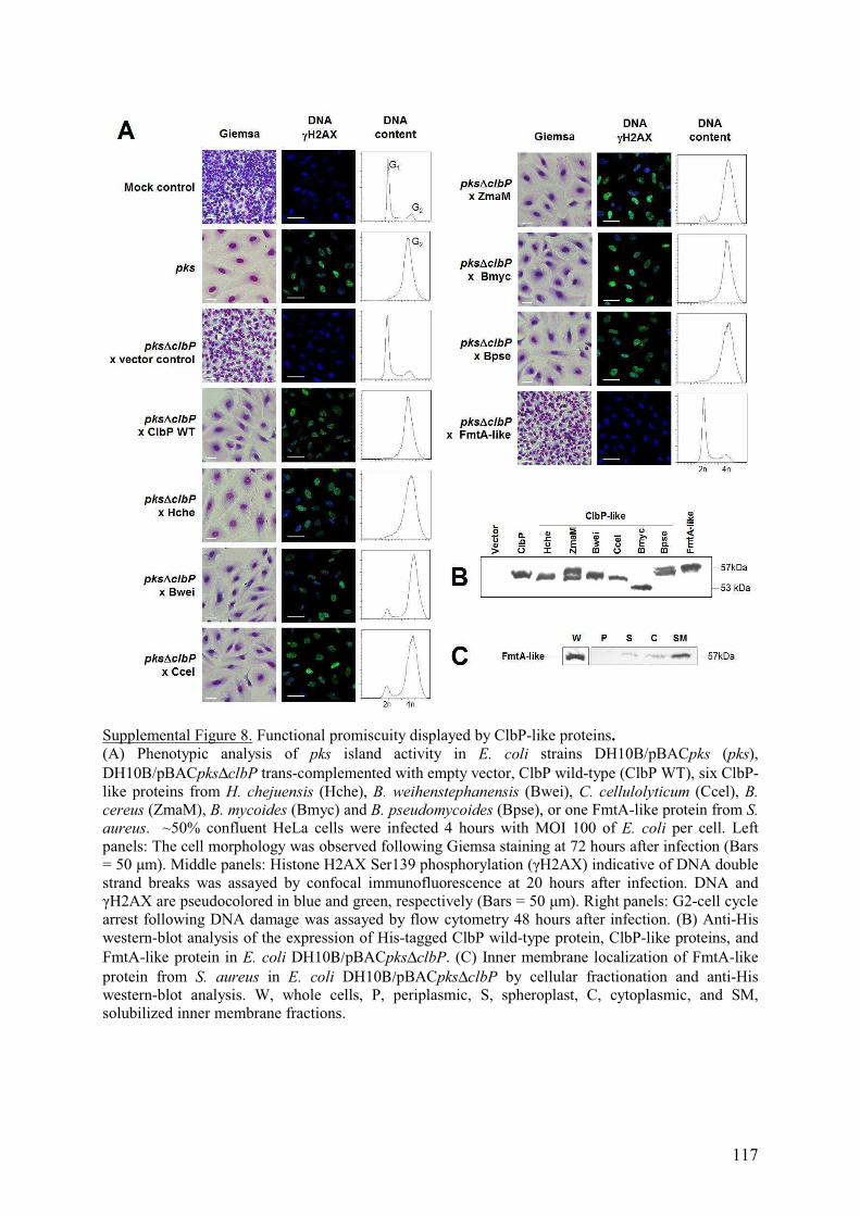
117
Supplemental Figure 8. Functional promiscuity displayed by ClbP-like proteins. (A) Phenotypic analysis of pks island activity in E. coli strains DH10B/pBACpks (pks), DH10B/pBACpks∆clbP trans-complemented with empty vector, ClbP wild-type (ClbP WT), six ClbP-like proteins from H. chejuensis (Hche), B. weihenstephanensis (Bwei), C. cellulolyticum (Ccel), B. cereus (ZmaM), B. mycoides (Bmyc) and B. pseudomycoides (Bpse), or one FmtA-like protein from S. aureus. ~50% confluent HeLa cells were infected 4 hours with MOI 100 of E. coli per cell. Left panels: The cell morphology was observed following Giemsa staining at 72 hours after infection (Bars = 50 �m). Middle panels: Histone H2AX Ser139 phosphorylation (�H2AX) indicative of DNA double strand breaks was assayed by confocal immunofluorescence at 20 hours after infection. DNA and �H2AX are pseudocolored in blue and green, respectively (Bars = 50 �m). Right panels: G2-cell cycle arrest following DNA damage was assayed by flow cytometry 48 hours after infection. (B) Anti-His western-blot analysis of the expression of His-tagged ClbP wild-type protein, ClbP-like proteins, and FmtA-like protein in E. coli DH10Β/pBACpks∆clbP. (C) Inner membrane localization of FmtA-like protein from S. aureus in E. coli DH10Β/pBACpks∆clbP by cellular fractionation and anti-His western-blot analysis. W, whole cells, P, periplasmic, S, spheroplast, C, cytoplasmic, and SM, solubilized inner membrane fractions.

118
ClbP ......aalstlihqrmqeakvpals.vs.vtikg.vr.q.rfvygvadv 39 ClbP-like H. chejuensis ......qtvselt.rrwdasqiptgg.ls.vvvng.qa.h.tlrlgaqea 37 ClbP-like B. cereus (ZmaM) .......kiqkhieeqisqagipgls.vvivkgne.ti.y.kknygyadi 39 ClbP-like B. weihenstephanensis KBAB4 .......qldslikkqmqeakipgms.vi.vvkgd.qa.vykksfgysnl 39 ClbP-like C. cellulolyticum 2 .......riedhihkymksgkipgls.vv.vvkdd.rdiy.kssfgysdl 39 ClbP-like B. cereus BL .......qidqfikeqqelsqipgis.vv.ivekg.kt.vyqkgfgyadl 39 ClbP-like B. mycoides .......aiqallddacrisgvpgms.is.iladd.ev.f.yfssgyadr 38 ClbP-like C. cellulolyticum BL .......kideliqkqmdkskiqglslvi.vsgdk.av.y.kkcygyaei 39 ClbP-like B. brevis .......aieqlvnqtmesekipgaa.vv.vvkeg.ktiy.kqsfgfsdt 39 ClbP-like C. botulinum .......kiekfveenmdkgkipgls.vt.ivkgd.kt.vyqkgfgysdi 39 ClbP-like P. larvae .......kmerwieeqmdkagipgls.vv.isgkd.st.lyqkgfgyagl 39 ClbP-like B. cereus CPT .......kiekfieenmdkgkvpgla.vtlvkgdk.tv.y.qkgfgysdi 39 ClbP-like L. sphaericus .......nielfieeqqaiskipgls.vt.ivdkg.vt.vyqkgfgyani 39 FmtA-like S. aureus .......qidkiiehdlqkghipgas.il.ivkngkvf.l.nkgygyqdv 39 AmpC E. coli apqqindivhrtitplieqqkipgmavaviy.....qgkpyyftwgyadi 45 Peptidase Pab87 ...mdvgklesfivekmaerkvpgisisiik..dg..dvvyakgfgyrnv 43
Motif 1 Motif 7 ClbP asqkantldtvYELGSMSKAFTglvvqiliqegrlrqgDDIITYLPEMrl 88 ClbP-like H. chejuensis .........gvFELASCSKAFTglliavleqegalsrqDAITRWIPELnq 78 ClbP-like B. cereus (ZmaM) dnkrqvtnetlFELGSTSKAFTalavlqlekegkiklsDSLTKYVSWFem 88 ClbP-like B. weihenstephanensis KBAB4 etkqrvtektlFEIGSNSKAFTalaiyqlvqkglidlkDPVSKYLQWFqm 88 ClbP-like C. cellulolyticum 2 krkekvtentlFELGSNSKAFTgigilqlqekglikleDSINKYIPWLkl 88 ClbP-like B. cereus BL ktkkpvnedtlFELASTSKAFTglailqlekegllkqaDDITKFLPWLql 88 ClbP-like B. mycoides ekglsasentlYELASVSKAFTgmgimlleeqgllsmtDPVQKYLPWFtl 87 ClbP-like C. cellulolyticum BL etkkpvtedtlFELGSTSKAFTalgiykledkglinldDSVSKYIPWLkl 88 ClbP-like B. brevis ekqepvtsdtlFEIGSNSKAFTalavhqlveqkrlsldDPVQNYIPWFsv 88 ClbP-like C. botulinum elqksvdseslFEIGSNSKAFTalgildlqknglikfdDEVTKYIPWLkv 88 ClbP-like P. larvae nnkrpvtgrtlFELGSTSKAFTglavlqlqdqgiirlsDPVSAYLPWFkm 88 ClbP-like B. cereus CPT dakkpvtskslFEIGSNSKAVTalgifnleksgqikleDEVTKYIPWLkv 88 ClbP-like L. sphaericus ktktpvtsdtlFELGSTSKAFTglailqlekqgllrrsDDIQTYIPWLtl 88 FmtA-like S. aureus dkkvkaspttkYEIASNTKAFTglailklaqegrlnlnDDVSKHVPHFkm 88 AmpC E. coli akkqpvtqqtlFELGSVSKTFTgvlggdaiargeiklsDPTTKYWPELt. 94 Peptidase Pab87 earlpstpetiYgigsitksftalaimklveegglsldDPVEKFVN.Ikl 91 ♦♦♦♦
Motif 4 ClbP nyqg........r........pa...sltvaDFLYHTSG.....LP.... 111 ClbP-like H. chejuensis rpel........g........ys...airlnNLLFHSSG.....LS.... 101 ClbP-like B. cereus (ZmaM) eykg........k........py...eitlyDLLHHTSG.....IP.... 111 ClbP-like B. weihenstephanensis KBAB4 iyegnykgqqlnk........nv...eitleQLLYHTSG.....IP.... 119 ClbP-like C. cellulolyticum 2 kykg........e........av...ditveQFLHHTSG.....IP.... 111 ClbP-like B. cereus BL tynd........k........pv...kinlnHLLYHTSG.....IP.... 111 ClbP-like B. mycoides kyqg........k........pvdmqsltlnNFLHHTSG.....LTnirh 116 ClbP-like C. cellulolyticum BL nykg........k........sa...dltlnHFLHHTSG.....VS.... 111 ClbP-like B. brevis tykg........q........qe...tilikHLLYHTSG.....IP.... 111 ClbP-like C. botulinum kykg........k........ev...pitieQLVHHTSG.....VS.... 111 ClbP-like P. larvae hfkg........ehqgekidgdv...ditleQLLHHTSG.....IP.... 119 ClbP-like B. cereus CPT kykg........k........ea...sitieQLLHQTSG.....IP.... 111 ClbP-like L. sphaericus tyng........k........ke...iitiqQLLSHTSG.....IP.... 111 FmtA-like S. aureus nyng........q........ne...titikQLLAQTSG.....IPs... 112 AmpC E. coli .....................akqwngitllHLATYTAGglplqVPdevk 122 Peptidase Pab87 r.....................pfgepvtvhhllthssg.....IPslgy 115
ClbP ..fstlarlenpmp.gsav.....a...qq...lr...ne.....n...l 135 ClbP-like H. chejuensis ..gdtldllrpsda.anal.....e...qlpgllr...dt.....p...l 128 ClbP-like B. cereus (ZmaM) ..ekaigyipitng.kdsi.....e...kt...ir...nvmp..ig...l 138 ClbP-like B. weihenstephanensis KBAB4 ..fhtigdipisnd.ndal.....e...nt...vreilnq.....k...l 146 ClbP-like C. cellulolyticum 2 ..fesigyikpdqr.enalent..v...kt...lv...gs.....e...l 138 ClbP-like B. cereus BL ..sttissipissa.ndal.....ettikt...lk...qt.....k...l 138 ClbP-like B. mycoides ..tqnipqgntpdm.lqkt.....v...em...lv...da.....e...l 141 ClbP-like C. cellulolyticum BL ..fltledikpsta.enal.....e...qt...vr...tl..qeie...l 138 ClbP-like B. brevis ..fesigsipisss.enai.....e...et...vr...ai.....nhsel 138 ClbP-like C. botulinum ..fntidkipvsne.dnal..yetv...kt...li...gi.....e...l 138 ClbP-like P. larvae ..fetikdipqgdg.ddslq.r.tv...kn...lv...nr.....e...l 146 ClbP-like B. cereus CPT ..fktidkipvsne.dnal.....e...et...vktlini.....e...l 138 ClbP-like L. sphaericus ..snsivqipesta.knal..eltv...kt...ll...dq.....t...l 138 FmtA-like S. aureus .ditsedavtnknnrlndv.....t...ra...im...gd.....e...l 138 AmpC E. coli sssdllrfyqnwqp.................................... 137 Peptidase Pab87 aeafidgmvggdnwlpvstpeetiafardmekw................a 149

119
Motif 2 ClbP lfapgakfsYASANYDVLGavienvtgktfteviaerltqplgmsatvav 185 ClbP-like H. chejuensis aypvgareeYATLNYSLLGlaaeratgksfavllrekiflplgmkdtfve 178 ClbP-like B. cereus (ZmaM) nrepgssfeYSTVNYDLLGlviekatnqsferyvqdhvlskldllstyag 188 ClbP-like B. weihenstephanensis KBAB4 etypgeqfnYASINYDILGlviqkvtnqsfeqyvqnnilnqfnmgntflf 196 ClbP-like C. cellulolyticum 2 qyypgteysYATINYDVLGllikivtgesyekymhdnivktyglnntflf 188 ClbP-like B. cereus BL nhkpgssfeYATINYDILGlviekvtkkpyevyikqnildqigmknslvg 188 ClbP-like B. mycoides afppgeqynYGTVNYDVLGlvieivsrqsyedfmkeqvfqplglhqtyvy 191 ClbP-like C. cellulolyticum BL dtppgkqfqYATINYDVLGlviekvtgksfedymqdevllpcglnntftt 188 ClbP-like B. brevis vrkpgteyeYATINYDILGyivekvtgetyeqyvqqqviaklgltqtf.l 187 ClbP-like C. botulinum dnepgksfqYATINYDVLGliiqevtgksyenyieenilkpmglsntyly 188 ClbP-like P. larvae dfypgeqfqYATINYDVLGlvieevtgssfetyvrthvldtlglketflf 196 ClbP-like B. cereus CPT dsepgrdfqYATINYDVLGlviekvtgksyegyieenvlkplglhntyly 188 ClbP-like L. sphaericus nrkpgssfeYATINYDVLGliieqvtklpydqyvkqkilqpigmyqsfvg 188 FmtA-like S. aureus hhkpgeefeYSNMNYDLLGliiqnvtkqsytkyitnswlkplhmthtsfk 188 AmpC E. coli awapgtqrlYANSSIGLFGalavkpsglsfeqamqtrvfqplklnhtwi. 186 Peptidase Pab87 vakpgerffYLNTGYVLLGkiiekvsgvsyeeyikkkileplgmnrsyff 199 ♦♦♦
Motif 3 ClbP kgdeiivnk...asgyklg.f.gkpv.aah.ap.lARNHVPAAYIhstlp 227 ClbP-like H. chejuensis gdpaatapr...isgykls.ftvarp.y.e.ap.rYLQNTPAGYIlstpe 220 ClbP-like B. cereus (ZmaM) remapldnm...aqgykrn...flqs.lpydap.nYRGNTPAGYFisnin 230 ClbP-like B. weihenstephanensis KBAB4 rkdvakydm...skgykig.f.lkpief.n.ap.iYRGNTPAGYFisnne 238 ClbP-like C. cellulolyticum 2 reqaskkgv..iakgykvg...flrp.leynap.iYRGNTPAGYLisnin 231 ClbP-like B. cereus BL ylqghpnem...atgyklr.fmrpyp.y.e.qp.iYRGNTPAGYIisntn 230 ClbP-like B. mycoides kedaqatgql..aqgyrssff.mttp.f.k.ap.dYAGNKPAGYIisntk 234 ClbP-like C. cellulolyticum BL rnqavesrm...aagykvs..lmrpv.k.ynap.vFLGNIPAGYFitnin 230 ClbP-like B. brevis kaqapvegm...arghkie.ffspkv.y.d.ap.dYRGNTPAGYVvsnvn 229 ClbP-like C. botulinum rnenvnkhm...akgykvs.flrphe.y.d.ap.iYRGNKPAGYIisnge 230 ClbP-like P. larvae rqetagrdm...adgykhg...fmqs.ltynap.mYRGDTPAGYFitnan 238 ClbP-like B. cereus CPT kneivneyi...akgykle.flkndv.y.d.ap.vYRGNKPAGYIissae 230 ClbP-like L. sphaericus ihqvafphm...asgykig...lmre.lpftpp.iYRGNIPAGYIisnan 230 FmtA-like S. aureus qtnnkskhd...aigyelq.g.stpv.v.s.kpefNLWDTPSAYMmtste 230 AmpC E. coli ..nvppaeeknyawgyreg.......kavhvspgaL..DAEAYGVkstie 224 Peptidase Pab87 keevekdk..dvamgyildkegr...lvpqpfpygITAD...GGLlssvl 241
Motif 6 ClbP DMEIWIDAWLh.rkalpatlrqgmsnswrgnsdv................ 260 ClbP-like H. chejuensis DMARWLQFLLq.ppklsdsrldvlqwa.sqqakr................ 252 ClbP-like B. cereus (ZmaM) DMERWLKLQLg.vekprmfpeelindsheanrkv................ 263 ClbP-like B. weihenstephanensis KBAB4 DMEKWLRMQLg.iyglsddhqkaiysthipnrsv................ 271 ClbP-like C. cellulolyticum 2 DIAGWLKIQM..gtsnsvndkkliqeshipnrsv................ 263 ClbP-like B. cereus BL DLAKWMNLQLg.ieinnsihtqlisdshipdktv................ 263 ClbP-like B. mycoides DMARWMGIQMgivqdipeifhtvikkshr.gdm................. 266 ClbP-like C. cellulolyticum BL DVQRWLSLHLg.siklpsidyklmeksheidtsvephdi........... 268 ClbP-like B. brevis DMERWLQIQLg.le.asafrpelvrasqlpdrsv................ 261 ClbP-like C. botulinum DMGKWLKIQMg.tlsdlkfhknlieeshnandti................ 263 ClbP-like P. larvae DMSKWLQIQLg.sgdgginrligqshs.p.drtv................ 269 ClbP-like B. cereus CPT DMAKWLKIQLg.tlndlkfdkdivgksqeanrtv................ 263 ClbP-like L. sphaericus DIAKWLKLQLg.nseiysidattikeshnpdqtv................ 263 FmtA-like S. aureus DLEHWIKFQLn.ppdkykslvq..q.shknlsst................ 260 AmpC E. coli DMARWVQSNLkpldinektlqqgiqlaqsrywqtgdmyqglgwemldwpv 274 Peptidase Pab87 DLAKYLKMYIerdesivs..keyiekmetsyik................v 273
Motif 8 Motif 5 ClbP pla.....adn....riLYASGW..FIDQn..qgpyisHGGQNPNFSSci 297 ClbP-like H. chejuensis pyg.....gds....daGYAYGW..DVEAg..attswsHPGQNPNAGAyv 289 ClbP-like B. cereus (ZmaM) apd.....n.n....gaSYAAGW..EVYQs..gggeisHGGSNPNYSSyi 299 ClbP-like B. weihenstephanensis KBAB4 pps.....edg.....sSYAGGW..QVFQn..gpgeisHAGSNPNFSSfv 307 ClbP-like C. cellulolyticum 2 rpy.....vdg.....sSYAAGW..SVFQs..gggelsHGGSNPNYSSfi 299 ClbP-like B. cereus BL qpi.....knn.....dYYASGW..IVRQe..dtksilHSGENPNFSSfi 299 ClbP-like B. mycoides svs....av.n....gmYYAAGW..SVNAe...qtiieHTGGNPNFRTev 302 ClbP-like C. cellulolyticum BL gvl.....ydk....geRYGAGW..ERAEg..rngf.fHGGANPNYSSym 304 ClbP-like B. brevis apa.....adg.....fSYASGW..MIYQd..gggqisHGGNNPAFSSym 297 ClbP-like C. botulinum apl.....rdg.....sSYASGW..LVYEk..gsreisHAGNNPNYSSfi 299 ClbP-like P. larvae ppa.....edg.....sSYAAGW..SVYQl..gsgmlsHSGSNPNYSSql 305 ClbP-like B. cereus CPT rpl.....sdg.....sFYASGW..FIYPk..senqisHAGNNPNYSSfi 299 ClbP-like L. sphaericus epf.....d.t....ntYYATGWA.VVEKe..nkpyiyHAGENPTFTSyf 300 FmtA-like S. aureus ig.....ep.n....anAYASGW..FTN.n..dehlvfHSGTLDNFSSfi 295 AmpC E. coli npdsiingsdnkialaaRPVKAITPPTPAv..raswvhKTGATGGFGSyv 322 Peptidase Pab87 p...........weifgGEGYGYGLIIYPnflgeklvgHSGSVGMYTGyi 312 ♦♦♦

120
ClbP alrpdqqigivalan.mns...n...l...ilql...cadidnyl..... 330 ClbP-like H. chejuensis afdpqtgvgvallgn.sns...p...q...viql...gravfehl..... 322 ClbP-like B. cereus (ZmaM) gfrdeeqigvvvlan.lns...n...y...tyyi...gssllnmm...id 333 ClbP-like B. weihenstephanensis KBAB4 vfhpqeklgvavman.mns...d...y...tqni...gqaimdtl.vge. 343 ClbP-like C. cellulolyticum 2 vfrpeeklgvavlan.lns...s...y...vqtl...gqgiidiild.r. 335 ClbP-like B. cereus BL imqpnkqtgvavlsn.mks...a...vttsigqg...vidlwngk..... 335 ClbP-like B. mycoides aillnertavsllsngant...n...i...nmvlk..vkdilegn..... 337 ClbP-like C. cellulolyticum BL firpdqgmgigmlcn.fgs...d...t...iqilgknlvdiidgn..... 340 ClbP-like B. brevis vfrpeeklgavvltn.lnslnas...v...iaeg...avnlvlgk..... 333 ClbP-like C. botulinum lfrpedkvgvailsn.tss...q...y...ipai...tqgineil.q.g. 334 ClbP-like P. larvae vllpgeeigiavlan.lns...d...y...tevi...gngiaail..... 338 ClbP-like B. cereus CPT vfnpkdkvgvavlsn.tns...q...y...veli...gqginkil..... 332 ClbP-like L. sphaericus imqpdeqigvailan.mnt...sstta...igqg...imdlwegk..... 336 FmtA-like S. aureus llnpkqnygivvlan.lns...e...y...vpkl...vehlntqi..... 328 AmpC E. coli afipekelgivmlanknyp...........nparvdaawqilnalq.... 358 Peptidase Pab87 gyipekkigvavlenssg..........yppsyiamyalalllgkn.... 349
ClbP .r.igk......y... 335 ClbP-like H. chejuensis .r.gas.p..qpl... 330 ClbP-like B. cereus (ZmaM) kevk.p......l... 340 ClbP-like B. weihenstephanensis KBAB4 .s.vvt......n... 348 ClbP-like C. cellulolyticum 2 .e.taa......p... 340 ClbP-like B. cereus BL .n.isd......t... 340 ClbP-like B. mycoides .l.tqs......y... 342 ClbP-like C. cellulolyticum BL .g.eps......l... 345 ClbP-like B. brevis .e.apv......v... 338 ClbP-like C. botulinum .rvynk......d... 340 ClbP-like P. larvae .q.gka..pep.l... 346 ClbP-like B. cereus CPT .e.egt......ynkn 340 ClbP-like L. sphaericus .t.ttp......l... 341 FmtA-like S. aureus .v.nhkry..s.t... 336 AmpC E. coli ................ Peptidase Pab87 ................
Supplemental Figure 9. Sequence alignment of NRP-associated peptidases with FmtA-like protein, AmpC β-lactamase and Pab87 peptidase. The sequences corresponding to motifs are in upper case. Residues emphasized by black shading are 100% conserved in NRP-associated peptidases. PRP specific motifs are indicated by black diamonds.
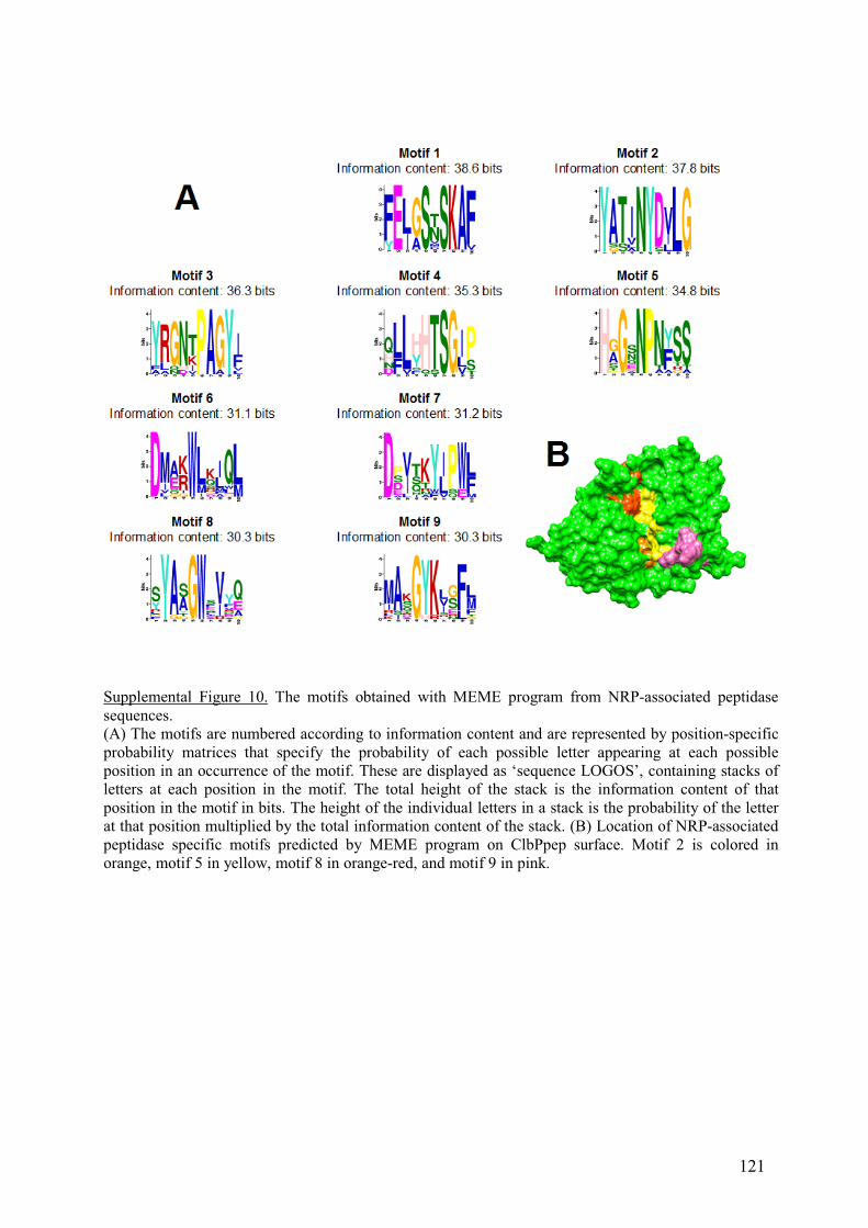
121
Supplemental Figure 10. The motifs obtained with MEME program from NRP-associated peptidase sequences. (A) The motifs are numbered according to information content and are represented by position-specific probability matrices that specify the probability of each possible letter appearing at each possible position in an occurrence of the motif. These are displayed as ‘sequence LOGOS’, containing stacks of letters at each position in the motif. The total height of the stack is the information content of that position in the motif in bits. The height of the individual letters in a stack is the probability of the letter at that position multiplied by the total information content of the stack. (B) Location of NRP-associated peptidase specific motifs predicted by MEME program on ClbPpep surface. Motif 2 is colored in orange, motif 5 in yellow, motif 8 in orange-red, and motif 9 in pink.

122
Supplemental Figure 11. Electrostatic potential surface map of MEROPS S12 peptidases, and their putative and known ligands including the crystal structures of ClbPpep (A), Pab87 peptidase (B) (23), aminopeptidase (C) (24), R61 carboxypeptidase (D) (19), and AmpC � -lactamase of E. coli (E) , the homology models of NRP-associated peptidases of H. chejuensis (F), B. cereus (ZmaM) (G), B. weihenstephanensis (H), C. cellulolyticum (I), B. pseudomycoides (J), B. mycoides (K), Lysinibacillus sphaericus C3-41 (L), and of FmtA-like protein of S. aureus (M), and the NRP-associated peptidase putative ligands (N, O) and known ligands of the MEROPS S12 enzymes AmpC and carboxypeptidases (P-S): (N) zwittermicin A, (O) pre-zwittermicin, (P) benzylpenicillin, (Q) cephalothin, (R) NAG-NAM-pentapeptides of Gram negative bacteria, and (S) NAG-NAM-pentapeptides of Gram positive bacteria. The catalytic pockets are indicated by green circles. Electrostatic surface potential in blue is positive, negative in red and neutral in white.

123
Supplemental Figure 12. Model of secretion and maturation of NRP compounds involving NRP-associated peptidases. As a two-component resistance mechanism, after translocation of an immature inactive NRP compound from cytoplasm to periplasm via efflux pumps such as Na+/H+ energy-driven MATE exporters or ATP-driven ABC exporters, the periplasmic location of the NRP-associated peptidase main domain may allow the maturation of the fully active NRP toxin, away from the potential cytoplasmic target of its producer cell. ABC exporters may be linked covalently to NRP peptidase as with B. cereus ZmaM. (Illustration made with the assistance of Servier Medical Art.)

124
Supplemental Tables
Supplemental Table 1. Data collection and structure determination statistics for ClbPpep crystal structure.
Data collection Space group C2 Cell dimensions a, b, c (Å)� 103.9, 149.9, 87.3 α, β, γ (°) 90.0, 123.9, 90.0 Resolution (Å) 20 - 2.4 (2.5 – 2.4)a
Rmerge (%)b 8.6 (28.0) a
[I / σ(I)] (°) 11.5 (3.7) a
Completness (%) 90.9 (94.3) a
Redundancy 3 (3) a
Refinement No. reflections 39,788 Rwork / Rfree 19.85/ 22.47 No. atoms Protein 7719 Water 249 B-factors Protein 22.6 Water 19.2 R.m.s deviations Bond lengths (Å) 0.005 Bond angle (o) 0.919 Ramanchandran plot: Favored region (%) Allowed region (%)
97.2 2.8
a Values in parentheses represent the highest resolution shells.

125
Supplemental Table 2. The 20 best non-redondant neighbours of ClbPpep structure recovered from DALI server.
PDP IDa Z-scoreb RMSDc
(Å) Aligned
residuesdSequence
identity (%) Description
2r9w 37.6 2.1 311 25 AmpC beta-lactamase 2qmi 37.2 2.6 324 24 Pab87 peptidase 2zc7 37.1 2.1 310 25 AmpC beta-lactamase 1fr1 36.9 2.2 311 24 AmpC beta-lactamase 2qz6 36.7 2.2 306 23 AmpC beta-lactamase 1zkj 36.1 2.4 310 21 AmpC beta-lactamase 1ga0 35.3 2.4 311 24 AmpC beta-lactamase 1ei5 34.5 2.5 310 22 D-Aminopeptidase 1ci8 34.0 2.6 315 21 Carboxylesterase 1cef 33.6 2.6 306 22 D-Ala-D-Ala Carboxypeptidase 2efu 33.0 2.8 306 18 D-Amino Acid Amidase 2dns 33.0 2.7 309 18 D-Amino Acid Amidase 1wyc 26.7 3.0 283 21 6-Aminohexanoate-dimer hydrolase 3i7j 25.0 2.7 248 20 AmpC beta-lactamase 1e25 16.7 3.3 217 11 PER-1 beta-lactamase 1hzo 15.9 3.3 212 8 PVUL beta-lactamase 1iyq 15.8 2.9 206 11 TOHO-1 beta-lactamase 1ylj 15.8 3.1 207 12 CTX-M-9 beta-lactamase 3byd 15.7 3.0 205 12 OXY-1 beta-lactamase 3bfd 15.6 3.0 204 11 SED-1 beta-lactamase a, PDP identity of the subject structure in RCSB protein data bank; b, quality of alignement ; c, root-mean square deviation of Cα atoms; d, Number of aligned residues.

126
Supplemental Table 3. Strains used in this study.
Strain or Plasmid Genotype/Description Source E. coli
DH5αF’ F´/endA1 hsdR17 (rK–mK+) glnV44 thi-1recA1 gyrA (Nalr) relA1 �(lacIZYAargF)U169 deoR (�80dlac�(lacZ)M15)
Invitrogen, France
BL21(DE3) F– ompT gal [dcm] [lon] hsdSB (rB – mB –; an E. coli B strain) with DE3, a � prophage carrying the T7 RNA polymerase gene
Novagen, Darmstadt, Germany
TG1 ∆(lac-pro) supE thi hsdD5/F' traD36 proA+B+ lacIq lacZ∆M15
Amersham Pharmacia biotech, France
IHE3034 Extra-intestinal pathogenic E. coli O18:K1:H7 (1) DH10B pVector DH10B containing the BAC vector pBeloBAC11 1
DH10B pBACpks DH10B containing pBACpks (1)
DH10B pBACpks∆clbP
DH10B containing pBACpks∆clbP (1)
MC4100A F’lac∆(lacZYA-argF)U169 araD139 rpsL150 thi flbB5301 deoC7 ptsF25 relA1 rbsR ara+
(5)
HPT406A ∆SRP isogenic mutant of MC4100A (25) CU164A secY39-cs, zhd-33::Tn10, ara � + (5) MC1060 F- DE(codB-lacI)3 galK16 galE15 LAM- e14- mcrA0 relA1
rpsL150(strR) spoT1 mcrB1 hsdR2 (3)
MYC-cs cold-sensitive yidC derivative of MC1060 (3) MC1000 araD139 (ara, leu)7697 lacX74 galU galK atrA (26) YLS-3 ∆mreB isogenic mutant of MC1000 (26) YLS-24 ∆ftsZ isogenic mutant of MC1000 (21)
Bacillus
weihenstephanensis KBAB4
bearing clbP-like gene (locus tag “BcerKBAB4_1783”) (27)
Bacillus cereus UW85 bearing clbP-like gene (locus tag “zmaM”) (17) Hahella chejuensis KCTC 2396T
bearing clbP-like gene (locus tag “HCH_03446”) (28)
Bacillus mycoides DSM 2048
bearing clbP-like gene (locus tag “bmyco0001_51990”) DSMZ
Bacillus pseudomycoides
DSM 12442 Bearing clbP-like gene (locus tag “bpmyx0001_2150”) DSMZ
Clostridium
cellulolyticum H10 bearing clbP-like genes (locus tag “Ccel_0973” and“Ccel_2389”)
DSMZ
Staphylococcus aureus
Mu50 bearing fmtA-like gene (locus tag “SAV2441”) CIP

127
Supplemental Table 4. Plasmids used in this study.
Plasmids Description Source pBeloBAC11 (or pVector) CmR New England
BioLabs pBACpks pBeloBAC11 harboring the pks island (1) pBACpks∆clbP pBACpks island mutated for clbP (1) p8761 pBAD24 encoding TorA twin-arginine signal peptide + C-
terminal in-frame GFPmut2, Para
(4)
p61ClbPssGFP pBAD24 encoding ClbP signal peptide plus 6 amino acid (1-36) + C-terminal in-frame GFPmut2, Para
This study
pET9a T7 promoter, KanR Novagen pClbPpep pET9a encoding ClbP peptidase domain (ClbPpep : amino acid
35 to 375) This study
pClbPpepY186E pET9a encoding the mutant Y186E of ClbPpep This studypMB702 pCRScript derivative encoding clbP plus 570 bp upstream (1) pBRSK pBR328 derivative containing Plac and PT7 promoters, and the
multiple cloning site from the Bluescript SK vector (Stratagene) (2)
pOB901 pBRSK encoding ClbP This study pOB902 pOB901 derivative encoding the mutant S95A of ClbP This study pOB903 pOB901 derivative encoding the mutant K98T of ClbP This study pOB904 pOB901 derivative encoding the mutant Y186G of ClbP This study pASK-IBA33plus Tetracycline promoter, C-terminal in-frame RGS6His tag, AmpR IBA GmbH,
Germany p33ClbP pASK-IBA33plus encoding clbP- RGS6His This study pS95A p33ClbP derivative encoding the mutant S95A of ClbP This study pE159K p33ClbP derivative encoding the mutant E159K of ClbP This study pY186E p33ClbP derivative encoding the mutant Y186E of ClbP This studypS188A p33ClbP derivative encoding the mutant S188A of ClbP This study pH257A p33ClbP derivative encoding the mutant H257A of ClbP This study pF316R p33ClbP derivative encoding the mutant G328S F316R of ClbP This study pG328S p33ClbP derivative encoding the mutant G328S of ClbP This study pN331A p33ClbP derivative encoding the mutant N331A of ClbP This study pC337A p33ClbP derivative encoding the mutant C337A of ClbP This study p33BweihenstephanensisT pASK-IBA33plus encoding ClbP-like-encoding sequence of the
locus tag BcerKBAB4_1783 This study
p33ZmaMT pASK-IBA33plus encoding ClbP-like-encoding sequence of the locus tag zmaM
This study
p33HchejuensisT pASK-IBA33plus encoding ClbP-like-encoding sequence of the locus tag HCH_03446
This study
p33Ccellulolyticum2T pASK-IBA33plus encoding ClbP-like-encoding sequence of the locus tag Ccel_2389
This study
p33Bmycoides1 pASK-IBA33plus encoding ClbP-like-encoding sequence of the locus tag bmyco0001_51990
This study
p33Bpseudomycoides pASK-IBA33plus encoding ClbP-like-encoding sequence of the locus tag bpmyx0001_2150
This study
p33FmtA-like pASK-IBA33plus encoding ClbP-like-encoding sequence of the locus tag SAV2441
This study

128
Supplemental Table 5. Primers used in this study for clonings (with restriction sites bolded and underlined).
Primer name Sequence Target
clbPMK-f AACCAATTGGTACCATGACAATAATGGAACACGTTAGC ClbP-encoding sequence
clbPBN-r AGCGGATCCGCTAGCCTCATCGTCCCACTCCTTGTTG ClbP-encoding sequence
clbPssBN-r AGCGGATCCGCTAGCGATAGGCTCATGTTCTTGCGCC ClbP signal sequence-encoding sequence
clbP-NdeI2-f GGAATTCCATATGCCTATCGGGGCGCAAGATG ClbPpep-encoding sequence
clbP-NotI2-r AGCATAGTTTAGCGGCCGCCTTAATATTTGCCAATGCGCAGA ClbPpep-encoding sequence
KBAB4_1783-MfeI-f AACCAATTGGTACCATGATGAGAGTAAAAATTGTTTTG locus tag BcerKBAB4_1783
KBAB4_1783-XhoI-r AGCCTCGAGGCTAGCATTTTTCTTATTATGCAAAGG locus tag BcerKBAB4_1783
ZmaM-EcoRI-f AACGAATTCGTACCATGAAGTTAAACATATGGTTGAAG locus tag zmaM
ZmaM-BamHI-r AGCGGATCCGCTAGCAAGCGACTTTCCCTTTTGTCTTGG locus tag zmaM
HCH_3446-EcoRI-f AACGAATTCGTACCATGAATAATGTCGGCTTAATCC locus tag HCH_03446
HCH_3446-BamHI-r AGCGGATCCGCTAGCCAGCGACGAAAAGGTTTTGCTG locus tag HCH_03446
Ccel_2389-MfeI-f AACCAATTGGTACCATGAAATTAACAAAAAATGG locus tag Ccel_2389
Ccel_2389-BamHI-r AGCGGATCCGCTAGCAAAGAGAAAGTTCAAATT locus tag Ccel_2389
Bmy_51990-EcoRI-f AACGAATTCGTACCATGAAAAAGATTATTG locus tag bmyco0001_51990
Bmy_51990-BamHI-r AGCGGATCCGCTAGCTCGGTGAGTGTATACAAACC locus tag bmyco0001_51990
Bpmy_2150-EcoRI-f AACGAATTCGTACCATGAATAAAAAGCAAAGAAAAC locus tag bpmyx0001_2150
Bpmy_2150-BamHI-r AGCGGATCCGCTAGCAGAAATAAATCTATTCATTTC locus tag bpmyx0001_2150
SAV_2441-EcoRI-f AACGAATTCGTACCATGACTACTAAAAAACTG locus tag SAV2441
SAV_2441-BamHI-r AGCGGATCCGCTAGCTGTCTTCGTTATCTTTG locus tag SAV2441

129
Supplemental Table 6. Primers used in this study for site-directed mutagenesis of clbP gene (with mutated bases bolded and underlined).
Mutation Primer name Sequence
S95A IHAPJPN105 gatgagtaaggcgtttaccgg
IHAPJPN106 gctcccagctcgtaaactgtg
K98T IHAPJPN107 tgcgtttaccggacttgtggtg
IHAPJPN108 gtactcatcgatcccagctcgta
E159K clbP-E159Kf caacactggctcggctgaaaaaccctatgcctgg
clbP-E159Kr ccaggcatagggtttttcagccgagccagtgttg
Y186G IHAPOB3 Y186G cgcctccgccaattatgatgtgttgggcgcggtgattgaaaatgtgacgggaaaaacctttac
IHAPOB4 ccgctaaacttagcacccggagcaaacagcagattctcgttgcgcagttg
Y186E clbP-Y186Ef gaagtttagcgaagcctccgccaattatgatgtgttgggc
clbP-Y186Er cataattggcggaggcttcgctaaacttcgcacccggcgc
S188A clbP-S188Af gtttagctatgccgccgccaattatgatgtgttgggcgcgg
clbP-S188Ar catcataattggcggcggcatagctaaacttcgcacccggcgc
H257A clbP-H257Af ggcccggaacgctgttcctgccgcctatatccatagcactctgcc
clbP-H257Ar ggcggcaggaacagcgttccgggccagaggcgcatgaaacagaacgg
F316R/G328S clbP-G328Ss cccttacatcagtcacagtgggcagaatccaaacttttcttcttgcattgcg
clbP-G328SF316Rr cccactgtgactgatgtaagggccctgattctggtcgatcctccaaccgc
G328S clbP-G328Sf gcccttacatcagtcacagtgggcagaatccaaac
clbP-G328Sr gtttggattctgcccactgtgactgatgtaagggc
N331A clbP-N331Af cacggtgggcaggctccaaacttttcttcttgcattgcgttgcgaccg
clbP-N331Ar gaagaaaagtttggagcctgcccaccgtgactgatgtaagggccttgattctgg
C337A clbP-C337Af gcagaatccaaacttttcttctgccattgcgttgcgaccggatcagc
clbP-C337Ar gctgatccggtcgcaacgcaatggcagaagaaaagtttggattctg
130
Supplemental References
1. Nougayrede, J. P., Homburg, S., Taieb, F., Boury, M., Brzuszkiewicz, E., Gottschalk, G., Buchrieser, C., Hacker, J., Dobrindt, U., and Oswald, E. (2006) Science 313(5788), 848-851
2. Schmitt, C. K., Darnell, S. C., Tesh, V. L., Stocker, B. A., and O'Brien, A. D. (1994) J
Bacteriol 176(2), 368-377 3. Chen, M., Xie, K., Jiang, F., Yi, L., and Dalbey, R. E. (2002) Biol Chem 383(10), 1565-1572 4. Pradel, N., Delmas, J., Wu, L. F., Santini, C. L., and Bonnet, R. (2009) Antimicrob Agents
Chemother 53(1), 242-248 5. Santini, C. L., Ize, B., Chanal, A., Muller, M., Giordano, G., and Wu, L. F. (1998) Embo J
17(1), 101-112 6. Bailey, T. L., and Elkan, C. (1994) Proc Int Conf Intell Syst Mol Biol 2, 28-36 7. Bailey, T. L., Williams, N., Misleh, C., and Li, W. W. (2006) Nucleic Acids Res 34(Web
Server issue), W369-373 8. Bailey, T. L., and Gribskov, M. (1998) Bioinformatics 14(1), 48-54 9. Eswar, N., Webb, B., Marti-Renom, M. A., Madhusudhan, M. S., Eramian, D., Shen, M. Y.,
Pieper, U., and Sali, A. (2006) Curr Protoc Bioinformatics Chapter 5, Unit 5 6 10. Sali, A., and Blundell, T. L. (1993) J Mol Biol 234(3), 779-815 11. Dolinsky, T. J., Nielsen, J. E., McCammon, J. A., and Baker, N. A. (2004) Nucleic Acids Res
32(Web Server issue), W665-667 12. Hinsen, K. (2000) J Comp Chem 21, 79-85 13. Pettersen, E. F., Goddard, T. D., Huang, C. C., Couch, G. S., Greenblatt, D. M., Meng, E. C.,
and Ferrin, T. E. (2004) J Comput Chem 25(13), 1605-1612 14. Case, D. A., Darden, T. A., Cheatham, I., T.E. , Simmerling, C. L., Wang, J., Duke, R. E.,
Luo, R., Crowley, M., Walker, R. C., Zhang, W., Merz, K. M., Wang, B., Hayik, S., Roitberg, A., Seabra, G., Kolossváry, I., Wong, K. F., Paesani, F., Vanicek, J., Wu, X., Brozell, S. R., Steinbrecher, T., Gohlke, H., Yang, L., Tan, C., Mongan, J., Hornak, V., Cui, G., Mathews, D. H., Seetin, M. G., Sagui, C., Babin, V., and Kollman, P. A. (2008) AMBER 10. In., University of California, San Francisco, CA, USA
15. Laskowski, R. A., MacArthur, M. W., Moss, D. S., and Thornton, J. M. (1993) J App Cryst26, 283-291
16. Baker, N. A., Sept, D., Joseph, S., Holst, M. J., and McCammon, J. A. (2001) Proc Natl Acad
Sci U S A 98(18), 10037-10041 17. Kevany, B. M., Rasko, D. A., and Thomas, M. G. (2009) Appl Environ Microbiol 75(4), 1144-
1155 18. Gasteiger, J., Rudolph, C., and Sadowski, J. (1990) Tetrahedron Comp Methodology 3, 537-
547 19. Beadle, B. M., Trehan, I., Focia, P. J., and Shoichet, B. K. (2002) Structure 10(3), 413-424 20. Delfosse, V., Girard, E., Birck, C., Delmarcelle, M., Delarue, M., Poch, O., Schultz, P., and
Mayer, C. (2009) PLoS One 4(3), e4712 21. Yuan, J., Zweers, J. C., van Dijl, J. M., and Dalbey, R. E. (2010) Cell Mol Life Sci 67(2), 179-
199 22. Thomas, J. D., Daniel, R. A., Errington, J., and Robinson, C. (2001) Mol Microbiol 39(1), 47-
53 23. Bompard-Gilles, C., Remaut, H., Villeret, V., Prange, T., Fanuel, L., Delmarcelle, M., Joris,
B., Frere, J., and Van Beeumen, J. (2000) Structure 8(9), 971-980 24. Lee, W., McDonough, M. A., Kotra, L., Li, Z. H., Silvaggi, N. R., Takeda, Y., Kelly, J. A.,
and Mobashery, S. (2001) Proc Natl Acad Sci U S A 98(4), 1427-1431 25. Tian, H., and Beckwith, J. (2002) J Bacteriol 184(1), 111-118 26. Shih, Y. L., Kawagishi, I., and Rothfield, L. (2005) Mol Microbiol 58(4), 917-928 27. Lapidus, A., Goltsman, E., Auger, S., Galleron, N., Segurens, B., Dossat, C., Land, M. L.,
Broussolle, V., Brillard, J., Guinebretiere, M. H., Sanchis, V., Nguen-The, C., Lereclus, D., Richardson, P., Wincker, P., Weissenbach, J., Ehrlich, S. D., and Sorokin, A. (2008) Chem Biol Interact 171(2), 236-249

131
28. Jeong, H., Yim, J. H., Lee, C., Choi, S. H., Park, Y. K., Yoon, S. H., Hur, C. G., Kang, H. Y., Kim, D., Lee, H. H., Park, K. H., Park, S. H., Park, H. S., Lee, H. K., Oh, T. K., and Kim, J. F. (2005) Nucleic Acids Res 33(22), 7066-7073

113

133
II. Article 2 – Les cyclomodulines des E. coli d’urosepsis
Cyclomodulins in Urosepsis Strains of Escherichia coli.
Damien Dubois, Julien Delmas, Anne Cady, Frédéric Robin, Adeline Sivignon, Eric Oswald
et Richard Bonnet.
Journal of Clinical Microbiology (2010), Vol. 48 (6), p2122-9.
L’ilot pks est quasi exclusivement restreint aux souches de E. coli de groupe
phylogénétique B2. Les ExPEC et en particulier les UPEC appartiennent majoritairement au
phylogroupe B2 et dans une moindre mesure au phylogroupe D [21]. En raison de la
découverte récente de cet ilot génomique pks, aucune donnée épidémiologique n’avait été
rapportée dans un contexte d’infection urinaire, E. coli restant le premier micro-organisme
responsable d’infections urinaires nosocomiales comme communautaires.
Nous avons déterminé la prévalence des gènes codant pour les quatre types de
cyclomodulines (CM) retrouvés chez E. coli, à savoir l’ilot pks codant la colibactine, les
différents types de cnf et de cdtB et le gène cif, dans une série de 146 souches isolées
d’hémocultures de patients ayant présenté un épisode d’urosepsis communautaire et admis
aux CHU de Clermont-Ferrand ou de Rennes, comparée à une série de 51 souches fécales
commensales. Ces dernières étaient issues d’individus sains communautaires de la région
clermontoise n’ayant présenté aucun signe d'infection aiguë, ou de troubles bénins ou malins
du tractus gastro-intestinal et n'ayant reçu aucun antibiotique dans le mois précédent le
prélèvement.
Le groupe phylogénétique de chaque souche de E. coli a été déterminé par PCR
multiplex. Le phylogroupe B2 était prédominant dans les souches isolées d’urosepsis (54%),
suivi par les phylogroupes D (23%), A (18%) et B1 (5%), comme précédemment observé
dans plusieurs études. Les isolats commensaux appartenaient le plus fréquemment au
phylogroupe A (37%), suivi des phylogroupes B1 et B2 (24 % pour les deux groupes),
résultats concordants avec certaines études précédentes mais différant d’autres études
françaises et d’autres pays industrialisés où le phylogroupe B2 prédominait dans les isolats
commensaux. Ainsi le phylogroupe A, et dans une moindre mesure le phylogroupe B1 étaient

134
significativement plus prévalents parmi les souches commensales que parmi les souches
d’origine urinaire (37% versus 18%, p=0,011 ; et 24% versus 5% p<0,001). En revanche, le
phylogroupe B2 était significativement prédominant dans les souches urinaires (54% versus
24%, p<0,001).
Les gènes codant les différentes CM ont été dépistés par hybridation d’ADN dot-blot à
l’aide de quatre sondes ou mélange de sondes détectant les gènes médians clbJ-K de l’ilot pks,
le gène cif et les différents types de gènes de cnf et de cdtB. Les gènes détectés par
hybridation ont été confirmés par PCR en ciblant les gènes des extrémités de l’ilot pks, clbA
et clbQ, le gène cif, et en typant les gènes cnf (par PCR multiplex mise au point au cours de
cette étude) et les gènes cdtB. La distribution des gènes des CM a été analysée en fonction des
phylogroupes. Les gènes codant les différentes CM les plus fréquents dans la cohorte étaient
l’ilot pks (27%) et le cnf1 (18%), qui restaient fortement liés au phylogroupe B2. Ces deux
facteurs de virulence se retrouvaient fréquemment associés dans le même isolat. Les gènes
ctdB (6%) et cif (1%) étaient faiblement réprésentés et ne présentaient pas de distribution
phylogénétique particulièrement significative. Les gènes cnf2 et cnf3 étaient absents.
L’ilot pks, le gène cnf1 et l’association de ces deux facteurs de virulence étaient
prédictifs d’une origine uroseptique de l’isolat (p=0,005, p<0,001 et p=0,001 respectivement).
Cependant, la prévalence de l’ilot pks parmi les isolats du phylogroupe B2 commensaux et
uroseptiques n’était significativement pas différente, contrairement à celle du gène cnf1,
suggérant que la colibactine est importante pour le processus de colonisation des épithélia et
CNF1 pour la virulence. En outre, l’absence de gènes codant les différentes CM associée aux
phylogroupes B1 ou A était significativement prédictive d’une origine commensale (p=0,003
et p=0,018 respectivement). Les gènes cdtB et cif ne présentaient pas de distribution clinique
particulière.
L’expression phénotypique des CM a été investiguée sur culture cellulaire. Les isolats
possédant les gènes cnf1 et cdtB produisaient une CM fonctionnelle. Un seul des deux isolats
cif-positifs était cytopathogène. En raison du caractère hémolytique de nombreuses souches
pks-positives, seulement 40% de ces souches étaient analysables. Parmi celles-ci, 14% soit 3
souches sur 21 ont présenté un ilot pks non fonctionnel ; deux d’entre elles provenaient des
fécès, soit 33% des souches pks-positives isolées de fécès. Ce défaut de fonctionnalité de l’ilot
pks peut induire une surestimation de la prévalence du facteur de virulence « colibacine » lors
des analyses génétiques.
135
Enfin, nous avons analysé la prévalence des résistances aux antibiotiques
habituellement utilisés en pratique clinique parmi les isolats d’origine urinaire, la résistance
étant décrite par plusieurs auteurs comme opposée à la virulence. Les isolats des
phylogroupes A étaient significativement plus résistants aux fluoroquinolones, à
l’amoxicilline et aux céphalosporines de 3ème génération. Les isolats des phylogroupes B2
étaient significativement plus sensibles à ces mêmes antibiotiques et au cotrimoxazole. De
même, les isolats pks-positifs et cnf1-positifs étaient significativement plus sensibles aux
fluoroquinolones, à l’amoxicilline et au cotrimoxazole, et à l’amoxicilline et au cotrimoxazole,
respectivement.

136

JOURNAL OF CLINICAL MICROBIOLOGY, June 2010, p. 2122–2129 Vol. 48, No. 60095-1137/10/$12.00 doi:10.1128/JCM.02365-09Copyright © 2010, American Society for Microbiology. All Rights Reserved.
Cyclomodulins in Urosepsis Strains of Escherichia coli£
Damien Dubois,1,2 Julien Delmas,1,2 Anne Cady,3 Frederic Robin,1,2 Adeline Sivignon,2
Eric Oswald,4,5,6,7 and Richard Bonnet1,2*
CHU Clermont-Ferrand, Centre de Biologie, Laboratoire de Bacteriologie, Clermont-Ferrand F-63003,1 Clermont Universite,Universite d’Auvergne, Evolution des bacteries pathogenes et susceptibilite de l’hote, JE 2526 usc INRA 2018, Clermont-Ferrand F-63001,2
CHU Rennes, Hopital Pontchaillou, Pole des Micro-organismes, Service de Bacteriologie, Virologie et Hygiene Hospitaliere,Rennes F-35033,3 Universite de Toulouse, UPS, Faculte de Medecine, Toulouse F-31400,4 Laboratoire de Bacteriologie-Hygiene,
CHU de Toulouse, Institut Federatif de Biologie, Toulouse F-31059,5 INRA, UMR 1225, Toulouse F-31076,6 andUniversite de Toulouse, ENVT, UMR 1225, Toulouse F-31076,7 France
Received 3 December 2009/Returned for modification 15 January 2010/Accepted 29 March 2010
Determinants of urosepsis in Escherichia coli remain incompletely defined. Cyclomodulins (CMs) are agrowing functional family of toxins that hijack the eukaryotic cell cycle. Four cyclomodulin types are actuallyknown in E. coli: cytotoxic necrotizing factors (CNFs), cycle-inhibiting factor (Cif), cytolethal distending toxins(CDTs), and the pks-encoded toxin. In the present study, the distribution of CM-encoding genes and thefunctionality of these toxins were investigated in 197 E. coli strains isolated from patients with community-acquired urosepsis (n 5 146) and from uninfected subjects (n 5 51). This distribution was analyzed in relationto the phylogenetic background, clinical origin, and antibiotic resistance of the strains. It emerged from thisstudy that strains harboring the pks island and the cnf1 gene (i) were strongly associated with the B2phylogroup (P, <0.001), (ii) frequently harbored both toxin-encoded genes in phylogroup B2 (33%), and (iii)were predictive of a urosepsis origin (P, <0.001 to 0.005). However, the prevalences of the pks island amongphylogroup B2 strains, in contrast to those of the cnf1 gene, were not significantly different between fecal andurosepsis groups, suggesting that the pks island is more important for the colonization process and the cnf1gene for virulence. pks- or cnf1-harboring strains were significantly associated with susceptibility to antibiotics(amoxicillin, cotrimoxazole, and quinolones [P, <0.001 to 0.043]). Otherwise, only 6% and 1% of all strainsharbored the cdtB and cif genes, respectively, with no particular distribution by phylogenetic background,antimicrobial susceptibility, or clinical origin.
The bacterial species Escherichia coli comprises a wide di-versity of strains belonging to the commensal intestinal flora ofhumans and warm-blooded animals. Among these strains, sev-eral pathogenic variants cause intestinal or extraintestinal in-fections in humans and animals (33). Population genetic stud-ies based on multilocus enzyme electrophoresis and variousDNA markers (10, 20, 44) classify the E. coli strains into fourmajor phylogenetic groups (A, B1, B2, and D). The groups arediversely associated with certain ecological niches and propen-sities to cause disease.
Extraintestinal pathogenic E. coli (ExPEC) strains are fac-ultative pathogens that are not yet fully described. They arereported to belong mainly to phylogroups B2 and D, and theypossess high numbers of virulence genes that belong to a flex-ible gene pool (43, 53). Among ExPEC strains, uropathogenicE. coli (UPEC) strains take advantage of host behavior andsusceptibility by employing virulence factors that facilitate bac-terial growth and persistence in the urinary tract (5, 28–30).Important virulence mechanisms are adhesion, invasion, sub-version of host defenses, and direct interference with hostcellular functions via secreted effectors (33, 69).
These effectors include the cyclomodulins (CMs), a func-tional class of toxins that hijack the cell cycle, a fundamental
host cell process (48). In the species E. coli, four kinds of CMshave been identified: the rho GTPase-targeting toxins CNF-1to CNF-3 (cytotoxic necrotizing factors) (34), the cycle-inhib-iting factor (Cif) (38), and two kinds of genotoxins, cytolethaldistending toxins (CDTs) I to V (19) and the recently discov-ered colibactin (41). Colibactin is probably a hybrid polyketide-nonribosomal peptide toxin, whose activity is encoded by thegenomic pks island (41). CDTs, Cif, and colibactin block mi-tosis, whereas CNFs promote DNA replication without cyto-kinesis. CM production can therefore be detected by the anal-ysis of the cytopathic effects induced (41, 50).
E. coli CMs are encoded by mobile elements (genomic is-lands, plasmids, and bacteriophages) that belong to the flexiblegene pool of E. coli (22). The aim of the present study was tocompare the prevalences of CMs in E. coli strains that differedin their clinical origins (community-acquired urosepsis or fe-ces), phylogroups, and susceptibilities to antimicrobial agents.
MATERIALS AND METHODS
Recruitment of patients and strains. One hundred forty-six E. coli strains were
collected from blood cultures of 146 adults with community-acquired urosepsis
between May 2006 and July 2008 in two university hospitals in France (Clermont-
Ferrand [n 5 86] and Rennes [n 5 60]). Community-acquired urosepsis was
defined as the association of a urinary tract infection (.104 leukocytes/ml and
.105 CFU/ml) with bacteremia due to the same E. coli strain in patients who had
not previously been admitted to hospital and whose hospital stay did not exceed
48 h. Patients with histories of urine disorders (previous hospitalization in a
urology or nephrology unit, urethral instrumentation, or nephrostomy) were
excluded. Of the 146 isolates, 123 came from samples taken in emergency wards,
5 in intensive care units, and 18 in other medical units. The median age of
* Corresponding author. Mailing address: Laboratoire de Bacteri-ologie, 28 place H. Dunant, 63001 Clermont-Ferrand, France. Phone:33 4 73 75 49 20. Fax: 33 4 73 75 49 22. E-mail: [email protected].
£ Published ahead of print on 7 April 2010.
2122

patients with urosepsis was 76 years (range, 20 to 96 years). There were 38 males
(26%) (median age, 71 years; range, 22 to 93 years) and 108 females (74%)
(median age, 76 years; range, 20 to 96 years).
Fecal isolates (n 5 51) were collected by anal swabs from adults in the
community who had no evidence of acute infection or other disorders of the
gastrointestinal tract and who had not received antibiotics in the preceding
month. The healthy individuals had no health care association within the past 6
months. Healthy individuals who had had urinary tract infections in the previous
12 months and patients with benign or malignant gastrointestinal disease were
excluded. The median age of the 51 patients was 46.5 years (range, 23 to 62
years). There were 18 males (35%) (median age, 30.5 years; range, 24 to 59
years) and 33 females (65%) (median age, 50 years; range, 23 to 62 years). One
arbitrarily selected E. coli colony per sample was analyzed. Previous data show
that there is an 86% probability that an arbitrarily selected fecal E. coli colony
represents the quantitatively predominant clone in the sample (35).
The E. coli strains were identified with the automated Vitek II system
(bioMerieux). The experimental guidelines of the authors’ institutions were
followed in the conduct of clinical research. Epidemiological and laboratory
results for each episode were recorded anonymously in a computer database
in accordance with French law. E. coli archetypal reference strains and the E.
coli K-12 strain DH5a, which are used as positive and negative controls,
respectively, for genotypic and phenotypic analyses, are given in Table 1. All
strains were stored on 15% glycerol-supplemented Luria-Bertani (LB) me-
dium at 280°C.
DNA extraction. Template DNA was extracted by the boiling-water method.
Briefly, three to five bacterial colonies from a freshly grown culture were sus-
pended in 150 ml sterile distilled water and were incubated for 15 min at 95°C.
After chilling on ice, bacterial debris was removed by centrifugation at 15,000 3
g for 5 min at 4°C. The supernatant was transferred to a fresh microcentrifuge
tube and was stored at 220°C until required.
PCR phylogenetic grouping and fingerprint profiling. E. coli strains were
classified according to the Escherichia coli Reference Collection (ECOR) system
(23) into phylogenetic groups A, B1, B2, and D by using a multiplex PCR
technique (10). The uidA gene was used as an internal control of amplification,
and the anonymous gene svg was detected to distinguish the B21 ribotyping/ST29
multilocus sequence type (MLST) isolates from the B2 strains (3). Three sub-
types of phylogroup B2 strains were differentiated by this technique and were
designated B21, Tsp-negative B2, and Tsp-positive B2. A DNA positive control
was performed with strain RS218, which harbors all the genes targeted by the
multiplex PCR. In order to investigate the clonal relationships among the pks-
and cnf1-positive strains, fingerprint profiles had been generated for each isolate
using the enterobacterial repetitive intergenic consensus (ERIC)-PCR scheme
reported by Adamus-Bialek et al. (1).
Detection of CM-producing genes by dot blot DNA hybridization. CM-encod-
ing genes were detected by dot blot DNA hybridization experiments. The probes
were obtained by PCR with previously published primers (Table 2) by using the
PCR DIG probe synthesis kit (Roche Molecular Biochemicals, France) accord-
ing to the manufacturer’s instructions. Two-microgram DNA samples were fixed
onto positively charged nylon membranes by UV illumination for 20 min. Hy-
bridization was performed using the Roche labeling and detection kit (Roche
Molecular Biochemicals) as indicated by the manufacturer. Each spot was
checked with a 16S rRNA gene probe. The pks island, which contains the
TABLE 1. Archetypal E. coli control strains used in this study
E. coli straina Gene(s) orclassification method
Host originSource
orreference
28C hly, cnf1, cdt-IV, clbA,b
clbK,b clbJ,b clbQbPorcine septicemia 13
1404 cnf2, cdt-III Bovine septicemia 47IHE3034 DclbP cdt-I, clb DclbP Newborn meningitis 41DH5a(pCP2123) cdt-II Laboratory strain 54C48a cnf3, eae, cif b Healthy lamb 45DH10b(pBACpks) clb Laboratory strain 41E22 cif, eae Rabbit, EPEC 38RS218 PCR-based phylotyping Newborn meningitis 61EDL933 eae, stx1, stx2 EHEC 51DH5a Laboratory strain Novagen
a Strain C48a was kindly provided by José Antonio Orden, and strain RS218was kindly provided by Philippe Bidet and Edouard Bingen.
b The clbA, clbK, clbJ, clbQ, and cif genes were detected during this study.
TABLE 2. Primers used in this study
Primer Oligonucleotide sequence (59–39)Tm of PCR
(°C)Specificity
PCR productsize (bp)
Sourceor reference
91E TCAAA(G,T)GAATTGACGGGGGC 54 16S rRNA gene 473 5813B GCCCGGGAACGTATTAC 18pksORF9-10.1KJ ATTCGATAGCGTCACCCAAC 58 clbK-clbJ 2,119 41pksORF9-10.2KJ TAAGCGTCTGGAATGCAGTGIHAPJPN42 CAGATACACAGATACCATTCA 55 clbA 1,002 27IHAPJPN46 CTAGATTATCCGTGGCGATTCIHAPJPN55 TTATCCTGTTAGCTTTCGTTC 55 clbQ 821 27IHAPJPN56 CTTGTATAGTTACACAACTATTTCCNF-1s GGGGGAAGTACAGAAGAATTA 48 cnf1 1,112 64CNF-1as TTGCCGTCCACTCTCACCAGTCNF-2s TATCATACGGCAGGAGGAAGCACC 48 cnf2 1,241 66CNF-2as GTCACAATAGACAATAATTTTCCGCNF3-3D TAACGTAATTAGCAAAGA 48 cnf3 757 45CNF-3as GTCTTCATTACTTACAGT This studyCDT-s1 GAAAGTAAATGGAATATAAATGTCCG 60 cdtB-II, cdtB-III, cdtB-V 467 64CDT-as1 AAATCACCAAGAATCATCCAGTTACDT-IIasa TTTGTGTTGCCGCCGCTGGTGAAA 62 cdtB-II 556 64CDT-IIIasa TTTGTGTCGGTGCAGCAGGGAAAA 62 cdtB-III, cdtB-V 555 64CDT-s2 GAAAATAAATGGAACACACATGTCCG 56 cdtB-I, cdtB-IV 467 64CDT-as2 AAATCTCCTGCAATCATCCAGTTACDT-Is CAATAGTCGCCCACAGGA 56 cdtB-I 411 64CDT-Ias ATAATCAAGAACACCACCACCDT-IVs CCTGATGGTTCAGGAGGCTGGTTC 56 cdtB-IV 350 64CDT-IVas TTGCTCCAGAATCTATACCTP105 GTCAACGAACATTAGATTAT 49 cdtC-V 748 25c2767r ATGGTCATGCTTTGTTATATcif-int-s AACAGATGGCAACAGACTGG 55 cif 383 38cif-int-as AGTCAATGCTTTATGCGTCAT
a Used with primer CDT-s1.
VOL. 48, 2010 CYCLOMODULINS IN UROSEPSIS STRAINS OF ESCHERICHIA COLI 2123

colibactin-producing gene cluster clb, was screened with a probe overlapping the
clbK and clbJ genes. The cnf genes were detected with a mixture of probes
specific to cnf1, cnf2, and cnf3. The cdtB genes were detected by two hybridiza-
tion experiments with the cdtB-II–cdtB-III and cdtB-I–cdtB-IV probe mixtures.
The cif gene was detected using an internal specific probe. The sensitivities and
specificities of the probes were checked on each membrane by spotting DNA
extracts of all CM control strains.
Identification of the CM-producing genes and other virulence factors by PCR
assays. Positive hybridizations with a CM probe were subjected to confirmatory
PCR assays using the primers given in Table 2. The reaction mixture contained
50 ng DNA sample, 0.2 mM each deoxynucleoside triphosphate (dNTP), 0.4 mM
each primer, 3 mM MgCl2, and 1.0 U RedGoldStar DNA polymerase (Euro-
gentec, France) in the corresponding reaction buffer. Primers located in the 59
and 39 regions of the pks island (the clbA and clbQ genes) were used to confirm
the full presence of the colibactin-producing island. A multiplex PCR was used
to distinguish the cnf1, cnf2, and cnf3 genes. Figure 1 shows the standardization
of the cnf multiplex PCR. cdtB-I, cdtB-II, cdtB-III/cdtB-V, and cdtB-IV were
differentiated by simplex PCR. The cdt-III and cdt-V operons were differentiated
by cdtC gene sequencing (Cogenics, Meylan, France) (Table 2) (4). Isolates
harboring cdt or cif were further tested by PCR for the additional virulence factor
genes stx1, stx2, and eae, as previously described (8).
Phenotypic tests. The cytopathic effects of CNF and CDTs were investigated
in all strains, and those of colibactin and Cif were investigated in nonhemolytic
strains, as previously described (41, 50). Briefly, the effects of CDT and CNF
were detected with a cell-lysate-interacting test. After a 48-h culture at 37°C with
shaking in Luria-Bertani broth medium, bacterial cells were sonicated and were
sterile filtered separately using 0.22-mm-pore-size filters. HeLa cells were treated
with the sterile sonicated lysates until analysis. The effects of colibactin and Cif
were detected using a cell-bacterium-interacting test, which was based on the
interaction between HeLa cells and bacteria. Overnight Luria-Bertani broth
cultures of E. coli were diluted in interaction medium, and HeLa cell cultures
were infected at multiplicities of infection (numbers of bacteria per cell at the
onset of infection) of 100 and 200. Cells were washed 3 to 6 times 4 h after
inoculation and were incubated in cell culture medium with 200 mg/ml gentami-
cin until analysis. After 72 h of incubation at 37°C under a 5% CO2 atmosphere,
the medium was removed by several washes of the HeLa cell monolayers. The
morphological changes characteristic of CDT, CNF, colibactin, and Cif were
observed after staining with Giemsa stain, as described elsewhere (41).
The detection of alpha-hemolysin was performed for all strains studied by
overnight growth at 37°C on Columbia sheep blood (5%) agar (Oxoid, Dardilly,
France). Antibiotic susceptibility tests were performed by the disc diffusion
method (Bio-Rad), using CA-SFM interpretative criteria (7). E. coli 25922
(ATCC) was used as the reference strain.
Statistical analysis. Statistical analysis was performed using the Fisher exact
and chi-square tests. (For multiple-group comparisons, an initial chi-square test
for heterogeneity was done, and only if this yielded a P value of ,0.05 were the
individual pairwise comparisons tested.) The threshold for statistical significance
was a P value of ,0.05.
No statistical difference in the distribution of phylogenetic groups, CM geno-
types, or antimicrobial resistance phenotypes was observed between strain
groups of the same clinical origin from different geographic sources (except for
resistance to extended-spectrum cephalosporins [C3G], where the P value was
0.041). The strains were therefore grouped together and were analyzed according
to their uroseptic or fecal origin.
RESULTS AND DISCUSSION
E. coli phylogroups and clinical origin. E. coli strains iso-lated from the fecal and urosepsis groups differed significantlyin the prevalences of phylogenetic groups A, B1, and B2 butnot in that of phylogroup D (Table 3). Phylogroup B2 waspredominant in urosepsis strains (54%), followed by phylo-groups D (23%), A (18%), and B1 (5%), as previously ob-served (5, 28–30). Fecal isolates belonged most frequently tophylogenetic group A (37%), followed by phylogroups B1 andB2, with identical prevalences (24%). These results are differ-ent from those of other studies, which found B2 strains pre-dominant in fecal samples of healthy subjects, notably in in-dustrialized countries (14, 16, 17, 29, 42, 71). This differencemight be explained by the impact of geographic/climatic con-ditions, dietary factors, and/or the use of antibiotics or hostgenetic factors on the commensal flora (14, 16, 62). Hence,phylogroup A, and to a lesser extent phylogroup B1, was sig-nificantly more prevalent among fecal strains than among uro-sepsis strains (37% versus 18% [P, 0.011] and 24% versus 5%[P, ,0.001], respectively). In contrast, phylogroup B2, knownto encompass the most virulent ExPEC strains (15), was sig-nificantly more predominant in urosepsis strains than in fecalstrains (54% versus 24% [P, ,0.001]).
Distribution of CM-encoding genes according to phyloge-
netic background. Table 4 shows a clear heterogeneity in the
FIG. 1. Standardization of cnf gene typing by multiplex PCR.Lanes: 1, strain 1404 (cnf2); 2, strain 28C (cnf1); 3, strain C48a (cnf3);4, mixture of equal quantities of 1404, 28c, and C48a DNAs (cnf1, cnf2,cnf3); 5, DNA ladder (Eurogentec).
TABLE 3. Distribution of phylogenetic groups among 197E. coli isolates recovered from patients with urosepsis
and from the feces of healthy individuals
Phylogeneticgroup
No. (%) of E. coli isolatesPa
Fecal (n 5 51) Urosepsis (n 5 146)
A 19 (37) 27 (18) (0.011)B1 12 (24) 7 (5) (,0.001)B2 12 (24) 79 (54) ,0.001D 8 (16) 33 (23)
a P values (by Fisher’s exact test) are shown where P is ,0.05. P values givenin parentheses indicate negative associations between the prevalence of theurosepsis strains and the particular phylogroup.
TABLE 4. Phylogenetic distribution of cyclomodulin genes and Hlyamong 197 E. coli isolates recovered from patients with urosepsis
and from the feces of healthy individuals
Virulencefactor
No. (%) of E. coli isolates
Pa
Total(n 5 197)
Group A(n 5 46)
Group B1(n 5 19)
Group B2(n 5 91)
Group D(n 5 41)
pks 53 (27) 0 0 53 (58) 0 ,0.001cnf1-Hly 36 (18) 1 (2) 1 (5) 34 (37) 0 ,0.001Hly alone 15 (8) 5 (11) 0 8 (9) 2 (5)cnf1-Hly, pks 30 (15) 0 0 30 (33) 0 ,0.001cdtB-I 5 (2) 1 (2) 0 4 (4) 0cdtB-IV 6 (3) 0 0 5 (5) 1 (2)cdtB-V 1 (1) 0 1 (5) 0 0cif 2 (1) 0 2 (11) 0 0
a P values (by Fisher’s exact test) are shown where P is ,0.05 and are forcomparisons of group B2 versus all other groups combined.
2124 DUBOIS ET AL. J. CLIN. MICROBIOL.

prevalences of CM genes, of which the most frequent were thepks island (27%) and cnf1 (18%), and in that of alpha-hemo-lysin expression (26%), three traits that were strongly associ-ated with phylogroup B2.
All pks-harboring strains belonged to phylogroup B2, aspreviously observed (27, 41, 56). This association was deep-seated (P, ,0.001 for B2 versus A, B1, and D combined orseparately). Fifty-eight percent of B2 strains possessed thistrait, which was observed in all B2 subtypes according to phy-logenetic grouping by PCR (3 among 12 B21 strains, 5 among7 Tsp-negative B2 strains, and 45 among 75 Tsp-positive B2strains).
Multiplex PCR-based cnf typing revealed only cnf1 genes.No cnf2 or cnf3 genes, which were first observed in E. coli
strains isolated from animals (12, 45), were detected in ourhuman strains, a finding consistent with the absence or veryweak prevalence of these genes observed in previous studies (9,12, 32, 37, 55, 63, 64). These results suggest that cnf2- andcnf3-positive strains are almost entirely absent in humans andthus are probably involved only in animal diseases. cnf1-har-boring strains were highly associated with phylogroup B2, in-cluding 37% of E. coli B2 strains, but were barely present in theother phylogroups (P, ,0.001 for B2 versus other groups), aspreviously observed (5, 26, 29, 30). Moreover, 33% of B2strains harbored both the pks island and the cnf1 gene (P,,0.001 for B2 versus other groups), as previously observed(27).
All cnf1-positive strains exhibited the alpha-hemolytic phe-notype. This association was probably due to the presence ofthe pathogenicity island (PAI) IIJ96-like domain, in which thecnf1 gene is located just downstream of the hlyCABD operon,which encodes the alpha-hemolytic phenotype (6, 34). We ob-served two isolates from one patient, one hemolytic cnf1-pos-itive and one nonhemolytic cnf1-negative isolate, sharing thesame randomly amplified polymorphic DNA (RAPD) 1283typing and antimicrobial susceptibility profiling, suggesting thespontaneous loss of a PAI IIJ96-like domain in the secondisolate, as a result of the instability of the PAI (39). Only thehemolytic cnf1-positive isolate was retained for statistical anal-ysis. Only 8% of strains exhibited an alpha-hemolytic pheno-type without the cnf1 gene. These strains, which accounted for29% of the alpha-hemolytic strains, had no particular phylo-genetic distribution pattern. They probably possessed another
PAI containing the hly operon, similar to the PAI ICFT073-likedomain (21).
cdtB genes were observed in 6% of strains, with only onecdtB subtype per strain. Although 75% of cdtB-positive strainsbelonged to phylogroup B2, no significant phylogenetic distri-bution pattern clearly emerged, even among cdtB subtypes. Inour study, cdtB-I (n 5 5) and cdtB-IV (n 5 6) types wereoverrepresented compared to the cdtB-V (n 5 1) type. Incontrast, no cdt-II- or cdt-III-positive strains were found. Ofthe cdt genes, cdt-I and cdt-IV are the most closely homolo-gous, and both genes, framed with lambdoid prophage genes(65), might have been acquired from a common ancestor byphage transduction. cdt genes did not possess any particularassociation with the pks island or the cnf1 gene (see Table 6).Since cdt genes have been extensively investigated in Shigatoxin-producing E. coli (STEC) strains, cdt-harboring strainswere screened for the eae and stx genes (4, 25, 46, 49). No eae
or stx genes were detected in our cdt-encoding strains.Two cif-harboring strains were observed. They belonged to
phylogroup B1 and harbored no other CM-encoding gene. Cifis an effector protein of the type 3 secretion system (T3SS)encoded by the locus of enterocyte effacement pathogenicityisland, and previous observations of the cif gene were restrictedto enteropathogenic E. coli (EPEC) and enterohemorrhagicE. coli (EHEC) strains (36, 38). The two cif-positive strains ofthis study were confirmed as EPEC strains by detection of theeae gene but not of stx genes.
Distribution of CM-encoding genes according to clinical
origin. Overall, the urosepsis strains had a significantly higherprevalence of the pks island than the fecal strains (32% versus12%; P, 0.005) (Table 5). The pks island could therefore beinvolved in uropathogenesis; however, the prevalences of thepks island among phylogroup B2 strains were not significantlydifferent between the fecal and urosepsis groups. The urosepsisstrains were also significantly more likely to harbor Hly(P, ,0.001) and cnf1 associated with Hly (P, ,0.001) than thefecal strains. Likewise, the B2 urosepsis strains harbored cnf1
associated with Hly significantly more frequently than their B2fecal counterparts (P, 0.028).
The weak prevalence of cdtB genes in urosepsis strains (4%)has been documented elsewhere (28, 29, 31). These observa-tions suggest that CDTs are not major virulence factors forurosepsis. cdt genes in E. coli urosepsis strains were repre-
TABLE 5. Distribution of cyclomodulin genes and Hly among 197 E. coli isolates according to phylogenetic group and clinical origin
CM gene(s)
No. (%) of allisolates (n 5 197) P value
for allisolatesa
No. (%) of isolates
P value forgroup B2a
No. (%) of group Disolates (n 5 41)Group A (n 5 46) Group B1 (n 5 19) Group B2 (n 5 91)
Fecal(n 5 51)
Urosepsis(n 5 146)
Fecal(n 5 19)
Urosepsis(n 5 27)
Fecal(n 5 12)
Urosepsis(n 5 7)
Fecal(n 5 12)
Urosepsis(n 5 79)
Fecal(n 5 8)
Urosepsis(n 5 33)
pks 6 (12) 47 (32) 0.005 0 0 0 0 6 (50) 47 (59) 0 0cnf1-Hly 1 (2) 35 (24) ,0.001 0 1 (4) 0 1 (14) 1 (8) 33 (42) 0.028 0 0Hly alone 3 (6) 12 (8) 2 (11) 3 (11) 0 0 1 (8) 7 (9) 0 2 (6)cnf1-Hly, pks 1 (2) 29 (20) 0.001 0 0 0 0 1 (8) 29 (37) 0 0cdtB-I 2 (4) 3 (2) 1 (5) 0 0 0 1 (8) 3 (4) 0 0cdtB-IV 3 (6) 3 (2) 0 0 0 0 2 (17) 3 (4) 1 (12) 0cdtB-V 1 (2) 0 0 0 1 (8) 0 0 0 0 0cif 0 2 (1) 0 0 0 2 (29) 0 0 0 0
a P values (by Fisher’s exact test) are shown where P is ,0.05.
VOL. 48, 2010 CYCLOMODULINS IN UROSEPSIS STRAINS OF ESCHERICHIA COLI 2125
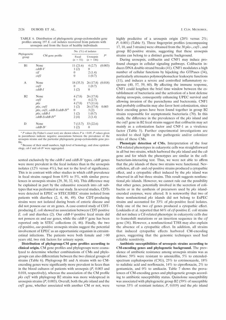
sented exclusively by the cdtB-I and cdtB-IV types. cdtB geneswere more prevalent in the fecal isolates than in the urosepsisisolates (12% versus 4%), but not to the level of significance.This is in contrast with other studies in which cdtB prevalencein fecal strains ranged from 0.9% to 5%, with similar preva-lences in urosepsis strains (29, 30, 32, 64). This difference maybe explained in part by the exhaustive research into cdt sub-types that was performed in our study. In several studies, CDTswere detected in EPEC or STEC/EHEC strains isolated frompatients with diarrhea (2, 4, 46, 49). Our CDT-producingstrains were not isolated during bouts of enteric disease anddid not possess eae or stx genes. A case-control study of CDT-producing E. coli showed no association between CDT-positiveE. coli and diarrhea (2). Our cdtB-V-positive fecal strain didnot possess stx and eae genes, while the cdtB-V gene has beenreported only in STEC collections (4, 46). Finally, the twocif-positive, eae-positive urosepsis strains suggest the potentialinvolvement of EPEC as an opportunistic organism in extraint-estinal infections. The patients were both female and .80years old, two risk factors for urinary sepsis.
Distribution of phylogroup-CM gene profiles according to
clinical origin. CM gene profiles and phylogroups were coana-lyzed to determine whether combinations of CMs and phylo-groups can also differentiate between the two clinical groups ofstrains (Table 6). Phylogroup B1 and A strains with no CM-encoding genes were significantly more prevalent in feces thanin the blood cultures of patients with urosepsis (P, 0.003 and0.018, respectively), whereas the association of the CM profilepks cnf1 with phylogroup B2 strains was more widespread inurosepsis strains (P, 0.003). Overall, both the pks island and thecnf1 gene, whether associated with another CM or not, were
highly predictive of a urosepsis origin (20% versus 2%;P, 0.001) (Table 5). Three fingerprint profiles (encompassing17, 10, and 3 strains) were obtained from the 30 pks-, cnf1-, andgroup B2-positive strains, suggesting that these urosepsisstrains can belong to a distinct genetic background.
During urosepsis, colibactin and CNF1 may induce pro-found changes in cellular signaling pathways. Colibactin in-duces DNA double-strand breaks (41). CNF1 modulates a highnumber of cellular functions by hijacking rho GTPases (34),particularly attenuates polymorphonuclear leukocyte functions(11), and induces a severe and controlled inflammatory re-sponse (40, 57, 59, 60). By affecting the immune response,CNF1 could lengthen the brief time window between the es-tablishment of bacteriuria and the activation of a host defenseduring urosepsis, consequently enhancing UPEC survival andallowing invasion of the parenchyma and bacteremia. CNF1and probably colibactin may also favor host colonization, sincetheir encoding genes have been found together in group B2strains responsible for asymptomatic bacteriuria (70). In thisstudy, the difference in the prevalences of the pks island andthe cnf1 gene in B2 fecal strains suggest that colibactin may actmainly as a colonization factor and CNF-1 as a virulencefactor (Table 5). Further experimental investigations areneeded to shed light on the pathogenic and/or colonizerroles of these CMs.
Phenotypic detection of CMs. Interpretation of the fourCM-related phenotypes in eukaryotic cells was straightforwardin all but two strains, which harbored the pks island and the cdt
gene and for which the phenotypes are similar in the cell-bacterium-interacting test. Thus, we were not able to affirmthat the pks islands of these two strains were functional. Nev-ertheless, all cdt- and cnf-positive strains produced a cytopathiceffect, and a cytopathic effect induced by the pks island wasobserved in all but three strains. This result suggests nonfunc-tional pks islands. However, we cannot rule out the possibilitythat other genes, potentially involved in the secretion of coli-bactin or in the synthesis of precursors used by pks island-encoded enzymes, were altered. It is noteworthy that two ofthese nonfunctional pks islands did not belong to ExPECstrains and accounted for 33% of pks-positive fecal isolates.Only one of the two cif genes produced a cytopathic effect.Loukiadis et al. reported that 66% of cif-positive E. coli strainsdid not induce a Cif-related phenotype in eukaryotic cells dueto frameshift mutations or an insertion sequence in the cif
gene (36). However, a nonfunctional T3SS may also explainthe absence of a cytopathic effect. In addition, all strainsthat induced cytopathic effects harbored CM-encodinggenes, suggesting that the genomic techniques used hadreliable sensitivity.
Antibiotic susceptibilities of urosepsis strains according to
CM-encoding genes and phylogenetic background. The prev-alence of antibiotic resistance among urosepsis strains was asfollows: 59% were resistant to amoxicillin, 5% to extended-spectrum cephalosporins (C3G), 25% to cotrimoxazole, 18%to nalidixic acid and norfloxacin, 14% to ciprofloxacin, 2% togentamicin, and 0% to amikacin. Table 7 shows the preva-lences of CM-encoding genes and phylogenetic groups accord-ing to antibiotic susceptibility status. Quinolone susceptibilitywas associated with phylogenetic group B2 (59% of susceptibleversus 33% of resistant isolates; P, 0.019) and the pks island
TABLE 6. Distribution of phylogenetic group-cyclomodulin geneprofiles among 197 E. coli isolates recovered from patients with
urosepsis and from the feces of healthy individuals
Phylogeneticgroup
CM gene profile
No. (%) of isolates
Pa
Fecal(n 5 51)
Urosepsis(n 5 146)
B1 None 11 (21.6) 4 (2.7) (0.003)cdtB-V 1 (2) 0cif 0 2 (1.4)cnf1 0 1 (0.7)
A None 18 (35.3) 26 (17.8) (0.018)cnf1 0 1 (0.7)cdtB-I 1 (2) 0
B2 None 4 (7.8) 26 (17.8)cnf1 0 4 (2.7)pks 4 (7.8) 17 (11.6)pks, cnf1 1 (2) 26 (17.8) 0.003pks, cnf1, cdtB-I/cdtB-IVb 0 3 (2)pks, cdtB-I 1 (2) 1 (0.7)cdtB-I/cdtB-IVb 2 (4) 2 (1.4)
D None 7 (13.7) 33 (22.6)cdtB-IV 1 (2) 0
a P values (by Fisher’s exact test) are shown where P is ,0.05. P values givenin parentheses indicate negative associations between the prevalence of theurosepsis strains and the particular phylogenetic group-cyclomodulin gene pro-file.
b Because of their small numbers, high level of homology, and close epidemi-ology, cdt-I and cdt-IV were aggregated.
2126 DUBOIS ET AL. J. CLIN. MICROBIOL.

(37% versus 11%; P, 0.011), whereas quinolone resistance wasassociated with group A (41% of resistant versus 13% of sus-ceptible isolates; P, 0.002). Likewise, amoxicillin susceptibilitywas associated with group B2 (78% versus 37%; P, ,0.001),the combination of the alpha-hemolytic phenotype and thecnf1 gene (35% versus 16%; P, 0.011), and the pks island (48%versus 21%; P, ,0.001), whereas amoxicillin resistance wasassociated with groups A and D (28% versus 5% [P, ,0.001]and 29% versus 13% [P, 0.028], respectively). Cotrimoxazolesusceptibility was also associated with group B2 (60% versus36%; P, 0.02), the alpha-hemolytic phenotype (36% versus17%; P, 0.024), the combination of the alpha-hemolytic phe-notype and the cnf1 gene (28% versus 11%; P, 0.043), and thepks island (41% versus 6%; P, ,0.001). C3G susceptibility wasassociated with group B2 (57% versus 0%; P, 0.003), whereasC3G resistance (six strains with overexpression of chromo-some-mediated cephalosporinases and one with an extended-spectrum b-lactamase, CTX-M-14) was associated with groupA (86% versus 15%; P, ,0.001).
These results suggest that resistance to quinolones is asso-ciated with the less virulent phylogenetic groups and with aweak prevalence of virulence factors, as previously observed(24, 26, 52, 67, 68). We obtained similar results for resistanceagainst cotrimoxazole, amoxicillin, and C3G. The acquisitionof antibiotic resistance by horizontal gene transfers or muta-tions may therefore require a particular genetic background, asobserved for virulence factors (15). Some studies have sug-gested that the low-virulence group A isolates are more ex-posed to antibiotic selection pressure within the intestinal tract(24, 26), which indicates the possible importance of environ-mental factors.
Conclusion. The distribution of CM-encoding genes, includ-ing the recently described pks genomic island, and the func-tionality of these toxins were investigated in E. coli strains inrelation to their phylogenetic background, clinical origin, andantibiotic resistance. One finding to emerge from the presentstudy was the frequent association of the pks island with the
cnf1 gene and the alpha-hemolytic phenotype, and their pres-ence in amoxicillin-, cotrimoxazole-, and quinolone-susceptibleE. coli strains of the B2 phylogenetic background isolated frompatients with urosepsis. The widespread diffusion of the pks
island and the cnf1 gene in E. coli help to distinguish ExPECfrom commensal strains and reinforce the idea that these genesof the E. coli flexible gene pool are involved in pathogenicityand/or in the ability to survive in new ecological niches, such asthe human urinary tract.
ACKNOWLEDGMENTS
This work was supported by le Ministere Francais de l’EducationNationale, de la Recherche et de la Technologie (grant JE2526) and byl’Institut National de la Recherche Agronomique (grant USC-2018).
We thank Rolande Perroux and Marlene Jan for helpful technicalassistance.
REFERENCES
1. Adamus-Bialek, W., A. Wojtasik, M. Majchrzak, M. Sosnowski, and P.Parniewski. 2009. (CGG)4-based PCR as a novel tool for discrimination ofuropathogenic Escherichia coli strains: comparison with enterobacterial re-petitive intergenic consensus-PCR. J. Clin. Microbiol. 47:3937–3944.
2. Albert, M. J., S. M. Faruque, A. S. Faruque, K. A. Bettelheim, P. K. Neogi,N. A. Bhuiyan, and J. B. Kaper. 1996. Controlled study of cytolethal dis-tending toxin-producing Escherichia coli infections in Bangladeshi children.J. Clin. Microbiol. 34:717–719.
3. Bidet, P., A. Metais, F. Mahjoub-Messai, L. Durand, M. Dehem, Y. Aujard,E. Bingen, X. Nassif, and S. Bonacorsi. 2007. Detection and identification byPCR of a highly virulent phylogenetic subgroup among extraintestinal patho-genic Escherichia coli B2 strains. Appl. Environ. Microbiol. 73:2373–2377.
4. Bielaszewska, M., M. Fell, L. Greune, R. Prager, A. Fruth, H. Tschape, M. A.Schmidt, and H. Karch. 2004. Characterization of cytolethal distending toxingenes and expression in Shiga toxin-producing Escherichia coli strains ofnon-O157 serogroups. Infect. Immun. 72:1812–1816.
5. Bingen-Bidois, M., O. Clermont, S. Bonacorsi, M. Terki, N. Brahimi, C.Loukil, D. Barraud, and E. Bingen. 2002. Phylogenetic analysis and preva-lence of urosepsis strains of Escherichia coli bearing pathogenicity island-likedomains. Infect. Immun. 70:3216–3226.
6. Blum, G., V. Falbo, A. Caprioli, and J. Hacker. 1995. Gene clusters encodingthe cytotoxic necrotizing factor type 1, Prs-fimbriae and alpha-hemolysinform the pathogenicity island II of the uropathogenic Escherichia coli strainJ96. FEMS Microbiol. Lett. 126:189–195.
7. CA-SFM. 2007. Comite de l’Antibiogramme de la Societe Francaise deMicrobiologie, communique 2007. CA-SFM, Paris, France.
8. Chassagne, L., N. Pradel, F. Robin, V. Livrelli, R. Bonnet, and J. Delmas.
TABLE 7. Distribution of antibiotic susceptibility according to cyclomodulin genes/Hly and phylogenetic background among 143 E. coliisolates recovered from patients with urosepsisa
Variable
Quinolones–fluoroquinolonesb Amoxicillin C3Gc Gentamicin Cotrimoxazole
No. (%) of isolates
Pd
No. (%) of isolates
Pd
No. (%) of isolates
Pd
No. (%) of isolates No. (%) of isolates
Pd
R(n 5 27)
S(n 5 119)
R(n 5 86)
S(n 5 60)
R(n 5 7)
S(n 5 139)
R(n 5 3)
S(n 5 143)
R(n 5 36)
S(n 5 110)
Phylogeneticgroups
A 11 (41) 16 (13) (0.002) 24 (28) 3 (5) (,0.001) 6 (86) 21 (15) (,0.001) 2 (67) 25 (17) 9 (25) 18 (16)B1 2 (7) 5 (4) 5 (6) 2 (3) 1 (14) 6 (4) 1 (33) 6 (4) 3 (8) 4 (4)B2 9 (33) 70 (59) 0.019 32 (37) 47 (78) ,0.001 0 (0) 79 (57) 0.003 0 (0) 79 (55) 13 (36) 66 (60) 0.020D 5 (19) 28 (24) 25 (29) 8 (13) (0.028) 0 (0) 33 (24) 0 (0) 33 (23) 11 (31) 22 (20)
CMspks 3 (11) 44 (37) 0.011 18 (21) 29 (48) ,0.001 0 47 (34) 0 47 (33) 2 (6) 45 (41) ,0.001cnf1-Hly 7 (26) 28 (24) 14 (16) 21 (35) 0.011 2 (29) 33 (24) 0 35 (24) 4 (11) 31 (28) 0.043Hly alone 1 (4) 11 (9) 8 (9) 4 (7) 0 12 (9) 0 12 (8) 2 (6) 10 (9)cdtB-I 0 3 (3) 1 (1) 2 (3) 0 3 (2) 0 3 (2) 0 3 (3)cdtB-IV 0 3 (3) 1 (1) 2 (3) 0 3 (2) 0 3 (2) 0 3 (3)cif 0 2 (2) 1 (1) 1 (2) 0 2 (1) 0 2 (1) 0 2 (2)
a R, resistant; S, susceptible.bAll resistant isolates were resistant both to quinolone (nalidixic acid) and to fluoroquinolones (either norfloxacin alone or norfloxacin and ciprofloxacin).c C3G, extended-spectrum cephalosporins.d P values (by Fisher’s exact test) are shown where P is ,0.05. P values shown in parentheses indicate negative associations between the prevalence of the susceptible
strains and the particular trait (phylogenetic group or CM).
VOL. 48, 2010 CYCLOMODULINS IN UROSEPSIS STRAINS OF ESCHERICHIA COLI 2127

2009. Detection of stx1, stx2, and eae genes of enterohemorrhagic Escherichiacoli using SYBR Green in a real-time polymerase chain reaction. Diagn.Microbiol. Infect. Dis. 64:98–101.
9. Clark, C. G., S. T. Johnson, R. H. Easy, J. L. Campbell, and F. G. Rodgers.2002. PCR for detection of cdt-III and the relative frequencies of cytolethaldistending toxin variant-producing Escherichia coli isolates from humans andcattle. J. Clin. Microbiol. 40:2671–2674.
10. Clermont, O., S. Bonacorsi, and E. Bingen. 2000. Rapid and simple deter-mination of the Escherichia coli phylogenetic group. Appl. Environ. Micro-biol. 66:4555–4558.
11. Davis, J. M., H. M. Carvalho, S. B. Rasmussen, and A. D. O’Brien. 2006.Cytotoxic necrotizing factor type 1 delivered by outer membrane vesicles ofuropathogenic Escherichia coli attenuates polymorphonuclear leukocyte an-timicrobial activity and chemotaxis. Infect. Immun. 74:4401–4408.
12. De Rycke, J., A. Milon, and E. Oswald. 1999. Necrotoxic Escherichia coli(NTEC): two emerging categories of human and animal pathogens. Vet.Res. 30:221–233.
13. Dozois, C. M., S. Clement, C. Desautels, E. Oswald, and J. M. Fairbrother.1997. Expression of P, S, and F1C adhesins by cytotoxic necrotizing factor1-producing Escherichia coli from septicemic and diarrheic pigs. FEMS Mi-crobiol. Lett. 152:307–312.
14. Duriez, P., O. Clermont, S. Bonacorsi, E. Bingen, A. Chaventre, J. Elion, B.Picard, and E. Denamur. 2001. Commensal Escherichia coli isolates arephylogenetically distributed among geographically distinct human popula-tions. Microbiology 147:1671–1676.
15. Escobar-Paramo, P., O. Clermont, A. B. Blanc-Potard, H. Bui, C. Le Bou-guenec, and E. Denamur. 2004. A specific genetic background is required foracquisition and expression of virulence factors in Escherichia coli. Mol. Biol.Evol. 21:1085–1094.
16. Escobar-Paramo, P., K. Grenet, A. Le Menac’h, L. Rode, E. Salgado, C.Amorin, S. Gouriou, B. Picard, M. C. Rahimy, A. Andremont, E. Denamur,and R. Ruimy. 2004. Large-scale population structure of human commensalEscherichia coli isolates. Appl. Environ. Microbiol. 70:5698–5700.
17. Escobar-Paramo, P., A. Le Menac’h, T. Le Gall, C. Amorin, S. Gouriou, B.Picard, D. Skurnik, and E. Denamur. 2006. Identification of forces shapingthe commensal Escherichia coli genetic structure by comparing animal andhuman isolates. Environ. Microbiol. 8:1975–1984.
18. Gauduchon, V., L. Chalabreysse, J. Etienne, M. Celard, Y. Benito, H. Lepidi,F. Thivolet-Bejui, and F. Vandenesch. 2003. Molecular diagnosis of infectiveendocarditis by PCR amplification and direct sequencing of DNA from valvetissue. J. Clin. Microbiol. 41:763–766.
19. Ge, Z., D. B. Schauer, and J. G. Fox. 2008. In vivo virulence properties ofbacterial cytolethal-distending toxin. Cell. Microbiol. 10:1599–1607.
20. Gordon, D. M., O. Clermont, H. Tolley, and E. Denamur. 2008. AssigningEscherichia coli strains to phylogenetic groups: multi-locus sequence typingversus the PCR triplex method. Environ. Microbiol. 10:2484–2496.
21. Guyer, D. M., J. S. Kao, and H. L. Mobley. 1998. Genomic analysis of apathogenicity island in uropathogenic Escherichia coli CFT073: distributionof homologous sequences among isolates from patients with pyelonephritis,cystitis, and catheter-associated bacteriuria and from fecal samples. Infect.Immun. 66:4411–4417.
22. Hacker, J., U. Hentschel, and U. Dobrindt. 2003. Prokaryotic chromosomesand disease. Science 301:790–793.
23. Herzer, P. J., S. Inouye, M. Inouye, and T. S. Whittam. 1990. Phylogeneticdistribution of branched RNA-linked multicopy single-stranded DNAamong natural isolates of Escherichia coli. J. Bacteriol. 172:6175–6181.
24. Houdouin, V., S. Bonacorsi, P. Bidet, M. Bingen-Bidois, D. Barraud, and E.Bingen. 2006. Phylogenetic background and carriage of pathogenicity island-like domains in relation to antibiotic resistance profiles among Escherichiacoli urosepsis isolates. J. Antimicrob. Chemother. 58:748–751.
25. Janka, A., M. Bielaszewska, U. Dobrindt, L. Greune, M. A. Schmidt, and H.Karch. 2003. Cytolethal distending toxin gene cluster in enterohemorrhagicEscherichia coli O157:H2 and O157:H7: characterization and evolutionaryconsiderations. Infect. Immun. 71:3634–3638.
26. Jaureguy, F., E. Carbonnelle, S. Bonacorsi, C. Clec’h, P. Casassus, E. Bin-gen, B. Picard, X. Nassif, and O. Lortholary. 2007. Host and bacterialdeterminants of initial severity and outcome of Escherichia coli sepsis. Clin.Microbiol. Infect. 13:854–862.
27. Johnson, J. R., B. Johnston, M. A. Kuskowski, J. P. Nougayrede, and E.Oswald. 2008. Molecular epidemiology and phylogenetic distribution of theEscherichia coli pks genomic island. J. Clin. Microbiol. 46:3906–3911.
28. Johnson, J. R., M. A. Kuskowski, A. Gajewski, S. Soto, J. P. Horcajada, M. T.Jimenez de Anta, and J. Vila. 2005. Extended virulence genotypes andphylogenetic background of Escherichia coli isolates from patients with cys-titis, pyelonephritis, or prostatitis. J. Infect. Dis. 191:46–50.
29. Johnson, J. R., K. Owens, A. Gajewski, and M. A. Kuskowski. 2005. Bacterialcharacteristics in relation to clinical source of Escherichia coli isolates fromwomen with acute cystitis or pyelonephritis and uninfected women. J. Clin.Microbiol. 43:6064–6072.
30. Johnson, J. R., F. Scheutz, P. Ulleryd, M. A. Kuskowski, T. T. O’Bryan, andT. Sandberg. 2005. Host-pathogen relationships among Escherichia coli iso-
lates recovered from men with febrile urinary tract infection. Clin. Infect.Dis. 40:813–822.
31. Johnson, J. R., and A. L. Stell. 2000. Extended virulence genotypes ofEscherichia coli strains from patients with urosepsis in relation to phylogenyand host compromise. J. Infect. Dis. 181:261–272.
32. Kadhum, H. J., D. Finlay, M. T. Rowe, I. G. Wilson, and H. J. Ball. 2008.Occurrence and characteristics of cytotoxic necrotizing factors, cytolethaldistending toxins and other virulence factors in Escherichia coli from humanblood and faecal samples. Epidemiol. Infect. 136:752–760.
33. Kaper, J. B., J. P. Nataro, and H. L. Mobley. 2004. Pathogenic Escherichiacoli. Nat. Rev. Microbiol. 2:123–140.
34. Lemonnier, M., L. Landraud, and E. Lemichez. 2007. Rho GTPase-activat-ing bacterial toxins: from bacterial virulence regulation to eukaryotic cellbiology. FEMS Microbiol. Rev. 31:515–534.
35. Lidin-Janson, G., B. Kaijser, K. Lincoln, S. Olling, and H. Wedel. 1978. Thehomogeneity of the faecal coliform flora of normal school-girls, character-ized by serological and biochemical properties. Med. Microbiol. Immunol.164:247–253.
36. Loukiadis, E., R. Nobe, S. Herold, C. Tramuta, Y. Ogura, T. Ooka, S.Morabito, M. Kerouredan, H. Brugere, H. Schmidt, T. Hayashi, and E.Oswald. 2008. Distribution, functional expression, and genetic organizationof Cif, a phage-encoded type III-secreted effector from enteropathogenicand enterohemorrhagic Escherichia coli. J. Bacteriol. 190:275–285.
37. Mainil, J. G., E. Jacquemin, and E. Oswald. 2003. Prevalence and identity ofcdt-related sequences in necrotoxigenic Escherichia coli. Vet. Microbiol. 94:159–165.
38. Marches, O., T. N. Ledger, M. Boury, M. Ohara, X. Tu, F. Goffaux, J. Mainil,I. Rosenshine, M. Sugai, J. De Rycke, and E. Oswald. 2003. Enteropatho-genic and enterohaemorrhagic Escherichia coli deliver a novel effector calledCif, which blocks cell cycle G2/M transition. Mol. Microbiol. 50:1553–1567.
39. Middendorf, B., B. Hochhut, K. Leipold, U. Dobrindt, G. Blum-Oehler, andJ. Hacker. 2004. Instability of pathogenicity islands in uropathogenic Esch-erichia coli 536. J. Bacteriol. 186:3086–3096.
40. Munro, P., G. Flatau, A. Doye, L. Boyer, O. Oregioni, J. L. Mege, L. Land-raud, and E. Lemichez. 2004. Activation and proteasomal degradation of rhoGTPases by cytotoxic necrotizing factor-1 elicit a controlled inflammatoryresponse. J. Biol. Chem. 279:35849–35857.
41. Nougayrede, J. P., S. Homburg, F. Taieb, M. Boury, E. Brzuszkiewicz, G.Gottschalk, C. Buchrieser, J. Hacker, U. Dobrindt, and E. Oswald. 2006.Escherichia coli induces DNA double-strand breaks in eukaryotic cells. Sci-ence 313:848–851.
42. Nowrouzian, F. L., I. Adlerberth, and A. E. Wold. 2006. Enhanced persis-tence in the colonic microbiota of Escherichia coli strains belonging to phy-logenetic group B2: role of virulence factors and adherence to colonic cells.Microbes Infect. 8:834–840.
43. Nowrouzian, F. L., A. E. Wold, and I. Adlerberth. 2005. Escherichia colistrains belonging to phylogenetic group B2 have superior capacity to persistin the intestinal microflora of infants. J. Infect. Dis. 191:1078–1083.
44. Ochman, H., and R. K. Selander. 1984. Standard reference strains of Esch-erichia coli from natural populations. J. Bacteriol. 157:690–693.
45. Orden, J. A., G. Dominguez-Bernal, S. Martinez-Pulgarin, M. Blanco, J. E.Blanco, A. Mora, J. Blanco, and R. de la Fuente. 2007. NecrotoxigenicEscherichia coli from sheep and goats produce a new type of cytotoxicnecrotizing factor (CNF3) associated with the eae and ehxA genes. Int.Microbiol. 10:47–55.
46. Orth, D., K. Grif, M. P. Dierich, and R. Wurzner. 2006. Cytolethal distendingtoxins in Shiga toxin-producing Escherichia coli: alleles, serotype distributionand biological effects. J. Med. Microbiol. 55:1487–1492.
47. Oswald, E., J. de Rycke, P. Lintermans, K. van Muylem, J. Mainil, G. Daube,and P. Pohl. 1991. Virulence factors associated with cytotoxic necrotizingfactor type two in bovine diarrheic and septicemic strains of Escherichia coli.J. Clin. Microbiol. 29:2522–2527.
48. Oswald, E., J. P. Nougayrede, F. Taieb, and M. Sugai. 2005. Bacterial toxinsthat modulate host cell-cycle progression. Curr. Opin. Microbiol. 8:83–91.
49. Pandey, M., A. Khan, S. C. Das, B. Sarkar, S. Kahali, S. Chakraborty, S.Chattopadhyay, S. Yamasaki, Y. Takeda, G. B. Nair, and T. Ramamurthy.2003. Association of cytolethal distending toxin locus cdtB with enteropatho-genic Escherichia coli isolated from patients with acute diarrhea in Calcutta,India. J. Clin. Microbiol. 41:5277–5281.
50. Peres, S. Y., O. Marches, F. Daigle, J. P. Nougayrede, F. Herault, C. Tasca,J. De Rycke, and E. Oswald. 1997. A new cytolethal distending toxin (CDT)from Escherichia coli producing CNF2 blocks HeLa cell division in G2/Mphase. Mol. Microbiol. 24:1095–1107.
51. Perna, N. T., G. Plunkett III, V. Burland, B. Mau, J. D. Glasner, D. J. Rose,G. F. Mayhew, P. S. Evans, J. Gregor, H. A. Kirkpatrick, G. Posfai, J.Hackett, S. Klink, A. Boutin, Y. Shao, L. Miller, E. J. Grotbeck, N. W. Davis,A. Lim, E. T. Dimalanta, K. D. Potamousis, J. Apodaca, T. S. Ananthara-man, J. Lin, G. Yen, D. C. Schwartz, R. A. Welch, and F. R. Blattner. 2001.Genome sequence of enterohaemorrhagic Escherichia coli O157:H7. Nature409:529–533.
52. Piatti, G., A. Mannini, M. Balistreri, and A. M. Schito. 2008. Virulence
2128 DUBOIS ET AL. J. CLIN. MICROBIOL.

factors in urinary Escherichia coli strains: phylogenetic background and quin-olone and fluoroquinolone resistance. J. Clin. Microbiol. 46:480–487.
53. Picard, B., J. S. Garcia, S. Gouriou, P. Duriez, N. Brahimi, E. Bingen, J.Elion, and E. Denamur. 1999. The link between phylogeny and virulence inEscherichia coli extraintestinal infection. Infect. Immun. 67:546–553.
54. Pickett, C. L., D. L. Cottle, E. C. Pesci, and G. Bikah. 1994. Cloning,sequencing, and expression of the Escherichia coli cytolethal distending toxingenes. Infect. Immun. 62:1046–1051.
55. Pickett, C. L., R. B. Lee, A. Eyigor, B. Elitzur, E. M. Fox, and N. A.Strockbine. 2004. Patterns of variations in Escherichia coli strains that pro-duce cytolethal distending toxin. Infect. Immun. 72:684–690.
56. Putze, J., C. Hennequin, J. P. Nougayrede, W. Zhang, S. Homburg, H.Karch, M. A. Bringer, C. Fayolle, E. Carniel, W. Rabsch, T. A. Oelschlaeger,E. Oswald, C. Forestier, J. Hacker, and U. Dobrindt. 2009. Genetic structureand distribution of the colibactin genomic island among Enterobacteriaceae.Infect. Immun. 77:4696–4703.
57. Real, J. M., P. Munro, C. Buisson-Touati, E. Lemichez, P. Boquet, and L.Landraud. 2007. Specificity of immunomodulator secretion in urinary sam-ples in response to infection by alpha-hemolysin and CNF1 bearing uro-pathogenic Escherichia coli. Cytokine 37:22–25.
58. Relman, D. A. 1993. The identification of uncultured microbial pathogens.J. Infect. Dis. 168:1–8.
59. Rippere-Lampe, K. E., M. Lang, H. Ceri, M. Olson, H. A. Lockman, andA. D. O’Brien. 2001. Cytotoxic necrotizing factor type 1-positive Escherichiacoli causes increased inflammation and tissue damage to the prostate in a ratprostatitis model. Infect. Immun. 69:6515–6519.
60. Rippere-Lampe, K. E., A. D. O’Brien, R. Conran, and H. A. Lockman. 2001.Mutation of the gene encoding cytotoxic necrotizing factor type 1 (cnf1)attenuates the virulence of uropathogenic Escherichia coli. Infect. Immun.69:3954–3964.
61. Silver, R. P., W. Aaronson, A. Sutton, and R. Schneerson. 1980. Comparativeanalysis of plasmids and some metabolic characteristics of Escherichia coliK1 from diseased and healthy individuals. Infect. Immun. 29:200–206.
62. Skurnik, D., D. Bonnet, C. Bernede-Bauduin, R. Michel, C. Guette, J. M.
Becker, C. Balaire, F. Chau, J. Mohler, V. Jarlier, J. P. Boutin, B. Moreau,D. Guillemot, E. Denamur, A. Andremont, and R. Ruimy. 2008. Character-istics of human intestinal Escherichia coli with changing environments. En-viron. Microbiol. 10:2132–2137.
63. Tavechio, A. T., L. R. Marques, C. M. Abe, and T. A. Gomes. 2004. Detectionof cytotoxic necrotizing factor types 1 and 2 among fecal Escherichia coliisolates from Brazilian children with and without diarrhea. Mem. Inst.Oswaldo Cruz 99:81–83.
64. Toth, I., F. Herault, L. Beutin, and E. Oswald. 2003. Production of cytolethaldistending toxins by pathogenic Escherichia coli strains isolated from humanand animal sources: establishment of the existence of a new cdt variant (typeIV). J. Clin. Microbiol. 41:4285–4291.
65. Toth, I., J. P. Nougayrede, U. Dobrindt, T. N. Ledger, M. Boury, S. Morabito,T. Fujiwara, M. Sugai, J. Hacker, and E. Oswald. 2009. Cytolethal distendingtoxin type I and type IV genes are framed with lambdoid prophage genes inextraintestinal pathogenic Escherichia coli. Infect. Immun. 77:492–500.
66. Van Bost, S., E. Jacquemin, E. Oswald, and J. Mainil. 2003. Multiplex PCRsfor identification of necrotoxigenic Escherichia coli. J. Clin. Microbiol. 41:4480–4482.
67. Velasco, M., J. P. Horcajada, J. Mensa, A. Moreno-Martinez, J. Vila, J. A.Martinez, J. Ruiz, M. Barranco, G. Roig, and E. Soriano. 2001. Decreasedinvasive capacity of quinolone-resistant Escherichia coli in patients with uri-nary tract infections. Clin. Infect. Dis. 33:1682–1686.
68. Vila, J., K. Simon, J. Ruiz, J. P. Horcajada, M. Velasco, M. Barranco, A.Moreno, and J. Mensa. 2002. Are quinolone-resistant uropathogenic Esch-erichia coli less virulent? J. Infect. Dis. 186:1039–1042.
69. Wiles, T. J., R. R. Kulesus, and M. A. Mulvey. 2008. Origins and virulencemechanisms of uropathogenic Escherichia coli. Exp. Mol. Pathol. 85:11–19.
70. Zdziarski, J., C. Svanborg, B. Wullt, J. Hacker, and U. Dobrindt. 2008.Molecular basis of commensalism in the urinary tract: low virulence orvirulence attenuation? Infect. Immun. 76:695–703.
71. Zhang, L., B. Foxman, and C. Marrs. 2002. Both urinary and rectal Esche-richia coli isolates are dominated by strains of phylogenetic group B2. J. Clin.Microbiol. 40:3951–3955.
VOL. 48, 2010 CYCLOMODULINS IN UROSEPSIS STRAINS OF ESCHERICHIA COLI 2129

145
III. Article 3 - Les cyclomodulines des E. coli de tumeurs
Prévalence des cyclomodulines de Escherichia coli
isolées de tumeurs colo-rectales, de diverticuloses et
de lésions iléales de maladie de Crohn.
Damien Dubois, Emmanuel Buc, Pierre Sauvanet, Nathalie Pradel, Marie-Agnès Bringer,
Arlette Darfeuille-Michaud, Denis Pezet et Richard Bonnet
A soumettre

146
Prévalence des cyclomodulines de Escherichia coli
isolées de tumeurs colorectales, de diverticuloses
et de lésions iléales de maladie de Crohn
Damien Duboisa,c, Emmanuel Bucb,c, Pierre Sauvanetc, Nathalie Pradelc, Marie-
Agnès Bringerc, Arlette Darfeuille-Michaudc, Denis Pezetb,c et Richard Bonneta,c
aCHU Clermont-Ferrand, Hôpital G. Montpied, Centre de Biologie, Laboratoire de bactériologie
clinique, Clermont-Ferrand, F-63003, France.
bCHU Clermont-Ferrand, Hôpital Estaing, Service de chirurgie générale et digestive, Clermont-
Ferrand, F-63003, France.
cClermont Université, Université d'Auvergne, Evolution des bactéries pathogènes et susceptibilité de
l’hôte, JE 2526, USC-INRA 2018, Clermont-Ferrand, F-63001, France.

147
Résumé
De nombreuses études ont évalué l'association entre le cancer colorectal et des
agents infectieux. Bien que l'implication des cyclomodulines de E. coli ait été suggérée,
aucune étude n'a abordé la colonisation du côlon par des E. coli produisant ces toxines chez
les patients atteints de cancer colorectal.
La diversité clonale quantitative et phylogénétique de E. coli et la prévalence des
gènes des cyclomodulines et leur fonctionnalité ainsi que la prévalence de l’alpha-
hémolysine de E. coli ont été investiguées dans des isolats issus de biopsies de tumeurs
coliques (n=39) et rectales (n=38), et de biopsies de diverticuloses (contrôles, n=30). De
plus, la prévalence de ces facteurs de virulence a été déterminée chez des isolats de
biopsies iléales de maladie de Crohn. Ces prévalences ont été comparées entre elles et à
celle d’isolats commensaux précédemment rapportées.
Dix isolats de chaque biopsie ont été typés par deux techniques génomiques.
Chaque clone déterminé a été ensuite étudié. Le groupe phylogénétique de chaque clone a
été déterminé par PCR. Les gènes des cyclomodulines, c’est à dire. l’ilot pks codant la
colibactine et les gènes cdtB, cnf1-3 et cif ont été détectés par hybridation d’ADN dot-blot et
confirmés ou typés par PCR. La fonctionnalité de ces cyclomodulines a été évaluée sur
cultures cellulaires. Les conclusions majeures de cette étude sont :
(1) la prévalence d’un clone de E. coli du phylogroupe B2 significativement supérieure dans
les biopsies de tumeurs coliques comparé aux autres origines cliniques (p, <0,001 à 0,015),
chez des patients de même origine géographique,
(2) la prévalence de l’ilot pks significativement supérieure dans la série de tumeurs coliques
comparé aux autres origines cliniques (p, <0,001 à 0,009),
(3) la prévalence de l’opéron hly-cnf1 significativement supérieure dans les tumeurs coliques
et rectales que dans les fèces (p<0,001 et p=0,019, respectivement),
(4) la prévalence de l’alpha-hémolysine significativement supérieure dans les tumeurs
coliques que dans les fèces, les diverticuloses et la MC (p<0,001, p=0,019 et p=0,042,
respectivement),
(5) la prévalence du gène cdtB significativement plus élevée dans les tumeurs coliques que
dans les diverticules (p=0,030),
(6) la présence de trois E. coli EntéroPathogènes présentant un gène cif fonctionnel isolés
de trois tumeurs coliques.
La majorité de ces facteurs de virulence étaient fonctionnels.
Notre étude épidémiologique montre que les différents facteurs de virulence de E. coli
investigués pourraient être impliqués dans la tumorigenèse colorectale.

148
Introduction
Escherichia coli est une espèce d’Entérobactérie diversifiée et versatile. Des études
de génétique des populations reposant principalement sur l’analyse du core-génome ont
permis de classer les souches de cette espèce en quatre groupes phylogénétiques majeurs
A, B1, B2 et D [1]. Ces groupes sont associés à la capacité de ces souches à coloniser des
niches écologiques et ont des capacités différentes à induire une pathologie. Aussi, les
souches de E. coli peuvent également être classées selon leur comportement vis-à-vis de
l’hôte. On peut distinguer les souches commensales non pathogènes, les souches
pathogènes intestinales et les souches pathogènes extra-intestinales.
Les souches de E. coli non pathogènes et son hôte humain coexistent pendant des
décennies avec un bénéfice mutuel. La colonisation de l’hôte se fait les heures qui suivent la
naissance et leur niche préférentielle est la couche muqueuse du côlon. E. coli est un micro-
organisme très compétitif sur ce site très peuplé et constitue la bactérie anaérobie facultative
la plus abondante de la microflore intestinale humaine. Ces souches de E. coli commensales
entrainent rarement une maladie, sauf chez les sujets immunodéprimés ou lorsque la
barrière digestive normale est altérée, comme dans la péritonite [2].
Parmi les pathogènes intestinaux, six pathovars sont bien caractérisés et
responsables de diarrhées : les E. coli entéropathogènes (EPEC), les E. coli
entérohémorragiques (EHEC), les E. coli entérotoxiques (ETEC), les E. coli
entéroaggrégatifs (EAEC), les E. coli entéroinvasifs (EIEC) et les E. coli d’adhésion diffuse
(DAEC). Un ensemble hétérogène de souches appelées E. coli adhérents et invasifs (AIEC)
[3] est associé à la maladie de Crohn (MC). Elles se répliquent dans les macrophages et
induisent la libération de grandes quantités de TNF-�, une caractéristique qui pourrait
contribuer à l'inflammation intestinale chronique observée dans la MC. Les E. coli
pathogènes extra-intestinales (ExPEC) constituent un ensemble de souches encore mal
défini qui comprend notamment les E. coli uropathogènes (UPEC) et les E. coli associés aux
méningites néonatales (MNEC) [2].
Ces souches de E. coli pathogènes se distinguent principalement par l’acquisition de
gènes impliqués dans la virulence par transfert horizontal via des plasmides, des
bactériophages et/ou des ilots génomiques. Ce pool de gènes flexible de E. coli représente
au moins la moitié du nombre total de gènes présents dans chaque génome, soit 2 000 à 3
400 gènes, et environ 88% des gènes recensés chez E. coli à ce jour [4]. Les facteurs de
virulence codés par ces gènes du pool flexible peuvent affecter une grande variété de
processus cellulaires critiques de l’hôte, comme la synthèse des protéines, les signaux de
transduction, les fonctions du cytosquelette, la sécrétion d'ions, la transcription, l'apoptose, la

149
fonction mitochondriale et la division cellulaire [2]. Les protéines effectrices qui perturbent
ces processus sont accompagnées par de nombreux facteurs de colonisation et de
« fitness » qui permettent aux bactéries d'adhérer et/ou d'envahir les cellules hôtes. Parmi
ces effecteurs, il a été observé une classe croissante de toxines appelées cyclomodulines
(CM) [5]. Elles agissent sur le cycle cellulaire eucaryote, un processus fondamental pour
l’homéostasie cellulaire et tissulaire, et ainsi induisent des effets cytopathiques qui peuvent
être mis à profit pour leur détection [6, 7]. Quatre types de CM ont été identifiées puis
caractérisées chez E. coli : les toxines protéiques CNF-1 à CNF-3 (facteurs nécrosants
cytotoxiques) ciblant les Rho GTPases [8, 9], le facteur d'inhibition du cycle (protéine Cif)
ciblant les complexes ubiquitine ligases cullin-RING [10, 11], et deux types de génotoxines,
les toxines de distension cytoléthales (protéines CDT de cinq types, I à V) [12] et la
colibactine, de découverte récente [6]. La colibactine est probablement une toxine de type
polycétide-peptide non ribosomal hybride, dont l'activité est codée par l'ilot génomique pks
[6]. Les toxines CDT, Cif et la colibactine bloquent la mitose, alors les toxines CNF favorisent
la réplication de l'ADN sans cytocinèse. Ces toxines peuvent affecter le système immunitaire
de l’hôte mais aussi le développement de cancer.
Actuellement, le cancer colorectal est le troisième cancer le plus fréquent, chez
l’homme et la femme, et la quatrième cause de décès par cancer à travers le monde, avec
1,2 million de cas estimés et 609.000 décès estimés en 2008 [13]. Basé sur les tendances
démographiques, l'incidence annuelle devrait augmenter de près de 80% à 2,2 millions de
cas au cours des deux prochaines décennies et la plupart de cette augmentation se produira
dans les régions les moins développées du monde (62%), le taux de ce cancer augmentant
avec l'industrialisation et l'urbanisation [14]. Dans les pays industrialisés, le cancer du côlon
est deux fois plus fréquent que celui du rectum. Environ 70% des patients atteints de cancer
du côlon ont plus de 65 ans. Le cancer colorectal survient le plus souvent de façon
sporadique et il est héréditaire ou familial dans seulement 5% des cas [15]. Les habitudes
alimentaires, le mode de vie et d'autres facteurs non génétiques tels que le tabac, l'utilisation
chronique d’anti-inflammatoires non-stéroïdiens sont considérés comme ayant un fort impact
sur le risque de cancer colorectal. De même, certaines conditions physiopathologiques telles
que le syndrome métabolique et les maladies inflammatoires de l'intestin (la MC et la recto-
colite ulcéro-hémorragique) augmentent le risque de cancer du côlon [13, 16, 17].
De nombreuses études ont évalué l'association entre le cancer colorectal et les
agents infectieux [18]. Des études utilisant des modèles animaux suggèrent que les micro-
organismes sont impliqués dans le développement du cancer colorectal [19]. Bien que
l'implication des CM de E. coli ait été suggérée [20-22], aucune étude n'a abordé la
colonisation de E. coli produisant ces toxines chez les patients atteints de cancer colorectal.
Afin d'étudier une possible association entre la production de CM de E. coli et le

150
développement du cancer colorectal, nous avons exploré la prévalence des gènes et
l’expression fonctionnelle des CM chez des E. coli isolées de tumeurs colorectales, de
diverticuloses humaines et de muqueuses iléales de patients atteints de MC.
Matériel et Méthodes
Souches bactériennes de référence
Les souches de E. coli de référence archétypes des facteurs de virulence étudiés et
la souche E. coli K-12 DH5� utilisés comme contrôles positifs et négatif respectivement sont
présentées dans le tableau 1 [23].
Tableau 1. Souches de E. coli archétypes utilisées dans cette étude.
Souche de E. coliCaractéristique
génétique Origine
Source ou
référence
28C hly, cnf1, cdt-IV, clbA, clbK, clbJ, clbQ
Septicémie porcine [24]
1404 cnf2, cdt-III Septicémie bovine [25]
IHE3034 ∆clbP cdt-I, clb ∆clbP Méningite de nouveau-né [6]
DH5α (pCP2123) cdt-II Souche de laboratoire [26]
C48a cnf3, eae, cif Agneau sain [9]
DH10β (pBACpks) clb Souche de laboratoire [6]
E22 cif, eae Lapin, EPEC [10]
RS218 chuA, yjaA, svg,TSPE4.C2, uidA (Phylogroupe B2.1)
Méningite de nouveau-né [27]
EDL933 eae, stx1, stx2 EHEC [28]
DH5α Souche de laboratoire Novagen
Patients
Les souches de E. coli incluses dans cette étude ont été isolées à partir
d’échantillons de muqueuses de patients du Centre Hospitalier Universitaire de Clermont-
Ferrand. Entre mars 2007 et novembre 2009, 77 échantillons ont été prélevés au niveau de
la tumeur de patients présentant un adénocarcinome colorectal (CCR) et 30 échantillons ont
été prélevés au niveau de diverticuloses. Des souches issues de lésions de l’iléon de
patients atteints de MC ont également été incluses [29, 30] ainsi que 51 souches de E. coli
commensales [23]. Tous les patients ont donné leur consentement éclairé et l'approbation du
comité d'éthique local a été obtenue.

151
Soixante-dix-sept échantillons d’adénocarcinomes colorectaux ont été prélevés lors
d’une intervention chirurgicale pour exérèse. L'âge médian des patients était de 69 ans
(extrêmes : 22 à 95 ans) ; le sexe ratio de la cohorte était de 1,26 (tableau 2). La cohorte
inclut 39 échantillons de cancers coliques (21 échantillons du côlon droit et 18 échantillons
du colon gauche) et 38 échantillons de cancers rectaux. Tous les patients ont reçu de la
céfoxitine (2 g par voie intraveineuse) au moment de l'incision.
Trente échantillons de diverticuloses ont été prélevés lors d’une intervention
chirurgicale pour exérèse. L'âge médian des patients était de 58 ans (extrêmes : 34 à 81
ans) ; le sexe ratio de la cohorte était de 0,72 (tableau 2). Tous les patients ont reçu de la
céfoxitine (2 g par voie intraveineuse) au moment de l'incision.
Tableau 2. Données démographiques des patients ayant eu une biopsie de tumeur ou de diverticulose.
TR TDG TDGR Divert.
n= 38 39 77 30
âge médian 64,5 71 69 58
âge moyen 65 67 66 60
écarts d’âge 22-87 35-95 22-95 34-81
hommes 23 20 43 13
femmes 15 19 34 18
sexe ratio 1,53 1,05 1,26 0,72
TR, groupe tumeurs du rectum ; TDG, groupe tumeurs du côlon ; TDGR, groupe tumeurs du colon et du rectum ; Divert., groupe diverticuloses
Les résections iléales de lésions chroniques de 9 patients présentant une MC (MCC)
qui avaient subi une iléocolectomie pour la première fois ont été incluses. Les échantillons
ont été prélevés au niveau de la muqueuse de l'iléon réséqué avec une pince endoscopique
[29, 30]. Les biopsies iléales de lésions récurrentes précoces de MC (MCP) ont été obtenues
à partir de l'iléon néoterminal (à partir de 10 cm de l'anastomose) par endoscopie chez 11
patients qui avaient subi une iléocolectomie avec anastomose iléo-colique de bout en bout.
Aucun des patients n’avait reçu d’antibiotiques dans les 4 semaines précédant le
prélèvement [29, 30].
Les isolats fécaux commensaux avaient été recueillis par écouvillonnages anaux
d’adultes dans la communauté [23]. Ces personnes ne présentaient aucun signe d'infection
aiguë ou de troubles bénins ou malins du tractus gastro-intestinal et n'avaient pas reçu
d'antibiotiques dans le mois précédant le prélèvement. Une colonie de E. coli choisie
arbitrairement par échantillon avait été analysée. L'âge médian des 51 patients était de 46,5
ans (extrêmes : 23 à 62 ans) ; le sexe ratio de la cohorte était de 0,54.

152
Traitement des biopsies
Les biopsies ont été prélevées dans un flacon stérile contenant une solution saline
tamponnée au phosphate (PBS). Chaque échantillon a été divisé en deux parties et pesé
(environ 5 à 10 mg chacune).
La première partie des échantillons a été lavée en PBS, puis broyée (Ultra-Turrax,
IKA) et incubée 15 minutes en présence de Triton 0,1X. Des dilutions de raison 10 du lysat
ont ensuite été réalisées et étalées sur milieu gélosé sélectif pour entérobactéries Drigalski
(bioMérieux) et sur milieu gélosé chromogénique chromID® CPS3 (bioMérieux). Les milieux
ont été incubés à 37°C pendant 48 heures en atmosph ère aérobie. La seconde partie des
échantillons a été incubée une heure à 37°C en mili eu EMEM (PAA) additionnée de 10µg/mL
de gentamicine (Euromedex) afin d’éliminer les bactéries extracellulaires adhérentes, puis
broyée, diluée et ensemencée de manière similaire à la première partie.
Les colonies isolées ont été repiquées en plaques 96 puits en milieu Luria Bertani
additionné de 15% de glycérol, incubées 24 heures à 37°C puis stockées à -80°C. Les
souches de E. coli ont été identifiées à l’aide du milieu gélosé chromogénique chromID®
CPS3 (bioMérieux, France) et du système automatisé Vitek II® (bioMérieux) en deuxième
intention. Au moins 10 colonies par prélèvement ont été retenues pour les études suivantes.
Etude de la clonalité des isolats et groupage phylogénétique
Les expérimentations ont été conduites comme indiqué précédemment [23].
Brièvement, l’ADN des isolats a été extrait par la méthode de l'eau bouillante. Les isolats de
E. coli ont été classés d’après le système de la collection de référence de E. coli ECOR [31]
dans les groupes phylogénétiques A, B1, B2 et D en fonction de la présence de trois gènes
(chuA, yjaA et svg) et d’un fragment d'ADN (TSPE4.C2), en utilisant une technique de PCR
multiplex incluant le gène uidA comme contrôle interne [32].
Deux techniques de typage génomique ont été utilisées : une « Enterobacterial
Repetitive Intergenic Consensus sequence » (ERIC)-PCR utilisant l’amorce ERIC2 (5�-AAG
TAA GTG ACT GGG GTG AGC G-3�) et une « Random Amplified Polymorphic DNA »
(RAPD) PCR utilisant l’amorce RAPD 1283 (5�-GCG ATC CCC A-3�). Ces amorces ont été
utilisées comme indiqué précédemment [33, 34]. Les isolats représentatifs d’un clone ont été
stockés à -80°C en milieu Luria-Bertani supplémenté avec 15% de glycérol.

153
Identification des gènes codant les cyclomodulines
La détection des gènes codant la production de cyclomodulines a été réalisée par
hybridation d’ADN selon la méthode du dot-blot. Chaque dépôt a été vérifié avec une sonde
du gène codant l’ARNr 16S de E. coli. L'ilot pks, qui contient le groupe de gènes clb
permettant la production de la colibactine a été détecté à l’aide d’une sonde chevauchant les
gènes clbK et clbJ. Les 3 types de gènes cnf ont été détectés avec un mélange de sondes
spécifiques de cnf1, cnf2, et cnf3. Les 5 types de gènes cdtB ont été détectés par deux
expériences d'hybridation avec une sonde spécifique des gènes cdtB-II, cdtB-III et cdtB-V, et
une sonde spécifique des gènes cdtB-I et cdtB-IV. Le gène cif a été détecté en utilisant une
sonde interne spécifique. La sensibilité et la spécificité des sondes ont été vérifiées sur
chaque membrane en déposant des extraits d'ADN de toutes les souches contrôles.
Les hybridations positives ont été soumises à confirmation par PCR en utilisant des
amorces spécifiques des gènes clbA et clbQ de l’ilot pks par PCR multiplex, des gènes cnf1,
cnf2 et cnf3 par PCR multiplex, des gènes cdtB-I, cdtB-II, cdtB-III/cdtB-V, et cdtB-IV, et du
gène cif par PCR simplex, comme précédemment décrit [23].
Les gènes cdt et cif étant fréquemment associés aux gènes stx1 et stx2 des E coli
entérohémorragiques (EHEC) et eae des E coli entéropathogènes (EPEC) respectivement,
tous les isolats ont été testés par PCR pour les gènes de virulence stx1, stx2, et eae, comme
décrit précédemment [35].
Détection de l’effet cytopathogène induit par les cyclomodulines
Les effets cytopathiques des CNF et des CDT ont été étudiés dans toutes les
souches, et ceux de la colibactine et de Cif ont été étudiés dans les souches non
hémolytiques, comme décrit précédemment [6, 7]. Brièvement, les effets des CNF et des
CDT ont été détectés par un test d’interaction lysat bactérien-culture cellulaire. Après une
culture de 48 h à 37°C sous agitation dans un milie u liquide de Luria-Bertani, les cellules
bactériennes ont été soniquées puis filtrées stérilement à l'aide de filtres de 0,22 µm. Des
cultures cellulaires HeLa ont été traitées avec les lysats bactériens stériles jusqu'à l'analyse.
Les effets de la colibactine et de Cif ont été détectés par un test d’interaction bactéries-
culture cellulaire. Des cultures de E. coli d’une nuit en bouillon Luria-Bertani ont été diluées
dans le milieu d'interaction, et des cultures de cellules HeLa ont été infectées à des
multiplicités d'infection (nombre de bactéries par cellule au début de l'infection) de 100 et
200. Les cultures cellulaires ont été lavées 3 à 6 fois 4 heures après infection et ont été
incubées dans un milieu de culture cellulaire contenant 200 µg/mL de gentamicine jusqu'à
l'analyse. Après 72 h d'incubation à 37 ° C sous un e atmosphère à 5% de CO2 le milieu a été

154
éliminé par plusieurs lavages des monocouches de cellules HeLa. Les modifications
morphologiques caractéristiques des CNF, des CDT, de la colibactine et de Cif ont été
observées après coloration par le Giemsa, comme décrit par ailleurs [6]. La détection de
l'alpha-hémolysine a été effectuée pour toutes les souches étudiées par croissance d'une
nuit à 37°C sur milieu gélosé Columbia contenant 5% de sang de mouton (Oxoid).
Analyses statistiques
Un premier test du chi-carré a été réalisé afin d’évaluer l'hétérogénéité entre les
groupes, et les comparaisons individuelles par paires ont été testées seulement si ce test
donnait une valeur de P <0,05. Les analyses statistiques ont été ensuite effectuées en
utilisant le test exact de Fisher. Le seuil de signification statistique était P <0,05.
Résultats et Discussion
Distribution des gènes codant les CM dans les phylogroupes de E. coli
La prévalence des gènes codant les CM chez les E. coli isolés est hétérogène
(tableau 3). Les gènes codant les CM les plus fréquents étaient l’ilot pks (18,3%) et cnf1
(13,5%), ainsi le gène codant pour l’alpha-hémolysine (16,9%), trois traits fortement associés
au phylogroupe B2.
Toutes les souches possédant l’ilot pks appartenaient au phylogroupe B2 (P <0,001
pour le groupe B2 versus les groupes A, B1 et D, combinés ou séparés). 50.6% des isolats
B2 possédaient ce trait, qui était observé dans tous les sous-types B2 ; un isolat sur cinq
B2.1, deux isolats sur quatre B2 TSPE4.C2-négatif et 35 isolats sur 66 B2 TSPE4.C2-positif.
Ces résultats sont en accord avec les études précédentes [6, 23, 36, 37]
Les gènes cnf étaient de type 1 dans 13,5% des isolats et de type 2 dans 0,5% des
isolats. Le seul isolat positif cnf2 ne possédait pas le gène cnf1. Les souches de E. coli
présentant cnf2 et cnf3 ont été principalement observées chez des souches d’origine
animale [9, 38]. Toutes les souches possédant le gène cnf1 appartenaient au phylogroupe
B2, et correspondaient à 37,3% de celles-ci. Comme pour l’ilot pks, cette association était
forte (P, <0,001 pour le groupe B2 versus les groupes A, B1 et D, combinés ou séparés) et
33,3% des isolats B2 possédaient l’ilot pks et le gène cnf1 (P, <0,001 pour le groupe B2
versus les groupes A, B1 et D, combinés ou séparés), comme précédemment observé [37].
Toutes les souches portant cnf1 sauf trois présentaient le phénotype alpha-
hémolytique. Cette association est probablement due à la présence de l'ilot de pathogénicité

155
(PAI) IIJ96-like, qui porte le gène cnf1 en aval de l'opéron hlyCABD de l’alpha-hémolysine
HlyA [39]. Les trois isolats non hémolytiques portant cnf1 possédaient néanmoins le gène
hlyC, laissant supposer une anomalie du PAI IIJ96-like telle que des mutations ponctuelles ou
des délétions empêchant la production de l’alpha-hémolysine. Ces deux isolats ont été
considérés cnf1- et Hly-positifs pour la suite de l’analyse. Seulement 3,4% des isolats
présentaient un phénotype alpha-hémolytique sans le gène cnf1. Parmi ces isolats, soit 20%
des souches alpha-hémolytiques, aucune association n’a été observée de manière
significative avec un groupe phylogénétique. Ces isolats possédaient probablement un autre
PAI contenant l'opéron hlyCABD, analogue au PAI ICFT073-like [40].
Les gènes cdtB ont été observés dans 4,3% des souches, avec un seul sous-type
cdtB par souche. Bien que 67% des souches cdtB-positives appartenaient au phylogroupe
B2, aucune association à un groupe phylogénétique n’a été observée de manière
significative, même en considérant les différents sous-types de cdtB. Les gènes cdtB-I/cdtB-
IV (n=7) était surreprésentés par rapport au groupe de gènes cdtB-II/cdtB-III/cdtB-V (n=2).
Les gènes cdtB-I et cdtB-IV sont deux homologues proches alors que les gènes cdtB-II,
cdtB-III et cdtB-V forment un autre sous-groupe [41]. L’unique isolat cdtB-III-positif possédait
aussi le seul gène cnf2 observé. Cette association de gènes est rapportée comme fréquente
et portée par un plasmide nommé pVir essentiellement présent chez des isolats d’origine
bovine [38, 42]. Les gènes cdtB ne possédaient pas d’association particulière avec l'ilot pks
ou le gène cnf1. Les gènes cdt ayant été largement étudiés dans les souches de E. coli
produisant des toxines Shiga (STEC) [43-45], les souches cdtB-positives ont été étudiées
pour les gènes eae et stx [4, 25, 46, 49, 51-53]. Aucun gène eae ou stx n’a été détecté dans
les isolats positifs cdtB.
Le gène cif a été détecté dans trois souches. Elles appartenaient aux phylogroupes A
(n=1) et B1 (n=2) et ne présentaient pas d’autres gènes codant pour les CM. Cif est une
protéine effectrice du système de sécrétion de type 3 (SST3) codée par le locus de l'ilot de
pathogénicité d’effacement des entérocytes. Le gène cif n’a été rapporté que dans les EPEC
et EHEC [10, 46]. Les trois isolats de cette étude possédaient le gène eae mais pas stx1 ou
stx2 et appartenaient donc au pathotype EPEC.

156
Tableau 3. Distribution des gènes codant les cyclomodulines et l’alpha-hémolysine HlyA en fonction du phylogroupe dans 207 isolats de E. coli issus de diverticuloses, de tumeurs
colorectales et de lésions iléales de Maladie de Crohn.
Nombre (%) d'isolats de E. coli
Groupes
Total A B1 B2 D Facteurs de virulence
(n= 207) (n= 57) (n= 28) (n= 75) (n= 47)
Pa
pks 38 (18,3) 38 (50,6) <0,001
cnf1-HlyA 28 (13,5) 28* (37,3) <0,001
HlyA non associé à cnf1 7 (3,4) 2 (7,1) 4 (5,3) 1 (2,1)
cnf1-HlyA, pks 25 (12,1) 25* (33,3) <0,001
cnf2-cdtB-III 1 (0,5) 1 (3,6)
cdtB 9 (4,3) 1 (1,7) 2 (7,1) 6 (8)
cdtB-I 2 (1) 2 (2,6)
cdtB-IV 5 (2,4) 1 (1,7) 4 (5,3)
cdtB-V 1 (0,5) 1 (3,6)
cif 3 (1,5) 1 (1,7) 2 (7,1) a les valeurs de P (par le test exact de Fischer) sont montrées lorsque P est <0,05.
* 3 isolats cnf1+ non hémolytiques mais présentant le gène hlyC
Détection phénotypique des cyclomodulines
La fonctionnalité d’une majorité de CM et de l’opéron codant l’alpha-hémolysine a été
confirmé dans les isolats étudiés (tableau 4). Toutes les souches qui ont induit un effet
cytopathogène possédaient un ou des gènes codant des CM connues pour induire un effet
compatible avec celui observé. L’interprétation des différents phénotypes cytopathogènes
liés aux CM ou à leur combinaison était aisée pour tous les isolats, sauf pour une souche
possédant l'ilot pks et un gène cdtB-I, des gènes produisant des CM qui induisent des effets
cytopathogènes similaires sur les tests cellulaires réalisés. Ainsi, nous n'avons pas été en
mesure d'affirmer que l’ilot pks et les gènes cdt de cette souche étaient fonctionnels.
Comme précédemment rapporté [23], des isolats pks-positifs étaient non
fonctionnels. Ils représentaient 31% des isolats possédant l’ilot pks et pouvant être explorés
(n= 13, isolats non hémolytiques et ne présentant pas cdtB). L’absence de production de
colibactine peut être due à une altération de l’ilot pks ou de gènes situés en dehors de l’ilot et
participant à la synthèse de précurseurs ou à la sécrétion de la colibactine.
La souche positive cnf2 a présenté un effet cytopathogène caractéristique
(multinucléation cellulaire) des CNF. Seul un isolat hémolytique provenant d’un diverticule et
un isolat positif cdtB-IV non hémolytique présentant le gène hlyC provenant d’une lésion
MCC portaient un gène cnf1 non fonctionnels, soit 7,1% des isolats positifs cnf1. Trois isolats
ont présenté un opéron hlyCABD non fonctionnel, soit 10,7% des isolats positifs cnf1. Parmi

157
les souches cdtB-positives, 22,2% (n=2) n’ont pas présenté d’effet cytopathogène
caractéristique. L’absence de cytotoxicité de ces isolats positifs pour cnf1, hly ou cdtB peut
avoir pour origine des mutations ou des séquences interrompues par des séquences
d’insertion dans les séquences codantes ou régulatrices de ces gènes, des défauts dans le
système de sécrétion de HlyA ou l’absence de production des deux autres protéines du
complexe hétérotrimérique formant la toxine CDT active, à savoir CdtA et CdtC, nécessaires
à l’entrée de CdtB dans la cellule eucaryote [12]. Un effet cytopathogène caractéristique de
Cif a été observé pour les trois isolats possédant le gène correspondant.
Tableau 4. Détection phénotypique des cyclomodulines et de l’alpha-hémolysine HlyA de 207 isolats de E. coli issus d’échantillons de diverticuloses et de tumeurs colorectales et de
lésions iléales de Maladie de Crohn.
Nombre (%) d'isolats de E. coliFacteurs de virulence Total F NF NA I
pks 38 9 (23,7) 4 (10,5) 24 (63,1) 1 (2,6)
cnf1 28 26 (92,8) 2* (7,1)
cnf2 1 1 (100)
hly 35 32 (91,4) 3** (8,6)
Hly associé à cnf1 28 25 (89,3) 3** (10,7)
cdtB 9 7 (77,8) 2*** (22,2)
cif 3 3 (100)
F, fonctionnel ; NF, non fonctionnel ; NA, non applicable ; I, indéterminé * un isolat hémolytique provenant d’un diverticule et un isolat cdtB-IV-positif non hémolytique
présentant le gène hlyC provenant d’une lésion CDC ** trois isolats cnf1-positifs mais présentant le gène hlyC (MCC, n=2 ; tumeur rectale, n=1)
*** un isolat cdtB-V-positif provenant d’une tumeur rectale et l’isolat cdtB-IV-positif non hémolytique présentant le gène hlyC provenant d’une lésion CDC
Diversité des clones de E. coli issus de cancers colorectaux et de diverticuloses
La diversité des clones de E. coli isolés de cancers colorectaux et de diverticuloses a
été comparée (tableau 5). Le nombre d’échantillons ne présentant pas de E. coli était
significativement supérieur dans la série diverticuloses comparé à la série tumeurs (19,3%
versus 5,2%, p=0,031). Ceci suggère que des isolats de E. coli seraient capables de
coloniser plus facilement les tumeurs colorectales que les diverticules. Il a été rapporté que
l’administration de bactéries anaérobies facultatives, comme Salmonella typhimurium,
Shigella flexneri et E. coli à des souris porteuses de tumeurs hépatique et splénique
conduisait à une accumulation préférentielle et la prolifération des micro-organismes dans
les tumeurs solides [47]. De plus, Martin et al ont mis en évidence que les isolats de E. coli
exprimaient des hémagglutinines dans environ 40% des souches issues de MC et de
cancers colorectaux, mais seulement dans 4% des contrôles. Cette expression

158
d’hémagglutinines était significativement corrélée avec l’adhésion à des cultures de cellules
intestinales embryonnaires (I407) et de cellules d'adénocarcinome du côlon (HT29) [48].
Swidsinski et al ont observé que les isolats de E. coli associés aux muqueuses sont
fréquemment trouvés dans la muqueuse colique des patients présentant un adénocarcinome
colorectal, mais rarement chez les témoins sains. De plus, la muqueuse colique des patients
atteints de CCR, mais pas la muqueuse colique normale était colonisée par des E. coli
intracellulaires [49]. Il est possible que la présence de ces E. coli lors de la MC et du cancer
colorectal puisse être favorisée par des changements de la muqueuse associés à la maladie.
Par ailleurs, Carvalho et al. ont récemment montré que les AIEC associés à la MC colonisent
et induisent une inflammation intestinale forte chez les souris transgéniques exprimant
CEACAM6, une molécule d’adhérence humaine [50]. CEACAM6 agit comme un récepteur
pour les pili de type 1 produites par les AIEC [51], et sa surexpression est observée dans les
polypes hyperplasiques et les adénomes précoces [52].
Tableau 5. Diversité quantitative des clones de E. coli issus de 77 échantillons de cancers colorectaux (côlon droit, colon gauche et rectum) et de 31 échantillons de diverticuloses.
Nombre (%) de patients
Divertic. TDGR TR TDG TD TG
Nombres de clones de E. coli /
échantillon (n=31) (n=77) (n=38) (n=39) (n=21) (n=18)
Pa
Divert. versusTDGR
0 6 (19,3) 4 (5,2) 2 (5,3) 2 (5,1) 1 (4,8) 1 (5,5) 0,031
1 8 (25,8) 29 (37,7) 13 (34,2) 16 (41) 10 (47,6) 6 (33,3)
2 14 (45,2) 28 (36,4) 15 (39,5) 13 (33,3) 8 (38,1) 5 (27,8)
3 2 (6,4) 12 (15,6) 6 (15,8) 6 (15,4) 2 (9,5) 4 (22,2)
4 1 (3,2) 1 (1,3) 1 (2,6)
5 2 (2,6) 2 (5,1) 2 (11,1)
6 1 (1,3) 1 (2,6)
Divert., groupe diverticuloses ; TDGR, groupe tumeurs du côlon et du rectum ; TDG, groupe tumeurs du côlon ; TD, groupe tumeurs du côlon droit ; TG, groupe tumeurs du côlon gauche ;
TR, groupe tumeurs du rectum a les valeurs de P (par le test exact de Fischer) sont montrées lorsque P est <0,05.
Les phylogroupes des E. coli issus de cancers colorectaux, de diverticuloses, de
biopsies iléales lésées de MC et de E. coli fécaux commensaux ont été comparés (tableau
6). Aucune différence n’ayant été observée entre les séries de biopsies de tumeurs coliques
droites et gauches, celles-ci ont été associées en une seule série. Bien qu’un seul clone ait
été isolé des séries de MC et fécale, il faut noter que la prévalence d’un clone de E. coli du
phylogroupe B2 est significativement plus élevée dans les biopsies de tumeurs coliques que
dans les biopsies de diverticuloses, de tumeurs rectales, de lésions iléales de MC et dans

159
les fèces (p, <0,001 à 0,015). De plus, la prévalence d’un clone de E. coli du phylogroupe D
est significativement plus élevée dans la série de tumeurs rectales que dans la série de
souches fécales commensales (p=0,027). 60% des isolats issus de biopsies de MC
appartiennent aux phylogroupes B2+D ; bien que non significative, cette prédominance a été
précédemment rapportée par Kotlowski et al [53].
Tableau 6. Diversité phylogénétique des clones de E. coli issus de 77 échantillons de cancers colorectaux (côlon droit, côlon gauche et rectum), de 31 échantillons de
diverticuloses, de 20 isolats de E. coli issus d’échantillons d’iléums lésés de maladie de Crohn et de 51 isolats de E. coli fécaux commensaux.
Nombre (%) de patients présentant un clone de E. coli
P a P
a
Divert. TDG TR Fecal Crohn TDG versusTR
versus
Phylo-groupes
de E. coli
(n=31) (n=39) (n=38) (n=51) (n=20) Divert. TR Fecal Crohn Fecal
A 13 (41,9) 11 (28,2) 14 (36,8) 19 (37) 4 (20)
B1 2 (6,4) 9 (23,1) 9 (23,7) 12 (24) 4 (20)
B2 13 (41,9) 28 (71,8) 16 (42,1) 12 (24) 6 (30) 0,015 0,011 <0,001 0,005
D 10 (32,3) 10 (25,6) 14 (36,8) 8 (16) 6 (30) 0,027
Divert., groupe diverticuloses ; TDG, groupe tumeurs du côlon ; TR, groupe tumeurs du rectum ; Fecal, groupe fécaux commensaux ; Crohn, groupe biopsies iléales de Crohn
a les valeurs de P (par le test exact de Fischer) sont montrées lorsque P est <0,05.
Distribution des gènes codant les cyclomodulines selon l’origine physiopathologique des souches de E. coli
La distribution des gènes codant les cyclomodulines et l’alpha-hémolysine a été
analysée selon le contexte physiopathologique (tableau 7). Quatre facteurs de virulence,
l’ilot pks, le gène cnf1, la protéine HlyA et les gènes cdtB ont été retrouvés préférentiellement
et de manière significative chez les patients présentant une tumeur.
L’ilot pks était significativement plus prévalent dans la série de tumeurs coliques que
dans les autres séries. Cuevas-Ramos et al ont montré que la colibactine produite par des
souches de E. coli possédant l’ilot pks avait des propriétés génotoxiques in vitro et in vivo
[54]. Ces souches sont mutagènes, induisent des dommages chromosomiques et une
instabilité génétique in vitro de manière dose-dépendante. Cette instabilité génétique est
retrouvée dans 85% des cancers colorectaux sporadiques [55]. Une infection de quelques
heures d’entérocytes dans un modèle d’anse intestinale ligaturée de souris par une souche
de E. coli produisant la colibactine induisait des cassures double-brin de l’ADN
correspondant à une irradiation de 0,5 Grays. [54]. De plus, les lignées cellulaires humaines

160
infectées transitoirement par des E. coli produisant la colibactine montrent la formation de
colonies cellulaires indépendantes de l’ancrage, un processus intervenant dans la diffusion
des métastases du cancer colorectal [54]. Ces données suggèrent une activité mutagène et
de transformation cellulaire des E. coli possédant l’ilot pks. Par son action génotoxique, la
colibactine pourrait être impliquée dans les différentes étapes du développement tumoral
colorectal.
La prévalence de l’opéron hly-cnf1 était significativement plus élevée dans les
tumeurs coliques et rectales que dans les fèces (p<0,001 et p=0,019, respectivement). CNF
est un promoteur de la prolifération cellulaire [8]. Cette toxine active les Rho-GTPases,
déclenchant la transition de phase G1-S du cycle cellulaire et induisant la réplication de
l'ADN sans cytocinèse . De plus, il a été montré que CNF1 inhibe l'apoptose et induit des
ségrégations chromosomiques anormales [56, 57].
La prévalence de l’alpha-hémolysine était significativement supérieure dans les
tumeurs coliques que dans les fèces, les diverticuloses et la MC (p<0,001, p=0,019 et
p=0,042, respectivement). L’ilot pks et l’opéron hly-cnf1 étant presque exclusivement
retrouvés dans les isolats du phylogroupe B2, on retrouve une prévalence significativement
plus élevée de ces facteurs de virulence dans les tumeurs coliques et rectales que dans les
fèces (p<0,001 et p=0,039, respectivement). Il a été rapporté que l’alpha-hémolysine de E.
coli augmente la perméabilité du côlon [58]. Ce processus représente une voie de
translocation bactérienne facilitant la colonisation d’un tissu tumoral et la dissémination
d’autres toxines bactériennes comme CNF1 et la colibactine. Néanmoins, l’alpha-hémolysine
pourrait aussi être un facteur anti-tumoral en favorisant la lyse des cellules néoplasiques.
La prévalence du gène cdtB était significativement supérieure dans les tumeurs
coliques que dans les diverticules (p=0,030). Ce gène était aussi présent chez des E. coli
issus de tumeurs rectales, de lésions iléales de Crohn et fécaux. CdtB présente une activité
de type DNAse et induit des cassures simple et/ou double-brin de l’ADN des cellules
eucaryotes [12]. La toxine CDT produite par Helicobacter hepaticus, une bactérie hépato-
carcinogène pour la souris favorise le développement de dysplasie hépatique dans un
modèle murin [59]. De plus, H. hepaticus a montré un pouvoir promoteur de la tumorigenèse
colique dans différents modèles murins [60-62]. L’activité génotoxique de CDT favoriserait
l’acquisition d’évènements mutationnels pouvant activer des gènes oncogènes et/ou
inactiver des gènes suppresseurs de tumeurs. Comme pour la colibactine, CDT pourrait ainsi
être impliquée, par son action génotoxique dans les différentes étapes du développement
tumoral colorectal. Les gènes cdt sont présents chez diverses bactéries entériques
pathogènes pour l’homme [59], comme les espèces de Campylobacter spp. qui sont une des

161
premières causes de gastro-entérites dans les pays industrialisés. En outre, Campylobacter
jejuni a été associé à certains lymphomes de l’intestin grêle [63].
La présence de trois isolats présentant les gènes cif et eae, et donc à ce titre classés
comme des EPEC a été observée dans la série de tumeurs coliques. Le gène eae des
EPEC est codé par l’ilot de pathogénicité LEE, qui provoque des lésions d’attachement et
d’effacement au niveau de la muqueuse intestinale [2]. Ainsi, les EPEC sont une cause
majeure de diarrhée infantile dans les pays en développement [7]. Dans les pays développés
l'incidence des diarrhées induites par les EPEC a diminué, et leur dépistage systématique
n'est plus effectué [64]. Toutefois, des études récentes en Europe et en Australie ont identifié
les EPEC chez 2,5 à 10,9% des enfants en bonne santé [65, 66], ce qui suggère qu'un
pourcentage important de la population en bonne santé des pays développés sont porteurs
de EPEC. Les travaux de Maddocks et al suggèrent que les EPEC sont plus fréquents dans
les tissus d’adénocarcinomes coliques que dans des tissus coliques normaux appariés, avec
une prévalence respective de 25% et 0% (n=20) [67]. Le locus LEE code un système de
sécrétion bactérien de type III (SST3) capable d’injecter des effecteurs bactériens qui
pourraient favoriser la cancérogenèse en agissant sur différents processus comme celui de
la réparation des lésions de l’ADN [67, 68]. Un des effecteur pouvant intervenir est le produit
du gène cif, qui altère le processus d’ubiquitination et donc la dégradation de protéines
intervenant dans de nombreux processus cellulaires comme la régulation du cycle cellulaire
ou du cytosquelette [10, 11]. Par ailleurs, Citrobacter rodentium, qui porte le locus LEE et
provoque des lésions d’attachement et d’effacement, est responsable d’hyperplasies
coliques transmissibles [69], réduit la période de latence de l'apparition de tumeurs induites
chimiquement [70], et favorise la formation adénome colique chez la souris Min [71].

162
Tableau 7. Présence chez les patients des gènes codant les CM dans des isolats de E. coliissus de 77 échantillons de cancers colorectaux (côlon droit, côlon gauche et rectum), de 31 échantillons de diverticuloses, et dans 20 isolats de E. coli issus d’échantillons d’iléums lésés
de maladie de Crohn et 51 isolats de E. coli fécaux commensaux.
Nombre (%) de patients présentant un clone de E. colipossédant le facteur de virulence
P a P
a
Divert. TDG TR Fecal Crohn TDG versusTR
versus
Facteurs de virulence de E. coli
(n=31) (n=39) (n=38) (n=51) (n=20) Divert R Fecal Crohn Fecal
pks 6 (19,4) 20 (51,3) 8 (21) 6 (12) 2 (10) 0,007 0,009 <0,001 0,002
cnf1-Hly 4 (12,9) 14 (35,9) 7 (18,4) 1 (2) 2* (10) <0,001 0,019
Hly non associé à
cnf11 (3,2) 3 (7,7) 1 (2,6) 3 (6) 1 (5)
Hly 5 (16,1) 17 (43,6) 8 (21,1) 4 (7,8) 3 (15) 0,019 <0,001 0,042
cnf1-Hly, pks 4 (12,9) 12 (30,8) 6 (15,8) 1 (2) 2* (10) <0,001 0,039
cnf2-cdtB-III 0 1 (2,6) 0 0 0
cdtB 0 6 (15,4) 2 (5,3) 6 (12) 1 (5) 0,030
cif 0 3 (7,7) 0 0 0
Divert., groupe diverticuloses ; TDG, groupe tumeurs du côlon ; TR, groupe tumeurs du rectum ; Fecal, groupe fécaux commensaux ; Crohn, groupe biopsies iléales de Crohn
a les valeurs de P (par le test exact de Fischer) sont montrées lorsque P est <0,05.
Les bactéries, pathogènes ou commensales peuvent avoir un rôle dans la
carcinogenèse, notamment colorectale [18, 21, 22]. Cependant, le développement tumoral
est un processus long et multifactoriel. Notre étude épidémiologique montre que des facteurs
de virulence de E. coli pourraient être impliqués dans la tumorigenèse colorectale.
Néanmoins, la présence de ces facteurs dans la tumeur ne présume pas de leur présence et
de leur implication dans le processus tumoral entamé ultérieurement. Clarifier le rôle
possible de ces facteurs de virulence dans la carcinogenèse colorectal nécessiterait une
étude observationnelle longitudinale de population, prenant en compte les nombreux
facteurs de risques et facteurs confondants (génétiques et environnementaux) chez les
patients. Se concentrer sur plusieurs sous-ensembles homogènes de la maladie pourrait
permettre d'augmenter la sensibilité des études.

163
Références
1. Gordon, D.M., et al., Assigning Escherichia coli strains to phylogenetic groups: multi-locus sequence typing versus the PCR triplex method. Environ Microbiol, 2008. 10(10): p. 2484-96.
2. Kaper, J.B., J.P. Nataro, and H.L. Mobley, Pathogenic Escherichia coli. Nat Rev Microbiol, 2004. 2(2): p. 123-40.
3. Darfeuille-Michaud, A., Adherent-invasive Escherichia coli: a putative new E. coli pathotype associated with Crohn's disease. Int J Med Microbiol, 2002. 292(3-4): p. 185-93.
4. Touchon, M., et al., Organised genome dynamics in the Escherichia coli species results in highly diverse adaptive paths. PLoS Genet, 2009. 5(1): p. e1000344.
5. Oswald, E., et al., Bacterial toxins that modulate host cell-cycle progression. Curr Opin Microbiol, 2005. 8(1): p. 83-91.
6. Nougayrede, J.P., et al., Escherichia coli induces DNA double-strand breaks in eukaryotic cells. Science, 2006. 313(5788): p. 848-51.
7. Peres, S.Y., et al., A new cytolethal distending toxin (CDT) from Escherichia coli producing CNF2 blocks HeLa cell division in G2/M phase. Mol Microbiol, 1997. 24(5): p. 1095-107.
8. Lemonnier, M., L. Landraud, and E. Lemichez, Rho GTPase-activating bacterial toxins: from bacterial virulence regulation to eukaryotic cell biology. FEMS Microbiol Rev, 2007. 31(5): p. 515-34.
9. Orden, J.A., et al., Necrotoxigenic Escherichia coli from sheep and goats produce a new type of cytotoxic necrotizing factor (CNF3) associated with the eae and ehxA genes. Int Microbiol, 2007. 10(1): p. 47-55.
10. Marches, O., et al., Enteropathogenic and enterohaemorrhagic Escherichia coli deliver a novel effector called Cif, which blocks cell cycle G2/M transition. Mol Microbiol, 2003. 50(5): p. 1553-67.
11. Jubelin, G., et al., Pathogenic bacteria target NEDD8-conjugated cullins to hijack host-cell signaling pathways. PLoS Pathog, 2010. 6(9).
12. Ge, Z., D.B. Schauer, and J.G. Fox, In vivo virulence properties of bacterial cytolethal-distending toxin. Cell Microbiol, 2008. 10(8): p. 1599-607.
13. Labianca, R., et al., Colon cancer. Crit Rev Oncol Hematol, 2010. 74(2): p. 106-33. 14. Karsa, L., et al., The dimensions of the CRC problem. Best Pract Res Clin
Anaesthesiol, 2010. 24(4): p. 381-96. 15. Kwak, E.L. and D.C. Chung, Hereditary colorectal cancer syndromes: an overview.
Clin Colorectal Cancer, 2007. 6(5): p. 340-4. 16. von Roon, A.C., et al., The risk of cancer in patients with Crohn's disease. Dis Colon
Rectum, 2007. 50(6): p. 839-55. 17. Eaden, J.A., K.R. Abrams, and J.F. Mayberry, The risk of colorectal cancer in
ulcerative colitis: a meta-analysis. Gut, 2001. 48(4): p. 526-35. 18. Burnett-Hartman, A.N., P.A. Newcomb, and J.D. Potter, Infectious agents and
colorectal cancer: a review of Helicobacter pylori, Streptococcus bovis, JC virus, and human papillomavirus. Cancer Epidemiol Biomarkers Prev, 2008. 17(11): p. 2970-9.
19. Uronis, J.M. and C. Jobin, Microbes and colorectal cancer: is there a relationship?Curr Oncol, 2009. 16(4): p. 22-4.
20. Travaglione, S., A. Fabbri, and C. Fiorentini, The Rho-activating CNF1 toxin from pathogenic E. coli: a risk factor for human cancer development? Infect Agent Cancer, 2008. 3: p. 4.
21. Lax, A.J., New genotoxin shows diversity of bacterial attack mechanisms. Trends Mol Med, 2007. 13(3): p. 91-3.

164
22. Mager, D.L., Bacteria and cancer: cause, coincidence or cure? A review. J Transl Med, 2006. 4: p. 14.
23. Dubois, D., et al., Cyclomodulins in urosepsis strains of Escherichia coli. J Clin Microbiol, 2010. 48(6): p. 2122-9.
24. Dozois, C.M., et al., Expression of P, S, and F1C adhesins by cytotoxic necrotizing factor 1-producing Escherichia coli from septicemic and diarrheic pigs. FEMS Microbiol Lett, 1997. 152(2): p. 307-12.
25. Oswald, E., et al., Virulence factors associated with cytotoxic necrotizing factor type two in bovine diarrheic and septicemic strains of Escherichia coli. J Clin Microbiol, 1991. 29(11): p. 2522-7.
26. Pickett, C.L., et al., Patterns of variations in Escherichia coli strains that produce cytolethal distending toxin. Infect Immun, 2004. 72(2): p. 684-90.
27. Silver, R.P., et al., Comparative analysis of plasmids and some metabolic characteristics of Escherichia coli K1 from diseased and healthy individuals. Infect Immun, 1980. 29(1): p. 200-6.
28. Perna, N.T., et al., Genome sequence of enterohaemorrhagic Escherichia coli O157:H7. Nature, 2001. 409(6819): p. 529-33.
29. Darfeuille-Michaud, A., et al., High prevalence of adherent-invasive Escherichia coli associated with ileal mucosa in Crohn's disease. Gastroenterology, 2004. 127(2): p. 412-21.
30. Darfeuille-Michaud, A., et al., Presence of adherent Escherichia coli strains in ileal mucosa of patients with Crohn's disease. Gastroenterology, 1998. 115(6): p. 1405-13.
31. Ochman, H. and R.K. Selander, Standard reference strains of Escherichia coli from natural populations. J Bacteriol, 1984. 157(2): p. 690-3.
32. Bidet, P., et al., Detection and identification by PCR of a highly virulent phylogenetic subgroup among extraintestinal pathogenic Escherichia coli B2 strains. Appl Environ Microbiol, 2007. 73(7): p. 2373-7.
33. Versalovic, J., T. Koeuth, and J.R. Lupski, Distribution of repetitive DNA sequences in eubacteria and application to fingerprinting of bacterial genomes. Nucleic Acids Res, 1991. 19(24): p. 6823-31.
34. Wang, G., et al., RAPD (arbitrary primer) PCR is more sensitive than multilocus enzyme electrophoresis for distinguishing related bacterial strains. Nucleic Acids Res, 1993. 21(25): p. 5930-3.
35. Chassagne, L., et al., Detection of stx1, stx2, and eae genes of enterohemorrhagic Escherichia coli using SYBR Green in a real-time polymerase chain reaction. Diagn Microbiol Infect Dis, 2009. 64(1): p. 98-101.
36. Putze, J., et al., Genetic structure and distribution of the colibactin genomic island among members of the family Enterobacteriaceae. Infect Immun, 2009. 77(11): p. 4696-703.
37. Johnson, J.R., et al., Molecular epidemiology and phylogenetic distribution of the Escherichia coli pks genomic island. J Clin Microbiol, 2008. 46(12): p. 3906-11.
38. De Rycke, J., A. Milon, and E. Oswald, Necrotoxic Escherichia coli (NTEC): two emerging categories of human and animal pathogens. Vet Res, 1999. 30(2-3): p. 221-33.
39. Blum, G., et al., Gene clusters encoding the cytotoxic necrotizing factor type 1, Prs-fimbriae and alpha-hemolysin form the pathogenicity island II of the uropathogenic Escherichia coli strain J96. FEMS Microbiol Lett, 1995. 126(2): p. 189-95.
40. Guyer, D.M., J.S. Kao, and H.L. Mobley, Genomic analysis of a pathogenicity island in uropathogenic Escherichia coli CFT073: distribution of homologous sequences among isolates from patients with pyelonephritis, cystitis, and Catheter-associated bacteriuria and from fecal samples. Infect Immun, 1998. 66(9): p. 4411-7.
41. Toth, I., et al., Cytolethal distending toxin type I and type IV genes are framed with lambdoid prophage genes in extraintestinal pathogenic Escherichia coli. Infect Immun, 2009. 77(1): p. 492-500.

165
42. Johnson, T.J., et al., Pyrosequencing of the Vir plasmid of necrotoxigenic Escherichia coli. Vet Microbiol, 2010. 144(1-2): p. 100-9.
43. Bielaszewska, M., et al., Characterization of cytolethal distending toxin genes and expression in shiga toxin-producing Escherichia coli strains of non-O157 serogroups.Infect Immun, 2004. 72(3): p. 1812-6.
44. Orth, D., et al., Cytolethal distending toxins in Shiga toxin-producing Escherichia coli: alleles, serotype distribution and biological effects. J Med Microbiol, 2006. 55(Pt 11): p. 1487-92.
45. Pandey, M., et al., Association of cytolethal distending toxin locus cdtB with enteropathogenic Escherichia coli isolated from patients with acute diarrhea in Calcutta, India. J Clin Microbiol, 2003. 41(11): p. 5277-81.
46. Loukiadis, E., et al., Distribution, functional expression, and genetic organization of Cif, a phage-encoded type III-secreted effector from enteropathogenic and enterohemorrhagic Escherichia coli. J Bacteriol, 2008. 190(1): p. 275-85.
47. Stritzker, J., et al., Tumor-specific colonization, tissue distribution, and gene induction by probiotic Escherichia coli Nissle 1917 in live mice. Int J Med Microbiol, 2007. 297(3): p. 151-62.
48. Martin, H.M., et al., Enhanced Escherichia coli adherence and invasion in Crohn's disease and colon cancer. Gastroenterology, 2004. 127(1): p. 80-93.
49. Swidsinski, A., et al., Association between intraepithelial Escherichia coli and colorectal cancer. Gastroenterology, 1998. 115(2): p. 281-6.
50. Carvalho, F.A., et al., Crohn's disease adherent-invasive Escherichia coli colonize and induce strong gut inflammation in transgenic mice expressing human CEACAM.J Exp Med, 2009. 206(10): p. 2179-89.
51. Barnich, N., et al., CEACAM6 acts as a receptor for adherent-invasive E. coli, supporting ileal mucosa colonization in Crohn disease. J Clin Invest, 2007. 117(6): p. 1566-74.
52. Scholzel, S., et al., Carcinoembryonic antigen family members CEACAM6 and CEACAM7 are differentially expressed in normal tissues and oppositely deregulated in hyperplastic colorectal polyps and early adenomas. Am J Pathol, 2000. 156(2): p. 595-605.
53. Kotlowski, R., et al., High prevalence of Escherichia coli belonging to the B2+D phylogenetic group in inflammatory bowel disease. Gut, 2007. 56(5): p. 669-75.
54. Cuevas-Ramos, G., et al., Escherichia coli induces DNA damage in vivo and triggers genomic instability in mammalian cells. Proc Natl Acad Sci U S A, 2010. 107(25): p. 11537-42.
55. Vogelstein, B., et al., Genetic alterations during colorectal-tumor development. N Engl J Med, 1988. 319(9): p. 525-32.
56. Fiorentini, C., et al., Toxin-induced activation of Rho GTP-binding protein increases Bcl-2 expression and influences mitochondrial homeostasis. Exp Cell Res, 1998. 242(1): p. 341-50.
57. Malorni, W. and C. Fiorentini, Is the Rac GTPase-activating toxin CNF1 a smart hijacker of host cell fate? Faseb J, 2006. 20(6): p. 606-9.
58. Troeger, H., et al., Escherichia coli alpha-haemolysin induces focal leaks in colonic epithelium: a novel mechanism of bacterial translocation. Cell Microbiol, 2007. 9(10): p. 2530-40.
59. Ge, Z., et al., Bacterial cytolethal distending toxin promotes the development of dysplasia in a model of microbially induced hepatocarcinogenesis. Cell Microbiol, 2007. 9(8): p. 2070-80.
60. Nagamine, C.M., et al., Helicobacter hepaticus promotes azoxymethane-initiated colon tumorigenesis in BALB/c-IL10-deficient mice. Int J Cancer, 2008. 122(4): p. 832-8.
61. Nagamine, C.M., et al., Helicobacter hepaticus infection promotes colon tumorigenesis in the BALB/c-Rag2(-/-) Apc(Min/+) mouse. Infect Immun, 2008. 76(6): p. 2758-66.

166
62. Fox, J.G., et al., Gut microbes define liver cancer risk in mice exposed to chemical and viral transgenic hepatocarcinogens. Gut, 2010. 59(1): p. 88-97.
63. Lecuit, M., et al., Immunoproliferative small intestinal disease associated with Campylobacter jejuni. N Engl J Med, 2004. 350(3): p. 239-48.
64. Nataro, J.P. and J.B. Kaper, Diarrheagenic Escherichia coli. Clin Microbiol Rev, 1998. 11(1): p. 142-201.
65. Afset, J.E., et al., Association of atypical enteropathogenic Escherichia coli (EPEC) with prolonged diarrhoea. J Med Microbiol, 2004. 53(Pt 11): p. 1137-44.
66. Beutin, L., et al., HEp-2 cell adherence, actin aggregation, and intimin types of attaching and effacing Escherichia coli strains isolated from healthy infants in Germany and Australia. Infect Immun, 2003. 71(7): p. 3995-4002.
67. Maddocks, O.D., et al., Attaching and effacing Escherichia coli downregulate DNA mismatch repair protein in vitro and are associated with colorectal adenocarcinomas in humans. PLoS One, 2009. 4(5): p. e5517.
68. Vogelmann, R. and M.R. Amieva, The role of bacterial pathogens in cancer. Curr Opin Microbiol, 2007. 10(1): p. 76-81.
69. Schauer, D.B., et al., Genetic and biochemical characterization of Citrobacter rodentium sp. nov. J Clin Microbiol, 1995. 33(8): p. 2064-8.
70. Barthold, S.W. and A.M. Jonas, Morphogenesis of early 1, 2-dimethylhydrazine-induced lesions and latent period reduction of colon carcinogenesis in mice by a variant of Citrobacter freundii. Cancer Res, 1977. 37(12): p. 4352-60.
71. Newman, J.V., et al., Bacterial infection promotes colon tumorigenesis in Apc(Min/+) mice. J Infect Dis, 2001. 184(2): p. 227-30.

167
Chapitre III
Discussion
et
Perspectives

168
169
Les résultats présentés précédemment mettent en évidence l’originalité de la protéine
ClbP et de ses homologues au sein de la famille des enzymes S12 avec notamment leur
association aux ilots de PKS-NRPS comme pks et l implication potentielle de ce dernier dans
les relations hôtes-bactéries. D’une part, l’activité peptidase de ClbP a été montrée par
l’analyse de la structure cristalline de son domaine enzymatique, sa caractérisation
biochimique et des travaux de mutagenèse qui ont confirmé le rôle essentiel de cette activité
pour la fonctionnalité de l’ilot pks. De plus, ClbP présente une promiscuité fonctionnelle avec
des homologues présents dans d’autres ilots synthétisant des composés NRP bactériens.
D’autre part, les études épidémiologiques ont révélé une plus forte prévalence de l’ilot pks
chez des isolats de E. coli issus d’urosepsis comparés à des isolats de E. coli commensaux, et
chez des isolats de E. coli issus de biopsies de tumeurs coliques comparés à des isolats de E.
coli commensaux ou issus de biopsies de diverticuloses ou de tumeurs rectales.
I. Fonction biochimique de ClbP et de ses orthologues
A. ClbP, une peptidase des composés NRP
Notre travail suggère que la colibactine synthétisée par les enzymes de l’ilot pks est
maturée par la peptidase ClbP dans le périplasme. L’orthologue ZmaM serait impliqué de
même dans la synthèse de la Zwittermicine A (ZmA), un composé PK-NRP de structure
connue (aminopolyol linéaire), qui est produit par un ilot génomique de Bacillus du groupe
cereus (B. cereus, B. thuringiensis et B. mycoides) [382]. ZmA est biosynthétisée sous forme
d’un métabolite secondaire de grande taille, qui serait clivé au niveau d’une liaison peptidique
pour former le composé actif ZmA [381]. La seule enzyme codée par l’ilot de biosynthèse
susceptible de catalyser cette réaction est la protéine ClbP-like ZmaM. Ces données
confortent nos résultats qui suggèrent un rôle conservé des protéines ClbP-like dans la
maturation de composés de type NRP ou PK-NRP. Nous proposons donc de désigner ces
protéines ClbP-like sous le terme de « peptidases associées aux NRP » (PaNRP).
B. Recherche d’un motif clivé par ClbP et ses orthologues
Le domaine PaNRP de ZmaM cliverait un métabolite « pré-Zma » entre une D-
asparagine et une D-sérine, libérant un acide gras-D-asparagine et le composé ZmA portant le

170
résidu D-sérine dont la stéréospécificité est un déterminant important de l’activité biologique
du composé [381, 383]. De manière intéressante, des analyses bioinformatiques de la
spécificité du substrat montrent que les ilots génomiques produisant une PaNRP codent aussi
pour des domaines d’adénylation A incorporant une asparagine ou un résidu apparenté, la
glutamine. De plus, toutes les peptidases de la famille MEROPS S12 caractérisées
biochimiquement comme les β-lactamases AmpC et la protéase Pab87 possèdent une
spécificité pour les substrats porteurs d’une stéréospécificité D [379, 384-387]. Des résidus
asparagine et glutamine de stéréospécificité D pourraient donc être un motif impliqué dans la
reconnaissance du substrat par les PaNRP.
C. Localisation de ClbP, de ses orthologues et de la colibactine
Au sein des clusters de biosynthèse, les PaNRP sont les seules protéines qui possèdent
une séquence signal de translocation périplasmique et un domaine catalytique prédits en
position périplasmique. Les PaNRP interviendraient donc dans le périplasme lors de la phase
finale de synthèse, après l’assemblage du squelette carboné dans le cytoplasme. Cependant,
l’isolement et la détermination de la structure de la colibactine ou de ZmA avant, après ou
pendant l‘action de ClbP ou d’une autre PaNRP comme ZmaM sont nécessaires pour avoir la
preuve directe de cette fonction.
Le principal écueil à ce travail est l’isolement de la colibactine. A ce jour, elle n’a
jamais été mise en évidence directement. Nous avons mené une analyse phylogénétique des
domaines de condensation C des enzymes NRPS codées par l’ilot pks. Les résultats suggèrent
que le domaine C de ClbN soit un domaine « starter C» habituellement observé dans des
clusters synthétisant des lipopeptides (figure 36). Typiquement, il acyle le premier acide
aminé intégré dans le composé avec un acide β-hydroxy-carboxylique ou acide gras β-
hydroxylé et favorise ainsi l’ancrage du composé aux membranes [277]. La colibactine
pourrait donc porter un acide gras responsable de sa rétention dans la membrane externe des
bactéries, à la manière des lipoprotéines de Braun, du viscosinamide, ou du composé PDIM
membranaire de M. tuberculosis [373, 388-390]. Cet aspect doit être intégré dans la stratégie
d’extraction et de purification de la colibactine. Cette hypothèse expliquerait aussi la nécessité
d’un contact étroit entre la bactérie produisant la colibactine et la cellule cible pour observer
l’effet cytopathogène de la colibactine. Ainsi, la colibactine serait délivrée aux cellules
eucaryotes par contact [24].

171
Figure 36 - Arbre phylogénétique des sous-types de domaines de condensation C de NRPS (domaines LCL, DCL, Starter, Dual E/C, Epi : Epimerisation et Cyc : Hétérocyclisation). Modifié d’après Rausch et al [277]. Les séquences peptidiques des domaines C de l’ilot pks ont été alignées à l’aide de COBALT un alignement de domaines C généré par Rausch et al.. La phylogénie a ensuite été reconstruite en utilisant PhyML avec le modèle de substitution WAG (Gamma et Sites invariables). Les valeurs de bootstrap, générées à partir de 100 arbres, sont indiquées pour les principales branches. Les domaines C de l’ilot pks sont en bleu et désignés par le nom de la protéine puis le numéro de domaine sur la protéine.

172
ClbP est associée à la membrane cytoplasmique. Cette localisation membranaire est
observée pour d’autres protéines intervenant dans la synthèse de composés PK, NRP, PK-
NRP ou apparentés. Une protéine PKS intervenant dans la synthèse la myxovirescine A est
localisée dans la membrane cytoplasmique de Myxococcus xanthus [391]. Les protéines de
l’ilot de biosynthèse du bacillaene s’assemblent en un mégacomplexe de mégasynthases,
formant un organite unique et juxtaposé à la membrane cellulaire, une organisation
probablement maintenue par l’association des lignes d'assemblage à un domaine membranaire
[392]. La synthèse des peptides lantibiotiques fait intervenir des complexes enzymatiques
ancrés sur la face cytoplasmique de la membrane bactérienne [393, 394]. Un couplage entre la
synthèse et l’exporteur du composé PDIM, un PK-acide gras a été rapporté chez M.
tuberculosis : le domaine cytoplasmique de l’exporteur MmpL7 de la famille des RND
(Résistance, Nodulation et Division cellulaire) perméases interagit avec l’enzyme PKS PpsE
qui est requise pour l’étape finale de la biosynthèse du phthiocerol [395]. La localisation
membranaire de ClbP est probablement due à ses trois hélices transmembranaires C-
terminales putatives. Ces hélices sont conservées chez les protéines PaNRP. De plus, de
nombreuses protéines PaNRP portent des domaines ABC exporteurs, connus pour être
également membranaires. La localisation membranaire de ClbP est donc probablement
conservée chez les orthologues. Cependant, l’importance de cette localisation et les
éventuelles interactions associées, notamment avec les pompes d’efflux et la chaine
d’assemblage PK-NRP restent à explorer.

173
II. Rôle biologique de ClbP et de ses orthologues
A. ClbP et la synthèse d’un composé NRP bioactif
Retrouvés principalement chez des souches de Firmicutes (genres Bacillus,
Clostridium et apparentés), les gènes codant les PaNRP sont souvent associés à des clusters
de biosynthèse codant des NRPS. Cependant, certains gènes des PaNRP ne sont pas
génétiquement liés à de tels clusters, alors qu’ils sont capables de restaurer l’activité
biologique de l’ilot pks défectif pour le gène clbP. Cette observation pose le problème du rôle
biologique des protéines PaNRP non liées génétiquement à un cluster de biosynthèse de NRP.
Dans le génome de Streptomycètes, le nombre de voies de biosynthèse nécessitant une
phosphopantéthéinylation dépassent celui des enzymes PPTases codées [396]. Une PPTase
est donc utilisée par de multiples voies de biosynthèse et leurs gènes peuvent être codés dans
une région distincte de l’ilot principal de biosynthèse. Les PaNRP non liées génétiquement à
des clusters NRPS pourraient donc être codées à distance des principaux gènes de la chaine
d’assemblage du NRP. Cette hypothèse est soutenue par l’observation de un à trois ilots de
biosynthèse de NRP putatifs dans 15 des 16 génomes possédant une PaNRP non liée
génétiquement à des ilots de NRPS.
Par ailleurs, les gènes de ces PaNRP chez Bacillus sp. B14905 et Lysinibacillus
sphaericus C3-41 sont dans le voisinage de gènes codant des homologues du système de
quorum-sensing « agr », des enzymes de synthèse de lantibiotiques ou de protéines de
germination de spores. Aussi nous ne pouvons pas éliminer la possibilité que ces PaNRP
puissent être impliquées dans la biosynthèse d’autres types de molécules naturelles portant
des liaisons peptidiques, comme des molécules de communication tels que les lantibiotiques
et d’autres molécules du quorum-sensing [335].
La source majeure de diversité des composés NRP provient principalement des
domaines d’adénylation A. Ces derniers sont responsables de la diversité des acides aminés
incorporés par les modules NRPS [257]. Les enzymes PaNRP pourraient être utilisées comme
un outil biotechnologique intéressant pour augmenter la diversité au sein des petites
molécules synthétisées au laboratoire, comme cela a été proposé pour d’autres enzymes de
finition intervenant la biosynthèse de métabolites secondaires [397, 398].

174
B. ClbP clive une séquence d’adressage ?
Les bactériocines et autres peptides ribosomaux antibiotiques tels que la microcine V
de E. coli et les lantibiotiques des bactéries à Gram positif sont produits sous forme de pré-
peptides incluant un peptide signal N-terminal. Ces pré-peptides maintiennent ces
bactériocines dans une forme inactive dans la cellule productrice et facilitent les interactions
avec les enzymes de modifications (hétérocyclases, oxidases) et les exporteurs. La séquence
signal N-terminale est habituellement clivée pendant l’export par des cystéine protéases
fusionnées à des transporteurs de type ABC ou après l’export par des sérine peptidases
périplasmiques dédiées [338, 399, 400]. Par analogie, les PaNRP pourraient éliminer une
partie du composé NRP impliqué dans les interactions avec des enzymes de synthèse et/ou
des exporteurs.
C. ClbP et protection de la cellule productrice
L’activité peptidase de ClbP est indispensable à l’activité biologique de l’ilot pks. De
même, la maturation de ZmA par une peptidase, probablement la protéine PaNRP ZmaM
permet de libérer le résidu D-Ser nécessaire à l’activité biologique du composé ZmA.
L’analyse topologique de ces protéines in silico et les données expérimentales recueillies pour
ClbP suggèrent que ces peptidases activent le composé NRP dans le périplasme. Par ailleurs,
de nombreuses PaNRP comme ZmaM possèdent en C-terminal un domaine exporteur de type
ABC. Cinq des sept exporteurs de type ABC fusionnés aux PaNRP appartiennent à la famille
des exporteurs de peptides cycliques (accession InterProScan IPR005898). Ces exporteurs
interviendraient dans le transport de composés NRP [401, 402]. ClbP et d’autres PaNRP liées
génétiquement à des ilots de NRPS ne sont pas fusionnés à des domaines exporteurs.
Cependant, les clusters de biosynthèse correspondants codent pour un exporteur de la famille
MATE, comme ClbM dans l’ilot pks, ou d’autres familles de pompes d’efflux. La fusion
d’une pompe d’efflux avec les PaNRP ne semble donc pas indispensable. De plus, le gène de
la pompe ClbM n’est pas indispensable à l’ilot pks et peut probablement être suppléé par
d’autres gènes du core-génome, les pompes d’efflux ayant souvent peu de spécificité pour les
substrats. Après translocation d’un composé NRP inactif du cytoplasme vers le périplasme
par des pompes d’efflux, la localisation périplasmique du domaine peptidase des PaNRP

175
pourrait permettre la maturation du composé NRP à distance d’une éventuelle cible
cytoplasmique. L’association PaNRP-pompes d’efflux peut donc être considérée comme un
mécanisme de protection à deux composants contre l’activité cytotoxique potentielle générée
par certains ilots génomiques synthétisant des composés NRP.
D. ClbP et protection contre des NRP exogènes
La peptidase MlrB de Sphingomonas sp. ACM-3962 (44,3 kDa) est une peptidase de
la famille MEROPS S12 qui intervient dans la dégradation de la microcystine LR, un NRP
heptapeptidique toxique synthétisé par la cyanobactérie Microcystis aeruginosa. MlrB
dégrade la microcystine LR en un tétrapeptide, en catalysant l’hydrolyse de la liaison
peptidique D-alanyl-L-leucine [403]. Les PaNRP et notamment celles non liées
génétiquement à des ilots de NRPS pourraient aussi avoir un rôle dans la dégradation de
métabolites secondaires de type NRP ou proches, et ainsi permettre une résistance à des
molécules antibiotiques produites par d’autres organismes. Par ailleurs, des centaines de
bactéries du sol, phylogénétiquement diverses et souvent proches des pathogènes de l’homme
ont la capacité de croître avec comme seule source de carbone des antibiotiques produits par
des bactéries du sol ou d’origine synthétique [404]. Les PaNRP pourraient également se
comporter comme des enzymes du métabolisme primaire en permettant à la bactérie d’utiliser
les composés NRP comme source de carbone et d’azote.

176
III. Distribution des gènes codant les PaNRP
Les PaNRP sont retrouvées chez les Firmicutes essentiellement et des γ-
Protéobactéries ; Hahella chejuensis et, pour ClbP, certaines Entérobactéries comme E. coli,
K. pneumoniae et C. koseri [246]. Les PaNRP sont présentes chez des pathogènes de
mammifères (E. coli, Clostridium botulinum) et des pathogènes provoquant des intoxications
alimentaires (Bacillus pumilus et groupe ‘B. cereus’, synthétisant des NRP tels que la toxine
émétisante céréulide) [260, 405]. Les PaNRP sont aussi retrouvées chez le pathogène des
abeilles Paenibacillus larvae [406] et chez des souches étudiées dans le cadre de recherches
appliquées : H. chejuensis [407], Brevibacillus brevis [408] et Clostridium cellulolyticum
[409]. Cependant, le seul ilot génomique codant une PaNRP et conduisant à la production
d’un composé NRP caractérisé est l’ilot de synthèse de la ZmA observé dans la souche de B.
cereus UW85 et d’autres souches du groupe ‘B. cereus’ [381].
La mobilité de l’ilot pks est supportée par la présence de gènes codant des intégrases,
des transposases et l’analyse de l’environnement génétique chez E. coli et d’autres espèces
d’Entérobactéries (cf revue bibliographique). Cette mobilité est une caractéristique commune
à de nombreux clusters de biosynthèse de métabolites secondaires, notamment de composés
NRPK. Ainsi, des ilots génomiques apparentés sont retrouvés dans des taxons microbiens qui
ont divergé depuis des centaines de millions d’années. Par exemple, les ilots de synthèse des
aminosides et des thiopeptides sont observés chez les Actinomycètes et les Bacillus spp. Les
ilots de synthèse des céphalosporines sont retrouvés chez les Actinomycètes, les �-
Protéobactéries et les champignons avec des groupes de gènes de biosynthèse
remarquablement similaires [410]. Ces données suggèrent le transfert en bloc de ces gènes par
voie horizontale. Cette capacité à transférer pose le problème de l’origine de ces ilots
génomiques et notamment de l’ilot pks. L’analyse de la distribution des gènes codant les
PaNRP au sein des phylums bactériens fournit un indice. Ces derniers sont principalement
observés dans les genres Bacillus et Clostridium (Firmicutes) et n’ont pas été retrouvés chez
les Actinobactéries et les β-Protéobactéries, alors que les gènes des mégasynthases sont
principalement observés chez ces groupes bactériens [260]. Cette surreprésentation des gènes
codant les PaNRP chez les genres Bacillus et Clostridium suggère une origine de ces gènes et
donc de l’ilot pks chez les Firmicutes.

177
IV. Fonctions biologiques de la colibactine
L’ilot pks de 54kb représente environ 1% du génome de E. coli et une seule copie de
celui-ci code des mégasynthases et des protéines accessoires qui formeraient un complexe de
1,8 MDa, proche de la taille du ribosome (2.5 MDa). Bien que l’activité de la colibactine ne
soit pas encore bien caractérisée, les ressources génétiques et métaboliques consacrées à sa
production sous-tendent un rôle biologique important. Nougayrède et al. proposent que la
colibactine puisse ralentir le renouvellement de l’épithélium intestinal chez les animaux et
prolonger ainsi la colonisation de cet épithélium par la bactérie productrice en bloquant le
cycle cellulaire par son action génotoxique [24]. D’autres fonctions pourraient lui être
attribuées.
Comme certains lipopeptides, la colibactine pourrait être impliquée dans la motilité
cellulaire et la formation de biofilm afin de favoriser le développement de colonies dans une
niche écologique particulière [388]. La production de la colibactine comme celle des
lipopeptides tel que le viscosinamide, lors de la phase de multiplication cellulaire jouerait
probablement un rôle dans l’étape de colonisation d’une niche écologique. Les lipopeptides,
dont la chaine d’acide gras est importante pour leurs activité biologiques présentent tous à des
degrés divers des propriétés de solubilité, biosurfactantes et d’affinité membranaire [388].
Cette propriété biosurfactante potentielle de la colibactine pourrait aider E. coli à coloniser les
surfaces et à utiliser les substrats liés à ces surfaces, dans un environnement libre comme aux
surfaces muqueuses intestinales des animaux et de l’homme.
La colibactine pourrait aussi posséder un rôle d’antagonisme contre d’autres micro-
organismes en perturbant des compétiteurs de la microflore, comme les macrolides inhibent le
quorum-sensing de P. aeruginosa [411], ou présenter une action antimicrobienne [335]. E.
coli peut vivre librement dans un environnement aquatique extérieur, y survivre longtemps et
parcourir de longues distances : il est à ce titre un indicateur majeur de la contamination
fécale de l’eau [412]. Certains environnements riches peuvent abriter de grandes populations
de nématodes et de protozoaires, et par conséquent exercer une force sélective potentiellement
puissante sur les microorganismes. La colibactine pourrait permettre à E. coli d’échapper à
ces prédateurs, en les intoxiquant grâce à son action génotoxique ou par une possible action
repellente, comme cela a été mis en évidence pour certains lipopeptides tel que le
viscosinamide [388, 413].

178
V. Rôle de la colibactine dans le processus infectieux
Notre étude épidémiologique comparant une série de souches de E. coli issues
d’urosepsis et une série de souches de E. coli souches isolées de fécès a mis en évidence une
prévalence significativement supérieure de l’ilot pks parmi les souches urinaires. Ceci suggère
que la colibactine serait un facteur de virulence dans le processus infectieux urinaire.
Néanmoins la fonctionnalité de l’ilot a été vérifiée dans seulement 40 % de souches et un
nombre non négligeable d’ilots était non fonctionnel dans les conditions expérimentales
utilisées. De plus, si on compare les isolats appartenant au phylogroupe B2 entre les deux
séries cliniques, la différence de prévalence n’est pas significative. Ainsi, d’un point de vue
épidémiologique, la colibactine pourrait être tout aussi bien considérée comme un facteur de
« fitness » ou de colonisation des épithélia intestinaux et urinaires.
Par ailleurs, la colibactine semble affecter le système immunitaire de l’hôte. Une
lymphotoxicité de souches de E. coli pks-positives a été observée sur des cultures primaires
mises en culture, avec des cassures double-brin de l’ADN, un arrêt du cycle cellulaire et une
mort par apoptose [414]. De plus, il a été montré dans un modèle murin de sepsis avec
injection dans le coussinet plantaire que la production de colibactine entraînait une
aggravation de la lymphopénie et une apoptose massive des thymocytes [414]. L’exacerbation
de la lymphopénie par des souches de E. coli pks-positives pourrait ainsi constituer un facteur
de gravité lors d’un sepsis sévère. Néanmoins, la production de colibactine ne semblait pas
influencer le taux de mortalité des souris. Il faut noter que le modèle animal de sepsis utilisé
dans cette étude ne permet pas une analyse complète de la physiopathologie de l’infection
urinaire et notamment la porte d’entrée usuelle de l’infection.
La colibactine favoriserait la colonisation de l’arbre urinaire en atténuant les réactions
inflammatoires et immunitaires innées qui ne font pas intervenir les lymphocytes. Il serait
ainsi intéressant d’évaluer l’action de souches de E. coli pks-positives sur le tractus urinaire -
colonisation et altération de l’épithélium -, et les autres cellules du système immunitaire. Les
cellules de l’immunitée innée comme les polynucléaires neutrophiles et les monocytes-
macrophages étant les premiers acteurs cellulaires face à l’infection urinaire, l’évaluation de
la cytoxicité de la colibactine sur ces cellules en culture pourrait être une approche dans
l’étude de l’implication de la colibactine dans le processus infectieux. La colibactine pourrait
permettre aux bactéries phagocytées d’entraîner une mort cellulaire rapide des polynucléaires
neutrophiles et/ou des monocytes-macrophages. La colibactine serait alors une arme de

179
« résistance à la phagocytose » contre les cellules phagocytaires, permettant une survie et un
réensemencement bactérien à partir de ces cellules immunitaires favorisant une infection
sévère telle que le sepsis.
VI. Rôle de la colibactine dans le cancer colorectal
Le cancer colorectal (CCR) est le troisième cancer le plus fréquent, chez l’homme
comme chez la femme, et la quatrième cause de décès par cancer à travers le monde, avec 1,2
million de cas estimés et 609.000 décès estimés en 2008. En Europe, environ 250 000
nouveaux cas de tumeurs coliques sont diagnostiqués chaque année, comptant pour environ
9% de toutes les tumeurs malignes [415]. Basé sur les tendances démographiques, l'incidence
annuelle devrait augmenter de près de 80% à 2,2 millions de cas au cours des deux prochaines
décennies et la plupart de cette augmentation se produira dans les régions les moins
développées du monde (62%), le taux de ce cancer augmentant avec l'industrialisation et
l'urbanisation [416]. Dans les pays industrialisés, le cancer du côlon est deux fois plus
fréquent que le cancer du rectum, alors que dans les pays en voie de développement, les taux
sont généralement similaires. Environ 70% des patients atteints de cancer du côlon ont plus de
65 ans [415].
Les CCR surviennent le plus souvent de façon sporadique et sont héréditaires dans
seulement 5% des cas [417]. Ces derniers sont dus à des altérations transmissibles du gène
APC (adenomatous polyposis coli) ou de gènes de réparation de l'ADN. Divers facteurs de
risque ont été identifiés pour les CCR sporadiques comme un régime alimentaire riche en
viande rouge et en viandes de préparations industrielles, la consommation importante d’alcool
(plus de 30g par jour), le tabagisme, l'utilisation chronique d’anti-inflammatoires non-
stéroïdiens et certaines conditions physiopathologiques telles que le syndrome métabolique et
les maladies inflammatoires chroniques de l'intestin (la maladie de Crohn et la recto-colite
ulcéro-hémorragique) [415, 418, 419].
Différentes étapes du développement tumoral colorectal peuvent être observées. Cette
séquence d’évènements bien définie – de la prolifération de cryptes aberrantes ou
hyperplasies en adénomes bénins, puis en carcinomes in situ et finalement en carcinomes
métastatiques – atteste de la progression par étape de ce cancer, qui se produit souvent sur de
nombreuses années. Pendant ce développement séquentiel, des mutations somatiques se
produisent dans un ordre prévisible, jouant également un rôle clé dans le développement du

180
CCR, notamment des mutations précoces du gène APC, souvent suivies de mutations dans les
gènes de la famille RAS et des altération du gène TP53 plus tardivement [420, 421].
Enfin l’inflammation chronique, d’origine infectieuse ou non, induit de nombreux
processus associés à la cancérogenèse, notamment la production de monoxyde d’azote (NO),
d’espèces réactives de l’oxygène (ROS), et l'élévation des niveaux de la cyclo-oxygénase
inductible COX2. Ces processus entrainent notamment des dommages directs de l'ADN,
l'inhibition de l'apoptose, la stimulation de la prolifération, l'augmentation de l'angiogenèse et
une immunodépression [422].
Le lien entre infection et cancer fait l’objet de nombreuses études depuis les années
quatre-vingts. Parmi les agents infectieux incriminés les plus étudiés, on trouve les
papillomavirus qui induisent le cancer du col de l’utérus, le virus d’Epstein-Barr impliqué
dans divers lymphomes, les virus des hépatites B et C qui sont responsables de cancers
hépatiques, le parasite Schistosoma haematobium impliqué dans le cancer de la vessie,
Salmonella enterica serovar Typhi liée au cancer des voies biliaires et H. pylori responsable
de cancers et de lymphomes gastriques. Les mécanismes exacts déclenchant la formation du
tissu malin ne sont pas toujours bien compris [423-425]. Cependant, l'inflammation chronique
engendrée par la persistance des infections bactériennes [422], et la production de toxines et
de métabolites secondaires par l'agent infectieux seraient responsables de dommages à l’ADN,
de modifications du cycle et de la prolifération cellulaire et d’échappement tumoral au
système immunitaire, qui conduisent au CCR [426]. Les agents infectieux seraient ainsi
responsables d’environ 18% des cancers [423]. Une observation fascinante reliant les
bactéries au développement du CCR a été faite il y a plus d'une décennie grâce des animaux
gnotobiotiques11. Dove et al. ont observé que le développement des tumeurs chez des souris
Min (mutées pour le gène APC) était réduit de moitié par rapport à celle des souris élevées
dans des conditions exemptes de microbes [427].
Nos études épidémiologiques portaient principalement sur la prévalence des
cyclomodulines et en particulier de l’ilot pks chez des souches de E. coli isolées de biopsies
de tumeurs coliques. Cette étude a mis en évidence une association entre l’ilot pks et les
cancers coliques : la prévalence de l’ilot pks était significativement supérieure dans la série de
tumeurs coliques comparé aux autres origines cliniques (p, <0,001 à 0,009). Cependant notre
étude comporte plusieurs limites. La taille de la cohorte était petite. Le développement du
11 Animaux nés et maintenus en l'absence de micro-organismes

181
CCR est un processus long. Le délai entre la colonisation bactérienne, et l’apparition du
cancer peut demander des années, voire des dizaines d’années La présence d’isolats de E. coli
possédant l’ilot pks au moment du prélèvement au sein d’une biopsie tumorale ne présume
donc pas de leur présence plusieurs années avant, ni de leur implication dans le processus
tumoral. Clarifier le rôle possible des micro-organismes porteurs de l’ilot pks dans le CCR
nécessite donc sur le plan épidémiologique une étude observationnelle et longitudinale de la
population et la prise en compte des facteurs génétiques et environnementaux associés au
risque de CCR. En outre, un ilot pks fonctionnel ayant été rapporté chez d’autres espèces
d’Entérobactéries [246], notamment commensales, celui-ci devrait être recherché sans
restriction d’espèce. Vu l’important réservoir microbien que constitue le côlon humain, une
« association de malfaiteurs » correspondant à différentes toxines comme les cyclomodulines,
à d’autres agents microbiens ou à une association microbe-agent chimique peut être impliquée
dans la tumorigenèse.
L’ilot pks a une forte prévalence dans les souches de E. coli commensales (12 à 32%)
et la colibactine est produite presque exclusivement par des E. coli du groupe phylogénétique
B2 [24, 246, 247, 428], dont la proportion dans les flores intestinales humaines est en
augmentation depuis ces 20 dernières années en Europe, au Japon et aux Etats-Unis [429],
régions où la prévalence du CCR est élevée [415]. Les souches de E. coli du groupe B2 sont
d’excellents colonisateurs et il a été observé qu’elles pouvaient persister plus durablement
dans le côlon que d’autres souches de E. coli [430]. Cette prédominance du groupe B2
s’observe même dès la naissance puisque plus de 50% des souches de E. coli isolées à partir
de selles d’enfants sont des B2 [431], et l’analyse du microbiome de 13 Japonais en bonne
santé a montré que les gènes codant pour la colibactine sont présents dans près de 40% des
selles des individus étudiés [432]. Il apparaît donc que les E. coli possédant l’ilot pks sont très
présents dans les flores intestinales humaines et ont la capacité de coloniser l’homme dès la
naissance puis de se maintenir durablement dans le côlon. Le fait d’héberger une souche de E.
coli produisant la colibactine pourrait donc représenter une exposition génotoxique chronique
au niveau de l’épithélium intestinal. Même à un faible niveau, cette génotoxicité pourrait être
significative car une seule cassure double-brin de l’ADN peut déclencher des changements
dégénératifs au niveau chromosomique [433]. Ainsi chez l’individu génétiquement
susceptible, souffrant de problèmes intestinaux chroniques, immunodéficient ou ayant des
habitudes alimentaires favorisant la flore colibacillaire, la colibactine pourrait jouer un rôle
dans le développement du cancer.
182
Cuevas-Ramos et al ont montré que la colibactine produite par des souches de E. coli
possédant l’ilot pks possédait des propriétés génotoxiques in vitro et in vivo [250]. Une
infection de quelques heures d’entérocytes dans un modèle d’anse intestinale ligaturée de
souris par une souche de E. coli produisant la colibactine induisait des cassures double-brin de
l’ADN correspondant à une irradiation de 0,5 Grays [250]. Les lignées cellulaires humaines
infectées transitoirement par des E. coli produisant la colibactine montrent aussi des lésions
chromosomiques et la formation de colonies cellulaires indépendantes de l’ancrage, un
processus intervenant dans le développement de métastases [250]. Ces souches sont donc
mutagènes, induisent des dommages chromosomiques et une instabilité génomique in vitro de
manière dose-dépendante. Cette instabilité génomique est retrouvée dans 85% des cancers
colorectaux sporadiques [420]. L’instabilité génomique est proposée depuis longtemps
comme une constante de la transformation cancéreuse.
Il faut néanmoins souligner que l’effet génotoxique de la colibactine en culture n’est
observable que lorsque la bactérie productrice est en contact direct avec la cellule hôte [24].
Par ailleurs, le modèle de carcinogenèse colorectale communément admis propose que les
cellules tumorales colorectales proviennent de mutations des cellules souches ou de leurs
progéniteurs précoces [434, 435]. L’organisation histologique du côlon montre que les
cellules souches et les cellules progénitrices en division rapide sont localisées à la base des
cryptes, un site normalement stérile. Pour être un facteur initiateur du processus tumoral, la
bactérie productrice de colibactine doit donc accéder à cette zone (figure 37). Par ailleurs, la
colibactine pourrait aussi être un facteur promoteur de tumeur. Les cellules souches
épithéliales ont besoin de temps pour accumuler des mutations et se transformer
progressivement. Avec un dommage à l’ADN constant et répété, consécutif à la présence
continue de bactéries génotoxiques, il est possible que ces cellules puissent acquérir les
changements nécessaires à leur transformation.

183
Figure 37 – Modèle hypothétique d’initiation de la tumorigenèse colique par la colibactine. Pour que la colibactine soit un facteur initiateur du processus tumoral colique, les souches de E. coli
commensales du groupe phylogénétique B2 portant l’ilot pks devraient :(1) franchir la couche de mucus interne si celle-ci n’est pas préalablement altérée, et (2) par contact cellulaire, délivrer ensuite la colibactine au niveau des cellules souches ou des cellules progénitrices épithéliales des cryptes coliques.
Une lymphotoxicité de souches de E. coli produisant la colibactine a été observée sur
des cultures primaires et dans un modèle murin de sepsis - aggravation de la lymphopénie et
apoptose massive des thymocytes [414]. Ces souches de E. coli pourraient favoriser
l’échappement des cellules tumorales à la surveillance du système immunitaire, les
lymphocytes semblant plus sensibles à l’action de la colibactine que les cellules épithéliales
tumorales.
Nos résultats épidémiologiques associés aux études expérimentales réalisées à ce jour
ne permettent pas encore d’affirmer l’existence d’un rôle des souches mutagènes de E. coli
dans la cancérogenèse. Il apparaît néanmoins plausible que des souches de E. coli possédant
l’ilot pks puissent modifier le renouvellement et la physiologie de l’épithélium intestinal pour
induire ou favoriser à long terme un cancer.
Des expérimentations animales pourraient aider à cerner le rôle potentiel de la
colibactine dans la carcinogenèse colorectale. Les souris Min semblent intéressantes car la

184
colonisation de leur tube digestif par des Bacteroides fragilis entérotoxigéniques déclenche
une colite inflammatoire avec une promotion rapide de tumeurs coliques distales, reproduisant
le cancer colique humain [436]. L’évaluation de l’effet de la colibactine seule ou en
association avec des B. fragilis entérotoxigéniques sur le tube digestif de ces souris peut
apporter de nouveaux éléments de réponses. Elle pourrait favoriser l’émergence de cellules
tumorales mais aussi favoriser l’élimination des cellules initiées ou transformées, car ces
dernières sont en général plus sensibles aux dommages à l’ADN.
D’autres organes que le côlon sont aussi exposés aux ExPEC produisant la colibactine.
Il serait intéressant d’étudier l’association et l’impact de ces bactéries génotoxiques dans les
cancers de l’arbre urinaire. En effet, le principal microorganisme isolé dans les infections
urinaires humaines (80 à 90%) est E. coli. La prévalence de souches de E. coli issues
d’urosepsis portant l’ilot pks était de 32% dans une de nos études [428]. Les UPEC sont
connues pour former des colonies bactériennes intracellulaires [437, 438]. Pour cette raison,
elles sont difficiles à éradiquer et elles provoquent ainsi des infections récurrentes. Les
individus ayant présenté plus de deux infections urinaires ont un risque plus important d’avoir
un carcinome de la vessie, principalement chez les femmes [439]. E. coli peut induire une
hyperplasie réactionnelle dans un modèle murin de prostatite chronique [440] et a été proposé
comme agent étiologique de l’inflammation chronique carcinogène dans le cancer de la
prostate [441]. La colibactine pourrait jouer un rôle chez les individus génétiquement
susceptibles, où un polymorphisme génétique a été associé au cancer de la vessie [442]. Les
cellules des individus génétiquement susceptibles sont en général affectées dans les systèmes
de réparation de l’ADN et sont donc moins efficaces pour réparer des cassures double-brin.
Face à des infections chroniques et/ou récurrentes à E. coli produisant la colibactine,
l’épithélium du tractus urinaire pourrait initier ou accélérer un processus tumoral.
En conclusion, le cancer gastrique et H. pylori est la meilleure association étudiée
entre une infection bactérienne et le cancer. Il est actuellement établi que d’autres bactéries
pathogènes ou « commensales » peuvent avoir un rôle direct dans la carcinogenèse,
notamment colorectale. Comprendre le lien entre cancer et bactérie(s) est un enjeu majeur
pour la prévention de ces pathologies. Cependant, les mécanismes impliqués sont
multifactoriels et font intervenir la réponse inflammatoire, des effecteurs bactériens, la
susceptibilité de l’hôte et probablement des facteurs environnementaux dont l’exploration
reste un défi pour les années à venir.

185
VII. Autres perspectives relatives à l’étude de l’ilot pks
L’absence d’activité cytopathogène dans le surnageant des souches de E. coli ayant un
ilot pks fonctionnel suggère que la colibactine présente une caractéristique physico-chimique
particulière la retenant dans une structure bactérienne externe (membrane externe, capsule),
et/ou qu’elle soit instable dans le milieu extérieur. Le lipopeptide bacillaene produit par B.
subtilis est sensible à la lumière et à la température [306]. Les structures oléfines12 de ce
composé sont probablement responsables de son instabilité. Des éléments structuraux
équivalents pourraient être générés dans la colibactine par les réductases de la mégasynthase
ClbB et/ou du complexe enzymatique probable ClbD-G, et donc engendrer des problèmes de
stabilité. Ces problèmes et la nature probablement lipopeptidique de la colibatine doivent être
intégrés dans les approches visant à purifier la colibactine et à déterminer sa structure.
Chez B. subtilis, les mégasynthases synthétisant la surfactine présentent une
organisation diffuse dans le cytoplasme alors que celles produisant le bacillaene s’assemblent
en une structure unique de 10 à 100 MDa associée à la membrane [392]. Cette organisation
serait mieux adaptée à la synthèse de composés instables en favorisant leur protection au sein
d’une structure qui s’apparente à un organite. La localisation et l’organisation de la chaine
d’assemblage de la colibactine reste à étudier.
La colibactine induit des cassures double-brin de l’ADN, qui peuvent résulter soit
d’une action directe de la colibactine sur l’ADN comme pour de nombreux composés NRPK
tels que la bléomycine ou la thiocoraline, soit d’une interférence avec les systèmes impliqués
dans la synthèse ou la réparation de l’ADN. Le composé ZmA, qui est maturé par une enzyme
proche de ClbP interagirait avec les sous-unités de l’ARN polymérase car des mutations dans
les gènes correspondants induisent une résistance à son action antibiotique chez E. coli [443].
Le mécanisme d’action de la colibactine reste à explorer sur ce plan. Une lignée cellulaire
eucaryote haploïde combinée à une mutagénèse aléatoire par transposons pourrait être utilisée
pour identifier des facteurs de l’hôte nécessaires à l’action de la colibactine (récepteur, voie
d’entrée dans le cellule eucaryote et cibles), comme déjà effectué pour la toxine diphtérique et
la CM CDT de E. coli [444].
12 Liaisons carbone-carbone insaturées, qui, lorsqu’elles sont conjuguées sont fréquemment responsables de réactions de dégradation et de destruction.

186
VIII. Conclusion
L’ilot génomique pks de Escherichia coli et d’autres Enterobacteriaceae code des
NRPK synthases qui permettent l’assemblage d’un composé hybride PK-NRP putatif nommé
colibactine, qui induit des cassures double-brin de l’ADN des cellules eucaryotes.
Nous avons montré que la protéine ClbP, essentielle à l’activité génotoxique de l’ilot
pks, possède une partie N-terminale catalytique et périplasmique, et une partie C-terminale
associée à la membrane cytoplasmique. La structure cristalline de ClbP et des expériences de
mutagenèse ont révélé un site actif à sérine et des caractéristiques structurelles originales
compatibles avec une activité peptidase, confirmée par des analyses biochimiques. Dix
homologues de ClbP ont été identifiés in silico dans des ilots génomiques de synthases de
peptides non ribosomaux d’espèces bactériennes proches et éloignées. Tous les homologues
testés ont présenté une promiscuité fonctionnelle avec ClbP. Nous avons proposé finalement
de désigner cette nouvelle sous-famille de peptidases, probablement impliquée dans la
maturation finale de composés NRP bioactifs chargés positivement, les peptidases associées
aux NRP (PaNRP). Cette fonction devra être confirmée en isolant la colibactine et/ou son
précurseur direct, ou en étudiant d’autres PaNRP codées dans d’autres ilots génomiques.
Par ailleurs, nous avons exploré le rôle de l’ilot pks au travers de deux études
épidémiologiques des cyclomodulines de E. coli, dans deux contextes physiopathologiques,
l’urosepsis et les cancers coliques et rectaux. L’ilot pks était significativement associé aux
souches issues d’urosepsis comparé à des souches commensales, et aux souches issues de
biopsies de tumeurs coliques comparé à des souches commensales et issues de biopsies de
tumeurs rectales, de diverticuloses et de lésions iléales de maladie de Crohn. Le rôle de la
colibactine devra néanmoins être investigué par d’autres études épidémiologiques et à l’aide
de modèles expérimentaux animaux reproduisant au mieux les conditions
physiopathologiques.

187
Chapitre IV
Autres
publications

188

189
I. Article 4 – CTX-M et virulence de E. coli K1
CTX-M �-lactamase production and virulence
of Escherichia coli K1
Damien Dubois, Nemani V. Prasadarao, Rahul Mittal, Laurent Bret, Marie Roujou-Gris et
Richard Bonnet
Emerging Infectious Diseases (2009), Vol. 15 (12), p1988-90.

CTX-M
-Lactamase
Production and
Virulence of
Escherichia coli K1
Damien Dubois, Nemani V. Prasadarao,
Rahul Mittal, Laurent Bret, Marie Roujou-Gris,
and Richard Bonnet
We report a patient with neonatal meningitis caused by
a CTX-M-1–producing Escherichia coli K1 strain. The inß u-
ence of CTX-M production on virulence was investigated
in cell culture and a newborn mouse model of meningitis.
CTX-M production had no inß uence on virulence but was a
major factor in clinical outcome.
Escherichia coli is the second most common cause
of neonatal meningitis. Neonatal meningitis E. coli
(NMEC) belong mainly to phylogenetic group B2 and har-
bor numerous virulence factors (1).
Since the beginning of the 21st century, an explosive
spread of CTX-M–type extended-spectrum -lactamases
(ESBLs) in E. coli has occurred (2). These enzymes con-
fer resistance to nearly all -lactam antimicrobial drugs,
including third-generation cephalosporins, the Þ rst-line
treatment for patients with serious E. coli infections. How-
ever, CTX-M–type ESBLs have been observed mainly in
E. coli strains with few virulence factors or in strains caus-
ing minor infections (3–5). In addition, bacterial resistance
to antimicrobial drugs is frequently reported as difÞ cult to
reconcile with bacterial virulence (6). Highly pathogenic
E. coli such as NMEC are therefore considered susceptible
to antimicrobial drugs (7). We report a clinical case of neo-
natal meningitis caused by CTX-M–producing NMEC and
the inß uence of CTX-M production on virulence.
The Study
In April 2007, a 39-year-old pregnant woman with am-
niotic sac rupture was admitted to a hospital in Orleans,
France, at 28 weeks and 4 days of gestation. Treatment
was started with betamethasone (12 mg 1×/d) for fetal lung
maturation and amoxicillin (1g 3×/d) for 4 days. Because of
a high serum level of C-reactive protein, antimicrobial drug
therapy was switched to amoxicillin with clavulanic acid
(1g 3×/d) for 1 day. A cesarean delivery was performed at
29 weeks and 2 days of gestation. A lumbar puncture sam-
ple of the low-weight (1,560 g) newborn female was tinged
with blood. Cerebrospinal ß uid (CSF) protein and glucose
values were 4.00 g/L and 3.5 mmol/L, respectively. Results
of CSF Gram staining were negative.
The infant was admitted to the neonatal critical care
unit and received amoxicillin (150 mg), cefotaxime (120
mg), and netilmicin (8 mg) 2×/d for 2 days. Culture of pla-
centa, maternal and infant blood, and infant gastric ß uid
yielded E. coli. The isolate was resistant to antimicrobial
drugs, including third-generation cephalosporins. Imipen-
em/cilastatin (25 mg 4×/d) and amikacin (15 mg 2×/d) were
given for 2 days. Treatment with imipenem/cilastatin was
given for 15 days and then stopped because of the infant’s
clinical improvement and return of C-reactive protein to
the reference level. Similar drug treatment was adminis-
trated to the mother.
One week after drug treatment was discontinued, the
infant showed signs of septicemia. A second lumbar punc-
ture sample had protein and glucose levels of 4.56 g/L and
0.1 mmol/L, respectively, and a leukocyte count of 4,700
cells/ L (54% polymorphonuclear cells). E. coli were iso-
lated from blood and CSF cultures and showed a resistance
pattern identical to that of the previous isolate. Meningitis
was a complication of the initial sepsis or a relapse of initial
unapparent meningitis (8).
Treatment was started with imipenem/cilastatin (30
mg 4×/d) for 25 days and amikacin (15 mg 2×/d) for 5 days.
Because the infant had a seizure, phenobarbital (22.5 mg)
and ciproß oxacin (15 mg 2×/d) were prescribed for 5 ad-
ditional days. Her condition gradually improved and blood
and CSF values returned to reference levels. The infant was
discharged from the hospital 1 month later and treatment
with the anticonvulsant was discontinued. She showed
normal psychomotor development at a regular follow-up
pediatric visit.
E. coli strains isolated from the mother and infant
were indistinguishable by enterobacterial repetitive in-
tergenic consensus sequence 2 PCR, random ampliÞ ed
polymorphic DNA analysis, and typing with a MALDI
BioTyper (Bruker Daltonique, Wissembourg, France)
(9). Thus, the isolates corresponded to the same strain,
designated Orl-1. PCR-based phylogenetic analysis and
serotyping showed that the strain belonged to group B2
and serotype O7:K1:H7, a major O antigen encountered
worldwide in NMEC (1).
Resistance of the Orl-1 strain (MICs 128 g/mL for
cefotaxime and 8 g/mL for ceftazidime) was caused by
DISPATCHES
1988 Emerging Infectious Diseases • www.cdc.gov/eid • Vol. 15, No. 12, December 2009
Author afÞ liations: University of Auvergne, Clermont-Ferrand,
France (D. Dubois, R. Bonnet); Teaching Hospital, Clermont-
Ferrand (D. Dubois, R. Bonnet); Childrens Hospital, Los Angeles,
California, USA (N.V. Prasadarao, R. Mittal); University of Southern
California, Los Angeles (N.V. Prasadarao); and La Source Hospital,
Orléans, France (L. Bret, M. Roujou-Gris)
DOI: 10.3201/eid1512.090928

CTX-M -Lactamase and Virulence of E. coli K1
the gene encoding CTX-M-1. This strain was also resis-
tant to tetracycline, trimethoprim, and sulfamethoxazole
and susceptible to cefoxitin, imipenem, aminoglycosides,
quinolones, chloramphenicol, and fosfomycin. It harbored
the major E. coli genes associated with neonatal meningitis
(Table 1) (1,10).
Plasmids from Orl-1 were used to transform E. coli
K-12 DH5 . Three resistance proÞ les that enabled detec-
tion of 3 plasmids were obtained. On the basis of screening
of plasmid transformants and Orl-1, most virulence factors
genes were presumably chromosomally encoded. Three
virulence factors (aer, iss, and a second copy of iroN) were
mediated by a tetracycline-resistant, large ( 180 kb), con-
jugative plasmid (pOrl-1-Te). The 2 other plasmids were
pOrl-1-CTX-M-1, the CTX-M-1–encoding large ( 150
kb) conjugative plasmid carrying resistance to trimethop-
rim and sulfamethoxazole, and pOrl-1-TEM-1, a TEM–1-
encoding small (<40 kb) plasmid.
A derivative strain that did not harbor the 3 plasmids
(Orl-c) was obtained by plasmid elimination with ethidium
bromide. Orl-1, Orl-c, and E. coli DH5 harboring pOrl-1-
CTX-M-1 were tested for invasiveness in human brain mi-
crovascular endothelial cells (10) and in a newborn mouse
(C57BL/6 wild-type) model of meningitis (R. Mittal et
al., unpub. data) to investigate the inß uence of CTX-M-1
production on virulence. E. coli strain E44, a rifampicin-
resistant mutant of archetypical NMEC K1 strain RS218,
was used as a positive control (10).
Orl-1 and Orl-c strains exhibited 3.5× lower inva-
siveness than strain E44. However, their ability to invade
human brain microvascular endothelial cells was 400×
higher than that of strain DH5 –CTX-M1. In the mouse
model, DH5 –CTX-M1 did not cause bacteremia or men-
ingitis. In contrast, Orl-1, Orl-c, and E44 induced menin-
gitis (prevalences of 100%, 84%, and 85%, respectively)
(Table 2).
The difference between Orl-1 and Orl-c in the mouse
model may be explained by loss of plasmid pOrl-1-Te from
Orl-c. Plasmid pOrl-1-Te is likely similar to pS88-related
plasmids of NMEC because they share 3 virulence factor
genes (iss, aer, and iroN) and are large and conjugative.
These plasmids contribute to virulence of NMEC (11).
Orl-1 and Orl-c showed similar behaviors, which suggest-
ed that the CTX-M-1–encoding plasmid pOrl-1-CTX-M-1
does not alter virulence of the strain.
Mice with E44- and Orl-1–induced neonatal menin-
gitis were treated with the third-generation cephalosporin
cefotaxime, as recommended for humans. Despite antimi-
crobial drug treatment, Orl-1, but not strain E44, caused
meningitis, suggesting that drug resistance is a major factor
in clinical outcomes.
Conclusions
Studies have reported emergence of E. coli as the pre-
dominant organisms responsible for sepsis at any gesta-
tional age and for increased rates of drug-resistant E. coli
caused by intrapartum drug prophylaxis (12). Spread of
ESBLs in E. coli and intrapartum exposure to antimicrobial
drugs may favor emergence of NMEC strains resistant to
third-generation cephalosporins.
Two other well-characterized E. coli K1 strains pro-
ducing ESBLs have been isolated from patients with neo-
natal meningitis in Algeria and France. The ESBL was
identiÞ ed as CTX-M-15 in both patients, and 1 infection
was lethal (13,14). Other putative ESBL-producing E. coli
K1 have been recently isolated, especially in developing
countries (15).
Emergence of ESBL-producing E. coli strains, which
are frequently resistant to ß uoroquinolone (2), highlights
the need for possible alternatives to third-generation ce-
Emerging Infectious Diseases • www.cdc.gov/eid • Vol. 15, No. 12, December 2009 1989
Table 2. Incidence of meningitis in a newborn mouse model by Escherichia coli strain, France*
Bacterial strain No. animals Mean ± SD bacteremia, log CFU/mL blood No. positive CSF cultures (% meningitis)
E44 20 6.95 ± 0.6 17 (85)†
Orl-1 16 6.75 ± 0.8 16 (100)†
Orl-c 17 6.60 ± 0.5 14 (82)†
DH5 -CTX-M1 10 0.10 ± 0.1 0
*CSF, cerebrospinal fluid. †p<0.005, significantly higher than the incidence of meningitis by DH5 -CTX-M1 by !
2 test.
Table 1. Virulence factors of Orl-1 Escherichia coli K1 strain, France
Integrativeelements* (1) Virulence genes Present
PAI III536–like iroN Yes
sfa/foc Yes
GimA-like ibeA (gimA4), ptnC (gimA1) Yes
PAI IIJ96–like hra Yes
hlyC, cnf1 No
PAI ICFT073–like hlyC No
aer (iucC) Yes
HPI-like fyuA, irp-2 Yes
GimB-like gimB Yes
pks island† clbA, clbK-J, clbP, clbQ Yes
Others chuA Yes
ompA Yes
hek Yes
iss Yes
malX Yes
cdtB-I to -V No
*PAI, pathogenicity island; HPI, high-pathogenicity island. †Positive cytopathogen effect with transient infection of HeLa cells.

phalosporins for treatment of patients with infected with
NMEC. Carbapenems are usually recommended for treat-
ment of infections with ESBL-producing E. coli (2). How-
ever, this case report shows the role of treatment duration
and the need for additional pharmacokinetic and safety
studies in neonates and for adjunctive therapies (8).
This characterization of a CTX-M-1–producing NMEC
strain highlights the emergence of CTX-M–type ESBL in
highly virulent E. coli. Because of worldwide spread of
CTX-Ms, caution should be exercised in the management
of patients with NMEC, and Þ rst-line treatment for neona-
tal meningitis may need to be reconsidered.
Acknowledgments
We thank Marlène Jan and Rolande Perroux for technical
assistance.
This study was supported by le Ministère Français de
l’Education Nationale, de la Recherche et de la Technologie grant
JE2526, l’Institut National de la Recherche Agronomique grant
USC-2018 to R.B., and National Institutes of Health grant AI
40567 to N.V.P.
Dr Dubois is a medical microbiologist at the Teaching Hos-
pital in Clermont-Ferrand, France. His main research interests are
pathogenesis and drug resistance of E. coli.
References
1. Bonacorsi S, Clermont O, Houdouin V, Cordevant C, Brahimi N,
Marecat A, et al. Molecular analysis and experimental virulence
of French and North American Escherichia coli neonatal menin-
gitis isolates: identiÞ cation of a new virulent clone. J Infect Dis.
2003;187:1895–906. DOI: 10.1086/375347
2. Pitout JD, Laupland KB. Extended-spectrum beta–lactamase-pro-
ducing Enterobacteriaceae: an emerging public-health concern.
Lancet Infect Dis. 2008;8:159–66. DOI: 10.1016/S1473-3099
(08)70041-0
3. Lavigne JP, Blanc-Potard AB, Bourg G, Moreau J, Chanal C, Bouzi-
ges N, et al. Virulence genotype and nematode-killing properties of
extra-intestinal Escherichia coli producing CTX-M beta-lactamases.
Clin Microbiol Infect. 2006;12:1199–206. DOI: 10.1111/j.1469-
0691.2006.01536.x
4. Pitout JD, Laupland KB, Church DL, Menard ML, Johnson JR. Vir-
ulence factors of Escherichia coli isolates that produce CTX-M-type
extended-spectrum beta-lactamases. Antimicrob Agents Chemother.
2005;49:4667–70. DOI: 10.1128/AAC.49.11.4667-4670.2005
5. Branger C, ZamÞ r O, Geoffroy S, Laurans G, Arlet G, Thien HV, et
al. Genetic background of Escherichia coli and extended-spectrum
beta-lactamase type. Emerg Infect Dis. 2005;11:54–61.
6. Andersson DI. The biological cost of mutational antibiotic resistance:
any practical conclusions? Curr Opin Microbiol. 2006;9:461–5.
DOI: 10.1016/j.mib.2006.07.002
7. Houdouin V, Bonacorsi S, Bidet P, Bingen E. Antimicrobial sus-
ceptibility of 136 Escherichia coli isolates from cases of neonatal
meningitis and relationship with virulence. Clin Microbiol Infect.
2007;13:1207–10. DOI: 10.1111/j.1469-0691.2007.01838.x
8. Galiza EP, Heath PT. Improving the outcome of neonatal men-
ingitis. Curr Opin Infect Dis. 2009;22:229–34. DOI: 10.1097/
QCO.0b013e32832ad49e
9. Jonas D, Spitzmuller B, Weist K, Ruden H, Daschner FD. Compari-
son of PCR-based methods for typing Escherichia coli. Clin Microbi-
ol Infect. 2003;9:823–31. DOI: 10.1046/j.1469-0691.2003.00661.x
10. Prasadarao NV, Wass CA, Weiser JN, Stins MF, Huang SH, Kim
KS. Outer membrane protein A of Escherichia coli contributes to
invasion of brain microvascular endothelial cells. Infect Immun.
1996;64:146–53.
11. Peigne C, Bidet P, Mahjoub-Messai F, Plainvert C, Barbe V, Medi-
gue C, et al. The plasmid of neonatal meningitis Escherichia coli
strain S88 (O45:K1:H7) is closely related to avian pathogenic E.
coli plasmids and is associated with high level bacteremia in a neo-
natal rat meningitis model. Infect Immun. 2009;77:2272–84. DOI:
10.1128/IAI.01333-08
12. Bizzarro MJ, Dembry LM, Baltimore RS, Gallagher PG. Chang-
ing patterns in neonatal Escherichia coli sepsis and ampicillin re-
sistance in the era of intrapartum antibiotic prophylaxis. Pediatrics.
2008;121:689–96. DOI: 10.1542/peds.2007-2171
13. Boyer-Mariotte S, Duboc P, Bonacorsi S, Lemeland JF, Bingen E,
Pinquier D. CTX-M-15–producing Escherichia coli in fatal neo-
natal meningitis: failure of empirical chemotherapy. J Antimicrob
Chemother. 2008;62:1472–4. DOI: 10.1093/jac/dkn362
14. Ramdani-Bouguessa N, Mendonca N, Leitao J, Ferreira E, Tazir
M, Canica M. CTX-M-3 and CTX-M-15 extended-spectrum beta-
lactamases in isolates of Escherichia coli from a hospital in Al-
giers, Algeria. J Clin Microbiol. 2006;44:4584–6. DOI: 10.1128/
JCM.01445-06
15. Sehgal R, Gaind R, Chellani H, Agarwal P. Extended-spectrum
beta lactamase–producing gram-negative bacteria: clinical proÞ le
and outcome in a neonatal intensive care unit. Ann Trop Paediatr.
2007;27:45–54. DOI: 10.1179/146532807X170501
Address for correspondence: Richard Bonnet, Laboratoire de
Bactériologie, Faculté de Médecine, Université d’Auvergne, 28 Place
Henri Dunant, Clermont-Ferrand, F-63001, France; email: rbonnet@chu-
clermontferrand.fr
DISPATCHES
1990 Emerging Infectious Diseases • www.cdc.gov/eid • Vol. 15, No. 12, December 2009
All material published in Emerging Infectious Diseases is in the
public domain and may be used and reprinted without special
permission; proper citation, however, is required.
Search past issues of EID at www.cdc.gov/eid

193
II. Article 5 – Identification des Staphylococcus par spectrométrie de masse
Identification of a variety of Staphylococcus species
by matrix-assisted laser desorption ionization-time
of flight mass spectrometry
Damien Dubois, David Leyssene, Jean-Paul Chacornac, Markus Kostrzewa, Pierre-
OlivierSchmit, Régine Talon, Richard Bonnet et Julien Delmas
J Clin Microbiol (2010) Mar, Vol. 48 (3), p941-5.

JOURNAL OF CLINICAL MICROBIOLOGY, Mar. 2010, p. 941–945 Vol. 48, No. 30095-1137/10/$12.00 doi:10.1128/JCM.00413-09Copyright © 2010, American Society for Microbiology. All Rights Reserved.
Identification of a Variety of Staphylococcus Species by Matrix-AssistedLaser Desorption Ionization–Time of Flight Mass Spectrometry£
Damien Dubois,1,2 David Leyssene,1,2 Jean Paul Chacornac,3 Markus Kostrzewa,4
Pierre Olivier Schmit,5 Regine Talon,3 Richard Bonnet,1,2 and Julien Delmas1,2*
CHU Clermont-Ferrand, Centre de Biologie, Laboratoire de Bacteriologie Clinique, Clermont-Ferrand F-63003, France1;Universite d’Auvergne Clermont-1, Faculte de Medecine, Laboratoire de Bacteriologie, JE2526, USC-INRA 2018,
Clermont-Ferrand F-63001, France2; INRA, Centre de Clermont-Ferrand-Theix, UR 454, Microbiologie,63122 Saint-Genes Champanelle, France3; Bruker Daltonik GmbH, Leipzig, Germany4; and
Bruker Daltonique, Wissembourg, France5
Received 25 February 2009/Returned for modification 5 August 2009/Accepted 11 December 2009
Whole-cell fingerprinting by matrix-assisted laser desorption ionization–time-of-flight mass spectrometry(MALDI-TOF MS) in combination with a dedicated bioinformatic software tool (MALDI Biotyper 2.0) wasused to identify 152 staphylococcal strains corresponding to 22 staphylococcal species. Spectra of the 152isolates, previously identified at the species level using a sodA gene-based oligonucleotide array, were analyzedagainst the main spectra of 3,030 microorganisms. A total of 151 strains out of 152 (99.3%) were correctlyidentified at the species level; only one strain was identified at the genus level. The MALDI-TOF MS methodrevealed different clonal lineages of Staphylococcus epidermidis that were of either human or environmentalorigin, which suggests that the MALDI-TOF MS method could be useful in the profiling of staphylococcalstrains. The topology of the dendrogram generated by the MALDI Biotyper 2.0 software from the spectra of 120Staphylococcus reference strains (representing 36 species) was in general agreement with that inferred from the16S rRNA gene-based analysis. Our findings indicate that the MALDI-TOF MS technology, associated with abroad-spectrum reference database, is an effective tool for the swift and reliable identification of Staphylococci.
Most staphylococci are harmless and reside normally on theskin and mucous membranes of humans and other organisms(16, 22, 34). Staphylococcal strains are isolated from variousfood products in which they are involved in fermentation (18,29). Staphylococcus species can cause a wide variety of diseasesin humans and other animals (2, 22, 30–32, 35). S. aureus is amajor pathogen in human infections (31). Several other Staph-
ylococcus species have also been implicated in human infec-tions, notably S. saprophyticus, S. epidermidis, S. lugdunensis,and S. schleiferi (4, 16, 31, 34). Coagulase-negative staphylo-cocci (CoNS) have emerged as predominant pathogens in hos-pital-acquired infections (4, 16, 31, 34). One of the majorchallenges of daily diagnostic work is therefore to identifyStaphylococcus species.
Several manual and automated methods based on pheno-typic characteristics have been developed for the identificationof Staphylococci (12, 24). Unfortunately, these systems havetheir limitations, mostly due to phenotypic differences betweenstrains from the same species (6, 10, 19, 21). Over the last 10years, many genotypic methods based on the analysis of se-lected DNA targets have been designed for species-level iden-tification of most common isolated CoNS (20, 26, 33). Thesequence polymorphism of the sodA gene has significant dis-criminatory power (20) and allows the development of assaysbased on DNA chip technologies (“Staph array”) (8). Re-cently, matrix-assisted laser desorption ionization–time of
flight mass spectrometry (MALDI-TOF MS) using protein“fingerprints” was used for the identification of microorgan-isms (1, 3, 5, 9, 11, 14, 25, 36). In the present study, we assessedthe ability of the MALDI Biotyper system (Bruker Daltonique,Wissembourg, France) to identify Staphylococcus species ofclinical and environmental origins previously identified bysodA gene-based oligonucleotide array (8).
MATERIALS AND METHODS
Bacterial strains. A total of 152 blind-coded Staphylococcus strains from our
collection, isolated from clinical (n 5 60) and food and plant samples (n 5 92)
and representing 22 species, were studied (Table 1). All strains were previously
identified as belonging to a specific species by the oligonucleotide “Staph array”
(8). This system associates PCR amplification of the sodA gene with an oligo-
nucleotide-based array to efficiently discriminate between the Staphylococcus
species. Identifications found with this array were identical to those obtained by
sequencing the internal fragment sodA (sodAint). In addition, the Vitek 2 system
has been used with the Gram-positive (GP) identification card (bioMerieux,
Marcy l’Etoile, France) to identify Staphylococcus strains. If the GP card indi-
cated the possibility of two or three species, supplementary tests (pigmentation,
hemolysis, or novobiocin resistance) were then performed to determine the
identification.
Sample preparation. Sample preparation for MALDI-TOF MS was per-
formed as previously described (14). Briefly, a few colonies of a fresh overnight
culture grown on Columbia blood agar at 37°C under aerobic conditions were
suspended in 300 ml distilled water to which 900 ml absolute ethanol was added.
The mixture was centrifuged at 12,000 3 g for 2 min, and the supernatant was
discarded. Ten microliters of formic acid (70%) was added to the pellet and
mixed thoroughly by pipetting before the addition of 10 ml of acetonitrile to the
mixture. The mixture was centrifuged at 12,000 3 g for 2 min. One microliter of
the supernatant was placed onto a steel target plate and air dried at room
temperature. Each sample was overlaid with 2 ml saturated solution of a-cyano-
4-hydroxycinnamic acid in 50% acetonitrile–2.5% trifluoroacetic acid and air
dried at room temperature. MALDI-TOF MS measurements were performed
with a Bruker Ultraflex II MALDI-TOF/TOF (tandem TOF) instrument
equipped with 200-Hz Smartbeam laser technology. Spectra were recorded in the
* Corresponding author. Mailing address: Laboratoire de Bacteri-ologie, 28 place H. Dunant, 63001 Clermont-Ferrand, France. Phone:33 4 73 75 49 20. Fax: 33 4 73 75 49 22. E-mail: [email protected].
£ Published ahead of print on 23 December 2009.
941
by R
ichard
Bonnet o
n J
une 1
8, 2
010
jcm
.asm
.org
Dow
nlo
aded fro
m

positive linear mode within a mass range of 2,000 to 20,000 Da. Five hundred
laser shots were recorded for each spectrum.
Data analysis. Raw spectra of the Staphylococcus strains were analyzed by
MALDI Biotyper 2.0 software (Bruker Daltonique) with default settings. The
whole process from MALDI-TOF MS measurement to identification was per-
formed automatically without any user intervention. The peak lists generated
were used for matches against the reference library directly using the integrated
pattern-matching algorithm of the software. The software provides a log score,
and the cutoff log score of 2 was used to validate identification at the species
level, as recommended by the manufacturer. For strain classification, the cre-
ation of the dendrogram is based on cross-wise minimum spanning tree (MSP)
matching. Similar MSPs result in a high matching score value. Each MSP is
matched against all MSPs of the analyzed set. The list of score values is used to
calculate normalized distance values between the analyzed species, resulting in a
matrix of matching scores. The visualization of the respective relationship be-
tween the MSPs is displayed in a dendrogram using the standard settings of the
MALDI Biotyper 2.0 software. Species with distance levels under 500 have been
described as reliably classified (25).
RESULTS AND DISCUSSION
Classification of staphylococcal reference strains. A score-oriented dendrogram was generated on the basis of the 120reference strains in the Bruker MALDI Biotyper 2.0 database(Fig. 1). This dendrogram revealed, with the default criticaldistance level of 500, 4 of the 11 cluster groups defined byPoyart et al. from the phylogenetic analysis based on the sodA
gene (20): S. sciuri, S. warneri, S. lugdunensis, and S. auricularis
(Fig. 1). These cluster groups were also in accordance withthose established by the phylogenetic analysis of the 16S rRNAgene (28). With a critical distance level of 750, the dendrogramrevealed overall the cluster groups of 16S rRNA genes, withsome variations (Fig. 1) (17, 28). The 16S rRNA gene-basedcluster group S. haemolyticus including S. haemolyticus and S.
hominis was divided into branches which linked at a distancelevel of 850. Unlike in other phylogenetic analyses, in which S.
kloosii belonged to S. saprophyticus cluster group, this specieswas clustered within the S. simulans group in the MALDI-TOFMS-based dendrogram (13, 20, 28). Inside the S. saprophyticus
cluster group, the two strains S. equorum subsp. equorum DSM20675 and DSM 20674 belonged to two distinct clusters. It isnoteworthy that the S. lutrae and S. schleiferi species eachconstituted a well-defined clade inside the S. hyicus-S. interme-
dius cluster group, like S. succinus, S. xylosus, S. cohnii, and S.
arlettae in the S. saprophyticus cluster group and S. capitis in theS. epidermidis cluster group.
Although some differences were observed, the topology ofthe MALDI-TOF MS-based phylogenetic tree was in moregeneral agreement with that inferred from the analysis of the16S rRNA gene sequences than that based on the sodA gene(20, 28). The proteins analyzed by MALDI Biotyper corre-spond to predominant proteins, such as ribosomal proteins.The probable coevolution of the ribosomal proteins andrRNAs may explain the similarities between 16S rRNA geneand MALDI-TOF-based dendrograms (23).
MALDI-TOF MS could thus be a valuable tool in phylopro-teomics. It might serve as a technique for protein profiling,which is recommended as an additional test for the descriptionof new staphylococcal species (7).
Species identification of staphylococcal isolates. Our den-drogram showed that the 36 reference Staphylococcus specieswere correctly distinguished. S. vitulinus and S. pulvereri, whichhave recently been shown to be one single species (27), werenot differentiated by the MALDI-TOF MS technique. Thistechnique seemed to be suitable for the differentiation ofStaphylococcus isolates at the species level.
MALDI-TOF MS spectra were then obtained for all clinicaland environmental staphylococcal isolates. A total of 151strains out of 152 (99.3%) were identified at the species levelby the MALDI Biotyper. One S. saprophyticus strain out of 16was identified as S. xylosus, with a log score of 1.995. It wastherefore identified at the genus level. The system identifiedboth clinical (14 different species) and environmental (17 dif-ferent species) strains without any complementary tests.
Carbonnelle et al. used the MALDI-TOF technology toidentify a clinical collection of Staphylococcus strains consti-tuted by the four major clinically relevant Staphylococcus spe-cies (S. aureus, S. epidermidis, S. warneri, and S. haemolyticus)(3). Their reference database contained 22 staphylococci ref-erence strains, including 17 staphylococcal species. In ourstudy, we identified 22 Staphylococcus species from theMALDI Biotyper database, which comprised the spectra of3,030 microorganisms, corresponding to Gram-negative andGram-positive bacteria, whose 120 staphylococcal strains rep-resented 36 staphylococcus species. A fortiori, the MALDI Bio-typer did not require any initial assessment such as Gramstaining or a catalase test, unlike the technology developed byCarbonnelle et al., which requires a presumptive identificationof Staphylococcus at the genus level.
The MALDI Biotyper database comprised 36 staphylococcalspecies. The Staphylococcus genus comprises 39 validly de-scribed species (J. P. Euzeby, List of bacterial names withstanding in nomenclature, 5 November 2008, posting date;http://www.bacterio.cict.fr/). The three species S. pettenkoferi,S. pseudintermedius, and S. gallinarum were absent from theMALDI Biotyper database. The MALDI Biotyper database
TABLE 1. Clinical and environmental staphylococcal strains usedin this study
Species (“Staph array”identification)
No. of isolates
Clinical Environmental Total
S. arlettae 0 1 1S. aureus 6 1 7S. capitis 5 2 7S. caprae 1 0 1S. carnosus 0 7 7S. cohnii subsp. ureal 2 2 4S. delphini 1 0 1S. epidermidis 11 8 19S. equorum 0 13 13S. fleurettii 0 2 2S. haemolyticus 5 1 6S. hominis 5 1 6S. lugdunensis 5 0 5S. pasteuri 0 8 8S. saprophyticus 6 10 16S. schleiferi 2 0 2S. sciuri 1 2 3S. simulans 4 0 4S. succinus 0 9 9S. vitulinus 0 5 5S. warneri 6 6 12S. xylosus 0 14 14
Total 60 92 152
942 DUBOIS ET AL. J. CLIN. MICROBIOL.
by R
ichard
Bonnet o
n J
une 1
8, 2
010
jcm
.asm
.org
Dow
nlo
aded fro
m

FIG. 1. Classification of staphyloccal reference strains. Shown is a score-oriented dendrogram of staphylococcal reference strains included inthe database. The terms “500-level cg” and “750-level cg” define cluster groups (cg) based on the branching pattern using critical distance levelsof 500 and 750, respectively. #, cluster groups as defined by the phylogenetic analysis of 16S rRNA genes.
943
by R
ichard
Bonnet o
n J
une 1
8, 2
010
jcm
.asm
.org
Dow
nlo
aded fro
m

contains four species (S. pasteuri, S. delphini, S. fleurettii, and S.
succinus) absent from the Vitek 2 Gram-positive (GP) carddatabase (bioMerieux, La-Balme les Grottes, France). More-over, according to the previously published data from ourstrain collection, the Vitek 2 system had misidentified or notidentified 1 out of 7 S. capitis, 2 out of 7 S. carnosus, 11 out of13 S. equorum, 1 out of 4 S. simulans, and 1 out of 5 S. vitulinus
strains (6). This suggests that the MALDI-TOF MS techniqueis better fitted to the identification of staphylococcal strainsthat are rarely isolated in clinical bacteriology than automatedbiochemical methods. In addition, sample preparation andanalysis with the MALDI-TOF MS technique are easier andmore time-saving (approximately 20 min for one identification)than with genotypical methods, and result acquisition is fasterthan with biochemical methods.
Biodiversity of S. epidermidis species. The dendrogram ofFig. 2 shows protein “fingerprint” heterogeneity of clinical andenvironmental (salt meat originated) S. epidermidis strains.Clinical isolates are gathered within two cluster groups and twoisolated strains, whereas meat origin isolates displayed onedistinct cluster group and one isolated strain. Diversity withinthe S. epidermidis species has been observed for human originS. epidermidis and animal origin S. epidermidis (15). TheMALDI-TOF MS method might have enough discriminatorypower to group isolates below the species level.
Conclusion. MALDI-TOF MS associated with MALDI Bio-typer software appears a reliable and accurate tool for theidentification of Staphylococcus species. However, furtherstudies are required to test this technology with a large collec-tion of staphylococci of diverse origins. The speed and thesimplicity of sample preparation and result acquisition associ-ated with minimal consumable costs make this method wellsuited for routine and high-throughput use. Hence, it is anexcellent alternative to traditional methods in food processingand medical care laboratories. It may also be used for theanalysis of clonal and/or taxonomic relationships.
REFERENCES
1. Barbuddhe, S. B., T. Maier, G. Schwarz, M. Kostrzewa, H. Hof, E. Domann,
T. Chakraborty, and T. Hain. 2008. Rapid identification and typing of Lis-teria species using matrix-assisted laser desorption ionization–time of flightmass spectrometry. Appl. Environ. Microbiol. 74:5402–5407.
2. Bergonier, D., R. de Cremoux, R. Rupp, G. Lagriffoul, and X. Berthelot.
2003. Mastitis of dairy small ruminants. Vet. Res. 34:689–716.3. Carbonnelle, E., J. L. Beretti, S. Cottyn, G. Quesne, P. Berche, X. Nassif, and
A. Ferroni. 2007. Rapid identification of staphylococci isolated in clinicalmicrobiology laboratories by matrix-assisted laser desorption ionization–time of flight mass spectrometry. J. Clin. Microbiol. 45:2156–2161.
4. Choi, S. H., J. H. Woo, J. Y. Jeong, N. J. Kim, M. N. Kim, Y. S. Kim, and J.
Ryu. 2006. Clinical significance of Staphylococcus saprophyticus identifiedon blood culture in a tertiary care hospital. Diagn. Microbiol. Infect. Dis.56:337–339.
5. Claydon, M. A., S. N. Davey, V. Edwards-Jones, and D. B. Gordon. 1996. Therapid identification of intact microorganisms using mass spectrometry. Nat.Biotechnol. 14:1584–1586.
6. Delmas, J., J. P. Chacornac, F. Robin, P. Giammarinaro, R. Talon, and R.
Bonnet. 2008. Evaluation of the Vitek 2 system with a variety of Staphylo-coccus species. J. Clin. Microbiol. 46:311–313.
7. Freney, J., W. E. Kloos, V. Hajek, J. A. Webster, M. Bes, Y. Brun, and C.
Vernozy-Rozand. 1999. Recommended minimal standards for description ofnew staphylococcal species. Subcommittee on the taxonomy of staphylococciand streptococci of the International Committee on Systematic Bacteriology.Int. J. Syst. Bacteriol. 49:489–502.
8. Giammarinaro, P., S. Leroy, J. P. Chacornac, J. Delmas, and R. Talon. 2005.Development of a new oligonucleotide array to identify staphylococcalstrains at species level. J. Clin. Microbiol. 43:3673–3680.
9. Grosse-Herrenthey, A., T. Maier, F. Gessler, R. Schaumann, H. Bohnel, M.
Kostrzewa, and M. Kruger. 2008. Challenging the problem of clostridialidentification with matrix-assisted laser desorption/ionization-time-of-flightmass spectrometry (MALDI-TOF MS). Anaerobe 14:242–249.
10. Ieven, M., J. Verhoeven, S. R. Pattyn, and H. Goossens. 1995. Rapid andeconomical method for species identification of clinically significant coagu-lase-negative staphylococci. J. Clin. Microbiol. 33:1060–1063.
11. Keys, C. J., D. J. Dare, H. Sutton, G. Wells, M. Lunt, T. McKenna, M.
McDowall, and H. N. Shah. 2004. Compilation of a MALDI-TOF massspectral database for the rapid screening and characterisation of bacteriaimplicated in human infectious diseases. Infect. Genet. Evol. 4:221–242.
12. Kim, M., S. R. Heo, S. H. Choi, H. Kwon, J. S. Park, M. W. Seong, D. H. Lee,
K. U. Park, J. Song, and E. C. Kim. 2008. Comparison of the MicroScan,VITEK 2, and Crystal GP with 16S rRNA sequencing and MicroSeq 500 v2.0analysis for coagulase-negative staphylococci. BMC Microbiol. 8:233.
13. Kwok, A. Y., S. C. Su, R. P. Reynolds, S. J. Bay, Y. Av-Gay, N. J. Dovichi, and
A. W. Chow. 1999. Species identification and phylogenetic relationshipsbased on partial HSP60 gene sequences within the genus Staphylococcus.Int. J. Syst. Bacteriol. 49:1181–1192.
14. Mellmann, A., J. Cloud, T. Maier, U. Keckevoet, I. Ramminger, P. Iwen, J.
Dunn, G. Hall, D. Wilson, P. Lasala, M. Kostrzewa, and D. Harmsen. 2008.Evaluation of matrix-assisted laser desorption ionization-time-of-flight massspectrometry in comparison to 16S rRNA gene sequencing for species iden-tification of nonfermenting bacteria. J. Clin. Microbiol. 46:1946–1954.
15. Nagase, N., A. Sasaki, K. Yamashita, A. Shimizu, Y. Wakita, S. Kitai, and J.
Kawano. 2002. Isolation and species distribution of staphylococci from ani-mal and human skin. J. Vet. Med. Sci. 64:245–250.
16. Otto, M. 2009. Staphylococcus epidermidis—the ‘accidental’ pathogen. Nat.Rev. Microbiol. 7:555–567.
17. Pantucek, R., I. Sedlacek, P. Petras, D. Koukalova, P. Svec, V. Stetina, M.
Vancanneyt, L. Chrastinova, J. Vokurkova, V. Ruzickova, J. Doskar, J.
Swings, and V. Hajek. 2005. Staphylococcus simiae sp. nov., isolated fromSouth American squirrel monkeys. Int. J. Syst. Evol. Microbiol. 55:1953–1958.
18. Papamanoli, E., P. Kotzekidou, N. Tzanetakis, and E. Litopoulou-Tzan-
etakis. 2002. Characterization of Micrococcaceae isolated from dry fer-mented sausage. Food Microbiol. 19:441–449.
19. Perl, T. M., P. R. Rhomberg, M. J. Bale, P. C. Fuchs, R. N. Jones, F. P.
Koontz, and M. A. Pfaller. 1994. Comparison of identification systems forStaphylococcus epidermidis and other coagulase-negative Staphylococcusspecies. Diagn. Microbiol. Infect. Dis. 18:151–155.
20. Poyart, C., G. Quesne, C. Boumaila, and P. Trieu-Cuot. 2001. Rapid andaccurate species-level identification of coagulase-negative staphylococci byusing the sodA gene as a target. J. Clin. Microbiol. 39:4296–4301.
21. Rhoden, D. L., and J. M. Miller. 1995. Four-year prospective study ofSTAPH-IDENT system and conventional method for reference identifica-tion of Staphylococcus, Stomatococcus, and Micrococcus spp. J. Clin. Mi-crobiol. 33:96–98.
22. Rich, M. 2005. Staphylococci in animals: prevalence, identification and an-timicrobial susceptibility, with an emphasis on methicillin-resistant Staphy-lococcus aureus. Br. J. Biomed. Sci. 62:98–105.
FIG. 2. Score-oriented dendrogram of clinical (C) and environ-mental (E) S. epidermidis isolates.
944 DUBOIS ET AL. J. CLIN. MICROBIOL.
by R
ichard
Bonnet o
n J
une 1
8, 2
010
jcm
.asm
.org
Dow
nlo
aded fro
m

23. Ryzhov, V., and C. Fenselau. 2001. Characterization of the protein subsetdesorbed by MALDI from whole bacterial cells. Anal. Chem. 73:746–750.
24. Sampimon, O. C., R. N. Zadoks, S. De Vliegher, K. Supre, F. Haesebrouck,
H. W. Barkema, J. Sol, and T. J. Lam. 2009. Performance of API Staph ID32 and Staph-Zym for identification of coagulase-negative staphylococciisolated from bovine milk samples. Vet. Microbiol. 136:300–305.
25. Sauer, S., A. Freiwald, T. Maier, M. Kube, R. Reinhardt, M. Kostrzewa, and
K. Geider. 2008. Classification and identification of bacteria by mass spec-trometry and computational analysis. PLoS One 3:e2843.
26. Sivadon, V., M. Rottman, S. Chaverot, J. C. Quincampoix, V. Avettand, P. de
Mazancourt, L. Bernard, P. Trieu-Cuot, J. M. Feron, A. Lortat-Jacob, P.
Piriou, T. Judet, and J. L. Gaillard. 2005. Use of genotypic identification bysodA sequencing in a prospective study to examine the distribution of co-agulase-negative Staphylococcus species among strains recovered duringseptic orthopedic surgery and evaluate their significance. J. Clin. Microbiol.43:2952–2954.
27. Svec, P., M. Vancanneyt, I. Sedlacek, K. Engelbeen, V. Stetina, J. Swings,
and P. Petras. 2004. Reclassification of Staphylococcus pulvereri Zakrze-wska-Czerwinska et al. 1995 as a later synonym of Staphylococcus vitulinusWebster et al. Int. J. Syst. Evol. Microbiol. 54:2213–2215.
28. Takahashi, T., I. Satoh, and N. Kikuchi. 1999. Phylogenetic relationships of38 taxa of the genus Staphylococcus based on 16S rRNA gene sequenceanalysis. Int. J. Syst. Bacteriol. 49:725–728.
29. Talon, R., S. Leroy-Setrin, and S. Fadda. 2002. Bacterial starters involved inthe quality of fermented meat products, p. 175–191. In F. Toldra (ed.),
Research advances in quality of meat and meat products. Research Signpost,Trivandrum, Kerala, India.
30. Taponen, S., and S. Pyorala. 2009. Coagulase-negative staphylococci ascause of bovine mastitis—not so different from Staphylococcus aureus? Vet.Microbiol. 134:29–36.
31. Tenover, F. C., and R. P. Gayne. 2000. The epidemiology of Staphylococcusinfections, p. 414–421. In J. I. Rood (ed.), Gram-positive pathogens. Amer-ican Society for Microbiology, Washington, DC.
32. van Duijkeren, E., D. J. Houwers, A. Schoormans, M. J. Broekhuizen-Stins,
R. Ikawaty, A. C. Fluit, and J. A. Wagenaar. 2008. Transmission of methi-cillin-resistant Staphylococcus intermedius between humans and animals.Vet. Microbiol. 128:213–215.
33. Vliegen, I., J. A. Jacobs, E. Beuken, C. A. Bruggeman, and C. Vink. 2006.Rapid identification of bacteria by real-time amplification and sequencing ofthe 16S rRNA gene. J. Microbiol. Methods 66:156–164.
34. von Eiff, C., G. Peters, and C. Heilmann. 2002. Pathogenesis of infectionsdue to coagulase-negative staphylococci. Lancet Infect. Dis. 2:677–685.
35. Weese, J. S., J. Rousseau, J. L. Traub-Dargatz, B. M. Willey, A. J. McGeer,
and D. E. Low. 2005. Community-associated methicillin-resistant Staphylo-coccus aureus in horses and humans who work with horses. J. Am. Vet. Med.Assoc. 226:580–583.
36. Winkler, M. A., J. Uher, and S. Cepa. 1999. Direct analysis and identificationof Helicobacter and Campylobacter species by MALDI-TOF mass spectrom-etry. Anal. Chem. 71:3416–3419.
VOL. 48, 2010 IDENTIFICATION OF STAPHYLOCOCCUS BY MALDI-TOF MS 945
by R
ichard
Bonnet o
n J
une 1
8, 2
010
jcm
.asm
.org
Dow
nlo
aded fro
m

199
Chapitre V
Références
bibliographiques

200

201
1. Dethlefsen, L., et al., The pervasive effects of an antibiotic on the human gut
microbiota, as revealed by deep 16S rRNA sequencing. PLoS Biol, 2008. 6(11): p. e280.
2. Kaper, J.B., J.P. Nataro, and H.L. Mobley, Pathogenic Escherichia coli. Nat Rev Microbiol, 2004. 2(2): p. 123-40.
3. Macdonald, T.T. and G. Monteleone, Immunity, inflammation, and allergy in the gut.
Science, 2005. 307(5717): p. 1920-5. 4. Hooper, L.V., T. Midtvedt, and J.I. Gordon, How host-microbial interactions shape
the nutrient environment of the mammalian intestine. Annu Rev Nutr, 2002. 22: p. 283-307.
5. Berg, R.D., The indigenous gastrointestinal microflora. Trends Microbiol, 1996. 4(11): p. 430-5.
6. Hacker, J. and E. Carniel, Ecological fitness, genomic islands and bacterial
pathogenicity. A Darwinian view of the evolution of microbes. EMBO Rep, 2001. 2(5): p. 376-81.
7. Touchon, M., et al., Organised genome dynamics in the Escherichia coli species
results in highly diverse adaptive paths. PLoS Genet, 2009. 5(1): p. e1000344. 8. Selander, R.K., D.A. Caugant, and T.S. Whittam, Genetic structure and variation in
natural populations of Escherichia coli, in Escherichia coli and Salmonella
typhimurium: cellular and molecular biology, F.C. Neidhardt, et al., Editors. 1987, American Society of Microbiology: Washington, DC. p. 1625–1648.
9. Ochman, H. and R.K. Selander, Standard reference strains of Escherichia coli from
natural populations. J Bacteriol, 1984. 157(2): p. 690-3. 10. Herzer, P.J., et al., Phylogenetic distribution of branched RNA-linked multicopy
single-stranded DNA among natural isolates of Escherichia coli. J Bacteriol, 1990. 172(11): p. 6175-81.
11. Desjardins, P., et al., Sex in Escherichia coli does not disrupt the clonal structure of
the population: evidence from random amplified polymorphic DNA and restriction-
fragment-length polymorphism. J Mol Evol, 1995. 41(4): p. 440-8. 12. Clermont, O., S. Bonacorsi, and E. Bingen, Rapid and simple determination of the
Escherichia coli phylogenetic group. Appl Environ Microbiol, 2000. 66(10): p. 4555-8. 13. Wirth, T., et al., Sex and virulence in Escherichia coli: an evolutionary perspective.
Mol Microbiol, 2006. 60(5): p. 1136-51. 14. Gordon, D.M., et al., Assigning Escherichia coli strains to phylogenetic groups: multi-
locus sequence typing versus the PCR triplex method. Environ Microbiol, 2008. 10(10): p. 2484-96.
15. Escobar-Paramo, P., et al., Decreasing the effects of horizontal gene transfer on
bacterial phylogeny: the Escherichia coli case study. Mol Phylogenet Evol, 2004. 30(1): p. 243-50.
16. Escobar-Paramo, P., et al., A specific genetic background is required for acquisition
and expression of virulence factors in Escherichia coli. Mol Biol Evol, 2004. 21(6): p. 1085-94.
17. Picard, B., et al., The link between phylogeny and virulence in Escherichia coli
extraintestinal infection. Infect Immun, 1999. 67(2): p. 546-53. 18. Johnson, J.R., et al., Phylogenetic distribution of extraintestinal virulence-associated
traits in Escherichia coli. J Infect Dis, 2001. 183(1): p. 78-88. 19. Boyd, E.F. and D.L. Hartl, Chromosomal regions specific to pathogenic isolates of
Escherichia coli have a phylogenetically clustered distribution. J Bacteriol, 1998. 180(5): p. 1159-65.

202
20. Darfeuille-Michaud, A., Adherent-invasive Escherichia coli: a putative new E. coli
pathotype associated with Crohn's disease. Int J Med Microbiol, 2002. 292(3-4): p. 185-93.
21. Wiles, T.J., R.R. Kulesus, and M.A. Mulvey, Origins and virulence mechanisms of
uropathogenic Escherichia coli. Exp Mol Pathol, 2008. 85(1): p. 11-9. 22. Russo, T.A. and J.R. Johnson, Proposal for a new inclusive designation for
extraintestinal pathogenic isolates of Escherichia coli: ExPEC. J Infect Dis, 2000. 181(5): p. 1753-4.
23. Nougayrede, J.P., et al., Cyclomodulins: bacterial effectors that modulate the
eukaryotic cell cycle. Trends Microbiol, 2005. 13(3): p. 103-10. 24. Nougayrede, J.P., et al., Escherichia coli induces DNA double-strand breaks in
eukaryotic cells. Science, 2006. 313(5788): p. 848-51. 25. Fischbach, M.A. and C.T. Walsh, Assembly-line enzymology for polyketide and
nonribosomal Peptide antibiotics: logic, machinery, and mechanisms. Chem Rev, 2006. 106(8): p. 3468-96.
26. Pommier, Y. and K.W. Kohn, [Cell cycle and checkpoints in oncology: new
therapeutic targets]. Med Sci (Paris), 2003. 19(2): p. 173-86. 27. Norbury, C. and P. Nurse, Animal cell cycles and their control. Annu Rev Biochem,
1992. 61: p. 441-70. 28. Malumbres, M. and M. Barbacid, Mammalian cyclin-dependent kinases. Trends
Biochem Sci, 2005. 30(11): p. 630-41. 29. Vermeulen, K., D.R. Van Bockstaele, and Z.N. Berneman, The cell cycle: a review of
regulation, deregulation and therapeutic targets in cancer. Cell Prolif, 2003. 36(3): p. 131-49.
30. Morgan, D.O., Principles of CDK regulation. Nature, 1995. 374(6518): p. 131-4. 31. Hochegger, H., S. Takeda, and T. Hunt, Cyclin-dependent kinases and cell-cycle
transitions: does one fit all? Nat Rev Mol Cell Biol, 2008. 9(11): p. 910-6. 32. Pines, J., Cyclins: wheels within wheels. Cell Growth Differ, 1991. 2(6): p. 305-10. 33. Evans, T., et al., Cyclin: a protein specified by maternal mRNA in sea urchin eggs that
is destroyed at each cleavage division. Cell, 1983. 33(2): p. 389-96. 34. Glotzer, M., A.W. Murray, and M.W. Kirschner, Cyclin is degraded by the ubiquitin
pathway. Nature, 1991. 349(6305): p. 132-8. 35. Diehl, J.A., F. Zindy, and C.J. Sherr, Inhibition of cyclin D1 phosphorylation on
threonine-286 prevents its rapid degradation via the ubiquitin-proteasome pathway.
Genes Dev, 1997. 11(8): p. 957-72. 36. Sherr, C.J., G1 phase progression: cycling on cue. Cell, 1994. 79(4): p. 551-5. 37. King, R.W., et al., A 20S complex containing CDC27 and CDC16 catalyzes the
mitosis-specific conjugation of ubiquitin to cyclin B. Cell, 1995. 81(2): p. 279-88. 38. Lew, D.J. and S. Kornbluth, Regulatory roles of cyclin dependent kinase
phosphorylation in cell cycle control. Curr Opin Cell Biol, 1996. 8(6): p. 795-804. 39. Jeffrey, P.D., et al., Mechanism of CDK activation revealed by the structure of a
cyclinA-CDK2 complex. Nature, 1995. 376(6538): p. 313-20. 40. Fisher, R.P. and D.O. Morgan, A novel cyclin associates with MO15/CDK7 to form
the CDK-activating kinase. Cell, 1994. 78(4): p. 713-24. 41. Boutros, R., C. Dozier, and B. Ducommun, The when and wheres of CDC25
phosphatases. Curr Opin Cell Biol, 2006. 18(2): p. 185-91. 42. Booher, R.N., P.S. Holman, and A. Fattaey, Human Myt1 is a cell cycle-regulated
kinase that inhibits Cdc2 but not Cdk2 activity. J Biol Chem, 1997. 272(35): p. 22300-6.

203
43. Parker, L.L. and H. Piwnica-Worms, Inactivation of the p34cdc2-cyclin B complex by
the human WEE1 tyrosine kinase. Science, 1992. 257(5078): p. 1955-7. 44. Sherr, C.J. and J.M. Roberts, Inhibitors of mammalian G1 cyclin-dependent kinases.
Genes Dev, 1995. 9(10): p. 1149-63. 45. Carnero, A. and G.J. Hannon, The INK4 family of CDK inhibitors. Curr Top Microbiol
Immunol, 1998. 227: p. 43-55. 46. Polyak, K., et al., Cloning of p27Kip1, a cyclin-dependent kinase inhibitor and a
potential mediator of extracellular antimitogenic signals. Cell, 1994. 78(1): p. 59-66. 47. Harper, J.W., et al., Inhibition of cyclin-dependent kinases by p21. Mol Biol Cell,
1995. 6(4): p. 387-400. 48. Lee, M.H., I. Reynisdottir, and J. Massague, Cloning of p57KIP2, a cyclin-dependent
kinase inhibitor with unique domain structure and tissue distribution. Genes Dev, 1995. 9(6): p. 639-49.
49. Hengst, L. and S.I. Reed, Inhibitors of the Cip/Kip family. Curr Top Microbiol Immunol, 1998. 227: p. 25-41.
50. Waga, S., R. Li, and B. Stillman, p53-induced p21 controls DNA replication.
Leukemia, 1997. 11 Suppl 3: p. 321-3. 51. Toyoshima, F., et al., Nuclear export of cyclin B1 and its possible role in the DNA
damage-induced G2 checkpoint. Embo J, 1998. 17(10): p. 2728-35. 52. Heald, R., M. McLoughlin, and F. McKeon, Human wee1 maintains mitotic timing by
protecting the nucleus from cytoplasmically activated Cdc2 kinase. Cell, 1993. 74(3): p. 463-74.
53. Liu, F., et al., The human Myt1 kinase preferentially phosphorylates Cdc2 on
threonine 14 and localizes to the endoplasmic reticulum and Golgi complex. Mol Cell Biol, 1997. 17(2): p. 571-83.
54. Peng, C.Y., et al., Mitotic and G2 checkpoint control: regulation of 14-3-3 protein
binding by phosphorylation of Cdc25C on serine-216. Science, 1997. 277(5331): p. 1501-5.
55. Yang, J., et al., Maintenance of G2 arrest in the Xenopus oocyte: a role for 14-3-3-
mediated inhibition of Cdc25 nuclear import. Embo J, 1999. 18(8): p. 2174-83. 56. Kato, J., et al., Direct binding of cyclin D to the retinoblastoma gene product (pRb)
and pRb phosphorylation by the cyclin D-dependent kinase CDK4. Genes Dev, 1993. 7(3): p. 331-42.
57. Buchkovich, K., L.A. Duffy, and E. Harlow, The retinoblastoma protein is
phosphorylated during specific phases of the cell cycle. Cell, 1989. 58(6): p. 1097-105. 58. Nevins, J.R., Toward an understanding of the functional complexity of the E2F and
retinoblastoma families. Cell Growth Differ, 1998. 9(8): p. 585-93. 59. Girard, F., et al., Cyclin A is required for the onset of DNA replication in mammalian
fibroblasts. Cell, 1991. 67(6): p. 1169-79. 60. Zindy, F., et al., Cyclin A is required in S phase in normal epithelial cells. Biochem
Biophys Res Commun, 1992. 182(3): p. 1144-54. 61. Bradbury, E.M., R.J. Inglis, and H.R. Matthews, Control of cell division by very lysine
rich histone (F1) phosphorylation. Nature, 1974. 247(439): p. 257-61. 62. Buttinelli, M., et al., The role of histone H1 in chromatin condensation and
transcriptional repression. Genetica, 1999. 106(1-2): p. 117-24. 63. Voitenleitner, C., E. Fanning, and H.P. Nasheuer, Phosphorylation of DNA
polymerase alpha-primase by cyclin A-dependent kinases regulates initiation of DNA
replication in vitro. Oncogene, 1997. 14(13): p. 1611-5. 64. Ubersax, J.A., et al., Targets of the cyclin-dependent kinase Cdk1. Nature, 2003.
425(6960): p. 859-64.
204
65. Sancar, A., et al., Molecular mechanisms of mammalian DNA repair and the DNA
damage checkpoints. Annu Rev Biochem, 2004. 73: p. 39-85. 66. Zhou, B.B. and S.J. Elledge, The DNA damage response: putting checkpoints in
perspective. Nature, 2000. 408(6811): p. 433-9. 67. Jazayeri, A., et al., ATM- and cell cycle-dependent regulation of ATR in response to
DNA double-strand breaks. Nat Cell Biol, 2006. 8(1): p. 37-45. 68. Kastan, M.B. and J. Bartek, Cell-cycle checkpoints and cancer. Nature, 2004.
432(7015): p. 316-23. 69. Harrison, J.C. and J.E. Haber, Surviving the breakup: the DNA damage checkpoint.
Annu Rev Genet, 2006. 40: p. 209-35. 70. Lee, J.H. and T.T. Paull, ATM activation by DNA double-strand breaks through the
Mre11-Rad50-Nbs1 complex. Science, 2005. 308(5721): p. 551-4. 71. Branzei, D. and M. Foiani, Regulation of DNA repair throughout the cell cycle. Nat
Rev Mol Cell Biol, 2008. 9(4): p. 297-308. 72. Fernandez-Capetillo, O., A. Celeste, and A. Nussenzweig, Focusing on foci: H2AX
and the recruitment of DNA-damage response factors. Cell Cycle, 2003. 2(5): p. 426-7. 73. Messick, T.E. and R.A. Greenberg, The ubiquitin landscape at DNA double-strand
breaks. J Cell Biol, 2009. 187(3): p. 319-26. 74. Taylor, W.R. and G.R. Stark, Regulation of the G2/M transition by p53. Oncogene,
2001. 20(15): p. 1803-15. 75. Zeng, Y., et al., Replication checkpoint requires phosphorylation of the phosphatase
Cdc25 by Cds1 or Chk1. Nature, 1998. 395(6701): p. 507-10. 76. Falck, J., et al., The ATM-Chk2-Cdc25A checkpoint pathway guards against
radioresistant DNA synthesis. Nature, 2001. 410(6830): p. 842-7. 77. Bartek, J. and J. Lukas, Mammalian G1- and S-phase checkpoints in response to DNA
damage. Curr Opin Cell Biol, 2001. 13(6): p. 738-47. 78. Harper, J.W., et al., The p21 Cdk-interacting protein Cip1 is a potent inhibitor of G1
cyclin-dependent kinases. Cell, 1993. 75(4): p. 805-16. 79. Hermeking, H., et al., 14-3-3 sigma is a p53-regulated inhibitor of G2/M progression.
Mol Cell, 1997. 1(1): p. 3-11. 80. Bartek, J., C. Lukas, and J. Lukas, Checking on DNA damage in S phase. Nat Rev Mol
Cell Biol, 2004. 5(10): p. 792-804. 81. Musacchio, A. and E.D. Salmon, The spindle-assembly checkpoint in space and time.
Nat Rev Mol Cell Biol, 2007. 8(5): p. 379-93. 82. Schang, L.M., The cell cycle, cyclin-dependent kinases, and viral infections: new
horizons and unexpected connections. Prog Cell Cycle Res, 2003. 5: p. 103-24. 83. Caprioli, A., et al., Partial purification and characterization of an escherichia coli
toxic factor that induces morphological cell alterations. Infect Immun, 1983. 39(3): p. 1300-6.
84. Orden, J.A., et al., Necrotoxigenic Escherichia coli from sheep and goats produce a
new type of cytotoxic necrotizing factor (CNF3) associated with the eae and ehxA
genes. Int Microbiol, 2007. 10(1): p. 47-55. 85. Lockman, H.A., et al., Yersinia pseudotuberculosis produces a cytotoxic necrotizing
factor. Infect Immun, 2002. 70(5): p. 2708-14. 86. De Rycke, J., A. Milon, and E. Oswald, Necrotoxic Escherichia coli (NTEC): two
emerging categories of human and animal pathogens. Vet Res, 1999. 30(2-3): p. 221-33.
87. Lemonnier, M., L. Landraud, and E. Lemichez, Rho GTPase-activating bacterial
toxins: from bacterial virulence regulation to eukaryotic cell biology. FEMS Microbiol Rev, 2007. 31(5): p. 515-34.

205
88. Lemichez, E., et al., Molecular localization of the Escherichia coli cytotoxic
necrotizing factor CNF1 cell-binding and catalytic domains. Mol Microbiol, 1997. 24(5): p. 1061-70.
89. Fabbri, A., M. Gauthier, and P. Boquet, The 5' region of cnf1 harbours a translational
regulatory mechanism for CNF1 synthesis and encodes the cell-binding domain of the
toxin. Mol Microbiol, 1999. 33(1): p. 108-18. 90. Pei, S., A. Doye, and P. Boquet, Mutation of specific acidic residues of the CNF1 T
domain into lysine alters cell membrane translocation of the toxin. Mol Microbiol, 2001. 41(6): p. 1237-47.
91. Buetow, L., et al., Structure of the Rho-activating domain of Escherichia coli cytotoxic
necrotizing factor 1. Nat Struct Biol, 2001. 8(7): p. 584-8. 92. Schmidt, G., et al., The Rho-deamidating cytotoxic necrotizing factor 1 from
Escherichia coli possesses transglutaminase activity. Cysteine 866 and histidine 881
are essential for enzyme activity. J Biol Chem, 1998. 273(22): p. 13669-74. 93. Blum, G., et al., Gene clusters encoding the cytotoxic necrotizing factor type 1, Prs-
fimbriae and alpha-hemolysin form the pathogenicity island II of the uropathogenic
Escherichia coli strain J96. FEMS Microbiol Lett, 1995. 126(2): p. 189-95. 94. Landraud, L., et al., Expression of cnf1 by Escherichia coli J96 involves a large
upstream DNA region including the hlyCABD operon, and is regulated by the RfaH
protein. Mol Microbiol, 2003. 47(6): p. 1653-67. 95. Johnson, T.J., et al., Pyrosequencing of the Vir plasmid of necrotoxigenic Escherichia
coli. Vet Microbiol, 2010. 144(1-2): p. 100-9. 96. Leeds, J.A. and R.A. Welch, RfaH enhances elongation of Escherichia coli hlyCABD
mRNA. J Bacteriol, 1996. 178(7): p. 1850-7. 97. Kouokam, J.C., et al., Active cytotoxic necrotizing factor 1 associated with outer
membrane vesicles from uropathogenic Escherichia coli. Infect Immun, 2006. 74(4): p. 2022-30.
98. Yu, H. and K.S. Kim, Ferredoxin is involved in secretion of cytotoxic necrotizing
factor 1 across the cytoplasmic membrane in Escherichia coli K1. Infect Immun, 2010. 78(2): p. 838-44.
99. Davis, J.M., et al., Cytotoxic necrotizing factor type 1 delivered by outer membrane
vesicles of uropathogenic Escherichia coli attenuates polymorphonuclear leukocyte
antimicrobial activity and chemotaxis. Infect Immun, 2006. 74(8): p. 4401-8. 100. Kim, K.J., J.W. Chung, and K.S. Kim, 67-kDa laminin receptor promotes
internalization of cytotoxic necrotizing factor 1-expressing Escherichia coli K1 into human brain microvascular endothelial cells. J Biol Chem, 2005. 280(2): p. 1360-8.
101. Chung, J.W., et al., 37-kDa laminin receptor precursor modulates cytotoxic
necrotizing factor 1-mediated RhoA activation and bacterial uptake. J Biol Chem, 2003. 278(19): p. 16857-62.
102. Contamin, S., et al., The p21 Rho-activating toxin cytotoxic necrotizing factor 1 is
endocytosed by a clathrin-independent mechanism and enters the cytosol by an acidic-
dependent membrane translocation step. Mol Biol Cell, 2000. 11(5): p. 1775-87. 103. McNichol, B.A., et al., Two domains of cytotoxic necrotizing factor type 1 bind the
cellular receptor, laminin receptor precursor protein. Infect Immun, 2007. 75(11): p. 5095-104.
104. Grande, K.K., et al., Cytotoxic necrotizing factor type 1-neutralizing monoclonal
antibody NG8 recognizes three amino acids in a C-terminal region of the toxin and
reduces toxin binding to HEp-2 cells. Infect Immun, 2009. 77(1): p. 170-9.
206
105. Blumenthal, B., et al., The cytotoxic necrotizing factors from Yersinia
pseudotuberculosis and from Escherichia coli bind to different cellular receptors but
take the same route to the cytosol. Infect Immun, 2007. 75(7): p. 3344-53. 106. Stoll, T., et al., A new member of a growing toxin family--Escherichia coli cytotoxic
necrotizing factor 3 (CNF3). Toxicon, 2009. 54(6): p. 745-53. 107. Knust, Z., et al., Cleavage of Escherichia coli cytotoxic necrotizing factor 1 is
required for full biologic activity. Infect Immun, 2009. 77(5): p. 1835-41. 108. Oswald, E., et al., Cytotoxic necrotizing factor type 2 produced by virulent
Escherichia coli modifies the small GTP-binding proteins Rho involved in assembly of
actin stress fibers. Proc Natl Acad Sci U S A, 1994. 91(9): p. 3814-8. 109. Hoffmann, C., et al., The Yersinia pseudotuberculosis cytotoxic necrotizing factor
(CNFY) selectively activates RhoA. J Biol Chem, 2004. 279(16): p. 16026-32. 110. Etienne-Manneville, S. and A. Hall, Rho GTPases in cell biology. Nature, 2002.
420(6916): p. 629-35. 111. Schmidt, G., et al., Gln 63 of Rho is deamidated by Escherichia coli cytotoxic
necrotizing factor-1. Nature, 1997. 387(6634): p. 725-9. 112. Flatau, G., et al., Toxin-induced activation of the G protein p21 Rho by deamidation of
glutamine. Nature, 1997. 387(6634): p. 729-33. 113. Lerm, M., et al., Identification of the region of rho involved in substrate recognition
by Escherichia coli cytotoxic necrotizing factor 1 (CNF1). J Biol Chem, 1999. 274(41): p. 28999-9004.
114. Lerm, M., et al., Deamidation of Cdc42 and Rac by Escherichia coli cytotoxic
necrotizing factor 1: activation of c-Jun N-terminal kinase in HeLa cells. Infect Immun, 1999. 67(2): p. 496-503.
115. Lerm, M., et al., Proteasomal degradation of cytotoxic necrotizing factor 1-activated
rac. Infect Immun, 2002. 70(8): p. 4053-8. 116. Doye, A., et al., Ubiquitin-mediated proteasomal degradation of Rho proteins by the
CNF1 toxin. Methods Enzymol, 2006. 406: p. 447-56. 117. Doye, A., et al., CNF1 exploits the ubiquitin-proteasome machinery to restrict Rho
GTPase activation for bacterial host cell invasion. Cell, 2002. 111(4): p. 553-64. 118. Caprioli, A., et al., A cell division-active protein from E. coli. Biochem Biophys Res
Commun, 1984. 118(2): p. 587-93. 119. Fiorentini, C., et al., Cytoskeletal changes induced in HEp-2 cells by the cytotoxic
necrotizing factor of Escherichia coli. Toxicon, 1988. 26(11): p. 1047-56. 120. Fiorentini, C., et al., Activation of rho GTPases by cytotoxic necrotizing factor 1
induces macropinocytosis and scavenging activity in epithelial cells. Mol Biol Cell, 2001. 12(7): p. 2061-73.
121. Falzano, L., et al., Induction of phagocytic behaviour in human epithelial cells by
Escherichia coli cytotoxic necrotizing factor type 1. Mol Microbiol, 1993. 9(6): p. 1247-54.
122. Falzano, L., et al., Epithelial cells challenged with a Rac-activating E. coli cytotoxin
acquire features of professional phagocytes. Toxicol In Vitro, 2002. 16(4): p. 421-5. 123. Malorni, W. and C. Fiorentini, Is the Rac GTPase-activating toxin CNF1 a smart
hijacker of host cell fate? Faseb J, 2006. 20(6): p. 606-9. 124. Lacerda, H.M., et al., Cytotoxic necrotizing factor 1 from Escherichia coli and
dermonecrotic toxin from Bordetella bronchiseptica induce p21(rho)-dependent
tyrosine phosphorylation of focal adhesion kinase and paxillin in Swiss 3T3 cells. J Biol Chem, 1997. 272(14): p. 9587-96.
125. Falzano, L., et al., Interaction of Escherichia coli cytotoxic necrotizing factor type 1
(CNF1) with cultured cells. Cytotechnology, 1993. 11 Suppl 1: p. S56-8.

207
126. Boyer, L., et al., Rac GTPase instructs nuclear factor-kappaB activation by conveying
the SCF complex and IkBalpha to the ruffling membranes. Mol Biol Cell, 2004. 15(3): p. 1124-33.
127. Johnson, J.R. and A.L. Stell, Extended virulence genotypes of Escherichia coli strains
from patients with urosepsis in relation to phylogeny and host compromise. J Infect Dis, 2000. 181(1): p. 261-72.
128. Johnson, J.R., et al., Host-pathogen relationships among Escherichia coli isolates
recovered from men with febrile urinary tract infection. Clin Infect Dis, 2005. 40(6): p. 813-22.
129. Bingen-Bidois, M., et al., Phylogenetic analysis and prevalence of urosepsis strains of
Escherichia coli bearing pathogenicity island-like domains. Infect Immun, 2002. 70(6): p. 3216-26.
130. Bonacorsi, S. and E. Bingen, Molecular epidemiology of Escherichia coli causing
neonatal meningitis. Int J Med Microbiol, 2005. 295(6-7): p. 373-81. 131. Kadhum, H.J., et al., Occurrence and characteristics of cytotoxic necrotizing factors,
cytolethal distending toxins and other virulence factors in Escherichia coli from
human blood and faecal samples. Epidemiol Infect, 2008. 136(6): p. 752-60. 132. Mainil, J.G., et al., Comparison of necrotoxigenic Escherichia coli isolates from farm
animals and from humans. Vet Microbiol, 1999. 70(1-2): p. 123-35. 133. Kavitha, K., et al., Isolation of necrotoxigenic Escherichia coli from paediatric
patients with acute diarrhoea. J Med Microbiol, 2010. 59(Pt 4): p. 503-4. 134. Van Bost, S., et al., Characteristics of necrotoxigenic Escherichia coli isolated from
septicemic and diarrheic calves between 1958 and 1970. Vet Microbiol, 2001. 82(4): p. 311-20.
135. Sannes, M.R., et al., Virulence factor profiles and phylogenetic background of
Escherichia coli isolates from veterans with bacteremia and uninfected control
subjects. J Infect Dis, 2004. 190(12): p. 2121-8. 136. Andreu, A., et al., Urovirulence determinants in Escherichia coli strains causing
prostatitis. J Infect Dis, 1997. 176(2): p. 464-9. 137. Mitsumori, K., et al., Virulence characteristics of Escherichia coli in acute bacterial
prostatitis. J Infect Dis, 1999. 180(4): p. 1378-81. 138. Johnson, J.R., et al., Phylogenetic and pathotypic comparison of concurrent urine and
rectal Escherichia coli isolates from men with febrile urinary tract infection. J Clin Microbiol, 2005. 43(8): p. 3895-900.
139. Johnson, J.R. and T.A. Russo, Molecular epidemiology of extraintestinal pathogenic (uropathogenic) Escherichia coli. Int J Med Microbiol, 2005. 295(6-7): p. 383-404.
140. Real, J.M., et al., Specificity of immunomodulator secretion in urinary samples in
response to infection by alpha-hemolysin and CNF1 bearing uropathogenic
Escherichia coli. Cytokine, 2007. 37(1): p. 22-5. 141. Terai, A., et al., Escherichia coli virulence factors and serotypes in acute bacterial
prostatitis. Int J Urol, 1997. 4(3): p. 289-94. 142. Kanamaru, S., et al., Distribution and genetic association of putative uropathogenic
virulence factors iroN, iha, kpsMT, ompT and usp in Escherichia coli isolated from
urinary tract infections in Japan. J Urol, 2003. 170(6 Pt 1): p. 2490-3. 143. Yamamoto, S., Molecular epidemiology of uropathogenic Escherichia coli. J Infect
Chemother, 2007. 13(2): p. 68-73. 144. Johnson, J.R., et al., Extended virulence genotypes and phylogenetic background of
Escherichia coli isolates from patients with cystitis, pyelonephritis, or prostatitis. J Infect Dis, 2005. 191(1): p. 46-50.
208
145. Johnson, J.R., et al., Phylogenetic distribution of virulence-associated genes among
Escherichia coli isolates associated with neonatal bacterial meningitis in the
Netherlands. J Infect Dis, 2002. 185(6): p. 774-84. 146. Bonacorsi, S., et al., Molecular analysis and experimental virulence of French and
North American Escherichia coli neonatal meningitis isolates: identification of a new
virulent clone. J Infect Dis, 2003. 187(12): p. 1895-906. 147. Okeke, I.N., et al., Characterization of Escherichia coli strains from cases of
childhood diarrhea in provincial southwestern Nigeria. J Clin Microbiol, 2000. 38(1): p. 7-12.
148. Elliott, S.J., et al., Characterization of the roles of hemolysin and other toxins in
enteropathy caused by alpha-hemolytic Escherichia coli linked to human diarrhea.
Infect Immun, 1998. 66(5): p. 2040-51. 149. Paciorek, J., Virulence properties of Escherichia coli faecal strains isolated in Poland
from healthy children and strains belonging to serogroups O18, O26, O44, O86, O126
and O127 isolated from children with diarrhoea. J Med Microbiol, 2002. 51(7): p. 548-56.
150. Blanco, J., et al., Enterotoxigenic and necrotizing Escherichia coli in human
diarrhoea in Spain. Eur J Epidemiol, 1992. 8(4): p. 548-52. 151. Oswald, E., et al., Virulence factors associated with cytotoxic necrotizing factor type
two in bovine diarrheic and septicemic strains of Escherichia coli. J Clin Microbiol, 1991. 29(11): p. 2522-7.
152. Van Bost, S., et al., Putative roles of the CNF2 and CDTIII toxins in experimental
infections with necrotoxigenic Escherichia coli type 2 (NTEC2) strains in calves.
Microbes Infect, 2003. 5(13): p. 1189-93. 153. Khan, N.A., et al., Outer membrane protein A and cytotoxic necrotizing factor-1 use
diverse signaling mechanisms for Escherichia coli K1 invasion of human brain
microvascular endothelial cells. Microb Pathog, 2003. 35(1): p. 35-42. 154. Mysorekar, I.U. and S.J. Hultgren, Mechanisms of uropathogenic Escherichia coli
persistence and eradication from the urinary tract. Proc Natl Acad Sci U S A, 2006. 103(38): p. 14170-5.
155. Mills, M., K.C. Meysick, and A.D. O'Brien, Cytotoxic necrotizing factor type 1 of
uropathogenic Escherichia coli kills cultured human uroepithelial 5637 cells by an
apoptotic mechanism. Infect Immun, 2000. 68(10): p. 5869-80. 156. Falzano, L., et al., Cytotoxic necrotizing factor 1 enhances reactive oxygen species-
dependent transcription and secretion of proinflammatory cytokines in human uroepithelial cells. Infect Immun, 2003. 71(7): p. 4178-81.
157. Rippere-Lampe, K.E., et al., Cytotoxic necrotizing factor type 1-positive Escherichia
coli causes increased inflammation and tissue damage to the prostate in a rat
prostatitis model. Infect Immun, 2001. 69(10): p. 6515-9. 158. Rippere-Lampe, K.E., et al., Mutation of the gene encoding cytotoxic necrotizing
factor type 1 (cnf(1)) attenuates the virulence of uropathogenic Escherichia coli.
Infect Immun, 2001. 69(6): p. 3954-64. 159. Smith, Y.C., et al., Hemolysin of uropathogenic Escherichia coli evokes extensive
shedding of the uroepithelium and hemorrhage in bladder tissue within the first 24
hours after intraurethral inoculation of mice. Infect Immun, 2008. 76(7): p. 2978-90. 160. Uhlen, P., et al., Alpha-haemolysin of uropathogenic E. coli induces Ca2+ oscillations
in renal epithelial cells. Nature, 2000. 405(6787): p. 694-7. 161. Wullt, B., et al., The role of P fimbriae for colonization and host response induction in
the human urinary tract. J Infect Dis, 2001. 183 Suppl 1: p. S43-6.

209
162. Hofman, P., et al., Escherichia coli cytotoxic necrotizing factor 1 effaces microvilli
and decreases transmigration of polymorphonuclear leukocytes in intestinal T84
epithelial cell monolayers. Infect Immun, 1998. 66(6): p. 2494-500. 163. Davis, J.M., S.B. Rasmussen, and A.D. O'Brien, Cytotoxic necrotizing factor type 1
production by uropathogenic Escherichia coli modulates polymorphonuclear
leukocyte function. Infect Immun, 2005. 73(9): p. 5301-10. 164. Munro, P., et al., Activation and proteasomal degradation of rho GTPases by
cytotoxic necrotizing factor-1 elicit a controlled inflammatory response. J Biol Chem, 2004. 279(34): p. 35849-57.
165. Bos, J.L., ras oncogenes in human cancer: a review. Cancer Res, 1989. 49(17): p. 4682-9.
166. Caygill, C.P., et al., The association between typhoid carriage, typhoid infection and
subsequent cancer at a number of sites. Eur J Cancer Prev, 1995. 4(2): p. 187-93. 167. Schlumberger, M.C. and W.D. Hardt, Triggered phagocytosis by Salmonella:
bacterial molecular mimicry of RhoGTPase activation/deactivation. Curr Top Microbiol Immunol, 2005. 291: p. 29-42.
168. Basseres, D.S. and A.S. Baldwin, Nuclear factor-kappaB and inhibitor of kappaB
kinase pathways in oncogenic initiation and progression. Oncogene, 2006. 25(51): p. 6817-30.
169. Thomas, W., et al., Cytotoxic necrotizing factor from Escherichia coli induces RhoA-
dependent expression of the cyclooxygenase-2 Gene. Infect Immun, 2001. 69(11): p. 6839-45.
170. Marx, J., Cancer research. Inflammation and cancer: the link grows stronger. Science, 2004. 306(5698): p. 966-8.
171. Fiorentini, C., et al., Toxin-induced activation of Rho GTP-binding protein increases
Bcl-2 expression and influences mitochondrial homeostasis. Exp Cell Res, 1998. 242(1): p. 341-50.
172. Fiorentini, C., et al., Rho-dependent cell spreading activated by E.coli cytotoxic
necrotizing factor 1 hinders apoptosis in epithelial cells. Cell Death Differ, 1998. 5(11): p. 921-9.
173. Miraglia, A.G., et al., Cytotoxic necrotizing factor 1 prevents apoptosis via the
Akt/IkappaB kinase pathway: role of nuclear factor-kappaB and Bcl-2. Mol Biol Cell, 2007. 18(7): p. 2735-44.
174. Hopkins, A.M., et al., Constitutive activation of Rho proteins by CNF-1 influences
tight junction structure and epithelial barrier function. J Cell Sci, 2003. 116(Pt 4): p. 725-42.
175. Min, C., et al., NF-kappaB and epithelial to mesenchymal transition of cancer. J Cell Biochem, 2008. 104(3): p. 733-44.
176. Travaglione, S., A. Fabbri, and C. Fiorentini, The Rho-activating CNF1 toxin from
pathogenic E. coli: A risk factor for human cancer development? Infect Agent Cancer, 2008. 3: p. 4.
177. Peres, S.Y., et al., A new cytolethal distending toxin (CDT) from Escherichia coli
producing CNF2 blocks HeLa cell division in G2/M phase. Mol Microbiol, 1997. 24(5): p. 1095-107.
178. Ge, Z., D.B. Schauer, and J.G. Fox, In vivo virulence properties of bacterial
cytolethal-distending toxin. Cell Microbiol, 2008. 10(8): p. 1599-607. 179. Toth, I., et al., Production of cytolethal distending toxins by pathogenic Escherichia
coli strains isolated from human and animal sources: establishment of the existence of
a new cdt variant (Type IV). J Clin Microbiol, 2003. 41(9): p. 4285-91.

210
180. Janka, A., et al., Cytolethal distending toxin gene cluster in enterohemorrhagic
Escherichia coli O157:H- and O157:H7: characterization and evolutionary
considerations. Infect Immun, 2003. 71(6): p. 3634-8. 181. De Rycke, J. and E. Oswald, Cytolethal distending toxin (CDT): a bacterial weapon to
control host cell proliferation? FEMS Microbiol Lett, 2001. 203(2): p. 141-8. 182. Toth, I., et al., Cytolethal distending toxin type I and type IV genes are framed with
lambdoid prophage genes in extraintestinal pathogenic Escherichia coli. Infect Immun, 2009. 77(1): p. 492-500.
183. Friedrich, A.W., et al., Cytolethal distending toxin in Escherichia coli O157:H7:
spectrum of conservation, structure, and endothelial toxicity. J Clin Microbiol, 2006. 44(5): p. 1844-6.
184. Asakura, M., et al., An inducible lambdoid prophage encoding cytolethal distending
toxin (Cdt-I) and a type III effector protein in enteropathogenic Escherichia coli. Proc Natl Acad Sci U S A, 2007. 104(36): p. 14483-8.
185. Lara-Tejero, M. and J.E. Galan, CdtA, CdtB, and CdtC form a tripartite complex that
is required for cytolethal distending toxin activity. Infect Immun, 2001. 69(7): p. 4358-65.
186. Nesic, D., Y. Hsu, and C.E. Stebbins, Assembly and function of a bacterial genotoxin.
Nature, 2004. 429(6990): p. 429-33. 187. Lee, R.B., et al., Interactions of Campylobacter jejuni cytolethal distending toxin
subunits CdtA and CdtC with HeLa cells. Infect Immun, 2003. 71(9): p. 4883-90. 188. Nesic, D. and C.E. Stebbins, Mechanisms of assembly and cellular interactions for the
bacterial genotoxin CDT. PLoS Pathog, 2005. 1(3): p. e28. 189. Elwell, C.A. and L.A. Dreyfus, DNase I homologous residues in CdtB are critical for
cytolethal distending toxin-mediated cell cycle arrest. Mol Microbiol, 2000. 37(4): p. 952-63.
190. Lara-Tejero, M. and J.E. Galan, A bacterial toxin that controls cell cycle progression
as a deoxyribonuclease I-like protein. Science, 2000. 290(5490): p. 354-7. 191. Scott, D.A. and J.B. Kaper, Cloning and sequencing of the genes encoding
Escherichia coli cytolethal distending toxin. Infect Immun, 1994. 62(1): p. 244-51. 192. Pickett, C.L., et al., Cloning, sequencing, and expression of the Escherichia coli
cytolethal distending toxin genes. Infect Immun, 1994. 62(3): p. 1046-51. 193. Ueno, Y., et al., Biogenesis of the Actinobacillus actinomycetemcomitans cytolethal
distending toxin holotoxin. Infect Immun, 2006. 74(6): p. 3480-7. 194. Lindmark, B., et al., Outer membrane vesicle-mediated release of cytolethal
distending toxin (CDT) from Campylobacter jejuni. BMC Microbiol, 2009. 9: p. 220. 195. McSweeney, L.A. and L.A. Dreyfus, Carbohydrate-binding specificity of the
Escherichia coli cytolethal distending toxin CdtA-II and CdtC-II subunits. Infect Immun, 2005. 73(4): p. 2051-60.
196. Mise, K., et al., Involvement of ganglioside GM3 in G(2)/M cell cycle arrest of human
monocytic cells induced by Actinobacillus actinomycetemcomitans cytolethal
distending toxin. Infect Immun, 2005. 73(8): p. 4846-52. 197. Boesze-Battaglia, K., et al., Cholesterol-rich membrane microdomains mediate cell
cycle arrest induced by Actinobacillus actinomycetemcomitans cytolethal-distending
toxin. Cell Microbiol, 2006. 8(5): p. 823-36. 198. Cortes-Bratti, X., et al., Cellular internalization of cytolethal distending toxin from
Haemophilus ducreyi. Infect Immun, 2000. 68(12): p. 6903-11. 199. McSweeney, L.A. and L.A. Dreyfus, Nuclear localization of the Escherichia coli
cytolethal distending toxin CdtB subunit. Cell Microbiol, 2004. 6(5): p. 447-58.

211
200. Guerra, L., et al., Cellular internalization of cytolethal distending toxin: a new end to
a known pathway. Cell Microbiol, 2005. 7(7): p. 921-34. 201. Nishikubo, S., et al., An N-terminal segment of the active component of the bacterial
genotoxin cytolethal distending toxin B (CDTB) directs CDTB into the nucleus. J Biol Chem, 2003. 278(50): p. 50671-81.
202. Haghjoo, E. and J.E. Galan, Salmonella typhi encodes a functional cytolethal
distending toxin that is delivered into host cells by a bacterial-internalization pathway.
Proc Natl Acad Sci U S A, 2004. 101(13): p. 4614-9. 203. Spano, S., J.E. Ugalde, and J.E. Galan, Delivery of a Salmonella Typhi exotoxin from
a host intracellular compartment. Cell Host Microbe, 2008. 3(1): p. 30-8. 204. Li, L., et al., The Haemophilus ducreyi cytolethal distending toxin activates sensors of
DNA damage and repair complexes in proliferating and non-proliferating cells. Cell Microbiol, 2002. 4(2): p. 87-99.
205. Hassane, D.C., R.B. Lee, and C.L. Pickett, Campylobacter jejuni cytolethal distending
toxin promotes DNA repair responses in normal human cells. Infect Immun, 2003. 71(1): p. 541-5.
206. Hassane, D.C., et al., Cytolethal distending toxin demonstrates genotoxic activity in a
yeast model. Infect Immun, 2001. 69(9): p. 5752-9. 207. Frisan, T., et al., The Haemophilus ducreyi cytolethal distending toxin induces DNA
double-strand breaks and promotes ATM-dependent activation of RhoA. Cell Microbiol, 2003. 5(10): p. 695-707.
208. Elwell, C., et al., Escherichia coli CdtB mediates cytolethal distending toxin cell cycle
arrest. Infect Immun, 2001. 69(5): p. 3418-22. 209. Shenker, B.J., et al., A novel mode of action for a microbial-derived immunotoxin: the
cytolethal distending toxin subunit B exhibits phosphatidylinositol 3,4,5-triphosphate
phosphatase activity. J Immunol, 2007. 178(8): p. 5099-108. 210. Kitagawa, T., H. Hoshida, and R. Akada, Genome-wide analysis of cellular response
to bacterial genotoxin CdtB in yeast. Infect Immun, 2007. 75(3): p. 1393-402. 211. Alby, F., et al., Study of the cytolethal distending toxin (CDT)-activated cell cycle
checkpoint. Involvement of the CHK2 kinase. FEBS Lett, 2001. 491(3): p. 261-5. 212. Escalas, N., et al., Study of the cytolethal distending toxin-induced cell cycle arrest in
HeLa cells: involvement of the CDC25 phosphatase. Exp Cell Res, 2000. 257(1): p. 206-12.
213. Cortes-Bratti, X., et al., The Haemophilus ducreyi cytolethal distending toxin induces
cell cycle arrest and apoptosis via the DNA damage checkpoint pathways. J Biol Chem, 2001. 276(7): p. 5296-302.
214. Comayras, C., et al., Escherichia coli cytolethal distending toxin blocks the HeLa cell
cycle at the G2/M transition by preventing cdc2 protein kinase dephosphorylation and
activation. Infect Immun, 1997. 65(12): p. 5088-95. 215. Smith, J.L. and D.O. Bayles, The contribution of cytolethal distending toxin to
bacterial pathogenesis. Crit Rev Microbiol, 2006. 32(4): p. 227-48. 216. Aragon, V., K. Chao, and L.A. Dreyfus, Effect of cytolethal distending toxin on F-
actin assembly and cell division in Chinese hamster ovary cells. Infect Immun, 1997. 65(9): p. 3774-80.
217. Johnson, J.R., et al., Bacterial characteristics in relation to clinical source of
Escherichia coli isolates from women with acute cystitis or pyelonephritis and
uninfected women. J Clin Microbiol, 2005. 43(12): p. 6064-72. 218. Albert, M.J., et al., Controlled study of cytolethal distending toxin-producing
Escherichia coli infections in Bangladeshi children. J Clin Microbiol, 1996. 34(3): p. 717-9.

212
219. Ge, Z., et al., Bacterial cytolethal distending toxin promotes the development of
dysplasia in a model of microbially induced hepatocarcinogenesis. Cell Microbiol, 2007. 9(8): p. 2070-80.
220. Ge, Z., et al., Cytolethal distending toxin is essential for Helicobacter hepaticus
colonization in outbred Swiss Webster mice. Infect Immun, 2005. 73(6): p. 3559-67. 221. Young, V.B., et al., In vitro and in vivo characterization of Helicobacter hepaticus
cytolethal distending toxin mutants. Infect Immun, 2004. 72(5): p. 2521-7. 222. Pratt, J.S., et al., Modulation of host immune responses by the cytolethal distending
toxin of Helicobacter hepaticus. Infect Immun, 2006. 74(8): p. 4496-504. 223. McAuley, J.L., et al., MUC1 cell surface mucin is a critical element of the mucosal
barrier to infection. J Clin Invest, 2007. 117(8): p. 2313-24. 224. Fox, J.G., et al., Gastroenteritis in NF-kappaB-deficient mice is produced with wild-
type Camplyobacter jejuni but not with C. jejuni lacking cytolethal distending toxin
despite persistent colonization with both strains. Infect Immun, 2004. 72(2): p. 1116-25.
225. Jain, D., et al., Differences in virulence attributes between cytolethal distending toxin
positive and negative Campylobacter jejuni strains. J Med Microbiol, 2008. 57(Pt 3): p. 267-72.
226. Purdy, D., et al., Characterisation of cytolethal distending toxin (CDT) mutants of
Campylobacter jejuni. J Med Microbiol, 2000. 49(5): p. 473-9. 227. Wising, C., et al., The cytolethal distending toxin of Haemophilus ducreyi aggravates
dermal lesions in a rabbit model of chancroid. Microbes Infect, 2005. 7(5-6): p. 867-74.
228. Ohara, M., et al., Caspase-2 and caspase-7 are involved in cytolethal distending toxin-
induced apoptosis in Jurkat and MOLT-4 T-cell lines. Infect Immun, 2004. 72(2): p. 871-9.
229. Shenker, B.J., et al., Actinobacillus actinomycetemcomitans immunosuppressive
protein is a member of the family of cytolethal distending toxins capable of causing a
G2 arrest in human T cells. J Immunol, 1999. 162(8): p. 4773-80. 230. Fernandes, K.P., et al., Inhibition of interferon-gamma-induced nitric oxide
production in endotoxin-activated macrophages by cytolethal distending toxin. Oral Microbiol Immunol, 2008. 23(5): p. 360-6.
231. Nougayrede, J.P., et al., Type III secretion-dependent cell cycle block caused in HeLa
cells by enteropathogenic Escherichia coli O103. Infect Immun, 2001. 69(11): p. 6785-95.
232. Marches, O., et al., Enteropathogenic and enterohaemorrhagic Escherichia coli
deliver a novel effector called Cif, which blocks cell cycle G2/M transition. Mol Microbiol, 2003. 50(5): p. 1553-67.
233. Loukiadis, E., et al., Distribution, functional expression, and genetic organization of
Cif, a phage-encoded type III-secreted effector from enteropathogenic and
enterohemorrhagic Escherichia coli. J Bacteriol, 2008. 190(1): p. 275-85. 234. Jubelin, G., et al., Cycle inhibiting factors (CIFs) are a growing family of functional
cyclomodulins present in invertebrate and mammal bacterial pathogens. PLoS One, 2009. 4(3): p. e4855.
235. Yao, Q., et al., A bacterial type III effector family uses the papain-like hydrolytic
activity to arrest the host cell cycle. Proc Natl Acad Sci U S A, 2009. 106(10): p. 3716-21.
236. Charpentier, X. and E. Oswald, Identification of the secretion and translocation
domain of the enteropathogenic and enterohemorrhagic Escherichia coli effector Cif,

213
using TEM-1 beta-lactamase as a new fluorescence-based reporter. J Bacteriol, 2004. 186(16): p. 5486-95.
237. Hsu, Y., et al., Structure of the cyclomodulin Cif from pathogenic Escherichia coli. J Mol Biol, 2008. 384(2): p. 465-77.
238. Crow, A., et al., Crystal structures of Cif from bacterial pathogens Photorhabdus
luminescens and Burkholderia pseudomallei. PLoS One, 2009. 4(5): p. e5582. 239. Taieb, F., et al., Escherichia coli cyclomodulin Cif induces G2 arrest of the host cell
cycle without activation of the DNA-damage checkpoint-signalling pathway. Cell Microbiol, 2006. 8(12): p. 1910-21.
240. Samba-Louaka, A., et al., Bacterial cyclomodulin Cif blocks host cell cycle by
stabilizing cyclin dependent kinase inhibitors p21(waf1) and p27(kip1). Cell Microbiol, 2008.
241. Samba-Louaka, A., et al., The enteropathogenic Escherichia coli effector Cif induces
delayed apoptosis in epithelial cells. Infect Immun, 2009. 77(12): p. 5471-7. 242. Cui, J., et al., Glutamine deamidation and dysfunction of ubiquitin/NEDD8 induced by
a bacterial effector family. Science, 2010. 329(5996): p. 1215-8. 243. Rabut, G. and M. Peter, Function and regulation of protein neddylation. 'Protein
modifications: beyond the usual suspects' review series. EMBO Rep, 2008. 9(10): p. 969-76.
244. Oswald, E., et al., Bacterial toxins that modulate host cell-cycle progression. Curr Opin Microbiol, 2005. 8(1): p. 83-91.
245. Homburg, S., et al., Expression analysis of the colibactin gene cluster coding for a
novel polyketide in Escherichia coli. FEMS Microbiol Lett, 2007. 275(2): p. 255-62. 246. Putze, J., et al., Genetic structure and distribution of the colibactin genomic island
among members of the family Enterobacteriaceae. Infect Immun, 2009. 77(11): p. 4696-703.
247. Johnson, J.R., et al., Molecular epidemiology and phylogenetic distribution of the
Escherichia coli pks genomic island. J Clin Microbiol, 2008. 46(12): p. 3906-11. 248. Hacker, J., U. Hentschel, and U. Dobrindt, Prokaryotic chromosomes and disease.
Science, 2003. 301(5634): p. 790-3. 249. De Rycke, J., et al., Mitotic block and delayed lethality in HeLa epithelial cells
exposed to Escherichia coli BM2-1 producing cytotoxic necrotizing factor type 1.
Infect Immun, 1996. 64(5): p. 1694-705. 250. Cuevas-Ramos, G., et al., Escherichia coli induces DNA damage in vivo and triggers
genomic instability in mammalian cells. Proc Natl Acad Sci U S A, 2010. 107(25): p. 11537-42.
251. Hutter, R., Overproduction of microbial metabolites, in Biotechnology, Microbial
Products, H.J. Rehm and J. Reed, Editors. 1986, Verlag chemie: Weinheim, Germany. p. 3-17.
252. Clardy, J. and C. Walsh, Lessons from natural molecules. Nature, 2004. 432(7019): p. 829-37.
253. Fischbach, M.A., C.T. Walsh, and J. Clardy, The evolution of gene collectives: How
natural selection drives chemical innovation. Proc Natl Acad Sci U S A, 2008. 105(12): p. 4601-8.
254. Nolan, E.M. and C.T. Walsh, How nature morphs peptide scaffolds into antibiotics.
Chembiochem, 2009. 10(1): p. 34-53. 255. Li, J.W. and J.C. Vederas, Drug discovery and natural products: end of an era or an
endless frontier? Science, 2009. 325(5937): p. 161-5. 256. Harvey, A.L., Natural products in drug discovery. Drug Discov Today, 2008. 13(19-
20): p. 894-901.

214
257. Walsh, C.T. and M.A. Fischbach, Natural products version 2.0: connecting genes to
molecules. J Am Chem Soc, 2010. 132(8): p. 2469-93. 258. Flores-Sanchez, I.J. and R. Verpoorte, Plant polyketide synthases: a fascinating group
of enzymes. Plant Physiol Biochem, 2009. 47(3): p. 167-74. 259. Zaslaver, A., et al., Just-in-time transcription program in metabolic pathways. Nat
Genet, 2004. 36(5): p. 486-91. 260. Donadio, S., P. Monciardini, and M. Sosio, Polyketide synthases and nonribosomal
peptide synthetases: the emerging view from bacterial genomics. Nat Prod Rep, 2007. 24(5): p. 1073-109.
261. Meier, J.L. and M.D. Burkart, The chemical biology of modular biosynthetic enzymes.
Chem Soc Rev, 2009. 38(7): p. 2012-45. 262. Keating, T.A. and C.T. Walsh, Initiation, elongation, and termination strategies in
polyketide and polypeptide antibiotic biosynthesis. Curr Opin Chem Biol, 1999. 3(5): p. 598-606.
263. Moore, B.S. and C. Hertweck, Biosynthesis and attachment of novel bacterial
polyketide synthase starter units. Nat Prod Rep, 2002. 19(1): p. 70-99. 264. Schwarzer, D., R. Finking, and M.A. Marahiel, Nonribosomal peptides: from genes to
products. Nat Prod Rep, 2003. 20(3): p. 275-87. 265. Samel, S.A., M.A. Marahiel, and L.O. Essen, How to tailor non-ribosomal peptide
products--new clues about the structures and mechanisms of modifying enzymes. Mol Biosyst, 2008. 4(5): p. 387-93.
266. Lambalot, R.H., et al., A new enzyme superfamily - the phosphopantetheinyl
transferases. Chem Biol, 1996. 3(11): p. 923-36. 267. Austin, M.B. and J.P. Noel, The chalcone synthase superfamily of type III polyketide
synthases. Nat Prod Rep, 2003. 20(1): p. 79-110. 268. Mootz, H.D., R. Finking, and M.A. Marahiel, 4'-phosphopantetheine transfer in
primary and secondary metabolism of Bacillus subtilis. J Biol Chem, 2001. 276(40): p. 37289-98.
269. Tang, Y., S.C. Tsai, and C. Khosla, Polyketide chain length control by chain length
factor. J Am Chem Soc, 2003. 125(42): p. 12708-9. 270. Kohli, R.M. and C.T. Walsh, Enzymology of acyl chain macrocyclization in natural
product biosynthesis. Chem Commun (Camb), 2003(3): p. 297-307. 271. Staunton, J. and K.J. Weissman, Polyketide biosynthesis: a millennium review. Nat
Prod Rep, 2001. 18(4): p. 380-416. 272. Aron, Z.D., et al., Characterization of a new tailoring domain in polyketide
biogenesis: the amine transferase domain of MycA in the mycosubtilin gene cluster. J Am Chem Soc, 2005. 127(43): p. 14986-7.
273. Tang, L., et al., Cloning and heterologous expression of the epothilone gene cluster.
Science, 2000. 287(5453): p. 640-2. 274. Smith, S. and S.C. Tsai, The type I fatty acid and polyketide synthases: a tale of two
megasynthases. Nat Prod Rep, 2007. 24(5): p. 1041-72. 275. Kao, C.M., L. Katz, and C. Khosla, Engineered biosynthesis of a complete
macrolactone in a heterologous host. Science, 1994. 265(5171): p. 509-12. 276. Felnagle, E.A., et al., Nonribosomal peptide synthetases involved in the production of
medically relevant natural products. Mol Pharm, 2008. 5(2): p. 191-211. 277. Rausch, C., et al., Phylogenetic analysis of condensation domains in NRPS sheds light
on their functional evolution. BMC Evol Biol, 2007. 7: p. 78. 278. Chooi, Y.H. and Y. Tang, Adding the lipo to lipopeptides: do more with less. Chem
Biol, 2010. 17(8): p. 791-3.
215
279. Keating, T.A., et al., Chain termination steps in nonribosomal peptide synthetase
assembly lines: directed acyl-S-enzyme breakdown in antibiotic and siderophore
biosynthesis. Chembiochem, 2001. 2(2): p. 99-107. 280. Sieber, S.A. and M.A. Marahiel, Learning from nature's drug factories: nonribosomal
synthesis of macrocyclic peptides. J Bacteriol, 2003. 185(24): p. 7036-43. 281. Gaitatzis, N., B. Kunze, and R. Muller, In vitro reconstitution of the myxochelin
biosynthetic machinery of Stigmatella aurantiaca Sg a15: Biochemical
characterization of a reductive release mechanism from nonribosomal peptide
synthetases. Proc Natl Acad Sci U S A, 2001. 98(20): p. 11136-41. 282. Stachelhaus, T. and C.T. Walsh, Mutational analysis of the epimerization domain in
the initiation module PheATE of gramicidin S synthetase. Biochemistry, 2000. 39(19): p. 5775-87.
283. Clugston, S.L., et al., Chirality of peptide bond-forming condensation domains in
nonribosomal peptide synthetases: the C5 domain of tyrocidine synthetase is a
(D)C(L) catalyst. Biochemistry, 2003. 42(41): p. 12095-104. 284. Balibar, C.J., F.H. Vaillancourt, and C.T. Walsh, Generation of D amino acid residues
in assembly of arthrofactin by dual condensation/epimerization domains. Chem Biol, 2005. 12(11): p. 1189-200.
285. Hoffmann, K., et al., Purification and characterization of eucaryotic alanine
racemase acting as key enzyme in cyclosporin biosynthesis. J Biol Chem, 1994. 269(17): p. 12710-4.
286. Gehring, A.M., et al., The nonribosomal peptide synthetase HMWP2 forms a
thiazoline ring during biogenesis of yersiniabactin, an iron-chelating virulence factor
of Yersinia pestis. Biochemistry, 1998. 37(33): p. 11637-50. 287. Du, L., et al., An oxidation domain in the BlmIII non-ribosomal peptide synthetase
probably catalyzing thiazole formation in the biosynthesis of the anti-tumor drug
bleomycin in Streptomyces verticillus ATCC15003. FEMS Microbiol Lett, 2000. 189(2): p. 171-5.
288. Schneider, T.L., B. Shen, and C.T. Walsh, Oxidase domains in epothilone and
bleomycin biosynthesis: thiazoline to thiazole oxidation during chain elongation.
Biochemistry, 2003. 42(32): p. 9722-30. 289. Reimmann, C., et al., Essential PchG-dependent reduction in pyochelin biosynthesis
of Pseudomonas aeruginosa. J Bacteriol, 2001. 183(3): p. 813-20. 290. Miller, D.A., C.T. Walsh, and L. Luo, C-methyltransferase and cyclization domain
activity at the intraprotein PK/NRP switch point of yersiniabactin synthetase. J Am Chem Soc, 2001. 123(34): p. 8434-5.
291. Hacker, C., et al., Mutational analysis of the N-methyltransferase domain of the
multifunctional enzyme enniatin synthetase. J Biol Chem, 2000. 275(40): p. 30826-32. 292. Du, L., et al., The biosynthetic gene cluster for the antitumor drug bleomycin from
Streptomyces verticillus ATCC15003 supporting functional interactions between
nonribosomal peptide synthetases and a polyketide synthase. Chem Biol, 2000. 7(8): p. 623-42.
293. Tang, G.L., Y.Q. Cheng, and B. Shen, Leinamycin biosynthesis revealing
unprecedented architectural complexity for a hybrid polyketide synthase and
nonribosomal peptide synthetase. Chem Biol, 2004. 11(1): p. 33-45. 294. Wu, K., et al., The FK520 gene cluster of Streptomyces hygroscopicus var.
ascomyceticus (ATCC 14891) contains genes for biosynthesis of unusual polyketide
extender units. Gene, 2000. 251(1): p. 81-90.

216
295. Miller, D.A., et al., Yersiniabactin synthetase: a four-protein assembly line producing
the nonribosomal peptide/polyketide hybrid siderophore of Yersinia pestis. Chem Biol, 2002. 9(3): p. 333-44.
296. Walsh, C.T., The chemical versatility of natural-product assembly lines. Acc Chem Res, 2008. 41(1): p. 4-10.
297. Neumann, C.S., D.G. Fujimori, and C.T. Walsh, Halogenation strategies in natural
product biosynthesis. Chem Biol, 2008. 15(2): p. 99-109. 298. Walsh, C.T., H.C. Losey, and C.L. Freel Meyers, Antibiotic glycosyltransferases.
Biochem Soc Trans, 2003. 31(Pt 3): p. 487-92. 299. Kim, B.S., et al., Biochemical evidence for an editing role of thioesterase II in the
biosynthesis of the polyketide pikromycin. J Biol Chem, 2002. 277(50): p. 48028-34. 300. Schwarzer, D., et al., Regeneration of misprimed nonribosomal peptide synthetases by
type II thioesterases. Proc Natl Acad Sci U S A, 2002. 99(22): p. 14083-8. 301. Wenzel, S.C. and R. Muller, Formation of novel secondary metabolites by bacterial
multimodular assembly lines: deviations from textbook biosynthetic logic. Curr Opin Chem Biol, 2005. 9(5): p. 447-58.
302. Keating, T.A., et al., Selectivity of the yersiniabactin synthetase adenylation domain in
the two-step process of amino acid activation and transfer to a holo-carrier protein
domain. Biochemistry, 2000. 39(9): p. 2297-306. 303. Piel, J., Biosynthesis of polyketides by trans-AT polyketide synthases. Nat Prod Rep,
2010. 27(7): p. 996-1047. 304. Cheng, Y.Q., G.L. Tang, and B. Shen, Type I polyketide synthase requiring a discrete
acyltransferase for polyketide biosynthesis. Proc Natl Acad Sci U S A, 2003. 100(6): p. 3149-54.
305. Carvalho, R., et al., The biosynthetic genes for disorazoles, potent cytotoxic
compounds that disrupt microtubule formation. Gene, 2005. 359: p. 91-8. 306. Butcher, R.A., et al., The identification of bacillaene, the product of the PksX
megacomplex in Bacillus subtilis. Proc Natl Acad Sci U S A, 2007. 104(5): p. 1506-9. 307. Beck, B.J., et al., The hidden steps of domain skipping: macrolactone ring size
determination in the pikromycin modular polyketide synthase. Chem Biol, 2002. 9(5): p. 575-83.
308. Wenzel, S.C., et al., Structure and biosynthesis of myxochromides S1-3 in Stigmatella
aurantiaca: evidence for an iterative bacterial type I polyketide synthase and for
module skipping in nonribosomal peptide biosynthesis. Chembiochem, 2005. 6(2): p. 375-85.
309. He, J. and C. Hertweck, Iteration as programmed event during polyketide assembly;
molecular analysis of the aureothin biosynthesis gene cluster. Chem Biol, 2003. 10(12): p. 1225-32.
310. Gaitatzis, N., et al., The biosynthesis of the aromatic myxobacterial electron transport
inhibitor stigmatellin is directed by a novel type of modular polyketide synthase. J Biol Chem, 2002. 277(15): p. 13082-90.
311. Olano, C., et al., Evidence from engineered gene fusions for the repeated use of a
module in a modular polyketide synthase. Chem Commun (Camb), 2003(22): p. 2780-2.
312. Ullrich, M. and C.L. Bender, The biosynthetic gene cluster for coronamic acid, an
ethylcyclopropyl amino acid, contains genes homologous to amino acid-activating
enzymes and thioesterases. J Bacteriol, 1994. 176(24): p. 7574-86. 313. Chen, H., et al., Aminoacyl-S-enzyme intermediates in beta-hydroxylations and
alpha,beta-desaturations of amino acids in peptide antibiotics. Biochemistry, 2001. 40(39): p. 11651-9.

217
314. Kleinkauf, H., J. Dittmann, and A. Lawen, Cell-free biosynthesis of cyclosporin A and
analogues. Biomed Biochim Acta, 1991. 50(10-11): p. S219-24. 315. Spivey, H.O. and J. Ovadi, Substrate channeling. Methods, 1999. 19(2): p. 306-21. 316. Lai, J.R., A. Koglin, and C.T. Walsh, Carrier protein structure and recognition in
polyketide and nonribosomal peptide biosynthesis. Biochemistry, 2006. 45(50): p. 14869-79.
317. Weissman, K.J. and R. Muller, Protein-protein interactions in multienzyme
megasynthetases. Chembiochem, 2008. 9(6): p. 826-48. 318. Lomakin, I.B., Y. Xiong, and T.A. Steitz, The crystal structure of yeast fatty acid
synthase, a cellular machine with eight active sites working together. Cell, 2007. 129(2): p. 319-32.
319. Asturias, F.J., et al., Structure and molecular organization of mammalian fatty acid
synthase. Nat Struct Mol Biol, 2005. 12(3): p. 225-32. 320. Jenni, S., et al., Architecture of a fungal fatty acid synthase at 5 A resolution. Science,
2006. 311(5765): p. 1263-7. 321. Khosla, C., et al., Structure and mechanism of the 6-deoxyerythronolide B synthase.
Annu Rev Biochem, 2007. 76: p. 195-221. 322. Tanovic, A., et al., Crystal structure of the termination module of a nonribosomal
peptide synthetase. Science, 2008. 321(5889): p. 659-63. 323. Sieber, S.A., et al., Evidence for a monomeric structure of nonribosomal Peptide
synthetases. Chem Biol, 2002. 9(9): p. 997-1008. 324. Hillson, N.J. and C.T. Walsh, Dimeric structure of the six-domain VibF subunit of
vibriobactin synthetase: mutant domain activity regain and ultracentrifugation studies.
Biochemistry, 2003. 42(3): p. 766-75. 325. Broadhurst, R.W., et al., The structure of docking domains in modular polyketide
synthases. Chem Biol, 2003. 10(8): p. 723-31. 326. Wu, N., D.E. Cane, and C. Khosla, Quantitative analysis of the relative contributions
of donor acyl carrier proteins, acceptor ketosynthases, and linker regions to
intermodular transfer of intermediates in hybrid polyketide synthases. Biochemistry, 2002. 41(15): p. 5056-66.
327. Hahn, M. and T. Stachelhaus, Selective interaction between nonribosomal peptide
synthetases is facilitated by short communication-mediating domains. Proc Natl Acad Sci U S A, 2004. 101(44): p. 15585-90.
328. Richter, C.D., et al., Multienzyme docking in hybrid megasynthetases. Nat Chem Biol, 2008. 4(1): p. 75-81.
329. Koglin, A., et al., Conformational switches modulate protein interactions in peptide
antibiotic synthetases. Science, 2006. 312(5771): p. 273-6. 330. Weissman, K.J., et al., Evidence for a protein-protein interaction motif on an acyl
carrier protein domain from a modular polyketide synthase. Chem Biol, 2006. 13(6): p. 625-36.
331. Waksman, S.A. and H.B. Woodruff, The Soil as a Source of Microorganisms
Antagonistic to Disease-Producing Bacteria. J Bacteriol, 1940. 40(4): p. 581-600. 332. Martinez, J.L., Antibiotics and antibiotic resistance genes in natural environments.
Science, 2008. 321(5887): p. 365-7. 333. Goh, E.B., et al., Transcriptional modulation of bacterial gene expression by
subinhibitory concentrations of antibiotics. Proc Natl Acad Sci U S A, 2002. 99(26): p. 17025-30.
334. Linares, J.F., et al., Antibiotics as intermicrobial signaling agents instead of weapons.
Proc Natl Acad Sci U S A, 2006. 103(51): p. 19484-9.

218
335. Fajardo, A. and J.L. Martinez, Antibiotics as signals that trigger specific bacterial
responses. Curr Opin Microbiol, 2008. 11(2): p. 161-7. 336. Keller, L. and M.G. Surette, Communication in bacteria: an ecological and
evolutionary perspective. Nat Rev Microbiol, 2006. 4(4): p. 249-58. 337. Kaufmann, G.F., et al., Revisiting quorum sensing: Discovery of additional chemical
and biological functions for 3-oxo-N-acylhomoserine lactones. Proc Natl Acad Sci U S A, 2005. 102(2): p. 309-14.
338. Cotter, P.D., C. Hill, and R.P. Ross, Bacteriocins: developing innate immunity for
food. Nat Rev Microbiol, 2005. 3(10): p. 777-88. 339. Pradel, E., et al., Detection and avoidance of a natural product from the pathogenic
bacterium Serratia marcescens by Caenorhabditis elegans. Proc Natl Acad Sci U S A, 2007. 104(7): p. 2295-300.
340. Mazzola, M., et al., Protozoan-induced regulation of cyclic lipopeptide biosynthesis is
an effective predation defense mechanism for Pseudomonas fluorescens. Appl Environ Microbiol, 2009. 75(21): p. 6804-11.
341. Matz, C., et al., Impact of violacein-producing bacteria on survival and feeding of
bacterivorous nanoflagellates. Appl Environ Microbiol, 2004. 70(3): p. 1593-9. 342. Raaijmakers, J.M., et al., Natural functions of lipopeptides from Bacillus and
Pseudomonas: more than surfactants and antibiotics. FEMS Microbiol Rev, 2010. 343. Ron, E.Z. and E. Rosenberg, Natural roles of biosurfactants. Environ Microbiol, 2001.
3(4): p. 229-36. 344. Tran, H., et al., Role of the cyclic lipopeptide massetolide A in biological control of
Phytophthora infestans and in colonization of tomato plants by Pseudomonas
fluorescens. New Phytol, 2007. 175(4): p. 731-42. 345. Nielsen, T.H., et al., Genes involved in cyclic lipopeptide production are important for
seed and straw colonization by Pseudomonas sp. strain DSS73. Appl Environ Microbiol, 2005. 71(7): p. 4112-6.
346. Braun, P.G., et al., Evidence and characterization of a gene cluster required for the
production of viscosin, a lipopeptide biosurfactant, by a strain of Pseudomonas
fluorescens. Can J Microbiol, 2001. 47(4): p. 294-301. 347. Julkowska, D., et al., Comparative analysis of the development of swarming
communities of Bacillus subtilis 168 and a natural wild type: critical effects of
surfactin and the composition of the medium. J Bacteriol, 2005. 187(1): p. 65-76. 348. Neu, T.R., Significance of bacterial surface-active compounds in interaction of
bacteria with interfaces. Microbiol Rev 1996. 60: p. 151-66. 349. Hall-Stoodley, L., J.W. Costerton, and P. Stoodley, Bacterial biofilms: from the
natural environment to infectious diseases. Nat Rev Microbiol, 2004. 2(2): p. 95-108. 350. Lopez, D., et al., Cannibalism enhances biofilm development in Bacillus subtilis. Mol
Microbiol, 2009. 74(3): p. 609-18. 351. Lopez, D., et al., Paracrine signaling in a bacterium. Genes Dev, 2009. 23(14): p.
1631-8. 352. Nitschke, M., et al., Surfactin reduces the adhesion of food-borne pathogenic bacteria
to solid surfaces. Lett Appl Microbiol, 2009. 49(2): p. 241-7. 353. Mireles, J.R., 2nd, A. Toguchi, and R.M. Harshey, Salmonella enterica serovar
typhimurium swarming mutants with altered biofilm-forming abilities: surfactin
inhibits biofilm formation. J Bacteriol, 2001. 183(20): p. 5848-54. 354. Straight, P.D., J.M. Willey, and R. Kolter, Interactions between Streptomyces
coelicolor and Bacillus subtilis: Role of surfactants in raising aerial structures. J Bacteriol, 2006. 188(13): p. 4918-25.

219
355. Hider, R.C. and X. Kong, Chemistry and biology of siderophores. Nat Prod Rep, 2010. 27(5): p. 637-57.
356. Fischbach, M.A., et al., How pathogenic bacteria evade mammalian sabotage in the
battle for iron. Nat Chem Biol, 2006. 2(3): p. 132-8. 357. Flo, T.H., et al., Lipocalin 2 mediates an innate immune response to bacterial
infection by sequestrating iron. Nature, 2004. 432(7019): p. 917-21. 358. Fischbach, M.A., et al., The pathogen-associated iroA gene cluster mediates bacterial
evasion of lipocalin 2. Proc Natl Acad Sci U S A, 2006. 103(44): p. 16502-7. 359. Bais, H.P., R. Fall, and J.M. Vivanco, Biocontrol of Bacillus subtilis against infection
of Arabidopsis roots by Pseudomonas syringae is facilitated by biofilm formation and
surfactin production. Plant Physiol, 2004. 134(1): p. 307-19. 360. Thrane, C., et al., Viscosinamide-producing Pseudomonas fluorescens DR54 exerts a
biocontrol effect on Pythium ultimum in sugar beet rhizosphere. FEMS Microbiol Ecol, 2000. 33(2): p. 139-146.
361. Ongena, M., et al., Surfactin and fengycin lipopeptides of Bacillus subtilis as elicitors
of induced systemic resistance in plants. Environ Microbiol, 2007. 9(4): p. 1084-90. 362. Partida-Martinez, L.P. and C. Hertweck, Pathogenic fungus harbours endosymbiotic
bacteria for toxin production. Nature, 2005. 437(7060): p. 884-8. 363. Partida-Martinez, L.P., et al., Endosymbiont-dependent host reproduction maintains
bacterial-fungal mutualism. Curr Biol, 2007. 17(9): p. 773-7. 364. George, K.M., et al., Mycolactone: a polyketide toxin from Mycobacterium ulcerans
required for virulence. Science, 1999. 283(5403): p. 854-7. 365. Pahlevan, A.A., et al., The inhibitory action of Mycobacterium ulcerans soluble factor
on monocyte/T cell cytokine production and NF-kappa B function. J Immunol, 1999. 163(7): p. 3928-35.
366. Coutanceau, E., et al., Selective suppression of dendritic cell functions by
Mycobacterium ulcerans toxin mycolactone. J Exp Med, 2007. 204(6): p. 1395-403. 367. Gooding, T.M., et al., Immune response to infection with Mycobacterium ulcerans.
Infect Immun, 2001. 69(3): p. 1704-7. 368. Marsollier, L., et al., Aquatic insects as a vector for Mycobacterium ulcerans. Appl
Environ Microbiol, 2002. 68(9): p. 4623-8. 369. Johnson, P.D., et al., Mycobacterium ulcerans in mosquitoes captured during outbreak
of Buruli ulcer, southeastern Australia. Emerg Infect Dis, 2007. 13(11): p. 1653-60. 370. Marsollier, L., et al., Colonization of the salivary glands of Naucoris cimicoides by
Mycobacterium ulcerans requires host plasmatocytes and a macrolide toxin, mycolactone. Cell Microbiol, 2005. 7(7): p. 935-43.
371. Hong, H., et al., Mycolactones: immunosuppressive and cytotoxic polyketides
produced by aquatic mycobacteria. Nat Prod Rep, 2008. 25(3): p. 447-54. 372. Daffe, M. and M.A. Laneelle, Distribution of phthiocerol diester, phenolic mycosides
and related compounds in mycobacteria. J Gen Microbiol, 1988. 134(7): p. 2049-55. 373. Brennan, P.J. and H. Nikaido, The envelope of mycobacteria. Annu Rev Biochem,
1995. 64: p. 29-63. 374. Camacho, L.R., et al., Identification of a virulence gene cluster of Mycobacterium
tuberculosis by signature-tagged transposon mutagenesis. Mol Microbiol, 1999. 34(2): p. 257-67.
375. Cox, J.S., et al., Complex lipid determines tissue-specific replication of
Mycobacterium tuberculosis in mice. Nature, 1999. 402(6757): p. 79-83. 376. Astarie-Dequeker, C., et al., Phthiocerol dimycocerosates of M. tuberculosis
participate in macrophage invasion by inducing changes in the organization of
plasma membrane lipids. PLoS Pathog, 2009. 5(2): p. e1000289.

220
377. Rousseau, C., et al., Production of phthiocerol dimycocerosates protects
Mycobacterium tuberculosis from the cidal activity of reactive nitrogen intermediates
produced by macrophages and modulates the early immune response to infection. Cell Microbiol, 2004. 6(3): p. 277-87.
378. Scholz-Schroeder, B.K., et al., The contribution of syringopeptin and syringomycin to
virulence of Pseudomonas syringae pv. syringae strain B301D on the basis of sypA
and syrB1 biosynthesis mutant analysis. Mol Plant Microbe Interact, 2001. 14(3): p. 336-48.
379. Delfosse, V., et al., Structure of the archaeal pab87 peptidase reveals a novel self-
compartmentalizing protease family. PLoS One, 2009. 4(3): p. e4712. 380. Beadle, B.M., et al., Structural milestones in the reaction pathway of an amide
hydrolase: substrate, acyl, and product complexes of cephalothin with AmpC beta-
lactamase. Structure, 2002. 10(3): p. 413-24. 381. Kevany, B.M., D.A. Rasko, and M.G. Thomas, Characterization of the complete
zwittermicin A biosynthesis gene cluster from Bacillus cereus. Appl Environ Microbiol, 2009. 75(4): p. 1144-55.
382. Zhou, Y., et al., Novel roles of Bacillus thuringiensis to control plant diseases. Appl Microbiol Biotechnol, 2008. 80(4): p. 563-72.
383. Rogers, E.W., D.S. Dalisay, and T.F. Molinski, (+)-Zwittermicin A: assignment of its
complete configuration by total synthesis of the enantiomer and implication of D-
serine in its biosynthesis. Angew Chem Int Ed Engl, 2008. 47(42): p. 8086-9. 384. Komeda, H. and Y. Asano, Gene cloning, nucleotide sequencing, and purification and
characterization of the D-stereospecific amino-acid amidase from Ochrobactrum
anthropi SV3. Eur J Biochem, 2000. 267(7): p. 2028-35. 385. Asano, Y., et al., Structural similarity of D-aminopeptidase to carboxypeptidase DD
and beta-lactamases. Biochemistry, 1992. 31(8): p. 2316-28. 386. Asano, Y., et al., An alkaline D-stereospecific endopeptidase with beta-lactamase
activity from Bacillus cereus. J Biol Chem, 1996. 271(47): p. 30256-62. 387. Goffin, C. and J.M. Ghuysen, Multimodular penicillin-binding proteins: an enigmatic
family of orthologs and paralogs. Microbiol Mol Biol Rev, 1998. 62(4): p. 1079-93. 388. Raaijmakers, J.M., et al., Natural functions of lipopeptides from Bacillus and
Pseudomonas: more than surfactants and antibiotics. FEMS Microbiol Rev, 2010. 34(6): p. 1037-62.
389. Nielsen, T.H., et al., Viscosinamide, a new cyclic depsipeptide with surfactant and
antifungal properties produced by Pseudomonas fluorescens DR54. J Appl Microbiol, 1999. 87(1): p. 80-90.
390. Braun, V. and K. Hantke, Biochemistry of bacterial cell envelopes. Annu Rev Biochem, 1974. 43(0): p. 89-121.
391. Simunovic, V., F.C. Gherardini, and L.J. Shimkets, Membrane localization of motility,
signaling, and polyketide synthetase proteins in Myxococcus xanthus. J Bacteriol, 2003. 185(17): p. 5066-75.
392. Straight, P.D., et al., A singular enzymatic megacomplex from Bacillus subtilis. Proc Natl Acad Sci U S A, 2007. 104(1): p. 305-10.
393. Kiesau, P., et al., Evidence for a multimeric subtilin synthetase complex. J Bacteriol, 1997. 179(5): p. 1475-81.
394. Engelke, G., et al., Biosynthesis of the lantibiotic nisin: genomic organization and
membrane localization of the NisB protein. Appl Environ Microbiol, 1992. 58(11): p. 3730-43.

221
395. Jain, M. and J.S. Cox, Interaction between polyketide synthase and transporter
suggests coupled synthesis and export of virulence lipid in M. tuberculosis. PLoS Pathog, 2005. 1(1): p. e2.
396. Lu, Y.W., A.K. San Roman, and A.M. Gehring, Role of phosphopantetheinyl
transferase genes in antibiotic production by Streptomyces coelicolor. J Bacteriol, 2008. 190(20): p. 6903-8.
397. Watanabe, K., Exploring the biosynthesis of natural products and their inherent
suitability for the rational design of desirable compounds through genetic engineering.
Biosci Biotechnol Biochem, 2008. 72(10): p. 2491-506. 398. Challis, G.L., Engineering Escherichia coli to produce nonribosomal peptide
antibiotics. Nat Chem Biol, 2006. 2(8): p. 398-400. 399. Duquesne, S., et al., Microcins, gene-encoded antibacterial peptides from
enterobacteria. Nat Prod Rep, 2007. 24(4): p. 708-34. 400. Havarstein, L.S., D.B. Diep, and I.F. Nes, A family of bacteriocin ABC transporters
carry out proteolytic processing of their substrates concomitant with export. Mol Microbiol, 1995. 16(2): p. 229-40.
401. Davidson, A.L., et al., Structure, function, and evolution of bacterial ATP-binding
cassette systems. Microbiol Mol Biol Rev, 2008. 72(2): p. 317-64, table of contents. 402. Saurin, W., M. Hofnung, and E. Dassa, Getting in or out: early segregation between
importers and exporters in the evolution of ATP-binding cassette (ABC) transporters.
J Mol Evol, 1999. 48(1): p. 22-41. 403. Bourne, D.G., et al., Characterisation of a gene cluster involved in bacterial
degradation of the cyanobacterial toxin microcystin LR. Environ Toxicol, 2001. 16(6): p. 523-34.
404. Dantas, G., et al., Bacteria subsisting on antibiotics. Science, 2008. 320(5872): p. 100-3.
405. Lapidus, A., et al., Extending the Bacillus cereus group genomics to putative food-
borne pathogens of different toxicity. Chem Biol Interact, 2008. 171(2): p. 236-49. 406. Qin, X., et al., Genome sequences of the honey bee pathogens Paenibacillus larvae
and Ascosphaera apis. Insect Mol Biol, 2006. 15(5): p. 715-8. 407. Jeong, H., et al., Genomic blueprint of Hahella chejuensis, a marine microbe
producing an algicidal agent. Nucleic Acids Res, 2005. 33(22): p. 7066-73. 408. Nakai, T., D. Yamauchi, and K. Kubota, Enhancement of linear gramicidin expression
from Bacillus brevis ATCC 8185 by casein peptide. Biosci Biotechnol Biochem, 2005. 69(4): p. 700-4.
409. Desvaux, M., Clostridium cellulolyticum: model organism of mesophilic cellulolytic
clostridia. FEMS Microbiol Rev, 2005. 29(4): p. 741-64. 410. Fischbach, M.A., Antibiotics from microbes: converging to kill. Curr Opin Microbiol,
2009. 12(5): p. 520-7. 411. Giamarellos-Bourboulis, E.J., Macrolides beyond the conventional antimicrobials: a
class of potent immunomodulators. Int J Antimicrob Agents, 2008. 31(1): p. 12-20. 412. Foppen, J.W. and J.F. Schijven, Evaluation of data from the literature on the transport
and survival of Escherichia coli and thermotolerant coliforms in aquifers under
saturated conditions. Water Res, 2006. 40(3): p. 401-26. 413. Schulenburg, H. and J.J. Ewbank, The genetics of pathogen avoidance in
Caenorhabditis elegans. Mol Microbiol, 2007. 66(3): p. 563-70. 414. Marcq, I., et al., Génotoxicité in vitro et in vivo des souches de E. coli produisant la
colibactine, in 8ème Congrès National de la Société Française de Microbiologie. 2010: Marseille.
415. Labianca, R., et al., Colon cancer. Crit Rev Oncol Hematol, 2010. 74(2): p. 106-33.

222
416. Karsa, L., et al., The dimensions of the CRC problem. Best Pract Res Clin Anaesthesiol, 2010. 24(4): p. 381-96.
417. Kwak, E.L. and D.C. Chung, Hereditary colorectal cancer syndromes: an overview.
Clin Colorectal Cancer, 2007. 6(5): p. 340-4. 418. von Roon, A.C., et al., The risk of cancer in patients with Crohn's disease. Dis Colon
Rectum, 2007. 50(6): p. 839-55. 419. Eaden, J.A., K.R. Abrams, and J.F. Mayberry, The risk of colorectal cancer in
ulcerative colitis: a meta-analysis. Gut, 2001. 48(4): p. 526-35. 420. Lievre, A. and P. Laurent-Puig, [Molecular biology in clinical cancer research: the
example of digestive cancers]. Rev Epidemiol Sante Publique, 2005. 53(3): p. 267-82. 421. de la Chapelle, A., Genetic predisposition to colorectal cancer. Nat Rev Cancer, 2004.
4(10): p. 769-80. 422. Karin, M. and F.R. Greten, NF-kappaB: linking inflammation and immunity to cancer
development and progression. Nat Rev Immunol, 2005. 5(10): p. 749-59. 423. Parkin, D.M., The global health burden of infection-associated cancers in the year
2002. Int J Cancer, 2006. 118(12): p. 3030-44. 424. Herrera, L.A., et al., Role of infectious diseases in human carcinogenesis. Environ
Mol Mutagen, 2005. 45(2-3): p. 284-303. 425. Blaser, M.J., Understanding microbe-induced cancers. Cancer Prev Res (Phila), 2008.
1(1): p. 15-20. 426. Lax, A.J., Opinion: Bacterial toxins and cancer--a case to answer? Nat Rev Microbiol,
2005. 3(4): p. 343-9. 427. Dove, W.F., et al., Intestinal neoplasia in the ApcMin mouse: independence from the
microbial and natural killer (beige locus) status. Cancer Res, 1997. 57(5): p. 812-4. 428. Dubois, D., et al., Cyclomodulins in urosepsis strains of Escherichia coli. J Clin
Microbiol, 2010. 48(6): p. 2122-9. 429. Escobar-Paramo, P., et al., Large-scale population structure of human commensal
Escherichia coli isolates. Appl Environ Microbiol, 2004. 70(9): p. 5698-700. 430. Nowrouzian, F.L., A.E. Wold, and I. Adlerberth, Escherichia coli strains belonging to
phylogenetic group B2 have superior capacity to persist in the intestinal microflora of
infants. J Infect Dis, 2005. 191(7): p. 1078-83. 431. Gordon, D.M., S.E. Stern, and P.J. Collignon, Influence of the age and sex of human
hosts on the distribution of Escherichia coli ECOR groups and virulence traits.
Microbiology, 2005. 151(Pt 1): p. 15-23. 432. Kurokawa, K., et al., Comparative metagenomics revealed commonly enriched gene
sets in human gut microbiomes. DNA Res, 2007. 14(4): p. 169-81. 433. Jeggo, P.A. and M. Lobrich, DNA double-strand breaks: their cellular and clinical
impact? Oncogene, 2007. 26(56): p. 7717-9. 434. Barker, N., et al., Crypt stem cells as the cells-of-origin of intestinal cancer. Nature,
2009. 457(7229): p. 608-11. 435. Zhu, L., et al., Prominin 1 marks intestinal stem cells that are susceptible to neoplastic
transformation. Nature, 2009. 457(7229): p. 603-7. 436. Wu, S., et al., A human colonic commensal promotes colon tumorigenesis via
activation of T helper type 17 T cell responses. Nat Med, 2009. 15(9): p. 1016-22. 437. Anderson, G.G., et al., Intracellular bacterial biofilm-like pods in urinary tract
infections. Science, 2003. 301(5629): p. 105-7. 438. Rosen, D.A., et al., Detection of intracellular bacterial communities in human urinary
tract infection. PLoS Med, 2007. 4(12): p. e329.

223
439. Anderson, G.G., et al., Intracellular bacterial communities of uropathogenic
Escherichia coli in urinary tract pathogenesis. Trends Microbiol, 2004. 12(9): p. 424-30.
440. Elkahwaji, J.E., et al., Chronic bacterial infection and inflammation incite reactive
hyperplasia in a mouse model of chronic prostatitis. Prostate, 2007. 67(1): p. 14-21. 441. De Marzo, A.M., et al., Inflammation in prostate carcinogenesis. Nat Rev Cancer,
2007. 7(4): p. 256-69. 442. Michiels, S., et al., Genetic polymorphisms in 85 DNA repair genes and bladder
cancer risk. Carcinogenesis, 2009. 30(5): p. 763-8. 443. Stabb, E.V. and J. Handelsman, Genetic analysis of zwittermicin A resistance in
Escherichia coli: effects on membrane potential and RNA polymerase. Mol Microbiol, 1998. 27(2): p. 311-22.
444. Carette, J.E., et al., Haploid genetic screens in human cells identify host factors used
by pathogens. Science, 2009. 326(5957): p. 1231-5.