
Light scattering by isolated nanoparticles with arbitrary ...
6
Light scattering by isolated nanoparticles with arbitrary shapes Cecilia Noguez, Iván O. Sosa, and Rubén G. Barrera Instituto de Física, Universidad Nacional Autónoma de México, Apartado Postal 20-364, México D.F., México. ABSTRACT Using the Discrete Dipole Approximation we have studied the optical properties of different isolated nanoparticles with arbitrary shapes. We have investigated the main features in the optical spectra, depending of the geometry and size of such nanoparticles. We present and discuss our results in terms of the scattering, extinction and absorption optical coefficients, which can be directly compared with experiments. The results are discussed in terms of the optical signature of each nanoparticle depending of its size and shape. INTRODUCTION In the last few years, a lot of effort has been made in the development of the science and technology at the nanometer scale, covering from growth and characterization to device processing. The fabrication of nanostructures requires a deeper understanding of physical phenomena involved at this scale. In particular, the shape and size of such low-dimensional structures are crucial parameters to determine their physical properties. For example, low- dimensional quantum structures have shown to have unique optical properties, which have been employed in the fabrication of new opto-electronic devices [1]. The estimation of the shape and size of nanoparticles can be done using several structural characterization techniques, such as Atomic Force Microscopy (AFM), Scanning Tunneling Microscopy (STM), Transmission Electron Microscopy (TEM), Reflection High-Energy Electron Diffraction (RHEED) [2-5], and optical spectroscopies such as absorption spectroscopy, Surface Enhanced Raman Scattering (SERS), and Differential Reflectance (DR) [6-8]. Different shapes of nanoparticles have been observed using these techniques, such as spheres, spheroids, lens-shaped, cone-shaped, pyramids with different facets, and truncated pyramids [9-12]. The predicted values of the quantum dot ground state and excited states will be obtained accurately only if the correct shape and size of the particle is known. In particular, a variety of size and shape dependent results are found in optical studies that relate the surface plasmon excitons, and significant enhancement in Raman intensities of the peaks in the absorption and Raman excitation profiles [13]. However, an exact experimental determination of size and shape parameters of a given particle at present is still controversial. Structural characterization techniques like AFM , RHEED, or TEM are useful tools to qualitatively characterize the shape and size parameters of nanoparticle. However, these tools have some limitations to resolve such parameters of nanoparticles. One of the main limitations is that in most cases the growth and characterization of nanoparticles are made in different ambient. This is a serious problem since the properties of the nanoparticles are ambient dependent. On the other hand, structural techniques can substantially modify the properties of the nanoparticle, and under some conditions these tools could destroy the samples. Furthermore, the growth and Mat. Res. Soc. Symp. Proc. Vol. 704 © 2002 Materials Research Society W9.24.1
Transcript of Light scattering by isolated nanoparticles with arbitrary ...

Light scattering by isolated nanoparticles with arbitrary shapes
Cecilia Noguez, Iván O. Sosa, and Rubén G. BarreraInstituto de Física, Universidad Nacional Autónoma de México, Apartado Postal 20-364, MéxicoD.F., México.
ABSTRACT
Using the Discrete Dipole Approximation we have studied the optical properties of differentisolated nanoparticles with arbitrary shapes. We have investigated the main features in theoptical spectra, depending of the geometry and size of such nanoparticles. We present anddiscuss our results in terms of the scattering, extinction and absorption optical coefficients,which can be directly compared with experiments. The results are discussed in terms of theoptical signature of each nanoparticle depending of its size and shape.
INTRODUCTION
In the last few years, a lot of effort has been made in the development of the science andtechnology at the nanometer scale, covering from growth and characterization to deviceprocessing. The fabrication of nanostructures requires a deeper understanding of physicalphenomena involved at this scale. In particular, the shape and size of such low-dimensionalstructures are crucial parameters to determine their physical properties. For example, low-dimensional quantum structures have shown to have unique optical properties, which have beenemployed in the fabrication of new opto-electronic devices [1].
The estimation of the shape and size of nanoparticles can be done using several structuralcharacterization techniques, such as Atomic Force Microscopy (AFM), Scanning TunnelingMicroscopy (STM), Transmission Electron Microscopy (TEM), Reflection High-EnergyElectron Diffraction (RHEED) [2-5], and optical spectroscopies such as absorption spectroscopy,Surface Enhanced Raman Scattering (SERS), and Differential Reflectance (DR) [6-8]. Differentshapes of nanoparticles have been observed using these techniques, such as spheres, spheroids,lens-shaped, cone-shaped, pyramids with different facets, and truncated pyramids [9-12]. Thepredicted values of the quantum dot ground state and excited states will be obtained accuratelyonly if the correct shape and size of the particle is known. In particular, a variety of size andshape dependent results are found in optical studies that relate the surface plasmon excitons, andsignificant enhancement in Raman intensities of the peaks in the absorption and Ramanexcitation profiles [13]. However, an exact experimental determination of size and shapeparameters of a given particle at present is still controversial.
Structural characterization techniques like AFM , RHEED, or TEM are useful tools toqualitatively characterize the shape and size parameters of nanoparticle. However, these toolshave some limitations to resolve such parameters of nanoparticles. One of the main limitations isthat in most cases the growth and characterization of nanoparticles are made in different ambient.This is a serious problem since the properties of the nanoparticles are ambient dependent. On theother hand, structural techniques can substantially modify the properties of the nanoparticle, andunder some conditions these tools could destroy the samples. Furthermore, the growth and
Mat. Res. Soc. Symp. Proc. Vol. 704 © 2002 Materials Research Society
W9.24.1

characterization of the parameters of such nanoparticles are made at different times, which canbe also an additional uncontrollable variable of the physical properties. All these limitationsmake desirable the development of a characterization tool that can accomplish its functions in thesame ambient conditions, at a real time, and in a non-destructive way. These attributes can beachieve employing optical characterization tools, since optical spectroscopies have been veryuseful within this context, due to their non-destructive and real-time character and in situpotentiality [6-8], which are not usually present in structural characterization techniques, such asAFM , RHEED, or TEM. These properties of optical characterization tools will allow to controlthe growth of nanoparticles at the same time, and it will possible to correct the shape and size ofsuch nanoparticle. In the future, this fact will be crucial in the development of the nanosciencesand its technological applications.
In this paper we present a theoretical study of the optical properties of silver and gold isolatednanoparticles with different shapes and sizes. Gold and silver nanometric size particles are ofinterest since their physical and chemical properties are very different from bulk metals. One ofour goals is to relate the main peaks of the optical spectra of these nanoparticles to shape and sizeparameters, as well as their material properties. We believe that this study can be help todetermine and optimize nanoparticle physical properties during and after growth.
FORMALISM
In this work, the nanoparticles of interest are typically large enough that we can accurately applythe classical electromagnetic theory to describe their interaction with light. But they are smallenough so we can observe strong variations in the optical properties with particle size, shape, andlocal environment. Because of the complexity of the systems being studied, efficientcomputational methods capable of treating large materials are essential. In the last few years,have been developed several numerical methods to find the optical properties of small particlessuch as the Discrete Dipole Approximation (DDA), T-matrix methods, Spectral Representationmethods (SR) and more. In this work we employed DDA, which is a well suitable technique forstudying scattering and absorption of electromagnetic radiation by particles with sizes of theorder of the wavelength or less. DDA has been applied to a broad range of problems, includinginterstellar dust grains, ice crystals in the atmosphere in the Earth, interplanetary dust, humanblood cells, surface features of semiconductors, metal nanoparticles and their aggregates, andmore. DDA was first introduced by Purcell and Pennypacker [14], and has been subjected toseveral improvements, in particular those made by Draine, and collaborators [15].
Discrete Dipole Approximation (DDA)
The main idea behind DDA is to approximate a target, in our case the nanoparticle, by an arrayof polarizable points or dipoles. Once the localization and polarizability of each dipole arespecified, the calculation of the scattering and absorption coefficients by the dipole array can bedone, depending only on the accuracy of the computational hardware.
Suppose we have an array of N polarizable points
†
Ri{ }, i =1, 2, K, N , each one characterizedby a polarizability complex tensor
†
t a i. The system is excited by a monochromatic incident wave
†
E inceiwt , where w is the angular frequency and t denotes time. Each dipole of the system is
W9.24.2
subject to an electric field that can be divided into two contributions: (i) the incident radiationfield, plus (ii) the radiation field resulting from all of the other induced dipoles. The sum of bothfields is the so called local field at each dipole given by
†
Ei, local = Ei, inc + Ei, dip = E0eik ⋅r - A ij ⋅ P j
i≠ j , (1)
where Pi is the dipole moment of the ith element, and Aij is the dipole-dipole interaction matrix,given by
†
A ij ⋅ P j =eikrij
rij3 k 2rij ¥ (rij ¥ P j ) +
(1- ikrij )rij
2 [rij2P j - 3rij (rij ⋅ P j )]
Ï Ì Ó
¸ ˝ ˛
. (2)
Here
†
k = w /c = 2p /l, rij = r j - ri, and rij = rij , and c is the speed of light, and l is the
wavelength of the incident light. Once we have solved the 3N-coupling complex linear equationsgiven by
†
Pi = ai ⋅ Ei, loc , have each dipole moment, and then we can find the extinction andabsorption cross sections for a target given by the following expressions
†
Cext =4pkE0
2 Im(Ei, inc* ⋅ Pi)
i=1
N
 , Cabs =4pkE0
2 Im(Pi ⋅ (a i-1)*Pi
*) -13
k 3 Pi2Ï
Ì Ó
¸ ˝ ˛ ,
i=1
N
 (3)
where (*) means complex conjugated. The scattering cross section can be obtained using thefollowing relation,
†
Csca = Cext -Cabs .
There is some arbitrariness in the construction of the array of dipole points that represent a solidtarget of a given geometry. For example, it is not obvious how many dipoles are required toadequately approximate the target, or which is the best choice of dipole polarizabilities. We canchoose the separation between dipoles d such that d<<l, such that we can asign the polarizabilityfor each particle i in vacuum, using the Clausius-Mossotti. Now the question is, how manydipoles we need to mimic the continuum particle with an array of discrete dipoles? The answer isnot straightforward, since we have to consider the convergence of the physical quantities as afunction of the dipole number. We found that for an arbitrary geometry
†
N ≥104 is a goodnumber. However, we have a matrix of (3N)2 complex elements which would require a largeamount of computational effort.
In this work we have employed the program adapted by Draine and Flatau to solve the complexlinear equations found from DDA. To directly solve the complex linear equations we wouldrequire a tremendous computer capabilities, however, we can use iterative techniques toapproximately compute P. This algorithm, called DDSCAT, uses a periodic lattice where thedipoles are located, then it is possible to use fast Fourier transform techniques to evaluate matrix-vector products, which allows the hole computation of the final P for a large number of dipoles.Finally, if we want a variation of the phase of the incoming field radiation of less than a radianbetween first-neighbors dipoles, we will need to satisfy the relation
†
e kd £1. Then, it is also
clear that we would require a large N or large wavelengths, or a small refractive index
†
e . For acomplete description of DDA and DDSCAT code, the reader can consult References [15,16].
W9.24.3

RESULTS AND DISCUSSION
We have calculated the extinction and absorption coefficients per surface unit area A, defined by
†
Qext/abs = Cext/abs / A , as a function of l for silver and gold nanoparticles with different geometrieslike spheres, ellipsoids and cubes, and different sizes. We have represented or mimic thenanoparticles with thousands of dipoles N>104, in order to have a good convergence of thephysical properties studied here.
In Fig. 1(a) we show Qext as a function of l is shown for silver spheres of for silver spheres of 50nm (solid line), 100 nm (dotted line), 150 nm (dashed line), and 200 nm (dashed-dotted line).The spheres are made of silver and mimic with 65,000 dipoles. In Fig. 1(a), we can observe thatat about 320 nm all the Qext spectra have a local minimum that corresponds to the wavelenhg atwhich intra-band electron transitions on silver start. These intra-band transitions give rise to thepeak in the Qabs spectra at about 350 nm, this is shown in Fig. 1(b). In the Qext spectra amaximum at 400 nm is observed which is more pronounced for the nanoparticle with thesmallest radio. This maximum diminishes as the size of the particle increases. However for largerwavelengths, the spectrum for the nanoparticle of 50 nm decays very quickly, while the spectraof the other nanoparticles do not show the same behavior.
In Fig. 1(b) we show the Qabs spectra as a function of l for the silver spheres described in Fig.1(a). In this case we can observe in the Qabs spectra, very large and sharp peaks above 450 nm forspheres of 100, 150 and 200 nm. These large peaks are due to the lack of convergence of thecalculations, such that they have no any physical explanation. As is shown in Fig. 1(c), whereQext for the sphere of 100 nm is plot as a function of l these large peaks are washed out as thenumber of dipoles in the calculation is increased dramatically from 65,000 to 221,000. This lackof convergence is particularly observed in metal particles due to the fact that for largewavelengths the dielectric function of silver, and in general for metals, is negative and verylarge. Taken into account these considerations, we can explain the Qext spectra for l> 350 nm aslight-scattering effects for the spheres larger than 50 nm. Furthermore, in Fig. 1(a), we can seethat light scattering is less intense for small nanospheres, as it is expected.
Figure 1. Qext and Qabs as a function of l for nanospeheres of different sizes.
In Fig. 2 we show Qext for (a) gold and (b) silver ellipsoidal nanoparticles where the incidentelectromagnetic field is taken perpendicular (solid line) and parallel (dashed line) to the majoraxis of the ellipsoid. The ellipsoidal nanoparticles have a major semiaxis of 3 nm, with a ratio2:1 between minor and major axis, and are mimic with 12,600 dipoles that resembles well thephysical properties of such nanoparticles. As it is expected, the main contribution to Qext comes
W9.24.4

from the excited surface plasmon, which its location and intensity depend on the particulargeometry of each nanoparticle. For both materials, the main contribution to Qext corresponds toan external field parallel to the major axis of the ellipsoid. For such small nanoparticles the peaksin the Qext spectra are dominated by light absorption processes due to the geometrical propertiesof the particle. On the other hand, the intensity and sharpness of the spectra is dominated by thematerial properties of the particle. Both ellipsoidal nanoparticles show a main peak for bothexternal fields. In particular, for a external field parallel to the major semiaxis the spectra show alarge peak at wavelengths above 450 nm, and also show a small peak or shoulder at wavelengthsbelow 300 nm. For the silver particle, the main peak is more intense and sharper than the peakcorresponding to the gold particle. The second peak or shoulder of the silver particle is ten timesless intense than the main peak, while for the gold nanoparticle the second peak is wider andmore intense in comparison to the main peak. For both nanoparticles, the structure at lowerwavelengths (l< 300 nm) is due to intra-band electron transitions on these metals.
Figure 2. Qext as a function of l for ellipsoidal nanoparticles of (a) gold and (b) silver.
In Fig. 3(a) and (b) we show Qext and Qabs respectively, as a function of l for cubic nanoparticlesmade of silver of 50 nm (solid line) and of 150 nm (dashed line) of side. Both cubicnanoparticles are mimic with 65,000 dipoles. The large peak in fig.3(b) at about l~720 nm is dueto the lack of convergence in the calculation. This peak can be washed out using a larger amountof dipoles, however it does not affect the discussion of the present results, since the main effectsare observed at shorter wavelengths. We can observe that the cubic shape of the nanoparticlesshow a rich structure in Qext and Qabs , which were not observed for the previous spherical andspheroidal geometries. These peaks are due to the several kind of surface plasmons excited in thecubic nanoparticle . In Fig. 3(a), corresponding to Qext of the silver nanocube, we can observethat the peaks from l=200 nm to l<450 nm are due to the particular geometry of thenanoparticle since they are present for both of cubic nanoparticle, and these structure arepredominant in the Qabs spectra in Fig. 3(b). In a cube, we have that several surface plasmons canbe excited in the faces of the cube. At larger l<450 nm, the spectrum has contributions from thelight scattering as well. As the size of the particle increases, this peak moves to largerwavelengths. The latter is confirmed in Fig.~3(b), since Qabs shows contributions to the spectraonly for l<450 nm for both naoparticles. In Fig. 3(c) we show Qext for a gold cubic nanoparticleof side of 6 nm mimic with 110,500 dipoles, which size is very small compared with thewavelength of the incident light. Therefore, the whole structure of the spectrum is due toabsorption effects only. In this case, the structure of several peaks found for silver nanoparticlesis repleaced by a wide structure from l=200 nm to about l=600nm. This wide structure is due tosurface plasmons which are affected by the intra-band transitions in gold which washed out the
W9.24.5
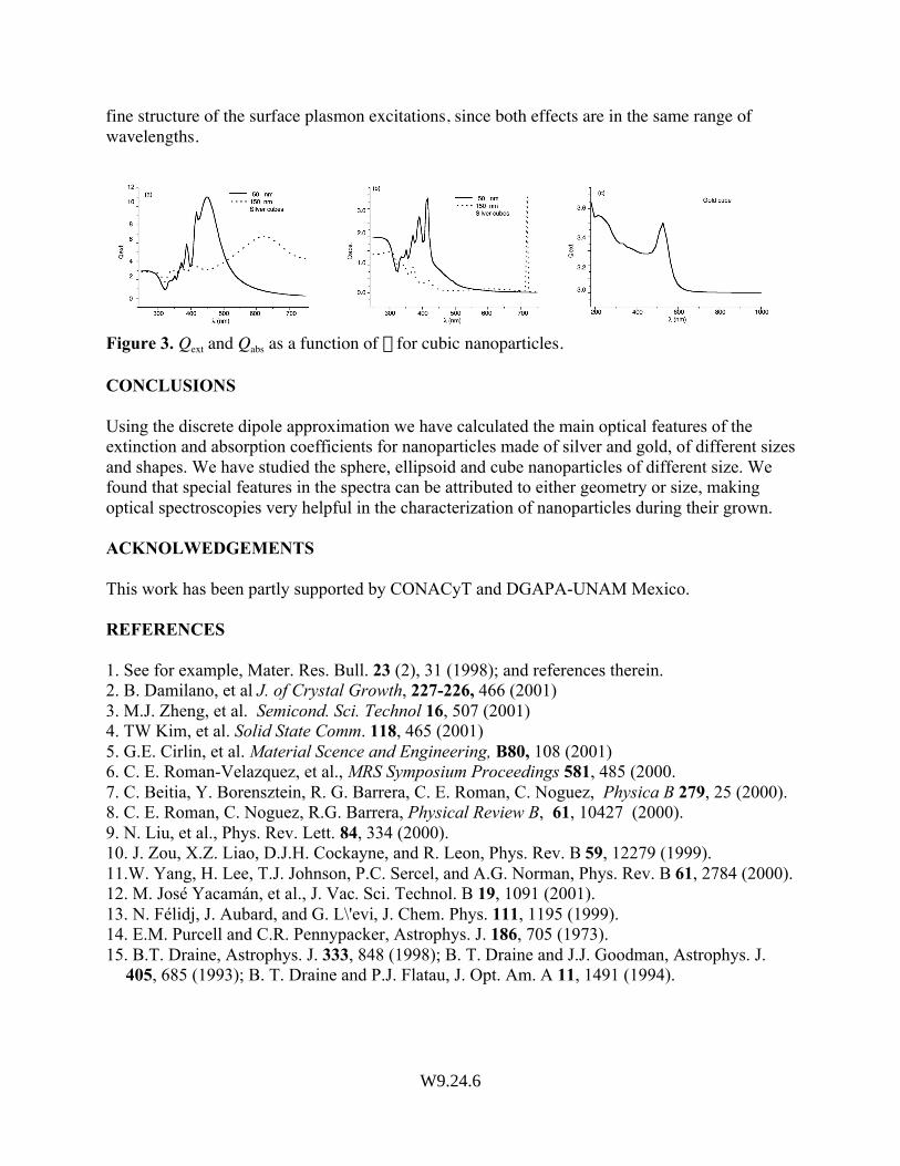
fine structure of the surface plasmon excitations, since both effects are in the same range ofwavelengths.
Figure 3. Qext and Qabs as a function of l for cubic nanoparticles.
CONCLUSIONS
Using the discrete dipole approximation we have calculated the main optical features of theextinction and absorption coefficients for nanoparticles made of silver and gold, of different sizesand shapes. We have studied the sphere, ellipsoid and cube nanoparticles of different size. Wefound that special features in the spectra can be attributed to either geometry or size, makingoptical spectroscopies very helpful in the characterization of nanoparticles during their grown.
ACKNOLWEDGEMENTS
This work has been partly supported by CONACyT and DGAPA-UNAM Mexico.
REFERENCES
1. See for example, Mater. Res. Bull. 23 (2), 31 (1998); and references therein.2. B. Damilano, et al J. of Crystal Growth, 227-226, 466 (2001)3. M.J. Zheng, et al. Semicond. Sci. Technol 16, 507 (2001)4. TW Kim, et al. Solid State Comm. 118, 465 (2001)5. G.E. Cirlin, et al. Material Scence and Engineering, B80, 108 (2001)6. C. E. Roman-Velazquez, et al., MRS Symposium Proceedings 581, 485 (2000.7. C. Beitia, Y. Borensztein, R. G. Barrera, C. E. Roman, C. Noguez, Physica B 279, 25 (2000).8. C. E. Roman, C. Noguez, R.G. Barrera, Physical Review B, 61, 10427 (2000).9. N. Liu, et al., Phys. Rev. Lett. 84, 334 (2000).10. J. Zou, X.Z. Liao, D.J.H. Cockayne, and R. Leon, Phys. Rev. B 59, 12279 (1999).11.W. Yang, H. Lee, T.J. Johnson, P.C. Sercel, and A.G. Norman, Phys. Rev. B 61, 2784 (2000).12. M. José Yacamán, et al., J. Vac. Sci. Technol. B 19, 1091 (2001).13. N. Félidj, J. Aubard, and G. L\'evi, J. Chem. Phys. 111, 1195 (1999).14. E.M. Purcell and C.R. Pennypacker, Astrophys. J. 186, 705 (1973).15. B.T. Draine, Astrophys. J. 333, 848 (1998); B. T. Draine and J.J. Goodman, Astrophys. J.
405, 685 (1993); B. T. Draine and P.J. Flatau, J. Opt. Am. A 11, 1491 (1994).
W9.24.6