Light Quality-Mediated Petiole Elongation in Arabidopsis during Shade Avoidance Involves Cell Wall...
13
Light Quality-Mediated Petiole Elongation in Arabidopsis during Shade Avoidance Involves Cell Wall Modification by Xyloglucan Endotransglucosylase/Hydrolases 1[C][W][OA] Rashmi Sasidharan, C.C. Chinnappa, Marten Staal, J. Theo M. Elzenga, Ryusuke Yokoyama, Kazuhiko Nishitani, Laurentius A.C.J. Voesenek, and Ronald Pierik* Plant Ecophysiology, Institute of Environmental Biology, Utrecht University, 3584 CA Utrecht, The Netherlands (R.S., L.A.C.J.V., R.P.); Department of Biological Sciences, University of Calgary, Calgary, Alberta, Canada T2N 1N4 (C.C.C.); Ecophysiology of Plants, University of Groningen, 9750AA Haren, The Netherlands (M.S., J.T.M.E.); and Department of Developmental Biology and Neurosciences, Graduate School of Life Sciences, Tohoku University, Sendai 980–8578, Japan (R.Y., K.N.) Some plants can avoid shaded conditions via rapid shoot elongation, thus growing into better lit areas in a canopy. Cell wall- modifying mechanisms promoting this elongation response, therefore, are important regulatory points during shade avoidance. Two major cell wall-modifying protein families are expansins and xyloglucan endotransglucosylase/hydrolases (XTHs). The role of these proteins during shade avoidance was studied in Arabidopsis (Arabidopsis thaliana). In response to two shade cues, low red to far-red light (implying neighbor proximity) and green shade (mimicking dense canopy conditions), Arabidopsis showed classic shade avoidance features: petiole elongation and leaf hyponasty. Measurement of the apoplastic proton flux in green shade-treated petioles revealed a rapid efflux of protons into the apoplast within minutes, unlike white light controls. This apoplastic acidification probably provides the acidic pH required for the optimal activity of cell wall- modifying proteins like expansins and XTHs. Acid-induced extension, expansin susceptibility, and extractable expansin activity were similar in petioles from white light- and shade-treated plants. XTH activity, however, was high in petioles exposed to shade treatments. Five XTH genes (XTH9, -15, -16, -17, and -19) were positively regulated by low red to far-red light conditions, while the latter four and XTH22 showed a significant up-regulation also in response to green shade. Consistently, knockout mutants for two of these XTH genes also had reduced or absent shade avoidance responses to these light signals. These results point toward the cell wall as a vital regulatory point during shade avoidance. Crowding in natural plant communities or in crop fields leads to resource limitation and competition for the same. In order to survive in such a situation, plants need to be able to outgrow competing vegetation to get to the light. Rapid shoot elongation, coupled with an upward movement of the leaves, are two obvious morphological characteristics displayed by plants that are being shaded. Other features include reduced branching and, when the shading is persistent, an acceleration of flowering to produce seeds and thus ensure reproduction. Collectively, this suite of re- sponses triggered by shade is referred to as the shade avoidance syndrome (SAS; Vandenbussche et al., 2005; Franklin, 2008). SAS is set into motion due to the modification of the spectral composition of light in a canopy. Light reflected from leaves gets enriched in far-red wavelengths due to the preferential absorption of red light. This reduction in red to far-red (R/FR) light is a very reliable signal of impending shade (Ballare et al., 1990). In closed canopies, light reflected from, as well as transmitted through, leaves has not just a low R/FR but also low blue fluence rates and a lower total light intensity. Perception of these light quality changes is possible in plants due to the pres- ence of Pr and Pfr (Smith, 2000; Ishimaru et al., 2007), blue light receptors, the cryptochromes, and the pho- totropins (Christie and Briggs, 2001). Rapid shoot elongation during SAS involves, primarily, cellular expansion fueled by turgor-driven uptake of water, leading to increased pressure within cells. In order to allow further water uptake, the cell walls yield to this pressure by becoming more extensible (Cosgrove, 2000). Cell wall loosening is defined as a process 1 This work was supported by the National Science and Engi- neering Council of Canada (Discovery Grant to C.C.C.) and the Netherlands Organization for Scientific Research (VENI grant no. 86306001 to R.P.). * Corresponding author; e-mail [email protected]. The author responsible for distribution of materials integral to the findings presented in this article in accordance with the policy described in the Instructions for Authors (www.plantphysiol.org) is: Rashmi Sasidharan ([email protected]). [C] Some figures in this article are displayed in color online but in black and white in the print edition. [W] The online version of this article contains Web-only data. [OA] Open Access articles can be viewed online without a sub- scription. www.plantphysiol.org/cgi/doi/10.1104/pp.110.162057 978 Plant Physiology Ò , October 2010, Vol. 154, pp. 978–990, www.plantphysiol.org Ó 2010 American Society of Plant Biologists www.plant.org on June 23, 2014 - Published by www.plantphysiol.org Downloaded from Copyright © 2010 American Society of Plant Biologists. All rights reserved.
Transcript of Light Quality-Mediated Petiole Elongation in Arabidopsis during Shade Avoidance Involves Cell Wall...
Light Quality-Mediated Petiole Elongation inArabidopsis during Shade Avoidance InvolvesCell Wall Modification by XyloglucanEndotransglucosylase/Hydrolases1[C][W][OA]
Rashmi Sasidharan, C.C. Chinnappa, Marten Staal, J. Theo M. Elzenga, Ryusuke Yokoyama,Kazuhiko Nishitani, Laurentius A.C.J. Voesenek, and Ronald Pierik*
Plant Ecophysiology, Institute of Environmental Biology, Utrecht University, 3584 CA Utrecht, TheNetherlands (R.S., L.A.C.J.V., R.P.); Department of Biological Sciences, University of Calgary, Calgary, Alberta,Canada T2N 1N4 (C.C.C.); Ecophysiology of Plants, University of Groningen, 9750AA Haren, TheNetherlands (M.S., J.T.M.E.); and Department of Developmental Biology and Neurosciences, Graduate Schoolof Life Sciences, Tohoku University, Sendai 980–8578, Japan (R.Y., K.N.)
Some plants can avoid shaded conditions via rapid shoot elongation, thus growing into better lit areas in a canopy. Cell wall-modifying mechanisms promoting this elongation response, therefore, are important regulatory points during shadeavoidance. Two major cell wall-modifying protein families are expansins and xyloglucan endotransglucosylase/hydrolases(XTHs). The role of these proteins during shade avoidance was studied in Arabidopsis (Arabidopsis thaliana). In response to twoshade cues, low red to far-red light (implying neighbor proximity) and green shade (mimicking dense canopy conditions),Arabidopsis showed classic shade avoidance features: petiole elongation and leaf hyponasty. Measurement of the apoplasticproton flux in green shade-treated petioles revealed a rapid efflux of protons into the apoplast within minutes, unlike whitelight controls. This apoplastic acidification probably provides the acidic pH required for the optimal activity of cell wall-modifying proteins like expansins and XTHs. Acid-induced extension, expansin susceptibility, and extractable expansinactivity were similar in petioles from white light- and shade-treated plants. XTH activity, however, was high in petiolesexposed to shade treatments. Five XTH genes (XTH9, -15, -16, -17, and -19) were positively regulated by low red to far-red lightconditions, while the latter four and XTH22 showed a significant up-regulation also in response to green shade. Consistently,knockout mutants for two of these XTH genes also had reduced or absent shade avoidance responses to these light signals.These results point toward the cell wall as a vital regulatory point during shade avoidance.
Crowding in natural plant communities or in cropfields leads to resource limitation and competition forthe same. In order to survive in such a situation, plantsneed to be able to outgrow competing vegetation to getto the light. Rapid shoot elongation, coupled with anupward movement of the leaves, are two obviousmorphological characteristics displayed by plants thatare being shaded. Other features include reducedbranching and, when the shading is persistent, an
acceleration of flowering to produce seeds and thusensure reproduction. Collectively, this suite of re-sponses triggered by shade is referred to as the shadeavoidance syndrome (SAS; Vandenbussche et al., 2005;Franklin, 2008). SAS is set into motion due to themodification of the spectral composition of light in acanopy. Light reflected from leaves gets enriched infar-red wavelengths due to the preferential absorptionof red light. This reduction in red to far-red (R/FR)light is a very reliable signal of impending shade(Ballare et al., 1990). In closed canopies, light reflectedfrom, as well as transmitted through, leaves has notjust a low R/FR but also low blue fluence rates and alower total light intensity. Perception of these lightquality changes is possible in plants due to the pres-ence of Pr and Pfr (Smith, 2000; Ishimaru et al., 2007),blue light receptors, the cryptochromes, and the pho-totropins (Christie and Briggs, 2001). Rapid shootelongation during SAS involves, primarily, cellularexpansion fueled by turgor-driven uptake of water,leading to increased pressure within cells. In order toallow further water uptake, the cell walls yield to thispressure by becoming more extensible (Cosgrove,2000). Cell wall loosening is defined as a process
1 This work was supported by the National Science and Engi-neering Council of Canada (Discovery Grant to C.C.C.) and theNetherlands Organization for Scientific Research (VENI grant no.86306001 to R.P.).
* Corresponding author; e-mail [email protected] author responsible for distribution of materials integral to the
findings presented in this article in accordance with the policydescribed in the Instructions for Authors (www.plantphysiol.org) is:Rashmi Sasidharan ([email protected]).
[C] Some figures in this article are displayed in color online but inblack and white in the print edition.
[W] The online version of this article contains Web-only data.[OA] Open Access articles can be viewed online without a sub-
scription.www.plantphysiol.org/cgi/doi/10.1104/pp.110.162057
978 Plant Physiology�, October 2010, Vol. 154, pp. 978–990, www.plantphysiol.org � 2010 American Society of Plant Biologists www.plant.org on June 23, 2014 - Published by www.plantphysiol.orgDownloaded from
Copyright © 2010 American Society of Plant Biologists. All rights reserved.
where molecular modifications of the cell wall makea rigid, inextensible wall extensible (Cleland, 1971;Cosgrove, 1999). This results from the action of differ-ent proteins on the cell wall matrix, which weakens thecell wall, allowing it to yield to turgor pressure. Twocell wall-modifying protein families that are wellcharacterized and implicated in cell expansion duringgrowth and development are the expansins and thexyloglucan endotransglucosylase/hydrolases (XTHs;Cosgrove, 1999, 2005; Rose et al., 2002). Expansins werefirst identified as the mediators of acid-induced exten-sion (AIE) in isolated cell walls. They are believed to actvia disruption of the noncovalent interactions betweencellulose and hemicelluloses (xyloglucan in most di-cots) in the cell wall, thus allowing cell wall loosening(McQueen-Mason et al., 1992; Cosgrove, 2000).Expansins are required for plant growth (Cho and
Kende, 1997b; Vreeburg et al., 2005) and in develop-mental processes where modification of the cell wall isrequired, such as fruit softening (Brummell et al.,1999), abscission (Belfield et al., 2005), and plant-pathogen interactions (Cantu et al., 2008). Althoughmanipulation of expansin gene expression has con-firmed the role of these proteins as important playersin the regulation of cell wall extensibility, in a fewinstances such studies have also revealed unexpectedand counterintuitive effects (Caderas et al., 2000;Rochange et al., 2001) where the correlation betweengrowth and expansin activity did not hold. XTHs areanother family of wall-modifying proteins that exist asa large gene family in most plant species. XTHs alsoact on the xyloglucan-cellulose network in the cell wall,but, unlike expansins, they employ enzymatic mecha-nisms (hydrolysis and/or transglucosylation) to mod-ify the cell wall (Nishitani and Tominaga, 1992; Roseet al., 2002). These proteins are also involved in plantgrowth and development, where wall modification isrequired (Campbell and Braam, 1999; Rose et al., 2002).In the model plant Arabidopsis (Arabidopsis thali-
ana), SAS and the importance of the regulation of cellwall extensibility by wall-modifying proteins havebeen studied extensively, albeit separately. Both ex-pansins and XTHs are present as large multigenefamilies in Arabidopsis (Rose et al., 2002; Sampedroand Cosgrove, 2005) and have been implicated inArabidopsis growth (Hyodo et al., 2003; Schopfer andLiszkay, 2006), in response to various environmentalstresses such as drought and salt tolerance (Schopferand Liszkay, 2006), and in response to pathogen/insectattack (Wieczorek et al., 2006; Divol et al., 2007).Rosette plants like Arabidopsis mostly respond toshade by moving their leaves (which normally havea prostrate orientation) upward, accompanied by anelongation of the petiole (Mullen et al., 2006; Djakovic-Petrovic et al., 2007). Numerous microarray studieshave shown that this response involves massive andrapid changes in gene expression (Devlin et al., 2003;Salter et al., 2003; Sessa et al., 2005). In addition totranscription factors and hormone regulatory genes, anumber of cell wall-related genes including XTHs and
expansins are also regulated following treatment withshade signals (Ma et al., 2001; Sessa et al., 2005; Roig-Villanova et al., 2006). The aim of this study was tomake use of this vast amount of available microarraydata as well as other genomic resources in order tostudy the regulation of cell wall properties mediatedby expansins and XTHs during shade avoidance re-sponses in Arabidopsis. We used two types of lightmanipulations to mimic different stages of shading. Alow R/FR indicates the presence of proximal neigh-bors and provides an early warning of impendingcanopy closure. Green shade replicates conditions thatwould occur in a dense canopy where there is alreadypersistent shading and crowding from neighboringplants. Using these two distinct shade cues allowedany differences in the detection and response to thesesignals to be observed. Furthermore, it was of interestto see which particular members of the expansin orXTH multigene families might be involved andwhether the regulation of these genes is light signalspecific even though they ultimately bring about thesame morphological response.
RESULTS
Arabidopsis Displays Typical Shade Avoidance
Characteristics in Response to Shade Signals
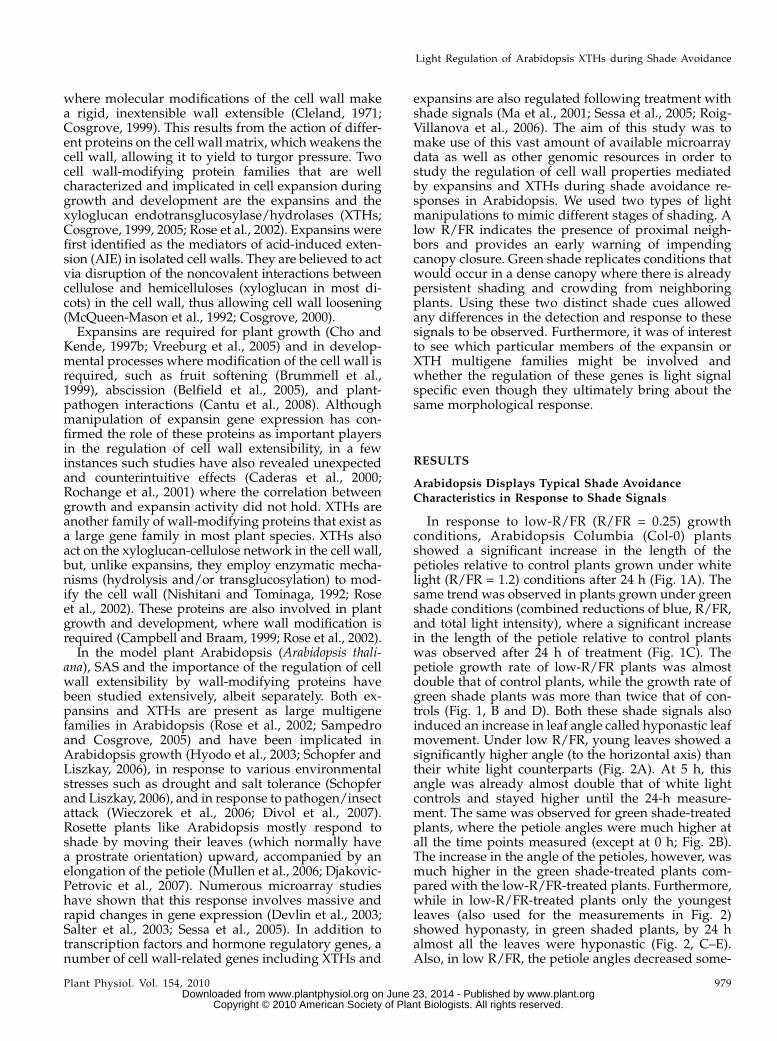
In response to low-R/FR (R/FR = 0.25) growthconditions, Arabidopsis Columbia (Col-0) plantsshowed a significant increase in the length of thepetioles relative to control plants grown under whitelight (R/FR = 1.2) conditions after 24 h (Fig. 1A). Thesame trend was observed in plants grown under greenshade conditions (combined reductions of blue, R/FR,and total light intensity), where a significant increasein the length of the petiole relative to control plantswas observed after 24 h of treatment (Fig. 1C). Thepetiole growth rate of low-R/FR plants was almostdouble that of control plants, while the growth rate ofgreen shade plants was more than twice that of con-trols (Fig. 1, B and D). Both these shade signals alsoinduced an increase in leaf angle called hyponastic leafmovement. Under low R/FR, young leaves showed asignificantly higher angle (to the horizontal axis) thantheir white light counterparts (Fig. 2A). At 5 h, thisangle was already almost double that of white lightcontrols and stayed higher until the 24-h measure-ment. The same was observed for green shade-treatedplants, where the petiole angles were much higher atall the time points measured (except at 0 h; Fig. 2B).The increase in the angle of the petioles, however, wasmuch higher in the green shade-treated plants com-pared with the low-R/FR-treated plants. Furthermore,while in low-R/FR-treated plants only the youngestleaves (also used for the measurements in Fig. 2)showed hyponasty, in green shaded plants, by 24 halmost all the leaves were hyponastic (Fig. 2, C–E).Also, in low R/FR, the petiole angles decreased some-
Light Regulation of Arabidopsis XTHs during Shade Avoidance
Plant Physiol. Vol. 154, 2010 979 www.plant.org on June 23, 2014 - Published by www.plantphysiol.orgDownloaded from
Copyright © 2010 American Society of Plant Biologists. All rights reserved.
what at 24 h relative to the 8-h reading, while the greenshade leaves retained their strongly hyponastic re-sponse until the 24-h time point (Fig. 2, A and B).
Shade Treatment Induces Rapid Apoplastic Acidificationin Arabidopsis Petioles
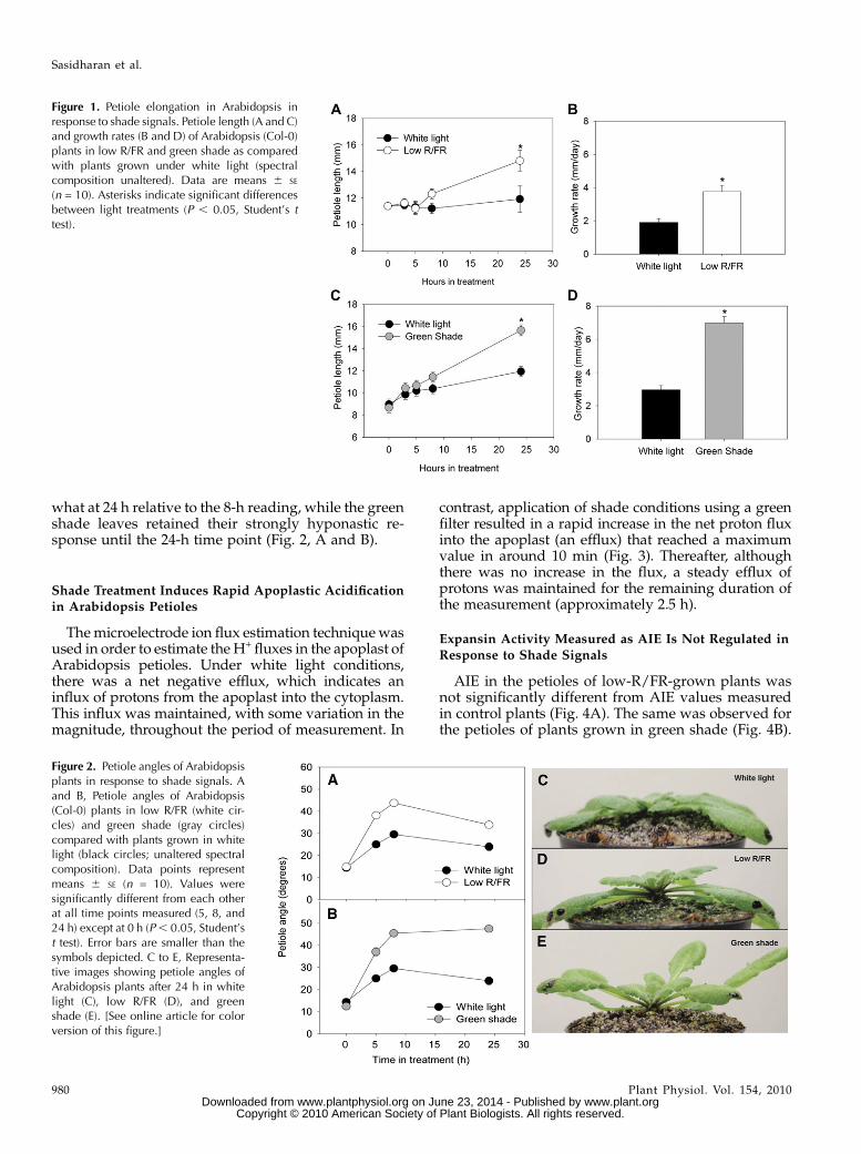
Themicroelectrode ion flux estimation techniquewasused in order to estimate theH+ fluxes in the apoplast ofArabidopsis petioles. Under white light conditions,there was a net negative efflux, which indicates aninflux of protons from the apoplast into the cytoplasm.This influx was maintained, with some variation in themagnitude, throughout the period of measurement. In
contrast, application of shade conditions using a greenfilter resulted in a rapid increase in the net proton fluxinto the apoplast (an efflux) that reached a maximumvalue in around 10 min (Fig. 3). Thereafter, althoughthere was no increase in the flux, a steady efflux ofprotons was maintained for the remaining duration ofthe measurement (approximately 2.5 h).
Expansin Activity Measured as AIE Is Not Regulated in
Response to Shade Signals
AIE in the petioles of low-R/FR-grown plants wasnot significantly different from AIE values measuredin control plants (Fig. 4A). The same was observed forthe petioles of plants grown in green shade (Fig. 4B).
Figure 1. Petiole elongation in Arabidopsis inresponse to shade signals. Petiole length (A and C)and growth rates (B and D) of Arabidopsis (Col-0)plants in low R/FR and green shade as comparedwith plants grown under white light (spectralcomposition unaltered). Data are means 6 SE
(n = 10). Asterisks indicate significant differencesbetween light treatments (P , 0.05, Student’s ttest).
Figure 2. Petiole angles of Arabidopsisplants in response to shade signals. Aand B, Petiole angles of Arabidopsis(Col-0) plants in low R/FR (white cir-cles) and green shade (gray circles)compared with plants grown in whitelight (black circles; unaltered spectralcomposition). Data points representmeans 6 SE (n = 10). Values weresignificantly different from each otherat all time points measured (5, 8, and24 h) except at 0 h (P, 0.05, Student’st test). Error bars are smaller than thesymbols depicted. C to E, Representa-tive images showing petiole angles ofArabidopsis plants after 24 h in whitelight (C), low R/FR (D), and greenshade (E). [See online article for colorversion of this figure.]
Sasidharan et al.
980 Plant Physiol. Vol. 154, 2010 www.plant.org on June 23, 2014 - Published by www.plantphysiol.orgDownloaded from
Copyright © 2010 American Society of Plant Biologists. All rights reserved.
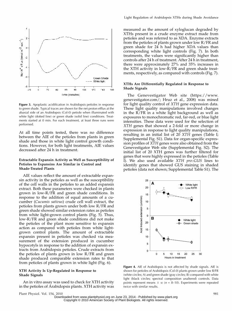
At all time points tested, there was no differencebetween the AIE of the petioles from plants in greenshade and those in white light control growth condi-tions. However, for both light treatments, AIE valuesdecreased after 24 h in treatment.
Extractable Expansin Activity as Well as Susceptibility of
Petioles to Expansins Are Similar in Control andShade-Treated Plants
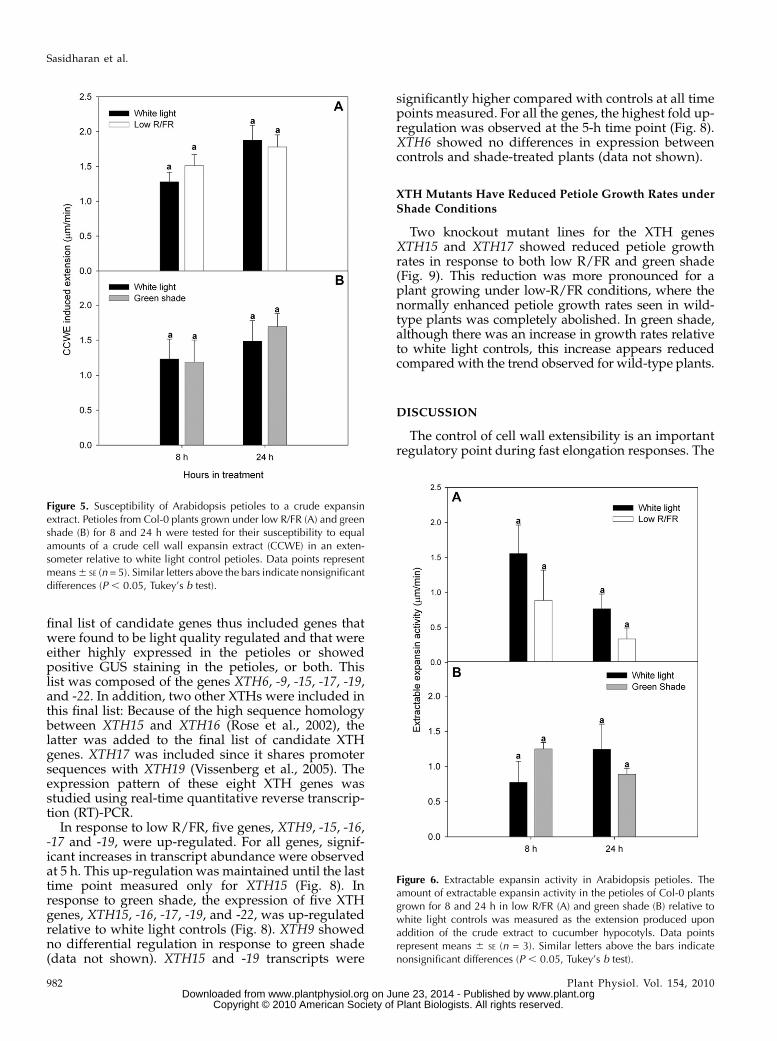
AIE values reflect the amount of extractable expan-sin activity in the petioles as well as the susceptibilityof the cell walls in the petioles to an added expansinextract. Both these parameters were checked in plantsgrown in low-R/FR and green shade conditions. Inresponse to the addition of equal amounts of a cu-cumber (Cucumis sativus) crude cell wall extract, thepetioles from plants grown under both low R/FR andgreen shade showed similar extension rates as petiolesfrom white light-grown control plants (Fig. 5). Thus,low-R/FR and green shade conditions did not makethe petioles of the plant more sensitive to expansinaction as compared with petioles from white light-grown control plants. The amount of extractableexpansin present in petioles was checked via mea-surement of the extension produced in cucumberhypocotyls in response to the addition of expansin ex-tracts from Arabidopsis petioles. Crude extracts fromthe petioles of plants grown in low R/FR and greenshade produced comparable extension rates to thatfrom petioles of plants grown in white light (Fig. 6).
XTH Activity Is Up-Regulated in Response toShade Signals
An in vitro assay was used to check for XTH activityin the petioles of Arabidopsis plants. XTH activity was
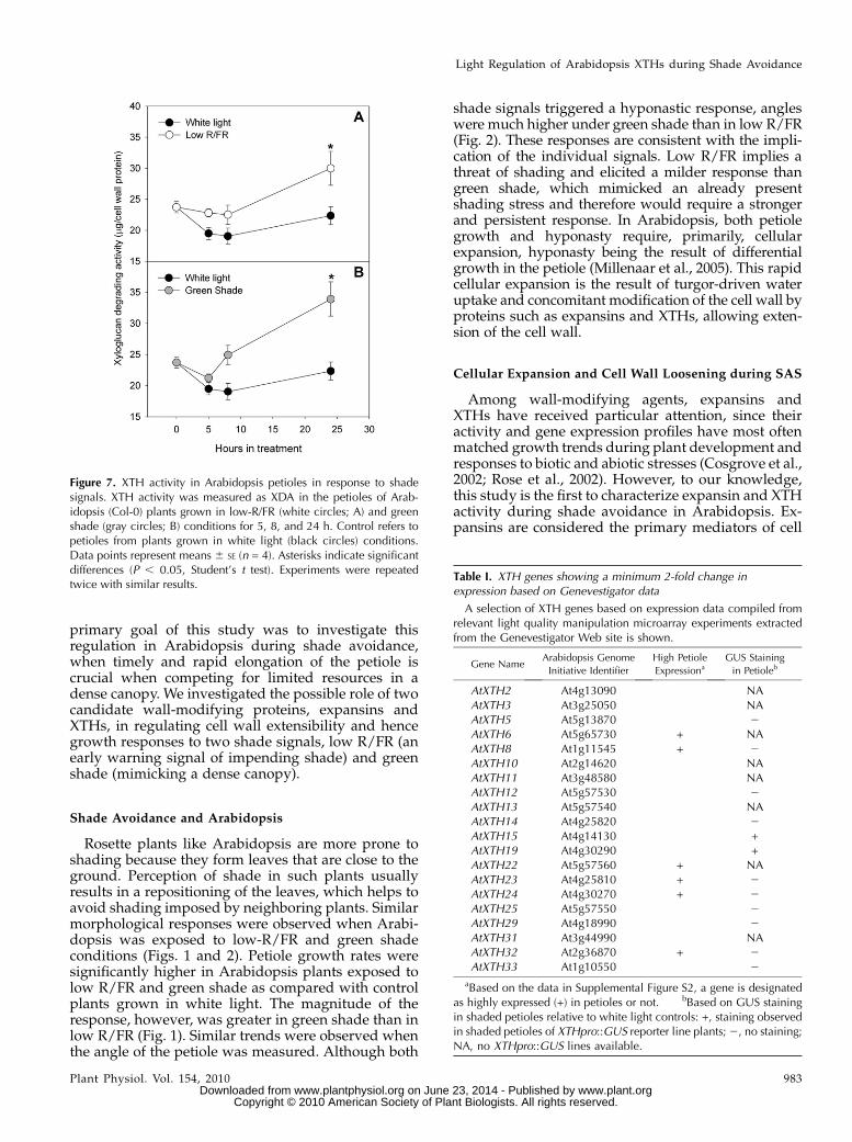
measured as the amount of xyloglucan degraded byXTHs present in a crude enzyme extract made frompetioles and was referred to as XDA. Enzyme extractsfrom the petioles of plants grown under low R/FR andgreen shade for 24 h had higher XDA values thancorresponding white light controls (Fig. 7). In bothtreatments, the values were significantly higher thancontrols after 24 h of treatment. After 24 h in treatment,there were approximately 27% and 35% increases inthe XTH activity in low-R/FR and green shade treat-ments, respectively, as compared with controls (Fig. 7).
XTHs Are Differentially Regulated in Response to
Shade Signals
The Genevestigator Web site (https://www.genevestigator.com/; Hruz et al., 2008) was minedfor light quality control of XTH gene expression data.These light quality manipulations involved changesin the R/FR in a white light background as well asexposures to monochromatic red, far-red, or blue lightintensities. These data were used for the selection ofXTH genes that showed a 2-fold or more change inexpression in response to light quality manipulations,resulting in an initial list of 20 XTH genes (Table I;Supplemental Fig. S1). Data for organ-specific expres-sion profiles of XTH genes were also obtained from theGenevestigator Web site (Supplemental Fig. S2). Theinitial list of 20 XTH genes was further filtered forgenes that were highly expressed in the petioles (TableI). We also used available XTH pro::GUS lines toidentify genes that showed GUS staining in shadedpetioles (data not shown; Supplemental Table S1). The
Figure 3. Apoplastic acidification in Arabidopsis petioles in responseto green shade. Typical traces are shown for the net proton efflux at theabaxial side of an Arabidopsis (Col-0) petiole when illuminated withwhite light (dotted line) or green shade (solid line) conditions. Treat-ments started at 0 min. For each treatment, at least three runs wereperformed.
Figure 4. AIE of Arabidopsis is not affected by shade signals. AIE isshown for petioles of Arabidopsis (Col-0) plants grown under low R/FR(white circles; A) and green shade (gray circles; B) compared with whitelight (black circles; spectral composition unaltered) controls. Datapoints represent means 6 SE (n = 8–10). Experiments were repeatedtwice with similar results.
Light Regulation of Arabidopsis XTHs during Shade Avoidance
Plant Physiol. Vol. 154, 2010 981 www.plant.org on June 23, 2014 - Published by www.plantphysiol.orgDownloaded from
Copyright © 2010 American Society of Plant Biologists. All rights reserved.
final list of candidate genes thus included genes thatwere found to be light quality regulated and that wereeither highly expressed in the petioles or showedpositive GUS staining in the petioles, or both. Thislist was composed of the genes XTH6, -9, -15, -17, -19,and -22. In addition, two other XTHs were included inthis final list: Because of the high sequence homologybetween XTH15 and XTH16 (Rose et al., 2002), thelatter was added to the final list of candidate XTHgenes. XTH17 was included since it shares promotersequences with XTH19 (Vissenberg et al., 2005). Theexpression pattern of these eight XTH genes wasstudied using real-time quantitative reverse transcrip-tion (RT)-PCR.
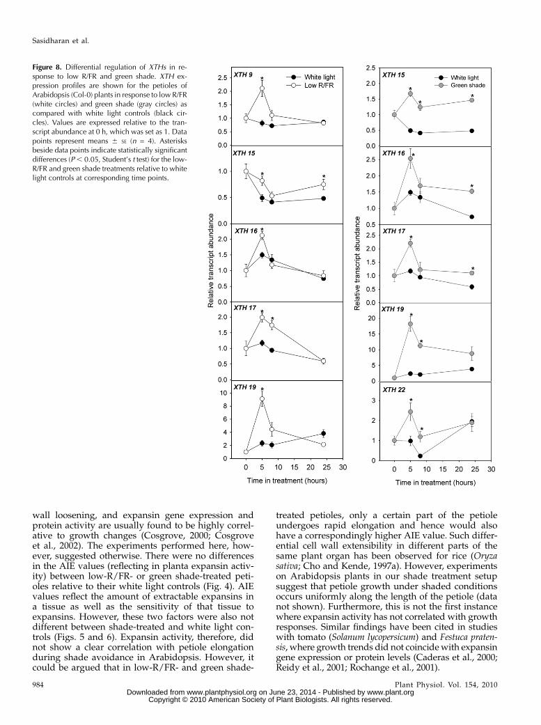
In response to low R/FR, five genes, XTH9, -15, -16,-17 and -19, were up-regulated. For all genes, signif-icant increases in transcript abundance were observedat 5 h. This up-regulation was maintained until the lasttime point measured only for XTH15 (Fig. 8). Inresponse to green shade, the expression of five XTHgenes, XTH15, -16, -17, -19, and -22, was up-regulatedrelative to white light controls (Fig. 8). XTH9 showedno differential regulation in response to green shade(data not shown). XTH15 and -19 transcripts were
significantly higher compared with controls at all timepoints measured. For all the genes, the highest fold up-regulation was observed at the 5-h time point (Fig. 8).XTH6 showed no differences in expression betweencontrols and shade-treated plants (data not shown).
XTH Mutants Have Reduced Petiole Growth Rates under
Shade Conditions
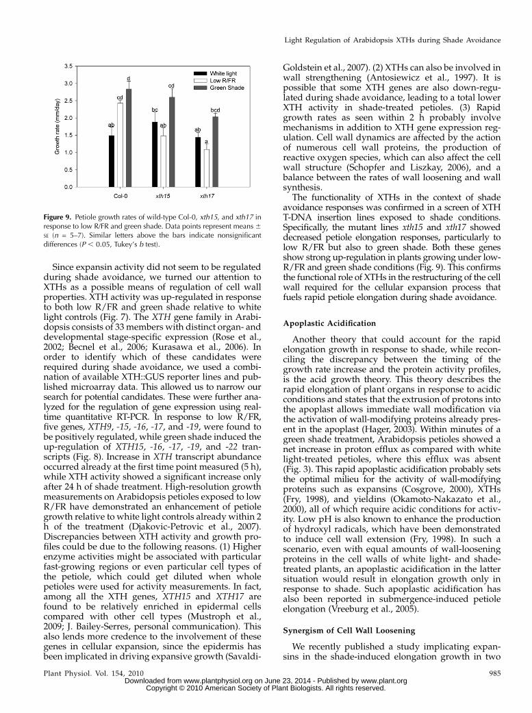
Two knockout mutant lines for the XTH genesXTH15 and XTH17 showed reduced petiole growthrates in response to both low R/FR and green shade(Fig. 9). This reduction was more pronounced for aplant growing under low-R/FR conditions, where thenormally enhanced petiole growth rates seen in wild-type plants was completely abolished. In green shade,although there was an increase in growth rates relativeto white light controls, this increase appears reducedcomparedwith the trend observed for wild-type plants.
DISCUSSION
The control of cell wall extensibility is an importantregulatory point during fast elongation responses. The
Figure 6. Extractable expansin activity in Arabidopsis petioles. Theamount of extractable expansin activity in the petioles of Col-0 plantsgrown for 8 and 24 h in low R/FR (A) and green shade (B) relative towhite light controls was measured as the extension produced uponaddition of the crude extract to cucumber hypocotyls. Data pointsrepresent means 6 SE (n = 3). Similar letters above the bars indicatenonsignificant differences (P , 0.05, Tukey’s b test).
Figure 5. Susceptibility of Arabidopsis petioles to a crude expansinextract. Petioles from Col-0 plants grown under low R/FR (A) and greenshade (B) for 8 and 24 h were tested for their susceptibility to equalamounts of a crude cell wall expansin extract (CCWE) in an exten-someter relative to white light control petioles. Data points representmeans6 SE (n = 5). Similar letters above the bars indicate nonsignificantdifferences (P , 0.05, Tukey’s b test).
Sasidharan et al.
982 Plant Physiol. Vol. 154, 2010 www.plant.org on June 23, 2014 - Published by www.plantphysiol.orgDownloaded from
Copyright © 2010 American Society of Plant Biologists. All rights reserved.
primary goal of this study was to investigate thisregulation in Arabidopsis during shade avoidance,when timely and rapid elongation of the petiole iscrucial when competing for limited resources in adense canopy. We investigated the possible role of twocandidate wall-modifying proteins, expansins andXTHs, in regulating cell wall extensibility and hencegrowth responses to two shade signals, low R/FR (anearly warning signal of impending shade) and greenshade (mimicking a dense canopy).
Shade Avoidance and Arabidopsis
Rosette plants like Arabidopsis are more prone toshading because they form leaves that are close to theground. Perception of shade in such plants usuallyresults in a repositioning of the leaves, which helps toavoid shading imposed by neighboring plants. Similarmorphological responses were observed when Arabi-dopsis was exposed to low-R/FR and green shadeconditions (Figs. 1 and 2). Petiole growth rates weresignificantly higher in Arabidopsis plants exposed tolow R/FR and green shade as compared with controlplants grown in white light. The magnitude of theresponse, however, was greater in green shade than inlow R/FR (Fig. 1). Similar trends were observed whenthe angle of the petiole was measured. Although both
shade signals triggered a hyponastic response, angleswere much higher under green shade than in low R/FR(Fig. 2). These responses are consistent with the impli-cation of the individual signals. Low R/FR implies athreat of shading and elicited a milder response thangreen shade, which mimicked an already presentshading stress and therefore would require a strongerand persistent response. In Arabidopsis, both petiolegrowth and hyponasty require, primarily, cellularexpansion, hyponasty being the result of differentialgrowth in the petiole (Millenaar et al., 2005). This rapidcellular expansion is the result of turgor-driven wateruptake and concomitant modification of the cell wall byproteins such as expansins and XTHs, allowing exten-sion of the cell wall.
Cellular Expansion and Cell Wall Loosening during SAS
Among wall-modifying agents, expansins andXTHs have received particular attention, since theiractivity and gene expression profiles have most oftenmatched growth trends during plant development andresponses to biotic and abiotic stresses (Cosgrove et al.,2002; Rose et al., 2002). However, to our knowledge,this study is the first to characterize expansin and XTHactivity during shade avoidance in Arabidopsis. Ex-pansins are considered the primary mediators of cell
Figure 7. XTH activity in Arabidopsis petioles in response to shadesignals. XTH activity was measured as XDA in the petioles of Arab-idopsis (Col-0) plants grown in low-R/FR (white circles; A) and greenshade (gray circles; B) conditions for 5, 8, and 24 h. Control refers topetioles from plants grown in white light (black circles) conditions.Data points represent means 6 SE (n = 4). Asterisks indicate significantdifferences (P , 0.05, Student’s t test). Experiments were repeatedtwice with similar results.
Table I. XTH genes showing a minimum 2-fold change inexpression based on Genevestigator data
A selection of XTH genes based on expression data compiled fromrelevant light quality manipulation microarray experiments extractedfrom the Genevestigator Web site is shown.
Gene NameArabidopsis Genome
Initiative Identifier
High Petiole
ExpressionaGUS Staining
in Petioleb
AtXTH2 At4g13090 NAAtXTH3 At3g25050 NAAtXTH5 At5g13870 2AtXTH6 At5g65730 + NAAtXTH8 At1g11545 + 2AtXTH10 At2g14620 NAAtXTH11 At3g48580 NAAtXTH12 At5g57530 2AtXTH13 At5g57540 NAAtXTH14 At4g25820 2AtXTH15 At4g14130 +AtXTH19 At4g30290 +AtXTH22 At5g57560 + NAAtXTH23 At4g25810 + 2AtXTH24 At4g30270 + 2AtXTH25 At5g57550 2AtXTH29 At4g18990 2AtXTH31 At3g44990 NAAtXTH32 At2g36870 + 2AtXTH33 At1g10550 2
aBased on the data in Supplemental Figure S2, a gene is designatedas highly expressed (+) in petioles or not. bBased on GUS stainingin shaded petioles relative to white light controls: +, staining observedin shaded petioles of XTHpro::GUS reporter line plants;2, no staining;NA, no XTHpro::GUS lines available.
Light Regulation of Arabidopsis XTHs during Shade Avoidance
Plant Physiol. Vol. 154, 2010 983 www.plant.org on June 23, 2014 - Published by www.plantphysiol.orgDownloaded from
Copyright © 2010 American Society of Plant Biologists. All rights reserved.
wall loosening, and expansin gene expression andprotein activity are usually found to be highly correl-ative to growth changes (Cosgrove, 2000; Cosgroveet al., 2002). The experiments performed here, how-ever, suggested otherwise. There were no differencesin the AIE values (reflecting in planta expansin activ-ity) between low-R/FR- or green shade-treated peti-oles relative to their white light controls (Fig. 4). AIEvalues reflect the amount of extractable expansins ina tissue as well as the sensitivity of that tissue toexpansins. However, these two factors were also notdifferent between shade-treated and white light con-trols (Figs. 5 and 6). Expansin activity, therefore, didnot show a clear correlation with petiole elongationduring shade avoidance in Arabidopsis. However, itcould be argued that in low-R/FR- and green shade-
treated petioles, only a certain part of the petioleundergoes rapid elongation and hence would alsohave a correspondingly higher AIE value. Such differ-ential cell wall extensibility in different parts of thesame plant organ has been observed for rice (Oryzasativa; Cho and Kende, 1997a). However, experimentson Arabidopsis plants in our shade treatment setupsuggest that petiole growth under shaded conditionsoccurs uniformly along the length of the petiole (datanot shown). Furthermore, this is not the first instancewhere expansin activity has not correlated with growthresponses. Similar findings have been cited in studieswith tomato (Solanum lycopersicum) and Festuca praten-sis, where growth trends did not coincidewith expansingene expression or protein levels (Caderas et al., 2000;Reidy et al., 2001; Rochange et al., 2001).
Figure 8. Differential regulation of XTHs in re-sponse to low R/FR and green shade. XTH ex-pression profiles are shown for the petioles ofArabidopsis (Col-0) plants in response to low R/FR(white circles) and green shade (gray circles) ascompared with white light controls (black cir-cles). Values are expressed relative to the tran-script abundance at 0 h, which was set as 1. Datapoints represent means 6 SE (n = 4). Asterisksbeside data points indicate statistically significantdifferences (P, 0.05, Student’s t test) for the low-R/FR and green shade treatments relative to whitelight controls at corresponding time points.
Sasidharan et al.
984 Plant Physiol. Vol. 154, 2010 www.plant.org on June 23, 2014 - Published by www.plantphysiol.orgDownloaded from
Copyright © 2010 American Society of Plant Biologists. All rights reserved.
Since expansin activity did not seem to be regulatedduring shade avoidance, we turned our attention toXTHs as a possible means of regulation of cell wallproperties. XTH activity was up-regulated in responseto both low R/FR and green shade relative to whitelight controls (Fig. 7). The XTH gene family in Arabi-dopsis consists of 33 members with distinct organ- anddevelopmental stage-specific expression (Rose et al.,2002; Becnel et al., 2006; Kurasawa et al., 2006). Inorder to identify which of these candidates wererequired during shade avoidance, we used a combi-nation of available XTH::GUS reporter lines and pub-lished microarray data. This allowed us to narrow oursearch for potential candidates. These were further ana-lyzed for the regulation of gene expression using real-time quantitative RT-PCR. In response to low R/FR,five genes, XTH9, -15, -16, -17, and -19, were found tobe positively regulated, while green shade induced theup-regulation of XTH15, -16, -17, -19, and -22 tran-scripts (Fig. 8). Increase in XTH transcript abundanceoccurred already at the first time point measured (5 h),while XTH activity showed a significant increase onlyafter 24 h of shade treatment. High-resolution growthmeasurements on Arabidopsis petioles exposed to lowR/FR have demonstrated an enhancement of petiolegrowth relative to white light controls already within 2h of the treatment (Djakovic-Petrovic et al., 2007).Discrepancies between XTH activity and growth pro-files could be due to the following reasons. (1) Higherenzyme activities might be associated with particularfast-growing regions or even particular cell types ofthe petiole, which could get diluted when wholepetioles were used for activity measurements. In fact,among all the XTH genes, XTH15 and XTH17 arefound to be relatively enriched in epidermal cellscompared with other cell types (Mustroph et al.,2009; J. Bailey-Serres, personal communication). Thisalso lends more credence to the involvement of thesegenes in cellular expansion, since the epidermis hasbeen implicated in driving expansive growth (Savaldi-
Goldstein et al., 2007). (2) XTHs can also be involved inwall strengthening (Antosiewicz et al., 1997). It ispossible that some XTH genes are also down-regu-lated during shade avoidance, leading to a total lowerXTH activity in shade-treated petioles. (3) Rapidgrowth rates as seen within 2 h probably involvemechanisms in addition to XTH gene expression reg-ulation. Cell wall dynamics are affected by the actionof numerous cell wall proteins, the production ofreactive oxygen species, which can also affect the cellwall structure (Schopfer and Liszkay, 2006), and abalance between the rates of wall loosening and wallsynthesis.
The functionality of XTHs in the context of shadeavoidance responses was confirmed in a screen of XTHT-DNA insertion lines exposed to shade conditions.Specifically, the mutant lines xth15 and xth17 showeddecreased petiole elongation responses, particularly tolow R/FR but also to green shade. Both these genesshow strong up-regulation in plants growing under low-R/FR and green shade conditions (Fig. 9). This confirmsthe functional role of XTHs in the restructuring of the cellwall required for the cellular expansion process thatfuels rapid petiole elongation during shade avoidance.
Apoplastic Acidification
Another theory that could account for the rapidelongation growth in response to shade, while recon-ciling the discrepancy between the timing of thegrowth rate increase and the protein activity profiles,is the acid growth theory. This theory describes therapid elongation of plant organs in response to acidicconditions and states that the extrusion of protons intothe apoplast allows immediate wall modification viathe activation of wall-modifying proteins already pres-ent in the apoplast (Hager, 2003). Within minutes of agreen shade treatment, Arabidopsis petioles showed anet increase in proton efflux as compared with whitelight-treated petioles, where this efflux was absent(Fig. 3). This rapid apoplastic acidification probably setsthe optimal milieu for the activity of wall-modifyingproteins such as expansins (Cosgrove, 2000), XTHs(Fry, 1998), and yieldins (Okamoto-Nakazato et al.,2000), all of which require acidic conditions for activ-ity. Low pH is also known to enhance the productionof hydroxyl radicals, which have been demonstratedto induce cell wall extension (Fry, 1998). In such ascenario, even with equal amounts of wall-looseningproteins in the cell walls of white light- and shade-treated plants, an apoplastic acidification in the lattersituation would result in elongation growth only inresponse to shade. Such apoplastic acidification hasalso been reported in submergence-induced petioleelongation (Vreeburg et al., 2005).
Synergism of Cell Wall Loosening
We recently published a study implicating expan-sins in the shade-induced elongation growth in two
Figure 9. Petiole growth rates of wild-type Col-0, xth15, and xth17 inresponse to low R/FR and green shade. Data points represent means 6SE (n = 5–7). Similar letters above the bars indicate nonsignificantdifferences (P , 0.05, Tukey’s b test).
Light Regulation of Arabidopsis XTHs during Shade Avoidance
Plant Physiol. Vol. 154, 2010 985 www.plant.org on June 23, 2014 - Published by www.plantphysiol.orgDownloaded from
Copyright © 2010 American Society of Plant Biologists. All rights reserved.
ecotypes of the Caryophyllaceae species Stellaria long-ipes (Sasidharan et al., 2008). It was expected that thebasic wall-modifying mechanism underlying elonga-tion responses to identical cues (low R/FR and greenshade) would be similar. Therefore, it was surprisingto observe that, unlike S. longipes, in Arabidopsis XTHsshow a much stronger correlation with shade-inducedelongation growth. Even though the cell wall containsnumerous enzyme activities, expansins have beenconsidered primary wall-loosening agents mostlydue to their ability to bring about extension of isolatedwall specimens (Cosgrove, 2005). Most studies inves-tigating elongation growth in response to environ-mental cues have supported this notion (Wu et al.,1996; Cho and Kende, 1997a; Vriezen et al., 2000;Cosgrove et al., 2002; Sasidharan et al., 2008). How-ever, there have also been a few studies where ex-pansin activity and/or gene expression have notmatched growth trends (Caderas et al., 2000; Rochangeet al., 2001). In addition, XTHs have recently beendemonstrated to be able to bring about wall-looseningactivity in vitro (Van Sandt et al., 2007). Therefore,XTHs, although sometimes considered secondarywall-loosening agents, do have the ability to be pri-mary candidates. This study also supports the role ofXTHs as primary wall-modifying agents during shadeavoidance in Arabidopsis. However, it is likely not tobe the sole mechanism. Cell wall properties are veryoften the result of coordinate activities of cell wallassembly, disassembly, polymer synthesis, and secre-tion. Most large-scale analyses of cell walls haverevealed a number of cell wall-related proteins to beregulated during different developmental stages andin response to environmental stresses. These proteinsact on specific parts of the cell wall and have charac-teristic effects on it. In all likelihood, wall looseningrequires a synergism between these different proteins.In Arabidopsis as well, the primary action of XTHs isprobably enhanced or supplemented by other wall-modifying agents such as expansins, endoglucanases,and free radicals.
During expansion, regulation occurs not just at thelevel of wall modification but also for the delivery ofwall components to the cell wall (Cosgrove, 2005).Furthermore, cell wall composition differs not justbetween species but also between different develop-mental stages and in response to different environ-
mental stresses, as has been demonstrated forArabidopsis roots (Freshour et al., 1996). Thus, thecomposition of a particular cell wall at a particulartime, and in a particular species and organ, wouldprobably dictate the kind of enzymatic activities re-quired. This suggestion is supported by large-scaletranscriptomic and proteomic studies that have re-vealed how not just different protein families, but alsospecific members within these protein families, arecoordinately expressed at different developmentalstages (Yokoyama et al., 2004; Imoto et al., 2005; Irshadet al., 2008). In addition, comparisons have beendrawn with cellulose-digesting microbes that utilizemultiprotein complexes to coordinate multiple proteinactivities while acting on their target. It is speculatedthat such a complex might exist in plants and wouldallow multiple proteins to be present together at theright location in the cell wall, thereby increasing theefficiency of wall modification (Rose and Bennett,1999). Finally, the activities of both expansins andXTHs can be affected by conditions in the cell wall,such as substrate availability, accessibility to the target,the presence of activators/inhibitors, and also the pHof the apoplast. There was a rapid efflux of protonsinto the apoplastic space in Arabidopsis petioles whenexposed to shaded conditions. Thus, the amount ofacidification can also determine which proteins areactive and to what extent. Therefore, coordinationwith other pathways that determine the optimal mi-lieu for enzyme activities would also be essential.
CONCLUSION
In conclusion, these results demonstrate that Arabi-dopsis can clearly distinguish between different shadesignals that imply either neighbor proximity (low R/FR)or shade (green shade). In response to each of thesesignals, Arabidopsis plants responded with characteris-tic morphological responses that differed in magnitudeand that corresponded with an increase in XTH activity.Different XTH genes were expressed in response to eachshade cue, and this increase in gene expression isprobably responsible for the observed increase in XTHactivity. Furthermore, the reduced ability of xth mutantsto enhance petiole elongation when shaded pointstoward a functional role for XTHs in regulating cell
Table II. Sequences (5#–3#) of primer combinations and annealing temperatures for the genes studied using real-time RT-PCR
Arabidopsis Genome
Initiative IdentifierGene Name Forward Primer Reverse Primer
Annealing
Temperature
�CAt4g03210 AtXTH9 TACCATGAATACAACACTGCGTTTACT TACCATGAATACAACACTGCGTTTACT 62At4g14130 AtXTH15 CGGCACCGTCACTGCTTAC GAAACTCAAAGTCTATCTCGTCATGTG 62At3g23730 AtXTH16 CCGGTAACTCCGCTGGAA TCTCGTCGTGTGTTGGTCCTT 62At1g65310 AtXTH17 ATGGGCTAATGGAAAATCATCTTGTT TACTTTGCACACCTTTCATTCTTGTC 62At4g30290 AtXTH19 TGCAGCTAAATGATTGATTCTTTGAT CCATTGAGTTACAAAGACAACGTCA 62At5g57560 AtXTH22 CTAAAGAGTGCTTAGCTGCATAGAGAGA CAAATCAATAAAATTCACGTGATCTACAA 62At4g05320 AtUBQ10 GGCCTTGTATAATCCCTGATGAATAAG AAAGAGATAACAGGAACGGAAACATAGT 62
Sasidharan et al.
986 Plant Physiol. Vol. 154, 2010 www.plant.org on June 23, 2014 - Published by www.plantphysiol.orgDownloaded from
Copyright © 2010 American Society of Plant Biologists. All rights reserved.
wall properties that are essential for elongation re-sponses during shade avoidance. Although expansinactivity appeared not to be regulated during responsesto shade, they may play a role in regulating wallextensibility through a collaborative or synergisticaction with other wall-modifying mechanisms, includ-ing XTHs. Rapid apoplastic acidification was initiatedupon green shade conditions and probably providesthe optimum environment for wall-modifying pro-cesses to occur.
MATERIALS AND METHODS
Plant Growth
Both wild-type and transgenic Arabidopsis (Arabidopsis thaliana) plants
used were in the Col-0 wild-type background. Seeds were sown on a moist
filter paper placed in sealed petri dishes. After a 4-d stratification period in the
dark at 4�C, the petri dishes were placed in a growth chamber for another 4 d
in order to allow seed germination. Seedlings were then transferred to 70-mL
pots filled with a soil and perlite (1:2, v/v) mixture containing 0.14 mg of
MgOCaO (17%; Vitasol) and 0.14 mg of slow-release fertilizer (Osmocote
“plus mini”; Scotts Europe). Prior to seedling transfer, the pots containing soil
were allowed to soak up 20 mL of nutrient solution containing 2.6 mM KNO3,
2.0 mM Ca(NO3)2, 0.6 mM KH2PO4, 0.9 mM MgSO4, 6.6 mM MnSO4, 2.8 mM
ZnSO4, 0.5 mM CuSO4, 66 mM H3BO3, 0.8 mM Na2MoO4, and 134 mM Fe-EDTA,
pH 5.8. All chemicals were analytical reagent grade and obtained fromMerck.
The pots were placed on irrigation mats that were automatically watered
every day in a short-day (9-h photoperiod, 200 mmol m22 s21 photosynthet-
ically active radiation [PAR]) growth chamber for 4 weeks before being used
for experiments.
Light Treatments
Four-week-old plants were used for experiments. Light quality manipu-
lations took place in a white light background (Philips Master HPI-T Plus 400
Wand Philips Plus Line Pro 150W). The R/FRwas reduced from 1.2 to 0.25 by
supplemental far-red light (730-nm light-emitting diode; Shinko Electronics
[http://www.shinkohelecs.com]). PAR for this light treatment was main-
tained at 140 mmol m22 s21. Green shading mimicking light conditions in a
dense canopy was achieved using two layers of Lee 122 Fern Green, which
reduced the PAR to 65 mmol m22 s21, the R/FR to 0.19, and the blue light
photon fluence rate to 2 mmol m22 s21. Wherever mentioned, white light
control refers to data from plants grown in light conditions with an un-
altered spectral composition and PAR of 140 mmol m22 s21. All light treat-
ments were started at approximately 10 AM each time the experiments were
performed.
Measurement of Plant Growth
Care was taken to choose similar-sized plants. The same leaf in each pot
was marked with ink, and the corresponding petiole was measured using a
digital caliper at relevant time points at the start and for the duration of
specific treatments. For each treatment, petiole lengths were measured from at
least 10 individual plants. Measurements were made for three independent
trials.
Measurement of Leaf Angle
The leaves to be measured were marked with ink before the start of the
treatments. Leaves obstructing the leaves to be measured were cut in order to
be able to make angle measurements. Angle measurements were made by
photographing the plants at relevant time points and then measuring the
angle X between the two marked leaves using the angle measurement tool of
the image software ImageJ (Abramoff et al., 2004). This angle Xwas then used
to estimate the angle of the marked leaf (Y) from the soil surface using the
formula Y = (180 – X)/2. At least 10 plants were used per treatment per time
point. The experiments were repeated twice.
Measurement of Apoplastic Proton Fluxes
The microelectrode ion flux estimation (Newman, 2001) technique was
used to measure net H+ fluxes in petioles from 28-d-old Arabidopsis plants.
Microelectrodes were pulled from borosilicate glass capillaries (GC150-10;
Harvard Apparatus) and silanized with tributylchlorosilane (Fluka). Before
use, the microelectrodes were backfilled with 15 mM NaCl plus 40 mM KH2PO4
and the tip was filled with Hydrogen Ionophore II (Cocktail A; Fluka). After
calibration of the microelectrode (pH 5.1–7.8), a young Arabidopsis leaf was
detached from the plant, its petiole was gently abraded using Carborundum,
and the leaf was then mounted in a 3-mL cuvette filled with 1 mM KCl. The
cuvette was then placed in front of an inverted microscope (Nikon), and the
H+ selective microelectrode was mounted vertically in a holder connected to a
three-way piezo-controlled micromanipulator (Luigs and Neumann) driven
by a computer-controlled motor (M061-CE08; Superior Electric). The electrode
was then brought into position next to the abaxial surface of the petiole at a
distance of 20 mm. During measurements, the distance between the micro-
electrode and the petiole surface changed from 20 to 60 mm with a frequency
of 0.1 Hz. For controls, measurements were performed with the leaf illumi-
nated using two fiber-optic cables (PAR of 180 mm m22 s21). For shade
treatments, two layers of green filter (Lee 122 Fern Green) were placed across
the two fiber-optic cables. The solution used to submerge the leaf was bubbled
overnight with CO2-free air, and during the experiment CO2-free air was
blown over the surface of the cuvette to minimize the effect of changing CO2
concentrations on the pH of the experimental solution. Chemical activity of
H+ was recorded at two distances, and these data were then used to generate
H+ flux values according to Newman (2001).
Measurement of AIE
Leaves with petioles that were at least 6 mm in length were marked before
the start of the light treatments. This was the minimal starting length required
in order to be able to place the petiole in the extensometer setup. Petioles were
harvested at relevant time points and immediately frozen in liquid nitrogen.
Thawed, abraded, and pressed petioles were clamped in the extensometer
cuvette under a constant weight of 20 g. The petioles were first bathed in 160
mL of a 50 mM HEPES (pH 6.8) buffer for 30 min, after which the buffer was
replaced with a 50 mM sodium acetate (pH 4.5) buffer for another 30 min. AIE
was measured as the difference in the slopes of lines fitted through 10-min
intervals before and after the observed bending point obtained upon the
change in pH of the buffer. For each treatment, at least six to eight petioles
from individual plants were measured. Experiments were repeated at least
two times.
Extraction of Cell Wall Proteins and Measurement ofExtractable Expansin Protein Activity
Crude cell wall extracts were made as described by Rochange et al. (2001)
and modified to extract in 1.5-mL tubes. Briefly, 30 Arabidopsis petioles (two
petioles per plant) were harvested and pooled per biological replicate, per
treatment, per time point. The petioles were immediately frozen and then
ground in a homogenization buffer. The cell walls were retained via centri-
fugation (21,000g, 1 min). The cell wall pellet was then washed several times
with a wash buffer. This was followed by the addition of the extraction buffer
containing 1 M NaCl. The cell walls were then allowed to incubate in this
extraction buffer, after which the supernatant was recovered via centrifuga-
tion. The proteins were precipitated via the addition of ammonium sulfate and
then recovered via centrifugation. Protein pellets were suspended in 50 mM
sodium acetate buffer, pH 4.5, and the amount of protein was estimated using
the method of Bradford (1976).
Susceptibility of Arabidopsis Petioles to Crude Cell WallExtracts from Cucumber Hypocotyls
Arabidopsis petioles were harvested from plants grown under different
light treatments (control, low R/FR, and green shade) and then frozen
immediately and stored at 280�C. Frozen, thawed petioles were then boiled
for 5 min to eliminate any endogenous expansin activity. These petioles were
then pressed for 2 min and mounted in the extensometer setup under a
constant pulling weight of 20 g. Extension was first measured for 30 min in an
acidic (50 mM sodium acetate, pH 4.5) buffer, after which the buffer was
Light Regulation of Arabidopsis XTHs during Shade Avoidance
Plant Physiol. Vol. 154, 2010 987 www.plant.org on June 23, 2014 - Published by www.plantphysiol.orgDownloaded from
Copyright © 2010 American Society of Plant Biologists. All rights reserved.
replaced with a crude cell wall cucumber (Cucumis sativus) extract in the same
acidic buffer. All petioles received an equal amount of the protein extract.
Susceptibility of the petioles to this crude protein extract was measured as the
difference in the slopes of the lines produced before and after the addition of
the protein extract.
Measurement of XDA
Petioles from Arabidopsis Col-0 plants grown under different light treat-
ments were harvested at different (0, 5, 8, and 24 h) time points. Harvested
material was immediately frozen in liquid nitrogen and stored at 280�C until
it was ready to be used. Enzyme extracts were prepared as described by Soga
et al. (1999). Briefly, frozen petioles were homogenized in ice-cold sodium
phosphate buffer (10 mM, pH 7). The homogenate was centrifuged, and the
supernatant was discarded. The remaining cell wall pellet was washed twice
with sodium phosphate buffer (10 mM, pH 7), after which the wall pellet was
resuspended in sodium phosphate buffer (10 mM, pH 6) containing 1 M NaCl.
The walls were then allowed to extract in this buffer for 24 h at 4�C before
centrifugation and removal of the supernatant. This supernatant was then
used as a crude enzyme extract to measure XDA as described by Sulova et al.
(1995). The reaction mixture contained 15 mL of the enzyme extract, 0.4 mg
mL21 xyloglucan (Megazyme International), and 0.2 mg mL21 xyloglucan
oligosaccharides (XGOs; Megazyme International) in 0.2 mL of 0.1 M sodium
phosphate buffer, pH 6. XGOs act as additional glycosyl acceptors for the
transglucosylation reaction and thereby stimulate the depolymerization of
xyloglucan by the enzyme. Following incubation of the reaction mixture for
1 h at 37�C, the reaction was terminated via the addition of 0.1 mL of 1 N HCl.
The remaining xyloglucan was then detected by the iodine staining method,
followed by spectrophotometric measurement of the resulting xyloglucan-
iodine complex as described by Sasidharan et al. (2008). The XDA measured
includes the transglucosylating activity and hydrolytic activity of XTHs as
well as the hydrolytic activity of nonspecific endoglucanases. However,
parallel assays run without XGOs (wherein the enzyme would function
only as a hydrolase) indicated that the measured values had negligible
hydrolytic activity at the termination of the assay. The values measured,
therefore, are indicative of XET activity and are expressed as percentage XDA
per microgram of cell wall protein. Protein estimation was performed using
the Bradford (1976) assay using a commercially available Bradford Reagent
(Bio-Rad). Twenty petioles from different plants were pooled together to
obtain a crude enzyme extract, and this constituted one biological replicate.
Each experiment used at least three biological replicates. Experiments were
repeated twice.
Selection of Candidate XTH Genes
In order to identify which of the 33 Arabidopsis XTH genes are regulated
during shade-induced petiole elongation, we mined the Genevestigator Web
site (https://www.genevestigator.com/; Hruz et al., 2008) for a comprehen-
sive analysis of XTH expression regulation. Expression data for the effect of
light quality (R/FR, red, far-red, and blue light wavelengths) manipulations
were compiled. The raw data from the Web site were used to generate a heat
map using the TM4 MeV software (download available from http://www.
tm4.org/; Saeed et al., 2006). The Genevestigator Web site was also used to
extract XTH organ-specific expression data, which were plotted using the
same software (TM4 MeV). We also screened available XTHpro::GUS lines
(Vissenberg et al., 2005; Becnel et al., 2006; Liu et al., 2007) for candidate genes
regulated upon shade treatment. The Genevestigator and XTHpro::GUS re-
porter data were combined to get the final list of XTH candidate genes. The
regulation of these XTH genes was studied using real-time RT-PCR.
Real-Time RT-PCR
Petioles from 4-week-old Arabidopsis Col-0 plants grown under different
light treatments (low R/FR, green shade, and control light conditions) were
harvested at different time points (5, 8, and 24 h) during the treatments. Two
petioles per plant were harvested, and at least 10 petioles were pooled to form
one biological replicate. The same two petioles were harvested from each
plant. Harvested material was immediately frozen and stored at 280�C. All
the data shown are means of four biological replicates. Total RNA from these
samples was extracted using the RNeasy plant mini kit (Qiagen). RT of total
RNA using random hexamers was performed as described above. Real-time
RT-PCRwas performed using Arabidopsis ubiquitin (AtUBQ10) as an internal
standard in a 20-mL reaction that contained 11 mL of SYBR Green Supermix
(Bio-Rad; catalog no. 170-8882), 30 ng of cDNA (0.1 ng for 18S rRNA), and
gene-specific primers (Table I). A Bio-Rad MyiQ single-color real-time PCR
detection system was used. The following program was used for all the genes
tested: 3 min at 95�C, followed by 40 cycles of 30 s at 95�C, 30 s at gene-specificannealing temperature, and 60 s at 72�C. The annealing temperature was
optimized for each primer pair to result in specific amplification of the
transcript of interest while avoiding the formation of primer dimers. Primer
sequences and annealing temperatures are given in Table II. In addition, for
every primer combination used, efficiency and melting curves were obtained.
PCR products were also resolved on 1% agarose gels in order to confirm single
products of the expected size. The cycle threshold (Ct) value for each gene was
normalized relative to the Ct value of AtUBQ10. Relative transcript levels
were calculated using the comparative Ct method (Livak and Schmittgen,
2001) and expressed relative to the average value at day 0, which was set as 1.
Mutant Screen
T-DNA insertion lines for several XTH genes were obtained from the Salk
Institute. xth17 (SALK_008429) has been previously characterized (Osato et al.,
2006). xth15 (SALK_039464) was subjected to the following analyses after three
backcrosses to the wild type. The genotype of the T-DNA insertion allele was
determined by PCR of genomic DNA using primer sets of an XTH15-specific
forward primer (5#-CTGGAAGCTTAATCACTATGTAGGAGGATGCG-3#), aT-DNA left border-specific primer (5#-GCGTGGACCGCTTGCTGCAACT-3#),and an XTH15-specific reverse primer (5#-CCAGGAATGCTTTATTGATCTT-
GAC-3#). Mutant lines were exposed to low R/FR and green shade conditions,
which were as described before. There were no size differences between wild-
type Col-0 and mutant plants. Petiole measurements were made at 0 and 24 h
after the start of the light treatments. Measurements were made as described
before. Experiments were repeated twice.
Supplemental Data
The following materials are available in the online version of this article.
Supplemental Figure S1. Genevestigator Arabidopsis XTH gene expres-
sion in response to light quality manipulations.
Supplemental Figure S2. Genevestigator XTH organ-specific expression.
Supplemental Table S1. XTH genes screened for shade-induced differen-
tial gene expression using XTHpro::GUS lines.
ACKNOWLEDGMENT
We thank Prof. Janet Braam (Rice University) for supplying XTH::GUS
lines that were used in screening for candidate XTH genes.
Received July 1, 2010; accepted August 2, 2010; published August 5, 2010.
LITERATURE CITED
Abramoff MD, Magelhaes PJ, Ram SJ (2004) Image processing with
ImageJ. Biophotonics International 11: 36–42
Antosiewicz DM, Purugganan MM, Polisensky DH, Braam J (1997)
Cellular localization of Arabidopsis xyloglucan endotransglycosylase-
related proteins during development and after wind stimulation. Plant
Physiol 115: 1319–1328
Ballare CL, Scopel AL, Sanchez RA (1990) Far-red radiation reflected from
adjacent leaves: an early signal of competition in plant canopies. Science
247: 329–332
Becnel J, Natarajan M, Kipp A, Braam J (2006) Developmental expression
patterns of Arabidopsis XTH genes reported by transgenes and Gene-
vestigator. Plant Mol Biol 61: 451–467
Belfield EJ, Ruperti B, Roberts JA, McQueen-Mason S (2005) Changes in
expansin activity and gene expression during ethylene-promoted leaflet
abscission in Sambucus nigra. J Exp Bot 56: 817–823
Bradford MM (1976) A rapid and sensitive method for the quantitation of
microgram quantities of protein utilizing the principle of protein-dye
binding. Anal Biochem 72: 248–254
Sasidharan et al.
988 Plant Physiol. Vol. 154, 2010 www.plant.org on June 23, 2014 - Published by www.plantphysiol.orgDownloaded from
Copyright © 2010 American Society of Plant Biologists. All rights reserved.
Brummell DA, Harpster MH, Dunsmuir P (1999) Differential expression of
expansin gene family members during growth and ripening of tomato
fruit. Plant Mol Biol 39: 161–169
Caderas D, Muster M, Vogler H, Mandel T, Rose JK, McQueen-Mason S,
Kuhlemeier C (2000) Limited correlation between expansin gene ex-
pression and elongation growth rate. Plant Physiol 123: 1399–1414
Campbell P, Braam J (1999) Xyloglucan endotransglycosylases: diversity of
genes, enzymes and potential wall-modifying functions. Trends Plant
Sci 4: 361–366
Cantu D, Vicente AR, Greve LC, Dewey FM, Bennett AB, Labavitch JM,
Powell ALT (2008) The intersection between cell wall disassembly,
ripening, and fruit susceptibility to Botrytis cinerea. Proc Natl Acad Sci
USA 105: 859–864
Cho HT, Kende H (1997a) Expression of expansin genes in rice is correlated
with growth. Plant Physiol (Suppl) 114: 810
Cho HT, Kende H (1997b) Expression of expansin genes is correlated with
growth in deepwater rice. Plant Cell 9: 1661–1671
Christie JM, Briggs WR (2001) Blue light sensing in higher plants. J Biol
Chem 276: 11457–11460
Cleland R (1971) Cell wall extension. Annu Rev Plant Physiol 22: 197–222
Cosgrove DJ (1999) Enzymes and other agents that enhance cell wall
extensibility. Annu Rev Plant Physiol 50: 391–417
Cosgrove DJ (2000) Expansive growth of plant cell walls. Plant Physiol
Biochem 38: 109–124
Cosgrove DJ (2005) Growth of the plant cell wall. Nat Rev Mol Cell Biol 6:
850–861
Cosgrove DJ, Li LC, Cho HT, Hoffmann-Benning S, Moore RC, Blecker D
(2002) The growing world of expansins. Plant Cell Physiol 43: 1436–1444
Devlin PF, Yanovsky MJ, Kay SA (2003) A genomic analysis of the shade
avoidance response in Arabidopsis. Plant Physiol 133: 1617–1629
Divol F, Vilaine F, Thibivilliers S, Kusiak C, Sauge MH, Dinant S (2007)
Involvement of the xyloglucan endotransglycosylase/hydrolases en-
coded by celery XTH1 and Arabidopsis XTH33 in the phloem response
to aphids. Plant Cell Environ 30: 187–201
Djakovic-Petrovic T, de Wit M, Voesenek LACJ, Pierik R (2007) DELLA
protein function in growth responses to canopy signals. Plant J 51:
117–126
Franklin KA (2008) Shade avoidance. New Phytol 179: 930–944
Freshour G, Clay RP, Fuller MS, Albersheim P, Darvill AG, Hahn MG
(1996) Developmental and tissue-specific structural alterations of the
cell-wall polysaccharides of Arabidopsis thaliana roots. Plant Physiol 110:
1413–1429
Fry SC (1998) Oxidative scission of plant cell wall polysaccharides by
ascorbate-induced hydroxyl radicals. Biochem J 332: 507–515
Hager A (2003) Role of the plasma membrane H+-ATPase in auxin-induced
elongation growth: historical and new aspects. J Plant Res 116: 483–505
Hruz T, Laule O, Szabo G, Wessendorp F, Bleuler S, Oertle L, Widmayer P,
Gruissem W, Zimmermann P (2008) Genevestigator V3: a reference
expression database for the meta-analysis of transcriptomes. Adv
Bioinformatics 2008: 420747
Hyodo H, Yamakawa S, Takeda Y, Tsuduki M, Yokota A, Nishitani K,
Kohchi T (2003) Active gene expression of a xyloglucan endotransglu-
cosylase/hydrolase gene, XTH9, in inflorescence apices is related to cell
elongation in Arabidopsis thaliana. Plant Mol Biol 52: 473–482
Imoto K, Yokoyama R, Nishitani K (2005) Comprehensive approach to
genes involved in cell wall modifications in Arabidopsis thaliana. Plant
Mol Biol 58: 177–192
Irshad M, Canut H, Borderies G, Pont-Lezica R, Jamet E (2008) A new
picture of cell wall protein dynamics in elongating cells of Arabidopsis
thaliana: confirmed actors and newcomers. BMC Plant Biol 8: 94
Ishimaru M, Smith DL, Gross KC, Kobayashi S (2007) Expression of three
expansin genes during development and maturation of Kyoho grape
berries. J Plant Physiol 164: 1675–1682
Kurasawa K, Matsui A, Saitou K, Yokoyama R, Nishitani K (2006) A
comprehensive analysis of all members of the group III subfamily of
xyloglucan endotransglucosylase/hydrolase (XTH) family of Arabi-
dopsis. Plant Cell Physiol 47: S72
Liu YB, Lu SM, Zhang JF, Liu S, Lu YT (2007) A xyloglucan endotrans-
glucosylase/hydrolase involved in growth of primary root and alters
the deposition of cellulose in Arabidopsis. Planta 226: 1547–1560
Livak KJ, Schmittgen TD (2001) Analysis of relative gene expression data
using real-time quantitative PCR and the 22DDCT method. Methods 25:
402–408
Ma L, Li J, Qu L, Hager J, Chen Z, Zhao H, Deng XW (2001) Light control of
Arabidopsis development entails coordinated regulation of genome
expression and cellular pathways. Plant Cell 13: 2589–2607
McQueen-Mason S, Durachko DM, Cosgrove DJ (1992) Two endogenous
proteins that induce cell wall extension in plants. Plant Cell 4: 1425–1433
Millenaar FF, Cox MCH, de Jong van Berkel YEM, Welschen RAM, Pierik
R, Voesenek LACJ, Peeters AJM (2005) Ethylene-induced differential
growth of petioles in Arabidopsis: analyzing natural variation, response
kinetics, and regulation. Plant Physiol 137: 998–1008
Mullen JL, Weinig C, Hangarter RP (2006) Shade avoidance and the
regulation of leaf inclination in Arabidopsis. Plant Cell Environ 29:
1099–1106
Mustroph A, Zanetti ME, Jang CJH, Holtan HE, Repetti PP, Galbraith
DW, Girke T, Bailey-Serres J (2009) Profiling translatomes of discrete
cell populations resolves altered cellular priorities during hypoxia in
Arabidopsis. Proc Natl Acad Sci USA 106: 18843–18848
Newman IA (2001) Ion transport in roots: measurement of fluxes using ion-
selective microelectrodes to characterize transporter function. Plant Cell
Environ 24: 1–14
Nishitani K, Tominaga R (1992) Endo-xyloglucan transferase, a novel class
of glycosyltransferase that catalyzes transfer of a segment of xyloglucan
molecule to another xyloglucan molecule. J Biol Chem 267: 21058–21064
Okamoto-Nakazato A, Takahashi K, Kido N, Owaribe K, Katou K (2000)
Molecular cloning of yieldins regulating the yield threshold of cowpea
cell walls: cDNA cloning and characterization of recombinant yieldin.
Plant Cell Environ 23: 155–164
Osato Y, Yokoyama R, Nishitani K (2006) A principal role for AtXTH18 in
Arabidopsis thaliana root growth: a functional analysis using RNAi
plants. J Plant Res 119: 153–162
Reidy B, McQueen-Mason S, Nosberger J, Fleming A (2001) Differential
expression of alpha- and beta-expansin genes in the elongating leaf of
Festuca pratensis. Plant Mol Biol 46: 491–504
Rochange SF, Wenzel CL, McQueen-Mason SJ (2001) Impaired growth in
transgenic plants over-expressing an expansin isoform. Plant Mol Biol
46: 581–589
Roig-Villanova I, Bou J, Sorin C, Devlin PF, Martinez-Garcia JF (2006)
Identification of primary target genes of phytochrome signaling: early
transcriptional control during shade avoidance responses in Arabidop-
sis. Plant Physiol 141: 85–96
Rose JKC, Bennett AB (1999) Cooperative disassembly of the cellulose-
xyloglucan network of plant cell walls: parallels between cell expansion
and fruit ripening. Trends Plant Sci 4: 176–183
Rose JKC, Braam J, Fry SC, Nishitani K (2002) The XTH family of enzymes
involved in xyloglucan endotransglucosylation and endohydrolysis:
current perspectives and a new unifying nomenclature. Plant Cell
Physiol 43: 1421–1435
Saeed AI, Bhagabati NK, Braisted JC, Liang W, Sharov V, Howe EA, Li
JW, Thiagarajan M, White JA, Quackenbush J (2006) TM4 microarray
software suite. In AR Kimmel, B Oliver, eds, DNA Microarrays, Part B:
Databases and Statistics, Vol 411. Elsevier Academic Press, San Diego,
pp 134–193
Salter MG, Franklin KA, Whitelam GC (2003) Gating of the rapid shade-
avoidance response by the circadian clock in plants. Nature 426: 680–683
Sampedro J, Cosgrove DJ (2005) The expansin superfamily. Genome Biol
6: 242
Sasidharan R, Chinnappa CC, Voesenek LACJ, Pierik R (2008) The
regulation of cell wall extensibility during shade avoidance: a study
using two contrasting ecotypes of Stellaria longipes. Plant Physiol 148:
1557–1569
Savaldi-Goldstein S, Peto C, Chory J (2007) The epidermis both drives and
restricts plant shoot growth. Nature 446: 199–202
Schopfer P, Liszkay A (2006) Plasma membrane-generated reactive oxygen
intermediates and their role in cell growth of plants. Biofactors 28: 73–81
Sessa G, Carabelli M, Sassi M, Ciolfi A, Possenti M, Mittempergher F,
Becker J, Morelli G, Ruberti I (2005) A dynamic balance between gene
activation and repression regulates the shade avoidance response in
Arabidopsis. Genes Dev 19: 2811–2815
Smith H (2000) Phytochromes and light signal perception by plants: an
emerging synthesis. Nature 407: 585–591
Soga K, Wakabayashi K, Hoson T, Kamisaka S (1999) Hypergravity
increases the molecular mass of xyloglucans by decreasing xyloglucan-
degrading activity in azuki bean epicotyls. Plant Cell Physiol 40:
581–585
Light Regulation of Arabidopsis XTHs during Shade Avoidance
Plant Physiol. Vol. 154, 2010 989 www.plant.org on June 23, 2014 - Published by www.plantphysiol.orgDownloaded from
Copyright © 2010 American Society of Plant Biologists. All rights reserved.
Sulova Z, Lednicka M, Farkas V (1995) A colorimetric assay for xyloglucan-
endotransglycosylase from germinating seeds. Anal Biochem 229:
80–85
Vandenbussche F, Pierik R, Millenaar FF, Voesenek LACJ, Van der
Straeten D (2005) Reaching out of the shade. Curr Opin Plant Biol 8:
462–468
Van Sandt VST, Suslov D, Verbelen JP, Vissenberg K (2007) Xyloglucan
endotransglucosylase activity loosens a plant cell wall. Ann Bot (Lond)
100: 1467–1473
Vissenberg K, Oyama M, Osato Y, Yokoyama R, Verbelen JP, Nishitani K
(2005) Differential expression of AtXTH17, AtXTH18, AtXTH19 and
AtXTH20 genes in Arabidopsis roots: physiological roles in specification
in cell wall construction. Plant Cell Physiol 46: 192–200
Vreeburg RAM, Benschop JJ, Peeters AJM, Colmer TD, Ammerlaan
AHM, Staal M, Elzenga TM, Staals RHJ, Darley CP, McQueen-Mason
SJ, et al (2005) Ethylene regulates fast apoplastic acidification and
expansin A transcription during submergence-induced petiole elonga-
tion in Rumex palustris. Plant J 43: 597–610
Vriezen WH, De Graaf B, Mariani C, Voesenek LACJ (2000) Submergence
induces expansin gene expression in flooding-tolerant Rumex palustris
and not in flooding-intolerant R. acetosa. Planta 210: 956–963
Wieczorek K, Golecki B, Gerdes G, Heinen P, Szakasits D, Durachko DM,
Cosgrove DJ, Kreil DP, Puzio PS, Bohlmann H, et al (2006) Expansins
are involved in the formation of nematode-induced syncytia in roots of
Arabidopsis thaliana. Plant J 48: 98–112
Wu Y, Sharp RE, Durachko DM, Cosgrove DJ (1996) Growth maintenance
of the maize primary root at low water potentials involves increases in
cell-wall extension properties, expansin activity, and wall susceptibility
to expansins. Plant Physiol 111: 765–772
Yokoyama R, Rose JKC, Nishitani K (2004) A surprising diversity and
abundance of xyloglucan endotransglucosylase/hydrolases in rice:
classification and expression analysis. Plant Physiol 134: 1088–1099
Sasidharan et al.
990 Plant Physiol. Vol. 154, 2010 www.plant.org on June 23, 2014 - Published by www.plantphysiol.orgDownloaded from
Copyright © 2010 American Society of Plant Biologists. All rights reserved.