Life-threatening bleeding from gastric dieulafoy’s lesion ... · hematemesis and melena. She was...
4
CASE REPORT Open Access Life-threatening bleeding from gastric dieulafoy’ s lesion in a pregnant woman with hellp syndrome: a case report and literature review Chen Si 1* , Zhu Xiuli 1 , Xie Li 1 , Jia Yong 1 , Zhou Ying 2 and Zhang Kaiguang 1 Abstract Background: Dieulafoy’s lesion (DL) is one of the rare causes of upper gastrointestional bleeding. This disease is characterized by small sub-mucosal arteriole that eroded the stomach mucosa and cause severe upper GI bleeding without obvious ulceration. The most common location is fundus area of stomach and usually affects patients over 50 years of age with multiple comorbidities. Case presentation: We report a case of life-threatening bleeding from DL during late pregnancy 31 weeks. Hemoclips were used twice through upper endoscopy with successful hemostasis. Unfortunately, she developed HELLP syndrome diagnosed 5 days after the GI bleeding was stopped. Her pregnancy had to be terminated with delivery of a premature infant. She recovered from her illness and discharged from hospital uneventfully. There is no current report in literature of DL in pregnant woman subsequently suffered HELLP syndrome. Conclusion: Endoscopic hemoclip application is an effective technique in the treatment of upper GI bleeding from DL. For this patient, laparoscopic surgery or combination therapy before pregnancy may have been a suitable treatment on preventing rebleeding. Keywords: Dieulafoy’s lesion, Pregnancy, HELLP syndrome Background Dieulafoy’ s lesion (DL) is one of the rare, but potentially life-threatening causes of massive upper gastrointestinal (GI) bleeding. It accounts for about 1–2% of acute upper GI bleeding and occur more common in gastric fundic area at all age [1]. The diagnosis in pregnant women has not been reported. Due to the hemodynamic changes during pregnancy, DL is likely increase the morbidities and mortalities of pregnant women. Here we report a case that a pregnant woman who had massive upper GI hemorrhage from Dieulafoy’ s lesion of stomach. She was treated successfully with hemoclips endoscopically with supportive care including fluid resuscitation, blood transfusion. Unfortunately, the patient eventually developed HELLP (hemolysis, elevated liver enzymes, and low platelets) syndrome. Termination of pregnancy prematurely to save the mother and baby’ s life was the only option. In 1982, Weinstein firstly described the HELLP syn- drome which including hemolysis, elevated liver en- zymes and low platelets [2]. The incidence of this syndrome is only 0.2%–0.6% in pregnant women and the exact etiology has not been clearly defined. There were case reports about Dieulafoy’ s disease and HELLP syndrome occurred in women. To our knowledge, se- quential occurrence of DL and HELLP syndrome were not reported so far. Case presentation A 27-year-old female patient was admitted to the gastroenterology department of our hospital due to hematemesis and melena. She was at 31 weeks and * Correspondence: [email protected] 1 Department of Gastroenterology, Affiliated Provincial Hospital of Anhui Medical University, Hefei, China Full list of author information is available at the end of the article © The Author(s). 2017 Open Access This article is distributed under the terms of the Creative Commons Attribution 4.0 International License (http://creativecommons.org/licenses/by/4.0/), which permits unrestricted use, distribution, and reproduction in any medium, provided you give appropriate credit to the original author(s) and the source, provide a link to the Creative Commons license, and indicate if changes were made. The Creative Commons Public Domain Dedication waiver (http://creativecommons.org/publicdomain/zero/1.0/) applies to the data made available in this article, unless otherwise stated. Si et al. BMC Gastroenterology (2017) 17:89 DOI 10.1186/s12876-017-0646-1
-
Upload
nguyendiep -
Category
Documents
-
view
216 -
download
0
Transcript of Life-threatening bleeding from gastric dieulafoy’s lesion ... · hematemesis and melena. She was...
CASE REPORT Open Access
Life-threatening bleeding from gastricdieulafoy’s lesion in a pregnant womanwith hellp syndrome: a case report andliterature reviewChen Si1*, Zhu Xiuli1, Xie Li1, Jia Yong1, Zhou Ying2 and Zhang Kaiguang1
Abstract
Background: Dieulafoy’s lesion (DL) is one of the rare causes of upper gastrointestional bleeding. This disease ischaracterized by small sub-mucosal arteriole that eroded the stomach mucosa and cause severe upper GI bleedingwithout obvious ulceration. The most common location is fundus area of stomach and usually affects patients over50 years of age with multiple comorbidities.
Case presentation: We report a case of life-threatening bleeding from DL during late pregnancy 31 weeks.Hemoclips were used twice through upper endoscopy with successful hemostasis. Unfortunately, she developedHELLP syndrome diagnosed 5 days after the GI bleeding was stopped. Her pregnancy had to be terminated withdelivery of a premature infant. She recovered from her illness and discharged from hospital uneventfully. There isno current report in literature of DL in pregnant woman subsequently suffered HELLP syndrome.
Conclusion: Endoscopic hemoclip application is an effective technique in the treatment of upper GI bleeding from DL.For this patient, laparoscopic surgery or combination therapy before pregnancy may have been a suitable treatmenton preventing rebleeding.
Keywords: Dieulafoy’s lesion, Pregnancy, HELLP syndrome
BackgroundDieulafoy’s lesion (DL) is one of the rare, but potentiallylife-threatening causes of massive upper gastrointestinal(GI) bleeding. It accounts for about 1–2% of acute upperGI bleeding and occur more common in gastric fundicarea at all age [1]. The diagnosis in pregnant women hasnot been reported. Due to the hemodynamic changesduring pregnancy, DL is likely increase the morbiditiesand mortalities of pregnant women. Here we report acase that a pregnant woman who had massive upper GIhemorrhage from Dieulafoy’s lesion of stomach. Shewas treated successfully with hemoclips endoscopicallywith supportive care including fluid resuscitation,blood transfusion. Unfortunately, the patient eventually
developed HELLP (hemolysis, elevated liver enzymes,and low platelets) syndrome. Termination of pregnancyprematurely to save the mother and baby’s life was theonly option.In 1982, Weinstein firstly described the HELLP syn-
drome which including hemolysis, elevated liver en-zymes and low platelets [2]. The incidence of thissyndrome is only 0.2%–0.6% in pregnant women andthe exact etiology has not been clearly defined. Therewere case reports about Dieulafoy’s disease and HELLPsyndrome occurred in women. To our knowledge, se-quential occurrence of DL and HELLP syndrome werenot reported so far.
Case presentationA 27-year-old female patient was admitted to thegastroenterology department of our hospital due tohematemesis and melena. She was at 31 weeks and
* Correspondence: [email protected] of Gastroenterology, Affiliated Provincial Hospital of AnhuiMedical University, Hefei, ChinaFull list of author information is available at the end of the article
© The Author(s). 2017 Open Access This article is distributed under the terms of the Creative Commons Attribution 4.0International License (http://creativecommons.org/licenses/by/4.0/), which permits unrestricted use, distribution, andreproduction in any medium, provided you give appropriate credit to the original author(s) and the source, provide a link tothe Creative Commons license, and indicate if changes were made. The Creative Commons Public Domain Dedication waiver(http://creativecommons.org/publicdomain/zero/1.0/) applies to the data made available in this article, unless otherwise stated.
Si et al. BMC Gastroenterology (2017) 17:89 DOI 10.1186/s12876-017-0646-1
4 days gestation. The patient had gastric DL diagnosedby esophagogastroduodenoscopy (EGD) 8 years agowhen she presented with frequent hematemesis andblack stools. EGD showed an blood vessel with bleedingover her gastric fundus about 6 cm from the gastro-esophageal junction (Fig. 1). She had three episodes ofupper GI bleeding from the same lesion without noovert symptoms.In this case, emergency EGD (GIF Q260, Olympus
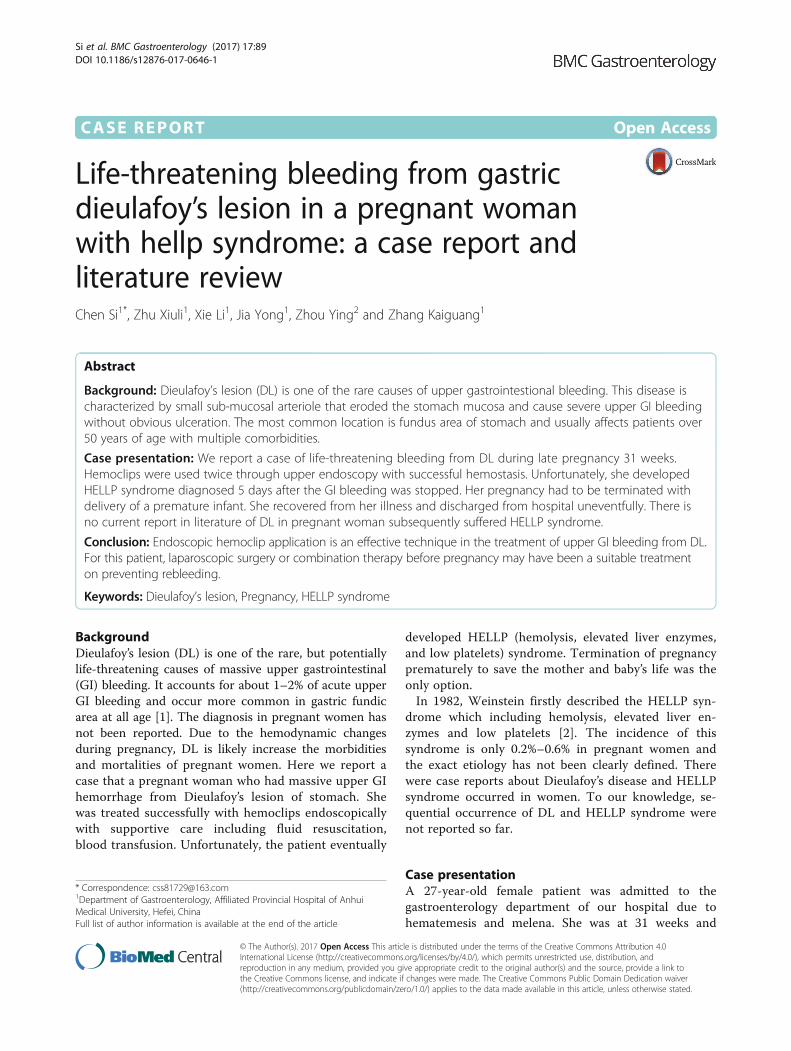
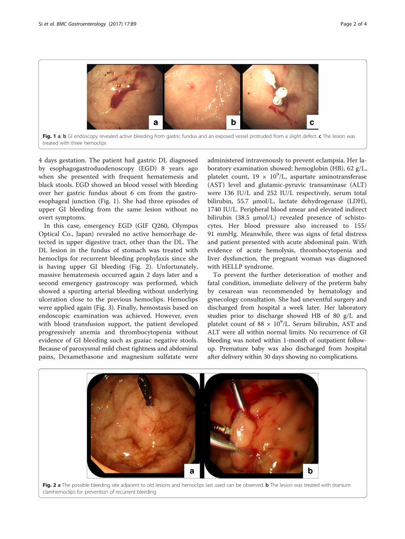
Optical Co., Japan) revealed no active hemorrhage de-tected in upper digestive tract, other than the DL. TheDL lesion in the fundus of stomach was treated withhemoclips for recurrent bleeding prophylaxis since sheis having upper GI bleeding (Fig. 2). Unfortunately,massive hematemesis occurred again 2 days later and asecond emergency gastroscopy was performed, whichshowed a spurting arterial bleeding without underlyingulceration close to the previous hemoclips. Hemoclipswere applied again (Fig. 3). Finally, hemostasis based onendoscopic examination was achieved. However, evenwith blood transfusion support, the patient developedprogressively anemia and thrombocytopenia withoutevidence of GI bleeding such as guaiac negative stools.Because of paroxysmal mild chest tightness and abdominalpains, Dexamethasone and magnesium sulfatate were
administered intravenously to prevent eclampsia. Her la-boratory examination showed: hemoglobin (HB), 62 g/L,platelet count, 19 × 109/L, aspartate aminotransferase(AST) level and glutamic-pyruvic transaminase (ALT)were 136 IU/L and 252 IU/L respectively, serum totalbilirubin, 55.7 μmol/L, lactate dehydrogenase (LDH),1740 IU/L. Peripheral blood smear and elevated indirectbilirubin (38.5 μmol/L) revealed presence of schisto-cytes. Her blood pressure also increased to 155/91 mmHg. Meanwhile, there was signs of fetal distressand patient presented with acute abdominal pain. Withevidence of acute hemolysis, thrombocytopenia andliver dysfunction, the pregnant woman was diagnosedwith HELLP syndrome.To prevent the further deterioration of mother and
fatal condition, immediate delivery of the preterm babyby cesarean was recommended by hematology andgynecology consultation. She had uneventful surgery anddischarged from hospital a week later. Her laboratorystudies prior to discharge showed HB of 80 g/L andplatelet count of 88 × 109/L. Serum bilirubin, AST andALT were all within normal limits. No recurrence of GIbleeding was noted within 1-month of outpatient follow-up. Premature baby was also discharged from hospitalafter delivery within 30 days showing no complications.
Fig. 1 a, b GI endoscopy revealed active bleeding from gastric fundus and an exposed vessel protruded from a slight defect. c The lesion wastreated with three hemoclips
Fig. 2 a The possible bleeding site adjacent to old lesions and hemoclips last used can be observed. b The lesion was treated with titaniumclamhemoclips for prevention of recurrent bleeding
Si et al. BMC Gastroenterology (2017) 17:89 Page 2 of 4

Discussion and conclusionDL is one of the rare causes of GI bleeding. It accountsfor about 1.5% of all GI hemorrhages [3, 4]. Due to thesubtlety of the lesion, endoscopic visual criteria havebeen established for diagnosis of DL [5]. In this case weobserved active arterial spurting from a small (<3 mm)defect in the gastric mucosa. DL associated upper GIbleeding during late pregnancy is extraordinarily rare inclinical practice. There was only one case report of duo-denal DL associated bleeding in the first trimester ofpregnancy in literature (Table 1) [6]. It can occur any-where in the gastrointestinal tract. Some lesions are extra-gastric including duodenum and esophageal [5, 6].Although there is no consensus on the best techniques
to treat DL, GI endoscopy is a vital tool [7, 8]. The com-mon endoscopic treatment for DL associated GI bleedinginclude thermal probe, regional injection of epinephrineand mechanical methods [9–11]. The use of hemoclipsproves to be easy and safe technique in treating DL GIbleeding and preventing rebleeding [11]. In our case, thepregnant woman suffered from pulsatile bleeding has beenmultiple hospitalizations for hemostasis which was con-trolled with emergency endoscopy two times. During latepregnancy, enlarged uterus can cause gastrointestinalcompression leading to delayed gastric emptying. Reten-tion of gastric juice and food may result in damage to themucous membranes; thus, the risk of DL rebleeding maybe higher in the third trimester.Surgery is needed in about 3% to 16% of cases in pa-
tients with DL bleeding due to rebleeding after endo-scopic therapies [12–14]. Laparoscopic surgery has been
reported to treat patient with DL bleeding with advan-tages of minimally invasive intervention, but accuratelocalization of the bleeding site can be challenged [15].For this patient rebleeding of DL occurred at the samesite of stomach, so wide wedge resection of laparoscopicsurgery with intraoperative endoscopy before pregnancycould have been a preferable treatment for patient withfrequent recurrence of DL bleeding. Combined modalitysuch as epinephrine injection and application of hemo-clips therapy can achieve higher rate of initial hemostasisand lower rate of rebleeding when compared withmonotherapy [12, 14]. It also may be a better option forour current case.Even with the success of endoscopic therapy, massive
hemorrhage has led to the progressive decline of PLTand eventually the HELLP syndrome occurred. It maybe associated with massive hemorrhage and repeatedlyerythrocyte transfusion. Moreover, hypoxia and heter-ologous related antigen during transfusion triggeredimmune reaction and inflammatory response that areboth included in the etiology and pathogenesis ofHELLP [16]. Damage to the vascular endothelial cell byactivated platelets and other factors may cause throm-botic microangiopathy. This is considered to be themain pathogenetic mechanism of HELLP [16, 17].The syndrome is typically observed in patients with se-
vere preeclampsia. Although there was no severe pre-eclampsia in our case, laboratory results were consistentwith the diagnosis of HELLP syndrome. The diagnosticcriteria of the HELLP syndrome was initially proposedby Tennessee Classification System [18]. It includes
Fig. 3 a A pulsatile bleeding in the upper stomach without underlying ulceration. b Resolution of hemorrhage following placement of hemoclips
Table 1 Bleeding DL in pregnant woman: literature review
Author GA at diagnosis (weeks) Treatment Complications Follow-up (months) Outcome
Benedetto et al. 9 Endoscopy with HCPs no 1 Normal
Chen et al. 31 Endoscopy with HCPs HELLP syndrome 2 Normal
GA gestational age, HCPs hemoclips
Si et al. BMC Gastroenterology (2017) 17:89 Page 3 of 4

presence of hemolysis, thrombocytopenia and hepaticchanges with increased levels of enzymes. Pregnancytermination is still considered to be the best HELLPtreatment method ensuring safety of the mother andinfant [17, 18]. Current case presents nonspecific symp-toms and stable delivery of the fetus by cesarean sec-tion. The mother and infant recovered uneventfullyfollowing effective treatment.The patient with DL followed by HELLP syndrome is
rare. Koji et al. have reported a case of HELLP syndromewith pituitary apoplexy [19]. Endoscopic therapy, espe-cially hemoclips, is an initial and main treatment ofbleeding caused by DL. However, both combined endo-scopic therapies and surgery might be needed in somepatients who are in a high risk of life-threateningrebleeding. Pathogenesis of HELLP syndrome in patientwith DL is unclear. Massive GI hemorrhage and inflam-matory response may be the triggering factors that leadto HELLP syndrome.
AbbreviationsDL: Dieulafoy’s lesion; GI: Gastrointestinal; HELLP: H (hemolysis)EL (elevated liver enzymes) LP (low platelet count); PLT: Platelet;EGD: Esophagogastroduodenoscopy
FundingThis report received no specific grant from any funding agency or institution.
Availability of data and materialsAll data generated or analyzed during this study are included in thispublished article.
Authors’ contributionsXL, JY, ZX and CS managed the patient; XL, ZK and CS worked on endoscopictherapy; ZY performed caesarian section; CS and ZX wrote the manuscript andZY supervised it. All authors approved the final manuscript.
Ethics approval and consent to participateThis article is a retrospective study and does not contain any studies withhuman subjects performed by any of the authors. So, the ethical approvalwas not necessary and Anhui Provincial Hospital medical ethics committeecan offer exempt ethical statement in support.
Consent for publicationWritten informed consent was obtained from the patient for publicationof this case report and any accompanying images. A copy of the writtenconsent is available for review by the Editor of this journal.
Competing interestsThe authors declare that they have no competing interests.
Publisher’s NoteSpringer Nature remains neutral with regard to jurisdictional claims inpublished maps and institutional affiliations.
Author details1Department of Gastroenterology, Affiliated Provincial Hospital of AnhuiMedical University, Hefei, China. 2Department of Obstetrics, AffiliatedProvincial Hospital of Anhui Medical University, Hefei, China.
Received: 24 January 2017 Accepted: 24 July 2017
References1. Baxter M, Aly EH. Dieulafoy’s lesion: current trends in diagnosis and
management. Ann R Coll Surg Engl. 2010;92:548–54.2. Benedetto C, Marozio L, Tancredi A. Biochemistry of HELLP syndrome.
Adv Clin Chem. 2011;53:85–104.3. Chaer R, Helton WS. Dieulafoy’s disease. Am Coll Surg. 2003;196:290–6.4. Veldhuyzen van Zanten SJ, Bartelsman JF, Schipper ME, et al. Recurrent
massive haematemesis from Dieulafoy vascular malformations–a reviewof 101 cases. Gut. 1986;27:213–22.
5. Malliaras GP, Carollo A, Bogen G. Esophageal Dieulafoy’s lesion: anexceedingly rare cause of massive upper GI bleeding. J Surg Case Rep.2016;6:1–2.
6. Mangiavillano B, Luigiano C, Basile S, et al. Is pregnancy a predisposingfactor for Dieulafoy's duodenal lesion or only a coincidence? Clin ResHepatol Gastroenterol. 2013;37(5):e119–20.
7. Nguyen DC, Jackson CS. The Dieulafoy’s lesion: an update on evaluation,diagnosis, and management. J Clin Gastroenterol. 2015;49(7):541–9.
8. Alis H, Oner OZ, Kalayci MU, et al. Is endoscopic band ligation superior toinjection therapy for Dieulafoy lesion? Surg Endosc. 2009;23(7):1465–9.
9. Schmulewitz N, Baillie J. Dieulafoy lesions: a review of 6 years of experienceat a tertiary referral center. Am J Gastroenterol. 2001;96:1688–94.
10. Iacopini F, Petruzziello L, Marchese M, et al. Haemostasis of Dieulafoy’s lesionby argon plasma coagulation. Gastrointest Endosc. 2007;66:20–6.
11. Yamaguchi Y, Yamato T, Katsumi N, et al. Short-term and long-term benefitsof endoscopic hemoclip application for Dieulafoy’s lesion in the upper GI tract.Gastrointest Endosc. 2003;57:653–6.
12. Eisenberg D, Bell R. Intraoperative endoscopy: a requisite tool forlaparoscopic resection of unusual gastrointestinal lesions–a case series.J Surg Res. 2009;155:318–20.
13. Norton ID, Petersen BT, Sorbi D, et al. Management and long-term prognosisof Dieulafoy lesion. Gastrointest Endosc. 1999;50:762–7.
14. Chung IK, Kim EJ, Lee MS, et al. Bleeding Dieulafoy's lesions and the choiceof endoscopic method: comparing the hemostatic efficacy of mechanicaland injection methods. Gastrointest Endosc. 2000;52(6):721–4.
15. Alva S, Abir F, Tran D. Laparoscopic gastric wedge resection for Dieulafoy’sdisease following pre-operative endoscopic localisation with India ink andendoscopic clips. J Soc Laparoendosc Surg. 2006;10:244–6.
16. Abildgaard U, Heimdal K. Pathogenesis of the syndrome of hemolysis,elevated liver enzymes, and low platelet count (HELLP): a review. Eur JObstet Gynecol Reprod Biol. 2013;166(2):117–23.
17. Dusse LM, Alpoim PN, Silv JT, et al. Revisiting HELLP syndrome. Clin ChimActa. 2015;45 (Pt B):117–20.
18. Barton JR, Sibai BM. Diagnosis and management of hemolysis, elevated liverenzymes, and low platelets syndrome. Clin Perinatol. 2004;31:807–33.
19. Murao K, Imachi H, Muraoka T, et al. Hemolysis, elevated liver enzymes, andlow platelet count (HELLP) syndrome with pituitary apoplexy. Fertil Steril.2011;96(1):260–1.
• We accept pre-submission inquiries
• Our selector tool helps you to find the most relevant journal
• We provide round the clock customer support
• Convenient online submission
• Thorough peer review
• Inclusion in PubMed and all major indexing services
• Maximum visibility for your research
Submit your manuscript atwww.biomedcentral.com/submit
Submit your next manuscript to BioMed Central and we will help you at every step:
Si et al. BMC Gastroenterology (2017) 17:89 Page 4 of 4