life sciences: animal tissue
35
Animal Tissues Prepared by: MOKWENA CATHY BUITUMELO
-
Upload
cathy-mokwena -
Category
Education
-
view
219 -
download
5
Transcript of life sciences: animal tissue
Animal Tissues
Prepared by:
MOKWENA CATHY BUITUMELO
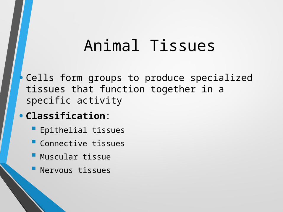
Animal Tissues
• Cells form groups to produce specialized tissues that function together in a specific activity
• Classification: Epithelial tissues
Connective tissues
Muscular tissue
Nervous tissues
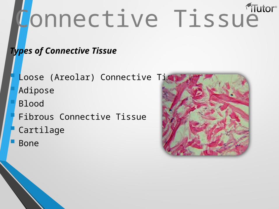
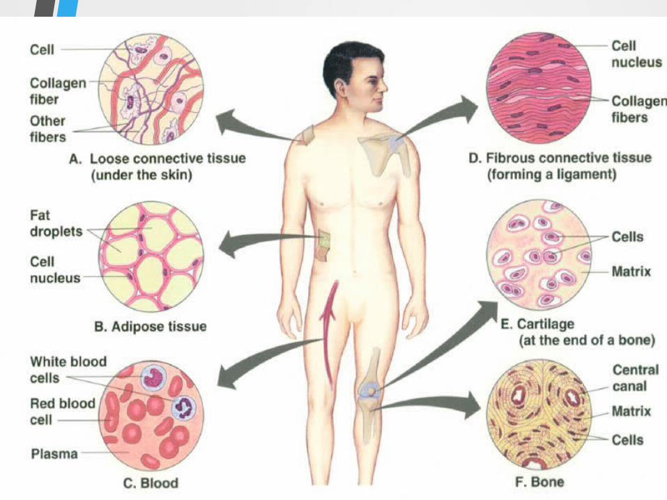
Connective TissueTypes of Connective Tissue
Loose (Areolar) Connective Tissue
Adipose
Blood
Fibrous Connective Tissue
Cartilage
Bone
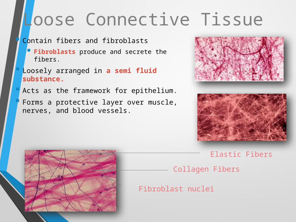
Loose Connective Tissue Contain fibers and fibroblasts
Fibroblasts produce and secrete the fibers.
Loosely arranged in a semi fluid substance.
Acts as the framework for epithelium.
Forms a protective layer over muscle, nerves, and blood vessels.
Elastic Fibers
Collagen Fibers
Fibroblast nuclei
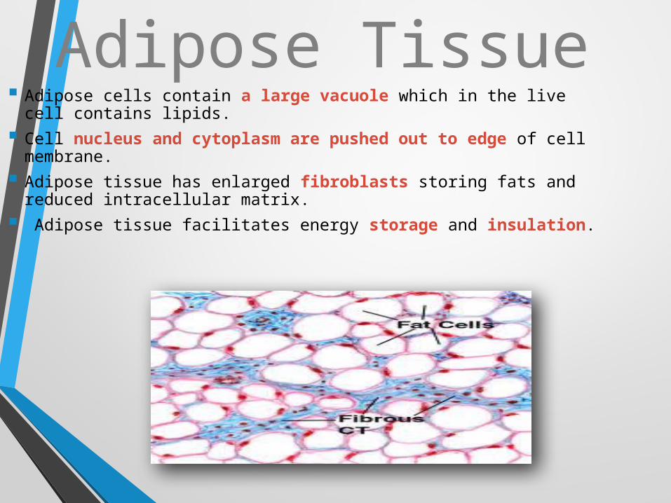
Adipose Tissue Adipose cells contain a large vacuole which in the live cell
contains lipids.
Cell nucleus and cytoplasm are pushed out to edge of cell membrane.
Adipose tissue has enlarged fibroblasts storing fats and reduced intracellular matrix.
Adipose tissue facilitates energy storage and insulation.
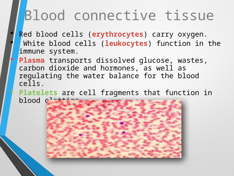
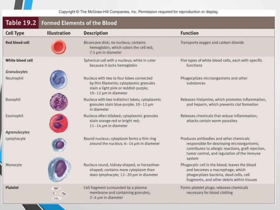
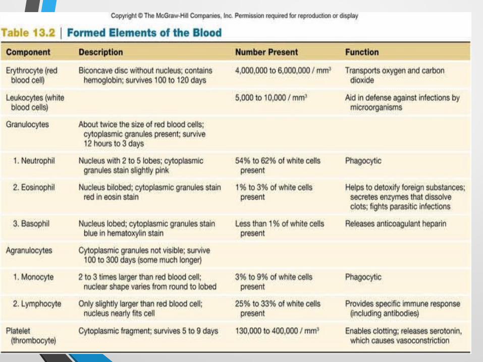
Blood connective tissue Red blood cells (erythrocytes) carry oxygen. White blood cells (leukocytes) function in the immune
system. Plasma transports dissolved glucose, wastes, carbon
dioxide and hormones, as well as regulating the water balance for the blood cells.
Platelets are cell fragments that function in blood clotting.
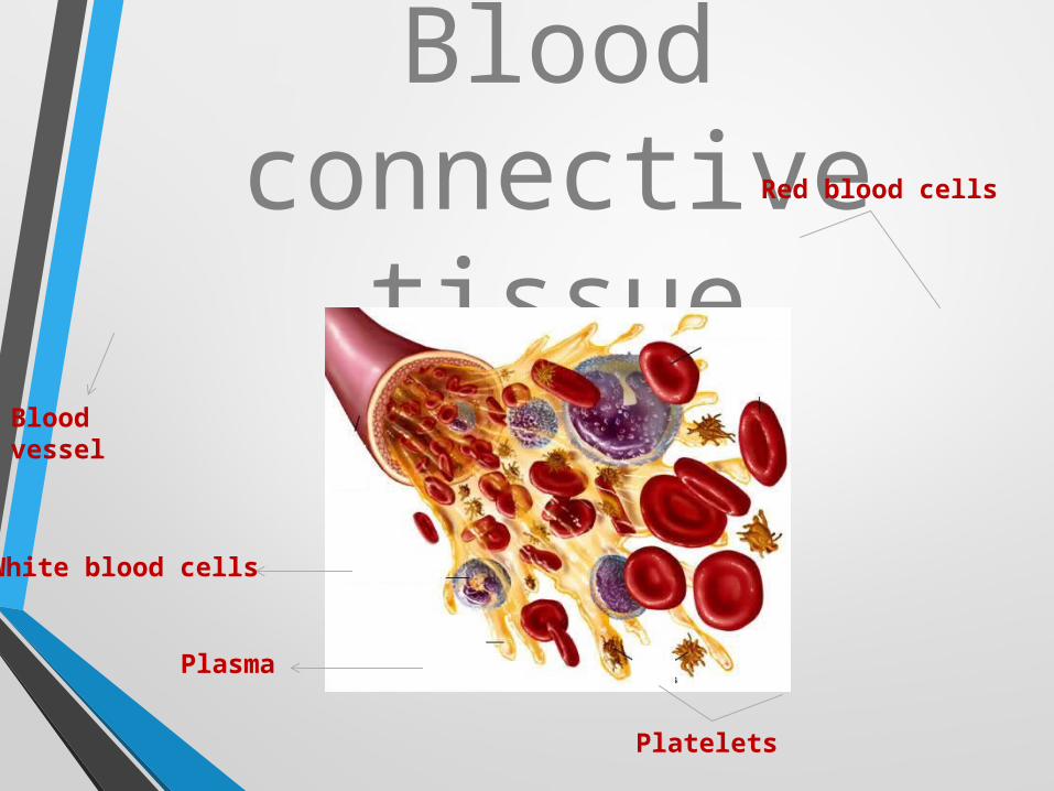
Blood connective
tissueBlood vessel
Red blood cells
White blood cells
Plasma
Platelets
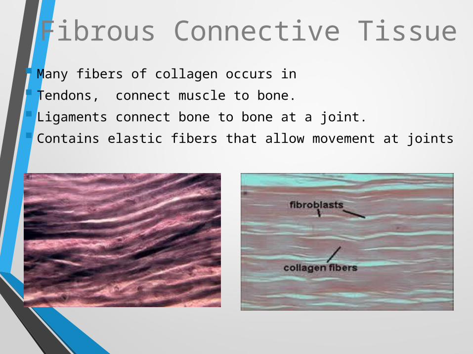
Fibrous Connective Tissue Many fibers of collagen occurs in
Tendons, connect muscle to bone.
Ligaments connect bone to bone at a joint.
Contains elastic fibers that allow movement at joints
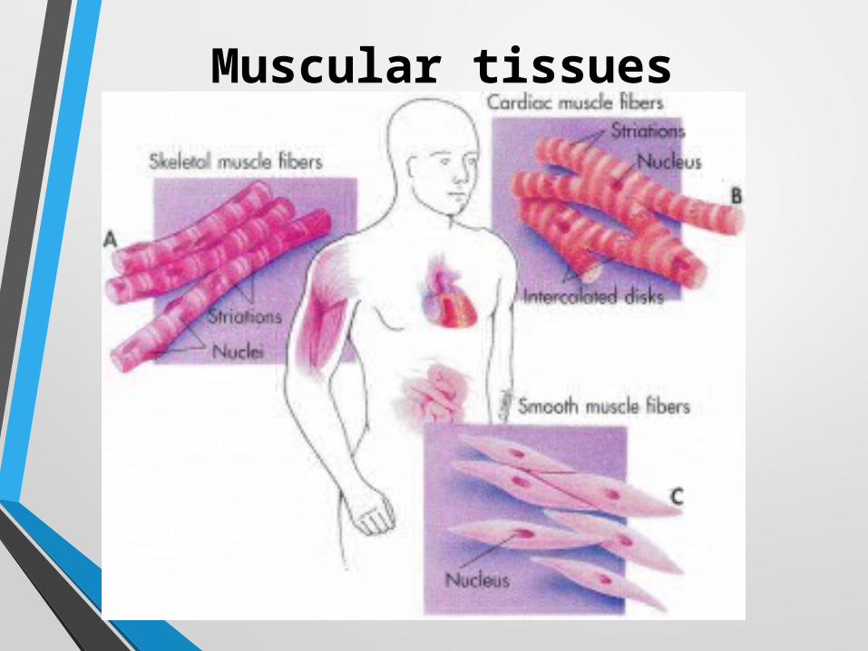
Muscular tissues
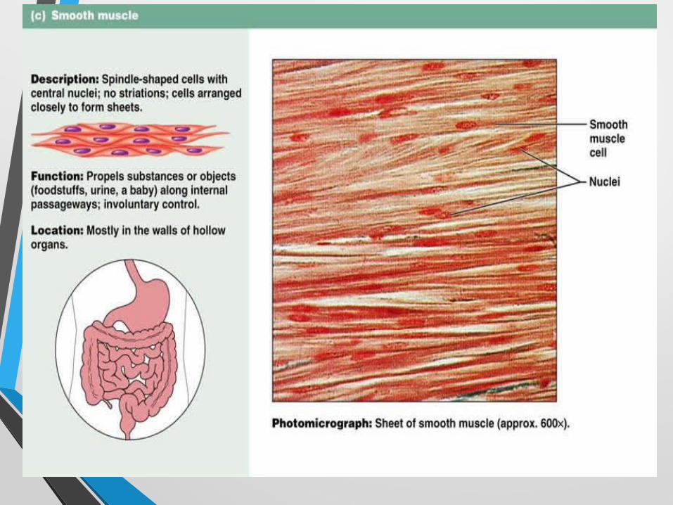
Smooth muscle
• These have long cells which have single nucleus.
• The cells of this muscle is spindle in shape.
• They are also called unstrained because we can not control them
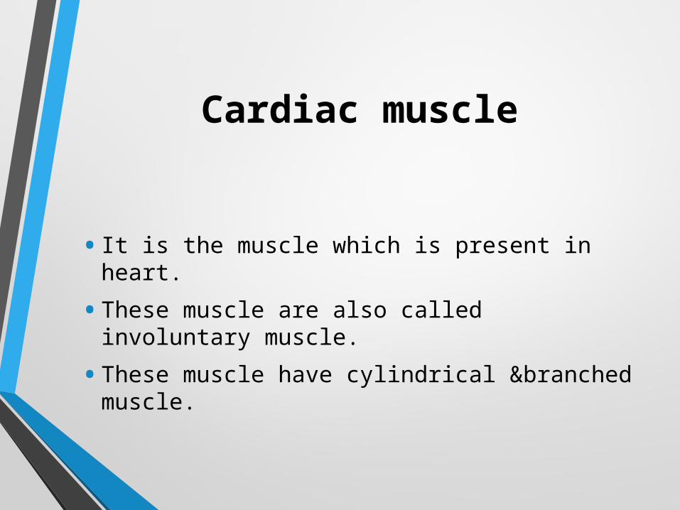
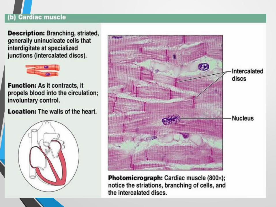
Cardiac muscle
• It is the muscle which is present in heart.
• These muscle are also called involuntary muscle.
• These muscle have cylindrical &branched muscle.
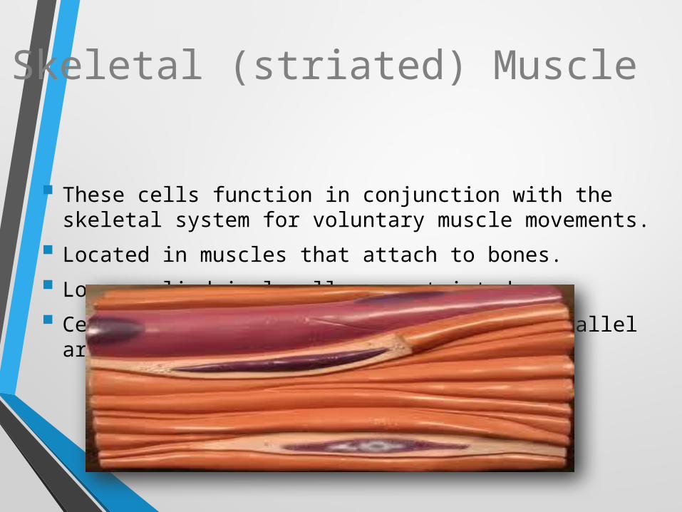
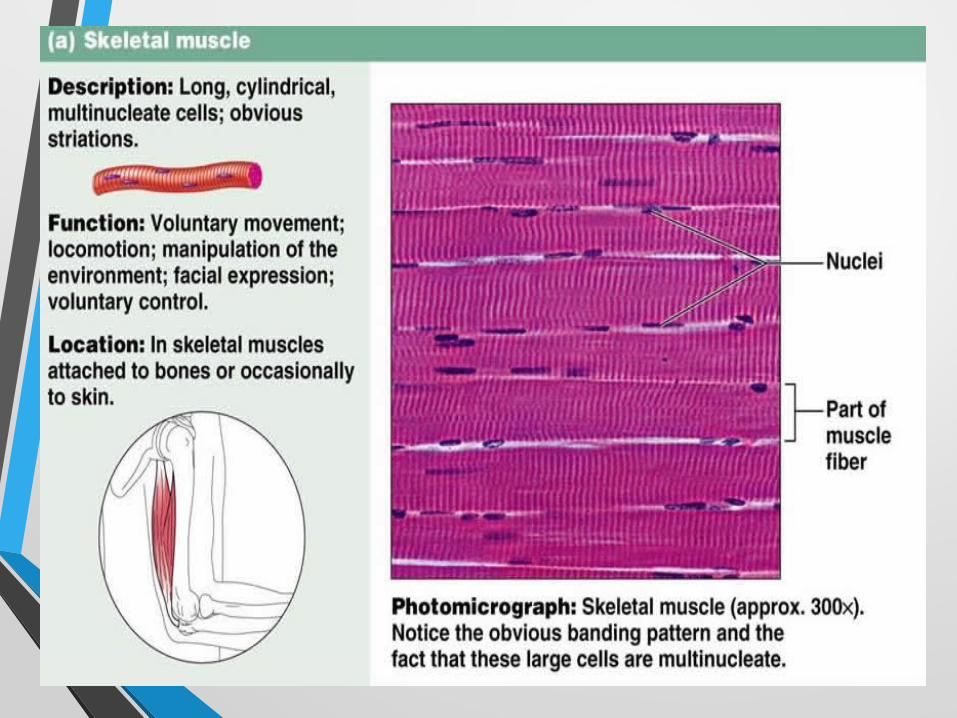
Skeletal (striated) Muscle
These cells function in conjunction with the skeletal system for voluntary muscle movements.
Located in muscles that attach to bones.
Long, cylindrical cells are striated.
Cells are bundled closely together in parallel arrays.
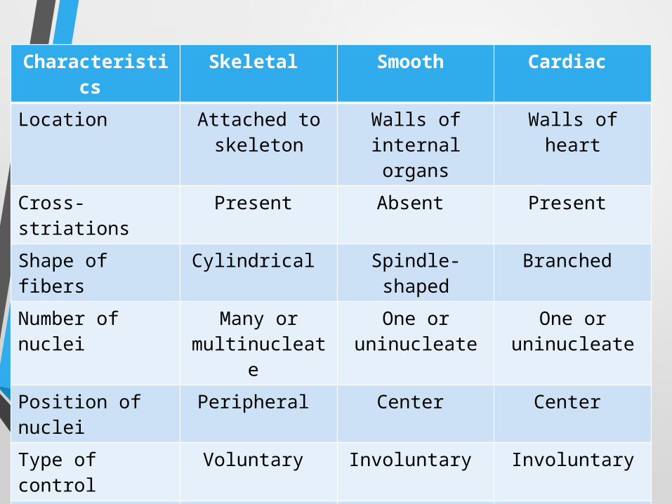
Characteristics
Skeletal Smooth Cardiac
Location Attached to skeleton
Walls of internal organs
Walls of heart
Cross-striations Present Absent Present
Shape of fibers Cylindrical Spindle-shaped
Branched
Number of nuclei
Many or multinucleate
One or uninucleate
One or uninucleate
Position of nuclei
Peripheral Center Center
Type of control Voluntary Involuntary Involuntary
Speed of contraction
Most rapid slowest Intermediate
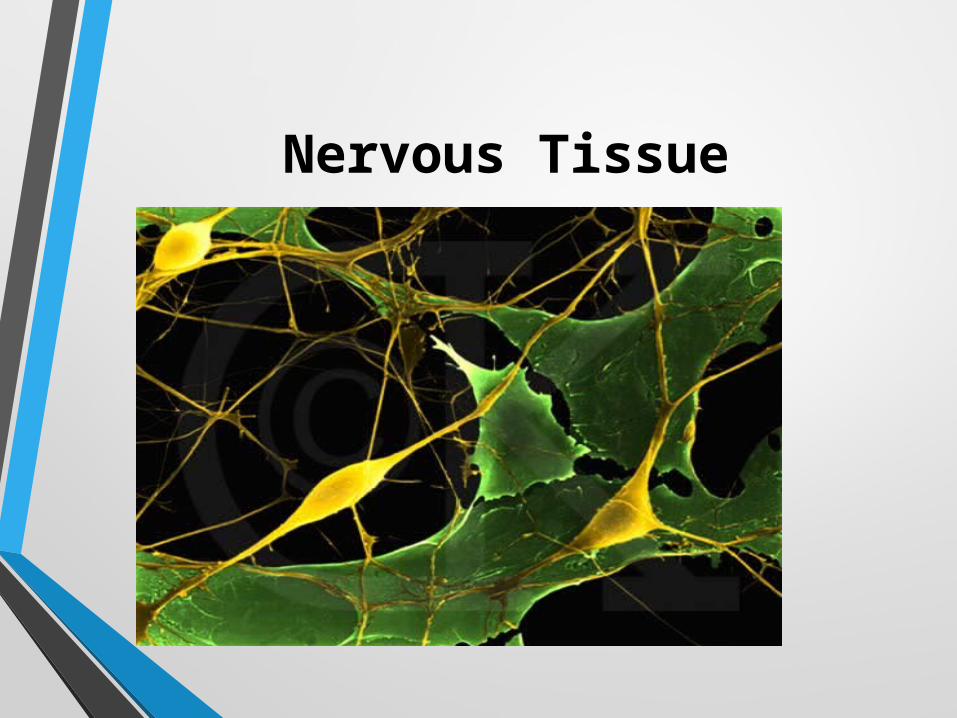
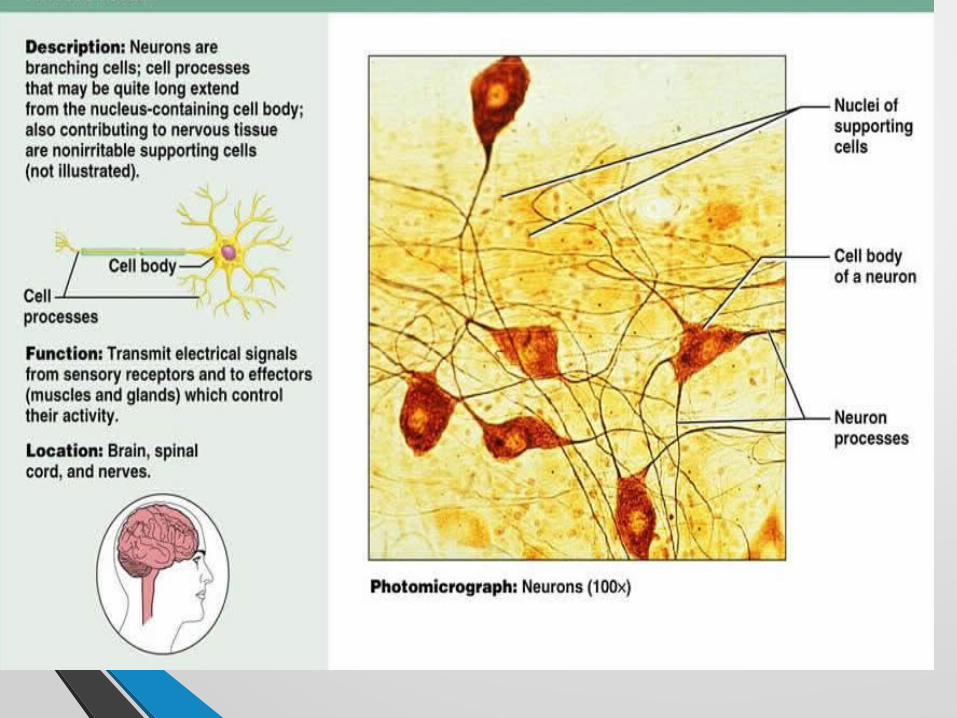
Nervous Tissue
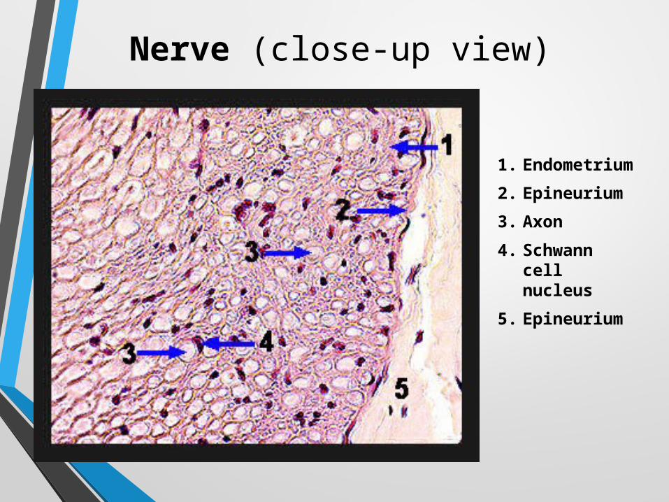
Nerve (close-up view)
1. Endometrium
2. Epineurium
3. Axon
4. Schwann cell nucleus
5. Epineurium
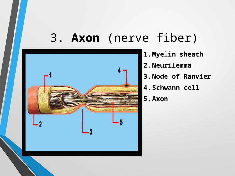
3. Axon (nerve fiber)1. Myelin sheath
2. Neurilemma
3. Node of Ranvier
4. Schwann cell
5. Axon
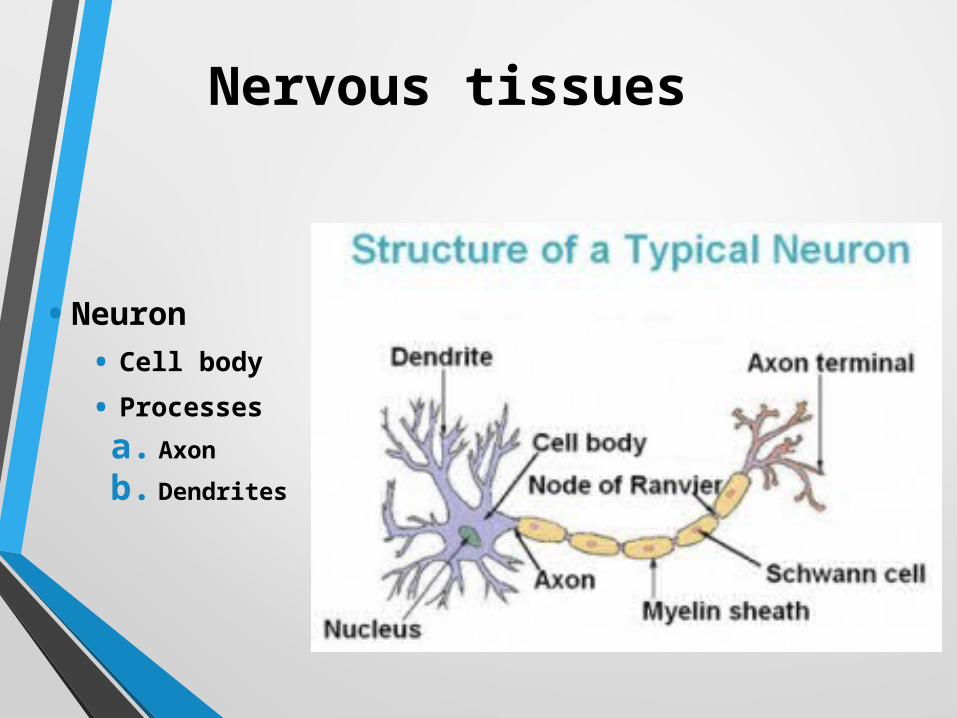
Nervous tissues
• Neuron
• Cell body
• Processes
a. Axon
b. Dendrites
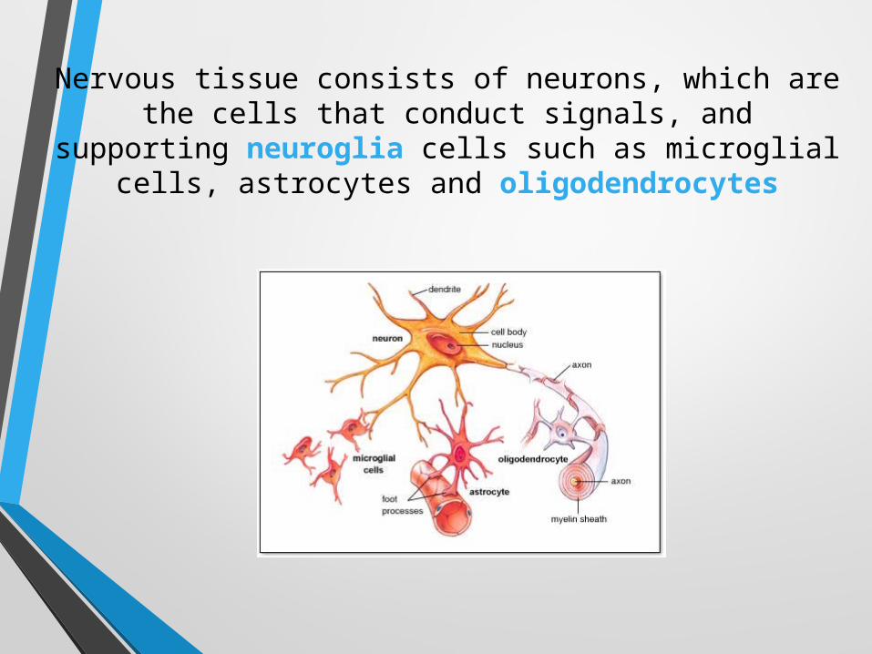
Nervous tissue consists of neurons, which are the cells that conduct signals, and supporting neuroglia
cells such as microglial cells, astrocytes and oligodendrocytes
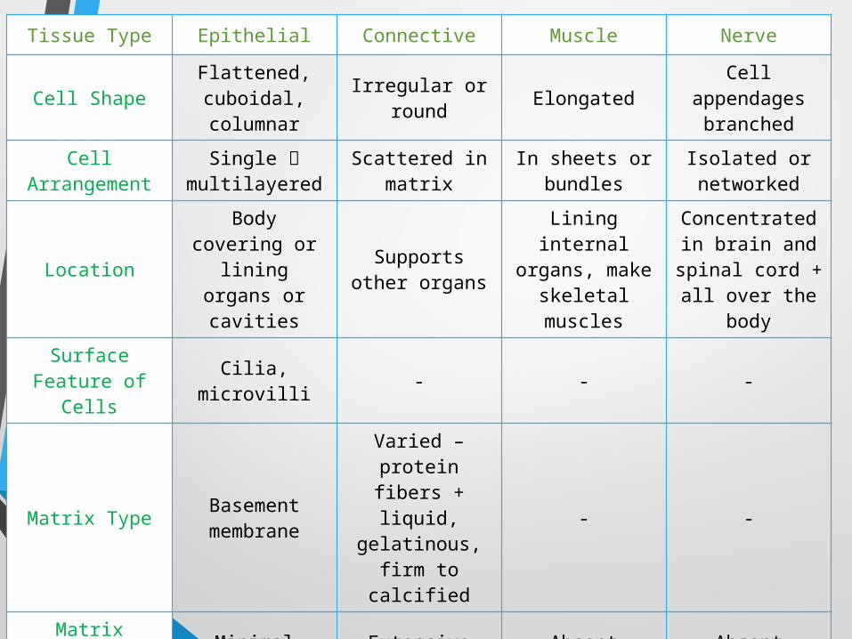
Tissue Type Epithelial Connective Muscle Nerve
Cell ShapeFlattened, cuboidal, columnar
Irregular or round Elongated
Cell appendages
branched
Cell Arrangement
Single multilayered
Scattered in matrix
In sheets or bundles
Isolated or networked
LocationBody covering or lining organs
or cavities
Supports other organs
Lining internal organs, make
skeletal muscles
Concentrated in brain and spinal cord + all over the
body
Surface Feature of Cells Cilia, microvilli - - -
Matrix Type Basement membrane
Varied – protein fibers
+ liquid, gelatinous,
firm to calcified
- -
Matrix Amount Minimal Extensive Absent Absent
Unique FeatureNo direct blood supply, except
for glands
Cartilage has no blood supply
Can generate electrical
signals, force and movement
Can generate electrical
signal
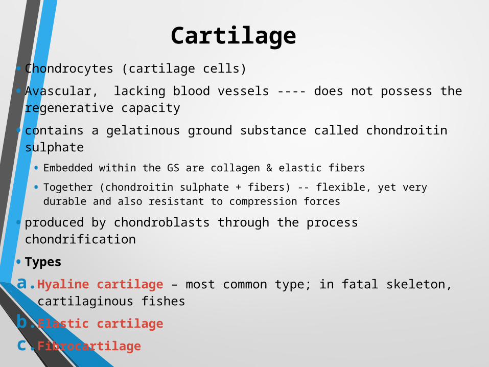
Cartilage • Chondrocytes (cartilage cells)
• Avascular, lacking blood vessels ---- does not possess the regenerative capacity
• contains a gelatinous ground substance called chondroitin sulphate
• Embedded within the GS are collagen & elastic fibers
• Together (chondroitin sulphate + fibers) -- flexible, yet very durable and also resistant to compression forces
• produced by chondroblasts through the process chondrification
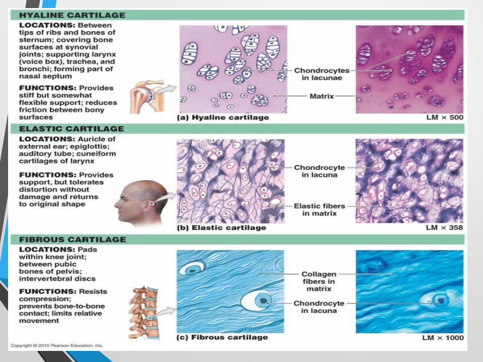
• Types
a. Hyaline cartilage – most common type; in fatal skeleton, cartilaginous fishes
b. Elastic cartilage
c. Fibrocartilage
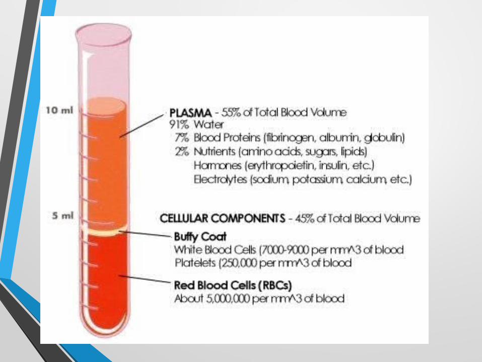
Blood

References
Bell, A. 1999. Anatomy 503 – Human Histology
http://faculty.une.edu/com/abell/histo/histolab2.htm
General Zoology Laboratory Manual, Biology Dept., Univ. of San Carlos
A/P Lab: A website for Human Anatomy and Physiology
http://bioweb.uwlax.edu/APlab/Index.htm
Ross, M., Romwell, L., and Kaye, G. 1995. Histology: A Text and Atlas. Williams and Wilkins, USA.
http://www.slideshare.net/itutor/animal-tissue-20032768?qid=9f7ec26b-d9b8-4566-9f10-b8593213dbd9&v=qf1&b=&from_search=4