Life Science Journal 2017;14(9) · foods containing the beneficial compounds especially mentioned...
24
Life Science Journal 2017;14(9) http://www.lifesciencesite.com 37 Toxicity of monosodium glutamate on the liver of chick embryos and the effectiveness of pomegranate peel extract in reducing this toxicity (to show scientific miracle in the Quran and Sunnah). Fawzyah A. M. Al-Ghamdi Biology Department, Science Faculty, King Abdul-Aziz University, Jeddah, Saudi Arabia. [email protected] Abstract: The monitor changes caused by monosodium glutamate MSG on both the morphological and histological structure of the liver of chicken and the preventive and therapeutic roles of Pomegranate peels extract PPE at (10- 12-14) days of incubation. 720 fertilized egg divided equally into6 groups; Control group (C- distilled water (0.1 ml/egg). PPE G (PPE-(0.3 ml/egg). MSG G (MSG-(0.1 ml/egg). Mixed group (PPE+ MSG): MSG (0.1 ml/egg) and PPE (0.3 ml/egg) together in (0-1) days. Preventive G (PPE-MSG: 0.3 ml/eggs per day) at (0-1 days) then with a MSG (0.1 ml/eggs per day) at (3-4 days). Therapeutic G (MSG-PPE: MSG (0.1 ml/eggs per day) at (0-1 days) then with PPE (0.3 ml/eggs per day) at (3-4 days). External examination revealed more of malformation and discrepancy in the weights and heights of the embryos. Morphometric measurements showed delayed or decreased in organs growth as height of neck, beak, eye diameters, liver weights, diameters of hepatocytes. Histological study appeared differences degeneration in blood sinusoids, hepatic cords and hepatocytes. Results showed that MSG group was mostly affected followed by MSG+PPE group then MSG-PPE group, meanwhile PPE-MSG group showed marked protection, also PPE group was like control group. We can concluded that MSG substance had oxidized effects that act on the cells and tissues of the body that depend on the amount and time of dose administration. We must eat foods containing the beneficial compounds especially mentioned in the Quran and Sunnah as the pomegranate fruit that has enormous benefits as it acts as antioxidant that protect cells and reduce the risk of various diseases. [Fawzyah A. M. Al-Ghamdi. Toxicity of monosodium glutamate on the liver of chick embryos and the effectiveness of pomegranate peel extract in reducing this toxicity (to show scientific miracle in the Quran and Sunnah).. Life Sci J 2017;14(9):37-60]. ISSN: 1097-8135 (Print) / ISSN: 2372-613X (Online). http://www.lifesciencesite.com . 4. doi:10.7537/marslsj140917.04 . Key words: Pomegranate Peel extract, Monosodium glutamate, Preventive, Therapeutic, malformation, degeneration. 1. Introduction: Fast food consumption was increased and became a common phenomenon all over the world because of the significant changes in the lifestyles. The living style had shifted from intake of natural or little treated foods to intake of fast foods that contains high energy and low dietary fibers (Bryant and Dundes 2008; Dongen et al. 2010). The fast food had different effects on health as they contained high proportions of saturated fats and sodium that leads to increased body weight gain and body mass index increased body mass index and weight increase (Serrano and Jedda 2009; Block et al., 2004). So the usage of the most common and widely used flavor enhancer, MSG salt was investigated to assess its side effects on the health of pregnant women and their fetuses and nursing mothers and their newborns and infants because those groups of society are morevulnerable to MSG salt due to the rapid evolution of their tissues and incomplete maturity of their immune system to get rid of toxins. Pomegranate fruit (Pomegranate) is one of the oldest fruits known to man because of high nutritional value as food, antioxidant effects and also its different parts as juice, peels, seeds and roots used in folk medicine in different countries of the world and for many diseases. Although pomegranate is rich in antioxidants, its shells, which are usually ignored, are twice as high in its content of antioxidants compared to grains, in addition to ellagic acid that is used in preparation of various drugs, anthocyanins (Noda et al., 2012). Therefore, attention has been paid to natural antioxidants that prevent the oxidation of plasma lipids that led to atherosclerosis (Gill et al., 2000), in addition to their interaction with free radicals. Glutamate was obtained by digestion of foods containing the appropriate amounts of free glutamate whether naturally present or added to foods as MSG then absorbed from digestive tractby active transport with amino acids. Glutamic acid is converted into free amino acids or peptides absorbed via mucous cells in small intestine, where peptides reacted with water and converted them into amino acids and some glutamates then metabolized in the liver. In the blood it’s converted to sodium and glutamic acid. Previous studies mentioned that injection of MSG intraperitoneal leads to transport of MSG through the epithelial cells actively to the blood to be metabolized as excreted outside of the body, meanwhile remaining
Transcript of Life Science Journal 2017;14(9) · foods containing the beneficial compounds especially mentioned...

Life Science Journal 2017;14(9) http://www.lifesciencesite.com
37
Toxicity of monosodium glutamate on the liver of chick embryos and the effectiveness of pomegranate peel extract in reducing this toxicity (to show scientific miracle in the Quran and Sunnah).
Fawzyah A. M. Al-Ghamdi
Biology Department, Science Faculty, King Abdul-Aziz University, Jeddah, Saudi Arabia.
Abstract: The monitor changes caused by monosodium glutamate MSG on both the morphological and histological structure of the liver of chicken and the preventive and therapeutic roles of Pomegranate peels extract PPE at (10-12-14) days of incubation. 720 fertilized egg divided equally into6 groups; Control group (C- distilled water (0.1 ml/egg). PPE G (PPE-(0.3 ml/egg). MSG G (MSG-(0.1 ml/egg). Mixed group (PPE+ MSG): MSG (0.1 ml/egg) and PPE (0.3 ml/egg) together in (0-1) days. Preventive G (PPE-MSG: 0.3 ml/eggs per day) at (0-1 days) then with a MSG (0.1 ml/eggs per day) at (3-4 days). Therapeutic G (MSG-PPE: MSG (0.1 ml/eggs per day) at (0-1 days) then with PPE (0.3 ml/eggs per day) at (3-4 days). External examination revealed more of malformation and discrepancy in the weights and heights of the embryos. Morphometric measurements showed delayed or decreased in organs growth as height of neck, beak, eye diameters, liver weights, diameters of hepatocytes. Histological study appeared differences degeneration in blood sinusoids, hepatic cords and hepatocytes. Results showed that MSG group was mostly affected followed by MSG+PPE group then MSG-PPE group, meanwhile PPE-MSG group showed marked protection, also PPE group was like control group. We can concluded that MSG substance had oxidized effects that act on the cells and tissues of the body that depend on the amount and time of dose administration. We must eat foods containing the beneficial compounds especially mentioned in the Quran and Sunnah as the pomegranate fruit that has enormous benefits as it acts as antioxidant that protect cells and reduce the risk of various diseases. [Fawzyah A. M. Al-Ghamdi. Toxicity of monosodium glutamate on the liver of chick embryos and the effectiveness of pomegranate peel extract in reducing this toxicity (to show scientific miracle in the Quran and Sunnah).. Life Sci J 2017;14(9):37-60]. ISSN: 1097-8135 (Print) / ISSN: 2372-613X (Online). http://www.lifesciencesite.com. 4. doi:10.7537/marslsj140917.04. Key words: Pomegranate Peel extract, Monosodium glutamate, Preventive, Therapeutic, malformation, degeneration. 1. Introduction:
Fast food consumption was increased and became a common phenomenon all over the world because of the significant changes in the lifestyles. The living style had shifted from intake of natural or little treated foods to intake of fast foods that contains high energy and low dietary fibers (Bryant and Dundes 2008; Dongen et al. 2010). The fast food had different effects on health as they contained high proportions of saturated fats and sodium that leads to increased body weight gain and body mass index increased body mass index and weight increase (Serrano and Jedda 2009; Block et al., 2004). So the usage of the most common and widely used flavor enhancer, MSG salt was investigated to assess its side effects on the health of pregnant women and their fetuses and nursing mothers and their newborns and infants because those groups of society are morevulnerable to MSG salt due to the rapid evolution of their tissues and incomplete maturity of their immune system to get rid of toxins. Pomegranate fruit (Pomegranate) is one of the oldest fruits known to man because of high nutritional value as food, antioxidant effects and also its different parts as juice, peels, seeds
and roots used in folk medicine in different countries of the world and for many diseases.
Although pomegranate is rich in antioxidants, its shells, which are usually ignored, are twice as high in its content of antioxidants compared to grains, in addition to ellagic acid that is used in preparation of various drugs, anthocyanins (Noda et al., 2012). Therefore, attention has been paid to natural antioxidants that prevent the oxidation of plasma lipids that led to atherosclerosis (Gill et al., 2000), in addition to their interaction with free radicals.
Glutamate was obtained by digestion of foods containing the appropriate amounts of free glutamate whether naturally present or added to foods as MSG then absorbed from digestive tractby active transport with amino acids. Glutamic acid is converted into free amino acids or peptides absorbed via mucous cells in small intestine, where peptides reacted with water and converted them into amino acids and some glutamates then metabolized in the liver. In the blood it’s converted to sodium and glutamic acid. Previous studies mentioned that injection of MSG intraperitoneal leads to transport of MSG through the epithelial cells actively to the blood to be metabolized as excreted outside of the body, meanwhile remaining

Life Science Journal 2017;14(9) http://www.lifesciencesite.com
38
part converted to glutamine by the liver cells that led to destruction of liver cells and their apoptosis (Ortiz et al., 2006).
MSG had many side effects on different body systems; studies reported that diets contained food additives affected brain neurotransmission, behaviors and psychological status (Weiss 1980; Zeisel 1986; Lowik 1996), memory (Pollitt et al. 1996) especially in children. In addition, eating different food that contained additives especially glutamic acid leads to severe disturbances in behavior and concentration (Lau et at.2006). Food additives can transport blood brain barrier and caused neurotoxicity as its lipid soluble that led to change in the nature of neural membrane (Khanna et al. 1980). Injection of rats newborn with dose 4mg/kg on days 2,4,6,8 and 10 led to significant decrease in pituitary gland weight by 30-40% that led to decrease secretion of hormones that affected other endocrine glands and its connection with hypothalamus ((Miskowiak and Partyka 2000). Newborns exposed to monosodium glutamate, resulting in damage to nerve cells, disordersin light- perception and colloidal cells (Husarova, and D. Ostatnikova (2013).
The administration of repeated doses of MSG for long periods on adult and newborn mice in dose of 2mg/g either oral or subcutaneous injection led to destruction of the function and structure of the retina as it destroyed the blood barrier that organized entry of substances to the retina thus preventing polarization of materials and stimulates their death as result of elevated glutamate inside cells (Swelim 2004). Study showed effect of MSG on chicken embryos agricultures network (in vitro) and reported significant damage to low concentration within hours and that there is a direct correlation between the concentration and the damage produced (Reif-Lehrer et al.,1975). The effect of oral MSG (4g/kg) to pregnant female rats at age of 100 days. The results showed sudden decrease in the ovaries weight in rats treated with MSG compared to the control group in addition to decrease in fetal weights and lengths and decreased diameters of placenta, as well as decreased in the level of follicle stimulating hormone. It was concluded that treatment with MSG during pregnancy has a negative effect on the fetuses. (Tawfeeq and AL-Badr, 2012).
The effect of MSG as a flavor enhancer and acrylamide, chemical compound that is naturally formed in a large group of foods when cooked and also used in water treatment, on mice liver tissue treated with a dose (30mg / kg), resulted in changes in the structure and toxic to the hepatocytes (AL-Mosaibih, 2013). Studies showed that mice treated with MSG at a dose of 0.6-1.6 mg / kg for 14 days resulted in increased body weight, enlarged liver, in addition to elevation of blood levels of some liver
enzymes as oxidative stress results in decrease in the liver ability to detoxify toxic substances (Tawfik and Al-Badr 2012). The results of the study of the toxic effects of MSG on the liver when mice injecting with a dose of 5 mg / kg during 28 days showed elevation of serum levels of hepatic enzymes, especially alanine aminotransferase (9.15%) and aspartate aminotransferase (66.86%) due to negative effect of excessive body weight gain on the liver (Egbuonu et al., 2009). The toxic effect of MSG on the liver when repeatedly intraperitoneal injected into the mice at (0-15-30-45) minutes that led to elevation of serum liver enzymes as aspartate aminotransferase and alanine aminotransferase at minute (30- 45) due to damage of the cell membrane in addition to deterioration of fat oxidation in the liver, that resulted in liberation of free radicles, and showed degenerative changes of hepatocytescase edema- degeneration-necrosis (Ortiz et al. 2006).
Pomegranate contains high amounts of antioxidants such as soluble polyphenols that protect the body's cells and organs such as heart, brain and bones, resist against various diseases and prevent break down of free radicles inside the body (Gil et al., 2000; Rosenblat 2006; Guo et al., 2008). Pomegranate has many applications and benefits, including reduce the risk of vascular tumor. Also, pomegranate contains hydrobenzoic acids Phenylpropanoids that inhibit special protein present in the bone marrow responsible for the movement of cancer cells to bones Hora 2003; Albrecht 2004; Adhami 2011). A study carried on 28 elderly people suffering from difficulty in memory reported that when these patients drinking 250 ml of pomegranate juice each day resulted in a marked improvement in verbal and visual memories and confirmed by studies and studies in mice confirmed that pomegranate had anti - Alzheimer 's protection role. (Faraj 2011; Chalfoun-Mounayar 2012; Jurenka 2008; Bookheimer et al. 2000). Experimental study showed that the antioxidant substances present in pomegranate especially Punicalagin plays an important role in the health of sperms through the regulation and maturity of spermsas sperms are more susceptible to damage caused by oxidation due to exposure to high concentrations of polyunsaturated fatty acids. (Ghaffari and Ahmadian, 2007; Jurenka, 2008; Akpinar-Bayizit, 2012). Pomegranate shells have positive effects on the liver and kidney tissues. Adult rats injected intraperitoneal with pomegranate (200 mg/kg) led to decrease lipid oxidation and elevated levels of antioxidant enzymes assuperoxide dismutase that led to restoration of cell viability, decreased cell damage and equalization of free radicals and usage of minerals as copper, zinc and manganese, in addition to increase activity of the enzyme catalase at natural harmless levels (Moneim,

Life Science Journal 2017;14(9) http://www.lifesciencesite.com
39
2012). Administration of pomegranate indirectly inhibits the enzymes caused by arthritis that led to joint cartilage protection from infection (Jurenka, 2008Akpinar-Bayizit, 2012). Also, acidic pomegranate compounds help well digestion of fat foods; burn accumulated fats and so prevents obesity and has an effective role in cleaning the intestines and expelling toxins Jurenka, 2008; Akpinar-Bayizit, 2012).; Faraj and Koyee, 2011). The mechanism of action of tannins and phenolic compounds that are present in pomegranate peels is explained by its ability to precipitate the proteins present in the cell membrane or within the living cells when they are consumed from the cell membrane, formation of hydrogen bonds between the free and multiple hydroxyl phenols, and nitrogen compounds or proteins, thus inhibitthe effectiveness of certain enzymes in the organism (Reeds et al, 1996; Covington, 1997). Some phenols also act as antioxidants by reducing hydrogen donation, suppress free radicals formation. Flavonoids are important antioxidants because they have various mechanisms as:
1. Inhibition of the enzymes responsible for superoxide anion and generation of reactive oxygen species production.
2. Flavonoids bind tiny metals that play an important role in oxygen metabolism. Due to low oxidation-reduction of flavonoids, thermodynamic flavonoids (FL-OH) have a high potential for free radical reduction and thus act as hydrogen atom donors (Hakkim et al, 2008).
The liver is one of the most important organs in the body as it stores carbohydrates, proteins, fats and fat soluble vitamins, regulates blood sugar levels and stores glucose as glycogen, secretion of bile that contribute to the digestion and absorption of fats, conversion of amino acids to urea, synthesis of many hormones, red blood cells formation and regeneration in the fetus, conservation of hormonal balance and removal of microbes and waste products from the cells. Eroschenko (2000) and Kierszenbaum (2002) reported that the liver is the largest gland in the body that composed of multiple lobules surrounded by a capsule formed from collagen elastic fibers. From the capsule hepatocytes radiated and directed to blood sinusoids. In this area connective tissues formed portal area that consists of: hepatic artery, portal vein, and bile duct. The capillaries and arteries accumulate first in the hepatic sinusoids in a sequential way towards the central vein. The blood coming from the general circulation enters through the hepatic vein. The blood sinusoids lining with endothelial cells that had regular nucleus beside it kupffer cells appear. which are large phagocytic cells that swallow small sized materials to prevent sinus obstruction, help in hemoglobin digestion, and formation of proteins responsible for
immune processes (AL-Ghamdi, 2007; Young et al., 2013). 2. Materials and methods: The experimental animals
720 fertilized chicken eggs were used in this study. Their mean weight was 60 ± 62 grams. They were obtained immediately at the time of laying the chicken and prior to their incubation process at Fakeeh Poultry Farms in Al Lis town in West region of Saudi Arabia. Chicken embryos used in experimental researches as they are highly sensitive to any nutrition deficiency provided to them and they give the congenital anomalies that produced by different drugs on mammalian embryos, as the nutritional needs of humans are very close to that of chicken embryos compared to mice. Lack of nutritive elements accompanied by changes in appearance and poor growth and performance of chicken embryo. Materials Monosodium Glutamate
MSG is white crystalline powder, fast-soluble in water, supplied by the Ayaz Packaging and Food Packaging Company in Jeddah. Monosodium glutamate is cheap and widely available in local markets and can be obtained from a number importers and exporters. It is one of the most common used flavor enhancers. The dose used of monosodium glutamate:
Each egg was injected with an effective dose of 0.1 ml of monosodium glutamate solution inside the air chamber before incubation (Al-Qudsi and Al-Jahdali, 2012). Pomegranate Peel
Pomegranate peel was important in traditional and modern medicine as it is used for cure for many disorders as they had many active functional compounds with beneficial effects proved by previous scientific studies. The pomegranate fruit was purchased from a private home farm in Wadi Baidah in Al Baha in the southern part of the Kingdom. The dose used from the pomegranate peel extract:
The therapeutic dose was adjusted according to that used in white mice males with a weight of 180 ± 200 grams to the dose that can be used in fertilized eggs with mean weight of 60 ± 62 grams. 0.5 ml was injected to each 100 grams of weight according to Al-Ghamdi (2007) and El-awdan et al. (2013) as the following dose was adopted = 0.3 ml \ egg. The hot aqueous extract was approved according to Hasan et al. (2016). Experimental groups:
1. Control (C) group. 2. Treatment with pomegranate peel extract
(PPE) group: Treated group with two repeated doses of PPE.

Life Science Journal 2017;14(9) http://www.lifesciencesite.com
40
3. Treatment with monosodium glutamate (MSG) group: Treated group with acute two repeated doses of MSG.
4. Mixed treated with monosodium glutamate and pomegranate peel together (MSG + PPE) group: Treated group with acute two repeated doses of MSG and PPE.
5. Protective treated with pomegranate and then monosodium glutamate (PPE-MSG) group: Treated group with chronic doses of PPE-MSG.
6. Treatment with monosodium glutamate and then pomegranate peel (MSG-PPE) group: Treated group with chronic doses of MSG-PPE. Morphological and Histological Studies: Morphological examination:
The embryos were examined in the control and therapeutic groups at the specified ages where: Phonography of embryos for gross deformities via camera (Canon Eso 600D) after their extraction from the egg, where the embryo was placed on special papers to visualized deformities with distance fixation for results accuracy. The embryos were filmed complete for all ages to measure fetal length, neck length, beak length, eye diameter where the head was photo from the left side to measure the left eye diameter as well as the right side and finally measurement of liver thickness. Morphometric methods:
The morphometric quantification was made using UTHSCA Image tool included estimating changes in measurements of fetal length, neck length, beak length, eye diameter at all ages (10-12-14) days of incubation by taking (12) readings. Measurements taken from the histological sectors were thickness of the hepatic cells at age 10 days to determine the extent of the change, either by stretch or shrinkage, where 12 readings were taken for each experimental group. The readings were recorded in Excel 2003. Histological methods:
Specimen preparation for histological study byoptical microscopy via making work successive paraffin transverse segments. The usual steps were made starting with fixation till staining with hematoxylin and eosin stain. Slide Photography:
The whole liver tissue segments of the embryos were imaged using Olympus OP72 camera connected to the Olympus BH-2 at King Fahad Medical Research Center. Statistical Analysis:
The results were collected on Excel 2003, and then data were transferred to SPSS version 19 where the normal distribution of the data was examined. The data were then statistically analyzed using One Way Analysis of Variance (ANOVA) test. The LSD multiple comparisons test was used to measure
differences between groups. A P<0.05 was considered significant and then graphically represented. 3. Results: Morphological Studies:
The results of this study showed that form the examination of the phenotype of chicken embryos, monosodium glutamate and pomegranate peel extract had different effects on congenital malformations at selected ages (10-12-14 days) of incubation. These changes were suitable fordoses and the specific injection period. Congenital malformations: Effect of pomegranate peel extract (PPE) on morphogenesis in chicken embryos:
Examination of the of chicken embryos treated with two recurrent doses of PPE (0.3 ml/ egg) at days (0-1) during the first week of incubation and at the specified ages (10-12-14 days) revealed no morphological changes of the embryos compared to control group. Effect of Monosodium glutamate [MSG] on the morphogenesis of chick embryos:
Examination of the phenotype of chicken embryos treated with MSG with two standard repetitive doses (0.1 ml MSG/ egg) at days (0-1) during the first week of incubation and at the specified ages (10-12-14 days) revealed multiple congenital malformations especially at 10 days compared to control group embryos as follows:
Age (10) days: Small size and deformation of the fetus. Abdominal distension and hernia Non-fusion of the abdominal wall and the appearance of organs, disappearance of the eye and widening of the peripheral area of the brain in the head.
Age (12) days: small size of the fetus and deformity of the limbs and swelling of the abdomen and the hernia and the appearance of organs from the abdomen.
Age (14) day: hernia and the appearance of abdominal organs and the extent of the peripheral area of the brain and its distorted. (Figure 1- A ). Effect of Monosodium glutamate and Pomegranate Peel Extract [MSG + PPE] on the morphogenesis of chick embryos:
Examination of the phenotype of chicken embryos treated with MSG and PPE together at the same time with two standard repetitive doses (0.1 ml of MSG / egg) and 0.3 ml of PPE/ egg at days (0-1) during the first week of incubation and at ages (10-12-14 days) revealed presence of congenital malformations, especially at day 10 compared to control group embryos.
At age (10) days: Abdominal hernia and non-fusion of the abdominal wall and the emergence of

Life Science Journal 2017;14(9) http://www.lifesciencesite.com
41
organs outside the abdomen and short neck of the embryo.
At age (12) and (14) days: observed abdominal hernia.. (Figure 1- B ). Effect of Pomegranate Peel Extract then Monosodium glutamate [PPE-MSG] on the morphogenesis of chick embryos:
Examination of the chicken embryo treated with two effective standard doses (0.3 mL PPE/ egg) at days (0-1) during the first week of incubation and then treated with two effective standard doses of MSG (0.1 ml MSG/ eggs) in the days (3-4) during the first week of incubation and at the specified ages (10-12-14 days)
revealed absence of significant congenital anomalies compared to control group embryos. Effect of Monosodium glutamate then Pomegranate Peel Extract [MSG-PPE] on the morphogenesis of chick embryos:
Examination of chicken embryos treated with MSG first with two effective standard doses (0.1 ml of MSG/ egg) at days (0-1) during the first week of incubation and then PPE with two standard doses (0.3 ml of PPE/ egg) at (3-4 days) during the first week of incubation at the specified age (10-12-14 days) revealed limited congenital malformations mostly hernia in the bowel except at age at 10 days compared to control group embryos.. (Figure 1- C ).
Statistical Studies: The effect on the length:
Examining of chicken embryos at all ages of the study revealed the effects of MSG and PPE on total length as following:
The lowest significant decrease in mean length of chicken embryos was found in group treated with PPE at all ages (-10-12-14-) day of incubation compared to mean length in control group (C) P = 0.00 and average of the difference was 0.517.
The highest decrease in mean length of chicken embryos was the group treated with MSG at all ages (-10-12-14-) days of incubation compared to
the average length in control group (C) P = 0.00, average of the difference was 0.842.
There was a significant decrease in the average length of the chicken embryos of the mixed group (MSG + PPE) compared to control group (C) P = 0.00, and the average difference 0.742 at the ages of 10-12-14 days of incubation.
There was also a significant decrease in the average length of the chicken embryos of the PPE-MSG group at all ages (10-12-14-) day of incubation compared to the control group (C) P = 0.00 and the average difference 0.550.

Life Science Journal 2017;14(9) http://www.lifesciencesite.com
42
There was a significant decrease in the average length of chicken embryos for the treatment group (MSG-PPE) at all ages (10-12-14-) days of incubation compared to control group (C) P = 0.00, and average difference in length was 0.667. This was illustrated in Table (1), histogram (1) and Figure (2). The effect on the total body weight:
Examination of chicken embryos at all ages of the study revealed effect of MSG and PPE on total body weight as followings:
The lowest significant decrease in average chicken embryo weights was in PPE treated group compared to control group (C), P =0.010, with difference between average weight 0.533 at ages (10-12-14) days of incubation.
The highest significant decrease in average body weights of the embryo was in the group treated with MSG compared to control group (C), P =0.000,
with difference between average weight 0.950 at ages (10-12-14) days of incubation.
There was significant decrease in average body weight of chicken embryos in MSG+PPE group compared to control group (C), P =0.000, with difference between average weight 0.808 at ages (10-12-14) days of incubation.
There was significant decrease in average body weight of chicken embryos in PPE-MSG group compared to control group (C), P =0.009, with difference between average weight 0.642 at ages (12) days of incubation.
There was significant decrease in average body weight of chicken embryos in MSG-bgb PPE group compared to control group (C), P =0.000, with difference between average weight 0.767 at ages (10-12-14) days of incubation. These changes were presented at table (2), histogram (2) and figure (2).

Life Science Journal 2017;14(9) http://www.lifesciencesite.com
43
Life Science Journal 2017;14(9) http://www.lifesciencesite.com
44
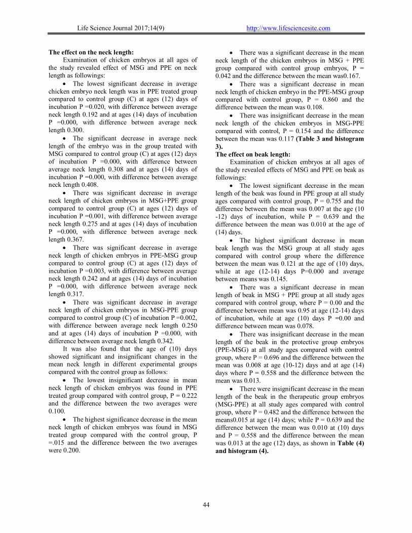
The effect on the neck length: Examination of chicken embryos at all ages of
the study revealed effect of MSG and PPE on neck length as followings:
The lowest significant decrease in average chicken embryo neck length was in PPE treated group compared to control group (C) at ages (12) days of incubation P =0.020, with difference between average neck length 0.192 and at ages (14) days of incubation P =0.000, with difference between average neck length 0.300.
The significant decrease in average neck length of the embryo was in the group treated with MSG compared to control group (C) at ages (12) days of incubation P =0.000, with difference between average neck length 0.308 and at ages (14) days of incubation P =0.000, with difference between average neck length 0.408.
There was significant decrease in average neck length of chicken embryos in MSG+PPE group compared to control group (C) at ages (12) days of incubation P =0.001, with difference between average neck length 0.275 and at ages (14) days of incubation P =0.000, with difference between average neck length 0.367.
There was significant decrease in average neck length of chicken embryos in PPE-MSG group compared to control group (C) at ages (12) days of incubation P =0.003, with difference between average neck length 0.242 and at ages (14) days of incubation P =0.000, with difference between average neck length 0.317.
There was significant decrease in average neck length of chicken embryos in MSG-PPE group compared to control group (C) of incubation P =0.002, with difference between average neck length 0.250 and at ages (14) days of incubation P =0.000, with difference between average neck length 0.342.
It was also found that the age of (10) days showed significant and insignificant changes in the mean neck length in different experimental groups compared with the control group as follows:
The lowest insignificant decrease in mean neck length of chicken embryos was found in PPE treated group compared with control group, P = 0.222 and the difference between the two averages were 0.100.
The highest significance decrease in the mean neck length of chicken embryos was found in MSG treated group compared with the control group, P =.015 and the difference between the two averages were 0.200.
There was a significant decrease in the mean neck length of the chicken embryos in MSG + PPE group compared with control group embryos, P = 0.042 and the difference between the mean was0.167.
There was a significant decrease in mean neck length of chicken embryo in the PPE-MSG group compared with control group, P = 0.860 and the difference between the mean was 0.108.
There was insignificant decrease in the mean neck length of the chicken embryos in MSG-PPE compared with control, P = 0.154 and the difference between the mean was 0.117 (Table 3 and histogram 3). The effect on beak length:
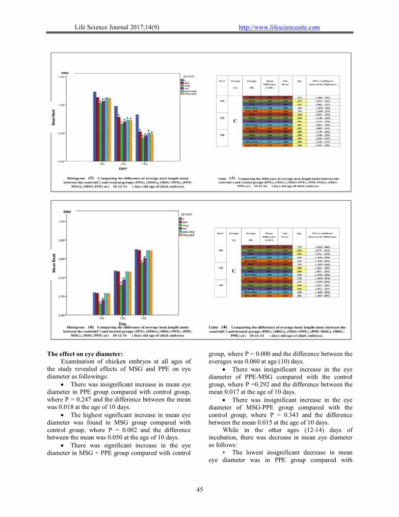
Examination of chicken embryos at all ages of the study revealed effects of MSG and PPE on beak as followings:
The lowest significant decrease in the mean length of the beak was found in PPE group at all study ages compared with control group, P = 0.755 and the difference between the mean was 0.007 at the age (10 -12) days of incubation, while P = 0.639 and the difference between the mean was 0.010 at the age of (14) days.
The highest significant decrease in mean beak length was the MSG group at all study ages compared with control group where the difference between the mean was 0.121 at the age of (10) days, while at age (12-14) days P=0.000 and average between means was 0.145.
There was a significant decrease in mean length of beak in MSG + PPE group at all study ages compared with control group, where P = 0.00 and the difference between mean was 0.95 at age (12-14) days of incubation, while at age (10) days P =0.00 and difference between mean was 0.078.
There was insignificant decrease in the mean length of the beak in the protective group embryos (PPE-MSG) at all study ages compared with control group, where P = 0.696 and the difference between the mean was 0.008 at age (10-12) days and at age (14) days where P = 0.558 and the difference between the mean was 0.013.
There were insignificant decrease in the mean length of the beak in the therapeutic group embryos (MSG-PPE) at all study ages compared with control group, where P = 0.482 and the difference between the means0.015 at age (14) days; while P = 0.639 and the difference between the mean was 0.010 at (10) days and P = 0.558 and the difference between the mean was 0.013 at the age (12) days, as shown in Table (4) and histogram (4).
Life Science Journal 2017;14(9) http://www.lifesciencesite.com
45
The effect on eye diameter:
Examination of chicken embryos at all ages of the study revealed effects of MSG and PPE on eye diameter as followings:
There was insignificant increase in mean eye diameter in PPE group compared with control group, where P = 0.247 and the difference between the mean was 0.018 at the age of 10 days.
The highest significant increase in mean eye diameter was found in MSG group compared with control group, where P = 0.002 and the difference between the mean was 0.050 at the age of 10 days.
There was significant increase in the eye diameter in MSG + PPE group compared with control
group, where P = 0.000 and the difference between the averages was 0.060 at age (10) days.
There was insignificant increase in the eye diameter of PPE-MSG compared with the control group, where P =0.292 and the difference between the mean 0.017 at the age of 10 days.
There was insignificant increase in the eye diameter of MSG-PPE group compared with the control group, where P = 0.343 and the difference between the mean 0.015 at the age of 10 days.
While in the other ages (12-14) days of incubation, there was decrease in mean eye diameter as follows:
• The lowest insignificant decrease in mean eye diameter was in PPE group compared with

Life Science Journal 2017;14(9) http://www.lifesciencesite.com
46
control, where P = 0.493 and the difference between the mean was0.011 at age (12-14) days.
• The highest significant decrease in eye diameter was in MSG group compared with control group, where P = 0.000 and the difference between mean 0.980 at age 12 days; while at age 14 days P =0.052 and the difference between means was0.900.
• There was significant decrease in the average eye diameter in MSG + PPE group compared with control group, where P = 0.001 and the difference between the averages was 0.052 at 12 days of incubation; at age 14 days P = 0.00 and the difference between the average was 0.880.
• There was significant decrease in the mean eye diameter in PPE-MSG compared with control group, where P = 0.016 and the mean difference was 0.38 at 12 days; at age 14 days P = 0.000 and the difference between the average was 0.69.
• There was significant decrease in the mean eye diameter in the therapeutic group embryos (MSG-PPE) compared with control group, where P = 0.001 and the difference between the mean was 0.051 at the
age of (12) days; while at age 14 days P = 0.000 and the difference between the averages was 0.078, as shown in Table (5) and histogram (5). The effect on liver weight:
After the dissection of chicken embryos and liver extraction at all ages of the study, no significant differences were found in the experimental groups compared to the control group at all ages (10-12-14) days of incubation as follows:
The mean weight of the liver in the experimental group at 10-12 days of incubation was constant compared with control group and the difference between the mean was 2.497, while the decrease was in MSG with difference between the two averages 0.010.
The mean liver weight in the experimental group at 14 days of incubation was constant compared to control group and the difference between mean was 3.728, while the decrease was in MSG group and the difference between the two averages was 0.010. This was illustrated in Table (6) and histogram (6).
The effect on hepatic cells thickness: The thickness of some hepatic cells was
measured in the histological sector of chicken embryos at 10 days of incubation. This age was selected only to represent the extent of hepatic cell thickness. The effect of monosodium glutamate and pomegranate extract was observed on liver cell thickness. The LSD was used to find the differences that reached the statistical significance level in the experimental groups compared with the control group at age 10 days as follows:
The least insignificant decrease in mean hepatic thickness was the PPE group at 10 days of incubation compared with control group, where P =
0.624 and the difference between the means was 0.244.
The highest significant decrease in mean hepatic thickness was the MSG group at 10 days of incubation compared with control group, where P = 0.000 and the difference between the two averages 2.563.
There was a significant decrease in the average thickness of hepatic cells of MSG + PPE group at 10 days of incubation compared with control group, where P = 0.00 and the mean difference was 2.459.
There was a significant decrease in the average thickness of PPE-MSG at 10 days of

Life Science Journal 2017;14(9) http://www.lifesciencesite.com
47
incubation compared with control group, where P = 0.558 and the difference between the mean was 0.292.
There was insignificant decrease in the average thickness of hepatic cells of MSG-PPE at 10
days of incubation compared to control group, where P = 0.506 and the difference between the mean 3.32, Table (7) and histogram (7).

Life Science Journal 2017;14(9) http://www.lifesciencesite.com
48
Histological Studies: **The normal histological structure of the
liver of the chick embryos (10) days old of the control group:
When examining the transverse tissue section in the liver of the chicken embryos of the control group at (10) days of incubation, we notice that the liver is surrounded by a thin fibrous tissue of connective tissue (C) composed of an integrated group of hepatic cells arranged in the form of ropes appeared in between them different blood vessels formed from, hepatic vein (HV) and central veins (CV). As the age progresses, the hepatic tissue becomes more dense than in the previous age as some hepatic cords (HC) appeared that radiated around the central veins (CV) and are close together. We can see some vacuolated hepatic cords (LC) that were formed of single raw of pyramid shaped hepatic cells that arranged so that the tops of these cells are directed toward a small cavity, and solid hepatic cords (SC) that are formed of one or two rows of the polygonal cells. Vacuolated hepatic rows were more than solid rows. Hepatic cells were characterized by having a specific cell membrane and acidophilic granulated cytoplasm and circular basic large two or more nuclei. The blood sinusoids (BS) appeared with narrow spaces and irregular edges between the hepatocellular ribbons that lining the blood vessels from the inside with flattened dark stained epithelial cells (EC) and larger, hyper pigmented spindle shaped cells called KC cells, as well as some red blood cells (RBCs). The histological structure of the liver of the chick embryo (10) days old of the Pomegranate Peel Extract Group:
Similarity was observed in the shape and structure of the general tissue shape compared with control group, except for some of the (BS). A few of them have a small hemorrhage that is simple and lined from (EC) and KC cells, as well as a few phagocytic cells and red blood cells (RBCs) (Figure3 ). The histological structure of the liver of the chick embryos (10) days old of the Monosodium Glutamate Group:
The structure of the tissue appeared to be different from control group with lost its normal form. Hepatocytes were less dense than the control group. The hepatic rows were deformed and undifferentiated both (LC) and (SC) rows. Varying degrees of degenerative changes were observed, including degeneration, pyknosis, and karyolisis. Some cells also appeared in the post-nuclear phase of lysis with pale and empty cytoplasm and a side nucleus known as apoptotic cells. Invagination of (CV) was observed in addition to the collapse of epithelial cells lining to it and fall into the cavity with hemorrhage, in addition to severe blood congestion of the hepatic vein and
fibrosis and appearance of some of small droplets (LD) appeared. (BS) appeared distorted shape with enlarged cavity and lined with epithelial cells and kupfer cells (KC). Many cells are also separated and fall into the blood sinusoids with appearance of red blood cells (RBCS) that were mostly affected (Figure3 ). The histological structure of the liver of the chick embryo (10) days old of the Mix Group:
Revealed that the liver structure was different compared with the control group. The hepatic rows appeared distorted in their apparent form that led to difficulty in distinguish between its two types. In addition, there were pyknosis, karyolisis and some cells showed apoptotic changes. In the sector, a large number of neutrophil cells (NC) are clustered around the (CV) and (BS) and some of them erupt inside the cavities, increase in number of lymphocytes, as well as destruction of endothelial lining of central veins and discrepancy in the size of LD and increase in their numbers. The blood sinusoids showed widening and hemorrhage inside them and separation of their endothelial lining and their falling inside (BS). This group is the relatively affected group (Figure3 ). The histological structure of the liver of the chick embryos (10) days old of the Preventive Group.
Histological structure of the liver of chicken embryo treated with PPE-MSG at age of 10 days after incubation revealed similarity of liver tissue structure to control group (Figure3 ). The histological structure of the liver of the chick embryos (10) days old of the Therapeutic Group.
Revealed difference in tissue structure compared to control group. As hepatic cords (HC) appeared less dense compared to control group and some degenerative changes appeared as pyknosis, karyolisis and hemorrhage in central veins as well as destruction and fibrosis of endothelial cells. Also, small number of small sized (LD) appeared. Also, (BS) appeared in between hepatic rows with different width that lined (EC) and appearance of kupfer cells, RBCs and phagocytic cells. In addition to destruction of endothelial lining of the blood sinusoids. This group is the least affected than MSG and MSG+PPE groups (Figure3 ). **The normal histological structure of the liver of the chick embryos (12) days old of the Control Group.
The hepatic tissue was more specific and denser than the previous ages. HC appeared closely related to each other and is radically arranged around the central veins. It is difficult to distinguish between the two types, LC type consisted of a single row of pyramidal shape hepatic cells that arranged in such a way that the tops of these cells are directed toward a small cavity. Solid hepatic cords (SD) consisted of a row or two of

Life Science Journal 2017;14(9) http://www.lifesciencesite.com
49
the polygonal cells. LC rows are more than SD rows. In between them different forms of blood vessels appeared as big (HV) and (CV) and (PA). Liver cells are characterized by having a well-defined cell membrane, acidic granulated cytoplasm, circular basic nucleus that contained two or more nucleoli, interspersed in them narrow (BS) with irregular edges lined with flattened EC lined side by side and in
between them large spindle shaped (KC), RBCs and in the first time in this age portal areas (PA) appeared between hepatic cords as false triangular shaped space consisted of branch of (PV), (HA) small sized with thick fibrous lining and branches of (BD) that surrounded by single layer of cuboidal epithelial cells or columnar small cells with rounded nucleus (Figure 4 ).
The histological structure of the liver of the chick embryos (12) days old of the Pomegranate Peel Extract Group.
Histological structure of liver cells of chicken embryo in PPE treated group at age 12 days after incubation showed similarity of liver tissue shape and structure compared to control group (Figure 4 ). The histological structure of the liver of the chick embryos (12) days old of the Monosodium Glutamate Group.
The structure of the tissue was different with loss of its normal shape compared to the control group. Hepatic tissue showed disorder and disintegration and less intense than the control group. The hepatic cords
were destructed and arranged radially around the CV. Varying degrees of degenerative changes were observed, including pyknosis as well as karyolisis. Some hepatic cells showed apoptotic changes, and a number of small sized fatty droplets (LD), uneven size of the sinuses (BS) as some of them were enlarged and others were compressed. The general shape of some of sinuses was disrupted and many of its cells are submerged and fall into the sinusoids with presence of RBCs and the remains of necrotic cells. The PA regions had the same structure as control sample but their walls were destructed. Fibrosis and damage to the surrounding liver cells and the separation of the lining cells of the PV were also observed. NC appeared

Life Science Journal 2017;14(9) http://www.lifesciencesite.com
50
around the PA. Deformity of the hepatic artery (HA), bile duct (BD) is addition to presence of LD which were the most damaging form (Figure 4). The histological structure of the liver of the chick embryos (12) days old of the Mix Group.
Showed affection of the hepatic tissue and its difference in shape and structure compared with control group. Some hepatocytes appeared healthy while others were affected. The presence of nuclear atrophy pyknosis and the breakdown of nuclear karyolisis and some cells showed apoptotic changes. Also we noticed detachment of some hepatic cells from the lining of the underlying membrane. Some of the (BS) appeared normal while, others were widened and congested and lined with EC and KC. Some of the cells lining the walls were separated and invaded into the sinuses with presence of RBCs and many other disintegrating cells as well as hemorrhage in some sinuses. The PA region had the same structure as control group but increased its wall thickness and presence of fibrosis was observed. Thickness and congestion of the (HA) was observed. The bile duct was distorted and damaged, as well as cellular filtration with neutrophils around bile duct and in
portal area, which is relatively affected group (Figure 4 ). The histological structure of the liver of the chick embryos (12) days old of the Preventive Group.
Revealed similarity in structure of liver cells to control group except presence of karyolisis in defragmented nucleus and presence of LD in between them BS with small narrow spaces and irregular edges that were lined with (EC) and (KC) and small RBCs. (Figure4 ). The histological structure of the liver of the chick embryos (12) days old of the Therapeutic Group.
Showed destruction of the liver cells as nuclei showed karyolisis and pyknosis and appearance of small sized LD and separation of endothelial lining of central vein with invagination of its lining. Also, BS appears with different shapes and with small narrow spaces with irregular edges that lined with (EC) and (KC) and RBCs. Also, invaginations of necrotic cells and their remaining inside the sinusoids with appearance of hemorrhage in some sinusoids. This is least affected group compared with MSG and MSG+PPE groups (Figure 4 ).
Life Science Journal 2017;14(9) http://www.lifesciencesite.com
51
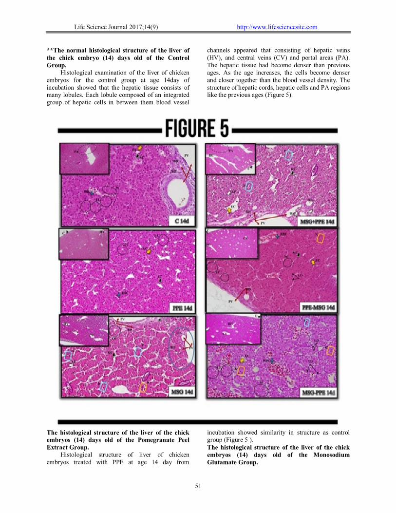
**The normal histological structure of the liver of the chick embryo (14) days old of the Control Group.
Histological examination of the liver of chicken embryos for the control group at age 14day of incubation showed that the hepatic tissue consists of many lobules. Each lobule composed of an integrated group of hepatic cells in between them blood vessel
channels appeared that consisting of hepatic veins (HV), and central veins (CV) and portal areas (PA). The hepatic tissue had become denser than previous ages. As the age increases, the cells become denser and closer together than the blood vessel density. The structure of hepatic cords, hepatic cells and PA regions like the previous ages (Figure 5).
The histological structure of the liver of the chick embryos (14) days old of the Pomegranate Peel Extract Group.
Histological structure of liver of chicken embryos treated with PPE at age 14 day from
incubation showed similarity in structure as control group (Figure 5 ). The histological structure of the liver of the chick embryos (14) days old of the Monosodium Glutamate Group.
Life Science Journal 2017;14(9) http://www.lifesciencesite.com
52
Different liver structure compared with control. Some HC appeared less dense compared with control and distorted shape with difficult to distinguish them. Many degenerative changes appeared as pyknosis, karyolisis and some cells showed apoptosis. Some hepatic cells separated from basement membrane lined them. BS with narrow spaced and showed abnormal shape with separation of cells lining their walls and rushes them inside with the presence of the remnants of decomposed cells. The PA appear the same structure but thickness of wall of PV appeared with detachment of the cells lining it from the inside and fibrosis as well as appearance of focal necrosis around the portal vein. The hepatic artery (HA) lost its normal shape and the thickness and narrowing of portalcanal that were mostly affected (Figure 5 ). The histological structure of the liver of the chick embryos (14) days old of the Mix Group.
Showed different liver structure compared with control. Liver cords appeared distorted. Different types of degenerative changes have been observed, including pyknosis, karyolisis, and some cells showed apoptosis with highly pigmented apoptotic nucleus surrounded by a halo of pure cytoplasm in between them deformed wide BS with the presence of RBCs, in addition to the separation of some of the cells lining and their fall into the sinus. The PA region appeared the same general structure with fibrosis to portal vein wall (PV) and the detachment of epithelial cells lining it from the inside. The hepatic artery (HA) distorted and lost its normal shape. The bile duct distorted with the nuclei of their cells showed apoptosis. The relatively affected group (Figure 5 ). The histological structure of the liver of the chick embryos (14) days old of the Preventive Group.
Histological structure of liver of chicken embryos treated with PPE-MSG at age 14 day from incubation showed similar liver structure compared with control (Figure 5 ). The histological structure of the liver of the chick embryos (14) days old of the Therapeutic Group.
Showed karyolisis, pyknosis and different sized LD in between them BS with different shapes with congestion of some of them in addition to separation of their lining and rushing of the remaining cells inside them. This is the least affected group the MSG and MSG+PPE groups (Figure 5 ). 4. Discussion
Congenital malformations: The results of the study showed that there were
different grades of congenital malformations in a number of embryos of the different experimental groups at selected ages. The group treated with MSG was the most affected group, followed by the mixed group (MSG + PPE) then (MSG-PPE). The
abnormalities were in the form of small fetal sizes and congestion of their body. Also, decrease in height and total body weight, neck length and beak length and eye diameter. Also decrease in liver weight in addition to the contraction of liver cells at the age 10 days. Distant abnormalities were small eye size and with increased age anophthalmia, brain hemorrhage and increased in the extent of the peripheral region of the brain. The visceral hernias were visible in all ages and groups compared to control group embryos. No congenital abnormalities were observed in PPE and PPE-MSG groups.
These results were in agreement with that obtained by other researches (Oforofuo et al.1997; Botey et.al. 1998; Al-Qudsi and Al-Jahdali 2012) as they reported that MSG had a prominent role in delayed and returned growth of the embryos. That the monosodium glutamate had prominent role in the delay and slow growth of embryos due to bleeding of the vessels and so embryonic cells were unable to obtain the necessary quantities of sufficient needs to support their growth, development and division efficiently, which led to the delay and slow growth of embryos and toxicity.
The results of this study were in agreement with others (Tawfeeq and Badr, (2012) in terms of slow and delayed growth as they reported that when female pregnant rats administrated orally MSG led to significant and noticeable changes in the hypothalamus region of infants and so decreased in its hormones especially growth hormone (GH) and so the length, weights and sizes of fetuses were decreased in addition to other side effects. Also, previous studies reported decreased in weights and heights of fetuses and newborns and claimed that to the ability of MSG to penetrate and cross the placenta of pregnant mothers and also its ability to cross the mammary gland barriers.
Also results obtained by this study supported the results of George et al. (2013) study, which observed a decrease in the weights of infants when the pregnant mothers given oral MSG for 20th day of pregnancy due to fetal resorption that led to small size, length and weight of the fetus as MSG had the ability to penetrate the immature blood-brain barriere specially in the fetuses, newborns and infants causing high toxicity. Also, MSG is highly soluble in lipids and so change the nature of the liquidity of nerve membranes and events damage the cells that led to neurotoxicity such as cell degradation, destruction of its branches and nuclei, and decrease in overall cell size, thus affecting the function of the hypothalamus pituitary axis that is responsible for the secretion of hormones, including growth hormone. As a result there has been a decrease in fetal weights and lengths in MSG where as in MSG-PPE MSG+PPE) were less affected because of

Life Science Journal 2017;14(9) http://www.lifesciencesite.com
53
antioxidants that were present in pomegranate peels that led to restore changes in the level of biochemical measurements resulting from oxidative stress of glutamate. In PPE and PPE-MSG groups antioxidants, organic acids, amino acids, vitamins and others that were found in the pomegranate extract play an important role in inhibiting the appearance of congenital malformations. So, the usage of pomegranate peel is safe on embryos during organogenesis, fetal growth and maturation period.
Our results were in agreed with Loren et al. (2005); that the antioxidants found in pomegranate peel can get ride of free radicals derived from MSG administration. Also, our results confirmed that obtained by (Tawfeeq and Badr 2012 who used vitamins, especially vitamin A, play an important role as antioxidant against the harmful effects of MSG because of its growth role and antioxidant that play an important role in the body as epithelial differentiation, vision, hematopoiesis, brain formation, formation pattern during embryogenesis, bone development and maintenance of immune system functions. Antioxidants and vitamin A play a role in preventing peroxidation of fat, DNA breakdown and conversion of antioxidant enzymes in the body's cells. The high ratios of polyphenols and alginic acid found in pomegranate peel and extract had important role in providing neurological protection of embryos from brain inertia as when pregnant mothers administered nutritional supplements consisting of special pomegranate extracts in the first trimester of pregnancy, or even in a period of lactation support and prevent the occurrence of any abnormalities of the nervous system, as they contribute to the inhibition of proteins associated with Alzheimer's disease in adults.
The results of this study also supported Jurenka (2008) and Vidal et al. (2003) that the active compounds extracted from pomegranate in all its components do not cause any harm at the specified doses because they are antioxidants contribute to the treatment of oxidative stress and liver disorders. These results were confirmed by histological examination of the tissues of mice treated with different parts of pomegranate, which proved the absence of any deformation effects or manifestations of developmental delay and toxicity because of the pomegranate peels contained phenol hydroxyl groups that enable them to eliminate the reactive oxygen species that affect the body's biological systems as well as their ability to interact with DNA, proteins and lipids, causing cell breakdown. Therefore, the phenol hydroxyl groups were responsible for antioxidant effects that protect the body from many free-radical-induced health problems. To prove this, 86 overweight people were given tablets of the most effective and necessary compounds derived from pomegranate
extracts during 28 days. As a result, these compounds contributed to weight loss due to its effect on pancreatic lipase enzyme, which works to digest fats as well as its ability to reduce the amount of calories and there were no reported side effects or negative changes in blood or urine.
The results obtained by this study were inconsistence with those obtained by others (Hermanussen and Tresguerres 2003; Hermanussen et al. 2006; Roman-Ramos et al., 2011). Their results showed that in rats ate MSG, there were an increase in total body weight and twice increased in body fat. Their results were explained by the ability of MSG to block leptin receptors in the hypothalamus. It has been reported that much of leptin hormone led to reduced sensitivity, which in turn leads to hunger and desire to eat and obesity. IN ADDITION, increase the production of resectin hormone and cytokines and stimulate the gene of TNF-α responsible for the formation of adipose tissue accompanied by a disturbance in levels of sugar and insulin, as well as increase in the concentration of liver enzymes associated with an increase in body weight.
The differences in the weight and length of embryos in the experimental groups may be due to the difference in the amount of doses injected, the time of the dose administration and the incubation time. As a result, monosodium glutamate and pomegranate peel extract resulted in different effects.
In general, MSG during organogenesis had a negative effect on the embryos, especially in early ages. This effect caused by a defect in the formation of two or more body organs led to apparent and functional abnormalities due to the lack of development and completeness of the body's defensive systems. It was observed that some of the embryos varied in resistance to MSG side effects and improved by PPE treatment. Some of the PPE peels treated were not able to improve side effects of MSG by the prescribed dose and duration of the study, especially in the MSG + PPE and MSG-PPE groups.
The current study showed no difference in liver weight for experimental groups compared to control group. The results were consistent with others (Gil-Loyzaga et al., 1993; Gill et al., 2008and Vladimila et al. 2009) where MSG and vitamin A were given together during pregnancy; there was insignificant difference in organs weight. Studies showed that pomegranate extract had positive effects in liver cells protection from any harmful effects caused by decreased consumption of free radicals by important enzymes, especially superoxide dismutase, which reduce the speed of cell destruction and restore vitality of the cells.
While embryos treated with MSG showed decrease in liver weight compared to the control

Life Science Journal 2017;14(9) http://www.lifesciencesite.com
54
group. This is due to the presence of glutamate receptors in different areas of the tissues such as hypothalamus, lung, heart, liver, endocrine system, kidneys, ovaries and uterus. These receptors are also affected by a certain mechanic that is believed to alter the permeability of cell membranes especially calcium and this change inhibit glutamate receptors and their effects. In contrary, Park et al. (2000) and Tawfik and Al-Badr (2012) reported an increase in organ weight when treated with MSG due to increase the effectiveness of inflammatory factors that negatively affected the tissue and lead to inflammation and thus an increase in the organs weight.
Our results was supported by Del Bigio and Vriend (1998) who found swelling at the front of the brain in mice as a result of its high contents of amino acids that act as nervous receptors as aspartate and glutamate and to confirm these effects further researches must be done.
The results of this study also showed that the eyes of the embryos treated with MSG at the age of (10) days were larger than others experimental groups as eye diameter at age 10 days was larger due to edema and swelling of brain of the embryos where the dorsal area of the frontal brain was larger compared with control group. While eye diameter was smaller at other ages and this consists with Cohen (1967) who reported that the eyes of MSG treated animals were smaller than the control group and anophthalmia of the eyes in some embryos may be due to elevated glutamate that led to inflammation of the optic nerve and closure of the central retinal vein and so defect formation of the eyes.
Al-Qudsi and Al-Jahdali (2012) and Olney (1980) agreed with our results in the presence of hemorrhage in chicken embryos injected with MSG and explained that by increased concentration of neurotransmitter glutamate affects the flow of large amounts of calcium ions within neural cells causing an imbalance in the ionic balance and a decrease in oxygen and affect the mitochondria inside the cell leading to increased generation of ROS and reduce the production of energy and appearance of free radicals that eliminate the cells. In addition, increase of glutamate cause increased concentration of ammonia ion, which contributes to the occurrence of cellular lesion of brain cells and cells apoptosis. - Morphological studies:
The appearance of congenital anomalies in the embryos as decrease size, enlarged abdominal region and abdominal hernia, anophalmia and decrease eye diameter, widening of the dorsal region of the brain and hemorrhagic. These congenital anomalies were mostly prominent in MSG treated followed by the MSG + PPE treatment group and finally the MSG-PPE treatment group. While, the PPE treated group
was similar to the control group and the PPE-MSG treated group showed a significant protective role. Statistical studies:
1. The differences in the lengths and weights of the embryos of the experimental groups were found. Generally, the decrease was dominant in most groups. The length and weight of the MSG-treated group embryos were lowest in height and weight followed by MSG + PPE and finally the MSG-PPE group. The length and weight of group embryos treated with PPE were the lowest in height and weight reduction, followed by PPE-MSG group embryos compared to control group embryos.
2. Morphometric studies confirmed that monosodium glutamate caused a delay and decrease in fetal and organ growth compared to control group in terms of neck length, beak length and eye diameter. There was also a decrease in liver weight and liver cell thickness. Pomegranate extract had an improved role in toxicity as much as possible. - Histological studies:
1. Histological studies of the liver showed differences in the thickness of the fibroblast and the difference in cellular density from one region to another, as well as the difficulty of distinguishing between both types of the hepatic cords and disorders in the radiographic arrangement of the hepatic rows around the congested deformed central veins as well as the variation in the area of the blood sinuses and their congestion and change the thickness of the walls and deformation of portal areas and their dispersion in addition to of vaculation in the tissue and fatty degeneration and accumulation of fat in the liver of the embryos, which appeared in particular in MSG treatment group followed by MSG + PPE treated group and finally the MSG-PPE treatment group.
2. Many degenerative changes in the treated tissue cells compared with the control group were observed as atrophy, dissociation, hypertrophy and decomposition of the nuclei as well as congestion and hemorrhage, the appearance of apoptotic cells and final liver cirrhosis.
The results were explained by that monosodium glutamate has an effect on tissues and cells from several aspects as the toxic effect on cells causing their death, the effect on the enzymatic activity of cells and then the disruption of cellular regulation, the effect on hormones regulating the tissues, thus affecting cellular functions. These interpretations are the result of many researchers and scientists in the same field of research and these results have agreed their views and suggestions.
The results of this study showed swelling accompanied by a tightening of the skin and hernia of abdominal organs in the experimental groups, as a result of exposure of the fetuses to repeated doses of

Life Science Journal 2017;14(9) http://www.lifesciencesite.com
55
MSG that led to weakness of the abdomen wall and development of specific opening in the abdomen that contained the hernia contents as the intestine or fat. Swelling due to the retention of body fluids caused by urinary system dysfunction (Biondo Simeses et al., 2010).
This may be a direct effect of MSG cell metabolism in embryos, as monosodium glutamate has the ability to pass easily through the membrane and affected embryos growth and development. Or due to its deformed effects on embryos that suggestion was supported by Noda et al. (2012), who confirmed that high glutamate concentration led to cell apoptosis and most of these cases were under the influence of the free radicles on the cells and the mono-glutamate metabolism results in the breakdown of the cell ribosome DNA. Pomegranate peels had a role in reducing these effects as it had antioxidant properties that led them to consumed free radicles and so maintained cell and body tissues.
Tamas et al. (2004) injected newborn rats with monosodium glutamate subcutaneous (2mg/gm) five times (1,3,5,7,9) after birth and second group consists from newly born infants subcutaneously injected with the same dose three times after birth (1,5,9). The results were similar in terms of maternal and neonatal weight loss and small size. Moreover, the first group was more affected with increased in fetal mortality rate due to repeated doses.
In agreement with our results about liver (Al-Gamdi2007; Abdel-Fatah 1992; EL Naggar et al., 1977) reported that in a 7-day-old fetus of age, the liver of the control group coated with a thin capsule of the connective tissue, the hepatic cells multiple and arranged to form hepatic rows in between them blood sinusoids. Hepatocytes radiated from the hepatic veins and separated by blood sinusoids. The hepatic cords were distinguished at ages (7-10) days into vaculated rows (LC) that consists of one layer of pyramidal cells that radiate around a space and solid rows (SC) that consists of two rows of polygonal cells. The hepatocytes had granulated acidic cytoplasmand rounded nucleus or two nuclei with basic cytoplasm. With increase age, liver increase in size gradually and the part of the medial segment of the blood sinusoids gradually blocked by developing hepatic rows. And from 12 to 16 days of incubation, hepatic rows became dense and close together and cannot be distinguished into hollow and solid cords. The study also revealed that the blood sinusoids appeared as wide gaps between the hepatic rows at first irregularly in shape as age progresses, their sizes decreased, the sinuses lining the flat epithelial cells with a flat dark nucleus, and kupfer cells, which are spindle cells with a small oval nucleus. As the age progresses, the blood
sinusoids appeared as slit as results of compression by developing hepatic rows.
In agreement with our results (Al-Qudsi and Al-Jahdali 2012; Al-Gamdi 2007; Abdel-Fatah 1992) in the appearance of the portal areas at the age of 12 days consisting of: the portal vein, a branch of the hepatic artery and tributaries of the bile duct. This led to scarcity of connective tissue and numerous portal areas that is an unreal hepatic model. Hamilton (1965) had suggested that the pyloric region at the day (10-14) may appear from one or more bile ducts, one portal vein, a small hepatic artery and a lymphatic vessel versus a few central veins.
This study showed that lipids drops were seen in the chicken embryos of the control group from day 7 of the growth and they increased in size and numbers as the age increased until the end of growth and as results of appearance of cytoplasm and capsulated hepatic cells. Others (Abdel-fatah 1992; Al-Gamdi, 2007; Al-qudsi and Al-Jahdali2012) supported our results in that lipid droplets for hepatocytes appeared from (5-7) days and at age 7 day of growth of chicken embryos there was association between functional activity with liver growth, growth of hepatic artery and increase in hepatocellular thickness due to fullness of gall bladder with cholesterol.
The histological examination showed that there was a clear and uneven effect in the liver of chicken embryo in different groups at the selected ages of the study. The MSG treated group was the most affected, followed by the mixed group (MSG + PPE) then therapeutic group (MSG-PPE). The degenerative changes were damage and variation of the shape of solid and hollow liver cords and the dominance of solid over the hollow live cords in addition to difficulty in distinguishing between them in advanced ages and these changes were manifestation of developmental delay caused by MSG toxicity. Also changes were manifested by widening in blood sinusoids and different degrees of congestion, hemorrhage and fibrosis and increased in lipid droplets. Also invagination in the wall of central vein with appearance of focal necrosis around it and portal area that showed also deformity and degenerative changes. The nucleus showed pyknosis, karyolysis and apoptosis and accumulation of neutrophils around the central veins and portal areas especially in the MSG and MSG + PPE, MSG-PPE groups. Liver segments of the PPE-MSG group showed resistance to the toxicity caused by sodium mono glutamate, where the damage was very rare while the liver segments of pomegranate peel extract treated group were healthy and formative compared with the control group. Kumar et al. (2003) stated that the biochemical reactions of the cell against damage appeared in different and complex ways that depend on the type of

Life Science Journal 2017;14(9) http://www.lifesciencesite.com
56
damage, time of occurrence and severity, so the small doses and short duration cause damage in the cell that can be repaired, while large repetitive doses for long time may cause cellular damage that cannot be recovered and led to cell death. Also damage of the cells depend on type of cells and their adaptation and its genetic structure, hormonal and physical status of the liver cells and its fullness with glycogen made its response strong.
Ortiz et al. (2006) reported that i.p. injection of MGS (4mg/kg) led to toxic effects on the body that was manifested by changes in growth picture and function of the liver these effects resulted from direct effects on organs metabolism of the organ and the hepatotoxicity was acute and serious after any drug use and after stop of drug intake that led to initial cirrhosis. The proposal was supported by other researchers, who explained that the degenerative changes in the liver in MSG, MSG + PPE and MSG-PPE groups at the ages of the study were growth delayed, variability in the density of liver cords, and deformed some of them, lysis and difficult to distinguish between them, as well as widening in the blood sinusoids and central veins and thickness of the wall due to MSG accumulation in the liver. Burkitt et al. (1993) explained cellular degeneration were due to inflammatory and immune reactions against damage caused by chemical compounds, parasitic and viral infections, and harmful agents against cells and tissues of the body.
In agreement with our results other researches (Al-Qudsi and Al-Jahdali 2012; Ortiz et al.2006; Hinoi et al.2004) examined the toxic effects of i.p. injection of MSG (4 mg/kg) in adult male rats after 15-30-45 minutes on the biochemical and histological changes of the liver and their results revealed that toxic effect on the cells led to tissue damage in addition to damage of cell membrane and excretion of the liver enzymes to the blood.
Overttreet et al. (2002) discussed the advanced toxicity in individuals treated with drugs, especially neurological drugs that the sub-acute doses leads to progressive destruction of liver cells in the repeated examination of liver cells, while the preliminary examination showed that about 50% of the hepatocytes showed decomposition and with sequential examination a massive hepatocellular nebula appeared. They explained their results by that MSG had the ability to cross and permeate the epithelial cell membranes in the peritoneal cavity to reach the blood and analyzed to sodium and glutamic acid as some of glutamic acid excreted while other part change to glutamate and during that hepatocyte repaired partially as glutamate inside them is high and they cannot get rid of them and that led to their
inflammation, degeneration and cell death in MSG treated groups.
Al-Qudsi and Al-Jahdali 2012; Ortiz et al.2006; Hinoi et al.2004 revealed that histological examination of hepatocytes showed nuclei with pyknotic chages at 15 minutes, lipid droplets at 30 minutes, changes in the liver cords and appearances of lange size and numbers of lipid droplets at 45 minutes in addition to congestion of blood sinusoids and central veins as results of hepatocytes inflammation and fibrosis. These results were in agreement with results of the present study that showed nuclear pyknosis, karyolisis, apoptoais and accumulation of neutrophils around the central veins and portal areas. Apoptosis may occur due to lack of important and necessary elements for its survival, DNA damage, treatment with cytotoxic drugs, exposure to radiation, loss of survival signals, or oxidative stress, all of which may lead to apoptosis that occurs as a result of cell response to internal and external signals (Dibartolomeis and Mone, 2003). Apoptosis also occurs when the cell is severely damaged or decreased its oxygen duet to decrease blood supply.. Cell bulging is a clear feature as internal organelles especially mitochondria and the entire cell swell and rupture. These changes occurs as the injury prevents the cells from adjusting their balance and ions, fluids as water and ions as sodium and calcium that normally pumped out of the cell but in the case of injury these ions influx inside the cells that led to cell rupture and expulsion of its contents to intracellular spaces (Van Cruchten and Van Den, 2002; Giorgio et al., 2005) that led to damage and apoptosis of neighboring cells by lysosomes in different degrees. So, toxic damage to the cells was divided into two main parts:
1) Changes on cellular structure, 2) changes on cellular functions. And both of these changes affect each other. On the other hand, the association of toxic chemical compounds with biomolecules has been divided into two parts:
1. Reversible combination that can be reversed so its effect is not often fatal because it is based on the nature of the cellular function.
2. Irreversible combination that that had toxic effects on the cells as they led to changes in cellular structure that led to cell death and apoptosis (Eisenbrand et al., 2002; Liu and Huang, 2006).
2) Changes that occurred in the cellular functions due to toxic substances can be divided into three divisions:
1. Changes in the cell membrane permeability that led to transport of the substances to and outside of the cells.
2. Changes in cell enzymatic activities that led to changes in internal respiration of the cells and so changes in energy storage.

Life Science Journal 2017;14(9) http://www.lifesciencesite.com
57
3. changes in cell division and formation of DNA and RNA and so changes in protein synthesis and mutation (Eisenbrand et al.2002); (Wang et al. 2004); (Liu and Huang 2006).
These results suggested that MSG had role in oxidative stress that led to cell death and degeneration and nuclear lysis. So, addition of pomegranate peel extract to MSG to decrease toxic effects of MSG on cell by apoptosis, destruction of DNA and RNA, decrease protein synthesis that is essential to form protein receptors that led to decrease in cell membrane thickness and disturbance in cell division and so delayed growth. Pomegranate peel extract removed free oxygen radicals and so maintained the cell structure and function especially in the growth stage as this stage is critical for cell division and this depend on the time and repetition of the dose.
Histological examination of liver of chicken embryos in MSG+PPE, MSG-PPE groups increase in the number and size of lipid droplets that led to lipid droplets accumulation due to toxins, malnutrition and anoxia. These results were confirmed by Nakanishi et al.2008 who reported that repetitive injection of adult male rats with MSG by 2 mg/kg for five days after delivery and then observation of the newborn for 6 and 12 months found that accumulation of fat droplets and inflammation of hepatocytes. Kumar et al. (2003) explained these lipid droplets accumulation in the liver was due to abnormal accumulation of triglyceride inside hepatocytes and these damage was temporary but damage that occurs due to cell apoptosis changes in lipid metabolism was irreversible. Inflammation also led to rapid changes in tissue response to damage. Curran and Crocker (2005) reported that presence of neutrophil and lymphocytes means chronic inflammation as accumulation of neutrophils around apoptotic cells lead to their engulf and decrease of infection and removal of damage as they circulate in the body by the blood but white blood cells activity may led to damage of healthy tissues and produced antioxidants and repair of cells. Increase in white blood cells numbers appears in blood vessels which were scattered in the connective tissue associated with edema caused by fluids leakage from the dilated and congested blood vessels. Edema also occurred due to increased permeability of blood vessels and increase lymphocytes migration from the blood vessels to surrounding tissues.
Also, researches showed that hepatocytes affected by MGS and PPE at 10 days of age by morphometric measurement compared with control group revealed decreased in hepatocytes thickness due to degeneration and apoptosis and shrinkage of hepatocytes due to treatment with MSG and significant and insignificant changes in different studied groups. These results was confirmed by
Anindita et al. (2012) who reported that shrinkage and decrease in hepatocyte thickness was due to degeneration and apoptosis due to treatment with MSG that led to loss of cell functions. Conclusions:
The results showed that sodium mono glutamate had side effects on hepatic tissue of chicken embryos based on dose and the timing given. The results showed that the pomegranate extract was completely safe and did not produce any side effects. Some of the most notable effects were. Reference: 1. Abdel-Fatah, A.A. (1992) Effect of tetracyclines
on the development of the liver in chick embryos, Ph.D. Thesis, Al-Azhar University, cairo-Egypt.
2. Adhami, V. M., et al. (2011). Prevention of Cancer with Pomegranate and Pomegranate Anthocyanins. Berries and Cancer Prevention, Springer: 209-226.
3. Albrecht, M., Jiang, W., Kumi-Diaka, J., Lansky, E.P., Gommersall, L.M., Patel, A., Mansel, R.E., Neeman, I., Geldof, A.A. and Campbell, M.J., (2004). Pomegranate extracts potently suppress proliferation, xenograft growth, and invasion of human prostate cancer cells. Journal of medicinal food, 7(3), pp.274-283.
4. Al-Gamdi.F.A (2007) Effect of Haloperidol (Hadoldecanoso) anti depressant drug on the development of some organs chick embryo, Ph.D. Thesis, King Abdul Aziz University, Girls Collage Education, Jeddah-KSA.
5. AL-Mosaibih, M. A. (2013). "Effects of monosodium glutamate and acrylamide on the liver tissue of adult Wistar rats." Life Sci. J 10(2s): 35-42.
6. Al-Qudsi, F. and A. Al-Jahdali (2012). "Effect of monosodium glutamate on chick embryo development." Journal of American Science 8(10).
7. Anindita, R., Soeprobowati, T.R. and Suprapti, N.H., (2012). Potensi Teh Hijau (Camelia sinensis L.) dalam Perbaikan Fungsi Hepar pada Mencit yang Diinduksi Monosodium Glutamat (MSG). BULETIN ANATOMI DAN FISIOLOGI dh SELLULA, 20(2), pp.15-23.
8. Biondo-Simões, M. d. L. P., et al. (2010). "Obesity and abdominal wound healing in rats. Acta cirurqica brasileira 25(1):86-92.
9. Block, J. P.; Scribner, R. A. and Desalvo, K. B. (2004). Fast food, Race/ethnicity, and income. Am. J. Prevt. Med. 27(3):211-217.
10. Bookheimer, S.Y., Strojwas, M.H., Cohen, M.S., Saunders, A.M., Pericak-Vance, M.A.,

Life Science Journal 2017;14(9) http://www.lifesciencesite.com
58
Mazziotta, J.C. and Small, G.W., (2000). Patterns of brain activation in people at risk for Alzheimer's disease. New England journal of medicine, 343(7), pp.450-456.
11. Botey, J., Cozzo, M., Marin, A. and Eseverri, J.L., (1987). Monosodium glutamate and skin pathology in pediatric allergology. Allergologia et immunopathologia, 16(6), pp.425-428.
12. Bryant, R. and Dundes, L. (2008). Fast food perceptions: A pilot study of college students in Spain and the United States. Appetite. 51:327-330..
13. Burkitt, H.G., Young, B. and Hath, J.W., (1993). Wheater 'Functional Histology" A txt and colour atlas. Hong Kong: Longmann Group, 1993.
14. Cohen, A.I., (1967). An electron microscopic study of the modification by monosodium glutamate of the retinas of normal and “rodless” mice. Developmental Dynamics, 120(2), pp.319-355.
15. Covington, A. D. (1997). "Modern tanning chemistry." Chemical Society Reviews 26(2): 111-126.
16. Curran, R.C. and Crocker, J. (2005): "Curran's Atlas of Histopathology"4th Revised Edition with 810 photomicrographs. Harvey Miller Publishers Oxford University Press.
17. Del Bigio, MR, J P Vriend. (1998) Monosodiume neurotransmitters and amino acids in cerebrum and striatum of immature rats with kaolin-induced hydrocephalus. Brain Research 798[1-2], 119-126.
18. Di Bartolomeis, S. M. and J. P. Moné (2003). "Apoptosis: a four-week laboratory investigation for advanced molecular and cellular biology students." Cell biology education 2(4): 275-295.
19. Dongen, M. V. V.; Kack, F. J. and Graaf, C. D. (2010). Eating rate of commonly consumed foods promotes food and energy intake. Appetite. 56:25-31.
20. Egbuonu, A.C.C., Obidoa, O., Ezeokonkwo, C.A. and Ejikeme, P.M., (2009). Hepatotoxic effects of low dose oral administration of monosodium glutamate in male albino rats. African Journal of Biotechnology, 8(13).
21. Eisenbrand, G., Pool-Zobel, B., Baker, V., Balls, M., Blaauboer, B.J., Boobis, A., Carere, A., Kevekordes, S., Lhuguenot, J.C., Pieters, R. and Kleiner, J., (2002). Methods of in vitro toxicology. Food and Chemical Toxicology, 40(2), pp.193-236.
22. El-Awdan, S.A., Jaleel, G.A.A. and Saleh, D.O., (2013). Grape seed extract attenuates hyperglycaemia-induced in rats by streptozotocin. Bulletin of Faculty of Pharmacy, Cairo University, 51(2), pp.203-209.
23. El-Naggar, M.I. (1977) comparative studies on the effect of cytotoxic drug on developing chick embryo, Ph.D. Thesis, Al-Azhar University, Cairo-Egypt.
24. Eroschenko, V.P. (2000): "Di Fiore's Atlas of histology with functional correltion". 9th ed. Lippincott Williams & Wilins.
25. Faraj, A. M. and Q. M. Koyee (2011). "A Coprological Diagnostic Comparison between Zinc Sulphate Floatation and Formalin: Ether Sedimentation with Two Natural Extracts (Pomegranate Molasses and Honey)."
26. George, K.R., Shibija, N.G. and Malini, N.A., (2013). Monosodium glutamate (MSG) induced developmental dysfunction in female albino rats (Rattus norvegicus). The Bioscan, 8, pp.73-6.
27. Ghaffari, M. and V. Ahmadian (2007). "Investigation of antioxidant and electron beam radiation effects on the thermal oxidation stability of low-density polyethylene." Radiation Physics and Chemistry 76(11): 1666-1670.
28. Gil, M.I., Tomás-Barberán, F.A., Hess-Pierce, B., Holcroft, D.M. and Kader, A.A., (2000). Antioxidant activity of pomegranate juice and its relationship with phenolic composition and processing. Journal of Agricultural and Food chemistry, 48(10), pp.4581-4589.
29. Gill, S.; Barker, M. & Pulido, O. (2008). Neuroexcitatory target in female reproductive system of the non human primates (Macaca fascicularis). Toxicol. Pathol., 36: 478-484. 25.
30. Gil-Loyzaga, P.; Hernandez-Ortiz, M. J.; Rodriguez-Benito, T. & Lasso Delavega, M. (1993). Dilitazem protect against neuro toxicity induced by excitotoxic amino acids on cochlear afferent fibers. ORL, J. Othorhinolaryngol. Relate. Spec., 55: 211-215.
31. Giorgio, M., Migliaccio, E., Orsini, F., Paolucci, D., Moroni, M., Contursi, C., Pelliccia, G., Luzi, L., Minucci, S., Marcaccio, M. and Pinton, P., (2005). Electron transfer between cytochrome c and p66 Shc generates reactive oxygen species that trigger mitochondrial apoptosis. Cell, 122(2), pp.221-233.
32. Guo, C., Wei, J., Yang, J., Xu, J., Pang, W. and Jiang, Y., (2008). Pomegranate juice is potentially better than apple juice in improving antioxidant function in elderly subjects. Nutrition Research, 28(2), pp.72-77.
33. Hakkim, F.L.;Arivazhagan, G. and Boopath, R. (2008). Antioxidant property of selected Ocimum species and their secondary metabolite content. J. Med. Plan. Res. 2: 250-25.
34. Hamilton, H. I. (1965) "lillie's Development of the chick" Third adition. Holt, Rinehart and

Life Science Journal 2017;14(9) http://www.lifesciencesite.com
59
Winston, New York. P.236-245; 252-297 & 471-479.
35. Hermanussen, M. & Tresguerres, J. A. F. (2003). “Does High Glutamate Intake Cause Obesity?,” Journal of Pediatric Endocrinology and Metabolism, 16(7), 965-8.
36. Hermanussen, M., Garcia, A.P., Sunder, M., Voigt, M., Salazar, V. and Tresguerres, J.A.F., (2006). Obesity, voracity, and short stature: the impact of glutamate on the regulation of appetite. European journal of clinical nutrition, 60(1), p.25.
37. Hinoi, E., Takeda, T., Ueshima, T., Tsuchihashi, Y. and Yoneda, Y. (2004) Glutamate signaling in peripheral tissues, Eur J Biochem;. 271:1-13.
38. Hora, J.J., Maydew, E.R., Lansky, E.P. and Dwivedi, C., (2003). Chemopreventive effects of pomegranate seed oil on skin tumor development in CD1 mice. Journal of medicinal food, 6(3), pp.157-161.
39. Husarova, V. and D. Ostatnikova (2013). "Monosodium glutamate toxic effects and their implications for human intake: a review." JMED Research 2013: 1-12.
40. Jurenka, J. (2008). "Therapeutic applications of pomegranate (Punica granatum L.): a review." Alternative medicine review: a journal of clinical therapeutic 13(2): 128-144.
41. Khanna, S. K.; Singh, G.B.And Krishna Murti, C.R. (1980)."Toxicity profile of some commonly encountered food colours." J.Fd.Sci. Technol., 17;95-103.
42. Kierszenbaum, A.L. (2002): "Histology and cell biology: An Introduction to Pathology". Mosby, Inc. USA.
43. Kumar, V.; Cotran, R. and Robbins, S. (2003):"Robbins "Basic Pathology". 7th edition. SAUNDERS. An Imprint of Elsevier.
44. Lau, K., McLean, W.G., Williams, D.P. and Howard, C.V., (2005). Synergistic interactions between commonly used food additives in a developmental neurotoxicity test. Toxicological Sciences, 90(1), pp.178-187.
45. Liu, J., and Huang,F. (2006). High Performance VMM-Bypass I/O in Virtual Machines. USENIX Annual Technical Conference, General Track.
46. Loren, D.J., Seeram, N.P., Schulman, R.N. and Holtzman, D.M., (2005). Maternal dietary supplementation with pomegranate juice is neuroprotective in an animal model of neonatal hypoxic-ischemic brain injury. Pediatric research, 57(6), pp.858-864.
47. Löwik, M. (1996). "Possible use of food consumption surveys to estimate exposure to additives." Food Additives & Contaminants 13(4): 427-441.
48. Miskowiak, B. & Partyka, M. (2000). “Neonatal Treatment withv Monosodium Glutamate (MSG): Structure of the TSHImmunoreactive Pituitary Cells,” Histology and Histopathology,15(2), 415-9.
49. Moneim, A. E. A. (2012). "Antioxidant activities of Punica granatum (pomegranate) peel extract on brain of rats." Journal of Medicinal Plants Research 6(2): 195-199.
50. Noda, Y., Kaneyuki, T., Mori, A. and Packer, L., (2002). Antioxidant activities of pomegranate fruit extract and its anthocyanidins: delphinidin, cyanidin, and pelargonidin. Journal of agricultural and food chemistry, 50(1), pp.166-171.
51. Oforofuo IAO, Onakewhor JUE and Idaewor PE. (1997) The effect of Chronic Admin. Of MSG on the histology of the Adult Wister rat Testes. Bioscience Research Communications; 9 (2): 1-2.
52. Olney, J.W., Labruyere, J. and De Gubareff, T. (1980) Brain damage in mice from voluntary ingestion of glutamate and aspartate. Neurobehav, Toxicol. 2: 125–129.
53. Ortiz, G.G., Bitzer-Quintero, O.K., Zárate, C.B., Rodríguez-Reynoso, S., Larios-Arceo, F., Velázquez-Brizuela, I.E., Pacheco-Moisés, F. and Rosales-Corral, S.A., (2006). Monosodium glutamate-induced damage in liver and kidney: a morphological and biochemical approach. Biomedicine & pharmacotherapy, 60(2), pp.86-91.
54. Ortiz, G.G., Bitzer-Quintero, O.K., Zárate, C.B., Rodríguez-Reynoso, S., Larios-Arceo, F., Velázquez-Brizuela, I.E., Pacheco-Moisés, F. and Rosales-Corral, S.A., (2006). Monosodium glutamate-induced damage in liver and kidney: a morphological and biochemical approach. Biomedicine & pharmacotherapy, 60(2), pp.86-91.
55. Overstreet, K.; Costnza, C.; Hassanin.T. and Masliah, E. (2002): "Fetal progressive hepatic necrosis associated with Lamotrigine treatment: a case report and literature review". Dig. Disc. Sci., 97(9);1921-1925.
56. Pollitt, E., Jacoby, E. and Cueto, S., (1996). School breakfast and cognition among nutritionally at-risk children in the Peruvian Andes. Nutrition reviews, 54(4), p.S22.
57. Reeds, P. J., Burrin, D. G., Jahoor, F., Wykes, L., Henry, J. & Frazer, E. M. (1996). “Enteral Glutamate is almost Completely Metabolized in First Pass by the Gastrointestinal Tract of Infant Pigs,” American Journal of Physiology, 270(3 Pt 1), E413-8.

Life Science Journal 2017;14(9) http://www.lifesciencesite.com
60
58. Reif-Lehrer, L., Bergenthal, J. and Hanninen, L., (1975). Effects of monosodium glutamate on chick embryo retina in culture. Investigative Ophthalmology & Visual Science, 14(2), pp.114-124.
59. Roman-Ramos, R., Almanza-Perez, J. C., Garcia-Macedo, R., Blancas-Flores, G., Fortis-Barrera, A., Jasso, E. I., Garcia-Lorenzana, M., Campos-Sepulveda, A. E., Cruz, M. & Alarcon-Aguilar, F. J. (2011). “Monosodium Glutamate Neonatal Intoxication Associated with Obesity in Adult Stage is Characterized by Chronic Inflammation and Increased mRNA Expression of Peroxisome Proliferator-Activated Receptors in Mice,” Basic & Clinical Pharmacology Toxicology, 108(6), 406-13.
60. Rosenblat, M., Volkova, N., Coleman, R. and Aviram, M., (2006). Pomegranate byproduct administration to apolipoprotein e-deficient mice attenuates atherosclerosis development as a result of decreased macrophage oxidative stress and reduced cellular uptake of oxidized low-density lipoprotein. Journal of agricultural and food chemistry, 54(5), pp.1928-1935.
61. Serrano, E. L. and Jedda, V. B. (2009). Comparison of fast-food and non-fast-food children's menu item. J. Nutr. Edu. Behavior.
62. Swelim, H. H. (2004) Monosodium glutamate (MSG) induced retinopathy in adult and neonatal CD-1 mic. Egypt. J. MED. LAB., (ESIC) vol. 13 No. 2.
63. Tamas, A., Gabriel, R., Racz, B., Denes, V., Kiss, P., Lubics, A., Lengvari, I. and Reglodi, D. (2004) Effect of pituitary adenylate cyclase activating polypeptide in retinal degeneration induced by monosodium-glutamate, Neuroscience letters. 372:110-113.
64. Tawfik, M. S. and N. Al-Badr (2012). "Adverse effects of monosodium glutamate on liver and
kidney functions in adult rats and potential protective effect of vitamins C and E." Food and Nutrition Sciences 3(5): 651.
65. Van Cruchten, S. and W. Van Den Broeck (2002). "Morphological and biochemical aspects of apoptosis, oncosis and necrosis." Anatomia, histologia, embryologia 31(4): 214-223.
66. Vidal, A., Fallarero, A., Peña, B.R., Medina, M.E., Gra, B., Rivera, F., Gutierrez, Y. and Vuorela, P.M., (2003). Studies on the toxicity of Punica granatum L. (Punicaceae) whole fruit extracts. Journal of ethnopharmacology, 89(2), pp.295-300.
67. Vladimila, B.; Zoran, B.; Stevo, N.; Todorka, S.; Vladimir, J.; Stasa, N. & Snezana, J. (2009). Dilrazem prevention of toxic effect of monosodium glutamate on ovaries in rats. Gen. Physiol. Biophys., 28: 149-154.
68. Vladimila, B.; Zoran, B.; Stevo, N.; Todorka, S.; Vladimir, J.; Stasa, N. & Snezana, J. (2009). Dilrazem prevention of toxic effect of monosodium glutamate on ovaries in rats. Gen. Physiol. Biophys., 28: 149-154.
69. Wang, Z. J. (2004). "The role of drag in insect hovering." Journal of Experimental Biology 207(23): 4147-4155.
70. Weiss, B., Williams, J.H., Margen, S., Abrams, B., Caan, B., Citron, L.J., Cox, C., McKibben, J., Ogar, D. and Schultz, S., (1980). Behavioral responses to artificial food colors. Science, 207(4438), pp.1487-1489.
71. Young, B., Woodford, P. and O'Dowd, G., (2013). Wheater's Functional Histology E-Book: A Text and Colour Atlas. Elsevier Health Sciences.
72. Zeisel, S. (1986). "Dietary influences on neurotransmission." Advances in pediatrics 33: 23.
9/17/2017