Life Science Journal 2014;11(10s) … · Muhammad Sohail Zafar1 Naseer Ahmed2 1. ... Al Madinah Al...
8
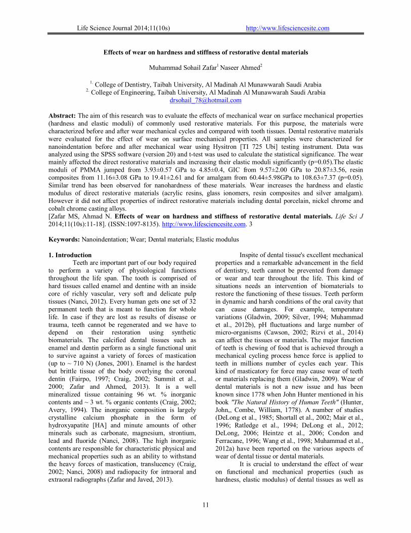
Life Science Journal 2014;11(10s) http://www.lifesciencesite.com 11 Effects of wear on hardness and stiffness of restorative dental materials Muhammad Sohail Zafar 1 Naseer Ahmed 2 1. College of Dentistry, Taibah University, Al Madinah Al Munawwarah Saudi Arabia 2. College of Engineering, Taibah University, Al Madinah Al Munawwarah Saudi Arabia [email protected] Abstract: The aim of this research was to evaluate the effects of mechanical wear on surface mechanical properties (hardness and elastic moduli) of commonly used restorative materials. For this purpose, the materials were characterized before and after wear mechanical cycles and compared with tooth tissues. Dental restorative materials were evaluated for the effect of wear on surface mechanical properties. All samples were characterized for nanoindentation before and after mechanical wear using Hysitron [TI 725 Ubi] testing instrument. Data was analyzed using the SPSS software (version 20) and t-test was used to calculate the statistical significance. The wear mainly affected the direct restorative materials and increasing their elastic moduli significantly (p=0.05).The elastic moduli of PMMA jumped from 3.93±0.57 GPa to 4.85±0.4, GIC from 9.57±2.00 GPa to 20.87±3.56, resin composites from 11.16±3.08 GPa to 19.41±2.61 and for amalgam from 60.44±5.98GPa to 108.63±7.37 (p=0.05). Similar trend has been observed for nanohardness of these materials. Wear increases the hardness and elastic modulus of direct restorative materials (acrylic resins, glass ionomers, resin composites and silver amalgam). However it did not affect properties of indirect restorative materials including dental porcelain, nickel chrome and cobalt chrome casting alloys. [Zafar MS, Ahmad N. Effects of wear on hardness and stiffness of restorative dental materials. Life Sci J 2014;11(10s):11-18]. (ISSN:1097-8135). http://www.lifesciencesite.com . 3 Keywords: Nanoindentation; Wear; Dental materials; Elastic modulus 1. Introduction Teeth are important part of our body required to perform a variety of physiological functions throughout the life span. The tooth is comprised of hard tissues called enamel and dentine with an inside core of richly vascular, very soft and delicate pulp tissues (Nanci, 2012). Every human gets one set of 32 permanent teeth that is meant to function for whole life. In case if they are lost as results of disease or trauma, teeth cannot be regenerated and we have to depend on their restoration using synthetic biomaterials. The calcified dental tissues such as enamel and dentin perform as a single functional unit to survive against a variety of forces of mastication (up to ~ 710 N) (Jones, 2001). Enamel is the hardest but brittle tissue of the body overlying the coronal dentin (Fairpo, 1997; Craig, 2002; Summit et al., 2000; Zafar and Ahmed, 2013). It is a well mineralized tissue containing 96 wt. % inorganic contents and ~ 3 wt. % organic contents (Craig, 2002; Avery, 1994). The inorganic composition is largely crystalline calcium phosphate in the form of hydroxyapatite [HA] and minute amounts of other minerals such as carbonate, magnesium, strontium, lead and fluoride (Nanci, 2008). The high inorganic contents are responsible for characteristic physical and mechanical properties such as an ability to withstand the heavy forces of mastication, translucency (Craig, 2002; Nanci, 2008) and radiopacity for intraoral and extraoral radiographs (Zafar and Javed, 2013). Inspite of dental tissue's excellent mechanical properties and a remarkable advancement in the field of dentistry, teeth cannot be prevented from damage or wear and tear throughout the life. This kind of situations needs an intervention of biomaterials to restore the functioning of these tissues. Teeth perform in dynamic and harsh conditions of the oral cavity that can cause damages. For example, temperature variations (Gladwin, 2009; Silver, 1994; Muhammad et al., 2012b), pH fluctuations and large number of micro-organisms (Cawson, 2002; Rizvi et al., 2014) can affect the tissues or materials. The major function of teeth is chewing of food that is achieved through a mechanical cycling process hence force is applied to teeth in millions number of cycles each year. This kind of masticatory for force may cause wear of teeth or materials replacing them (Gladwin, 2009). Wear of dental materials is not a new issue and has been known since 1778 when John Hunter mentioned in his book "The Natural History of Human Teeth" (Hunter, John,, Combe, William, 1778). A number of studies (DeLong et al., 1985; Shortall et al., 2002; Mair et al., 1996; Ratledge et al., 1994; DeLong et al., 2012; DeLong, 2006; Heintze et al., 2006; Condon and Ferracane, 1996; Wang et al., 1998; Muhammad et al., 2012a) have been reported on the various aspects of wear of dental tissue or dental materials. It is crucial to understand the effect of wear on functional and mechanical properties (such as hardness, elastic modulus) of dental tissues as well as
-
Upload
truongquynh -
Category
Documents
-
view
216 -
download
0
Transcript of Life Science Journal 2014;11(10s) … · Muhammad Sohail Zafar1 Naseer Ahmed2 1. ... Al Madinah Al...
Life Science Journal 2014;11(10s) http://www.lifesciencesite.com
11
Effects of wear on hardness and stiffness of restorative dental materials
Muhammad Sohail Zafar1 Naseer Ahmed2
1. College of Dentistry, Taibah University, Al Madinah Al Munawwarah Saudi Arabia
2. College of Engineering, Taibah University, Al Madinah Al Munawwarah Saudi Arabia [email protected]
Abstract: The aim of this research was to evaluate the effects of mechanical wear on surface mechanical properties (hardness and elastic moduli) of commonly used restorative materials. For this purpose, the materials were characterized before and after wear mechanical cycles and compared with tooth tissues. Dental restorative materials were evaluated for the effect of wear on surface mechanical properties. All samples were characterized for nanoindentation before and after mechanical wear using Hysitron [TI 725 Ubi] testing instrument. Data was analyzed using the SPSS software (version 20) and t-test was used to calculate the statistical significance. The wear mainly affected the direct restorative materials and increasing their elastic moduli significantly (p=0.05).The elastic moduli of PMMA jumped from 3.93±0.57 GPa to 4.85±0.4, GIC from 9.57±2.00 GPa to 20.87±3.56, resin composites from 11.16±3.08 GPa to 19.41±2.61 and for amalgam from 60.44±5.98GPa to 108.63±7.37 (p=0.05). Similar trend has been observed for nanohardness of these materials. Wear increases the hardness and elastic modulus of direct restorative materials (acrylic resins, glass ionomers, resin composites and silver amalgam). However it did not affect properties of indirect restorative materials including dental porcelain, nickel chrome and cobalt chrome casting alloys. [Zafar MS, Ahmad N. Effects of wear on hardness and stiffness of restorative dental materials. Life Sci J 2014;11(10s):11-18]. (ISSN:1097-8135). http://www.lifesciencesite.com. 3 Keywords: Nanoindentation; Wear; Dental materials; Elastic modulus 1. Introduction
Teeth are important part of our body required to perform a variety of physiological functions throughout the life span. The tooth is comprised of hard tissues called enamel and dentine with an inside core of richly vascular, very soft and delicate pulp tissues (Nanci, 2012). Every human gets one set of 32 permanent teeth that is meant to function for whole life. In case if they are lost as results of disease or trauma, teeth cannot be regenerated and we have to depend on their restoration using synthetic biomaterials. The calcified dental tissues such as enamel and dentin perform as a single functional unit to survive against a variety of forces of mastication (up to ~ 710 N) (Jones, 2001). Enamel is the hardest but brittle tissue of the body overlying the coronal dentin (Fairpo, 1997; Craig, 2002; Summit et al., 2000; Zafar and Ahmed, 2013). It is a well mineralized tissue containing 96 wt. % inorganic contents and ~ 3 wt. % organic contents (Craig, 2002; Avery, 1994). The inorganic composition is largely crystalline calcium phosphate in the form of hydroxyapatite [HA] and minute amounts of other minerals such as carbonate, magnesium, strontium, lead and fluoride (Nanci, 2008). The high inorganic contents are responsible for characteristic physical and mechanical properties such as an ability to withstand the heavy forces of mastication, translucency (Craig, 2002; Nanci, 2008) and radiopacity for intraoral and extraoral radiographs (Zafar and Javed, 2013).
Inspite of dental tissue's excellent mechanical properties and a remarkable advancement in the field of dentistry, teeth cannot be prevented from damage or wear and tear throughout the life. This kind of situations needs an intervention of biomaterials to restore the functioning of these tissues. Teeth perform in dynamic and harsh conditions of the oral cavity that can cause damages. For example, temperature variations (Gladwin, 2009; Silver, 1994; Muhammad et al., 2012b), pH fluctuations and large number of micro-organisms (Cawson, 2002; Rizvi et al., 2014) can affect the tissues or materials. The major function of teeth is chewing of food that is achieved through a mechanical cycling process hence force is applied to teeth in millions number of cycles each year. This kind of masticatory for force may cause wear of teeth or materials replacing them (Gladwin, 2009). Wear of dental materials is not a new issue and has been known since 1778 when John Hunter mentioned in his book "The Natural History of Human Teeth" (Hunter, John,, Combe, William, 1778). A number of studies (DeLong et al., 1985; Shortall et al., 2002; Mair et al., 1996; Ratledge et al., 1994; DeLong et al., 2012; DeLong, 2006; Heintze et al., 2006; Condon and Ferracane, 1996; Wang et al., 1998; Muhammad et al., 2012a) have been reported on the various aspects of wear of dental tissue or dental materials.
It is crucial to understand the effect of wear on functional and mechanical properties (such as hardness, elastic modulus) of dental tissues as well as
Life Science Journal 2014;11(10s) http://www.lifesciencesite.com
12
the dental materials being used to replace. Ideally, a very close match of mechanical properties of lost dental tissue and potential dental material is necessary for functional compatibility and longevity of the restoration. The aim of this research was to evaluate the effects of mechanical wear on surface mechanical properties (hardness and elastic moduli) of commonly used restorative materials. For this purpose, the materials were characterized before and after wear mechanical cycles and compared with tooth tissues. 2. Material and Methods 2.1. Sample preparation
The description of dental materials used in this study has been given in table 1.
Table 1 Description of dental materials used Materials Description Manufacturer Teeth Maxillary permanent
premolars Natural
(PMMA) BMS 016; Poly methyl methacrylate acrylic resin, Cadmium free
BMS dental Italy
GIC Capsulated glass ionomers cement, ChemFil Rock (A2)
DENTSPLY UK
Composite
Universal fluoride releasing resin composite, Heliomolar (A2)
Ivaclor vivadent USA
Amalgam
High copper spherical capsulated amalgam alloy Megalloy® EZ; [alloy to mercury mass ratio=1.3:1]
DENTSPLY UK
Ni-Cr Nickel chrome base metal alloy, Wiron® 99; [Ni (65%); Cr (22.5%); Mo (9.5%)]
BEGO medical Germany
Co-Cr Cobalt chrome base metal alloy Wironit® extrahart; [Co (63%); Cr (30%); Mo (5%)]
BEGO medical Germany
Porcelain High fusing feldspar ceramic, VITA VM®9
VIDENT company USA
For the evaluation of dental tissues, five
maxillary permanent premolars extracted for orthodontic or periodontal reasons were used. Teeth with carious lesion, wear or any kind of pathology were not included. Hydrogen peroxide solution (1 %) was used for disinfection of teeth (5 ºC for a day); followed by storage in plain water at 5 ºC until needed for testing. Teeth were transversely cut into flat disc shape samples using a 2.3 mm [standard grit cutting (ISO806104)] diamond disc. The flat surfaces were finished using motorised silicon carbide discs of
different grit sizes (200, 600 and 1200) in a set sequence. Diamond paste of decreasing grits (1.0 and 0.5 micron) was used for fine polishing.
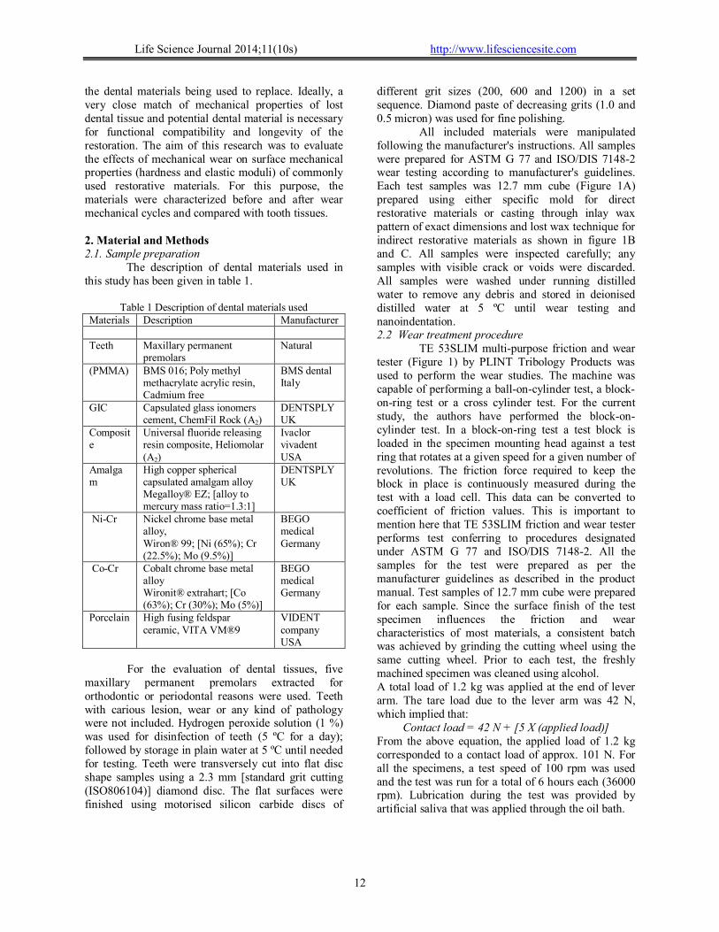
All included materials were manipulated following the manufacturer's instructions. All samples were prepared for ASTM G 77 and ISO/DIS 7148-2 wear testing according to manufacturer's guidelines. Each test samples was 12.7 mm cube (Figure 1A) prepared using either specific mold for direct restorative materials or casting through inlay wax pattern of exact dimensions and lost wax technique for indirect restorative materials as shown in figure 1B and C. All samples were inspected carefully; any samples with visible crack or voids were discarded. All samples were washed under running distilled water to remove any debris and stored in deionised distilled water at 5 ºC until wear testing and nanoindentation. 2.2 Wear treatment procedure
TE 53SLIM multi-purpose friction and wear tester (Figure 1) by PLINT Tribology Products was used to perform the wear studies. The machine was capable of performing a ball-on-cylinder test, a block-on-ring test or a cross cylinder test. For the current study, the authors have performed the block-on-cylinder test. In a block-on-ring test a test block is loaded in the specimen mounting head against a test ring that rotates at a given speed for a given number of revolutions. The friction force required to keep the block in place is continuously measured during the test with a load cell. This data can be converted to coefficient of friction values. This is important to mention here that TE 53SLIM friction and wear tester performs test conferring to procedures designated under ASTM G 77 and ISO/DIS 7148-2. All the samples for the test were prepared as per the manufacturer guidelines as described in the product manual. Test samples of 12.7 mm cube were prepared for each sample. Since the surface finish of the test specimen influences the friction and wear characteristics of most materials, a consistent batch was achieved by grinding the cutting wheel using the same cutting wheel. Prior to each test, the freshly machined specimen was cleaned using alcohol. A total load of 1.2 kg was applied at the end of lever arm. The tare load due to the lever arm was 42 N, which implied that:
Contact load = 42 N + [5 X (applied load)] From the above equation, the applied load of 1.2 kg corresponded to a contact load of approx. 101 N. For all the specimens, a test speed of 100 rpm was used and the test was run for a total of 6 hours each (36000 rpm). Lubrication during the test was provided by artificial saliva that was applied through the oil bath.
Life Science Journal 2014;11(10s) http://www.lifesciencesite.com
13
2.3. Nanoindentation testing
Nanoindentation was performed using a 3-sided pyramidal Berkovich (142.3 degree diamond probe) fitted in Hysitron [TI 725 Ubi] equipment. The system was placed on an anti-vibrational table to prevent any errors. The prepared samples were (tested before wear and after wear) installed to the sample holder using epoxy resin. The sample holder was firmly screwed to the sample table to prevent mobility during the indentation. The indentation was performed using a load of 1N. An optical camera (10X) was used to focus on the exact location of the indentation, after which the machine performs the indent. A minimum of five indentations were performed to get precise loading unloading curves and results. The system calculates absolute hardness and reduced modulus (Er). The reduced modulus can
be related with the elastic modulus of the specimen using following equation.
1
��=1 − �������
�
�������+1 − ���������
�
���������
For a standard diamond indenter probe, Eindenter is 1140 GPa and vindenter is 0.07. vsample has been approximated as 0.3 for the dental hard tissues (Sakaguchi and Powers, 2012) and can be used to calculate the Esample . The hardness has a nominal definition given by
� =�����
Where Pmax is the highest indentation force and � is the resulting projected contact area at that load. The medium of indentation was air. Data was analyzed using the SPSS software package (version 20) and statistical significance was calculated using the t-test.
(F)
(E)
12.7mm
[[ 12.7mm
12.7 mm
(A)
(G)
(C) (B)
(D)
(H)
Figure 1: Wear equipment and procedure; (A) Sample dimensions (B) Mold and wax patterns used to construct samples of dental materials (C) Prepared samples (D) Linear Variable Differential Transformer (LVDT) module for wear measurement (E) Test ring (F) Specimen mounting head with sample fitted (G) Loading arm (H) TE 53SLIM multi-purpose friction and wear tester
Life Science Journal 2014;11(10s) http://www.lifesciencesite.com
14
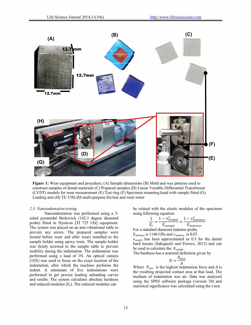
Figure 2: Direct restorative materials; representative loading, unloading nanoindentation curves and micrographs (10X) of post wear testing; (A) PMMA (B) GIC (C) Composite (D) Amalgam
(A)
(C)
(B)
(D)

Life Science Journal 2014;11(10s) http://www.lifesciencesite.com
15
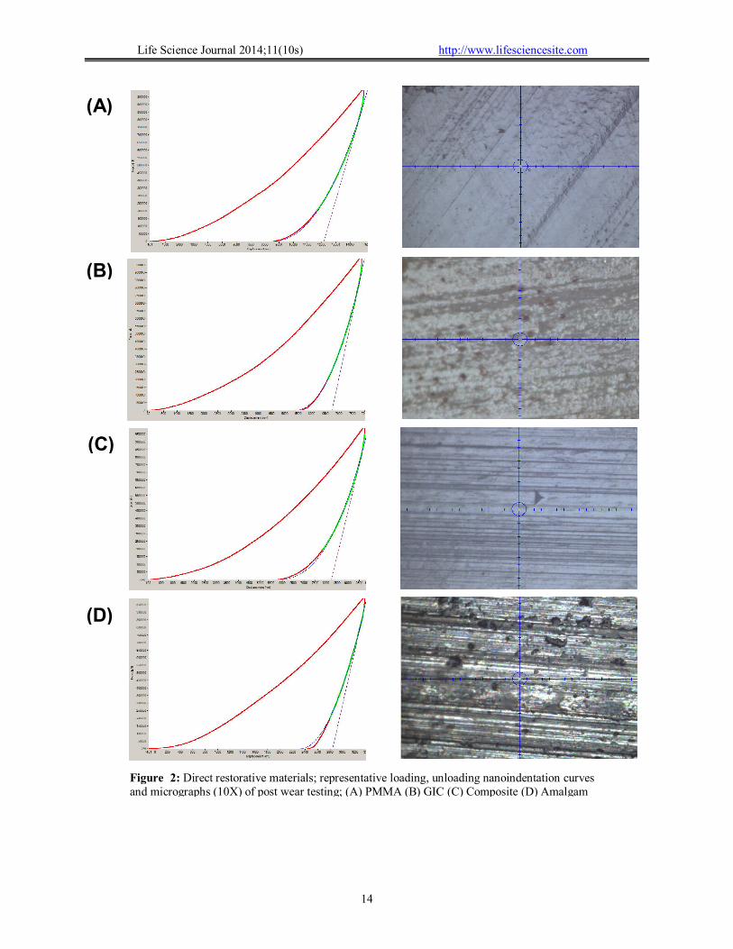
Figure 3: Indirect restorative materials; representative loading and unloading nanoindentation curves and micrographs (10X) of post wear testing; (A) Ni-Cr (B) Co-Cr (C) Porcelain (D) Example of defective loading and unloading curves
(A)
(C)
(B)
(D)
Life Science Journal 2014;11(10s) http://www.lifesciencesite.com
16
3. Results
Tooth tissues and commonly used dental restorative materials were tested for surface mechanical properties.
The effects of wear on restorative materials were also reported. The representative nanoindentation curves and corresponding digital micrographs (10X) for direct restorative materials (PMMA, GIC, composites and amalgam) and for indirect restorative materials (Ni-Cr, Co0Cr and
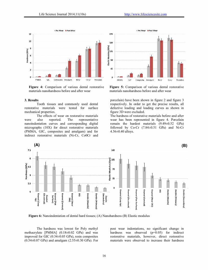
porcelain) have been shown in figure 2 and figure 3 respectively. In order to get the precise results, all defective loading and loading curves as shown in figure 3D were excluded. The hardness of restorative materials before and after wear has been represented in figure 4. Porcelain remain the hardest materials (9.49±0.52 GPa) followed by Co-Cr (7.84±0.51 GPa) and Ni-Cr 4.56±0.40 alloys.
The hardness was lowest for Poly methyl methacrylate [PMMA] (0.18±0.02 GPa) and was improved for GIC (0.34±0.05 GPa), resin composites (0.54±0.07 GPa) and amalgam (2.55±0.30 GPa). For
post wear indentations, no significant change in hardness was observed (p=0.05) for indirect restorative materials, however, direct restorative materials were observed to increase their hardness
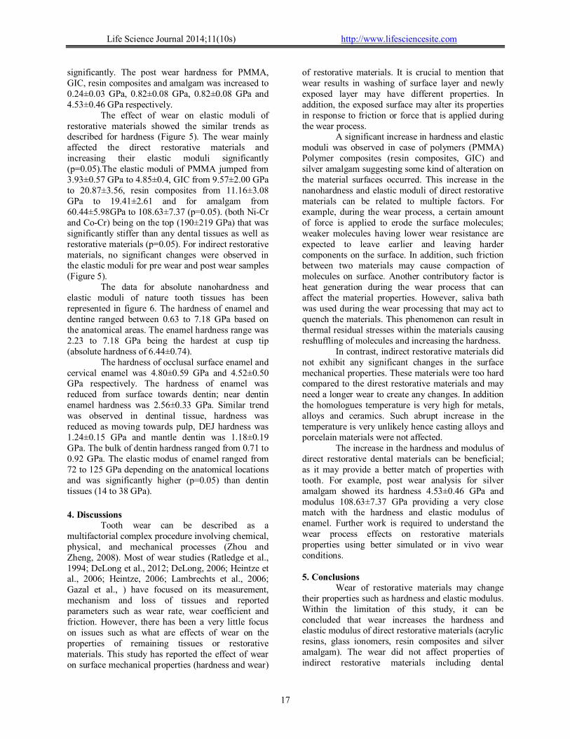
Figure 6: Nanoindentation of dental hard tissues; (A) Nanohardness (B) Elastic modulus
(B)
(A)
Figure 4: Comparison of various dental restorative materials nanohardness before and after wear
Figure 5: Comparison of various dental restorative materials nanohardness before and after wear
Life Science Journal 2014;11(10s) http://www.lifesciencesite.com
17
significantly. The post wear hardness for PMMA, GIC, resin composites and amalgam was increased to 0.24±0.03 GPa, 0.82±0.08 GPa, 0.82±0.08 GPa and 4.53±0.46 GPa respectively.
The effect of wear on elastic moduli of restorative materials showed the similar trends as described for hardness (Figure 5). The wear mainly affected the direct restorative materials and increasing their elastic moduli significantly (p=0.05).The elastic moduli of PMMA jumped from 3.93±0.57 GPa to 4.85±0.4, GIC from 9.57±2.00 GPa to 20.87±3.56, resin composites from 11.16±3.08 GPa to 19.41±2.61 and for amalgam from 60.44±5.98GPa to 108.63±7.37 (p=0.05). (both Ni-Cr and Co-Cr) being on the top (190±219 GPa) that was significantly stiffer than any dental tissues as well as restorative materials (p=0.05). For indirect restorative materials, no significant changes were observed in the elastic moduli for pre wear and post wear samples (Figure 5).
The data for absolute nanohardness and elastic moduli of nature tooth tissues has been represented in figure 6. The hardness of enamel and dentine ranged between 0.63 to 7.18 GPa based on the anatomical areas. The enamel hardness range was 2.23 to 7.18 GPa being the hardest at cusp tip (absolute hardness of 6.44±0.74).
The hardness of occlusal surface enamel and cervical enamel was 4.80±0.59 GPa and 4.52±0.50 GPa respectively. The hardness of enamel was reduced from surface towards dentin; near dentin enamel hardness was 2.56±0.33 GPa. Similar trend was observed in dentinal tissue, hardness was reduced as moving towards pulp, DEJ hardness was 1.24±0.15 GPa and mantle dentin was 1.18±0.19 GPa. The bulk of dentin hardness ranged from 0.71 to 0.92 GPa. The elastic modus of enamel ranged from 72 to 125 GPa depending on the anatomical locations and was significantly higher (p=0.05) than dentin tissues (14 to 38 GPa).
4. Discussions
Tooth wear can be described as a multifactorial complex procedure involving chemical, physical, and mechanical processes (Zhou and Zheng, 2008). Most of wear studies (Ratledge et al., 1994; DeLong et al., 2012; DeLong, 2006; Heintze et al., 2006; Heintze, 2006; Lambrechts et al., 2006; Gazal et al., ) have focused on its measurement, mechanism and loss of tissues and reported parameters such as wear rate, wear coefficient and friction. However, there has been a very little focus on issues such as what are effects of wear on the properties of remaining tissues or restorative materials. This study has reported the effect of wear on surface mechanical properties (hardness and wear)
of restorative materials. It is crucial to mention that wear results in washing of surface layer and newly exposed layer may have different properties. In addition, the exposed surface may alter its properties in response to friction or force that is applied during the wear process.
A significant increase in hardness and elastic moduli was observed in case of polymers (PMMA) Polymer composites (resin composites, GIC) and silver amalgam suggesting some kind of alteration on the material surfaces occurred. This increase in the nanohardness and elastic moduli of direct restorative materials can be related to multiple factors. For example, during the wear process, a certain amount of force is applied to erode the surface molecules; weaker molecules having lower wear resistance are expected to leave earlier and leaving harder components on the surface. In addition, such friction between two materials may cause compaction of molecules on surface. Another contributory factor is heat generation during the wear process that can affect the material properties. However, saliva bath was used during the wear processing that may act to quench the materials. This phenomenon can result in thermal residual stresses within the materials causing reshuffling of molecules and increasing the hardness.
In contrast, indirect restorative materials did not exhibit any significant changes in the surface mechanical properties. These materials were too hard compared to the direst restorative materials and may need a longer wear to create any changes. In addition the homologues temperature is very high for metals, alloys and ceramics. Such abrupt increase in the temperature is very unlikely hence casting alloys and porcelain materials were not affected.
The increase in the hardness and modulus of direct restorative dental materials can be beneficial; as it may provide a better match of properties with tooth. For example, post wear analysis for silver amalgam showed its hardness 4.53±0.46 GPa and modulus 108.63±7.37 GPa providing a very close match with the hardness and elastic modulus of enamel. Further work is required to understand the wear process effects on restorative materials properties using better simulated or in vivo wear conditions. 5. Conclusions
Wear of restorative materials may change their properties such as hardness and elastic modulus. Within the limitation of this study, it can be concluded that wear increases the hardness and elastic modulus of direct restorative materials (acrylic resins, glass ionomers, resin composites and silver amalgam). The wear did not affect properties of indirect restorative materials including dental

Life Science Journal 2014;11(10s) http://www.lifesciencesite.com
18
porcelain, nickel chrome and cobalt chrome casting alloys. Conflict of Interest
The authors declared no conflict of interest for conducting this research. Corresponding Author: Dr. Muhammad Sohail Zafar Assistant Professor (Dental Biomaterials) College of Dentistry, Taibah University Al Madinah Al Munawwarah Saudi Arabia E-mail: [email protected] References 1. Nanci A. Ten Cate's oral histology: development,
structure, and function. St. Louis, Mo.; London: Mosby. 2012
2. Jones FH. Teeth and bones: applications of surface science to dental materials and related biomaterials. Surface Science Reports. 2001; 42(3-5): 75-205.
3. Fairpo JEH. Heinemann dental dictionary. Oxford: Butterworth-Heinemann. 1997
4. Craig JR. Restorative dental materials. St. Louis, Mosby.; London: Mosby. 2002
5. Summit J, Robins W, Hilton T, Schwartz R. Fundamentals of operative dentistry: a contemporary approach. Chicago, Ill.; London: Quintessence Publishing. 2000
6. Zafar MS, Ahmed N. Nano-Mechanical Evaluation of Dental Hard Tissues Using Indentation Technique. World Applied Sciences Journal. 2013; 28(10): 1393-9.
7. Avery J. Oral development and histology. New York: Thieme Medical. 1994
8. Nanci A. Ten Cate's oral histology: development, structure, and function. St. Louis, Mo.; London: Mosby. 2008
9. Zafar MS, Javed E. Extraoral Radiography: An Alternative to Intraoral Radiography for Endodontic (Root Canal System) Length Determination. European Scientific Journal. 2013; 9(15): 51.
10. Gladwin MA. Clinical aspects of dental materials: theory, practice, and cases. Philadelphia, Pa.; London: Lippincott Williams & Wilkins. 2009
11. Silver FH. Biomaterials, medical devices and tissue engineering: an integrated approach: Chapman and Hall. 1994
12. Muhammad R, Ahmed N, Shariff YM, Silberschmidt VV. Finite-element analysis of forces in drilling of Ti-alloys at elevated temperature. Solid State Phenomena. 2012; 188: 250-5.
13. Cawson RA. Cawson's essentials of oral pathology and oral medicine. Edinburgh: Churchill Livingstone. 2002
14. Rizvi A, Zafar MS, Farid WM, Gazal G. Assessment of Antimicrobial Efficacy of MTAD, Sodium Hypochlorite,
EDTA and Chlorhexidine for Endodontic Applications: An In vitro Study. Middle-East Journal of Scientific Research. 2014; 21(2): 353-7.
15. Hunter, John,,Combe, William,,. The natural history of the human teeth: explaining their structure, use, formation, growth, diseases, illustrated with copper-plates. London: J. Johnson. 1778
16. DeLong R, Sakaguchi RL, Douglas WH, Pintado MR. The wear of dental amalgam in an artificial mouth: a clinical correlation. Dental Materials. 1985; 1(6): 238-42.
17. Shortall AC, Hu XQ, Marquis PM. Potential countersample materials for in vitro simulation wear testing. Dental Materials. 2002; 18(3): 246-54.
18. Mair LH, Stolarski TA, Vowles RW, Lloyd CH. Wear: mechanisms, manifestations and measurement. Report of a workshop. J.Dent. 1996; 24(1–2): 141-8.
19. Ratledge DK, Smith BGN, Wilson RF. The effect of restorative materials on the wear of human enamel. J.Prosthet.Dent. 1994; 72(2): 194-203.
20. DeLong R, Pintado MR, Douglas WH, Fok AS, Wilder AD, Swift EJ, Bayne SC. Wear of a dental composite in an artificial oral environment: A clinical correlation. Journal of Biomedical Materials Research Part B: Applied Biomaterials. 2012; 100B(8): 2297-306.
21. DeLong R. Intra-oral restorative materials wear: Rethinking the current approaches: How to measure wear. Dental Materials. 2006; 22(8): 702-11.
22. Heintze SD, Cavalleri A, Forjanic M, Zellweger G, Rousson V. A comparison of three different methods for the quantification of the in vitro wear of dental materials. Dental Materials. 2006; 22(11): 1051-62.
23. Condon JR, Ferracane JL. Evaluation of composite wear with a new multi-mode oral wear simulator. Dental Materials. 1996; 12(4): 218-26.
24. Wang W, DiBenedetto AT, Jon.Goldberg A. Abrasive wear testing of dental restorative materials. Wear. 1998; 219(2): 213-9.
25. Muhammad R, Ahmed N, Roy A, Silberschmidt VV. Turning of advanced alloys with vibrating cutting tool. Solid State Phenomena. 2012; 188: 277-84.
26. Sakaguchi RL, Powers JM,. Craig's restorative dental materials. Philadelphia, PA: Elsevier/Mosby. 2012
27. Zhou Z, Zheng J. Tribology of dental materials: a review. J.Phys.D. 2008; 41(11): 113001.
28. Heintze SD. How to qualify and validate wear simulation devices and methods. Dental Materials. 2006; 22(8): 712-34.
29. Lambrechts P, Debels E, Van Landuyt K, Peumans M, Van Meerbeek B. How to simulate wear?: Overview of existing methods. Dental Materials. 2006; 22(8): 693-701.
30. Gazal G, Fareed WM, Zafar MS, Al-Samadani KH. Pain and anxiety management for pediatric dental procedures using various combinations of sedative drugs: A review. Saudi Pharmaceutical Journal. Accepted manuscript.: 2014.
5/29/2014