
Life in the Serendipitous Lane: Excitement and ... · PDF fileLife in the Serendipitous Lane:...
90
Life in the Serendipitous Lane: Excitement and Gratification in Studying DNA Repair DNA Repair Interest Group History of DNA Repair June 20, 2006 Dr. Stuart Linn University of California, Berkeley
Transcript of Life in the Serendipitous Lane: Excitement and ... · PDF fileLife in the Serendipitous Lane:...

Life in the Serendipitous Lane:Excitement and Gratification in
Studying DNA RepairDNA Repair Interest Group
History of DNA RepairJune 20, 2006
Dr. Stuart Linn University of California, Berkeley

SerendipityCoined from The Three Princes of
Serindip (Sri Lanka), a Persian fairy tale in which the princes have an
aptitude for making fortunate discoveries accidentally

The formative years
Caltech 1958-1962Linus PaulingRichard FeynmanGeorge Beadle Norman HorowitzNorman DavidsonJerry VinogradHenry Borsook
Stanford 1962-1966Arthur KornbergPaul BergPhil HanawaltJoshua LederbergCharles YanofskyH. Gobind KhoranaI. R. Lehman

Postdoctoral and beyondGeneva (Cambridge) 1966-68Eduard KellenbergerRichard EpsteinWerner Arber(Sydney Brenner)(John Smith)
London 1974-75Robin Holliday
Oslo 1982Erling Seeberg
Berkeley 1968-Harrison (Hatch) EcholsBruce AmesA. John ClarkEdward PenhoetRobert MortimerSymore Fogel
Aviemore, June 1973Matthew MeselsonCharles RaddingBruce Alberts

RecBC(D)


RecBC(D) action with 1mM Ca2+
present


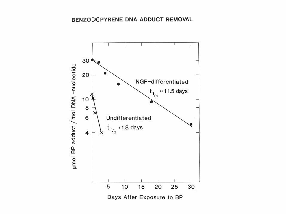
Coming of Age with Aging


S. W. Krauss &. S. Linn (1986) J. Cell Physiol. 126: 99-106


J. Barnard, M. LaBelle, & S. Linn Exp. Cell. Res. (1986)163: 500-508
MPC11 (Leukemic B line)
169 µg/106 cells
1650 U /106 cells
143 U /106 cells

Mouse primary skin fibroblasts
M. LaBelle & S. Linn (1984) Mut. Res.132: 51-61


L. M. Jensen & S. Linn (1988) Mol. Cell Biol. 8: 3964-3968


DNA polymerases
“The Kornberg Enzyme (E. coli pol I) is probably just a repair enzyme”--Mark Bretcher, Stanford 1967
(prior to the report of the polA mutant by De Lucia and Cairns in 1969)

Mosbaugh & Linn JBC 257:575 (1982)


AP and “UV” Endonucleases

_
_

Mosbaugh & Linn JBC 258:108 (1983)


Mosbaugh & Linn JBC 255:11743 (1980)


Both the protein and UV/AP endonucleases are immunodepleted by abs. against rat rpS3 and rat rpS3 produced in E. coli has the two activities.
J. Kim, et al. (1995) J. Biol. Chem.: 270: 13620-13629


The Human DNA Polymerases
The good old days…Pols α, β, γ
Αnd then there were five…Pols α, β, γ, δ, ε
And then came the “sloppier copiers”… We’re up to ≈17 eukaryotic pols and still counting
(and learning the Greek alphabet.)

Mosbaugh & Linn JBC 259:10247 (1984) Gap: 64 dNMP
Pol α incorp.: 48 dNMP/gap
Pol β incorp.: 15 dNMP/gap

Mosbaugh, Evans & Linn (1984) CSHSQB 49:581
D. H. Evans & S. Linn (1984) JBC: 259: 10252
SV40 chromatin substrate

Pol ε∗ subunit structure in humans and budding yeast
p17p12
p261 p59 POL2 DPB2
DPB4
DPB3
YeastHuman
*Mammalian pol ε was originally designated as pol δ2, pol δII or pol δ*

Catalytic subunit - p261
N C
• Large C-terminal domain important for protein-protein interactions
C-terminal domain
• N- and C- terminal domains are separated by a protease-sensitive site. A far N-terminal domain is also separated by a protease-sensitive site. Both sites are cleaved by Caspase 3 during apoptosis.
22851
• Polymerase and exonuclease motifs found in the N-terminal domain
pol and exo motifs
N-terminal domain

p261 C-terminal domain
C
Zn fingers
• Contains two zinc fingers
• Essential in both fission and budding yeast
11672285
• Necessary for protein-protein interactions- Mdm2 binding and stimulation- PCNA binding- Subunit binding
Mdm2 binding1878
• ** Necessary to sense replication blocks and delay entry into mitosis in budding, not fission yeast

p17 and p12 subunits
p17
p12
N C
N C
148
118
1
1
Histone-fold motifs
H2B family
H2A family

pol ε p17 is identical to huCHRAC p17
• CHRAC is a CHRomatin Accessibility Complex first isolated in Drosophila but conserved in humans
• Remodels chromatin in an ATP-dependent manner
• H2A histone-fold motif binding partner of huCHRAC p17 is not pol ε p12, but huCHRAC p15Drosophila similarly has distinct partners for p17.
• huCHRAC contains ACF1 which may target the complex to heterochromatin

p261
p59p12
p17Acf1p15
“hISWI”
Is there a pol ε : CHRAC complex?
p17
?
?
Apparently Not

In S. cerevisiae, pol ε replicates telomeres with maintenance of telomer-position effect epistatic states of the Sir complex. But
ISW2/yCHRAC binding at telomers promotes reversible switching between epigenetic
states.Ida & Araki, MCB 24:217 (2004)

G1
Early S Late S
G2/M
Mid S
Pol εPol δ
PCNA
BrdU
Foci
Polymerase foci are established
Repair? PIC assembly?
Euchromatin replication, δRepair δ/ε?
Heterochromatinreplication, δ/ε?
Only pol ε foci persist. Recombination/repair?
Cell cycle arrest, ε?
Repair?
Foci appearance and some putative functions of mammalian pols epsilon
and delta during the cell cycleRepair?

Outstanding questions
• What is/are the role(s) of the small pol ε foci?
• What is the significance of the sharing of p17 between CHRAC and pol ε?
• Is pol ε a repair and/or a replicative polymerase?
• Where do the error-prone pols localize during the cell cycle before and after DNA damage?

Oxidative DNA Damage
Stress responsesReactive Oxygen Species (ROS)
Fenton chemistry
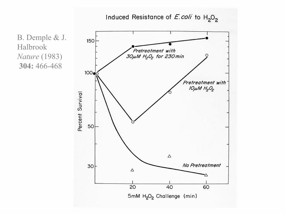
B. Demple & J. HalbrookNature (1983)304: 466-468


Responses to oxidative stress in E. coli and genes relevant to DNA repair
• OxyR regulon• Heat-shock response• SOS regulon
RecA (recA)• KatF regulon (sigma factor)
Exonuclease III (xthA)• SoxRS regulon
Endonuclease IV (nfo)

e e e eO2 O2 • H2O2 • OH H2O

Some reactions of oxygen radicalsE. S. Henle, Y. Luo & S. Linn (1996) Biochem.: 35, 12212-12219
O2•- + Fe 3+ O2 + Fe 2+
2 O2•- + 2H + O2 + H2O2
Fe 2 + + H2O2 Fe 3+ + OH- + •OH
•OH + DNA DNA •
DNA • + O2 DNAO2 •

J. A. Imlay & S. Linn (1986) J. Bact. 166: 519-527 & (1987) Ibid. 169: 2967-2976
(1988) Science 240: 640-642 & 1302-1309

Sequences of preferred duplex DNA cleavages by Fe2+/H2O2
Type I Oxidants (0.5 mM H2O2)RTGR
Type II Oxidants (50 mM H2O2)RGGG, TGGG (?)
Bold, underscored nucleotides are cleavage sites “R”, Purine; “Y”, Pyrimidine
E. S. Henle et al. (1999) J Biol. Chem. 274: 962-971

Conclusions of RTGR studiesFe2+ preferentially binds to RTGR sequences because of their unique
structure. That structure is not grossly perturbed upon binding.
Fe2+ is relatively loosely bound to RTGR; it is subject to oxidation by H2O2in the unbound state, giving rise to radicals which can be quenched by H2O2
*, thus explaining a peculiar dose response for “Mode I” damage.
*H2O2 + •OH HO2• + H2O
-----------------------------
Promoters of genes regulating Iron Metabolism, Responses to Oxygen Radical Stress or DNA Repair genes contain RTGR in essential motifs, usually as direct or inverted repeats. Is binding at RTGR sites in these promoter regions by Fe2+ but not by Fe3+ exploited for sensing iron and/or oxygen stress and consequently regulating these genes?
P. Rai et al. (2001) J. Mol. Biol. 312: 1089-1101

Conclusions of (R/T)GGG studiesGuanine N7 is the strongest DNA coordination site for transition metals.
Breaks occur 5’ to a deoxyguanosine with a 5’ --> 3’ polarity.
Binding of Fe2+ follows the same polarity.
Binding of Fe2+ at these sites causes a slight structural kink, somewhat stabilizing the binding and thus giving rise to the zero order dose response.
----------------------------------
RGGG is contained in the majority of telomer repeats and telomeric sequences are cleaved preferentially in vitro.
Is age-related telomere shortening contributed to by iron-mediated Fenton reactions?
Human genome recombination hotspots are CCTCCCT & CCCCACCCC.
P. Rai, D. E. Wemmer, & S. Linn (2005) Nuc. Ac. Res. 33: 497-510


NAD(P)H pools in E. coli 15 min after H2O2challenge
H2O2 Nucleotide Concentration (µM) RatioChallenge NADH NAD+ NADPH NADP+ NADPH/NADH
0.0 mM 700 230 24 210 0.030.5 200 630 33 270 0.175.0 4 770 11 240 2.75
10.0 <0.1 850 8 200 >80
J. L. Brumaghim, et al. (2003) J. Biol. Chem. 278: 42495-42504
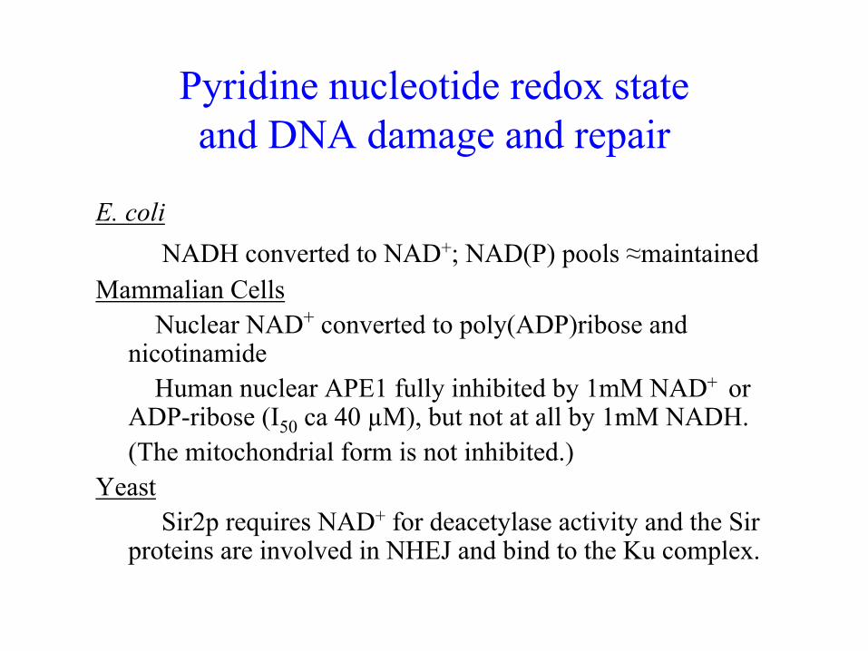
Pyridine nucleotide redox stateand DNA damage and repair
E. coliNADH converted to NAD+; NAD(P) pools ≈maintained
Mammalian CellsNuclear NAD+ converted to poly(ADP)ribose and
nicotinamideHuman nuclear APE1 fully inhibited by 1mM NAD+ or
ADP-ribose (I50 ca 40 µM), but not at all by 1mM NADH.(The mitochondrial form is not inhibited.)
YeastSir2p requires NAD+ for deacetylase activity and the Sir
proteins are involved in NHEJ and bind to the Ku complex.

Human Damage-specific DNA Binding Protein (DDB)
only in mammals(CS-A is similar)
Ubiquitous(except S.
cerevisiae)
Sequnce homologues
11p11-p1211q12-q13Gene map
10.44.9pI
48 kDa (sequence)41,000
127 kDa (sequence)124,000
Mr
DDB2 (XPE)DDB1

DDB binds tightly (Ka ≈ 1010) to some products of UV irradiation
T[t,s]T ≈ T[6,4]T >* T[Dewar]T > T[c,s]T* >TT
* the major UV photoproduct* > represents a ca. 3-fold difference
J. R. Reardon et al. (1993) J. Biol. Chem.: 268, 21301-21308
105-106 copies per cell

0
50
100
150
0 50 100
IMR-90 (lung)Hs-27 (skin)CCD-32sk (skin)CCD-33sk (skin)
Normal
0
50
100
150
0 50 100
XP3RO (XP-E)GM01389 (XP-E)Ops1 (XP-E)XP82TO (XP-E)XP2RO (XP-E)
XP-E
XP-E strains are resistant to UV-induced apoptosis
Hours Post-UV Irradiation
Dye exclusion
UV dose = 12 J/m2
Rel
ativ
e vi
abl e
cel
l num
b er (
%)

p53 regulatory pathway
Apoptosis
p53
CDK2E3 ligase Cyclin
dependent kinase
CDKN1A (p21)MDM2
Cytotoxic stresses
Cell cycle arrest
BAX
Negative feedback loop
No p53 mutations are found in XP-E cells.
CDKN1A (p21)

p127DDB1
1 1.1 .83 .89 .92 .80 .85
Normal XP-E
p21BAX
1 1.4 1.1 1.3 .45 .70 .56
p21CDKN1A1 .83 1.1 .53 .17 .060 .074
p48DDB2
actin1 1.0 .94 1.2 1.1 1.0 .93
p53 50k1 2.3 1.3 1.2 .080 .18 .10
20k
20k
40k
120k
40k
1 1.7 1.1 3.7 0 .090 .36
p90MDM2
p75MDM280k
1 1.2 1.8 1.3 .59 .47 .49
*IM
R-9
0H
s27
CC
D32
skC
CD
-33s
k
GM
0138
9
XP3R
OXP
82TO
Western blot
Basal protein levelsof p53 and p53-downsteam genes
T. Itoh, C. O’Shea & S. Linn (2003) Mol. Cell Bio. 23:7540-7553

Tentative proposal for carcinogenesis in XP-EUV damage
Abnormally high DDB2mRNA expression and/orlow DDB2 protein levels
Diminished p53 levels
Diminished apoptosis and cell cycle arrest after UV
Skin carcinogenesis
Ops1XP-E

Putative regulatory elements of intron 4 of the DDB2 gene

Effect of the three DDB2 expression constructs upon basal protein levels in an XP-E strain
p53
p21/WAF1
p48/DDB2
p127/DDB1
actin
1 1.1 1.1
1 1.1 1.1
1 .23 2.1
1 2.6 10
1 3.6 4.0
1 1.5 1.8p21/Bax
p90/MDM2
1 2 3
1: 5’UTR2: 5’UTR+hDDB2cDNA3: 5’UTR+cDNA+Intron 4
Western Blot

DDB2
Basal p48DDB2 level
p53
Basal p53 level
+
Regulation of basal levels
Basal DDB2 level is kept
low and stable.Post-transcriptional control -
+
+Other regulatory factors
DDB2 gene (RNA?)
p53
+
p90MDM2 p21BAX p21CDKN1A


DDB subunits bind to transcription and cell cycle regulators
X
XX
X
X
DDB2
Pradip Raychaudhuri & Yoshihiro Nakatani
Yue Xiong
X
X
Cullin 4A in COP9 signalosomeCDT1
Pradip Raychaudhuri & Vesna Rapic-Otrin Robert Roeder
XX
CBP/p300STAGA
Pradip RaychaudhuriE2F1
Robert LambXParamyxoviridae V proteins
Stuart LinnXEBV EBNA 2
Janet Butel & Pradip RaychaudhuriXHBV protein X
Reporting LaboratoriesDDB1Bound protein/complex

A possible scenario for DDB2 and global genomic repair*• After UV irradiation, DDB heterodimer binds DNA UV
damages.• The bound DDB recruits the STAGA and CBP/p300 protein
acetylase/chromatin remodeling complexes.• XPC or XPA is recruited to the site, depending on the damage.• Within the first ≈60 min repair takes place and DDB recruits the
Cullin 4A ubiquitin ligase off of the COP9 signalosome.• DNA repair factors, DDB, and chromatin remodeling complexes
are ubiquitinated by the recruited Cullin 4A and then degraded by the proteasome within ≈120 min. (CBP/p300 degradation would allow p53 to accumulate; STAGA and CBP/p300 degradation would limit chromatin remodeling.)
• DDB2 is restored after repair is complete (after 36 hr.)• DDB1 is transported to the nucleus, E2F1 and CDT1 are bound,
cell cycle progression resumes, and the DDB system is re-primed.*Based upon observations from the laboratories of P. Raychaudhuri, V. Rapic-Otrin, R. Roeder, Y. Nakatani, T. Matsunaga, K. Sugasawa, J. Ford, and others

Notes on the putative scheme• The time-ordering of early events may not be exact.
• It is not clear whether DDB1, DDB2, or the heterodimer binds the various complexes/proteins at various stages.
• If all lesions are repaired within 60 minutes, subsequent events, including those mediated by p53, do not occur.
• For transcription-coupled excision repair (TCR) a similar scenario appears to take place with CSA replacing DDB2 in some or all functions. ---Nakatani lab.
• If DDB2 were to function to coordinate the repair scenario with p53-mediated checkpoint and apoptosis responses, then the presence of DDB2 only in higher eucaryotes versus the ubiquitous presence of DDB1 would be explained.

Properties of DDB2-/- miceItoh et al. (2004) Proc. Natl. Acad. Sci. USA 10: 2052-2057
• F2 mice are viable, fertile, 92% of normal weight (8 wks.)(F6 mice are fertile, but becoming smaller.)
• Primary fibroblasts (MEFs) lack DDB2 expression as assayed by RT-PCR, activity, and immunoblotting.)
(Heterozygotes’ cells have ≈ 1/2 normal levels.)
• Primary fibroblasts (MEFs) resemble human XP-E fibroblasts(hyper-resistant to UV; reduced apoptosis and expression of p53-mediated effectors). (Heterozygotes’ cells are normal.)
• Predisposition to squamous cell carcinomas induced by UV, but not by DMBA (7,12-dimetylbenz[a]anthracene).

Properties of DDB2-/- miceItoh et al. unpublished (June 2006)
• UV-B caused cataracts only in -/- mice when mice treated with 2,500J/m2 for 5 days per week for up to 20 weeks.
• Life spans of -/-, -/+, +/+ mice not statistically different.
• -/- mice not abnormally sensitive to 400 rads of γ-radiation given at 7-1/2 to 8-1/2 weeks.
• E2F1 and p53 -/- phenotypes appear to be unaffected by DDB2 genotype.

For the future….
• Molecular details of the regulatory interactions between DDB2 and p53?
• Is DDB2 involved in signaling events in response to genotoxic stresses other than UV? (Possibly not, but cisplatin induces DDB2 roughly 4.5-fold.)
• Are there physiological functions of DDB1 in mammals in the absence of DDB2?[DDB1 (conditional?) KO mice?]

F. Zolezzi, et al. (2002) J. Biol. Chem. 277: 41183-41191

On the roles of pol ε and DDB1• Human pol ε and DDB co-purify and human DDB1
coIP’s with the pol ε catalytic subunit, p261.• DDB1 and pol ε have both been associated with
heterochromatin structure maintenance.• Do pol ε and DDB1 act jointly for regulating S phase
progression in mammals and perhaps other organisms?
• Does pol ε regulate S phase progression alone only in S. cerevisiae and other organisms that lack DDB1? (Has “the awesome power of yeast genetics” misguided us?)
• Or, does DDB1 alone regulate S phase progression in mammals and perhaps other organisms?
• But then, what of the interaction of DDB1and pol εin humans?

W. A. Deutsch & S. Linn (1979) PNAS 76: 141-144 & JBC 254: 12099-12103


Properties of Insertase• Specific apurinic (heat/acid treated) DNA Binding
– Does not bind to nicked apurinic sites (which inhibit)– G and A remove protein from bound sites– Activity heat- and cold-labile
• Insertion– G and A, but not T, C, dN, dNTP or NTP are substrates– Insertion makes sites stable to alkali and AP
endonuclease– Km 5µM for G; ≤5µM for A; inserts 40-400 purines/hr.– Product of G incorporation recovered as dGMP after
hydrolysis of the DNA with DNase and SVD.– Activity heat- and cold labile– Requires K+
W. A. Deutsch & S. Linn (1979) PNAS 76: 141-144 & JBC 254: 12099-12103

“There are two types of scientists in the world: turbidifiers and
clarifiers.”
---Sydney Brenner

Acknowledgements• My mentors• “We” --some 150 undergraduate & graduate
students, career researchers, postdocs, and visitors in our lab at Berkeley
• Collaborators worldwide• Colleagues (including competitors) worldwide
This talk is dedicated toAlex Karu (1943-2006)
Dale Mosbaugh (1953-2004)