Library Project –Part 2 ( questions 1 & 2) · Web viewThe two chains are joined together by two...
19
Library Project –Part 2 (questions 1 & 2) Maria Kyriakou 1) Literature search INSULIN “ Laughter is the best medicine- unless you’re diabetic, then insulin comes pretty high on the list” 3 Hexameric insulin. From ref. 1 From ref. 2 (A) So, what is a hormone really? Don’t just think of your sex hormones !! Hormones are chemical components that are produced in one part of the body and function at an entirely different one. 4 They include proteins, peptides and lipids, existing usually as precursor molecules. 5 In addition hormones have various functions such as the regulation of metabolism and its development. A major role is to act as messengers and carry information around tissues in the body by binding receptors on cell surfaces or within a cell. 5 Receptors are proteins on cell surfaces or within them with available sites for the signalling ligand molecule to bind on. The receptor’s major role is to act as the conductor for hormones and drugs to reach the body and the target they require. One of the main peptide hormones studied through years due to its importance and
Transcript of Library Project –Part 2 ( questions 1 & 2) · Web viewThe two chains are joined together by two...

Library Project –Part 2 (questions 1 & 2)
Maria Kyriakou
1) Literature search INSULIN
“ Laughter is the best medicine- unless you’re diabetic, then insulin comes pretty
high on the list”3
Hexameric insulin. From ref. 1 From ref. 2 (A)
So, what is a hormone really? Don’t just think of your sex hormones !!Hormones are chemical components that are produced in one part of the body and function at an entirely different one.4 They include proteins, peptides and lipids, existing usually as precursor molecules.5 In addition hormones have various functions such as the regulation of metabolism and its development. A major role is to act as messengers and carry information around tissues in the body by binding receptors on cell surfaces or within a cell.5 Receptors are proteins on cell surfaces or within them with available sites for the signalling ligand molecule to bind on. The receptor’s major role is to act as the conductor for hormones and drugs to reach the body and the target they require. One of the main peptide hormones studied through years due to its importance and necessity is insulin, whose structure, synthesis and function are discussed further below.
What about insulin’s structure? Ahem.. seems to be very complicated!!! Insulin was the first protein whose amino acid sequence was identified and the first to be synthesized. It is initially synthesized as a precursor molecule known as proinsulin and on cleavage it gives two peptides chains, one comprising of 21 amino acids and the other of 30 amino acids, being mostly conserved in humans and animals.6 (Figure 1). The two chains are joined together by two disulphide bonds between two cysteine residues, where a disulphide bond is the linkage bond between two sulphurs as shown below. Espinal6 declares in his book that x-ray analysis in 1926 showed that insulin exists as hexameric rhombohedral crystals and its dimeric form has a molecular weight of 12000 MW. Each hexamer is either bound to two zinc ions or to four zinc ions. There is also a calcium binding site, and finally any other available sites are

occupied by water molecules or any other ligand.7 The hexamer and some of its binding sites are indicated by Figure 2.
Figure 1. The two chains comprising the insulin structure, A and B, indicating also a disulphide bond between cysteines since they are very important in stabilizing the two chains together.
Chain structure from ref. 8
In addition, it is very important to realize that most of hormone functions are more potent once the 3D structure is obtained. Consequently modifications on any of the amino acids of the two chains or any conformational changes alter the activity as well.9 Finally, it is worth mentioning that achievements to build up newly modified structures have been done, as reported by Schuttler and Brandenburg10, by cross-linking, for example, insulin dimers together, since such alterations can be very helpful in obtaining more information on insulin-secretion mechanisms and action.
Figure 2. The insulin hexamer with two of the main binding sites shown next to it.From ref. 7 [Labels in diagrams are a bit too small and difficult to read]
Banting, Best, Macleod - The “inventors” of insulin!Starting from the very early times, in around 1889, Minkowski and Von Mering, used pancreas from dogs to produce and study the different effects that diabetics exhibited, since the pancreas is the major tissue where insulin is involved. Minkowski’s conclusions were mainly that the pancreas undergoes a secretion process, and that these secreted species could regulate the metabolic pathways of carbohydrates6, such as the oxidation of glucose, glucose generation and even the metabolism of lipids.
Around 1920, Frederick Banting who was working as a surgeon in London, on reading an article from Minkowiski suggested to Prof. of Physiology J.J.R. Macleod at the University of Toronto that the problems existing as far as the pancreas was

concerned, had to do with internal secretion abnormalities. Given permission, Banting worked with a science student, Charles Best, who extracted pancreas from dogs and analyzed blood sugar levels, especially on diabetics. By 1922, the first papers on pancreas secretion were published and in 1923 Banting and Prof. J.J.R. Macleod were awarded the Nobel prize for discovering insulin. Over the next years, insulin was produced in large amounts at the University of Toronto with great profits and in 1971 the International Diabetes Federation published an account based on insulin discovery.11
A B C
Figure 3. A) Frederick Banting12, B) Charles Best13, C) J.J.R Macleod13
How is insulin secreted from the pancreas? Quite complicated isn’t it?Insulin is secreted from the human pancreas and its release is mainly triggered when large concentrations of glucose are detected in the blood after a meal. Its main role is thus the uptake of glucose by increasing the rate of the glycolytic pathway, the process where glucose is converted to other carbohydrates that are used in the urea cycle or the fatty acids metabolism.
Glucose initially enters the β pancreatic cells by the help of transporters known as GLUT 2, that carry the sugar across the cell membrane, so the action of glucose seems to be responsible for most of the following changes undertaken. At the beginning, an increase in the ATP/ADP ratio14 is observed, the energy currency involved in all metabolic pathways. This rise selectively affects ATP–sensitive K+ ion channels by closing them and thus preventing K+ passage across the cell membrane (Figure 4). This decreases the charge difference that already exists between the inside and outside of the membrane, the effect known as depolarization. At the same time, an increasing electrical conductivity is observed, driving the opening of voltage-sensitive calcium-ion channels.14, 15 The opening of this channel allows the entrance of Ca2+ ions in the membrane which initially bind certain proteins such as calmodulins or synaptotagmins. As a result, this drives the densely packed vesicles of insulin to open and release insulin, by emerging through the membrane of β pancreatic cells.15

Figure 4. The insulin secretion mechanism in detail. [What do all the acronyms mean? P2Y? SUR1?] From ref. 16.
Interesting information on insulin secretion from their chemistry side!
Receptors, known as purinergic receptors, are responsible for ATP to act through them and trigger Ca2+ ion release observed16.
Very long-chain fatty acids, such as arachidonic acid made from 20 carbons, may regulate the action of the potassium ion channels involved or the calcium ion release.17
Imidazolines, with functional group shown, act as binding sites for essential proteins in the human brain18 and influence the secretion of insulin at the same time, by acting on KATP-channels, the most interesting bit being that regioisomers and enantiomers can have entirely different effects19.
Are you curious to know what insulin’s actions are? Here they are!! Overuse of the exclamation mark.
All insulin’s actions peak after feeding whereas on starving the hormone’s main role is glucagon production. There are three main sites to consider for insulin’s activities, those being the liver, muscle and adipose tissue.
Starting from blood glucose levels upon the fed state, insulin has the main role to increase the rate of glucose oxidation (glycolysis) in liver and muscle while at the same time it converts glucose concentrations to glycogen (storage fuel form).6
These processes are supported by an increase in the number of glucose transporters to the cell membrane which have a significant role, as described above, as well as by a variety of phosphorylation and dephosphorylation processes (Scheme 1) for activation and deactivation of enzymes (biological catalysts). In order for an enzyme to be phosphorylated, a PO3
2- group is introduced to it by adenosine triphosphate, ATP. The facilitated increase in the rate of glycolysis leads to pyruvate production which is then converted to Acetyl-CoA, one of the main components in biological processes since it supplies muscles with energy and it is used in lipid synthesis in the liver and adipose tissue6. Finally, it is worth mentioning that since glucose oxidation is stimulated by insulin the reverse process in the liver, gluconeogenesis, is inhibited. pyruvate
At low carbohydrate concentrations, fatty-acid breakdown in the liver is reduced, whereas synthesis is facilitated since triacylglycerols in the adipose tissue are converted to long chain fatty acids. This is favoured due to the larger availability of the responsible synthesizing enzyme, lipoprotein lipase20.

Finally, secondary actions of insulin include stimulation of protein synthesis20
as well as increased blood flow, vasodilatation, and hypotension21.
Scheme 1. The reactions of phosphorylation and dephosphorylation of enzymes, E, part of almost all biological processes. [What’s Pi?]
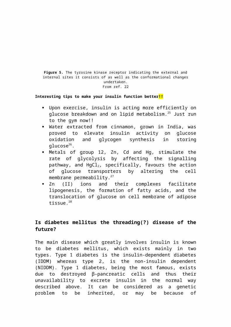
All of the actions of insulin are performed via insulin receptors, known as tyrosine kinase receptors. These are 2-subunit receptors and contain both an extracellular domain for insulin to bind as a ligand as well as an intracellular part, insulin protein kinase22 where all phosphorylation events take place. As with most polypeptide hormones, upon binding, conformational changes are undertaken on the two subunits and a series of phosphorylation events proceed through, leading to more of the actions hormones perform (Figure 5). Internalization of insulin upon binding keeps it in the cell followed by degradation at the end23. In addition, it is worth mentioning that all of these actions and signaling pathways are accompanied by the formation of second messengers such as cyclic adenosine phosphate, cAMP. Second messengers are molecules that disperse information around tissues. The decrease in the concentration of cAMP is the main cause for insulin’s activities and this is because of insulin suppressing the precursor molecule, adenylate cyclase, from which the second messenger is synthesized24.
Figure 5. The tyrosine kinase receptor indicating the external and internal sites it consists of as well as the conformational changes undertaken.
From ref. 22
Interesting tips to make your insulin function better!!
Upon exercise, insulin is acting more efficiently on glucose breakdown and on lipid metabolism.25 Just run to the gym now!!
Water extracted from cinnamon, grown in India, was proved to elevate insulin activity on glucose oxidation and glycogen synthesis in storing glucose26.
Metals of group 12, Zn, Cd and Hg, stimulate the rate of glycolysis by affecting the signalling pathway, and HgCl2, specifically, favours the action of glucose transporters by altering the cell membrane permeability.27

Zn (II) ions and their complexes facilitate lipogenesis, the formation of fatty acids, and the translocation of glucose on cell membrane of adipose tissue.28
Is diabetes mellitus the threading(?) disease of the future?
The main disease which greatly involves insulin is known to be diabetes mellitus, which exists mainly in two types. Type 1 diabetes is the insulin-dependent diabetes (IDDM) whereas type 2, is the non-insulin dependent (NIDDM). Type 1 diabetes, being the most famous, exists due to destroyed β-pancreatic cells and thus their unavailability to excrete insulin in the normal way described above. It can be considered as a genetic problem to be inherited, or may be because of environmental effects.29 Due to this insulin deficiency, glucose transporters are suppressed and glycogen synthesis is limited, whereas synthesis of glucose and protein degradation are facilitated,30 all being the opposite functions that normal insulin secretion produces. Usually it can be treated by replacing the pancreatic tissue through transplantation31 since it is greatly damaged, but most people nowadays perform injections or use insulin pumps. On the other hand, in type 2 diabetes the pancreatic β cells function quite perfectly, sometimes over-secreting insulin, but tissues around remain unaffected by it. It is also related to obesity. Finally gestational diabetes is another type of diabetes appearing only to pregnant women though, as a result of problematic insulin secretion.29
The term ‘mellitus’ can be translated as honey or sweet-tasting urine and as
Thomas Willis observed in the 17th century ‘diabetics piss a great deal’. Why not ask your diabetic friend for a fresh sample??32
Insulin resistance!! Obesity and differences between the two sexes!!
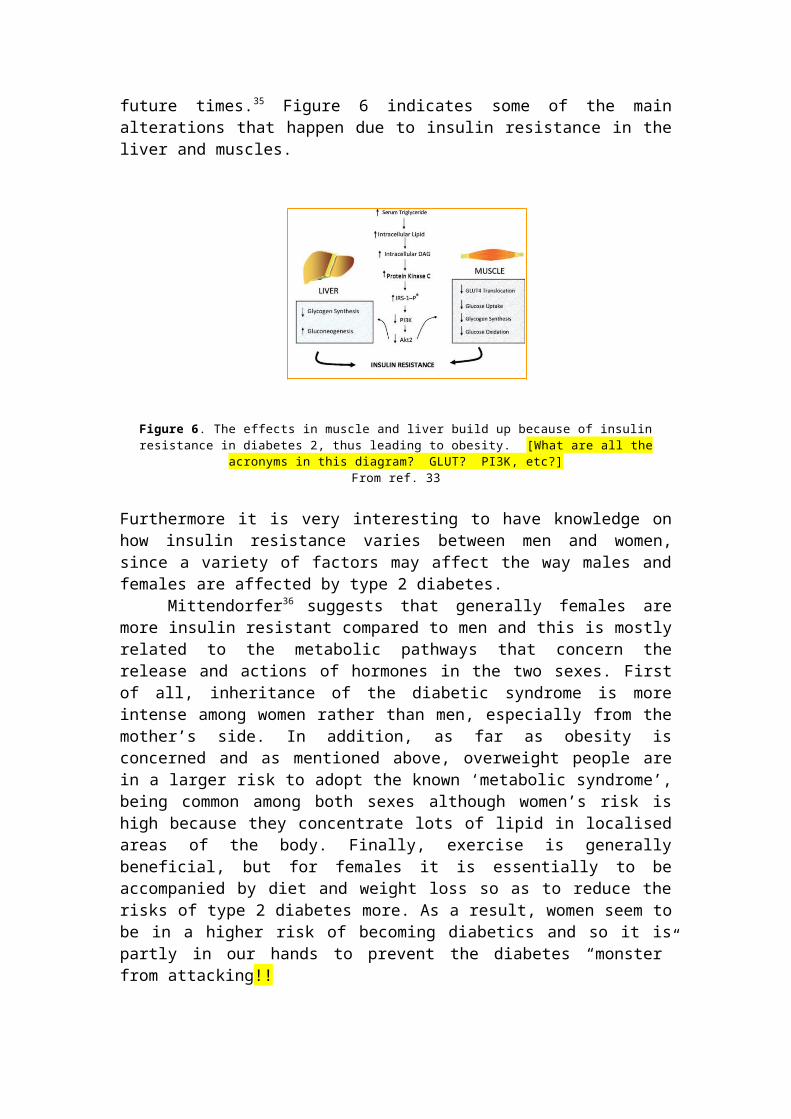
Insulin resistance appears to be a main condition for type 2 diabetes, defined as “reduced responsiveness to normal circulating levels of insulin”.33 This is directly related to obesity, and insulin resistance arises mostly due to adipose tissue abnormalities. As a result other tissues are now more exposed to fats and thus lots of them are concentrated at one position resulting to obesity after an extensive exposure.34 Taking the opposite condition into consideration, experiments have shown that obese people have a greater risk of becoming diabetics and using statistics made in From ref. 2 (B)England , as an example , it is thought that the current 2.35 million diabetics will increase to 2.5 million in future times.35 Figure 6 indicates some of the main alterations that happen due to insulin resistance in the liver and muscles.
Figure 6. The effects in muscle and liver build up because of insulin resistance in diabetes 2, thus leading to obesity. [What are all the acronyms in this diagram? GLUT? PI3K, etc?]
From ref. 33
Furthermore it is very interesting to have knowledge on how insulin resistance varies between men and women, since a variety of factors may affect the way males and females are affected by type 2 diabetes.
Mittendorfer36 suggests that generally females are more insulin resistant compared to men and this is mostly related to the metabolic pathways that concern the release and actions of hormones in the two sexes. First of all, inheritance of the diabetic syndrome is more intense among women rather than men, especially from the mother’s side. In addition, as far as obesity is concerned and as mentioned above, overweight people are in a larger risk to adopt the known ‘metabolic syndrome’, being common among both sexes although women’s risk is high because they concentrate lots of lipid in localised areas of the body. Finally, exercise is generally beneficial, but for females it is essentially to be accompanied by diet and weight loss so as to reduce the risks of type 2 diabetes more. As a result, women seem to be in a higher risk of becoming diabetics and so it is partly in our hands to prevent the diabetes “monster” from attacking!!
Notes and References
1. G.D. Smith, W.A. Pangborn, R.H. Blessing, The structure of T-6 bovine insulin, Acta Crystallogr.,Sect. D: Biol. Crystallogr.,2005,61,1476.
[ Reference found by keyword search on the Web of Science- ‘insulin structure- zinc ions- hexamer’.]
2. T. Garnero, Islets of humor from Defeat Diabetes, www.defeatdiabetes.org A) www.defeatdiabetes.org/E-Lerts/elerts0408.htm , October 2004,3( 10)B) www.diabetesdiabetes.org/E-Lerts/elerts0408.htm , August 2004, 3(8)
[ Picture found from Google images http://www.google.com using the keywords ‘ diabetes cartoons’]
3. Jasper Carrot, www.brainyquote.com/quotes/quotes/j/jaspercarr 2819A.html
from the Brainy quote webpage. [ Phrase found by searching Google, http://www.google.com with the
phrase ‘insulin quotes’.]
4. F.F. Bolander, Molecular Endocrinology, 3rd edn, Elseiver Academic Press, London, 2004.
[ Found by searching the shelves in the Medical Sciences Library in the endocrinology section.]
5. F. S Greenspan and D. G. Gardner, Basic & Clinical Endocrinology, 7th edn, McGraw-Hill Companies, New York, 2004.
[ Found using the same method as for reference [4].] 6. J. Espinal, Understanding Insulin Action: principles and molecular
mechanisms , Ellis Horwood, Chichester, 1989. (in Ellis Horwood series in Biochemistry and Biotechnology.
[ Reference book found by using the phrase ‘insulin action’ in the library catalogue search tool]
7. W. Kadima, Role of metal ions in the T-to-R allosteric transition in the insulin hexamer, Biochemistry, 1999, 38, 13443.
[ Reference found by same method as refernce [1].]
8. L. B. Chaykin, Insulin detemir and it’s unique mechanism of action, The Internet J. Endocrinol., 2007, 4, number 1.
[ Reference found by searching Google images, http://www.google.com and the phrase ‘structure of insulin’]
9. G. G. Dodson, S. Cutfield, E. Hoenjet, A. Wollmer, D. Branderburg, Crystal structure, aggregation and biological potency of beef insulin cross-linked at A1 and B29 by diaminosuberic acid, in Insulin: Chemistry, Structure and Function of Insulin and Related Hormones: Proceedings 2nd Int. Insulin Symposium, Aachen, 4-7 Sep 1979, eds. D. Branderburg and A. Wollmer, de Gruyter, Berlin, New York, 1980, p. 17-26.
[ Reference found by searching the library catalogue using the keywords ‘ insulin structure’.]
10. A. Schuttler & D. Brandenburg, Preparation of covalent insulin dimmers, in Insulin: Chemistry, Structure and Function of Insulin and Related Hormones: Proceedings 2nd Int. Insulin Symposium, Aachen, 4-7 Sep 1979, eds. D. Branderburg and A. Wollmer, de Gruyter, Berlin, New York, 1980, p.143- 149.
11. M. Bliss, The discovery of insulin, The University of Chicago Press, Chicago 1982.
[ Found by searching the library catalogue and the keywords ‘ insulin discovery’. The book was found in lockers and not available on the shelves.]
12. R. Madeb, L. G. Koniaris, S. I. Schwartz, The discovery of insulin: Rochester, New York, connection, Ann. Int. Med., 2005, 143, 907. [ Picture found by keyword search on the Web of Science for ‘insulin discovery’ and ‘Banting’.]
13. L. Rosenfeld, Insulin: Discovery and Controvery, Clin. Chem., 2002, 48,

2270. [Picture found by keyword search on the Web of Science for ‘insulin discovery’ and ‘Charles Best’.]
14. D. L. Cook & C. N. Hales, Intracellular ATP directly blocks K+ channels in
pancreatic β cells, Nature, 1984, 311, 271. [ Reference found in the reference section of chapter 4 of the book given by references [20] and [22].]
15. B. R. Gauthier & C. B. Wollheim, Synaptotagmins bind calcium to release insulin, Am. J. Physiol.- Endo. M., 2008, 295, E1279. [ Reference found by keyword search on the Web of Science for the years 2007-2008, with the words ‘ insulin secretion’ and ‘exocytosis’.]
16. I. Novak, Purinergic receptors in the endocrine and exocrine pancreas,
Purinergic signalling, 2008, 4, 237. [ Reference found by keyword search on the Web of Science for the recent 5 years, with the words ‘ purinergic receptors’ and ‘pancreas’. ]
17. Y. Sato & J.C. Henquin, The K+ - ATP channels- independent pathway of regulation of insulin secretion by glucose- In search of the underlying mechanism, Diabetes, 1998, 47, 1713. [ Found by searching the Web of Science for the keywords ‘insulin release’ and ‘ regulators’. ]
18. F. Gentili, C. Cardinaletti, C. Vespirini, F. Ghelfi, A. Farande, M. Giannella, A. Piergentili, W. Quaglia, L. Mattioli, M. Perfumi, A. Hudson, M. Pigini, Novel ligands rationally designed for characterizing I-2-imidazoline binding sites nature and function, J. Med. Chem., 2008, 51, 5130. [ Reference obtained by subject keyword search on the Web of Science with the phrase ‘ imidazolines functions’.]
19. P. Jakobsen, P. Madsen, H.S. Andersen, Imidazolines as efficacious glucose-dependent stimulators of insulin secretion, in European Journal of Medicinal Chemistry: Proceedings 17th Int. Symp. Of Medicinal Chemistry, Barcelona, 01-05 Sep 2002, Editions Scientifiques Medicales Elseveir, Paris, 2003, p. 357-362.
[ Reference obtained by searching on the Web of Science using the keywords ‘ stimulation- insulin- secretion’.] 20. E. A. Newsholme. S. J. Bevan, G. D. Dimitriadis, R. P. Kelly, in Insulin:
Molecular Biology to Pathology, eds. F. M. Ashcroft & S. J. H. Ashcroft, IRL Press, Oxford, 1992, p.155. [ Medical Sciences Library- diabetes section.]
21. C. Sartori, L. Trueb, P. Nicod, U. Scherrer, Effects of sympathectomy and nitric oxide synthase inhibition on vascular action of insulin in humans, Hypertention, 1999, 34, 586. [ Reference obtained by keyword search on the Web of Science for the variety of keywords ‘insulin action- glucose uptake- inhibitors’.]
22. K. Siddle, in Insulin:Molecular Biology to Pathology, eds. F. M. Ashcroft & S. J. Ashcroft , IRL Press, Oxford, 1992, p. 191. [ Found by same method as reference [20].]
23. B. Desbuquois & M. C. Postel- Vinay, Receptor-mediated internalisation of insulin, glucagon and growth hormone in intact rat liver- a biochemical study, in Insulin: Chemistry, Structure and Function of Insulin and Related Hormones: Proceedings 2nd Int. Insulin Symposium, Aachen, 4-7 Sep 1979, eds. D. Branderburg and A. Wollmer, de Gruyter, Berlin, New York, 1980. p. 285-292.
24. N. M. Pertseva, A.O. Shpakov, S. A. Plenseva, L. A. Kuznetsova, A novelview on the mechanism of action of insulin and other insulin superfamily peptides- involvement of adenylyl cyclase signalling system, Comp. Biochem. Phys. B., 2003, 134, 11. [ Found by keyword search on the Web of Science using the phrases ‘ insulin action’ , ‘ tyrosine kinase receptors’ and cyclic adenosine monophosphate.]
25. F. Shojaee – Moradie, K. C. R. Baynes, C. Pentecost, J. D. Bell, E. L.Thomas, N. C. Jackson, M. Stolinski, M. Whyte, D. Lorell, S. B. Bowes,J. Gibney, R. H. Jones, A. M. Umpleby, Exercise training reduces fatty acid availability and improves the insulin sensitivity of glucose, Diabetologia, 2007, 50, 404. [ Reference found by keyword search on the Web of Science with ‘insulin effects- muscles- adipose tissue- uptake glucose’.]
26. B. Roffey, A. Atwal, S. Kubow, , Cinamon water extracts increase glucose uptake but inhibit adiponectin secretion in 3T3-Li adipose cells, Mol. Nutr. Food Res., 2006, 50, 1739. [ Found using the same method as reference [21].]
27. D. M. Barnes & E. A. Kircher, Effects of mercury chloride on glucose transport in 3T3- Li adipocytes, Toxicol. in Vitro, 2005, 19, 207. [ Reference obtained using the same method as reference [ 21].]
28. Y. Yoshikawa, E. Ueda, Y. Kojima, H. Sakurai, The action mechanism of Zn(II) complexes with insulinomimetic activity in rat adipocytes, Life Sci.,2004, 75, 741. [ Reference obtained using the same method as reference [ 21].]
29. M. I. Harris & P. Zimmet, in International textbook of diabetes mellitus, eds K. G. M. Alberti, P. Zimmet, R. A. Defronzo, Wiley, Chichester, 2nd edn., 1997, vol. 1, p. 9. [ Medical Sciences library- diabetes section.]
30. P. J. Watkins, P. L . Drury, S. L. Howell, Diabetes and its management, 5th

edn, Blackwell Science Ltd, Oxford, 1996. [ Same as reference [ 29].]
31. F. Calcinaro, D. L. Wegmann, K. J. Lafferty, in Molecular Biology of Diabetes, eds. B. Daznin, D. LeRoith, N. J. Totowa, Humana Press, 1994, vol. 1, p. 69.
[ Same as reference [ 29].]
32. S. D. Sanders, Endocrine and reproductive systems, 2nd edn, Mosby, London, 2003. [ Same way as reference [ 29.] ]
33. G. Wolf, Role of fatty acids in the development of insulin resistance and type 2 diabetes mellitus, Nutr. News, 2008, 66, 597. [ Reference obtained by keyword search on the Web of Science for “diabetes- insulin- synthesis- obesity- glucose”.]
34. M. S. Westerterp, R. P. Mensink, Traces in metabolism and nutrition, Physiol. Behav. 2008, 94, 155. [ Reference obtained using the same method as reference [33].]
35. L. S. Aucott, Influence of weight loss on long-term diabetes outcomes, in Proceedings of the Nutrition Society: Summer Meeting of the Nutrition- Society, Coleraine, 16-19 Jul 2007, Cambridge University Press, Cambridge 2008, p. 54-59. [ Reference obtained using the same method as reference [33].]
36. B. Mitterndorfer, Insulin resistance : sex matters, Curr. Opin. in Clin. Nutr. and Metab. Care, 2005, 8, 367. [ Reference found by searching the Web of Science with the phrase ‘exercise improves insulin actions’.]
A very nice account of the subject, with a large number of relevant, properly formatted references. Be careful of using too many exclamation marks (!!!); it starts to look like a tabloid newspaper headline if you do. Nice use of diagrams and cartoons to make the article more fun to read. But be careful that you explain the contents of the diagrams properly in the captions. See Figs 2,4,6 and scheme 1.
19/25
2) Keeping-up-to-date with a subject.
Keeping-up-to-date with a topic, especially for people performing research for a long period of time, is extremely important. Although researching on a topic, such as the insulin molecule, a variety of facts discovered years ago are true and will always apply, there is new information given as time goes by and it is proved to be very useful. For example new ways on treating diabetes or methods to control the β
pancreatic cell secretion come to the surface all time and can be very useful for a researcher interested in such biological processes.
To begin with, most databases such as Web of Science, can provide students and researchers with an alert service that allows alert emails to be sent for specific journals that have been saved. The ‘Save History / Create alert’ options that exist as well as the use of specific keywords, such as “insulin action glycolysis”, allow new information discovered on that topic to be alerted through the alert email method. In addition to this, citation alerts can be set for a paper interested in or even set up searches to look automatically for specific authors that study a specific subject widely. Furthermore, current awareness is available from the powerful database Sci Finder, especially if interested in writing about chemical compounds, since their structure is to be analyzed and new discoveries structure features are often published.
As far as my topic is concerned, and more specifically the insulin molecule and its functions in the organism, I would suggest that the most suitable way to keep up-to-date is the email alert method provided by the Web of Science since most of the information collected for my project was from there. The best way to search for molecules is to use keywords on their discovery, their action and the effects they cause. I would suggest that it is a very general subject to write on and thus a variety of authors are involved, so author search for such a topic may not be the most suitable way of keeping-up-to date with the subject. In addition looking for newly published books in the library, especially symposiums, it can be very useful since analysis of new information on a molecule’s chemistry and biology through years can be provided by the large range of newly published books that is available.
For something as general interest as insulin, how about popular magazines, newspapers, or even the BBC news website as sources of current info?
4/5
Total: 19 + 4 = 23/30 PWM 10/3/09.